Note: Descriptions are shown in the official language in which they were submitted.
,4
CA 02672262 2009-06-10
- 1 -
_
DESCRIPTION
RADIOACTIVE DIAGNOSTIC IMAGING AGENT
TECHNICAL FIELD
[0001]
The present invention relates to a composition of a
radioactive fluorine-labeled amino acid compound. More
specifically, it relates to a composition of a radioactive
fluorine-labeled amino acid compound useful for detecting
tumors by positron emission tomography (PET).
BACKGROUND ART
[0002]
The radioactive diagnostic imaging agent is a medicine
directly administered to a human body and is a pharmaceutical
composition containing a compound labeled with a specific
radioisotope as an effective ingredient. The radioactive
diagnostic imaging agent enables diagnosis by administering
an agent to a subject and detecting a radiation emitted from
the compound, followed by imaging based on information
obtained from the radiation. The thus-conducted diagnosing
method is referred to as nuclear medicine examination, and
is effective in diagnosing a variety of diseases including
heart disease and cancer. Also, nuclear medicine
examination is characteristic in that it has not only high
specificity and sensitivity to diseases, but also has an
advantage of providing information on the functionality of
lesions, compared to other examination techniques.
[0003]
Compounds which are researched and developed as such
,4
CA 02672262 2009-06-10
- 2 -
radioactive diagnostic imaging agents include
1-amino-3- C8F]fluorocyclobutanecarboxylic acid
18F]
(hereinafter referred to as [
-FACBC). It is known that
[18F]-FACBC is taken up into a cell via an amino acid
transporter. Thus, [18F]-FACBC is expected to be developed
as a tumor diagnostic agent since it is largely taken up into
tumor cells which are highly proliferative and active in
protein synthesis.
[0004]
In radioactive diagnostic imaging agents, a problem
often arises such that compounds decompose by self-radiation
during delivery of the agents so as to cause decrease in
radiochemical purity due to so-called radiolysis.
Particularly, in PET agents for detection of positron
nuclides such as 18F, radiolysis often becomes more
problematic since the half-life of the nuclides used therein
is shorter than that of nuclides used in SPECT agents for
detection of gamma-ray emitting nuclides such as 99mTc, and
thus radioactivity upon shipment must be set larger than SPEC?
agents, thereby making the resulting radiation energy thereof
higher.
[0005]
For general pharmaceuticals, it is recommended in the
guideline of ICH that if impurities in an agent exceed 1.0%,
the impurities be subjected to structure determination when
the maximum daily dosage of an effective component thereof
is as small as not more than 1 mg (Non-Patent Document 1).
In most cases, the physical amount of impurities resulting
CA 02672262 2009-06-10
- 3 -
from the radiolysis which may be considered to be one aspect
of the decomposition of an agent is as small as about 10-12
mol, even if it exceeds 1.0%. Since the production amount
of impurities such as radioactive decomposed matters is
minute, structure determination of the impurities by NMR
analysis is difficult even though only determination of
molecular weight and presumption of their fragments can be
made by mass spectrometry which is excellent in detection
sensitivity. Also, it is very difficult to conduct
verification as to whether or not the impurities affect
effectiveness such as tumor accumulation of the agent.
Therefore, impurities in the radioactive diagnostic
imaging agent should be maintained as low as possible, and
it is preferable that radiolysis which may cause the
production of impurities should also be inhibited as much as
possible.
[0006]
Various methods for inhibiting radiolysis have been
examined focusing on application to [18F] -fluorodeoxyglucose
(hereinafter referred to as [18F]-FDG).
[0007]
International Publication No. W003/090789 pamphlet
discloses a method of reducing the radiolysis of [18F]-FDG
by adding a weak acid-based buffer to a [18F] -FDG solution
and an injection prepared by the method (Patent Document 1) .
Also, International Publication No. W004/043497 pamphlet
discloses adding ethanol to a [18F]-FDG solution to obtain
a composition of injection which may be reduced in radiolysis
.
CA 02672262 2009-06-10
k
- 4 -
- of [18F] -FDG to improve stability (Patent Document 2) .
[0008]
Japanese Patent Laid-open (Kokai) No. H10-147542
discloses a technique utilizing an organic compound high in
physiological acceptability such as monosaccharides,
disaccharides, organic acids and salts or esters thereof as
a radiation protecting agent (Patent Document 3). In this
publication, the organic compound high in physiological
acceptability and particularly effective as the radiation
protecting agent is defined to have a reaction rate constant
with OH radicals, H radicals or hydrated electrons in the
range of 1 x 10⁸ to 5 x 10¹⁰ molls'
.
[0009]
International Publication No. W004/056725 pamphlet
discloses a solid-phase synthesis method for 18F-labeled
tracers including [18F]-FACBC (Patent Document 4). In this
document, it is suggested that radiolysis of 18F-labeled
tracers is reduced by adding ascorbic acid to a composition
of injection.
Non-Patent Document 1: ICH HARMONISED TRIPARTTITE GUIDELINE,
IMPURITIES IN NEW DRUG PRODUCTS Q3B(R2) (page 7) (URL:
http://www.pmda.go.jp/ich/q/q3br2_06_7_3e.pdf)
Patent Document 1: International Publication No. W003/090789
pamphlet
Patent Document 2: International Publication No. W004/043497
pamphlet
Patent Document 3: Japanese Patent Laid-open (Kokai) No.
H10-147542
CA 02672262 2009-06-10
- 5 -
Patent Document 4: International Publication No. W004/056725
pamphlet
DISCLOSURE OF THE INVENTION
PROBLEMS TO BE SOLVED BY THE INVENTION
[0010]
As described above, International Publication No.
W003/090789 pamphlet and International Publication No.
W004/043497 pamphlet disclose conditions for preventing
radiolysis of [18F] -FDG in the solution. However, these
documents only disclose techniques for reducing radiolysis
of [18F]-FDG only, but do not disclose any technique for
reducing radiolysis of a series of radioactive
fluorine-labeled amino acid compounds such as [18F]_FACBC.
In addition, a technical feature of the invention
disclosed in International Publication No. W003/090789
pamphlet is adding a buffer, that is, increasing
radiochemical stability of[18F, i_FDG at a pH having a buffer
action, and it is indicated as Comparative Examples that the
radiochemical stability of[18F, i_FDG does not increase with
NaCl which has no buffer action.
[0011]
Japanese Patent Laid-open (Kokai) No. H10-147542
discloses a technique utilizing an organic compound high in
physiological acceptability as a radiation protecting agent
for radiopharmaceuticals. However, it is not apparent which
compound is selected as the organic compound high in
physiological acceptability or how much the compound is added
in order to prevent radiolysis of the series of radioactive
CA 02672262 2009-06-10
=
- 6 -
fluorine-labeled amino acid compounds such as [18F]-FACBC.
[0012]
International Publication No. W004/056725 pamphlet
proposes that the addition of ascorbic acid into a composition
of injection reduces the radiolysis of 18F-labeled tracers.
However, it does not contain any concrete disclosure of the
use of ascorbic acid as an additive for[18-t] _
FACBC. Also,
there is no disclosure as to on what condition it should be
used.
[0013]
The present invention has been made in view of the above
circumstances, and aimed at providing a composition
comprising a radioactive fluorine-labeled amino acid
compound, which can be reduced in radiolysis.
MEANS FOR SOLVING THE PROBLEM
[0014]
As a result of diligent researches, the inventors have
found that the radiolysis of [18F]-FACBC is reduced
dependently upon pH. Particularly, it has been found that
when the pH value is not more than 5.9, stability thereof is
maintained even if there exist no pharmaceutical additives
or buffers that prevent radiolysis.
Therefore, it has been found that decrease of
radiochemical purity of the radioactive diagnostic imaging
agent can be reduced by keeping the pH of the final agent at
2.0-5.9, and thus the present invention have been completed.
[0015]
According to one aspect of the present invention, there
,
CA 02672262 2009-06-10
s
¨ 7 -
is provided a radioactive diagnostic imaging agent which
comprises a solution containing a radioactive compound
represented by the following formula (1) as an effective
component:
[0016]
<0><712
ieF _________________
COOH
( 1 )
[0017]
wherein the solution has a pH value of 2.0-5.9. In a
preferable embodiment of the radioactive diagnostic imaging
agent according to the present invention, the above solution
can have a pH value of 2.0-4.9.
[0018]
The radioactive diagnostic imaging agent according to
the present invention may be one to which a pharmaceutical
additive is further added. As pharmaceutical additives,
various compounds which are generally accepted as additive
compounds can be used, including a pH regulator and a
dissolving aid as well as a sugar, a sugar alcohol, a sugar
lactone and the like. Preferably, a sugar alcohol can be
used.
[0019]
As the sugar alcohol, one or more compounds selected from
the group consisting of erythritol, xylitol, sorbitol and
mannitol can be used. Addition amount thereof is not limited
as long as it can additionally reduce radiolysis, but is
CA 02672262 2009-06-10
8 -
preferably not less than 0.5 pmol/mL, more preferably not less
than 1.0 pmol/mL, furthermore preferably not less than 5.0
pmol/mL, and particularly preferably not less than 10.0
pmol/mL. The upper limit of the addition amount needs to be
an amount that is acceptable for pharmaceutical additives;
for examples, the upper limit as a total daily dose is 200
mg for xylitol, 1.5 g for sorbitol, and 1.2 g for mannitol.
[0020]
In the radioactive diagnostic imaging agent according to
the present invention, radioactive concentration is not
particularly limited as long as a sufficient amount of
radioactivity can be ensured when used. More specifically,
the radioactive concentration in use is preferably 25-125
MBq/mL, and more preferably 25-100 MBq/mL.
[0021]
In the present specification, compounds acceptable as
pharmaceutical additives mean compounds that are approved as
pharmaceutical additives in the Japanese Pharmacopoeia, the
United States Pharmacopoeia, the European Pharmacopoeia, and
so on. In addition, sugar alcohol means a reduced form of
a sugar, and sugar lactone means a cyclic ester compound that
is derived by intramolecular dehydration condensation of a
sugar.
EFFECT OF THE INVENTION
[0022]
According to the present invention, a pH value of a
solution containing a radioactive fluorine-labeled amino
acid compound is regulated to 2.0-5.9, and thus a composition
CA 02672262 2009-06-10
- 9 -
of a radioactive fluorine-labeled amino acid compound is
provided which is reduced in radiolysis.
BEST MODE FOR CARRYING OUT THE INVENTION
[0023]
Hereinafter, the most preferable embodiments for the
composition of the radioactive fluorine-labeled amino acid
compound according to the present invention will be
described.
[0024]
The radioactive diagnostic imaging agent according to
the present invention is produced in 4 steps; a step of
imparting radioactive fluorine to a precursor (step 1) ; a step
of performing deprotection of the compound to which the
radioactive fluorine has been imparted (step 2); a step of
performing purification of a solution containing
anti-[18-t] _
FACBC after deprotection (step 3); and a step of
processing the purified anti-[18Y-] _
FACBC solution into a
preparation (step 4).
[0025]
Radioactive fluorine can be obtained by a known method,
for example, a method in which H2180 enriched water is used
as a target and exposed to proton bombardment. In this
instance, radioactive fluorine exists in the H2180 enriched
water used as a target. The H2180 enriched water containing
radioactive fluorine is allowed to pass through, for example,
an anion-exchange resin column so that the radioactive
fluorine is adsorbed and collected on the column, thereby
being separated from the H2180 enriched water. Thereafter,
CA 02672262 2009-06-10
- 10 -
a potassium carbonate solution is allowed to pass through the
column to elute the radioactive fluorine, and the eluate is
supplemented with a phase transfer catalyst and is evaporated
to dryness, thereby activating the radioactive fluorine.
[0026]
In step 1, the dried radioactive fluorine is dissolved
in acetonitrile, and ethyl cis-1-(N-tert-
butoxycarbonyl ) amino-3- [ ( trifluoromethyl ) sulfonyloxy] -
cyclobutanecarboxylate, as a precursor, is added to the
acetonitrile solution to allow them to react under heating.
As a result, radioactive fluorine is added to the precursor,
whereby ethyl trans-1-(N-tert-butoxycarbonyl)amino-
3_ ]
[18-t',
fluorocyclobutanecarboxylate is synthesized.
[0027]
In step 2, ethyl trans-1-(N-tert-
butoxycarbonyl)amino-3- [18F] ,
fluorocyclobutane carboxylate
obtained in step 1 is deprotected to yield a solution
containing anti- [1.8F] -FACBC as a target product. In this step,
the condition of deprotection is preferably being acidic.
For example, hydrochloric acid can be added to a solution
containing ethyl trans-1-(N-tert-butoxycarbonyl)amino-3-
[18F]fluorocyclobutane carboxylate to perform deprotection.
The amount of acid to be added needs not be restricted as long
as the amount can provide an acidic condition sufficient for
the deprotection.
[0028]
In step 3, purification of the solution containing
[18F]
anti- -FACBC which is obtained in step 2 is performed.
=
CA 02672262 2009-06-10
- 11 -
The purification process to be used includes various
processes such as a liquid-liquid extraction process and a
column separation process. For example, a process in which
the reaction solution is injected into HPLC to obtain a
18
fraction containing anti[F1
- -FACBC can be used. The
anti-[18F]-FACBC solution can be obtained in this step.
[0029]
The radioactive diagnostic imaging agent according to
the present invention can be obtained by subjecting the
anti-[18F]-FACBC solution obtained in step 3 to various
operations required for making a preparation, including an
operation of vaporizing organic solvents, an operation of
adding pharmaceutical additives, an operation of adjusting
pH, an operation of adjusting radioactive concentration, and
an operation of performing sterilization by means of an
autoclave, filtration or the like. In this step, it is
preferable that the pH value is controlled in the range of
2.0 to 5.9. For this purpose, it is preferable that the pH
value is previously controlled in the range of 2.0 to 5.9 in
step 3. Also, it is possible that the pH is controlled in
the range of 2.0 to 5.9 immediately after the anti- [18F] -FACBC
solution is obtained. By way of this step 4, a radioactive
diagnostic imaging agent can be obtained which contains the
anti- [18F]-FACBC as an effective ingredient and is adjusted
to the solution pH in the range of 2.0 to 5.9.
[0030]
Meanwhile, the radioactive diagnostic imaging agent
according to the present invention should have a
CA 02672262 2009-06-10
- 12 -
radioactivity enabling PET imaging when it is used, and thus
the radioactive concentration at the time of production is
adjusted to meet that radioactivity. For example, if it has
a radioactivity of 1.4 GBq in about 2 mL immediately after
production, it will have a radioactivitiy of 50-225 MBq when
it is used, thereby enabling a sufficient PFT imaging for
adults.
EXAMPLE
[0031]
Hereinafter, the present invention is described below
in more detail by way of Examples. However, these Examples
never limit the scope of the present invention.
[0032]
Examples 1-16, Comparative Examples 1-5: Relation between pH
and decrease of radiochemical purity
[18F] fluoride ion-containing H2180 was allowed to pass
through an anion-exchange resin column to adsorb and collect
[18-tj ,
fluoride ion on the column. Then, the column was washed
with water, and a mixture containing [18F]fluoride ion, a
potassium carbonate solution and a phase transfer catalyst
was obtained in accordance with the conventional method (for
example, a method described in the references (Radioisotopes,
50, (2001), p.205-227; Radioisotopes, 50, (2001), p.228-256;
"Production and quality control of radioactive agents for PET
- Handbook of synthesis and clinical use- (2nd edition)",
edited by PET Chemistry Workshop).
[0033]
The obtained mixture was heated in a reaction vessel to
CA 02672262 2009-06-10
- 13 -
evaporate water to dryness, and was subjected to azetropic
distillation with addition of acetonitrile, and a solution
of ethyl cis-l-(N-tert-butoxycarbonyl)amino-3-
[(trifluoromethyl)sulfonyloxy]-cyclobutane carboxylate in
acetonitrile was added thereto. The obtained solution was
heated under stirring so as not to evaporate acetonitrile,
thereby allowing nucleophilic substitution reaction to
proceed to obtain [18F] fluorine-labeled compound.
[0034]
After the reaction vessel was cooled to about 40 C, water
for injection was added to the reaction solution for dilution,
and the mixture was passed through a reversed phase silica
gel column to collect the [18-
f] fluorine-labeled compound.
This column was washed, and flashed with a flow of helium gas,
and then a 4 mol/L sodium hydroxide solution was filled in
the column, followed by closing the column exit. After 3
minutes, the column exit was opened, and an alkali solution
was eluated from the column and collected in a vial. This
operation was repeated twice, and washed with water, and then
the washings were combined with the alkali solution collected
as above.
[0035]
Next, to the solution collected as above, hydrochloric
acid was added, and heated to about 60 C to effect
deprotection reaction. The mixture was then passed through
an ion retardation resin column, an alumina column and a
reversed phase resin column in this order to perform
purification and obtain a stock solution of anti- [18F] -FACBC
CA 02672262 2009-06-10
- 14 -
The pH value of the stock solution of anti-['8F]-FACBC was
adjusted to about 3.5 by previously placing a hydrochloric
acid solution in the vessel that received the stock solution
of anti- [18F] -FACBC.
[0036]
Radioactivity of the obtained stock solution of
anti-[18F]-FACBC was measured, and then the stock solution
was diluted with a physiological saline solution so as to have
a radioactive concentration of about 510 MBq/mL at the time
when experiment was initiated (0 hour in Table 2). 2.23 mL
of this solution was aliquoted in a vial of 5mL in volume,
and a predetermined amount of a predetermined solution
indicated in Table 1 was added thereto, to obtain a sample
solution. The radioactive concentration of the sample
solutions immediately after preparation was 653-686 MBq/mL.
CA 02672262 2009-06-10
- 15 -
[0037]
Table 1: The solution added to each sample solution and its pH after
adjustment
Added solution (addition pH after
amount) adjustment
Example 1 500 mmol/L HC1 (40 pL) 2.00
500 mmol/L HC1 (40 pL),
Example 2 physiological saline 2.05
solution (40 pL)
Example 3 100 mmol/L HC1 (50 pL)
2.66
Physiological saline
Example 4 3.41
solution (80 pL)
Physiological saline
Example 5 3.46
solution (50 pL)
11 mmol/L NaOH (70 pL),
Example 6 physiological saline 3.97
solution (10 pL)
Example 7 10 mmol/L NaOH (70 pL)
4.04
12 mmol/L NaOH (70 pL),
Example 8 physiological saline 4.55
solution (10 pL)
Example 9 11 mmol/L NaOH (70 pL)
4.58
Example 10 12 mmol/L NaOH (70 pL)
4.88
17 mmol/L NaOH (60 pL),
Example 11 physiological saline
5.03
solution (20 pL)
Example 12 13 mmol/L NaOH (70 pL)
5.11
15 mmol/L NaOH (70 pl,),
Example 13 physiological saline
5.46
solution (10 pL)
Example 14 14.3 mmol/L NaOH (60 pL) 5.54
17 mmol/L NaOH (70 pL),
Example 15 physiological saline
5.90
solution (10 pL)
Example 16 14 mmol/L NaOH (70 pL) 5.94
18.5 mmol/L NaOH (60 pL),
Comparative Example 1 physiological saline
6.28
solution (20 pL)
Comparative Example 2 14.1 mmol/L NaOH (70 pL) 6.31
16 mmol/L NaOH (80 pL),
Comparative Example 3 physiological saline
6.57
solution (10 pL)
Comparative Example 4 15 mmol/L NaOH (70 pL)
6.83
17 mmol/L NaOH (80 pL),
Comparative Example 5 physiological saline
7.70
solution (0 pL)
[0038]
The sample solution was stored in an electric
thermostatic chamber adjusted to 25 C, TLC analysis was
performed on the following conditions at the time of
CA 02672262 2009-06-10
- 16 -
initiation of the experiment (0 hour) and 8.5 hours after the
initiation of the experiment, and a value of radiochemical
purity was calculated in accordance with the following
equation (1) . Measurement of the radiochemical purity was
repeated three times for each sample solution.
[0039]
TLC analysis conditions:
Mobile phase: acetonitrile/water/100% acetic acid = 4/1/1
TLC plate: Silica Gel 60F254 (trade name, thickness of
membrane: 0.25 mm, manufactured by Merck & Co., Inc.)
Mobile length: 10 cm
TLC scanner: Rita Star (manufactured by Raytest)
Number of analysis: Three times
[0040]
Radioactivity of [18 F]FACBC peak
Radiochemical purity (%)= x100 (1)
Total radioactivity on TLC plate
[0041]
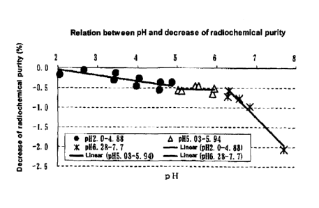
The results are shown in Table 2 and Fig. I.
CA 02672262 2009-06-10
,
- 17 -
[0042]
Table 2: Changes of radiochemical purity and decrease of radiochemical
purity of anti-[18F]-FACBC solution in different pH
Decrease*
Radiochemical purity (%)
pH (%)
0 hour 8.5 hours 8.5
hours
Example 1 2.00 99.41 99.44
0.03
Example 2 2.05 99.59 99.42 -
0.17
Example 3 2.66 99.38 99.33 -
0.05
Example 4 3.41 99.51 99.21 -
0.30
Example 5 3.46 99.39 99.26 -
0.13
Example 6 3.97 99.48 99.02 -
0.46
Example 7 4.04 99.38 99.11 -
0.27
Example 8 4.55 99.56 99.01 -
0.55
Example 9 4.58 99.35 98.98 -
0.37
Example 10 4.88 99.44 99.07 -
0.37
Example 11 5.03 99.46 98.89 -
0.57
Example 12 5.11 99.51 98.93 -
0.58
Example 13 5.46 99.52 99.06 -
0.46
Example 14 5.54 99.49 99.03 -
0.46
Example 15 5.90 99.51 98.86 -
0.65
Example 16 5.94 99.45 98.93 -
0.52
Comparative
6.28 99.53 98.81 -0.72
Example 1
Comparative
6.31 99.37 98.80 -0.57
Example 2
Comparative
6.57 99.43 98.67 -0.76
Example 3
Comparative
6.83 99.32 98.35 -0.97
Example 4
Comparative
7.70 99.37 97.31 -2.06
Example 5 _
* Decrease (%) = (radiochemical purity after 8.5 hours) - (radiochemical
purity after 0 hour)
[0043]
Referring to the relation between the pH and the decrease
of radiochemical purity, relatively mild decrease of
radiochemical purity was observed with the pH increase from
2.00 to 5.94. The slope based on linear approximation was
calculated, and as a result, the slope was -0.145 in the pH
range of 2.00-4.88, and was -0.010 in the pH range of
5.03-5.94.
On the other hand, when the pH value was not less than
6.28, sharp decrease of radiochemical purity occurred with
CA 02672262 2009-06-10
- 18 -
the pH increase. The slope based on linear approximation was
calculated, and as a result, it was -1.000. This value was
about 6.7 times the value in the pH range of 2.00-4.88, and
about 100 times the value in the pH range of 5.03-5.94. From
these, it was indicated that when the pH value is not less
than 6.28, a drastic decrease of radiochemical purity occurs
compared with the pH range of 2.00-5.94.
[0044]
Examples 17-28: Relation between mannitol concentration and
radiochemical purity at pH values of 3.44 and 4.78
A stock solution of anti- [18F] -FACBC was prepared in the
same manner as in Example 1 using [18F] fluoride
ion-containing H2180. Then, to the prepared anti-['8F] _ At F CBC
stock solution, a hydrochloric acid and a physiological
saline solution were added so as to have a radioactive
concentration of about 500 MBq/mL and a pH value of about 4.8
at the time when experiment was initiated (0 hour in Table
4) . 2.23 mL of the obtained solution was aliquoted in a vial
of 5 mL in volume, and a mannitol solution or a hydrochloric
acid at the concentration shown in Table 3 was added in an
amount shown in Table 3 to obtain a sample solution. The
radioactive concentration of the sample solutions
immediately after preparation was 553-565 MBq/mL.
=
CA 02672262 2009-06-10
- 19
[0045]
Table 3: Addition amount of mannitol solution in each sample solution
Mannnitol
concentration
pH Added solution (addition amount)
after
adjustment
(pmol/mL)
0.83 mg/mL Mannitol solution (50 pL)
Example 17 3.44 0.1
40 mmol/L HC1 (20 pL)
4.17 mg/mL Mannitol solution (50 pL)
Example 18 3.44 0.5
40 mmol/L HC1 (20 pL)
8.34 mg/mL Mannitol solution (50 pL)
Example 19 3.44 1.0
40 mmol/L HC1 (20 pL)
41.72 mg/mL Mannitol solution (50 pL)
Example 20 3.44 5.0
40 mmol/L HC1 (20 pL)
83.43 mg/mL Mannitol solution (50 pL)
Example 21 3.44
10.0
40 mmol/L HC1 (20 pL)
166.87 mg/mL Mannitol solution (50 pL)
Example 22 3.44
20.0
40 mmol/L HC1 (20 pL)
Example 23 4.78 0.83 mg/mL Mannitol solution (50 pL) 0.1
Example 24 4.78 4.17 mg/mL Mannitol solution (50 pL) 0.5
Example 25 4.78 8.34 mg/mL Mannitol solution (50 pL) 1.0
Example 26 4.78 41.72 mg/mL Mannitol solution (50 pL) 5.0
Example 27 4.78 83.43 mg/mL Mannitol solution (50 pL)
10.0
Example 28 4.78 166.87 mg/mL Mannitol solution (50 pL)
20.0
[0046]
The sample solution was stored in an electric
thermostatic chamber adjusted to 25 C, and the value of
radiochemical purity was calculated in the same manner as in
Example 1 at the time of initiation of the experiment (0 hour)
and 8.5 hours after the initiation of the experiment,.
Measurement of the radiochemical purity was repeated three
times for each sample solution.
[0047]
The results are shown in Table 4 and Fig. 2. In the all
Examples at the pH values of 3.44 and 4.78, decrease of
radiochemical purity was drastically reduced with increase
of mannitol concentration, and the reduction effect was
saturated at a mannitol concentration of not less than 5.0
CA 02672262 2009-06-10
- 20 -
pmol/mL.
Also, the decrease of radiochemical purity was more
inhibited at both mannitol concentrations at the pH value of
3.44 than 4.78.
From the above results, it was confirmed that the pH value
of the solution contributes to radiochemical stability.
Also, it was shown that radiolysis can be additionally reduced
by adding a mannitol
[0048]
Table 4: Change of radiochemical purity and decrease of radiochemical
purity of anti-r8F1-FACBC solution in the presence of mannitol
Mannitol Radiochemical purity
Decrease* (%)
pH Concentration (%)
(pmol/mL) 0 hour 8.5 hours 8.5
hours
Example 17 3.44 0.1 99.38 99.10 -0.28
Example 18 3.44 0.5 99.46 99.22 -0.24
Example 19 3.44 1.0 99.39 99.25 -0.14
Example 20 3.44 5.0 99.47 99.42 -0.05
Example 21 3.44 10.0 99.42 99.33 -0.09
Example 22 3.44 20.0 99.47 99.41 -0.06
Example 23 4.78 0.1 99.39 98.93 -0.46
Example 24 4.78 0.5 99.47 99.09 -0.38
Example 25 4.78 1.0 99.38 99.14 -0.24
Example 26 4.78 5.0 99.45 99.30 -0.15
Example 27 4.78 10.0 99.41 99.26 -0.15
Example 28 4.78 20.0 99.46 99.27 -0.19
*Decrease (%) = (radiochemical purity after 8.5 hours) - (radiochemical
purity after 0 hour)
[0049]
Examples 29-31, Comparative Examples 6-8: Relation between
decrease of radiochemical purity and radioactive
concentration
A stock solution of anti-[18F] -FACBC was prepared in the
same manner as in Example 1 using [18F] fluoride ion-containing
H2180. Radioactivity of the obtained stock solution of
anti- [18F] -FACBC was measured, and diluted and adjusted so
as to have a radioactive concentration of 507 MBq/mL and a
CA 02672262 2009-06-10
- 21 -
mannitol concentration of 10 pmol/mL (hereinafter, referred
to as standard solution for sample preparation in these
Examples and Comparative Examples). 2.23 mL of the obtained
standard solution for sample preparation was aliquoted in a
vial of 5 mL in volume, and a hydrochloric acid was added
thereto so that the pH value was adjusted to 3.94. From this
vial, a solution was fractionated in an amount shown in Table
5, and a physiological saline solution was added respectively
to make a sample solution of 1 ml in volume.
lo [0050]
Table 5: Diluting conditions in each sample
Radioactive
pH before Fractionated
concentration
Dilution rate
dilution amount mL
after dilution*
MBq/mL
Example 29 1 1 (no dilution) 573
Example 30 3.94 0.5 2 292
Example 31 0.1 10 61
*Radioactive concentration after dilution was calculated based on the
radioactivity measured about 10 minutes before initiation of experiment.
[0051]
Separately, 2.23 ml of the above prepared standard
solution for sample preparation was aliquoted in a vial of
5 ml in volume, and a sodium hydroxide solution was added to
adjust the pH value to 7.91. From this vial, a solution was
fractionated in an amount shown in Table 6, and a
physiological saline solution was added respectively to make
a sample solution of 1 mL in volume for use in Comparative
Examples 6-8.
CA 02672262 2009-06-10
- 22
[0052]
Table 6: Diluting conditions in each sample
Radioactive
pH before Fractionated
concentration
Dilution rate
dilution amount mL
after dilution*
MBq/mL
Comparative
1 1 (no dilution) 586
Example 6
Comparative
7.91 0.5 2 296
Example 7
Comparative
0.1 10 62
Example 8
*Radioactive concentration after dilution was calculated based on the
radioactivity measured about 10 minutes before initiation of experiment.
[0053]
The sample solution was stored in an electric
thermostatic chamber adjusted to 25 C, and the value of
radiochemical purity was calculated in the same manner as in
Example 1 at the time of initiation of the experiment (0 hour)
and 8.5 hours after the initiation of the experiment.
Measurement of the radiochemical purity was repeated three
times for each sample solution.
[0054]
The results are shown in Tables 7 and 8 . In Examples 29-31,
no matter what the radioactive concentration was, decrease
of radiochemical purity was hardly observed (Table 7). On
the other hand, in both Comparative Examples 6-8, time-course
decrease of radiochemical purity was observed, and it was
indicated that radiochemical purity tended to decrease with
increase of radioactive concentration (Table 8).
From the above results, at the pH value of 3.94, it was
confirmed that the decrease of radiochemical purity was
significantly reduced at a radioactive concentration of up
to 600 MBq/mL.
CA 02672262 2009-06-10
- 23 -
Also, the decrease of radiochemical purity was enhanced
with the increase of radioactive concentration at the pH value
of 7.91, and thus it was indicated that the time-course
decrease of radiochemical purity of anti- [18¨
r]_ FACBC was not
caused by decomposition of the anti- [18¨
t]_ FACBC due to lack
of chemical stability against the pH, but was caused by
decomposition of the anti- [18¨ _
FACBC due to radiolysis by
radiation.
[0055]
Table 7: Relation between radiochemical purity and radioactive
concentration in samples derived at pH 3.94
Radioactive concentration Radiochemical purity (%)
after dilution* MBq/mL 0 hour 6.5
hours decrease
Example 29 573 99.64 99.59 0.05
Example 30 292 99.60 99.56 , 0.04
Example 31 61 99.55 99.59 -0.04
*Radioactive concentration after dilution was calculated and measured
about 10 minutes before initiation of experiment.
[0056]
Table 8: Relation between radiochemical purity and radioactive
concentration in samples derived at pH 7.91
Radioactive concentration Radiochemical purity (%)
after dilution* MBq/mL 0 hour 6.5
hours decrease
Comparative
586 99.24 97.57 1.67
Example 6
Comparative
296 99.33 98.01 1.32
Example 7
Comparative
62 99.44 98.83 0.61
Example 8
*Radioactive concentration after dilution was calculated based on the
radioactivity measured about 10 minutes before initiation of experiment.
[0057]
Examples 32-33: Relation between addition of mannitol and
decrease of radiochemical purity
A stock solution of anti-[18F]-FACBC was prepared in the
same manner as in Example 1. The prepared stock solution of
anti- [18¨
Y]_ FACBC was diluted with a hydrochloric acid and a
physiological saline solution so as to have a radioactive
CA 02672262 2009-06-10
- 24
concentration of 568.1 MBq/mL at the predetermined time when
the experiment was initiated (0 hour in Table 9) (pH 3.98) .
This solution was aliquoted in an amount of 2.23 mL as a sample
solution (Example 32) . Separately, the solution adjusted to
507 MBq/mL was aliquoted in an amount of 2.23 mL, and a mannitol
solution was added thereto to prepare a solution adjusted to
have a mannitol concentration of 10 pmol/mL for use in the
experiment (Example 33) .
[0058]
The sample solution was stored in an electric
thermostatic chamber adjusted to 25 C, TLC analysis was
performed at the time of initiation of the experiment (0 hour) ,
2.5 hours later, 4.5 hours later, 6.5 hours later and 8.5 hours
later in the same manner as in Example 1, and the value of
radiochemical impurity was calculated in accordance with the
following equation (2) . Measurement of radiochemical
impurity was repeated three times for each sample solution.
[0059]
Radioactiveity of radiochemical impurity
Radiochemical impurity (%)= ________________________________ x100 (2)
Total radioactivity on TLC plate
[0060]
The results are shown in Table 9. In the sample solution
that was not blended with mannitol (Example 32) ,
radiochemical impurity was reduced to 1% or lower at all the
time points by virtue of the effect of the pH adjustment.
However, a tendency of time-course increase was indicated at
the time point until 6.5 hours after initiation of experiment.
On the other hand, in the sample solution that was blended
CA 02672262 2009-06-10
- 25 -
,
with mannitol (Example 33), no time-course increase of
radiochemical purity was observed.
From the above results, it was indicated that blending
mannitol enables the increase of radiochemical impurity by
radiolysis to be further inhibited. From this, it was
confirmed that the effect of stabilization of radiochemical
purity was more strengthened by the addition of mannitol.
[0061]
Table 9: Time-course change of radioactive impurity
Radiochemical impurity (%)
0 hour 2.5 hours 4.5 hours 6.5 hours
8.5 hours
Example 32 0.52 0.73 0.90 0.98 0.95
Example 33 0.50 0.52 0.48 0.56 0.49
INDUSTRIAL APPLICABILITY
[0062]
The present invention can reduce the radiolysis of
radioactive fluorine-labeled amino acid compounds useful as
PET agents, and is useful in the field of
radiopharmaceuticals.
BRIEF DESCRIPTION OF THE DRAWINGS
[0063]
Fig. 1 is a graph which shows a relation between the pH
and the decrease of radiochemical purity.
Fig. 2 is a graph which shows a relation between the
mannitol concentration and the decrease of radiochemical
purity.