Note: Descriptions are shown in the official language in which they were submitted.
CA 02672703 2009-06-15
WO 2008/071806 PCT/EP2007/064094
1
ANTIBODIES AGAINST HUMAN CYTOMEGALOVIRUS (HCMV)
TECHICAL FIELD
The invention relates to novel antibody sequences isolated from human B
cells having biological activities specific for a virus that infects human
cells.
BACKGROUND OF THE INVENTION
Human Cytomegalovirus (hCMV) is a widespread, highly species-specific
herpesvirus, causing significant morbidity and mortality in immunosuppressed
or
immunologically immature individuals.
Several recent reviews analyze hCMV biology and clinical manifestations
(Landolfo S et al., 2003; Gandhi M and Khanna R, 2004; Soderberg-Naucler C,
2006a). This viral pathogen infects the majority of the population worldwide
and is
acquired in childhood, following the contact with a bodily fluid, since the
virus
enters through endothelial cells and epithelial cells of the upper alimentary
or
respiratory systems, or through the genitourinary system. Seropositivity to
hCMV is
more prevalent in underdeveloped countries or in geographical areas with lower
income.
Following a primary infection, hCMV can persist in specific host cells of the
myeloid lineage in a latent state, replicating and disseminating in many
different cell
types (haematopoietic cells, epithelial cells, endothelial cells, or
fibroblasts) and
escaping the host immune system. The hCMV infection is generally asymptomatic
in
healthy people since hCMV infection and dissemination is maintained under
control
by the immune system, but total hCMV clearance is rarely achieved. In fact,
hCMV
virus has developed efficient mechanisms that allow viral genome to remain in
CA 02672703 2009-06-15
WO 2008/071806 PCT/EP2007/064094
2
selected sites in a latent state.
Any situation that weakens immune functions, such as stress conditions or
specific medical treatments, can lead to hCMV reactivation. Clinical
manifestations
of hCMV (such as retinitis, enterocolitis, pneumonitis, gastritis, or
hepatitis) can
occur following viral primary infection, reinfection, or reactivation. About
10% of
infants are infected by the age of 6 months following transmission from their
mothers via the placenta, during delivery, or by breastfeeding.
The hCMV virion consists of an icosahedral nucleocapsid which contains a
linear, 230 kb-long, double-stranded DNA genome. The expression of hCMV
genome is controlled by a complex cascade of transcriptional events that leads
to the
synthesis of more than 200 proteins involved in a large variety of biological
activities involved in viral infection, latency, and replication (Britt W and
Mach M,
1996).
The structural proteins form the virion envelope that is extremely complex
and still incompletely defined. It includes glycoproteins that are homologues
of
structural proteins identified in other herpesviridae and that can form
disulfide-
linked protein complexes within the virion: gCI (including only gB), gCII
(including
gM and gN) and gCIII (including gH, gL and g0). The gB, gH, and gN genes have
been also used for genotyping hCMV strains (Coaquette A et al.,2004; Dar L,
2007).
The glycoproteins gN and gM are the most abundant and, together with gH
and gB, have been shown to be essential for the initial interaction between
the
hCMV envelope and host cell surface, and consequently for the production of
infectious hCMV. For this reason, compounds targeting gB, gH, gN, and/or gM
can
efficiently inhibit hCMV infection by blocking the entry of circulating hCMV
CA 02672703 2009-06-15
WO 2008/071806 PCT/EP2007/064094
3
virions into the cells, following hCMV infection, reinfection, or
reactivation.
Treatment of hCMV infections is difficult because there are few therapeutic
options available. The presently available drugs compounds that inhibit viral
replication (Ganciclovir, Cidofivir, Foscarnet, Maribavir and others drugs
under
development) produce a significant clinical improvement, but may suffer from
poor
oral bio availability, low potency, the emergence of hCMV resistance (due to
mutations in the viral targets), and dose-limiting toxicities (De Clercq E,
2003;
Baldanti F and Gerna G, 2003; Gilbert C and Boivin G, 2005).
Novel means for preventing and treating hCMV infection are needed,
especially for immunocompromised individuals, in transplantation settings, and
in
prenatal prevention. In fact, hCMV is a clinically important opportunistic
pathogen
in HIV patients and in organ transplant recipients, where it contributes to
graft loss
independently from graft rejection, resulting in morbidity and mortality
(Puius Y
and Snydman D, 2007). The increasing number of bone marrow and solid organ-
transplant recipients raises the likelihood of hCMV clinical manifestations,
such as
hCMV retinitis (Wiegand T and Young L, 2006). Moreover, hCMV is the major
infectious cause of birth defects (such as hearing loss, delayed development,
or
mental retardation) which are due to a congenital or perinatal hCMV infection
transmitted by an hCMV-infected mother (Griffiths P and Walter S, 2005).
Thus, it is important to provide drugs for universal preemptive, prophylactic
hCMV-specific treatments, for example for the prevention of hCMV disease in
transplant recipients (Hebart H and Einsele H, 2004; Kalil A et al., 2005;
Snydman
D, 2006), in patients developing hCMV-related neuropathologies (Griffiths P,
2004)
or in at risk pregnancies (Schleiss M, 2003), to prevent the vertical
transmission and
CA 02672703 2009-06-15
WO 2008/071806 PCT/EP2007/064094
4
life-threatening hCMV infection to foetuses and neonates.
Moreover, pharmaceutical compositions against hCMV may be useful for the
treatment of other, more widespread diseases (such as cardiovascular and
autoimmune diseases, or some types of cancer), where hCMV is a possible
cofactor
and/or can be reactivated during immuniosuppressive treatments. For example,
hCMV is now a human pathogen of growing importance for disorders such as long-
term complications in tumour invasiveness and immune evasion since hCMV
infection may have oncomodulatory effects on cell apoptosis, differentiation,
and
migration. In autoimmune or vascular diseases, hCMV infection may alter immune
and inflammatory reactions (Cinatl J et al., 2004; Soderberg-Naucler C,
2006b).
An alternative way to prevent hCMV infection is vaccination, at the scope of
providing protection in an array of high-risk patient populations. However,
the
correlation between vaccination and the resulting immune response is not fully
understood and optimal hCMV vaccine strategy (using specific candidate
antigens or
live attenuated vaccines) seems depending on the patient population being
targeted
for protection. Therefore, prophylactic vaccination strategies are still under
evaluation (McLean G et al., 2006; Schleiss M, 2005).
In view of the present limitations of pharmacological strategies for hCMV
infections, the increasing knowledge of the host-hCMV relationship, and in
particular on the hCMV-specific immune response, makes immune-based therapies
good alternatives to substitute, or complement, existing therapies for the
successful
treatment of hCMV-associated complications (Gandhi M and Khanna R, 2004).
Recently, a long-term protection from the lethal course of CMV infection in
immunodeficient mice was achieved by transferring virus-specific memory B
cells,
CA 02672703 2009-06-15
WO 2008/071806 PCT/EP2007/064094
suggesting that such cells may have a therapeutic utility (Klenovsek K et al.,
2007).
An easier alternative to cell-based therapies can be passive immunotherapy,
consisting in the administration to individuals of pharmaceutical compositions
comprising therapeutic antibodies with a defined neutralizing activity against
a
5 human or viral antigen (e.g. hCMV).
This therapeutic approach has been designed on the antigen-binding and
biological features of antibodies and antibody fragments directed against
human or
viral therapeutic targets (Dunman P and Nesin M, 2003; Keller M and Stiehm E,
2000). Passive immunotherapy has been introduced into clinical practice,
rapidly
expanding the opportunities for the treatment of a wide variety of diseases
(including infectious diseases, immune-mediated diseases, and cancer). This
approach can be particularly effective in patients whose immune system is
unable to
produce antibodies in the amounts and/or with the specificity required to
block
and/or eliminate the targeted molecule (Chatenoud L, 2005; Laffly E and
Sodoyer R,
2005).
In the field of hCMV therapy, this approach is performed by administering
intravenously human immunoglobulin preparations that are obtained by pooling
human plasma with high titers of anti-hCMV antibodies, and commercialized for
clinical uses (under the name of Cytotect or CytoGam). However, these products
are
only a partially satisfactory solution for blocking hCMV infection. In fact,
this
treatment is used in immunocompromised patients, mostly for pre-emptive
treatment
and prophylaxis where antivirals are often co-administered (Marasco W and Sui
J,
2007; Nigro G et al., 2005; Bonaros N et al., 2004; Kocher A et al., 2003;
Kruger R
et al., 2003). Moreover, safety issues and shortage of such preparations are a
CA 02672703 2009-06-15
WO 2008/071806 PCT/EP2007/064094
6
growing concern, as reported in literature (Bayry J et al., 2007; Hamrock D,
2006).
Human recombinant antibodies that have high affinity for antigens expressed
on hCMV envelope and are able to neutralize the infection would represent more
appropriate drugs for passive immunization. In fact, several of the hCMV
glycoproteins elicit strong host immune responses, including the production of
virus-neutralizing antibodies, even though the stoichiometry of the envelope
proteins
is variable and may be altered to escape host immune response. This response
is
considered to be a key component of host immunity and represents a goal of
both
antibody and vaccine development.
Human monoclonal antibodies are the most preferable antibodies for clinical
applications, due to the intrinsic limitations of murine monoclonal
antibodies.
However, the development of previously identified human antibodies for hCMV
treatment (Matsumoto Y et al., 1986) has been interrupted since no clinical
benefits
were observed in studies that evaluated the efficacy of such antibodies, for
example,
in haematopoietic stem cell transplantation (Boeckh M et al., 2001), or in
retinitis
(Gilpin A et al., 2003). These failures trials warrant further studies aimed
at
selecting human monoclonal antibodies that more efficiently neutralize the
widest
variety of hCMV strains. The treatment of CMV infections would benefit from
having more potent pharmaceutical compositions comprising human monoclonal
antibodies that are purified from human B cells maintained in culture or
produced as
recombinant proteins that are expressed by human sequences introduced in
mammalian cell lines.
DISCLOSURE OF THE INVENTION
The present invention provides novel antibody sequences that bind and
CA 02672703 2009-06-15
WO 2008/071806 PCT/EP2007/064094
7
neutralize hCMV, and that can be used for detecting, treating, inhibiting,
preventing,
and/or ameliorating hCMV infection or an hCMV-related disease.
Human B cells were obtained from an hCMV-seropositive individual and
immortalized. This polyclonal population of immortalized, human B cells was
divided for generating subcultures that were tested for the presence of
antibodies
(Immunoglobulins G, IgG) in the cell culture supernatant neutralizing hCMV
infectivity in vitro. In particular, the type of neutralizing activity, the
isotype, and
the clonality were determined for the antibody secreted by the subculture
named
26A1. The antibody has been affinity-purified from both the original cell
culture
supernant and as a recombinant human monoclonal antibody, confirming the hCMV-
specific neutralizing activity using in vitro models for hCMV infection. This
antibody can be used for characterizing neutralizing antigens on hCMV
envelope.
The DNA sequences that encode the variable regions of the antibody secreted
by the 26A1 subculture were amplified, cloned, and sequenced. The
corresponding
protein sequences were analyzed to identify the Complementarity Determining
Regions (CDRs) that are responsible for the hCMV-specific biological activity.
These sequences can be used for producing proteins having hCMV-specific
binding
and neutralizing properties, in the form of full antibodies, antibody
fragments, or
any other format of functional protein (e.g. bioactive peptide, fusion
proteins) using
appropriate technologies for producing recombinant proteins.
Compositions having therapeutic, prophylactic, and/or diagnostic utility in
the management of hCMV infection and hCMV-related disorders can be prepared
using the proteins of the invention, either as recombinant proteins or as
natural
antibodies purified from cell cultures originated from the 26A1 subculture.
Such
CA 02672703 2009-06-15
WO 2008/071806 PCT/EP2007/064094
8
compositions may be used to supplement or replace present hCMV treatments
based
on antiviral compounds and/or intravenous immunoglobulins (IVIg) preparations.
Further embodiments of the present invention will be provided in the
following Detailed Description.
DESCRIPTION OF THE FIGURES
Figure 1: (A) Schematic representation of the CG3 antigen that has been
assembled and used in a gB-specific ELISA as described in the literature
(Rothe M
et al., 2001). The recombinant interstrain fusion CG3 antigen corresponds to a
combination of the gB Antigenic Domain 2 (AD2; SEQ ID NO: 1 and 2) from
hCMV strains AD169 (SwissProt Acc. No. P06473; amino acids 27-84) and Towne
(SwissProt Acc. No. P13201; amino acids 27-84). The AD2 region contains a site
(amino acids 70-81, underlined) that is conserved in different viral strains
and that
has been shown to be recognized by neutralizing antibodies (Qadri I et al.,
1992;
Kropff B et al., 1993). (B) Schematic representation of the gH antigen
included in
the gH(Ag)-GST fusion protein used for the gH-specific ELISA assay. The
recombinant antigen gH(Ag)-GST corresponds to an in-frame fusion between the
gH
amino terminal region (amino acids 16-144; SEQ ID NO: 3) from the hCMV strain
VR1814 (Revello M et al., 2001) and Glutathione-S-Transferase (GST). The amino
terminus of gH contains a linear antibody binding site (amino acids 34-43;
underlined) that is recognized by neutralizing antibodies (Urban M et al.,
1992).
Figure 2: overview of the selection process for identifying and characterizing
subcultures (wells) that contain IgG antibodies binding and neutralizing hCMV.
The
subcultures were obtained by immortalizing B cells from hCMV patient (CMV7)
using the EBV-based immortalization process disclosed in PCT/EP2005/056871.
CA 02672703 2009-06-15
WO 2008/071806 PCT/EP2007/064094
9
Supernatants from subcultures (wells) showing significant cell growth were
screened
directly in the hCMV microneutralization assay. Supernatants showing
neutralizing
activity were then screened using gB- and gH-specific ELISA. The number of
positive wells for each screening assay is indicated in the grey ovals.
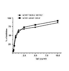
Figure 3: hCMV neutralizing activity of the natural 26A1 antibody, as
purified by affinity chromatography using Protein A from the supernatant of a
26A1
subculture-derived cell culture maintained in serum-free medium. The dose-
response
curve was performed in the hCMV neutralization assay including either human
embryonic fibroblasts (HELF) together with the hCMV strain AD169 (1,000
PFU/reaction; IC50 0.821.1g/flap, or human umbilical vein endothelial cells
(HUVEC) together with the hCMV strain VR1814 (1,000 PFU/reaction; IC50 0.67
lig/m1).
Figure 4: gH(Ag)-specific (A) and CG3 antigen-specific (B) binding activity
of IgG-containing supernatants from subcultures of immortalized human B cells.
The ELISA was performed using the cell culture medium only (medium, negative
control), or the supernatant from subcultures 26A1, 1F7 (identified in the
immortalized cells obtained from CMV5 donor, as described in the patent
application EP07111741), and 8C10 (identified in the immortalized cells
obtained
from CMV7 donor, as described in the present patent application and in patent
application EP07115410). The dotted line represents the threshold value (0.D.
=
0.1) for considering a subculture positive.
Figure 5: (A) Alignment of the DNA (lower case, 393 base pairs) and protein
(upper case, 131 amino acids) consensus sequence of the variable region for
the
heavy chain of the 26A1 IgG (VH 26A1; SEQ ID NO.: 4 and 5). (B) Protein
CA 02672703 2009-06-15
WO 2008/071806 PCT/EP2007/064094
consensus sequence for VH 26A1 with the indication of predicted CDRs (HCDR1,
HCDR2, and HCDR3; underlined; SEQ ID NO.: 6, 7, and 8). Alternative amino
acids that were encoded by the DNA sequences cloned in plasmid from isolated
E.
coli transformants are indicated below the consensus protein sequence.
5
Figure 6: (A) Alignment of the DNA (lower case, 330 base pairs) and protein
(upper case, 110 amino acids) consensus sequence of the variable region for
the light
chain of the 26A1 IgG (VL 26A1; SEQ ID NO.: 9 and 10). (B) Protein consensus
sequence for VL 26A1 with the indication of predicted CDRs of VL 26A1 (LCDR1,
LCDR2, and LCDR3; underlined; SEQ ID NO.: 11, 12, and 13).
10
Figure 7: Alignment of the DNA (lower case, 1449 base pairs; SEQ ID NO.:
14) and protein (upper case, 482 amino acids) consensus sequence of the heavy
chain of recombinant human 26A1 monoclonal antibody (SEQ ID NO.: 15). The
most likely cleavage site for signal peptide is between pos. 19 and 20 (VLS-
QV), as
determined using the SignalP 3.0 online prediction program (Bendtsen J et al.,
2004). The sequence originally identified in cDNA generated from cells in 26A1
subculture is underlined (see Fig. 5). Amino acids 153-482 correspond to human
IgG1 heavy chain constant region (SwissProt Acc. No. P01857).
Figure 8: Alignment of the DNA (lower case, 705 base pairs; SEQ ID NO.:
16) and protein (upper case, 234 amino acids) consensus sequence of the heavy
chain of recombinant human 26A1 IgG (SEQ ID NO.: 17). The most likely cleavage
site for signal peptide is between pos. 16 and 17 (CTG-SV), as determined
using the
SignalP 3.0 online prediction program (Bendtsen J et al., 2004). The sequence
originally identified in cells from 26A1 subculture is underlined (see Fig.
5). Amino
acids 1-19 and 131-234 correspond to 1-19 and 131-234 of human Ig lambda chain
CA 02672703 2009-06-15
WO 2008/071806 PCT/EP2007/064094
11
(SwissProt Ace. No. Q8N355).
Figure 9: hCMV-neutralizing activity of recombinant human 26A1 antibody
compared to Protein A-purified natural 26A1 antibody. The activity has been
tested
using HELF cells and AD169 hCMV strain in a microneutralization assay (A; 1000
PFU/reaction, 72 hour post-infection) or in a plaque reduction assay (B).
Figure 10: hCMV-neutralizing activity of recombinant human 26A1
monoclonal antibody compared to Protein A-purified natural 26A1 antibody. The
activity has been tested using HUVEC human cells and VR1814 hCMV strain in a
microneutralization assay (A; 1000 PFU/reaction), or using HELF human cells
and
AL-1 hCMV strain in a plaque reduction assay (B; 1000 PFU/reaction).
DETAILED DESCRIPTION OF THE INVENTION
The methods described in PCT/EP2005/056871 allow the efficient
immortalization of isotype-specific human B cells obtained from an individual,
whose blood contains antibodies having biological activities (e.g. binding
and/or
neutralizing a human or viral target), at the scope of obtaining polyclonal
populations of cells that secrete antibodies presenting such biological
activities
Extensive screening assays can be then performed using supernatants of
subcultures
obtained by these methods following a single step of cloning at low cell
density (e.g.
50, 20 cells or less per well). In this manner, it is possible to obtain
polyclonal
populations of immortalized B cells in which a large repertoire of IgG-
secreting
subcultures can be characterized and consequently a number of human monoclonal
IgG having the desired binding specificity for antigens and/or the biological
activity
can be identified.
In the present case, a polyclonal population of IgG-secreting, immortalized
CA 02672703 2009-06-15
WO 2008/071806 PCT/EP2007/064094
12
human B cells were obtained from the blood of an hCMV patient whose serum
presented, as biological activity, a strong hCMV-neutralizing activity. The
polyclonal population was used to generate, in a single subcloning step at 20
cells
per well and in appropriate culture conditions, thousands of subcultures that
contain
immortalized human B cells. The specific biological activity was then tested
in the
supernatant of hundreds of efficiently growing cell cultures at the scope of
selecting
those presenting the stronger activity, and then determining the isotype and,
if
possible, the epitope of the secreted antibody.
One of the most promising subcultures, named 26A1, was used to both purify
the natural human antibody from large scale cultures and to isolate the DNA
encoding such antibody from the immortalized B cells. The DNA sequence was
used
for producing the natural human antibody as a recombinant human antibody. The
natural and the recombinant human 26A1 monoclonal antibody were used for
performing more extensive biological assays and for assessing their potential
utility
in hCMV-related clinical applications.
The examples shows how the cell culture supernatant, the natural human
antibody, and the recombinant human antibody present the same biological
activity
determined in the original blood serum and polyclonal population of human EBV-
immortalized B cells. These evidences confirm that the methods described in
PCT/EP2005/056871 allow the identification, characterization, and the
production of
biologically active, isotype-specific, natural and recombinant human
monoclonal
antibodies. In fact, the complete process of cell immortalization and growth
in cell
culture conditions gives access to the repertoire of human antibodies in a
fast,
efficient and straightforward manner. Moreover, the cells resulting from the
process
CA 02672703 2009-06-15
WO 2008/071806 PCT/EP2007/064094
13
can be frozen and screened in a different moment, or in parallel for different
biological activities and/or antigens.
In one embodiment, the present invention provides proteins comprising a
sequence having at least 90% identity with the sequence of the HCDR3 (CDR3 of
the heavy chain variable region) of the 26A1 antibody (SEQ ID NO.: 8).
Together
with the HCDR1 and HCDR2 (SEQ ID NO.: 6 and SEQ ID NO.: 7), this HCDR3 is
included in the variable region of the heavy chain of the 26A1 antibody (VH
26A1;
Fig. 5; SEQ ID NO.: 5). This sequence is encoded by the DNA sequence (Fig. 5A;
SEQ ID NO.: 4) that was amplified and cloned using cells obtained from the
original
subculture secreting the 26A1 antibody. Thus a protein of the invention may
contain,
together with the HCDR3 of the 26A1 antibody (SEQ ID NO.: 8), the sequence of
the HCDR1 (SEQ ID NO.: 6) and/or HCDR2 (SEQ ID NO.: 7) of the 26A1 antibody
(Fig. 5B). Such a protein may comprise a sequence having at least 90% identity
with
the entire sequence of the variable region of the heavy chain of the 26A1
antibody.
The 26A1 antibody also contains a variable region of a light chain for which,
using the same approach, the DNA (SEQ ID NO.: 9) and the protein (SEQ ID NO.:
10) sequences, together with the specific LCDRs (SEQ ID NO.: 11, SEQ ID NO.:
12
and SEQ ID NO.: 13), have been determined (Fig. 6). Thus a protein of the
Invention can further comprises one or more sequences selected from the group
consisting of single LCDRs of the 26A1 antibody (SEQ ID NO.: 11, SEQ ID NO.:
12 and SEQ ID NO.: 13), which can be provided as a protein sequence comprising
a
sequence having at least 90% identity with VL 26A1 (Fig. 6B; SEQ ID NO.: 10).
This applies in particular when a human recombinant antibody, comprising both
the
natural VL 26A1 and VH 26A1 sequences as light and heavy chains (in the
natural
CA 02672703 2009-06-15
WO 2008/071806 PCT/EP2007/064094
14
conformation of a tetrameric complex comprising two light and two heavy
chains, or
in a single protein as recombinant variant of the natural antibody), is
desired.
Wherever a level of identity is indicated, this level of identity should be
determined on the full length of the relevant sequence of the invention.
The HCDR3 of the 26A1 antibody can be considered as characterizing the
antigen-binding portion of a specific human antibody that is capable of
binding and
neutralizing hCMV, as shown in the Examples. Even though, several or all CDRs
of
an antibody are generally required for obtaining an antigen-binding surface,
HCDR3
is the CDR showing the highest differences between antibodies not only with
respect
to sequence but also with respect to length. Such diversities are fundamental
components of binding regions for the recognition of essentially any antigen
by the
humoral immune system (Xu and Davis, 2000; Barrios Y et al. 2004; Bond C et
al.,
2003). Alternatively, combinations of CDRs can be linked to each other in very
short proteins that retain the original binding properties, as recently
reviewed
(Ladner R, 2007).
Thus, hCMV-neutralizing proteins can be generated using the HCDR3 of
26A1 antibody as hCMV binding moiety, in combination or not with other CDRs
from the 26A1 antibody, which can be expressed within an antibody protein
framework (Knappik A et al., 2000), or within a protein framework unrelated to
antibodies (Kiss C et al., 2006).
The variable region of the heavy and light chains forming 26A1 antibody (or
selected portions, such as the isolated HCDRs and LCDRs) can be included in
any
other protein format for functional antibody fragments, as described in the
literature
under different names such as Scfv (single-chain fragment variable), Fab
(variable
CA 02672703 2009-06-15
WO 2008/071806 PCT/EP2007/064094
heavy/light chain heterodimer), diabody, peptabody, VHH (variable domain of
heavy chain antibody), isolated heavy or light chains, bispecific antibodies,
and
other engineered antibody variants for non-/clinical applications (Jain M et
al., 2007;
Laffly E and Sodoyer R, 2005).
5
Alternative antibodies can be generated using the sequences of 26A1
antibody through a process of light-chain variable domain (VL) shuffling. In
fact,
several different antibodies can be generated and tested for hCMV-specific
activity
using a single heavy chain variable domain VH (such as the one of 26A1)
combined
with a library of VL domains, at the scope of determining VH/VL combinations
with
10
improved properties in terms of affinity, stability, and/or recombinant
production
(Ohlin M et al., 1996; Rojas G et al., 2004; Watkins N et al., 2004).
Novel approaches for developing new bioactive peptides also showed the
feasibility of synthesizing CDR-derived peptides that contain L-amino acids
and/or
D-amino acids, that maintain the original activity, and that may have a more
15
appropriate pharmacological profile (Smith J et al., 1995; Levi M et al.,
2000;
Wijkhuisen A et al., 2003).
Thus, the HCDR3 of the 26A1 antibody as well as sequences highly similar
to HCDR3 of 26A1 antibody, fusion proteins containing it, and synthetic
peptides
derived from them (e.g. containing L-amino acids, D-amino acids, in the normal
or
in the retro-inverso conformation), can be tested and used as hCMV-binding and
neutralizing proteins.
Moreover, it is known that antibodies may be modified in specific positions
in order to have antibodies with improved features, in particular for clinical
applications (such as better pharmacokinetic profile or higher affinity for an
CA 02672703 2009-06-15
WO 2008/071806 PCT/EP2007/064094
16
antigen). These changes can be made in the CDRs and/or framework of the 26A1
antibody and the sequence can be chosen by applying any of the dedicated
technologies for the rational design of antibodies that make use of affinity
maturation and other processes (Kim S et al., 2005; Jain M et al., 2007).
The proteins of the invention may be provided as antibodies in general, such
as fully human monoclonal antibodies having a specific isotype. The IgG
isotype,
for example, is the antibody format of almost all approved therapeutic
antibodies
(Laffly E and Sodoyer R, 2005). However, antigen binding portions isolated
from a
HIV-neutralizing IgG1 were transferred on a human IgA sequence and the
resulting
antibody is capable of inhibiting HIV infection as well (Mantis N et al.,
2007).
The protein of the invention may be also provided as antibody fragments,
bioactive peptides, or fusion proteins. All these alternative molecules should
maintain, if not enhance, the original hCMV binding and neutralization
properties
that were determined for the 26A1 antibody. In the case of fusion proteins,
the
heterologous protein sequences can be located in the N- or C-terminal position
to the
26A1-derived sequence, without affecting the correct expression and biological
activity of the hCMV-specific moiety (e.g. an antibody fragment).
The term "heterologous protein sequence" indicates a protein sequence that is
not naturally present in the N- or C-terminal position to the hCMV-specific
moiety
(e.g. an antibody fragment). The DNA sequence encoding this protein sequence
is
generally fused by recombinant DNA technologies and comprises a sequence
encoding at least 5 amino acids.
Such a heterologous protein sequence is generally chosen for providing
additional properties to the hCMV-specific antibody fragment for specific
diagnostic
CA 02672703 2009-06-15
WO 2008/071806 PCT/EP2007/064094
17
and/or therapeutic uses. Examples of such additional properties include:
better
means for detection or purification, additional binding moieties or biological
ligands, or post-translational modification of a fusion protein (e.g.
phosphorylation,
glycosylation, ubiquitination, SUMOylation, or endoproteolytic cleavage).
Alternatively (or additionally to the fusion with a heterologous protein
sequence), the activity of a protein of the invention may be improved with the
conjugation to different compound such as therapeutic, stabilizing, or
diagnostic
agents. Examples of these agents are detectable labels (e.g. a radioisotope, a
fluorescent compound, a toxin, a metal atom, a chemiluminescent compound, a
bioluminescent compound, or an enzyme) that can be bound using chemical
linkers
or polymers. The hCMV-specific biological activity may be improved by the
fusion
with another therapeutic protein, such as a protein or a polymer altering the
metabolism and/or the stability in diagnostic or therapeutic applications.
Means for choosing and designing protein moieties, ligands, and appropriate
linkers, as well as methods and strategies for the construction, purification,
detection
and use of fusion proteins are provided in the literature (Nilsson J et al.,
1997;
"Applications Of Chimeric Genes And Hybrid Proteins" Methods Enzymol. Vol.
326-328, Academic Press, 2000; WO 01/77137) and are commonly available in
clinical and research laboratories. For example, the fusion protein may
contain
sequences recognized by commercial antibodies (including tags such as
polyhistidine, FLAG, c-Myc, or HA tags) that can facilitate the in vivo and/or
in
vitro identification of the fusion protein, or its purification.
Other protein sequences can be identified by direct fluorescence analysis (as
in the case of Green Fluorescent Protein), or by specific substrates or
enzymes (e.g.
CA 02672703 2009-06-15
WO 2008/071806 PCT/EP2007/064094
18
using proteolytic sites). The stability of hCMV-specific antibodies, antibody
fragments, bioactive peptides, and fusion proteins may be improved with the
fusion
with a carrier protein, such as phage coat protein (cp3 or cp8), Maltose
Binding
Protein (MBP), Bovine Serum Albumin (BSA), or Glutathione-S-Transferase (GST).
The 26A1 antibody is a main object of the invention and it has been
characterized, within the supernatant of a specific subculture, as a human
IgG1
monoclonal antibody which has been selected due to the capability of
neutralizing
hCMV. This property have been determined by in vitro neutralization assays
using
cell culture supernatant (Table 1), and later as a Protein A-purified (Fig. 3)
and
-- recombinant (Fig. 9 and 10; SEQ ID NO.: 15 and 17) human monoclonal
antibody.
The specific hCMV antigen that is recognized by 26A1 antibody has not been
determined using a panel of known hCMV neutralizing epitopes in viral antigens
(see Fig. 1, 2, and 4). Consequently, this IgG antibody can be used for
defining an
hCMV-neutralizing epitope and proteins binding such antigen (e.g. in form of
the
antibodies, antibody fragments, bioactive peptides, fusion protein, or any
natural/recombinant proteins) that should be capable of neutralizing hCMV
infection
by recognizing such epitope.
In the past, ELISA or Western Blot using hCMV-specific truncated proteins
or synthetic peptides have been also used (Greijer A et al., 1999; Ohlin M et
al.,
-- 1993) and in this way antibodies directed to hCMV have been defined
according to
their antigen, being glycoprotein H (WO 94/16730, WO 94/09136, WO 92/11018),
glycoprotein B (EP248909, WO 93/21952) or glycoprotein M/glycoprotein N
(Shimamura M et al., 2006). Moreover, other components of the hCMV virion not
only affect viral tropism but can be targets of hCMV neutralizing antibodies,
as in
CA 02672703 2009-06-15
WO 2008/071806 PCT/EP2007/064094
19
the case of pUL130 and pUL128 (Wang D and Shenk T, 2005). Thus, the CMV
antigen / epitope recognized by the 26A1 antibodies can be identified by
different in
vitro assays based on the literature cited above
A further embodiment of the present invention is human IgG1 antibody
secreted by the 26A1 subculture, which can be provided as a Protein A-purified
natural antibody that binds and neutralized hCMV. This IgG1 antibody can be
used
for identifying competing proteins that can bind and neutralize hCMV as well.
Similar proteins are provided in the above description and in the Examples, in
particular as recombinant human antibodies and antibody fragments.
The mechanism of hCMV neutralization, that are associated to the viral
epitope recognized by the 26A1 antibody and the other proteins defined above,
can
be characterized using the available assays for specific structural hCMV
proteins
and/or strain, as shown in the literature using panels of human sera (Navarro
D et
al., 1997; Klein M et al., 1999; Weber B et al., 1993; Rasmussen L et al.,
1991;
Marshall G et al., 2000) or of monoclonal antibodies (Schoppel K et al., 1996;
Simpson J et al., 1993; Gicklhorn D et al., 2003).
Further objects of the inventions are the nucleic acids encoding any of the
antibodies, antibody fragments, fusion proteins, bioactive peptides, or
isolated
HCDRs and LCDRs defined above. The examples provide such sequences in
particular as encoding the full variable regions of the 26A1 heavy (SEQ ID
NO.: 4)
and light (SEQ ID NO.: 9) chains (Fig. 5A and 6A). These DNA sequences (or
selected portions, such as those encoding the specific HCDRs and LCDRs; Fig. 5
and 6) can be transferred in vectors for expressing them in one of the
alternative
formats for antibodies (e.g. full, affinity-matured, or CDR-grafted or
antibody
CA 02672703 2009-06-15
WO 2008/071806 PCT/EP2007/064094
fragments) or fusion proteins. These nucleic acids can comprise a sequence
having
at least 90% identity with SEQ ID NO.: 4, with or without a sequence further
comprising a sequence having at least 90% identity with SEQ ID NO.: 9,
depending
on whether sequences from only the heavy chain of 26A1 or both heavy and light
5 chain are needed.
When a fully human antibody is desirable, the antibody should further
comprise a heavy chain constant region selected from the group consisting of
IgGl,
IgG2, IgG3, IgG4, IgM, IgA and IgE constant regions. Preferably, the heavy
chain
constant region is a human IgA, IgG1 (as in the natural 26A1 antibody
characterized
10 from the 26A1 subculture), IgG2, or IgG4. The nucleic acid sequences
encoding the
full variable regions of 26A1 heavy and light chains have been cloned and
characterized by means of PCR reactions and vectors containing the resulting
PCR
products, which have used for transforming E coli cells. Such sequences can be
transferred (in part or in their entirety) into another vector, in particular
in the
15 expression cassette of a vector or of distinct vectors where they are
operably linked
to appropriate regulatory sequences (e.g. promoters, transcription
terminators).
The human 26A1 monoclonal antibody, or any other protein sequences
derived from such antibody, can be expressed as a recombinant protein using
such
vectors for transforming the appropriate host cells. The host cells comprising
the
20 nucleic acids of the invention can be prokaryotic or eukaryotic host
cells and should
allow the secretion of the desired recombinant protein. Methods for producing
such
proteins include culturing host cells transformed with the expression vectors
comprising their coding sequences under conditions suitable for protein
expression
and recovering the protein from the host cell culture. The vectors should
include a
CA 02672703 2009-06-15
WO 2008/071806 PCT/EP2007/064094
21
promoter, a ribosome binding site (if needed), the start/stop codons, and the
leader/secretion sequence, that can drive the expression of a mono or
bicistronic
transcript for the desired protein. The vectors should allow the expression of
the
recombinant protein in the prokaryotic or eukaryotic host cells. A cell line
substantially enriched in such cells can be then isolated to provide a stable
cell line.
The nucleic acids and host cells can be used for producing a protein of the
invention by applying common recombinant DNA technologies. Briefly, the
desired
DNA sequences can be either extracted by digesting the initial cloning vector
with
restriction enzymes, or amplified using such a vector as a template for a
Polymerase
Chain Reaction (PCR) and the PCR primers for specifically amplifying full
variable
regions of the heavy and light chains or only portions of them (e.g. the HCDR3
sequence). These DNA fragments can be then transferred into more appropriate
vectors for the expression into prokaryotic or eukaryotic host cells, as
described in
books and reviews on how to clone and produce recombinant proteins, including
titles in the series "A Practical Approach" published by Oxford Univ. Press
("DNA
Cloning 2: Expression Systems", 1995; "DNA Cloning 4: Mammalian Systems",
1996; "Protein Expression", 1999; "Protein Purification Techniques", 2001).
For eukaryotic hosts (e.g. yeasts, insect or mammalian cells), different
transcriptional and translational regulatory sequences may be employed,
depending
on the nature of the host. They may be derived from viral sources, such as
adenovirus, bovine Papilloma virus, Simian virus or the like, where the
regulatory
signals are associated with a particular gene which has a high level of
expression.
Examples are the TK promoter of the Herpes virus, the 5V40 early promoter, the
yeast gal4 gene promoter, etc. Transcriptional initiation regulatory signals
may be
CA 02672703 2009-06-15
WO 2008/071806 PCT/EP2007/064094
22
selected which allow for the transient (or constitutive) repression and
activation and
for modulating gene expression.
The sequence encoding the recombinant protein can be adapted and recloned
for making modifications at the DNA level only that can be determined, for
example, using software for selecting the DNA sequence in which the codon
usage
and the restriction sites are the most appropriate for cloning and expressing
a
recombinant protein in specific vectors and host cells (Grote A et al., 2005;
Carton J
et al., 2007).
During further cloning steps, protein sequences can be added in connection to
the desired antibody format (Scfv, Fab, antibody fragment, fully human
antibody,
etc.), or to the insertion, substitution, or elimination of one or more
internal amino
acids. These technologies can also be used for further structural and
functional
characterization and optimization of the therapeutic properties of proteins in
general,
and of antibodies in particular (Kim S et al., 2005), or for generating
vectors
allowing their stable in vivo delivery (Fang J et al., 2005). For example,
recombinant
antibodies can also be modified at the level of structure and/or activity by
choosing
a specific Fc region to be fused to the variable regions (Furebring C et al.,
2002;
Logtenberg T, 2007), by generating single chain antibody fragments (Gilliland
L et
al., 1996), and by adding stabilizing peptide sequences, (WO 01/49713),
polymers or
radiochemicals to chemically modified residues (Chapman A et al., 1999).
The DNA sequence coding for the recombinant protein, once inserted into a
suitable episomal or non-homologously or homologously integrating vector, can
be
introduced in the appropriate host cells by any suitable means
(transformation,
transfection, conjugation, protoplast fusion, electroporation, calcium
phosphate
CA 02672703 2009-06-15
WO 2008/071806 PCT/EP2007/064094
23
precipitation, direct microinjection, etc.) to transform them. Important
factors to be
considered when selecting a particular vector include: the ease with which
host cells
that contain the vector may be recognized and selected; the number of copies
of the
vector which are desired; and whether the vector is able to "shuttle" between
host
cells of different species.
The cells which have been stably transformed by the introduced DNA can be
selected by also introducing one or more markers which allow for selection of
host
cells which contain the expression vector. The marker may also provide for
phototrophy to an auxotropic host, biocide resistance, e.g. antibiotics, or
heavy
metals such as copper, or the like, and may be cleavable or repressed if
needed. The
selectable marker gene can either be directly linked to the DNA gene sequences
to
be expressed, or introduced into the same cell by co-transfection. Additional
transcriptional regulatory elements may also be needed for optimal expression.
Host cells may be either prokaryotic or eukaryotic. Amongst prokaryotic host
cells, the preferred ones are B. subtilis and E. coli. Amongst eukaryotic host
cells,
the preferred ones are yeast, insect, or mammalian cells. In particular, cells
such as
human, monkey, mouse, insect (using baculovirus-based expression systems) and
Chinese Hamster Ovary (CHO) cells (as shown in the Examples), provide post-
translational modifications to protein molecules, including correct folding or
certain
forms of glycosylation at correct sites. Also yeast cells can carry out post-
translational peptide modifications including glycosylation. A number of
recombinant DNA strategies exist which utilize strong promoter sequences and
high
copy number of plasmids that can be utilized for production of the desired
proteins
in yeast. Yeast recognizes leader sequences in cloned mammalian gene products
and
CA 02672703 2009-06-15
WO 2008/071806 PCT/EP2007/064094
24
secretes peptides bearing leader sequences (i.e., pre-peptides).
Mammalian cell lines available as hosts for expression are known in the art
and include many immortalized cell lines available from the American Type
Culture
Collection (ATCC) including, but not limited to, Chinese hamster ovary (CHO),
HeLa, baby hamster kidney (BHK), monkey kidney (COS), C127, 3T3, BHK, HEK
293, Per.C6, Bowes melanoma and human hepatocellular carcinoma (for example
Hep G2) cells and a number of other cell lines. In the baculovirus system, the
materials for baculovirus/insect cell expression systems are commercially
available
in kit form (e.g. commercialized by Invitrogen).
For long-term, high-yield production of a recombinant polypeptide, stable
expression is preferred. For example, cell lines which stably express the
polypeptide
of interest may be transformed using expression vectors which may contain
viral
origins of replication and/or endogenous expression elements and a selectable
marker gene on the same or on a separate vector. Following the introduction of
the
vector, cells may be allowed to grow for one or more days in an enriched media
before they are switched to selective media. The purpose of the selectable
marker is
to confer resistance to selection, and its presence allows growth and recovery
of
cells that successfully express the introduced sequences. Resistant clones of
stably
transformed cells may proliferate using tissue culture techniques appropriate
to the
cell type. A cell line substantially enriched in such cells can be then
isolated to
provide a stable cell line.
In the case of full recombinant human immunoglobulins, an important step is
the selection of the specific isotype and constant region. Vectors
specifically
designed for expressing antibodies with the desired isotype and subtype (for
CA 02672703 2009-06-15
WO 2008/071806 PCT/EP2007/064094
example, human IgA, IgGl, IgG2, or IgG4) are widely described in the
literature.
Then, the full antibodies or the fusion proteins can be expressed as
recombinant
proteins in prokaryotic organisms (e.g. Escherichia coli; Sorensen H and
Mortensen
K, 2005; Venturi M et al., 2002), plants (Ma J et al., 2005), or eukaryotic
cells, that
5 allow
a high level of expression as transient or stable transformed cells (Dinnis D
and James D, 2005). This would be required in particular when the
characterization
of the antibodies has to be performed using more sophisticated assays,
including in
vivo assays, where the half-life of the antibody can be determined. The host
cells can
be further selected on the basis of the expression level of the recombinant
protein.
10 In
addition, when the protein is expressed, especially as an antibody, in
eukaryotic host cells (mammalian cell lines, in particular), different vectors
and
expression systems have been designed for generating stable pools of
transfected
cell lines (Aldrich T et al., 2003; Bianchi A and McGrew J, 2003). High level,
optimized, stable expression of recombinant antibodies has been achieved
(Schlatter
15 S et
al., 2005), also due to optimization of cell culture conditions (Grunberg J et
al.,
2003; Yoon S et al., 2004) and by selecting or engineering clones with higher
levels
of antibody production and secretion (Bohm E et al., 2004; Butler M, 2005;).
The antibody, the antibody fragments, the bioactive peptide, the fusion
protein, and any other protein defined above as being capable of binding and
20
neutralizing hCMV can be purified using the well-established technologies that
allow the isolation of either non-/recombinant proteins from cell culture or
from
synthetic preparations. These technologies should provide a sufficient amount
of
protein (from the microgram to the milligram range) to perform a more
extensive
characterization and validation for hCMV-related prophylactic, diagnostic, and
CA 02672703 2009-06-15
WO 2008/071806 PCT/EP2007/064094
26
therapeutic uses. To this purpose, the preparations of recombinant or natural
proteins
can be tested in in vitro or in vivo assays (biochemical, tissue- or cell-
based assays,
disease models established in rodents or primates, biophysical methods for
affinity
measurements, epitope mapping, etc.), in particular using any of those
disclosed in
the Examples or in literature for studying hCMV pathogenesis and
immunobiology.
The antibodies, as purified preparations from human B cell supernatants or
expressed as recombinant proteins, can be further validated using organ- or
cell-
based in vitro assays known in the literature (Eggers M et al. 1998; Lam V et
al.,
2006; Reinhardt B et al., 2003; Forthal D et al., 2001; Goodrum F et al.,
2002).
Moreover, relevant pre-clinical tests can be made in hCMV-infected animals, in
particular in models where human host cells can be transplanted (Barry P et
al.,
2006; Gosselin J et al., 2005; Thomsen M et al., 2005).
The purification of the recombinant proteins of the invention can be carried
out by any of the conventional methods known for this purpose, i.e. any
procedure
involving extraction, precipitation, chromatography, or the like. In
particular,
methods for antibody purification can make use of immobilized gel matrices
contained within a column (Nisnevitch M and Firer M, 2001; Huse K et al.,
2002;
Horenstein A et al., 2003), exploiting the strong affinity of antibodies for
substrates
such Protein A, Protein G, or synthetic substrates (Verdoliva A et al., 2002;
Roque
A et al., 2004), or for specific antigens or epitopes (Murray A et al., 2002;
Jensen L
et al., 2004). After washing, the protein is eluted from the gel by a change
in pH or
ionic strength. Alternatively, HPLC (High Performance Liquid Chromatography)
can be used. The elution can be carried out using a water-acetonitrile-based
solvent
commonly employed for protein purification.
CA 02672703 2009-06-15
WO 2008/071806
PCT/EP2007/064094
27
The antibody, the antibody fragments, the bioactive peptides, the fusion
proteins, and any other compound defined above using 26A1 antibody sequences
can
be used for detecting, treating, inhibiting, preventing, and/or ameliorating
hCMV
infection. To this purpose, such compounds can be used for preparing
diagnostic,
therapeutic, or prophylactic compositions for the management of hCMV
infection.
In particular such compounds can be used for preparing pharmaceutical
compositions, together with any pharmaceutically acceptable vehicle or
carrier.
These compositions may further comprise any additional therapeutic or
prophylactic
agent, such as vaccines, hCMV-neutralizing antibody, intravenous
immunoglobulin
preparations, immunomodulating compounds, and/or antiviral compounds. The
literature provides some examples of such compounds acting on hCMV replication
(Foscarnet, Vanganciclovir, Fomivirsen, or Ganciclovir) and already tested in
humans, alone or in combination with intravenous immunoglobulin preparations
(De
Clercq E, 2003; Nigro G et al., 2005).
Moreover, recent literature suggets that human monoclonal antibodies can be
used for supplementing (and replacing, if possible) present treatments such as
intravenous immunoglobulin preparations and/or antiviral compounds, giving the
opportunity to reduce frequency and/or dosage of such pharmaceutical
compositions
(Bayry J et al., 2007).
These compositions may comprise an antibody, an antibody fragment, a
bioactive peptide, a fusion protein, and any other compound defined above on
the
basis of the sequence and activity of human 26A1 monoclonal antibody sequence.
The compositions may further comprise a different hCMV-neutralizing antibody,
an
intravenous immunoglobulins (IVIg) preparation and/or an antiviral compound.
The
CA 02672703 2009-06-15
WO 2008/071806 PCT/EP2007/064094
28
different hCMV-neutralizing antibody should be characterized by a different
epitope, such as the ones already described in the literature or in the patent
applications EP07114782, EP07115410, and EP07111741 (10B7, 8C10, and 1F7,
respectively) that are associated to gH, gB, or other hCMV antigens. In fact,
the
literature shows many examples in which, when two or more antibodies directed
to a
viral or human target are combined in a pharmaceutical composition, the
resulting
composition may have an improved therapeutic efficacy due not to a simple
additive
effect but to a specific synergic effect (Logtenberg T, 2007).
The compositions comprising any of the proteins (e.g. antibodies, antibody
fragments, fusion proteins, bioactive peptides) and of the nucleic acids
defined
above can be used and administered to an individual with a hCMV-related
diagnostic, therapeutic, or prophylactic purpose. These compositions can be
administered as means for hCMV-specific passive immunization which provide
therapeutic compounds (in particular therapeutic antibodies or therapeutic
antibodies
fragments) that, by targeting hCMV virions, can inhibit the propagation of the
virus
in the treated patient, and potentially block the outbreak of a viral
infection in the
population.
Depending on the specific use, the composition should provide the compound
to the human subject (in particular a pregnant woman or any other individual
that is
infected by hCMV or considered at risk for hCMV due to contact with an hCMV-
infected individual) for a longer or shorter period of time. To this purpose,
the
composition can be administered, in single or multiple dosages and/or using
appropriate devices, through different routes: intramuscularly, intravenously,
subcutaneously, topically, mucosally, by a nebulizer or an inhaler, as
eyedrops, in
CA 02672703 2009-06-15
WO 2008/071806 PCT/EP2007/064094
29
non-/biodegradable matrix materials, or using particulate drug delivery
systems. In
particular, the composition may allow topical or ocular administration, that
represent
a useful approach given the presence of hCMV in mucosae and eye. Moreover,
antibodies and antibody fragments can be effective when applied topically to
wounds (Streit M et al., 2006), cornea (Brereton H et al., 2005) or vagina
(Castle P
et al., 2002).
A pharmaceutical composition of the Invention should provide a
therapeutically or prophylactically effective amount of the compound to the
subject
that allows the compound to exert its activity for a sufficient period of
time. The
desired effect is to improve the status of the hCMV patient by controlling
hCMV
infection, reactivation, and/or re-infection, and by reducing at least some of
the
clinical manifestations of hCMV infection, such as retinitis or pneumonitis
(Landolfo S et al., 2003). For example, the composition should be administered
at an
effective amount from about 0.005 to about 50 mg/kg/body weight, depending on
the
route of administration, the number of administered doses, and the status of
the
individual.
In the case of compositions having diagnostic uses, the compound should be
detected using technologies commonly established in the clinical and research
laboratories for detecting virus in biological samples (e.g. ELISA or other
serological assays), or, when administered to a subject in vivo, at least 1,
2, 5, 10,
24, or more hours after administration. The detection of hCMV can be
performed,
using the proteins of the invention, in substitution or coupled to the known
means
and procedures that have been established for monitoring chronic or acute hCMV
infection in at risk populations of both immuno competent and immuno
compromised
CA 02672703 2009-06-15
WO 2008/071806 PCT/EP2007/064094
hosts, where a correlation between the data generated in vitro and the
clinical status
exists (Gilbert G, 2002; Gerna G and Lilleri D, 2006; Lazzarotto T et al.,
2007).
A method for treatment, prophylaxis, or diagnosis of hCMV, or of hCMV-
related disease can comprise the administration of a protein or of a nucleic
acid as
5 above defined. The method may further comprise the administration of a
different
hCMV-neutralizing antibody, an intravenous immunoglobulins (IVIg) preparation
and/or an antiviral compound.
Clinical development and use should be based on the pharmacokinetics and
pharmacodynamics of the antibody (Lobo E et al., 2004; Arizono H et al.,
1994), the
10 preclinical and clinical safety data (Tabrizi M and Riskos L, 2007), and
the
compliancy to international requirements for the production and quality
control of
monoclonal antibodies to be used for therapy and in vivo diagnosis in humans
(Harris R et al. 2004).
The proteins of the invention can be also used for the preparation of a
15 composition for detecting, treating, inhibiting, preventing, and/or
ameliorating other,
more widespread diseases (such as cardiovascular and autoimmune diseases, or
some types of cancer) that can be defined as hCMV-related or hCMV-associated
diseases. In these conditions, hCMV is considered as a possible cofactor since
it is
well-known that this virus is associated with cellular/immunological
inflammatory
20 processes (by stimulating the expression of Fc receptors, cell adhesion
molecules,
chemokines and cytokines), autoimmune activities (e.g. in atherosclerosis,
restenosis) and with alterations to the antigen-presentation pathways (by
inhibiting
MHC class I and II expression) leading to cell apoptosis, differentiation, and
migration, for example in blood vessels and in actively proliferating cells
(Cinatl J et
CA 02672703 2009-06-15
WO 2008/071806 PCT/EP2007/064094
31
al., 2004; Soderberg-Naucler C, 2006b).
Moreover, hCMV infection has been also found associated to alteration of
cellular metabolism (Munger J et al., 2007), depression (Phillips A et al.,
2007), or
risk factor for thrombotic events (Fridlender Z et al., 2007). Reactivation of
hCMV
and related complications has also been found in cancer patients (Sandherr M
et al.,
2006; Han X, 2007) or patients affected by inflammatory connective tissue
diseases
(Yoda Y et al., 2006), and in general in patients under immunosuppressive
treatments such as corticosteroids (Yamashita M et al., 2006), or chemotherapy
and
other antibody-based immunosuppressive regimens (O'Brien S et al., 2006;
Scheinberg P et al., 2007).
The invention will now be described by means of the following Examples,
which should not be construed as in any way limiting the present invention.
EXAMPLES
Example 1: Production of cell cultures secreting human monoclonal antibodies
that neutralize hCMV infectivity
Materials & methods
Selection of human donors that present IgG antibodies neutralizing hCMV in the
blood serum
These hCMV-specific assays have been performed as outlined in
PCT/EP2005/056871 or in the literature, as summarized below.
The hCMV-neutralizing antibodies were detected according to an hCMV
microneutralization assay based on human Embryo Lung Fibroblasts (HELF cells)
and hCMV AD169 strain (an hCMV laboratory strain from ATCC, cod. VR-538).
The hCMV microneutralization assays were also performed with the
CA 02672703 2009-06-15
WO 2008/071806 PCT/EP2007/064094
32
endotheliotropic hCMV VR1814 strain, a derivative of a clinical isolate
recovered
from a cervical swab of a pregnant woman (Revello M et al., 2001), and human
umbilical vein endothelial cells (HUVEC). These cells were obtained by
enzymatic
treatment of umbilical cord veins and cultured in endothelial growth medium
(EGM-
2, Cambrex Bio Science) supplemented with 2% Foetal Bovine Serum (FBS), human
recombinant vascular endothelial growth factor (VEGF), basic fibroblast growth
factor (bFGF), human epidermal growth factor (hEGF), insulin growth factor
(IGF-
1), hydrocortisone, ascorbic acid, heparin, gentamycin and amphotericin B, (1
pg/m1
each). Experiments were performed with cells at passage 2-6.
The use of HELF and HUVEC cells for studying hCMV infection and
replication using clinical and laboratory strains has been described in many
articles
(Gerna G et al., 2002). In the present case, the cells were plated (2.0-
2.5x10⁴/well)
onto flat-bottom wells of a 96-well plate in 100 1.11 of Growth Medium, which
contains Minimum Essential Medium (MEM; Gibco-BRL) with 10% Foetal Calf
Serum (FCS), 1 mM sodium pyruvate (NaP), and GPS (2 mM glutamine, 100 U/ml
penicillin and 100 pg/m1 streptomycin). Cells were cultured for 24 hours at
37'C.
Fifty 1.11 of antibody-containing samples (human serum, cell culture
supernatants, or of Protein A-purified natural or recombinant IgG at indicated
concentrations) were incubated with the laboratory strain hCMV AD169 [500
plaque
forming units (pfu) in 50 1.11 of MEM with 5% FCS; total volume of the mixture
was
100 pi] for 1 hour at 37 C. The mixture of antibody preparation and virus was
then
added to HELF cell monolayers (for hCMV AD169 and AL-1 strains) or HUVEC
cell monolayers (for hCMV VR1814) and incubated for 1 hour. The Growth Medium
was discarded from cell monolayers and replaced with the antibody-virus
mixture.
CA 02672703 2014-07-22
33
The plates were then centrifuged at 2,000 g for 30 minutes and incubated for
90
minutes at 37 C in 5% CO2. Growth Medium (100 ill) was added and the cultures
were maintained in the incubator for a further 72 hours.
The effect of B cell supernatants on hCMV infectivity was measured by
staining hCMV Intermediate Early Antigens (TEA, IE1 + 1E2) by indirect
immunoperoxidase staining. The cell monolayers were fixed with
acetone/methanol
solution (stored at -20 C) for 1 minute then washed with PBS. The cells were
permeabilized 111 0.1% TritonTm X-100 in PBS with 1% H202, 5 minutes on ice,
then
washed with PBS. Endogenous peroxidase was blocked with PBS with 50%
methanol and 0.6% H202, 30 minutes at room temperature in the dark and then
washed with PBS. Fifty 1.11 of Protein Blocking Agent (Ultra Tech HRP 500-600
Test; Streptavidin-Biotin Universal Detection System; PN IM2391) were added
for
10 minutes at room temperature, and then washed away with PBS. Mouse anti-
HCMV IEA (clone E13; Argene Biosoft; ref. 11-003) was added to wells for 60
minutes at room temperature. After washing, cells were incubated with 50 1.11
of
biotin-conjugated, secondary anti-human IgG (Ultra Tech HRP 500-600 Test;
Streptavidin-Biotin Universal Detection System; PN IM2391) or peroxidase-
conjugated goat anti-mouse IgG (Ultratech HRP).for 10 minutes. DAB substrate
(Merck; no. 1.02924.0001) in 0.1% H202 was added for 30-45 minutes at 20 C in
the dark and the reaction stopped by dilution with PBS. TEA-positive nuclei
were
counted under the microscope.
Medium only or cell culture supernatants containing irrelevant IgG antibodies
were used as a negative control. A commercial preparation of human IgG,
purified
from the serum of hCMV seropositive patients (Cytotect; Biotest), was used as
a
CA 02672703 2009-06-15
WO 2008/071806 PCT/EP2007/064094
34
positive control with progressive dilutions, starting at 125 [tg/ml.
Positivity was
defined as
40% inhibition of IEA-positive cells, compared to negative control
wells.
The 50%-inhibition endpoint calculated using the Reed-Munch method will
be considered the Neutralization Titre (NT):
NT = reciprocal antibody dilution [>50% inhibition] x [(% inhibition greater
than
50% - 50%)/(% inhibition greater than 50% - % inhibition less than 50%)]
Selection of human donors on the basis of the presence in the serum of IgG
that bind
to regions of the hCMV envelope glycoproteins gB or gH
The hCMV-specific binding assays have been performed as outlined in
PCT/EP2005/056871 or indicated by the Manufacturer, and validated with a
commercial mixture of IgG antibodies specific for CMV (Cytotect; Biotest). The
serum was tested in an ELISA specific for human IgG binding hCMV virion
proteins
that is commercially available (BEIA-CMV IgG Quant; Bouty, cod. 21465) and a
gB
(AD2) hCMV IgG ELISA, also commercially available (CG3 antigen Biotest AG,
cod. 807035; Fig.1A).
Briefly, breakable strips covered with an inactivated hCMV protein mixture
(derived from the laboratory strain AD169) were placed into microplates and
incubated with B cell supernatants diluted 1:81(10 IA of supernatants added to
800
iAl of sample diluents of the BEIA system), and the plate incubated at room
temperature for 30 minutes. After a washing cycle, pre-diluted monoclonal anti-
human IgG antibody conjugated with horseradish peroxidase (100 pl) was added
and
plate incubated at room temperature for a further 30 minutes. After a second
washing cycle, pre-diluted substrate-TMB solution (100 pl) was added and the
plate
CA 02672703 2014-07-22
was incubated at room temperature for 15 minutes. The reaction was stopped
using
the Stop Solution (100 pd/well) and the optical density was measured in bi-
chromatism at 450/620 nanometers.
Production of the culture of immortalized human B cells
5
Peripheral blood mononuclear cells (PBMCs) were obtained from a patient
recovered from an acute hCMV infection (CMV7) selected because of the presence
of hCMV-neutralizing antibodies in the serum. The EBV immortalization process
to
which PBMCs from CMV7 patient were subsequently exposed has been performed
according to the teachings of PCT/EP2005/056871. Briefly, PBMCs were purified
10 from peripheral blood by conventional density gradient centrifugation on
FicollTm/Hypaque. CD22-positive cells were isolated from fresh PBMC (>90%
purity)
with anti-human CD22-coated beads by the VarioMACS technique (Miltenyi Biotec
Inc.) as described by the manufacturer. The purified cells were stimulated
with a
combination of CpG2006 (Coley, 1 lig/m1) and IL-2 (Roche, 200U/m1). After a 4-
15 day
stimulation, cells were washed with fresh culture medium (RPMI-1640) and the
B cells were highly enriched in IgG-positive cells with anti-human IgG-coated
beads
by using the VarioMACS technique (Miltenyi Biotec Inc.), following the
manufacturer's instructions.
The selected and stimulated cells were suspended and maintained in RPMI-
20 1640
cell culture medium supplemented with 10% (v/v) heat-inactivated FCS (Foetal
Calf Serum), 1 mM sodium pyruvate, 100 p.g/m1 streptomycin and 100 U/ml
penicillin, in 24-well plates at 37 C and 5% CO2. The EBV immortalization was
performed using B95.8 cell supernatant (1:1 v/v for 16 hours).
At the end of the process, the immortalized cells were washed with fresh
CA 02672703 2014-07-22
36
culture medium (RPMI 1640 added with 10% Foetal Calf Serum) and put in culture
for 3 weeks at a density of 1.5 x 10⁶ cells/ml in 24 well plates with a feeder
layer
(irradiated allogeneic PBMC seeded at 5 x 10⁵ cells/well), without CpG2006
(and
not with CPG2006 as described in PCT/EP2006/069780 for the process started
from
PBMCs obtained from CMV5 donor).
Selection of subcultures of immortalized human B cells that secrete hCMV
neutralizing antibodies
Fifteen days after exposure to EBV, the hCMV neutralizing activity was
confirmed in the expanded, polyclonal cell culture with the AD169/HELF-based
microneutralization assay described above. Then, the cells were seeded at 20
cells/well on irradiated (30 Gy), allogeneic PBMCs (50,000/well) in 100 1.11
IMDM
added with 10% FCS and non essential amino acids (NEAA, diluted IX from a
100X commercial stock solution; EuroClone) in the absence of CpG2006 and IL-2.
A total of 4224 subcultures were generated and, after two weeks, 50 ill of the
same
medium were added. After a further 1-2 weeks of culture, the supernatants of
cell
cultures that presented growing and aggregated cells were tested in parallel
using the
hCMV neutralization assay based on HELF cells and hCMV strain AD169.
The supernatants of cell cultures that presented hCMV neutralizing activity
were tested using the ELISAs for detecting binding of human IgG to regions of
the
gB hCMV envelope glycoprotein, or total hCMV proteins described above.
Alternatively, gB- or gH-based antigens were generated as Glutathione-S-
Transferase (GST) fusion proteins. In the case of the gB(Ag)-GST antigen, the
gB
immunodominant region from hCMV strain C194 was fused to GST (BioDesign,
Cat. No. R18102; GS-4B SepharoseTM Affinity purified, 1 mg/ml). In the case of
CA 02672703 2014-07-22
37
gH(Ag)-GST antigen, the fragment of the gH glycoprotein of HCMV strain VR1814
was cloned by PCR, fused to GST gene, produced in E. coil and purified from
the
bacterial cell lysate on the basis of GST affinity. The recombinant gH(Ag)-GST
antigen corresponds to an in-frame fusion between the gH amino terminal region
(amino acids 16-144; Fig. 1B) from the hCMV strain VR1814 and GST. GST alone
was used as negative control.
These ELISA were performed by applying a common ELISA protocol in a
96- well format with minor modifications. Briefly, the antigen is diluted at 2
g/m1
in PBS and 50 1 of this protein solution (containing 100 ng of the
bacterially
expressed fusion protein) was used for coating ETA polystyrene plates (Nunc;
Cat
No. 469949). The coating of ELISA plates was performed overnight at 4 C, then,
after eliminating the protein solution, the plates were washed four times with
150 p.1
of Wash Buffer (PBS containing 0.05 of TweenTm20). A treatment for blocking
unspecific binding was performed by then dispensing 100 IA PBS containing 1%
of
milk in each well for 1 hour at 37 C. After four washing cycles with 150 1,i1
of Wash
Buffer, 50 I of supernatants from cell cultures were incubated in each well
for 2
hours at 37 C, using 50 l/well of the cell culture medium as negative
control. After
four washings cycles, 50 I of the secondary antibody [goat anti-human IgG (Fc
specific) antibody conjugated with horseradish peroxidase; diluted 1:30,000 in
wash
buffer; Sigma, Cat. No. A0170] were dispensed in each well and plates were
incubated for 1 hour at room temperature. After four additional washing
cycles, the
enzymatic reaction was developed by adding 50 1 of Substrate-TMB (3,3',5,5'
Tetramethylbenzidine; Sigma, Cat. No. T0440) in each well for further 30
minutes at
room temperature. The chromogenic reaction was stopped by dispensing 100 .1
of
CA 02672703 2009-06-15
WO 2008/071806 PCT/EP2007/064094
38
stop solution (1N Sulphuric acid) into each well and the optical density was
read at
450 nm.
Results
Human PBMCs were obtained from an hCMV patient (CMV7) presenting a
significant hCMV neutralization titre in serum (50% neutralization at 1:105
dilution), together with a strong reactivity in an ELISA (positive at 1:64
dilution a
sample is considered positive for the presence of IgG anti-gB at 1/4 or higher
dilutions) based on the binding to the AD2 domains of glycoprotein B, one of
the
hCMV antigens best characterized as eliciting serum neutralizing antibodies
(Qadri I
et al., 1992; Kropff B et al., 1993), and cloned within the CG3 antigen (Fig.
1A).
Moreover, the CMV7 sera was positive in another ELISA using the total hCMV
virion proteins, where an activity of 74 AU/ml was measured (a sample is
considered positive for the presence of anti-hCMV IgG when the result is at
least 10
AU/ml)
B cells from the CMV7 patient were used for generating an immortalized,
polyclonal cell culture highly enriched in B cells that secrete IgG using the
EBV-
based immortalization method disclosed in PCT/EP2005/056871 and
PCT/EP2006/069780. Compared to this latter document, disclosing the selection
of
anti-hCMV antibodies from another donor (CMV5), the subcultures were prepared
from the original bulk, polyclonal population of immortalized cells in the
absence of
CpG2006 and the supernatants were first selected for the presence of
antibodies
neutralizing hCMV infectivity by the microneutralization assay, and only after
for
antibodies binding to selected hCMV antigens.
The hCMV microneutralization assay was applied only to subcultures which
CA 02672703 2009-06-15
WO 2008/071806 PCT/EP2007/064094
39
proved to be actively growing with clusters of cells at 3 weeks of culture.
The
supernatants from the 324 wells were first screened in the hCMV neutralization
assay, 20 supernatants among these were found to reduce the infectivity of
hCMV
AD169 strain by at least 40%. When characterizing the hCMV binding activities
in
these wells, only two were found positive for gB (either as CG3 antigen or a
gB(Ag)-GST fusion protein), none for gH (as a gH(Ag)-GST fusion protein; Fig.
1B), and 18 wells for neither of them (Fig. 2).
Due to the low number of cells initially seeded in each well (20 cells/well),
each subculture presenting hCMV-neutralizing activity, should likely produce
monoclonal antibodies (i.e. secreted by cells clonally originated by a single,
specific
immortalized cell), especially given the low frequency of cells in the
immortalized,
polyclonal cell population that is expected to grow and secrete hCMV-
neutralizing
IgG. Further experimental activities were designed to confirm this assumption.
Example 2: Characterization of the 26A1 antibody
Materials and Methods
Expansion and characterization of the 26A1 subculture
The cells from the original subculture 26A1 were expanded on irradiated
allogeneic PBMC in IMDM medium (added with 10% FCS and NEAA), and the
hCMV neutralizing activity was confirmed at least twice during this expansion
step
using the hCMV microneutralization assay, as described in Example 1 (see Table
1).
The amount of antibody secreted by the 26A1 subculture was determined at
24, 48, and 72 hours using a commercial quantitative human IgG ELISA kit
(Immunotek; cod. 0801182; Zeptometrix Corp.) according to the manufacturer's
instructions. The subclass of the 26A1 antibody was determined using a
commercial
CA 02672703 2009-06-15
WO 2008/071806 PCT/EP2007/064094
assay (PeliClass human IgG subclass ELISA combi-kit; cod. #RDI-M1551cib, RDI
Divison of Fitzgerald Industries Intl.).
The cell culture was gradually expanded by seeding the cells contained in 1
well of a 96-well plate (z 1 x 10⁵) in one well of a 48 well plate on
irradiated
5 allogeneic PBMC in IMDM added with 5% FCS. After 5-7 days, cells were
expanded in one well of a 24-well plate in the absence of feeder layer, in
IMDM
added with 5% FCS. Then, cells (5 x 10⁵/m1) were plated in a 6 well plate in
the
absence of feeder layer in 50% IMDM and 50% Hybridoma-SFM (Gibco, cod.
12045-084) added with 2.5% FCS. Cells were cultured in these conditions for at
10 least one week. Exponentially growing cells were then washed and
cultured in T75
Flasks in Hybridoma-SFM at a concentration ranging from 5 x 10⁵ to 106/ml.
Cell
culture supernatant was collected, the IgG quantified and purified on Protein
A
columns, dialyzed against PBS buffer and filtered (0.21.1M).
Characterization of the antibody secreted by the 26A1 subculture
15 The BEIA-CMV, gB-based, and gH-based ELISA assays were described in
Example 1 (Fig.1 and 2). The HSV assay was performed according to the
literature
(Laquerre S et al., 1998).
The hCMV plaque reduction assay was performed using the hCMV AD169
strain. Briefly, the virus was diluted to 1000 PFU/reaction. Equal amounts
(0.1 ml)
20 of virus and each antibody or cell culture supernatant were mixed
and incubated at
37 C for 1 hour. The mixtures were added to HELF cells monolayers (in 24-well
plates) and allowed to adsorb for 1 hour at 37 C. Then the antibody-virus
mixture
was removed, and 1% methylcellulose overlayer-MEM-2% FCS was added to the
infected cells. Plaques will be counted as a measure of infectivity at day 10
or 12
CA 02672703 2009-06-15
WO 2008/071806 PCT/EP2007/064094
41
post-infection.
The 26A1 cell culture supernatant was tested in immunofluorescence on non-
infected HELF or HUVEC cells. Briefly, cells (7 x 10⁴/m1) were seeded on
gelatine-
coated glass-coverslips in 24-well plates in MEM added with 10% FCS and then
grown to semi-confluence. Cells were then washed twice with warm PBS, fixed
with
a pre-cooled (at -20 C) mixture of 50% acetone/50% methanol for 1 minute at
room
temperature (RT) and then washed with PBS. Fixed cells were permeabilized with
0.2% Triton X-100 in PBS for 20 minutes on ice, washed with PBS and incubated
for 15 minutes at RT with a blocking solution (PBS added with 2% FCS).
Alternatively, fixed cells were not permeabilized to determine the capability
of
antibodies to recognize cell surface components. In this case, fixed cells
were
washed with PBS, incubated for 15 minutes at RT with a blocking solution (PBS
added with 2% FCS) and incubated with 26A1 cell culture supernatant (80 ill),
for 2
hours at 37 C. Cells were then washed with warm PBS (3 times) and incubated
with
80 1.11 of FITC-conjugated rabbit anti-human IgG F(ab')2 (Jackson
ImmunoResearch), to track the human IgG staining as green colour. The
secondary
antibodies were diluted 1:50 in PBS added with 0.05% Tween 80 and added to the
cells for 1 hour at 37 C in the dark. Then, cells were washed with warm PBS
(3
times) and counter-stained with propidium iodide (0.25 pg/m1 in PBS; Sigma) .
The
coverslips were mounted on microscope-slide using the Mounting Medium (Vector
Laboratories). Images were recorded with an Olympus Fluoview-1X70 inverted
confocal laser scanning microscope.
Characterization of 26A1 IgG DNA/protein sequence and recombinant expression
An aliquot of the cell culture, resulting from the expansion of the initial
26A1
CA 02672703 2009-06-15
WO 2008/071806 PCT/EP2007/064094
42
cell culture, was used for sequencing of the variable regions of heavy chain
(VH)
and light chain (VL) of 26A1 antibody according the technology established by
Fusion Antibodies Ltd. Pellets of frozen cells (containing at least 50,000
cells) were
used for extracting total RNA. The corresponding cDNA was produced by reverse
transcription with an oligo(dT) primer. PCR reactions were set up to amplify
VH
region using a mix of IgG specific primers, and VL region with a mix of Igk/X,
primers. The PCR products of two amplification reactions were cloned using a
Eco
RI restriction site in a sequencing vector (pCR2.1; Invitrogen) and used for
transforming TOP10 E. coli cells.
At least ten selected colonies obtained from the two transformations were
picked and analyzed by sequencing. The resulting DNA sequences were aligned
and
translated into protein sequence generating a consensus DNA and protein
sequence
for VH 26A1 (SEQ ID NO.: 4 and SEQ ID NO.: 5, respectively) and VL 26A1 (SEQ
ID NO.: 9 and SEQ ID NO.: 10, respectively). The VH 26A1 and VL 26A1 protein
sequences were compared and aligned with sequences present in databases in the
public domain (using GenomeQuest, GeneSeq, and EBI databases). The CDRs
characterizing VH 26A1 (SEQ ID NO.: 5, 7, and 8) and VL 26A1 (SEQ ID NO.: 11,
12, and 13) protein sequences were predicted by the IMGT database (Giudicelli
V et
al., 2004).
Recombinant human 26A1 monoclonal antibody was expressed in eukaryotic
cells by cloning the consensus 26A1 heavy and light chain variable region
sequences
in the same expression vector, already containing the corresponding constant
regions
(for human IgG1 heavy chain and human Ig lambda chain) , and a dual promoter
vector allowing expression of both antibody chains. The complete sequence of
CA 02672703 2009-06-15
WO 2008/071806 PCT/EP2007/064094
43
recombinant human 26A1 monoclonal antibody in the vector was confirmed by
DNA sequencing and used to transiently transfect CHO DG44 cells (Derouazi M et
al., 2006) that were adapted to grow in serum-free suspension culture and
seeded at
1 x 10⁶ cells per ml in a 125 ml spinner flask. The transfection was performed
with a
mixture of 300 iug of expression vector with 900 iug of linear 25kDa
poly(ethylenimine). Cells were incubated at 37 C in 5% CO2 for 6 days in the
spinner flask before the media was harvested. The recombinant antibody was
purified using a Protein G column on an Akta Prime chromatography unit
following
the manufacturer's standard programme.
Results
Amongst the subcultures containing growing and IgG-secreting cells, the cell
culture supernatants of a few of them contained antibodies that neutralize
hCMV
infection but did not present a significant binding to the specific
recombinant
antigens gB and gH tested by ELISA (Fig. 1 and 2). In particular, the 26A1
subculture, that secretes an IgGl, showed the stronger and more reproducible
hCMV
neutralization activity, and it was chosen for a more detailed molecular and
biological characterization.
The binding of the IgG present in the supernatant from 26A1 subculture to
hCMV was confirmed using an ELISA containing a mixture of hCMV proteins
(BEIA-CMV). At the same time, this supernatant showed a significant hCMV
neutralizing activity against different hCMV strains in two human host cell
systems
(as shown in Table 1). This activity was not observed when the supernatant was
used
in a HSV-specific neutralization assay.
Moreover, the 26A1 subculture has been expanded and further subcloned at
CA 02672703 2009-06-15
WO 2008/071806 PCT/EP2007/064094
44
cells/well to confirm its monoclonality in terms of expression of hCMV-
neutralizing, human IgGl. In fact, amongst the wells showing cell growth, all
displayed a neutralizing activity ranging from 50 to 98% in the original AD169-
based assay, confirming the results obtained with the original 26A1
subculture.
5 The
cells in the original 26A1 subculture were used for scaling-up IgG
production, generating progressively larger cultures from which IgG can be
purified
and tested in hCMV assays. Larger cultures were generated by gradually
expanding
26A1 culture and eliminating some requirements for growth in cell culture
(such as
feeder layer, Foetal Calf Serum). Using this approach, it was demonstrated
that
10
larger cell cultures generated from the original 26A1 subculture secrete IgG1
at a
concentration of approx. 16 lig/m1/106 cells. These larger cultures showed a
doubling time of 4 days, even in the absence of feeder layer, and the hCMV
neutralizing activity was maintained in culture for more than 2 months.
The hCMV neutralization assays were repeated with the 26A1 antibody, in
the form of human IgG purified by affinity chromatography from large cell
culture.
The 26A1 IgG was tested in dose-response experiments to assess the
concentration
required for 50% inhibition (inhibitory concentration 50, or IC50) of hCMV
infectivity, using two assays with different combinations of hCMV strains and
human target cells. The results showed that the potent neutralizing activity
of the
Protein A-purified natural 26A1 IgG is neither cell-type nor virus-strain
specific,
and can be evaluated quantitatively as providing IC50 values of approx. 1
lig/m1 and
a neutralizing effect approaching 100% in both assays (Fig. 3).
When comparing the neutralizing activity of the Protein A-purified natural
26A1 IgG with that of a commercial preparation of hyperimmune IgG (IVIg)
against
CA 02672703 2009-06-15
WO 2008/071806 PCT/EP2007/064094
hCMV (Cytotect, Biotest) against the clinical isolate in HUVEC cells, the IC50
of
26A1 is 25 fold lower than that required for this commercial preparation,
demonstrating the potent neutralizing activity of 26A1 antibody.
In order to exclude that the neutralizing activity present in the supernatant
5 from the 26A1 subculture is due to the binding to a cell surface
molecule, the
supernatant was tested in immunofluorescence with uninfected HELF or HUVEC
cells. This assay showed that the IgG1 produced by 26A1 culture does not bind
to
the uninfected human cells. When the same cell supernatant is tested in ELISA
against relevant hCMV antigens, the antibody does not bind such proteins (Fig.
4),
10 indicating that the 26A1 antibody likely recognizes another,
undetermined antigen
on the hCMV envelope inducing the production of neutralizing antibodies.
The monoclonality of the hCMV neutralizing antibody secreted in the 26A1-
derived cell cultures was further confirmed by sequencing IgG-specific PCR
products obtained from this cell culture. Cell pellets were prepared for RNA
15 extraction and reverse transcription using cells originated from 26A1
subculture.
The resulting cDNA was then used for amplifying VH and VL sequences using
specific primers for the variable regions of human IgG heavy and light chains,
respectively. The PCR products were then cloned in plasmids that were used for
transforming bacterial cells. Bacterial transformants were randomly picked and
used
20 for sequencing the cloned PCR products. All the clones showed the same
DNA
sequence, apart from minor differences possibly due to PCR-induced error,
allowing
the determination of consensus sequences and CDRs for the variable regions of
the
heavy chain (Fig. 5) and light chain (Fig. 6) of the human 26A1 monoclonal
antibody.
CA 02672703 2009-06-15
WO 2008/071806 PCT/EP2007/064094
46
The sequences encoding the VH and VL regions of the 26A1 antibody can be
re-cloned in expression vectors for the appropriate expression of 26A1
variable
regions as an antibody fragment (Fab or ScFv) or within a fully human,
recombinant
antibody having a specific isotype and subclass (e.g. IgA, IgGl, or IgG4).
These
recombinant antibodies can than be tested for confirming the specific hCMV
neutralizing activity in the appropriate assays.
The recombinant human 26A1 monoclonal antibody was produced in
eukaryotic cells as a recombinant human IgG1 by cloning the DNA encoding the
heavy chain (Fig. 7) and light chain (Fig. 8) in an appropriate expression
vector that
has been used for transfecting CHO cells in transient expression experiments.
The
recombinant human 26A1 monoclonal antibody was affinity purified by cell
culture
supernatants and tested in different assays for hCMV neutralization. The tests
were
performed with the AD169/HELF cells system in both microneutralization and
plaque reduction assay. The recombinant human 26A1 monoclonal antibody
efficiently neutralized hCMV infectivity in a manner comparable with the
Protein A-
purified natural 26A1 antibody with a calculated IC50 of approx. 1 lig/m1
(Fig. 9).
The tests were pursued also in neutralization and plaque reduction assays
based on
different combinations of hCMV strains and target cells, all confirming the
comparable efficacy of both natural and recombinant human 26A1 monoclonal
antibody (Fig. 10).
Thus, the 26A1 antibody, in the form of either natural or recombinant human
monoclonal antibody, is an antibody that can be used for the clinical
management of
hCMV infection and of an hCMV-related disease.
CA 02672703 2009-06-15
WO 2008/071806
PCT/EP2007/064094
47
Table 1
hCMV Human Inhibition of hCMV infection using 26A1 cell
Strain Cell Line culture supernatanta
AD169 b HELF ++++
VR1814 c HELF ++
AL-1 d HELF +++
VR1814 c HUVEC ++++
a +, ++, +++, and ++++ correspond to 20-40%, 41-60%, 61-80%, and more than
80% of inhibition of the hCMV infection, respectively
b hCMV laboratory strain (from ATCC, code VR-538)
c
an endothelial cell-tropic derivative of a clinical isolate recovered from a
cervical swab of an hCMV-infected pregnant woman (Revello M et al., 2001)
d derivative of a clinical isolate recovered from a bronchoalveolar lavage
fluid of
an hCMV-infected lung transplant recipient (Luganini A et al., unpublished)
CA 02672703 2009-06-15
WO 2008/071806
PCT/EP2007/064094
48
REFERENCES
Aldrich T et al., (2003). Biotechnol Prog. 19: 1433-8.
Arizono H et al., (1994). Arzneimittelforschung. 44: 909-13.
Baldanti F and Gerna G, (2003). J Antimicrob Chemother. 52: 324-30.
Barrios Y et al., (2004). J Mol Recognit. 17: 332-8.
Barry P et al., (2006). ILAR J. 47: 49-64.
Bayry J et al., (2007). Nat Clin Pract Neurology. 3:120-1.
Bendtsen J et al., (2004). J Mol Biol. 340: 783-95.
Bianchi A and McGrew J (2003). Biotechnol Bioeng. 84 : 439-44.
Boeckh M et al., (2001). Biol. Blood Marrow Transplant. 7: 343-351.
Bohm E et al., (2004). Biotechnol Bioeng. 88: 699-706.
Bonaros N et al., (2004). Transplantation. 77: 890-7.
Bond C et al., (2003). J Mol Biol. 332: 643-55.
Brereton H et al., (2005). Br J Ophthalmol. 89: 1205-9.
Britt W and Mach M, (1996). Intervirology. 39: 401-12.
Butler M, (2005). Appl Microbiol Biotechnol. 68: 283-91.
Carton J et al., (2007). Protein Expr Purif. 55: 279-86.
Castle P et al., (2002). J Reprod Immunol. 56: 61-76.
Chapman A et al., (1999). Nat Biotechnol. 17: 780-3.
Chatenoud L, (2005). Methods Mol Med. 109: 297-328.
Cinatl J et al., (2004). FEMS Microbiol Rev. 28: 59-77.
Coaquette A et al., (2004). Clin Infect Dis. 39: 155-61.
Dar L, (2007). Indian J Med Res. 126: 99-100.
De Clercq E, (2003). J Antimicrob Chemother. 51: 1079-83.
CA 02672703 2009-06-15
WO 2008/071806
PCT/EP2007/064094
49
Derouazi M et al., (2006). Biochem Biophys Res Commun. 340: 1069-77.
Dinnis D and James D, (2005). Biotechnol Bioeng. 91: 180-9.
Dunman P and Nesin M, (2003). Curr Opin Pharmacol. 3: 486-96.
Eggers M et al., (1998). J Med Virol. 56: 351-8.
Fang J et al., (2005). Nat Biotechnol. 23: 584-90.
Forthal D et al., (2001). Transpl Infect Dis. 3 (Suppl 2):31-4.
Fridlender Z et al., (2007). Amer J Med Sci. 334: 111-4.
Furebring C et al., (2002). Mol Immunol. 38: 833-40.
Gandhi M and Khanna R, (2004). Lancet Infect Dis. 4: 725-38.
Gerna G et al., (2002). J Clin Microbiol. 40: 233-8.
Gerna G and Lilleri D, (2006). Herpes. 13: 4-11.
Gicklhorn D et al., (2003). J Gen Virol. 84: 1859-62.
Gilbert G, (2002). Med J Aust. 176: 229-36.
Gilbert C and Boivin G, (2005). Antimicrob Agents Chemother. 49: 873-83.
Gilliland L et al., (1996). Tissue Antigens. 47: 1-20.
Gilpin A et al., (2003). Control Clin Trials. 24: 92-8.
Giudicelli V et al., (2006). Nucleic Acids Res. 34: D781-4.
Goodrum F et al., (2002). PNAS. 99: 16255-60.
Gosselin J et al., (2005). J Immunol. 174: 1587-93.
Greijer A et al., (1999). J Clin Microbiol. 37: 179-88.
Griffiths P, (2004). Herpes. 11 (Suppl 2): 95A-104A.
Griffiths P and Walter S, (2005). Curr Opin Infect Dis. 18: 241-5.
Grote A et al., (2005). Nucleic Acids Res. 33 : W526-31.
Grunberg J et al., (2003). Biotechniques. 34 : 968-72.
CA 02672703 2009-06-15
WO 2008/071806
PCT/EP2007/064094
Hamrock D, (2006). Int Immunopharmcology. 6: 535-42.
Han X, (2007). J Clin MIcrob. 45: 1126-32.
Harris R et al., (2004). Drug Development Research. 61: 137-154.
Hebart H and Einsele H, (2004). Hum Immunol. 65: 432-6.
5 Horenstein A et al., (2003). J Immunol Methods. 275: 99-112.
Huse K et al., (2002). J Biochem Biophys Methods. 51: 217-31.
Jain M et al., (2007). Trends Biotechnol. 25: 307-16.
Jensen L et al., (2004). J Immunol Methods. 284: 45-54.
Kalil A et al., (2005). Ann Intern Med. 143: 870-80.
10 Keller M and Stiehm E, (2000). Clin Microbiol Rev. 13: 602-14.
Kim S et al., (2005). Mol Cells. 20: 17-29.
Kiss C et al., (2006). Nucleic Acids Res. 34: e132.
Klein M et al., (1999). J Virol. 73: 878-86.
Klenovsek K et al., (2007). Blood. 110: 3472-9.
15 Knappik A et al., (2000). J Mol Biol. 296: 57-86.
Kocher A et al., (2003). J Heart Lung Transpl. 22: 250-7.
Kropff B et al., (1993). J Med Virol. 39: 187-95.
Kruger R et al., (2003). J Heart Lung Transpl. 22: 754-63.
Ladner R, (2007). Nature Biotechnol. 25: 875-77.
20 Laffly E and Sodoyer R, (2005). Hum Antibodies. 14: 33-55.
Lam V et al., (2006). Biotechnol Bioeng. 93: 1029-39.
Landolfo S et al., (2003). Pharmacol Ther. 98: 269-97.
Laquerre Set al., (1998). J Virol. 72 :6119-6130.
Lazzarotto T et al., (2007). J. Clin Vir. Doi :10.1016/j.jcv.2007.10.015.
CA 02672703 2009-06-15
WO 2008/071806
PCT/EP2007/064094
51
Levi M et al., (2000). AIDS Res Hum Retroviruses. 16: 59-65.
Lobo E et al., (2004). J Pharm Sci. 93: 2645-68.
Logtenberg T, (2007). Trends Biotechnol. 25: 390-4.
Ma J et al., (2005). Vaccine. 23: 1814-8.
Mantis Net al., (2007). J Immunol. 179: 3144-52.
McLean G et al., (2006). Mol Immunol. 43: 2012-22.
Marasco W and Sui J, (2007). Nat Biotechnol. 25: 1421-34.
Marshall G et al., (2000). Viral Immunol. 13: 329-41.
Matsumoto Y et al., (1986). Bioch Biophy Res Commun. 137: 273-280.
Munger J et al., (2007). PLoS Pathogens. 2 : 1165-1175.
Murray A et al., (2002). J Chromatogr Sci. 40: 343-9.
Nilsson Jet al., (1997). Protein Expr Purif. 11: 1-16.
Navarro D et al., (1997). J Med Virol. 52: 451-9.
Nigro G et al., (2005). N Engl J Med. 353: 1350-62.
Nilsson Jet al., (1997). Protein Expr Purif. 11: 1-16.
Nisnevitch M and Firer M, (2001). J Biochem Biophys Methods. 49: 467-80.
O'Brien S et al., (2006). Clin Lymphoma Myeloma. 7: 125-30.
Ohlin M et al., (1993). J Virol. 67: 703-10.
Phillips A et al., (2007). Brain Behav Immun. Doi :10.1016/j.bbi.2007.06.012.
Puius Y and Snydman D, (2007). Curr Opin Infect Dis. 20: 419-24.
Qadri I et al., (1992). J Gen Virol. 73: 2913-21.
Rasmussen L et al., (1991). J Infect Dis. 164: 835-42.
Reinhardt B et al., (2003). J Virol Methods. 109: 1-9.
Revello M et al., (2001). J Infect Dis. 184: 1078-81.
CA 02672703 2009-06-15
WO 2008/071806
PCT/EP2007/064094
52
Rojas G et al., (2004). J Immunol Methods. 293: 71-83.
Roque A et al., (2004). Biotechnol Prog. 20 : 639-5.
Rothe M et al., (2001). J Med Virol. 65: 719-29.
Sandherr M et al., (2006). Ann Oncol. 17: 1051-9.
Scheinberg P et al., (2007)Blood. 109: 3219-24.
Schlatter S et al., (2005). Biotechnol Prog. 21: 122-33.
Schleiss M, (2003). Herpes. 10: 4-11.
Schleiss M, (2005). Herpes. 12: 66-75.
Schoppel K et al., (1996). Virology. 216: 133-45.
Schrader J and McLean G, (2007). Immunol Lett. 112: 58-60.
Shimamura M et al., (2006). J Virol. 80: 4591-600.
Simpson J et al., (1993). J Virol. 67: 489-96.
Smith J et al., (1995). J Biol Chem. 270: 30486-30490.
Snydman D, (2006). Rev Med Virol. 16: 289-95.
Soderberg-Naucler C, (2006a). Crit Rev Immunol. 26: 231-64.
Soderberg-Naucler C, (2006b). J Intern Med. 259: 219-46.
Sorensen H and Mortensen K, (2005). Microb Cell Fact. 4: 1.
Streit M et al., (2006). Int Wound J. 3: 171-9.
Tabrizi M and Riskos L, (2007). Drug Disc Today. 12: 540-7.
Thomsen M et al., (2005). Tissue Antigens. 66: 73-82.
Urban M et al., (1992). J Virol. 66: 1303-11.
Venturi M et al., (2002). J Mol Biol. 315: 1-8.
Verdoliva A et al., (2002). J Immunol Methods. 271: 77-8.
Xu J and Davis M, (2000). Immunity. 13: 37-45.
CA 02672703 2009-06-15
WO 2008/071806
PCT/EP2007/064094
53
Wang D and Shenk T, (2005). Proc Natl Acad Sci U S A. 102: 18153-8.
Wang X et al., (2005). Nature Med. 11: 515-521.
Watkins N et al., (2004). Tissue Antigens. 63: 345-54.
Weber B et al., (1993). Clin Investig. 71: 270-6.
Wiegand T and Young L, (2006). Int Ophthalmol Clin. 46: 91-110.
Wijkhuisen A et al., (2003). Eur J Pharmacol. 468: 175-82.
Yamashita M et al., (2006). Clin Exp Rheumatol. 24: 649-55.
Yoda Y et al., (2006). Mod Rheumatol. 16: 137-42.
Yoon S et al., (2004). Biotechnol Prog. 20: 1683-8.