Note: Descriptions are shown in the official language in which they were submitted.
CA 02672706 2009-06-15
WO 2008/088753 PCT/US2008/000420
OBTURATOR ASSEMBLY
BACKGROUND
1. Technical Field
[0001] The present invention relates to an apparatus for the penetration of
body tissue.
More particularly, the present invention relates to an optical obturator with
a retractable
penetrating end.
2. Background of Related Art
[0002] In endoscopic surgical procedures, surgery is performed in any hollow
viscus of
the bodythrough a small incision or through narrow endoscopic tubes (cannulas)
inserted
through a small entrance wound in the skin. In laparoscopic procedures,
surgery is performed in
the interior of the abdomen. Laparoscopic and endoscopic procedures often
require the surgeon
to act on organs, tissues and vessels far removed from the incision, thereby
requiring that any
instruments used in such procedures be of sufficient size and length to permit
remote operation.
100031 In laparoscopic procedures, in general, the surgical region is first
insufflated.
Thereafter, a trocar assembly, generally including a cannula and a stylet or
obturator having a
sharp tip for penetrating the body cavity, is typically used to create
percutaneous access.
Following puncture the cannula will remain in place during the procedure,
providing access for
additional instrumentation. An example of a known trocar is described in
commonly assigned
U.S. Patent No. 6,319,266 to Stellon, which issued Nov. 21, 2001, the contents
of which are
1
CA 02672706 2009-06-15
WO 2008/088753 PCT/US2008/000420
incorporated herein in its entirety by reference. With known trocars and
trocar assemblies,
advancement of the obturator through tissue is typically performed blind, that
is, without
visualization of the tissue being penetrated. Additionally, at present, most
currently used trocars
rely on protective tubes or relative retraction of the tip to prevent
inadvertent contact with tissue.
SUMMARY
The present disclosure relates to improvements in accessing and penetrating
body
tissue during endoscopic procedures, laparoscopic procedures and the like,
disclosing an
apparatus and a method of use thereof.
In one embodiment, an optical obturator for penetrating tissue includes
an outer member defining a longitudinal axis and having proximal and distal
ends, a leading
member disposed adjacent the distal end of the outer member and having an
optical window
adapted to permit passage of light therethrough for detection by a clinician,
and a penetrating
member mounted adjacent the leading member and having a penetrating surface
adapted to
facilitate penetrating of tissue. The leading member is adapted for
longitudinal movement
between a first longitudinal position and a second longitudinal position. The
penetrating surface
of the penetrating member is at least partially exposed upon movement of the
leading member
from the first longitudinal position to the second longitudinal position. The
first longitudinal
position of the leading member may correspond to an advanced position relative
to the outer
member, and the second longitudinal position of the leading member may
correspond to a
retracted position relative to the outer member. In this arrangement, the
leading member is
2
CA 02672706 2009-06-15
WO 2008/088753 PCT/US2008/000420
adapted to move from the first longitudinal position to the second
longitudinal position upon
engagement with tissue during passage of the leading member through the
tissue. The leading
member may be normally biased toward the first longitudinal position thereof.
A biasing
member may be adapted to operatively engage the leading member to normally
bias the leading
member toward the first longitudinal position thereof. The biasing member may
include a spring
member.
The penetrating member is operatively connected to the outer member. The
leading member may include a slot dimensioned to at least partially receive
the penetrating
member. The penetrating surface of the penetrating member is substantially
confined within the
slot when the leading member is in the first longitudinal position thereof and
is at least partially
exposed from the slot when the leading member is in the second longitudinal
position thereof.
The penetrating member may include a bladed knife.
The outer member preferably includes a longitudinal opening adapted for
reception of an endoscope. Alternatively, an imaging element may be associated
with the outer
member and adapted to transmit an image received through the optical window.
The leading member may define an arcuate configuration including, e.g., a
general
semi-hemispherical configuration.
In another embodiment, an optical obturator includes an outer sleeve member
defining a longitudinal axis, and having proximal and distal ends and a
longitudinal opening for
3
CA 02672706 2009-06-15
WO 2008/088753 PCT/US2008/000420
reception of an endoscope, an optical member disposed adjacent the distal end
of the outer sleeve
member and adapted to transfer an image of an object for detection by the
endoscope, and a
penetrating member operatively connected to the outer sleeve and being at
least partially
disposed within the optical member. The optical member is adapted for
longitudinal movement
from an advanced position to a retracted position upon engagement thereof with
tissue during
entry of the optical member through tissue. The penetrating member includes a
penetrating
surface adapted to penetrate through tissue. The penetrating surface is at
least partially exposed
from the optical member when the optical member is in the retracted position
thereof.
The penetrating member may be longitudinally fixed relative to the outer
sleeve
member. The optical member is adapted for reciprocal longitudinal movement
relative to the
penetrating member. A biasing member may be adapted to normally bias the
optical member
toward the advanced position thereof.
The optical member may include a slot adapted for at least partial reception
of the
penetrating member. The optical member defines a general hemispherical-shaped
configuration.
The penetrating member may be a knife blade having a piercing surface adapted
to pierce tissue.
[0004]
BRIEF DESCRIPTION OF THE DRAWINGS
[0005] The accompanying drawings, which are incorporated in, and constitute a
part of
this specification, illustrate embodiments of the disclosure and, together
with a general
4
CA 02672706 2009-06-15
WO 2008/088753 PCT/US2008/000420
description of the disclosure given above and the detailed description of the
embodiment(s) given
below, serve to explain the principles of the disclosure, wherein:
[0006] FIG. 1 is a perspective view of a surgical system in accordance with
the principles
of the present disclosure illustrating the optical obturator, an endoscope for
insertion within the
optical obturator, and a cannula assembly;
[0007] FIG. 2 is a perspective view of the optical obturator of the surgical
system;
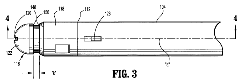
[0008] FIG. 3 is a side plan of the optical obturator of FIGS. 1-2 in an
initial advanced
longitudinal position;
[0009] FIG. 4 is a cross-sectional view of the optical obturator in the
initial advanced
position and taken along the lines 4-4 of FIG. 3;
[0010] FIG. 5 is an axial view of the optical obturator in the initial
advanced position;
[0011] FIG. 6 is a side plan view of the optical obturator in a retracted
longitudinal
position;
[0012] FIG. 7 is a cross-sectional view of the optical obturator in the
retracted position;
[0013] FIG. 8 is a view illustrating the optical obturator mounted to the
cannula
assembly, with the endoscope positioned therein, to permit visualization
during penetration of
tissue; and
CA 02672706 2009-06-15
WO 2008/088753 PCT/US2008/000420
[0014] FIG. 9 illustrates a methodology for using the apparatus of FIG. 1-8 in
accordance with the principles of the present invention.
DESCRIPTION OF EMBODIMENTS
[0015] Specific embodiments of the presently disclosed apparatus and method
will now
be described in detail with reference to the foregoing figures wherein like
reference numerals
identify similar or identical elements. In the figures and in the description
which follows, the
term "proximal", as is traditional will refer to the end of the apparatus or
instrument of the
present disclosure which is closest to the clinician, while the term "distal"
will refer to the end of
the device or instrument which is furthest from the clinician. In addition,
the term "transparent"
is to be interpreted as describing the ability to permit the passage of light
with or without clear
imaging capabilities. Moreover, any reference to any transparent material, or
to any material that
may be as transparent, includes any transparent or translucent material or any
material which is
not opaque to visible light or other radiation utilized for imaging purposes.
100161 Referring now to the drawings, FIG. 1 illustrates a surgical system in
accordance
with the present disclosure. System 10 has particular application in
laparoscopic procedures with
respect to accessing the abdominal cavity, and the like, and includes optical
obturator 100,
endoscope 200 and cannula assembly 1000, In general, endoscope 200 is at least
partially
positioned within optical obturator 100, and the assembled unit is received
within cannula
assembly 1000. The system 10 is applied against the abdominal wall whereby
optical obturator
6
CA 02672706 2009-06-15
WO 2008/088753 PCT/US2008/000420
100 punctures or penetrates the abdominal cavity under direct visualization
via endoscope 200,
thereby providing visual confirmation of entry. into the body cavity while
also substantially
minimizing any undesired contact or engagement with any underlying organs.
Obturator 100 and
endoscope 200 are then removed from cannula assembly 1000 to permit the
subsequent
introduction of surgical instrumentation utilized to carry out the remainder
of the procedure
through cannula assembly 1000. As an alternative, endoscope 200 may be
positioned within
optical obturator 100 after the optical obturator 100 has been inserted into
the body cavity
through cannula assembly 1000.
[0017] With reference to FIGS. 2-5, in conjunction with FIG. 1, optical
obturator 100
includes obturator housing 102 and sleeve or outer member 104 extending from
the housing 102.
Housing 102 is advantageously dimensioned for grasping by the clinician. In
one embodiment,
housing 102 may include locking collet 106 to secure endoscope 200 within
optical obturator 100
in, e.g., a similar manner as described in commonly assigned U.S. Patent
Application No.
11/103892 to Smith, the entire contents of which are hereby incorporated by
reference. Housing
102 may further define skirt 108 which mates with corresponding structure of
cannula assembly
1000. Outer member 104 defines proximal or trailing end 110 and leading or
distal end 112.
Outer member 100 further defines longitudinal axis "a" and has longitudinal
lumen 114
extending at least partially along the length of outer member 104. Housing 102
and outer
member 104 may be fabricated from any suitable biocompatible metal such as
stainless steel and
7
CA 02672706 2009-06-15
WO 2008/088753 PCT/US2008/000420
titanium and its alloys. Alternatively, these components may include a
polymeric material such
as polycarbonate, polystyrene, etc. and may be manufactured through known
molding.
[0018] Referring now to FIGS. 3-5, optical obturator 100 further includes
penetrating
end 116 adjacent leading or distal end 112 of outer member 104. Penetrating
end 116
incorporates penetrating housing 118, penetrating member 120 secured within
the penetrating
housing 118 and transparent or optical member 122 disposed about the
penetrating member 120.
Penetrating housing 118 is preferably secured to outer member 104 by
conventional means. In
one embodiment, penetrating housing 118 includes a pair of locking detents or
ribs 124 adjacent
corresponding recesses 126 within the penetrating housing 118. Locking ribs
124 are adapted for
reception within corresponding locking openings 128 in outer member 104 in
snap relation
therewith to secure penetrating housing 118 to the outer member 104. In the
alternative,
penetrating housing 118 may be secured to outer member 104 through other means
including
adhesives, cements, screw threading etc. As a further embodiment, penetrating
housing 118 may
be integrally or monolithically formed within outer member 104. Penetrating
housing 118 also
defines a pair of openings 130 adjacent its proximal end adapted for mounting
penetrating
member 120 within the penetrating housing 118 as will be discussed,
[0019] Penetrating member 120 may be any suitable element adapted to penetrate
and/or
pierce tissue including, e.g., a pyramidal or sharpened conical member, and
may or may not
incorporate sharpened edges or surfaces. In one embodiment, penetrating member
120 is a
8
CA 02672706 2009-06-15
WO 2008/088753 PCT/US2008/000420
planar bladed element secured within penetrating housing 118 and at least
partially extending
beyond outer member 104. In particular, penetrating member 120 includes
proximal legs 132
having locking detents 134 extending radially outwardly from the legs 132.
Locking detents 134
are dimensioned to be received within openings 130 of penetrating housing 118
in snap relation
to secure the penetrating member 120 relative to outer member 104.
Specifically, proximal legs
132 may be moved radially inwardly during insertion within penetrating housing
118 to permit
passage through the penetrating housing 118 and then released whereby locking
detents 134 are
received within openings 130. With this arrangement, penetrating member 120 is
generally
axially fixed relative to outer member 104. In the alternative, penetrating
member 120 may be
capable of longitudinal movement relative to outer member 104.
[0020] Penetrating member 120 further includes elongated blade portion 136
defining a
generally arcuate outer surface 138. Outer surface 138 may be sharpened to
facilitate piercing
through tissue or, alternatively, may be blunt or atraumatic to be devoid of
piercing capabilities.
[0021] Referring now to FIGS. 3-7, optical member 122 is mounted within
penetrating
housing 118 and is preferably'adapted for reciprocal longitudinal movement
relative to the
penetrating housing 118 and penetrating member 120 between an initial or first
advanced
longitudinal position depicted in FIGS. 3-5 and a second or retracted
longitudinal position
depicted in FIGS. 6-7. Optical member 122 includes proximal cylindrical
portion 140 which is
received within penetrating housing 118 and optical window or dome 142
extending from the
9
CA 02672706 2009-06-15
WO 2008/088753 PCT/US2008/000420
cylindrical portion 140. Any means for mounting optical member 122 within
penetrating housing
118 are envisioned. Optical member 122 includes longitudinal slot 144 (FIG. 5)
which bisects
the optical member 124 and is adapted for reception of penetrating member 120.
Optical
member 122 is distally biased toward the first longitudinal position by
biasing member 146. In
one embodiment, biasing member 146 is a coil spring which, at its proximal
end, engages
locking detents 134 of penetrating member 120 and, at its distal end, engages
proximal end 148
of optical member 122. In the first longitudinal position of optical member
122 depicted in
FIGS. 3-5, penetrating member 120 is preferably contained within the outer
boundary of the
optical member 122 thus avoiding any undesired contact of the penetrating
member 122 with the
clinician or tissue. In the second retracted position of optical member 120
depicted in FIGS. 6
and 7 as effected through a proximal force "F" (e.g., due to engagement with
tissue) on the
optical member 120, the penetrating member 120 is exposed to sever, incise, or
penetrate tissue.
[0022] Optical dome 142 is preferably transparent at least in part or defines
a window to
permit transmission of light and/or of an image. In one embodiment, optical
dome 142 is
generally semi-hemispherical in shape. Other configurations are also
envisioned including
conical, ogive, pyramidal etc. Optical dome 142 defines circumferential ledge
148. Ledge 148 is
adapted to engage distal end 150 of penetrating housing 118 upon movement to
the second
retracted longitudinal position thereby providing control of the degree of
retraction of optical
member 122. Preferably, the distance or spacing "k" between ledge 148 and
distal end 150 of
CA 02672706 2009-06-15
WO 2008/088753 PCT/US2008/000420
penetrating housing 118 is predetermined to permit sufficient exposure of
outer surface 138 of
penetrating member 120 to facilitate penetration through tissue.
[0023] Referring again to FIG. 1, endoscope 200 may be any conventional scope
suitable
for endoscopic applications including, e.g., a laparoscope, arthroscope,
colonoscope, etc. In one
embodiment, endoscope 200 may be the scope disclosed in commonly assigned U.S.
Patent No.
5,412,504 to Leiner (hereinafter "Leiner"), the entire contents of which are
hereby incorporated
by reference. Endoscope 200 incorporates an optical train or lens arrangement
capable of
transmitting an image from distal window 202 to eye piece 204 for viewing by
the surgeon and
may incorporate an illuminating system for providing light. Although FIG. 1
depicts endoscope
200 with eye piece 204, it is also contemplated that endoscope 200 may,
additionally or
alternatively, be connected to a monitor. Further details regarding endoscope
200 may be
ascertained by reference to Leiner.
[0024] The present disclosure also contemplates that the optical obturator 100
may be
fitted with an internal or integral illumination or imaging system thereby
avoiding the need for
endoscope 200, i.e., the illumination and imaging system would be built into
optical obturator
100. Those skilled in the art would appreciate the manner in which to modify
optical obturator
100 to incorporate an internal illumination or imaging system, into a single
unit.
[0025] Referring again to FIG. 1, cannula assembly 1000 of the system 10 will
now be
discussed. Cannula assembly 1000 may be any cannula assembly suitable for the
purpose of
11
CA 02672706 2009-06-15
WO 2008/088753 PCT/US2008/000420
accessing a body cavity. As an example, the apparatus of the present
disclosure may be used in a
laparoscopic surgical procedure where the peritoneal cavity is insufflated
with a suitable gas, e.g.,
C02, to separate the cavity wall from the internal organs housed therein. In
one embodiment,
cannula assembly 1000 includes cannula housing 1002 with cannula sleeve 1004
extending
therefrom. Either or both of cannula housing 1002 and cannula sleeve 1004 may
be opaque or
transparent, either wholly or in part, and may be fabricated from any
biocompatible material
including metals or polymers. Cannula sleeve 1004 defines an internal
longitudinal lumen 1006
dimensioned to permit the passage of surgical instrumentation. It is
contemplated that the
diameter of cannula sleeve 1004 may vary in diameter up to 15 mm, or larger,
dependent upon
the procedure in which it is employed and the corresponding size of the
instrument to be inserted
therein. Cannula assembly 1000 may include an internal seal or valve (not
shown), such as a
duck-bill valve or other zero closure valve, adapted to close in the absence
of a surgical
instrument to prevent passage of insufflation gases through the cannula
assembly 1000, as is
known in the art. An example of such an internal seal or valve is disclosed in
commonly
assigned U.S. Patent No. 5,820,600 to Carlson, et. al., the disclosure of
which is incorporated by
reference herein.
[0026] Cannula assembly 1000 may also include a seal assembly 2000 which may
be
releasably mounted to cannula housing 1002. Means for releasably connecting
seal assembly
2000 to cannula housing 1002 may include a bayonet coupling, threaded
connection, latch,
friction fit, tongue and groove arrangements, snap-fit, etc. Seal assembly
2000 includes at least
12
CA 02672706 2009-06-15
WO 2008/088753 PCT/US2008/000420
one internal seal or valve (not shown) adapted to form a fluid tight seal
about an instrument
inserted therethrough, as is known in the art. An example of one such suitable
seal is the fabric
seal disclosed in commonly assigned U.S. Patent 6,702,787 to Racenet et al.
(hereinafter
"Racenet"), the entire contents of which are incorporated herein by reference.
The seal disclosed
in the Racenet `787 patent may be a flat septum seal having a first layer of
resilient material and a
second fabric layer juxtaposed relative to the first layer. Further details of
the seal may be
ascertained by reference to Racenet. It is contemplated that seal assembly
2000 may or may not
be a component of cannula assembly 1000. For example, it is contemplated that
seal assembly
may be a separate, removable assembly. In the alternative, the seal assembly
may comprise an
integral part of the cannula assembly 1000, therefore not being removable.
[0027] Referring to FIGS. 1, 7 and 8, the use and function of the system 10
will now be
discussed. The peritoneal cavity is first insufflated with a suitable
biocompatible gas such as,
e.g., CO2 gas, such that the cavity wall is raised and lifted away from the
internal organs and
tissue housed therein, providing greater access thereto. The insufflation may
be performed with
an insufflation needle or similar device, as is conventional in the art.
Following insufflation,
endoscope 200 is positioned within optical obturator 100, specifically, first
through locking collet
106, then passed through longitudinal lumen 114 of outer member 104 and
advanced such that
distal window 202 of endoscope 200 is adjacent distal end 112 of outer member
104 specifically,
adjacent penetrating end 116. FIG. 7 depicts endoscope 200 positioned within
optical obturator
100. However, in FIG. 7, cannula 1000 is not shown. Endoscope 200 may be
secured within
13
CA 02672706 2009-06-15
WO 2008/088753 PCT/US2008/000420
optical obturator 100 through collet 106. Thereafter, optical obturator 100
and endoscope 200
are positioned within cannula assembly 1000 and advanced whereby skirt 108
mates with seal
assembly 2000, or if cannula assembly 1000 is devoid of seal assembly 2000,
the skirt 108 will
mate with cannula housing 1002. The present disclosure also contemplates that
endoscope 200
may be positioned within optical member 100 following the insertion of the
optical obturator 100
into cannula assembly 1000.
[0028] With the system 10 fully assembled, the targeted tissue is penetrated.
With
reference to FIG. 8, penetrating end 116 is applied against the tissue "t". As
optical member 122
engages the tissue "t", the optical member moves from the first longitudinal
position depicted in
FIGS. 3-5 to the second longitudinal position depicted in FIGS. 6-7 by the
force "F" applied by
the tissue against the bias of coil spring 146. In this condition, penetrating
member 120 is at least
partially exposed whereby outer surface 138 penetrates tissue. During
penetration, endoscope
200 permits constant visualization of any neighboring, underlying or
surrounding tissue during
the distal advancement of penetrating end 116 of optical obturator 100. This
allows the clinician
to confirm entry into the body cavity while also providing a way to monitor
the procedure,
thereby insuring that underlying tissue and organs do not engage or come into
contact with the
penetrating member 122 of penetrating end 116. In instances where a video
system is utilized,
the surgeon simply observes the penetration of body tissue "t" via any known
video monitor.
Once the penetrating end 116 passes through tissue, optical member 122 is no
longer constrained
by forces applied by the tissue and is free to move to the first longitudinal
position of FIGS. 3-5
14
CA 02672706 2009-06-15
WO 2008/088753 PCT/US2008/000420
under the influence of coil spring 146 to cover penetrating member 120.
Optical obturator 100
and endoscope 200 may then be removed from cannula assembly 1000 to permit the
introduction
of other instruments to perform the clinical surgical procedure.
[0029] FIG. 9 illustrates a methodology for performing a surgical procedure
according to
the principles of the present disclosure. The method incorporates the steps
of:
[0030] 1) positioning an endoscope 200 within optical obturator 100 (STEP
500);
2) advancing endoscope 200 to a position where the distal end thereof is
adjacent
penetrating end 116 of the optical obturator (STEP 502);
3) Optionally securing endoscope 200 within outer member 104 of the optical
obturator (STEP 504);
- 4) at least partially positioning optical obturator 100 endoscope 200 into
cannula
assembly 1000 (STEP 506);
5) advancing the system through tissue while visually monitoring with the
endoscope 200 (STEP 508);
6) removing optical obturator 100 and endoscope 200 from the cannula
assembly100 (STEP 510); and
7) performing a surgical procedure through the cannula assembly (STEP 512).
CA 02672706 2009-06-15
WO 2008/088753 PCT/US2008/000420
[00311 While the above is a complete description of the embodiments of the
present
disclosure, various alternatives, modifications and equivalents may be used.
Therefore, the
above description should not be construed as limiting, but rather as
illustrative of the principles
of the disclosure made herein. Those skilled in the art will envision other
modifications within
the scope and spirit of the claims appended hereto.
16