Note: Descriptions are shown in the official language in which they were submitted.
CA 02672961 2009-06-17
WO 2008/082529 PCT/US2007/025865
Gene Expression Profiling for Identification, Monitoring,
and Treatment of Ocular Disease
REFERENCE TO RELATED APPLICATIONS
This application claims the benefit of U.S. Provisional Application No.
60/876,098 filed
December 19, 2006, the contents of which are incorporated by reference in its
entirety.
FIELD OF THE INVENTION
The present invention relates generally to the identification of biological
markers
associated with the identification of ocular disease. More specifically, the
present invention
relates to the use of gene expression data in the identification, monitoring
and treatment of ocular
disease and in the characterization and evaluation of conditions induced by or
related to ocular
disease.
BACKGROUND OF THE INVENTION
Two leading causes of vision loss are glaucoma and age-related
maculodegenerative
disease (AMD). Glaucoma generally describes a group of diseases that damage
the optic nerve,
which transmits images from the light-sensitive inner back of the eye (retina)
to the brain for
interpretation. Because the optic nerve is unlikely to self repair, damage
tends to be permanent
and blindness can result. Glaucoma is a proliferative disease of the eye
affecting 2.2 million
patients in the U.S. and 65 million patients worldwide. It is related to the
production and removal
of the fluid in the eye known as the aqueous humor, a transparent fluid that
provides nutrition to
the lens and cornea and transmits light rays to the retina at the back of the
eye. Aqueous humor
leaves the eye through a sieve-like tissue called the trabecular meshwork, and
glaucoma is
believed to be caused by changes in the meshwork that prevent aqueous humor
from leaving the
eye. In the past, glaucoma was thought almost always to be related to high
intraocular pressure
that can result from problems such as a blocked fluid drainage system within
the eye. However,
CA 02672961 2009-06-17
WO 2008/082529 PCT/US2007/025865
evidence increasingly has shown that glaucoma can occur even when high
intraocular pressure is
absent.
There are several types of glaucoma, including primary open angle glaucoma
(POAG),
normal pressure glaucoma (NPG), and Pseudoexfoliative Glaucoma (PEX). POAG is
the most
common type of glaucoma often related to high intraocular pressure and the
second leading
cause of irreversible blindness in the United States. It is generally
characterized by a clinical
triad: (1) elevated intraocular pressure; (2) development of optic nerve
atrophy; and (3) loss of
peripheral field of vision, ultimately impairing central vision. The condition
usually develops
because the eye's drainage system functions improperly, sometimes due to
blockages or
constrictions that slowly cause fluid build-up. The term, open angle, is used
with this type of
glaucoma because the angle of the chamber where fluids build up to exit the
eye is normal and
not constricted.
NPG is a form of open angle glaucoma in which high intraocular pressure is
absent. With
NPG, vision loss tends to occur centrally rather than along the edges of the
field of view, as with
POAG. With PEX, a white, fiber-like material is deposited within the eye which
can lead to
blockages of the eye's drainage system, causing high intraocular pressure and
damage to the
optic nerve characteristic of open angle glaucoma. Reasons for formation of
these types of
deposits are unclear.
Age-related Maculodegenerative Disease (AMD) is a degenerative condition of
the
macula. It is the most common cause of vision loss in the United States in
those 50 years old or
older, and its prevalence increases with age. AMD is a major cause of visual
impairment in the
United States. Approximately 1.8 million Americans age 40 and older have
advanced AMD, and
another 7.3 million people with intermediate AMD are at substantial risk for
vision loss. AMD is
caused by hardening of the arteries that nourish the retina. This deprives the
retinal tissue of
oxygen and nutrients that it needs to function and thrive. As a result, the
central vision
deteriorates. AIVID is classified as either wet (neovascular) or dry (non-
neovascular), based on
the absence or the presence of abnormal growth of blood vessels under the
retina.
Wet AMD affects about 10% of patients who suffer from macular degeneration.
This
type occurs when new vessels form to improve the blood supply to oxygen-
deprived retinal
tissue. However, the new vessels are very delicate and break easily, causing
bleeding and
2
CA 02672961 2009-06-17
WO 2008/082529 PCT/US2007/025865
damage to surrounding tissue. The wet form can manifest in two types: classic
or occult. Over
70% of patients with the wet form have the occult type. To date, only the
classic wet type is
treated with conventional laser photocoagulation to stabilize vision or to
limit the growth of
abnormal blood vessels. The remaining majority of patients with wet AMD cannot
be treated
with the laser procedure. The current laser treatment does not improve vision
in most treated
eyes because the laser destroys not only the abnormal blood vessel but also
the overlying macula.
Dry AMD although more common, typically results in a less severe, more gradual
loss of
vision. It is characterized by drusen and loss of pigment in the retina.
Drusen are small,
yellowish deposits that form within the layers of the retina. The loss of
vision associated with dry
AMD tends to be milder and the disease progression is rather slow. There is no
currently proven
medical therapy for dry macular degeneration.
Glaucoma particularly is sight-threatening because, the disease often is
difficult to detect
in early stages due to a lack of symptoms, such as pain. In fact, glaucoma
often is diagnosed only
after vision already has been lost from optic nerve damage. Symptoms that do
present can
typically include gradual deterioration of vision, particularly loss of
peripheral vision, creating
tunnel vision and eventual blindness.
AMD also produces a slow loss of vision. Like glaucoma, both wet and dry A1VID
is
difficult to detect in early stages due to lack of initial symptoms. Early
signs of vision loss
associated with AMD can include seeing shadowy areas in your central vision or
experiencing
unusually fuzzy or distorted vision. The dry form of macular degeneration will
initially often
cause slightly blurred vision. The center of vision may then become blurred
and this region
grows larger as the disease progresses. No symptoms may be noticed if only one
eye is affected.
In wet macular degeneration, straight lines may appear wavy and central vision
loss can occur
rapidly.
Since individuals with glaucoma and AMD can live for several years
asymptomatic while
the disease progresses, regular screenings are essential to detect these
diseases at an early stage.
Early detection of ocular disease preserves vision longer and makes the
disease more
manageable without invasive procedures. Thus a need exists for better ways to
diagnose and
monitor the progression and treatment of ocular disease.
3
CA 02672961 2009-06-17
WO 2008/082529 PCT/US2007/025865
Additionally, information on any condition of a particular patient and a
patient's response
to types and dosages of therapeutic or nutritional agents has become an
important issue in
clinical medicine today not only from the aspect of efficiency of medical
practice for the health
care industry but for improved outcomes and benefits for the patients. Thus,
there is the need for
tests which can aid in the diagnosis and monitor the progression and treatment
of ocular disease.
SUMMARY OF THE INVENTION
The invention is in based in part upon the identification of gene expression
profiles
(Precision Profiles'T) associated with ocular disease. These genes are
referred to herein as ocular
disease associated genes. More specifically, the invention is based upon the
surprising discovery
that detection of as few as two ocular disease associated genes in a subject
derived sample is
capable of identifying individuals with or without ocular disease with at
least 75% accuracy.
More particularly, the invention is based upon the surprising discovery that
the methods provided
by the invention are capable of detecting ocular disease by assaying blood
samples.
In various aspects the invention provides methods of evaluating the presence
or absence
(e.g., diagnosing or prognosing) of ocular disease, based on a smple from the
subject, the sample
providing a source of RNAs, and determining a quantitative measure of the
amount of at least
one constituent of any constituent (e.g., ocular disease associated gene) of
any of Tables 1-5, 7-9,
and 11-13, and arriving at a measure of each constituent. In a particular
embodiment, the
invention provides a method for evaluating the presence of ocular disease in a
subject based on a
sample from the subject, the sample providing a source of RNAs, comprising: a)
determining a
quantitative measure of the amount of at least one constituent of any
constituent of any one table
selected from the group consisting of Table lA, Table 1B and Table 2 as a
distinct RNA
constituent in the subject sample, wherein such measure is obtained under
measurement
conditions that are substantially repeatable and the constituent is selected
so that measurement of
the constituent distinguishes between a normal subject and an ocular disease-
diagnosed subject
in a reference population with at least 75% accuracy; and b) comparing the
quantitative measure
of the constituent in the subject sample to a reference value.
4
CA 02672961 2009-06-17
WO 2008/082529 PCT/US2007/025865
Also provided by the invention is a method for assessing or monitoring the
response to
therapy (e.g., individuals who will respond to a particular therapy
("responders), individuals who
won't respond to a particular therapy ("non-responders"), and/or individuals
in which toxicity of
a particular therapeutic may be an issue), in a subject having ocular disease
or a condition related
to ocular disease, based on a sample from the subject, the sample providing a
source of RNAs,
the method comprising: i) determining a quantitative measure of the amount of
at least one
constituent of any panel of constituents in Tables 1-5, 7-9, and 11-13 as a
distinct RNA
constituent, wherein such measure is obtained under measurement conditions
that are
substantially repeatable to produce a patient data set; and ii) comparing the
patient data set to a
baseline profile data set, wherein the baseline profile data set is related to
the ocular disease, or
conditions related to ocular disease.
In a further aspect, the invention provides a method for monitoring the
progression of
ocular disease or a condition related to ocular disease in a subject, based on
a sample from the
subject, the sample providing a source of RNAs, the method comprising: a)
determining a
quantitative measure of the amount of at least one constituent of any
constituent of Tables 1-5, 7-
9, and 11-13 as a distinct RNA constituent in a sample obtained at a first
period of time to
produce a first patient data set; and determining a quantitative measure of
the amount of at least
one constituent of any constituent of Tables 1-5, 7-9, and 11-13, as a
distinct RNA constituent in
a sample obtained at a second period of time to produce a second profile data
set, wherein such
measurements are obtained under measurement conditions that are substantially
repeatable.
Optionally, the constituents measured in the first sample are the same
constituents measured in
the second sample. The first subject data set and the second subject data set
are compared
allowing the progression of ocular disease in a subject to be determined. The
second subject
sample is taken e.g., one day, one week, one month, two months, three months,
1 year, 2 years,
or more after first subject sample.
In various aspects the invention provides a method for determining a profile
data set, i.e.,
an ocular disease profile, for characterizing a subject with ocular disease or
conditions related to
ocular disease based on a sample from the subject, the sample providing a
source of RNAs, by
using amplification for measuring the amount of RNA in a panel of constituents
including at
least ons constituent from any of Tables 1-5, 7-9, and 11-13, and arriving at
a measure of each
5
CA 02672961 2009-06-17
WO 2008/082529 PCT/US2007/025865
constituent. The profile data set contains the measure of each constituent of
the panel.
Also provided by the invention is a method of characterizing ocular disease or
conditions
related to ocular disease in a subject, based on a sample from the subject,
the sample providing a
source of RNAs, by assessing a profile data set of a plurality of members,
each member being a
quantitative measure of the amount of a distinct RNA constituent in a panel of
constituents
selected so that measurement of the constituents enables characterization of
ocular disease.
In yet another aspect the invention provides a method of characterizing ocular
disease or
conditions related to ocular disease in a subject, based on a sample from the
subject, the sample
providing a source of RNAs, by determining a quantitative measure of the
amount of at least one
constituent from Tables 1-5, 7-9, and 11-13.
Additionally, the invention includes a biomarker for predicting individual
response to
ocular disease treatment in a subject having ocular disease or a condition
related to ocular
disease comprising at least one constituent of any constituent of Tables 1-5,
7-9, and 11-13.
The methods of the invention further include comparing the quantitative
measure of the
constituent in the subject derived sample to a reference value or a baseline
value, e.g. baseline
data set. The reference value is for example an index value. Comparison of the
subject
measurements to a reference value allows for the present or absence of ocular
disease to be
determined, response to therapy to be monitored or the progression of ocular
disease to be
determined. For example, a similarity in the subject data set compared to a
baseline data set
derived from a subject having ocular disease indicates the presence of ocular
disease or response
to therapy that is not efficacious. Whereas a similarity in the subject data
set compares to a
baseline data set derived from a subject not having ocular disease indicates
the absence of ocular
disease or response to therapy that is efficacious. In various embodiments,
the baseline data set
is derived from one or more other samples from the same subject, taken when
the subject is in a
biological condition different from that in which the subject was at the time
the first sample was
taken, with respect to at least one of age, nutritional history, medical
condition, clinical indicator,
medication, physical activity, body mass, and environmental exposure, and the
baseline profile
data set may be derived from one or more other samples from one or more
different subjects.
The baseline profile data set may be derived from one or more other samples
from the
same subject taken under circumstances different from those of the first
sample, and the
6
CA 02672961 2009-06-17
WO 2008/082529 PCT/US2007/025865
circumstances may be selected from the group consisting of (i) the time at
which the first sample
is taken (e.g., before, after, or during treatment for ocular disease), (ii)
the site from which the
first sample is taken, (iii) the biological condition of the subject when the
first sample is taken.
The measure of the constituent is increased or decreased in the subject
compared to the
expression of the constituent in the reference, e.g., normal reference sample
or baseline value.
The measure is increased or decreased 10%, 25%, 50% compared to the reference
level.
Alternately, the measure is increased or decreased 1, 2, 5 or more fold
compared to the reference
level.
In various aspects of the invention the methods are carried out wherein the
measurement
conditions are substantially repeatable, particularly within a degree of
repeatability of better than
ten percent, five percent or more particularly within a degree of
repeatability of better than three
percent, and/or wherein efficiencies of amplification for all constituents are
substantially similar,
more particularly wherein the efficiency of amplification is within ten
percent, more particularly
wherein the efficiency of amplification for all constituents is within five
percent, and still more
particularly wherein the efficiency of amplification for all constituents is
within three percent or
less.
In addition, the one or more different subjects may have in common with the
subject at
least one of age group, gender, ethnicity, geographic location, nutritional
history, medical
condition, clinical indicator, medication, physical activity, body mass, and
environmental
exposure. A clinical indicator may be used to assess ocular disease or
condition related to ocular
disease of the one or more different subjects, and may also include
interpreting the calibrated
profile data set in the context of at least one other clinical indicator,
wherein the at least one
other clinical indicator includes blood chemistry, molecular markers in the
blood, fluourescein
angiography, other chemical assays, and physical findings.
The panel of constituents are selected so as to distinguish from a normal and
a ocular
disease-diagnosed subject. Alternatively, the panel of constituents is
selected as to permit
characterizing the severity of ocular disease in relation to a normal subject
over time so as to
track movement toward normal as a result of successful therapy and away from
normal in
response to ocular disease recurrence. Thus, in some embodiments, the methods
of the invention
are used to determine efficacy of treatment of a particular subject.
7
CA 02672961 2009-06-17
WO 2008/082529 PCT/US2007/025865
Preferably, the panel of constituents are selected so as to distinguish, e.g.,
classify
between a normal and a ocular disease-diagnosed subject with at least 75%,
80%, 85%, 90%,
95%, 97%, 98%, 99% or greater accuracy. By "accuracy" is meant that the method
has the
ability to distinguish, e.g., classify, between subjects having ocular disease
or conditions
associated with ocular disease, and those that do not. Accuracy is determined
for example by
comparing the results of the Gene Precision Profiling'T' to standard accepted
clinical methods of
diagnosing ocular disease, e.g., one or more symptoms of ocular disease such
as gradual
deterioration of vision, loss of peripheral vision, tunnel vision, seeing
shadowy areas in your
central vision or experiencing unusually fuzzy or distorted vision, loss of
central vision, straight
lines appearing wavy, and blindness.
At least 1, 2, 3, 4, 5, 6, 7, 8, 9, 10, 15, 20, 25, 30, 40, 50, 60, or 70 or
more constituents
are measured. In one aspect, one or more constituents from Tables 1-5, 7-9,
and 11-13 is
measured. In a preferred embodibment, one or more constituents selected from
TGFB 1 and
MMP19 is measured. In another aspect, two or more constituents from Tables 1-
5, 7-9, and 11-
13 is measured. Preferably, two or more constituents selected from TGFB1; CRP,
MADD,
MMP19, CASP9,IVIlVIP13, NFKBIB, JUN, BCL3, BCL2L1, BAX, CD69, CD44, VDAC1,
NFKB1, TIMP3, CD4, NOS2A, TRAF2, BIRC3,IVIlVIP2, MAPK14, IL8, HSPAIA, BIK,
MMP9, M1VIP3,IVIlVIP12, PDCD8, C1QA, NOS1, TIMP 1, TNFSF12, BID, ECE1, IL1RN,
TNFRSFIB, TGFA, CD68, SAA1, GSR, BAD, SERPINA3, BAK1, CD3Z, TRADD, MAPK1,
PPARA, CASP3, TP53, TRAF3, MAP3K1, HLADRBI, SOD2, IFNG, PTGS2, PLAU,
ANXA11, LTA, APAF1, CASP1, TOSO, CD19,1VIlVIP15, TNFRSFIA, BIRC2, GSTA1,
PDCD8, and 1VIlVIP 1 is measured. Even more preferably, TGFB 1 and one or more
of the
following: SERPINB2, and CD69; ii) MMP 19; and iii) IVIlVP 19 and CD69 is
measured.
In some embodiments, the methods of the present invention are used in
conjunction with
standard accepted clinical methods to diagnose ocular disease. By ocular
disease or conditions
related to ocular disease is meant a disease, condition of, or injury to the
eye. The term ocular
disease encompasses glaucoma (e.g., primary open angle glaucoma, normal
pressure glaucoma,
and pseudoexfoliative glaucoma), and both wet and dry macular degeneration.
The sample is any sample derived from a subject which contains RNA. For
example the
sample is blood, a blood fraction, body fluid, a population of cells or tissue
from the subject.
8
CA 02672961 2009-06-17
WO 2008/082529 PCT/US2007/025865
Optionally one or more other samples can be taken over an interval of time
that is at least one
month between the first sample and the one or more other samples, or taken
over an interval of
time that is at least twelve months between the first sample and the one or
more samples, or they
may be taken pre-therapy intervention or post-therapy intervention. In such
embodiments, the
first sample may be derived from blood and the baseline profile data set may
be derived from
tissue or body fluid of the subject other than blood. Alternatively, the first
sample is derived from
tissue or bodily fluid of the subject and the baseline profile data set is
derived from blood.
Also included in the invention are kits for the detection of ocular disease in
a subject,
containing at least one reagent for the detection or quantification of any
constituent measured
according to the methods of the invention and instructions for using the kit.
Unless otherwise defined, all technical and scientific terms used herein have
the same
meaning as commonly understood by one of ordinary skill in the art to which
this invention
belongs. Although methods and materials similar or equivalent to those
described herein can be
used in the practice or testing of the present invention, suitable methods and
materials are
described below. All publications, patent applications, patents, and other
references mentioned
herein are incorporated by reference in their entirety. In case of conflict,
the present
specification, including definitions, will control. In addition, the
materials, methods, and
examples are illustrative only and not intended to be limiting.
Other features and advantages of the invention will be apparent from the
following
detailed description and claims.
Brief Description of the Drawings
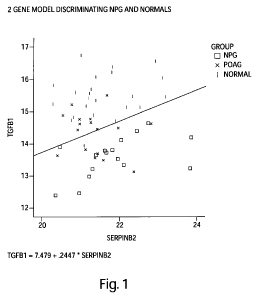
Figure 1 is a graphical representation of the 2-gene model TGFB 1 and SERPINB2
based
on the Precision Profile`m for Ocular disease (Table lA), capable of
distinguishing between
subjects afflicted with normal pressure glaucoma (NPG) and normal subjects,
with a
discrimination line overlaid onto the graph as an example of the Index
Function evaluated at a
particular logit value. Values above the line represent subjects predicted to
be in the normal
population. Values below the line represent subjects predicted to be in the
NPG
9
CA 02672961 2009-06-17
WO 2008/082529 PCT/US2007/025865
population.TGFB 1 values are plotted along the Y-axis, SERPINB2 values are
plotted along the
X-axis.
Figure 2 is a graphical representation of the 2-gene model NIlVIP19 and CD69,
based on
the Precision ProfileTM for Ocular disease (Table lA), capable of
distinguishing between subjects
afflicted with primary open angle glaucoma (POAG) and normal subjects, with a
discrimination
line overlaid onto the graph as an example of the Index Function evaluated at
a particular logit
value. Values above the line represent subjects predicted to be in the normal
population. Values
below the line represent subjects predicted to be in the POAG population..
MMP19 values are
plotted along the Y-axis, CD69 values are plotted along the X-axis.
Figure 3 is a graphical representation of the 2-gene model TGFB1 and CD69,
based on
the Precision ProfileTM for Ocular disease (Table 1 A), capable of
distinguishing between subjects
afflicted with normal pressure glaucoma (NPG) and primary open angle glaucoma
(POAG)
versus normal subjects, with a discrimination line overlaid onto the graph as
an example of the
Index Function evaluated at a particular logit value. Values above the line
represent subjects
predicted to be in the normal population. Values below the line represent
subjects predicted to be
in the NPG and POAG population. TGFB1 values are plotted along the Y-axis,
CD69 values.are
are plotted along the X-axis.
DETAILED DESCRIPTION
Definitions
The following terms shall have the meanings indicated unless the context
otherwise
requires:
"Accuracy" refers to the degree of conformity of a measured or calculated
quantity (a test
reported value) to its actual (or true) value. Clinical accuracy relates to
the proportion of true
outcomes (true positives (TP) or true negatives (TN)) versus misclassified
outcomes (false
positives (FP) or false negatives (FN)), and may be stated as a sensitivity,
specificity, positive
predictive values (PPV) or negative predictive values (NPV), or as a
likelihood, odds ratio,
among other measures.
CA 02672961 2009-06-17
WO 2008/082529 PCT/US2007/025865
"Algorithm" is a set of rules for describing a biological condition. The rule
set may be
defined exclusively algebraically but may also include alternative or multiple
decision points
requiring domain-specific knowledge, expert interpretation or other clinical
indicators.
An "agent" is a "composition" or a "stimulus", as those terms are defined
herein, or a
combination of a composition and a stimulus.
"Amplification" in the context of a quantitative RT-PCR assay is a function of
the number
of DNA replications that are required to provide a quantitative determination
of its concentration.
"Amplification" here refers to a degree of sensitivity and specificity of a
quantitative assay
technique. Accordingly, amplification provides a measurement of concentrations
of constituents
that is evaluated under conditions wherein the efficiency of amplification and
therefore the
degree of sensitivity and reproducibility for measuring all constituents is
substantially similar.
A "baseline profile data set" is a set of values associated with constituents
of a Gene
Expression Panel (Precision ProfileTM) resulting from evaluation of a
biological sample (or
population or set of samples) under a desired biological condition that is
used for mathematically
normative purposes. The desired biological condition may be, for example, the
condition of a
subject (or population or set of subjects) before exposure to an agent or in
the presence of an
untreated disease or in the absence of a disease. Alternatively, or in
addition, the desired
biological condition may be health of a subject or a population or set of
subjects. Alternatively,
or in addition, the desired biological condition may be that associated with a
population or set of
subjects selected on the basis of at least one of age group, gender,
ethnicity, geographic location,
nutritional history, medical condition, clinical indicator, medication,
physical activity, body
mass, and environmental exposure.
A "biological condition" of a subject is the condition of the subject in a
pertinent realm
that is under observation, and such realm may include any aspect of the
subject capable of being
monitored for change in condition, such as health; disease including ocular
disease; cancer;
trauma; aging; infection; tissue degeneration; developmental steps; physical
fitness; obesity, and
mood. As can be seen, a condition in this context may be chronic or acute or
simply transient.
Moreover, a targeted biological condition may be manifest throughout the
organism or
population of cells or may be restricted to a specific organ (such as skin,
heart, eye or blood), but
in either case, the condition may be monitored directly by a sample of the
affected population of
11
CA 02672961 2009-06-17
WO 2008/082529 PCT/US2007/025865
cells or indirectly by a sample derived elsewhere from the subject. The term
"biological
condition" includes a "physiological condition".
"Body fluid" of a subject includes blood, urine, spinal fluid, lymph, mucosal
secretions,
prostatic fluid, semen, haemolymph or any other body fluid known in the art
for a subject.
"Calibrated profile data set" is a function of a member of a first profile
data set and a
corresponding member of a baseline profile data set for a given constituent in
a panel.
A "clinical indicator" is any physiological datum used alone or in conjunction
with other
data in evaluating the physiological condition of a collection of cells or of
an organism. This
term includes pre-clinical indicators.
"Clinical parameters" encompasses all non-sample or non-Precision ProfilesTM
of a
subject's health status or other characteristics, such as, without limitation,
age (AGE), ethnicity
(RACE), gender (SEX), and family history of ocular disease.
A "composition" includes a chemical compound, a nutraceutical, a
pharmaceutical, a
homeopathic formulation, an allopathic formulation, a naturopathic
formulation, a combination
of compounds, a toxin, a food, a food supplement, a mineral, and a complex
mixture of
substances, in any physical state or in a combination of physical states.
To "derive" a profile data set from a sample includes determining a set of
values
associated with constituents of a Gene Expression Panel (Precision ProfileTM)
either (i) by direct
measurement of such constituents in a biological sample. "Distinct R1VA or
protein constituent"
in a panel of constituents is a distinct expressed product of a gene, whether
RNA or protein. An
"expression" product of a gene includes the gene product whether RNA or
protein resulting from
translation of the messenger RNA.
"FN" is false negative, which for a disease state test means classifying a
disease subject
incorrectly as non-disease or normal.
"FP" is false positive, which for a disease state test means classifying a
normal subject
incorrectly as having disease.
A` formula," "algorithm," or "modeP' is any mathematical equation,
algorithmic,
analytical or programmed process, statistical technique, or comparison, that
takes one or more
continuous or categorical inputs (herein called "parameters") and calculates
an output value,
sometimes referred to as an "index" or "index value." Non-limiting examples of
"formulas"
12
CA 02672961 2009-06-17
WO 2008/082529 PCT/US2007/025865
include comparisons to reference values or profiles, sums, ratios, and
regression operators, such
as coefficients or exponents, value transformations and normalizations
(including, without
limitation, those normalization schemes based on clinical parameters, such as
gender, age, or
ethnicity), rules and guidelines, statistical classification models, and
neural networks trained on
historical populations. Of particular use in combining constituents of a Gene
Expression Panel
(Precision ProfileTM) are linear and non-linear equations and statistical
significance and
classification analyses to determine the relationship between levels of
constituents of a Gene
Expression Panel (Precision ProfileTM) detected in a subject sample and the
subject's risk of
ocular disease. In panel and combination construction, of particular interest
are structural and
1o synactic statistical classification algorithms, and methods of risk index
construction, utilizing
pattern recognition features, including, without limitation, such established
techniques such as
cross-correlation, Principal Components Analysis (PCA), factor rotation,
Logistic Regression
Analysis (LogReg), Kolmogorov Smirnoff tests (KS), Linear Discriminant
Analysis (LDA),
Eigengene Linear Discriminant Analysis (ELDA), Support Vector Machines (SVM),
Random
Forest (RF), Recursive Partitioning Tree (RPART), as well as other related
decision tree
classification techniques (CART, LART, LARTree, FlexTree, amongst others),
Shrunken
Centroids (SC), StepAIC, K-means, Kth-Nearest Neighbor, Boosting, Decision
Trees, Neural
Networks, Bayesian Networks, Support Vector Machines, and Hidden Markov
Models, among
others. Other techniques may be used in survival and time to event hazard
analysis, including
Cox, Weibull, Kaplan-Meier and Greenwood models well known to those of skill
in the art.
Many of these techniques are useful either combined with a consituentes of a
Gene Expression
Panel (Precision ProfileTM) selection technique, such as forward selection,
backwards selection,
or stepwise selection, complete enumeration of all potential panels of a given
size, genetic
algorithms, voting and committee methods, or they may themselves include
biomarker selection
methodologies in their own technique. These may be coupled with information
criteria, such as
Akaike's Information Criterion (AIC) or Bayes Information Criterion (BIC), in
order to quantify
the tradeoff between additional biomarkers and model improvement, and to aid
in inimi~ing
overfit. The resulting predictive models may be validated in other clinical
studies, or cross-
validated within the study they were originally trained in, using such
techniques as Bootstrap,
Leave-One-Out (LOO) and 10-Fold cross-validation (10-Fold CV). At various
steps, false
13
CA 02672961 2009-06-17
WO 2008/082529 PCT/US2007/025865
discovery rates (FDR) may be estimated by value permutation according to
techniques known in
the art.
A "Gene Expression Panel" (Precision Profile'm) is an experimentally verified
set of
constituents, each constituent being a distinct expressed product of a gene,
whether RNA or
protein, wherein constituents of the set are selected so that their
measurement provides a
measurement of a targeted biological condition.
A "Gene Expression Profile" (Precision ProfileTM) is a set of values
associated with
constituents of a Gene Expression Panel resulting from evaluation of a
biological sample (or
population or set of samples).
A "Gene Expression Profile Inflammation Index" is the value of an index
function that
provides a mapping from an instance of a Gene Expression Profile into a single-
valued measure
of inflammatory condition.
A Gene Expression Profile Ocular Disease Index " is the value of an index
function that
provides a mapping from an instance of a Gene Expression Profile into a single-
valued measure
of an ocular disease condition.
The "health" of a subject includes mental, emotional, physical, spiritual,
allopathic,
naturopathic and homeopathic condition of the subject.
"Index" is an arithmetically or mathematically derived numerical
characteristic developed
for aid in simplifying or disclosing or informing the analysis of more complex
quantitative
information. A disease or population index may be determined by the
application of a specific
algorithm to a plurality of subjects or samples with a common biological
condition.
"Inflammation" is used herein in the general medical sense of the word and may
be an
acute or chronic; simple or suppurative; localized or disseminated; cellular
and tissue response
initiated or sustained by any number of chemical, physical or biological
agents or combination of
agents.
"Inflammatory state" is used to indicate the relative biological condition of
a subject
resulting from inflammation, or characterizing the degree of inflammation.
A "large number" of data sets based on a common panel of genes is a number of
data sets
sufficiently large to permit a statistically significant conclusion to be
drawn with respect to an
instance of a data set based on the same panel.
14
CA 02672961 2009-06-17
WO 2008/082529 PCT/US2007/025865
"Negative predictive value" or "NPV" is calculated by TN/(TN + FN) or the true
negative
fraction of all negative test results. It also is inherently impacted by the
prevalence of the disease
and pre-test probability of the population intended to be tested.
See, e.g., O'Marcaigh AS, Jacobson RM, "Estimating the Predictive Value of a
Diagnostic Test, How to Prevent Misleading or Confusing Results," Clin. Ped.
1993, 32(8): 485-
491, which discusses specificity, sensitivity, and positive and negative
predictive values of a test,
e.g., a clinical diagnostic test. Often, for binary disease state
classification approaches using a
continuous diagnostic test measurement, the sensitivity and specificity is
summarized by
Receiver Operating Characteristics (ROC) curves according to Pepe et al.,
"Limitations of the
Odds Ratio in Gauging the Performance of a Diagnostic, Prognostic, or
Screening Marker," Am.
J. Epidemiol 2004, 159 (9): 882-890, and summarized by the Area Under the
Curve (AUC) or c-
statistic, an indicator that allows representation of the sensitivity and
specificity of a test, assay,
or method over the entire range of test (or assay) cut points with just a
single value. See also,
e.g., Shultz, "Clinical Interpretation of Laboratory Procedures," chapter 14
in Teitz,
Fundamentals of Clinical Chemistry, Burtis and Ashwood (eds.), 4th edition
1996, W.B.
Saunders Company, pages 192-199; and Zweig et al., "ROC Curve Analysis: An
Example
Showing the Relationships Among Serum Lipid and Apolipoprotein Concentrations
in
Identifying Subjects with Coronory Artery Disease," Clin. Chem., 1992, 38(8):
1425-1428. An
alternative approach using likelihood functions, BIC, odds ratios, information
theory, predictive
values, calibration (including goodness-of-fit), and reclassification
measurements is summarized
according to Cook, "Use and Misuse of the Receiver Operating Characteristic
Curve in Risk
Prediction," Circulation 2007, 115: 928-935.
A"normaP' subject is a subject who is generally in good health, has not been
diagnosed
with ocular disease, or one who is not suffering from ocular disease, is
asymptomatic for ocular
disease, and lacks the traditional laboratory risk factors for ocular disease.
A "normative" condition of a subject to whom a composition is to be
administered means
the condition of a subject before administration, even if the subject happens
to be suffering from
a disease.
The term "ocular disease" is used to indicate a disease or condition of, or
injury to, the
eye. As defined herein, ocular disease encompasses glaucoma (e.g., primary
open angle
CA 02672961 2009-06-17
WO 2008/082529 PCT/US2007/025865
glaucoma, normal pressure glaucoma, pseudoexfoliative glaucoma, primary angle
closure
glaucoma, and pigmentary glaucoma), age-related macular degeneration (wet and
dry), retinal
detachment, retinoschisis, retinopathy (prematurity, hypertensive, diabetic,
and proliferative
vitreo-retinopathy), retinitis pigmentosa, macular edema, scleritis,
keratitis, corneal ulcer, Fuch's
dystrophy, iritis, keratoconus, keratoconjunctivitis sicca, uveitis,
conjunctivitis, and cataract.
A"panel" of genes is a set of genes including at least two constituents.
A "population of cells" refers to any group of cells wherein there is an
underlying
commonality or relationship between the members in the population of cells,
including a group
of cells taken from an organism or from a culture of cells or from a biopsy,
for example.
"Positive predictive value" or "PPV' is calculated by TP/(TP+FP) or the true
positive,
fraction of all positive test results. It is inherently impacted by the
prevalence of the disease and
pre-test probability of the population intended to be tested.
"Risk" in the context of the present invention, relates to the probability
that an event will
occur over a specific time period, and can mean a subject's "absolute" risk or
"relative" risk.
Absolute risk can be measured with reference to either actual observation post-
measurement for
the relevant time cohort, or with reference to index values developed from
statistically valid
historical cohorts that have been followed for the relevant time period.
Relative risk refers to the
ratio of absolute risks of a subject compared either to the absolute risks of
lower risk cohorts,
across population divisions (such as tertiles, quartiles, quintiles, or
deciles, etc.) or an average
population risk, which can vary by how clinical risk factors are assessed.
Odds ratios, the
proportion of positive events to negative events for a given test result, are
also commonly used
(odds are according to the formula p/(1-p) where p is the probability of event
and (1- p) is the
probability of no event) to no-conversion.
"Risk evaluation," or "evaluation of risle' in the context of the present
invention
encompasses making a prediction of the probability, odds, or likelihood that
an event or disease
state may occur, and/or the rate of occurrence of the event or conversion from
one disease state
to another, i.e., from a normal condition to ocular disease and vice versa.
Risk evaluation can
also comprise prediction of future clinical parameters, traditional laboratory
risk factor values, or
other indices of ocular disease results, either in absolute or relative terms
in reference to a
previously measured population. Such differing use may require different
consituentes of a Gene
16
CA 02672961 2009-06-17
WO 2008/082529 PCT/US2007/025865
Expression Panel (Precision ProfileTm) combinations and individualized panels,
mathematical
algorithms, and/or cut-off points, but be subject to the same aforementioned
measurements of
accuracy and performance for the respective intended use.
A "sample" from a subject may include a single cell or multiple cells or
fragments of
cells or an aliquot of body fluid, taken from the subject, by means including
venipuncture,
excretion, ejaculation, massage, biopsy, needle aspirate, lavage sample,
scraping, surgical
incision or intervention or other means known in the art. The sample is blood,
urine, spinal fluid,
lymph, mucosal secretions, prostatic fluid, semen, haemolymph or any other
body fluid known in
the art for a subject. The sample is also a tissue sample.
"Sensitivity" is calculated by TP/(TP+FN) or the true positive fraction of
disease subjects.
"Specificity" is calculated by TN/(TN+FP) or the true negative fraction of non-
disease or
normal subjects.
By "statistically significant", it is meant that the alteration is greater
than what might be
expected to happen by chance alone (which could be a "false positive").
Statistical significance
can be determined by any method known in the art. Commonly used measures of
significance
include the p-value, which presents the probability of obtaining a result at
least as extreme as a
given data point, assuming the data point was the result of chance alone. A
result is often
considered highly significant at a p-value of 0.05 or less and statistically
significant at a p-value
of 0.10 or less. Such p-values depend significantly on the power of the study
performed.
A "set" or "population" of samples or subjects refers to a defined or selected
group of
samples or subjects wherein there is an underlying commonality or relationship
between the
members included in the set or population of samples or subjects.
A "Signature Profile" is an experimentally verified subset of a Gene
Expression Profile
selected to discriminate a biological condition, agent or physiological
mechanism of action.
A "Signature PaneP' is a subset of a Gene Expression Panel (Precision
Profilethe
constituents of which are selected to permit discrimination of a biological
condition, agent or
physiological mechanism of action.
A "subject" is a cell, tissue, or organism, human or non-human, whether in
vivo, ex vivo
or in vitro, under observation. As used herein, reference to evaluating the
biological condition of
a subject based on a sample from the subject, includes using blood or other
tissue sample from a
17
CA 02672961 2009-06-17
WO 2008/082529 PCT/US2007/025865
human subject to evaluate the human subject's condition; it also includes, for
example, using a
blood sample itself as the subject to evaluate, for example, the effect of
therapy or an agent upon
the sample.
A "stimulus" includes (i) a monitored physical interaction with a subject, for
example
ultraviolet A or B, or light therapy for seasonal affective disorder, or
treatment of psoriasis with
psoralen or treatment of cancer with embedded radioactive seeds, other
radiation exposure, and
(ii) any monitored physical, mental, emotional, or spiritual activity or
inactivity of a subject.
"Therapy" includes all interventions whether biological, chemical, physical,
metaphysical, or combination of the foregoing, intended to sustain or alter
the monitored
biological condition of a subject.
"TN" is true negative, which for a disease state test means classifying a non-
disease or
normal subject correctly.
"TP" is true positive, which for a disease state test means correctly
classifying a disease
subj ect.
The PCT patent application publication number WO 01/25473, published April 12,
2001,
entitled "Systems and Methods for Characterizing a Biological Condition or
Agent Using
Calibrated Gene Expression Profiles," which is herein incorporated by
reference, discloses the
use of Gene Expression Panels (Precision Profiles`m) for the evaluation of (i)
biological condition
(including with respect to health and disease) and (ii) the effect of one or
more agents on
biological condition (including with respect to health, toxicity, therapeutic
treatment and drug
interaction).
In particular, the Gene Expression Panels (Precision ProfilesT) described
herein may be
used, without limitation, for measurement of the following: therapeutic
efficacy of natural or
synthetic compositions or stimuli that may be formulated individually or in
combinations or
mixtures for a range of targeted biological conditions; prediction of
toxicological effects and
dose effectiveness of a composition or mixture of compositions for an
individual or for a
population or set of individuals or for a population of cells; determination
of how two or more
different agents administered in a single treatment might interact so as to
detect any of
synergistic, additive, negative, neutral or toxic activity; performing pre-
clinical and clinical trials
by providing new criteria for pre-selecting subjects according to informative
profile data sets for
18
CA 02672961 2009-06-17
WO 2008/082529 PCT/US2007/025865
revealing disease status; and conducting preliminary dosage studies for these
patients prior to
conducting phase 1 or 2 trials. These Gene Expression Panels (Precision
ProfilesT) may be
employed with respect to samples derived from subjects in order to evaluate
their biological
condition.
The present invention provides Gene Expression Panels (Precision ProfilesT)
for the
evaluation or characterization of ocular disease and conditions related to
ocular disease in a
subject. In addition, the Gene Expression Panels described herein also provide
for the evaluation
of the effect of one or more agents for the treatment of ocular disease and
conditions related to
ocular disease.
The Gene Expression Panels (Precision Profiles") are referred to herein as the
"Precision
ProfileTm for Ocular Disease" and the "Precision Profile"" for Inflammatory
Response". A
Precision ProfileTM for Ocular Disease includes one or more genes, e.g.,
constituents, listed in
Tables 1, 3-5, 7-9, and 11-13, whose expression is associated with ocular
disease or conditions
related to ocular disease. A Precision ProfileT' for Inflammatory Response
includes one or more
genes, e.g., constituents, listed in Table 2, whose expression is associated
with inflammatory
response and ocular disease. Each gene of the Precision Profile'm for Ocular
Disease and
Precision ProfileTM for Inflammatory Response is refered to herein as an
ocular disease associated
gene or an ocular disease associated constituent.
It has been discovered that valuable and unexpected results may be achieved
when the
quantitative measurement of constituents is performed under repeatable
conditions (within a
degree of repeatability of measurement of better than twenty percent,
preferably ten percent or
better, more preferably five percent or better, and more preferably three
percent or better). For
the purposes of this description and the following claims, a degree of
repeatability of
measurement of better than twenty percent may be used as providing measurement
conditions
that are "substantially repeatable". In particular, it is desirable that each
time a measurement is
obtained corresponding to the level of expression of a constituent in a
particular sample,
substantially the same measurement should result for substantially the same
level of expression.
In this manner, expression levels for a constituent in a Gene Expression Panel
(Precision
ProfileTM) may be meaningfully compared from sample to sample. Even if the
expression level
measurements for a particular constituent are inaccurate (for example, say,
30% too low), the
19
CA 02672961 2009-06-17
WO 2008/082529 PCT/US2007/025865
criterion of repeatability means that all measurements for this constituent,
if skewed, will
nevertheless be skewed systematically, and therefore measurements of
expression level of the
constituent may be compared meaningfully. In this fashion valuable information
may be
obtained and compared concerning expression of the constituent under varied
circumstances.
In addition to the criterion of repeatability, it is desirable that a second
criterion also be
satisfied, namely that quantitative measurement of constituents is performed
under conditions
wherein efficiencies of amplification for all constituents are substantially
similar as defined
herein. When both of these criteria are satisfied, then measurement of the
expression level of
one constituent may be meaningfully compared with measurement of the
expression level of
another constituent in a given sample and from sample to sample.
The evaluation or characterization of ocular disease is defined to be
diagnosing ocular
disease, assessing the presence or absence of ocular disease, assessing the
risk of developing
ocular disease, or assessing the prognosis of a subject with ocular disease.
Similarly, the
evaluation or characterization of an agent for treatment of ocular disease
includes identifying
agents suitable for the treatment of ocular disease. The agents can be
compounds known to treat
ocular disease or compounds that have not been shown to treat ocular disease.
Ocular disease and conditions related to ocular disease is evaluated by
determining the
level of expression (e.g., a quantitative measure) of an effective number
(e.g., one or more) of
constituents of a Gene Expression Panel (Precision ProfileTM) disclosed herein
(i.e., Tables 1-2).
By an effective number is meant the number of constituents that need to be
measured in order to
discriminate between a normal subject and a subject having ocular disease.
Preferably the
constituents are selected as to discriminate between a normal subject and a
subject having ocular
disease with at least 75% accuracy, more preferably 80%, 85%, 90%, 95%, 97%,
98%, 99% or
greater accuracy.
The level of expression is determined by any means known in the art, such as
for
example quantitative PCR. The measurement is obtained under conditions that
are substantially
repeatable. Optionally, the qualitative measure of the constituent is compared
to a reference or
baseline level or value (e.g. a baseline profile set). In one embodiment, the
reference or baseline
level is a level of expression of one or more constituents in one or more
subjects known not to be
suffering from ocular disease (e.g., normal, healthy individual(s)).
Alternatively, the reference or
CA 02672961 2009-06-17
WO 2008/082529 PCT/US2007/025865
baseline level is derived from the level of expression of one or more
constituents in one or more
subjects known to be suffering from ocular disease. Optionally, the baseline
level is derived
from the same subject from which the first measure is derived. For example,
the baseline is
taken from a subject prior to receiving treatment or surgery for ocular
disease, or at different
time periods during a course of treatment. Such methods allow for the
evaluation of a particular
treatment for a selected individual. Comparison can be performed on test
(e.g., patient) and
reference samples (e.g., baseline) measured concurrently or at temporally
distinct times. An
example of the latter is the use of compiled expression information, e.g., a
gene expression
database, which assembles information about expression levels of ocular
disease associated
genes.
A reference or baseline level or value as used herein can be used
interchangeably and is
meant to be relative to a number or value derived from population studes,
including without
limitation, such subjects having similar age range, subjects in the same or
similar ethnic group,
sex, or, in female subjets, pre-menopausal or post-menopausal subjects, or
relative to the starting
sample of a subject undergoing treatment for ocular disease. Such reference
values can be
derived from statistical analyses and/or risk prediction data of populations
obtained from
mathematical algorithms and computed indices of ocular disease. Reference
indices can also be
constructed and used using algoriths and other methods of statistical and
structural classification.
In one embodiment of the present invention, the reference or baseline value is
the amount
of expression of an ocular disease associated gene in a control sample derived
from one or more
subjects who are both asymptomatic and lack traditional laboratory risk
factors for ocular
disease.
In another embodiment of the present invention, the reference or baseline
value is the
level of ocular disease associated genes in a control sample derived from one
or more subjects
who are not at risk or at low risk for developing ocular disease.
In a further embodiment, such subjects are monitored and/or periodically
retested for a
diagnostically relevant period of time ("longitudinal studies") following such
test to verify
continued absence from ocular disease. Such period of time may be one year,
two years, two to
five years, five years, five to ten years, ten years, or ten or more years
from the initial testing date
for determination of the reference or baseline value. Furthermore,
retrospective measurement of
21
CA 02672961 2009-06-17
WO 2008/082529 PCT/US2007/025865
ocular disease associated genes in properly banked historical subject samples
may be used in
establishing these reference or baseline values, thus shortening the study
time required,
presuming the subjects have been appropriately followed during the intervening
period through
the intended horizon of the product claim.
A reference or basline value can also comprise the amounts of ocular disease
associated
genes derived from subjects who show an improvement in ocular disease status
as a result of
treatments and/or therapies for the ocular disease being treated and/or
evaluated.
In another embodiment, the reference or baseline value is an index value or a
baseline
value. An index value or baseline value is a composite sample of an effective
amount of ocular
disease associated genes from one or more subjects who do not have ocular
disease.
For example, where the reference or baseline level is comprised of the amounts
of ocular
disease associated genes derived from one or more subjects who have not been
diagnosed with
ocular disease or are not known to be suffereing from ocular disease, a change
(e.g., increase or
decrease) in the expression level of a ocular disease associated gene in the
patient-derived
sample of an ocular disease associated gene compared to the expression level
of such gene in the
reference or baseline level indicates that the subject is suffering from or is
at risk of developing
ocular disease. In contrast, when the methods are applied prophylacticly, a
similar level of
expression in the patient-derived sample of an ocular disease associated gene
as compared to
such gene in the baseline level indicates that the subject is not suffering
from or at risk of
developing ocular disease.
Where the reference or baseline level is comprised of the amounts of ocular
disease
associated genes derived from one or more subjects who have been diagnosed
with ocular
disease, or are known to be suffereing from ocular disease, a similarity in
the expression pattern
in the patient-derived sample of an ocular disease associated gene compared to
the ocular disease
baseline level indicates that the subject is suffering from or is at risk of
developing ocular
disease.
Expression of an ocular disease associated gene also allows for the course of
treatment of
ocular disease to be monitored. In this method, a biological sample is
provided from a subject
undergoing treatment, e.g., if desired, biological samples are obtained from
the subject at various
time points before, during, or after treatment. Expression of an ocular
disease associated gene is
22
CA 02672961 2009-06-17
WO 2008/082529 PCT/US2007/025865
then determined and compared to a reference or baseline profile. The baseline
profile may be
taken or derived from one or more individuals who have been exposed to the
treatment.
Alternatively, the baseline level may be taken or derived from one or more
individuals who have
not been exposed to the treatment. For example, samples may be collected from
subjects who
have received initial treatment for ocular disease and subsequent treatment
for ocular disease to
monitor the progress of the treatment.
Differences in the genetic makeup of individuals can result in differences in
their relative
abilities to metabolize various drugs. Accordingly, the Precision ProfileTm
for Ocular Disease
(Table 1A and 1B) and the Precision Profile'T' for Inflammatory Response
(Table 2) disclosed
herein allow for a putative therapeutic or prophylactic to be tested from a
selected subject in
order to determine if the agent is a suitable for treating or preventing
ocular disease in the
subject. Additionally, other genes known to be associated with toxicity may be
used. By suitable
for treatment is meant determining whether the agent will be efficacious, not
efficacious, or toxic
for a particular individual. By toxic it is meant that the manifestations of
one or more adverse
effects of a drug when administered therapeutically. For example, a drug is
toxic when it
disrupts one or more normal physiological pathways.
To identify a therapeutic that is appropriate for a specific subject, a test
sample from the
subject is exposed to a candidate therapeutic agent, and the expression of one
or more of ocular
disease genes is determined. A subject sample is incubated in the presence of
a candidate agent
and the pattern of ocular disease associated gene expression in the test
sample is measured and
compared to a baseline profile, e.g., an ocular disease baseline profile or a
non-ocular disease
baseline profile or an index value. The test agent can be any compound or
composition. For
example, the test agent is a compound known to be useful in the treatment of
ocular disease.
Alternatively, the test agent is a compound that has not previously been used
to treat ocular
disease.
If the reference sample, e.g., baseline is from a subject that does not have
ocular disease a
similarity in the pattern of expression of ocular disease genes in the test
sample compared to the
reference sample indicates that the treatment is efficacious. Whereas a change
in the pattern of
expression of ocular disease genes in the test sample compared to the
reference sample indicates
a less favorable clinical outcome or prognosis. By "efficacious" is meant that
the treatment leads
23
CA 02672961 2009-06-17
WO 2008/082529 PCT/US2007/025865
to a decrease of a sign or symptom of ocular disease in the subject or a
change in the pattern of
expression of an ocular disease associated gene such that the gene expression
pattern has an
increase in similarity to that of a reference or baseline pattern. Assessment
of ocular disease is
made using standard clinical protocols. Efficacy is determined in association
with any known
method for diagnosing or treating ocular disease.
A Gene Expression Panel (Precision Profile'm) is selected in a manner so that
quantitative
measurement of RNA or protein constituents in the Panel constitutes a
measurement of a
biological condition of a subject. In one kind of arrangement, a calibrated
profile data set is
employed. Each member of the calibrated profile data set is a function of (i)
a measure of a
distinct constituent of a Gene Expression Panel (Precision Profile"m) and (ii)
a baseline quantity.
Additional embodiments relate to the use of an index or algorithm resulting
from
quantitative measurement of constituents, and optionally in addition, derived
from either expert
analysis or computational biology (a) in the analysis of complex data sets;
(b) to control or
normalize the influence of uninformative or otherwise minor variances in gene
expression values
between samples or subjects; (c) to simplify the characterization of a complex
data set for
comparison to other complex data sets, databases or indices or algorithms
derived from complex
data sets; (d) to monitor a biological condition of a subject; (e) for
measurement of therapeutic
efficacy of natural or synthetic compositions or stimuli that may be
formulated individually or in
combinations or mixtures for a range of targeted biological conditions; (f)
for predictions of
toxicological effects and dose effectiveness of a composition or mixture of
compositions for an
individual or for a population or set of individuals or for a population of
cells; (g) for
determination of how two or more different agents administered in a single
treatment might
interact so as to detect any of synergistic, additive, negative, neutral of
toxic activity (h) for
performing pre-clinical and clinical trials by providing new criteria for pre-
selecting subjects
according to informative profile data sets for revealing disease status and
conducting preliminary
dosage studies for these patients prior to conducting Phase 1 or 2 trials.
Gene expression profiling and the use of index characterization for a
particular condition
or agent or both may be used to reduce the cost of Phase 3 clinical trials and
may be used beyond
Phase 3 trials; labeling for approved drugs; selection of suitable medication
in a class of
medications for a particular patient that is directed to their unique
physiology; diagnosing or
24
CA 02672961 2009-06-17
WO 2008/082529 PCT/US2007/025865
determining a prognosis of a medical condition or an infection which may
precede onset of
symptoms or alternatively diagnosing adverse side effects associated with
administration of a
therapeutic agent; managing the health care of a patient; and quality control
for different batches
of an agent or a mixture of agents.
The subject
The methods disclosed herein may be applied to cells of humans, mammals or
other
organisms without the need for undue experimentation by one of ordinary skill
in the art because
all cells transcribe RNA and it is known in the art how to extract RNA from
all types of cells.
A subject can include those who have not been previously diagnosed as having
ocular
disease or a condition related to ocular disease. Alternatively, a subject can
also include those
who have already been diagnosed as having ocular disease or a condition
related to ocular
disease. Diagnosis of an ocular disease such as glaucoma is made, for example,
from any one or
combination of the following procedures:.l) measurement of intraolcular
pressure; 2)
examination of the appearance of the meshwork; 3) examination of the
appearance of the optic
nerve; 4) examination of the individual's visual field, particularly
peripheral vision. Diagnosis of
an ocular disease such as AlVID is made, for example, from any one or
combination of the
following procedures: a retinal examination, a visual test using an Amsler
grid which detects
changes in central vision (a sign of AMD if the grid appears distorted); and
fluorescein
angiography to specifically examine the retinal blood vessels surrounding the
macula.
Optionally, the subject has previously been treated with a therapeutic agent,
including but
not limited to therapeutic agents for the treatment of glaucoma, such as beta
blockers (e.g.,
Timoptic, Betoptic), topical beta-adrenergic receptor antagonists (e.g.,
timolol, levobunolol
(Betagan) , and betaxolol), carbonic anhydrase inhibitors (e.g., dorzolamide
(Trusopt),
brinzolamide (Azopt), and acetazolamide (Diamox)), alpha2-adrenergic agonists
(e.g.,
brimonidine (Alphagan)); prostaglandin (e.g., latanoprost (Xalatan),
bimatoprost (Lumigan) and
travoprost (Travatan)), sympathomimetics (e.g., epinephrine and dipivefi-in
(Propine)), miotic
agents (parasympathomimetics, e.g., pilocarpine), and marijuana; and
therapeutic agents for the
treatment of wet AMD, such as pegabtanib (Macugen), verteporfin (Visudyne),
bevacizumab
(Avastin), ranibizumab (Lucentis), anecortave (Retaane), squalamine (Evizon),
siRNA, and
antisense oligonucleotides iCo-007 (targeting the Raf-1 kinase). Optionally,
the therapeutic agent
CA 02672961 2009-06-17
WO 2008/082529 PCT/US2007/025865
is administered alone, or in combination, or in succession with a surgical
procedure for treating
ocular disease, including but not limited to laser surgery, photodynamic
therapy, open, incisional
surgery, radiation therapy (brachytherapy) and rheopheresis. For example, an
argon laser may be
used to perform a procedure called a trabeculoplasty, where the laser is
focused into the
meshwork where it alters cells there to let aqueous fluid leave the eye more
efficiently. A laser
may also be used to make a small hole in the colored part of the eye (the
iris) to allow the
aqueous fluid to flow more freely within in the eye. A laser or freezing
treatment may also be
used to destroy tissue in the eye that makes aqueous humor. Open, incisional
surgery may be
performed if medication and initial laser treatments are unsuccessful in
reducing pressure within
the eye. One type of surgery, a trabeculectomy, creates an opening in the wall
of the eye so that
aqueous humor can drain. Another type of surgery places a drainage tube into
the eye between
the cornea and iris. It exits at the junction of the cornea and sclera (the
white portion of the eye).
The tube drains to a plate that is sewn on the surface of the eye about
halfway back.
A subject can also include those who are suffering from, or at risk of
developing ocular
disease or a condition related to ocular disease, such as those who exhibit
known risk factors for
ocular disease or conditions related to ocular disease. For example, known
risk factors for ocular
disease such as glaucoma include but are not limited to: heredity, race (high
prevalence among
African Americans), suspicious optic nerve appearance (cupping > 50% or
assymetry), central
comeal thickness less than 555 microns (0.5 mm), gender (increased risk in
males), aging (being
older than 60), diabetes, high mypoia (nearsightedness), high blood pressure
(hypertension),
frequent migraines, an injury or surgery to the eye, and a history of steroid
use. Known risk
factors for developing AMD include aging, smoking, gender (women appear to be
at slightly
higher risk), obesity, hypertension, lighter eye color, heredity, and race.
There are also
suggestions that visible and ultraviolet light may damage the retina, and that
low consumption of
fruits and vegetables, which contain certain antioxidants may potentially
increase risk of AMD.
Selecting Constituents of a Gene Expression Panel (Precision Profile7)
The general approach to selecting constituents of a Gene Expression Panel
(Precision
Profile'"') has been described in PCT application publication number WO
01/25473, incorporated
herein by reference in its entirety. A wide range of Gene Expression Panels
(Precision ProfilesTM)
have been designed and experimentally validated, each panel providing a
quantitative measure of
26
CA 02672961 2009-06-17
WO 2008/082529 PCT/US2007/025865
biological condition that is derived from a sample of blood or other tissue.
For each panel,
experiments have verified that a Gene Expression Profile using the panel's
constituents is
informative of a biological condition. (It has also been demonstrated that in
being informative of
biological condition, the Gene Expression Profile is used, among other things,
to measure the
effectiveness of therapy, as well as to provide a target for therapeutic
intervention.).
Tables 1-5, 7-9, and 11-13 listed below, include relevant genes which may be
selected for
a given Precision Profile', such as the Precision ProfilesTM demonstrated
herein to be useful in
the evaluation of ocular disease and conditions related to ocular disease.
Tables 1A and 1B are
panels of 96 and 97 genes respectively, whose expression is associated with
ocular disease or
conditions related to ocular disease.
Table 2 is a panel of genes whose expression is associated with inflammatory
response.
Inflammation is known to play a critical role in many types of ocular
diseases. The earliest
events of inflammation are related to hyperemia and effusion of fluid from
blood vessels
responding to locally-generated inflammatory mediators. In most tissues such
serous effusion is
of little consequence, but the anatomy of the eye presents some special
problems. Serous
effusion from the choroid, for example, creates instantly blinding retinal
detachment that might
ultimately result in irreversible retinal damage because the retina is
separated from its nutritional
choroidal support. Alternatively, the leakage of protein into the aqueous
humor changes its
optical properties and results in aqueous flare, and the abnormal chemical
composition of the
aqueous is a potential cause for cataract because the lens depends entirely
upon the delivery of
quantitatively and qualitatively normal aqueous humor for its nutritional
health.
In some instances, the leakage of small molecular weight proteins from
reactive vessels is
followed by the leakage of larger proteins like fibrinogen, resulting in the
extravascular
accumulation of fibrin. The potential for adhesion between adjacent inflamed,
sticky surfaces is
little more than an inconvenience in most tissues, but within the globe the
adhesion of iris to lens
creates posterior synechia with the potential for pupillary block, iris bombe,
and secondary
glaucoma. Similarly, the accumulation and subsequent contraction of fibrin
within the vitreous
creates the risk of traction retinal detachment.
Additionally, leukocytes may accumulate and settle by gravity within the
anterior
chamber as they attempt to exit the globe via the trabecular meshwork
(hypopyon), or form
27
CA 02672961 2009-06-17
WO 2008/082529 PCT/US2007/025865
adherent clusters that stick to the comeal endothelium (keratic precipitates).
Because the globe
is a closed sphere, inflammatory mediators and various cytokines associated
with leucocytic
recruitment or subsequent events of wound healing are distributed throughout
the globe, so there
is really no such thing as localized intraocular inflammation. Although, for
example, the anterior
uveitis is clinically distinguishable from choroiditis, from a histologic
perspective all intraocular
inflammation is diffuse (i.e. endophthalmitis). As such, both the ocular
disease genes listed in
Tables 1A and 1B and the inflammatory response genes listed in Table 2 can be
used to detect
ocular disease and distinguish between subjects suffering from ocular disease
and normal
subjects.
Table 5 was derived from a study of the gene expression patterns described in
Example 1
below. Table 5 describes a multi-gene model based on genes from the Precision
ProfileTM for
Ocular Disease (Glaucoma) (shown in Table 1A), derived from latent class
modeling of the
subjects from this study using 1 and 2 gene models to distinguish between
subjects suffering
from normal pressure glaucoma (NPG) and normal subjects. Constituent models
selected from
Table 5 are capable of correctly classifying ocular disease-afflicted and/or
normal subjects with
at least 75% accuracy. For example, in Table 5, Gene Column 1, it can be seen
that the 1-gene
model, TGFB1, correctly classifies NPG-afflicted subjects with 100% accuracy,
and normal
subjects with 92% accuracy. In Table 5, Gene Column 2, it can be seen that the
2-gene model,
TGFB1 and SERPINB2, correctly classifies NPG-afflicted subjects with 100%
accuracy, and
normal subjects with 92% accuracy.
Table 9 was derived from a study of the gene expression patterns described in
Example 2
below. Table 9 also describes multi-gene models based on genes from the
Precision Profile." for
Ocular Disease (Glaucoma) (shown in Table 1 A), derived from latent class
modeling of the
subjects from this study using 1 and 2-gene models to distinguish between
subjects suffering
from primary open angle glaucoma (POAG) based on genes from the Precision
ProfileTm for
Ocular Disease (Table lA). Constituent models selected from Table 9 are
capable of correctly
classifying POAG-afflicted and/or normal subjects with at least 75% accuracy.
For example, in
Table 9, Gene Column 1, it can be seen that the 1-gene model,lVIlVIP19,
correctly classifies
POAG-afflicted subjects with 82% accuracy, and normal subjects with 83%
accuracy. In Table
28
CA 02672961 2009-06-17
WO 2008/082529 PCT/US2007/025865
9, Gene Column 2, it can be seen that the 2-gene model, M0IP19 and CD69,
correctly classifies
POAG-afflicted subjects with 94% accuracy, and normal subjects with 92%
accuracy.
Table 13 was derived from a study of the gene expression patterns described in
Example
3 below. Table 13 also describes multi-gene models based on genes from the
Precision ProfileTM
for Ocular Disease (Glaucoma) (shown in Table 1 A), derived from latent class
modeling of the
subjects from this study using 1 and 2-gene models to distinguish between
subjects suffering
from both normal pressure glaucoma (NPG) and primary open angle glaucoma
(POAG) based on
genes from the Precision ProfileTM for Ocular Disease (Table 1A). Constituent
models selected
from Table 13 are capable of correctly classifying NPG and POAG-afflicted
and/or normal
subjects with at least 75% accuracy. For example, in Table 13, Gene Column 1,
it can be seen
that the 1-gene model, TGFB1, correctly classifies NPG and POAG-afflicted
subjects with 85%
accuracy, and normal subjects with 92% accuracy. In Table 13, Gene Column 2,
it can be seen
that the 2-gene model, TGFB1 and CD69, correctly classifies NPG and POAG-
afflicted subjects
with 94% accuracy, and normal subjects with 92% accuracy.
In general, panels may be constructed and experimentally validated by one of
ordinary
skill in the art in accordance with the principles articulated in the present
application.
Design of assays
Typically, a sample is run through a panel in replicates of three for each
target gene
(assay); that is, a sample is divided into aliquots and for each aliquot the
concentrations of each
constituent in a Gene Expression Panel (Precision ProfileT) is measured. From
over thousands of
constituent assays, with each assay conducted in triplicate, an average
coefficient of variation
was found (standard deviation/average) * 100, of less than 2 percent among the
normalized ACT
measurements for each assay (where normalized quantitation of the target mRNA
is determined
by the difference in threshold cycles between the internal control (e.g., an
endogenous marker
such as 18S rRNA, or an exogenous marker) and the gene of interest. This is a
measure called
"intra-assay variability". Assays have also been conducted on different
occasions using the same
sample material. This is a measure of "inter-assay variability". Preferably,
the average
coefficient of variation of intra- assay variability or inter-assay
variability is less than 20%, more
preferably less than 10%, more preferably less than 5%, more preferably less
than 4%, more
preferably less than 3%, more preferably less than 2%, and even more
preferably less than 1%.
29
CA 02672961 2009-06-17
WO 2008/082529 PCT/US2007/025865
It has been determined that it is valuable to use the quadruplicate or
triplicate test results
to identify and eliminate data points that are statistical "outliers"; such
data points are those that
differ by a percentage greater, for example, than 3% of the average of all
three or four values.
Moreover, if more than one data point in a set of three or four is excluded by
this procedure, then
all data for the relevant constituent is discarded.
Measurement of Gene Expression for a Constituent in the Panel
For measuring the amount of a particular RNA in a sample, methods known to one
of
ordinary skill in the art were used to extract and quantify transcribed RNA
from a sample with
respect to a constituent of a Gene Expression Panel (Precision ProfileTM).
(See detailed protocols
below. Also see PCT application publication number WO 98/24935 herein
incorporated by
reference for RNA analysis protocols). Briefly, RNA is extracted from a sample
such as any
tissue, body fluid, cell, or culture medium in which a population of cells of
a subject might be
growing. For example, cells may be lysed and RNA eluted in a suitable solution
in which to
conduct a DNAse reaction. Subsequent to RNA extraction, first strand synthesis
may be
performed using a reverse transcriptase. Gene amplification, more specifically
quantitative PCR
assays, can then be conducted and the gene of interest calibrated against an
internal marker such
as 18S rRNA (Hirayama et al., Blood 92, 1998: 46-52). Any other endogenous
marker can be
used, such as 28S-25S rRNA and 5S rRNA. Samples are measured in multiple
replicates, for
example, 3 replicates. In an embodiment of the invention, quantitative PCR is
performed using
amplification, reporting agents and instruments such as those supplied
commercially by Applied
Biosystems (Foster City, CA). Given a defmed efficiency of amplification of
target transcripts,
the point (e.g., cycle number) that signal from amplified target template is
detectable may be
directly related to the amount of specific message transcript in the measured
sample. Similarly,
other quantifiable signals such as fluorescence, enzyme activity,
disintegrations per minute,
absorbance, etc., when correlated to a known concentration of target templates
(e.g., a reference
standard curve) or normalized to a standard with limited variability can be
used to quantify the
number of target templates in an unknown sample.
Although not limited to amplification methods, quantitative gene expression
techniques
may utilize amplification of the target transcript. Alternatively or in
combination with
amplification of the target transcript, quantitation of the reporter signal
for an internal marker
CA 02672961 2009-06-17
WO 2008/082529 PCT/US2007/025865
generated by the exponential increase of amplified product may also be used.
Amplification of
the target template may be accomplished by isothermic gene amplification
strategies or by gene
amplification by thermal cycling such as PCR.
It is desirable to obtain a definable and reproducible correlation between the
amplified
target or reporter signal, i.e., internal marker, and the concentration of
starting templates. It has
been discovered that this objective can be achieved by careful attention to,
for example,
consistent primer-template ratios and a strict adherence to a narrow
permissible level of
experimental amplification efficiencies (for example 80.0 to 100% +/- 5%
relative efficiency,
typically 90.0 to 100% +/- 5% relative efficiency, more typically 95.0 to 100%
+/- 2 %, and most
typically 98 to 100% +/- 1 % relative efficiency). In determining gene
expression levels with
regard to a single Gene Expression Profile, it is necessary that all
constituents of the panels,
including endogenous controls, maintain similar amplification efficiencies, as
defined herein, to
permit accurate and precise relative measurements for each constituent.
Amplification
efficiencies are regarded as being "substantially similar", for the purposes
of this description and
the following claims, if they differ by no more than approximately 10%,
preferably by less than
approximately 5%, more preferably by less than approximately 3%, and more
preferably by less
than approximately 1%. Measurement conditions are regarded as being
"substantially
repeatable, for the purposes of this description and the following claims, if
they differ by no
more than approximately +/- 10% coefficient of variation (CV), preferably by
less than
approximately +/- 5% CV, more preferably +/- 2% CV. These constraints should
be observed
over the entire range of concentration levels to be measured associated with
the relevant
biological condition. While it is thus necessary for various embodiments
herein to satisfy criteria
that measurements are achieved under measurement conditions that are
substantially repeatable
and wherein specificity and efficiencies of amplification for all constituents
are substantially
similar, nevertheless, it is within the scope of the present invention as
claimed herein to achieve
such measurement conditions by adjusting assay results that do not satisfy
these criteria directly,
in such a manner as to compensate for errors, so that the criteria are
satisfied after suitable
adjustment of assay results.
In practice, tests are run to assure that these conditions are satisfied. For
example, the
design of all primer-probe sets are done in house, experimentation is
performed to determine
31
CA 02672961 2009-06-17
WO 2008/082529 PCT/US2007/025865
which set gives the best performance. Even though primer-probe design can be
enhanced using
computer techniques known in the art, and notwithstanding common practice, it
has been found
that experimental validation is still useful. Moreover, in the course of
experimental validation,
the selected primer-probe combination is associated with a set of features:
The reverse primer should be complementary to the coding DNA strand. In one
embodiment, the primer should be located across an intron-exon junction, with
not more than
four bases of the three-prime end of the reverse primer complementary to the
proximal exon. (If
more than four bases are complementary, then it would tend to competitively
amplify genomic
DNA.)
In an embodiment of the invention, the primer probe set should amplify cDNA of
less
than 110 bases in length and should not amplify, or generate fluorescent
signal from, genomic
DNA or transcripts or cDNA from related but biologically irrelevant loci.
A suitable target of the selected primer probe is first strand cDNA, which in
one
embodiment may be prepared from whole blood as follows:
(a) Use of cell systems or whole blood for ex vivo assessment of a biological
condition.
Human blood is obtained by venipuncture and prepared for assay. The aliquots
of
heparinized, whole blood are mixed with additional test therapeutic compounds
and held at 37 C
in an atmosphere of 5% CO2 for 30 minutes. Cells are lysed and nucleic acids,
e.g., RNA, are
extracted by various standard means.
Nucleic acids, RNA and/or DNA are purified from cells, tissues or fluids of
the test
population of cells. Cells systems that may be used to study ocular disease
includes trabecular
meshwork (typically stimulated with TGFB2), retinal Ganglion cells (induction
of apoptosis via
neurotrophin deprivation and/or glutamate toxicity; induction of oxidative
stress via EGCG,
epigallocatechin gallate), optic nerve head cells and choroid epithelial cells
(laser induction of
neovascularization). RNA is preferentially obtained from the nucleic acid mix
using a variety of
standard procedures (or RNA Isolation Strategies, pp. 55-104, in RNA
Methodologies,
A
laboratory guide for isolation and characterization, 2nd edition, 1998, Robert
E. Farrell, Jr., Ed.,
Academic Press), in the present using a filter-based RNA isolation system from
Ambion
32
CA 02672961 2009-06-17
WO 2008/082529 PCT/US2007/025865
(RNAqueous TM Phenol-free Total RNA Isolation Kit, Catalog #1912, version
9908; Austin,
Texas).
(b) Amplification strategies.
Specific RNAs are amplified using message specific primers or random primers.
The
specific primers are synthesized from data obtained from public databases
(e.g., Unigene,
National Center for Biotechnology Information, National Library of Medicine,
Bethesda, MD),
including information from genomic and cDNA libraries obtained from humans and
other
animals. Primers are chosen to preferentially amplify from specific RNAs
obtained from the test
or indicator samples (see, for example, RT PCR, Chapter 15 in RNA Methodolo
egi s, A
Laboratory Guide for Isolation and Characterization, 2nd edition, 1998, Robert
E. Farrell, Jr.,
Ed., Academic Press; or Chapter 22 pp.143-151, RNA Isolation and
Characterization Protocols,
Methods in mMolecular Biology, Volume 86, 1998, R. Rapley and D. L. Manning
Eds., Human
Press, or 14 in Statistical refinement of primer design parameters, Chapter 5,
pp.55-72, PCR
Applications: Protocols for functional genomics, M.A.Innis, D.H. Gelfand and
J.J. Sninsky, Eds.,
1999, Academic Press). Amplifications are carried out in either isothermic
conditions or using a
thermal cycler (for example, a ABI 9600 or 9700 or 7900 obtained from Applied
Biosystems,
Foster City, CA; see Nucleic acid detection methods, pp. 1-24, in Molecular
Methods for Virus
Detection, D.L.Wiedbrauk and D.H., Farkas, Eds., 1995, Academic Press).
Amplified nucleic
acids are detected using fluorescent-tagged detection oligonucleotide probes
(see, for example,
Taqman'T' PCR Reagent Kit, Protocol, part number 402823, Revision A, 1996,
Applied
Biosystems, Foster City CA) that are identified and synthesized from publicly
known databases
as described for the amplification primers.
For example without limitation, amplified cDNA is detected and quantified
using
detection systems such as the ABI Prism 7900 Sequence Detection System
(Applied
Biosystems (Foster City, CA)), the Cepheid SmartCycler and Cepheid GeneXpert
Systems,
the Fluidigm BioMark'M System, and the Roche LightCycler 480 Real-Time PCR
System.
Amounts of specific RNAs contained in the test sample can be related to the
relative quantity of
fluorescence observed (see for example, Advances in Quantitative PCR
Technology: 5' nuclease
assays, Y.S. Lie and C.J. Petropolus, Current Opinion in Biotechnology, 1998,
9:43-48, or Rapid
33
CA 02672961 2009-06-17
WO 2008/082529 PCT/US2007/025865
Thermal Cycling and PCR Kinetics, pp. 211-229, chapter 14 in PCR Applications:
protocols for
functional genomics, M.A. Innis, D.H. Gelfand and J.J. Sninsky, Eds., 1999,
Academic Press).
As a particular implementation of the approach described here in detail is a
procedure for
synthesis of first strand cDNA for use in PCR. Examples of the procedure used
with several of
the above-mentioned detection systems are described below. In some
embodiments, these
procedures can be used for both whole blood RNA and RNA extracted from
cultured cells (e.g.,
trabecular meshwork, retinal Ganglion cells, optic nerve head cells and
choroid epithelial cells).
Methods herein may also be applied using proteins where sensitive quantitative
techniques, such
as an Enzyme Linked ImmunoSorbent Assay (ELISA) or mass spectroscopy, are
available and
well-known in the art for measuring the amount of a protein constituent (see
WO 98/24935
herein incorporated by reference).
An example of a procedure fo the synthesis of first strand cDNA for use in PCR
amplification is as follows:
Materials
1. Applied Biosystems TAQMAN Reverse Transcription Reagents Kit (P/N 808-
0234). Kit Components: l OX TaqMan RT Buffer, 25 mM Magnesium chloride,
deoxyNTPs
mixture, Random Hexamers, RNase Inhibitor, MultiScribe Reverse Transcriptase
(50 U/mL) (2)
RNase / DNase free water (DEPC Treated Water from Ambion (P/N 9915G), or
equivalent)
Methods
1. Place RNase Inhibitor and MultiScribe Reverse Transcriptase on ice
immediately.
All other reagents can be thawed at room temperature and then placed on ice.
2. Remove RNA samples from -80 C freezer and thaw at room temperature and
then place immediately on ice.
3. Prepare the following cocktail of Reverse Transcriptase Reagents for each
100
mL RT reaction (for multiple samples, prepare extra cocktail to allow for
pipetting error):
1 reaction (mL) 11X, e.g. 10 samples ( L)
l OX RT Buffer 10.0 110.0
25 mM Mg02 22.0 242.0
dNTPs 20.0 220.0
Random Hexamers 5.0 55.0
34
CA 02672961 2009-06-17
WO 2008/082529 PCT/US2007/025865
RNAse Inhibitor 2.0 22.0
Reverse Transcriptase 2.5 27.5
Water 18.5 203.5
Total: 80.0 880.0 (80 L per sample)
4. Bring each RNA sample to a total volume of 20 L in a 1.5 mL
microcentrifuge
tube (remove 10 L RNA and dilute to 20 L with RNase / DNase free water, for
whole blood
RNA use 20 L total RNA) and add 80 L RT reaction mix from step 5,2,3. Mix by
pipetting up
and down.
5. Incubate sample at room temperature for 10 minutes.
6. Incubate sample at 37 C for 1 hour.
7. Incubate sample at 90 C for 10 minutes.
8. Quick spin samples in microcentrifuge.
9. Place sample on ice if doing PCR immediately, otherwise store sample at -20
C
for future use.
10. PCR QC should be run on all RT samples using 18S and (3-actin.
Following the synthesis of first strand cDNA, one particular embodiment of the
approach
for amplification of first strand cDNA by PCR, followed by detection and
quantification of
constituents of a Gene Expression Panel (Precision Profile'.) is performed
using the ABI Prism
7900 Sequence Detection System as follows:
Materials
1. 20X Primer/Probe Mix for each gene of interest.
2. 20X Primer/Probe Mix for 18S endogenous control.
3. 2X Taqman Universal PCR Master Mix.
4. cDNA transcribed from RNA extracted from cells.
5. Applied Biosystems 96-Well Optical Reaction Plates.
6. Applied Biosystems Optical Caps, or optical-clear film.
7. Applied Biosystem Prism 7700 or 7900 Sequence Detector.
Methods
1. Make stocks of each Primer/Probe mix containing the Primer/Probe for the
gene
of interest, Primer/Probe for 18S endogenous control, and 2X PCR Master Mix as
follows. Make
CA 02672961 2009-06-17
WO 2008/082529 PCT/US2007/025865
sufficient excess to allow for pipetting error e.g., approximately 10% excess.
The following
example illustrates a typical set up for one gene with quadruplicate samples
testing two
conditions (2 plates).
1X (1 well) ( L)
2X Master Mix 7.5
20X 18S Primer/Probe Mix 0.75
20X Gene of interest Primer/Probe Mix 0.75
Total 9.0
2. Make stocks of cDNA targets by diluting 95 L of cDNA into 200011L of water.
The amount of cDNA is adjusted to give CT values between 10 and 18, typically
between
12 and 16.
3. Pipette 9 L of Primer/Probe mix into the appropriate wells of an Applied
Biosystems 384-Well Optical Reaction Plate.
4. Pipette 10 L of cDNA stock solution into each well of the Applied
Biosystems
384-Well Optical Reaction Plate.
5. Seal the plate with Applied Biosystems Optical Caps, or optical-clear film.
6. Analyze the plate on the ABI Prism 7900 Sequence Detector.
In another embodiment of the invention, the use of the primer probe with the
first strand
cDNA as described above to permit measurement of constituents of a Gene
Expression Panel
(Precision ProfileTM) is performed using a QPCR assay on Cepheid SmartCycler
and
GeneXpert Instruments as follows:
I. To run a QPCR assay in duplicate on.the Cepheid SmartCycler instrument
containing three
target genes and one reference gene, the following procedure should be
followed.
A. With 20X Primer/Probe Stocks.
Materials
1. SmartMixTm-HM lyophilized Master Mix.
2. Molecular grade water.
3. 20X Primer/Probe Mix for the 18S endogenous control gene. The endogenous
control gene will be dual labeled with VIC-MGB or equivalent.
36
CA 02672961 2009-06-17
WO 2008/082529 PCT/US2007/025865
4. 20X Primer/Probe Mix for each for target gene one, dual labeled with FAM-
BHQ 1 or
equivalent.
5. 20X Primer/Probe Mix for each for target gene two, dual labeled with Texas
Red-
BHQ2 or equivalent.
6. 20X Primer/Probe Mix for each for target gene three, dual labeled with
Alexa 647-
BHQ3 or equivalent.
7. Tris buffer, pH 9.0
8. cDNA transcribed from RNA extracted from sample.
9. SmartCycler 25 L tube.
10. Cepheid SmartCycler instrument.
Methods
1. For each cDNA sample to be investigated, add the following to a sterile 650
L tube.
SmartMixTM-HM lyophilized Master Mix 1 bead
20X 18S Primer/Probe Mix 2.5 L
20X Target Gene 1 Primer/Probe Mix 2.5 L
20X Target Gene 2 Primer/Probe Mix 2.5 L
20X Target Gene 3 Primer/Probe Mix 2.5 L
Tris Buffer, pH 9.0 2.5 L
Sterile Water 34.5 L
Total 47 L
Vortex the mixture for 1 second three times to completely mix the reagents.
Briefly
centrifuge the tube after vortexing.
2. Dilute the cDNA sample so that a 3 L addition to the reagent mixture above
will
give an 18S reference gene CT value between 12 and 16.
37
CA 02672961 2009-06-17
WO 2008/082529 PCT/US2007/025865
3. Add 3 L of the prepared cDNA sample to the reagent mixture bringing the
total
volume to 50 L. Vortex the mixture for 1 second three times to completely mix
the
reagents. Briefly centrifuge the tube after vortexing.
4. Add 25 L of the mixture to each of two SmartCycler tubes, cap the tube
and spin
for 5 seconds in a microcentrifuge having an adapter for SmartCycler tubes.
5. Remove the two SmartCycler tubes from the microcentrifuge and inspect for
air
bubbles. If bubbles are present, re-spin, otherwise, load the tubes into the
SmartCycler instrument.
6. Run the appropriate QPCR protocol on the SmartCycler , export the data and
analyze
the results.
B. With Lyophilized SmartBeadsTM
Materials
1. SmartMix'. -HM lyophilized Master Mix.
2. Molecular grade water.
3. SmartBeadsTm containing the 18S endogenous control gene dual labeled with
VIC-
MGB or equivalent, and the three target genes, one dual labeled with FAM-BHQI
or
equivalent, one dual labeled with Texas Red-BHQ2 or equivalent and one dual
labeled with Alexa 647-BHQ3 or equivalent.
4. Tris buffer, pH 9.0
5. cDNA transcribed from RNA extracted from sample.
6. SmartCycler 25 L tube.
7. Cepheid SmartCycler instrument.
Methods
1. For each cDNA sample to be investigated, add the following to a sterile 650
L tube.
SmartMixTM-HM lyophilized Master Mix 1 bead
SmartBeadTM containing four primer/probe sets 1 bead
Tris Buffer, pH 9.0 2.5 L
Sterile Water 44.5 L
Total 47 L
38
CA 02672961 2009-06-17
WO 2008/082529 PCT/US2007/025865
Vortex the mixture for 1 second three times to completely mix the reagents.
Briefly
centrifuge the tube after vortexing.
2. Dilute the cDNA sample so that a 3 L addition to the reagent mixture above
will
give an 18S reference gene CT value between 12 and 16.
3. Add 3 L of the prepared cDNA sample to the reagent mixture bringing the
total
volume to 50 L. Vortex the mixture for 1 second three times to completely mix
the
reagents. Briefly centrifuge the tube after vortexing.
4. Add 25 L of the mixture to each of two SmartCycler tubes, cap the tube
and spin
for 5 seconds in a microcentrifuge having an adapter for SmartCycler tubes.
5. Remove the two SmartCycler tubes from the microcentrifuge and inspect for
air
bubbles. If bubbles are present, re-spin, otherwise, load the tubes into the
SmartCycler instrument.
6. Run the appropriate QPCR protocol on the SmartCycler , export the data and
analyze
the results.
II. To run a QPCR assay on the Cepheid GeneXpert instrument containing three
target genes
and one reference gene, the following procedure should be followed. Note that
to do
duplicates, two self contained cartridges need to be loaded and run on the
GeneXpert
instrument.
Materials
1. Cepheid GeneXpert self contained cartridge preloaded with a lyophilized
SmartMixTM-HM master mix bead and a lyophilized SmartBeaC containing four
primer/probe sets.
2. Molecular grade water, containing Tris buffer, pH 9Ø
3. Extraction and purification reagents.
4. Clinical sample (whole blood, RNA, etc.)
5. Cepheid GeneXpert instrument.
Methods
1. Remove appropriate GeneXpert self contained cartridge from packaging.
2. Fill appropriate chamber of self contained cartridge with molecular grade
water with
Tris buffer, pH 9Ø
39
CA 02672961 2009-06-17
WO 2008/082529 PCT/US2007/025865
3. Fill appropriate chambers of self contained cartridge with extraction and
purification
reagents.
4. Load aliquot of clinical sample into appropriate chamber of self contained
cartridge.
5. Seal cartridge and load into GeneXpert instrument.
6. Run the appropriate extraction and amplification protocol on the GeneXpert
and
analyze the resultant data.
In yet another embodiment of the invention, the use of the primer probe with
the first
strand cDNA as described above to permit measurement of constituents of a Gene
Expression
Panel (Precision ProfileT) is performed using a QPCR assay on the Roche
LightCycler 480
Real-Time PCR System as follows:
Materials
1. 20X Primer/Probe stock for the 18S endogenous control gene. The endogenous
control gene may be dual labeled with either VIC-MGB or VIC-TAMRA.
2. 20X Primer/Probe stock for each target gene, dual labeled with either FAM-
TAMRA or FAM-BHQ 1.
3. 2X LightCycler 490 Probes Master (master mix).
4. 1X cDNA sample stocks transcribed from RNA extracted from samples.
5. 1X TE buffer, pH 8Ø
6. LightCycler 480 384-well plates.
7. Source MDx 24 gene Precision Profile'm 96-well intermediate plates.
8. RNase/DNase free 96-well plate.
9. 1.5 mL microcentrifuge tubes.
10. Beckman/Coulter Biomek 3000 Laboratory Automation Workstation.
11. Velocityl 1 BravoT' Liquid Handling Platform.
12. LightCycler 480 Real-Time PCR System.
Methods:
l. Remove a Source MDx 24 gene Precision Profile'.'~'96-well intermediate
plate
from the freezer, thaw and spin in a plate centrifuge.
2. Dilute four (4) 1X cDNA sample stocks in separate 1.5 mL microcentrifuge
tubes
with the total final volume for each of 540 L.
CA 02672961 2009-06-17
WO 2008/082529 PCT/US2007/025865
3. Transfer the 4 diluted cDNA samples to an empty RNase/DNase free 96-well
plate using the Biomek 3000 Laboratory Automation Workstation.
4. Transfer the cDNA samples from the cDNA plate created in step 3 to the
thawed
and centrifuged Source 1VIDx 24 gene Precision Profile"" 96-well intermediate
plate using
Biomek 3000 Laboratory Automation Workstation. Seal the plate with a foil
seal and spin in a
plate centrifuge.
5. Transfer the contents of the cDNA-loaded Source MDx 24 gene Precision
ProfileT' 96-well intermediate plate to a new LightCycler 480 384-well plate
using the Bravo'.
Liquid Handling Platform. Seal the 384-well plate with a LightCycler 480
optical sealing foil
and spin in a plate centrifuge for 1 minute at 2000 rpm.
6. Place the sealed in a dark 4 C refrigerator for a minimum of 4 minutes.
7. Load the plate into the LightCycler 480 Real-Time PCR System and start the
LightCycler 480 software. Chose the appropriate run parameters and start the
run.
8. At the conclusion of the run, analyze the data and export the resulting CP
values
to the database.
In some instances, target gene FAM measurements may be beyond the detection
limit of
the particular platform instrument used to detect and quantify constituents of
a Gene Expression
Panel (Precision ProfileTM). To address the issue of "undetermined" gene
expression measures as
lack of expression for a particular gene, the detection limit may be reset and
the "undetermined"
constituents may be "flagged". For example without limitation, the ABI Prism
7900HT
Sequence Detection System reports target gene FAM measurements that are beyond
the
detection limit of the instrument (>40 cycles) as "undetermined". Detection
Limit Reset is
performed when at least 1 of 3 target gene FAM CT replicates are not detected
after 40 cycles
and are designated as "undetermined". "Undetermined" target gene FAM CT
replicates are re-set
to 40 and flagged. CT normalization (0 CT) and relative expression
calculations that have used
re-set FAM CT values are also flagged.
Baseline profile data sets
The analyses of samples from single individuals and from large groups of
individuals
provide a library of profile data sets relating to a particular panel or
series of panels. These
profile data sets may be stored as records in a library for use as baseline
profile data sets. As the
41
CA 02672961 2009-06-17
WO 2008/082529 PCT/US2007/025865
term "baseline" suggests, the stored baseline profile data sets serve as
comparators for providing
a calibrated profile data set that is informative about a biological condition
or agent. Baseline
profile data sets may be stored in libraries and classified in a number of
cross-referential ways.
One form of classification may rely on the characteristics of the panels from
which the data sets
are derived. Another form of classification may be by particular biological
condition, e.g., ocular
disease. The concept of biological condition encompasses any state in which a
cell or population
of cells may be found at any one time. This state may reflect geography of
samples, sex of
subjects or any other discriminator. Some of the discriminators may overlap.
The libraries may
also be accessed for records associated with a single subject or particular
clinical trial. The
classification of baseline profile data sets may further be annotated with
medical information
about a particular subject, a medical condition, and/or a particular agent.
The choice of a baseline profile data set for creating a calibrated profile
data set is related
to the biological condition to be evaluated, monitored, or predicted, as well
as, the intended use
of the calibrated panel, e.g., as to monitor drug development, quality control
or other uses. It may
be desirable to access baseline profile data sets from the same subject for
whom a first profile
data set is obtained or from different subject at varying times, exposures to
stimuli, drugs or
complex compounds; or may be derived from like or dissimilar populations or
sets of subjects.
The baseline profile data set may be normal, healthy baseline.
The profile data set may arise from the same subject for which the first data
set is
obtained, where the sample is taken at a separate or similar time, a different
or similar site or in a
different or similar biological condition. For example, a sample may be taken
before stimulation
or after stimulation with an exogenous compound or substance, such as before
or after
therapeutic treatment. Alternatively the sample is taken before or include
before or after a
surgical procedure for ocular disease. The profile data set obtained from the
unstimulated
sample may serve as a baseline profile data set for the sample taken after
stimulation. The
baseline data set may also be derived from a library containing profile data
sets of a population
or set of subjects having some defining characteristic or biological
condition. The baseline
profile data set may also correspond to some ex vivo or in vitro properties
associated with an in
vitro cell culture. The resultant calibrated profile data sets may then be
stored as a record in a
database or library along with or separate from the baseline profile data base
and optionally the
42
CA 02672961 2009-06-17
WO 2008/082529 PCT/US2007/025865
first profile data set although the first profile data set would normally
become incorporated into a
baseline profile data set under suitable classification criteria. The
remarkable consistency of
Gene Expression Profiles associated with a given biological condition makes it
valuable to store
profile data, which can be used, among other things for normative reference
purposes. The
normative reference can serve to indicate the degree to which a subject
conforms to a given
biological condition (healthy or diseased) and, alternatively or in addition,
to provide a target for
clinical intervention.
Selected baseline profile data sets may be also be used as a standard by which
to judge
manufacturing lots in terms of efficacy, toxicity, etc. Where the effect of a
therapeutic agent is
being measured, the baseline data set may correspond to Gene Expression
Profiles taken before
administration of the agent. Where quality control for a newly manufactured
product is being
determined, the baseline data set may correspond with a gold standard for that
product. However,
any suitable normalization techniques may be employed. For example, an average
baseline
profile data set is obtained from authentic material of a naturally grown
herbal nutraceutical and
compared over time and over different lots in order to demonstrate
consistency, or lack of
consistency, in lots of compounds prepared for release.
Calibrated data
Given the repeatability achieved in measurement of gene expression, described
above in
connection with "Gene Expression Panels" (Precision Profiles" ) and "gene
amplification", it
was concluded that where differences occur in measurement under such
conditions, the
differences are attributable to differences in biological condition. Thus, it
has been found that
calibrated profile data sets are highly reproducible in samples taken from the
same individual
under the same conditions. Similarly, it has been found that calibrated
profile data sets are
reproducible in samples that are repeatedly tested. Also found have been
repeated instances
wherein calibrated profile data sets obtained when samples from a subject are
exposed ex vivo to
a compound are comparable to calibrated profile data from a sample that has
been exposed to a
sample in vivo.
Calculation of calibrated profile data sets and computational aids
The calibrated profile data set may be expressed in a spreadsheet or
represented
graphically for example, in a bar chart or tabular form but may also be
expressed in a three
43
CA 02672961 2009-06-17
WO 2008/082529 PCT/US2007/025865
dimensional representation. The function relating the baseline and profile
data may be a ratio
expressed as a logarithm. The constituent may be itemized on the x-axis and
the logarithmic
scale may be on the y-axis. Members of a calibrated data set may be expressed
as a positive
value representing a relative enhancement of gene expression or as a negative
value representing
a relative reduction in gene expression with respect to the baseline.
Each member of the calibrated profile data set should be reproducible within a
range with
respect to similar samples taken from the subject under similar conditions.
For example, the
calibrated profile data sets may be reproducible within 20%, and typically
within 10%. In
accordance with embodiments of the invention, a pattern of increasing,
decreasing and no change
in relative gene expression from each of a plurality of gene loci examined in
the Gene
Expression Panel (Precision Profile". ) may be used to prepare a calibrated
profile set that is
informative with regards to a biological condition, biological efficacy of an
agent treatment
conditions or for comparison to populations or sets of subjects or samples, or
for comparison to
populations of cells. Patterns of this nature may be used to identify likely
candidates for a drug
trial, used alone or in combination with other clinical indicators to be
diagnostic or prognostic
with respect to a biological condition or may be used to guide the development
of a
pharmaceutical or nutraceutical through manufacture, testing and marketing.
The numerical data obtained from quantitative gene expression and numerical
data from
calibrated gene expression relative to a baseline profile data set may be
stored in databases or
digital storage mediums and may be retrieved for purposes including managing
patient health
care or for conducting clinical trials or for characterizing a drug. The data
may be transferred in
physical or wireless networks via the World Wide Web, email, or internet
access site for
example or by hard copy so as to be collected and pooled from distant
geographic sites.
The method also includes producing a calibrated profile data set for the
panel, wherein
each member of the calibrated profile data set is a function of a
corresponding member of the
first profile data set and a corresponding member of a baseline profile data
set for the panel, and
wherein the baseline profile data set is related to the ocular disease or
conditions related to ocular
disease to be evaluated, with the calibrated profile data set being a
comparison between the first
profile data set and the baseline profile data set, thereby providing
evaluation of ocular disease or
conditions related to ocular disease of the subject.
44
CA 02672961 2009-06-17
WO 2008/082529 PCT/US2007/025865
In yet other embodiments, the function is a mathematical function and is other
than a
simple difference, including a second function of the ratio of the
corresponding member of first
profile data set to the corresponding member of the baseline profile data set,
or a logarithmic
function. In such embodiments, the first sample is obtained and the first
profile data set
quantified at a first location, and the calibrated profile data set is
produced using a network to
access a database stored on a digital storage medium in a second location,
wherein the database
may be updated to reflect the first profile data set quantified from the
sample. Additionally, using
a network may include accessing a global computer network.
In an embodiment of the present invention, a descriptive record is stored in a
single
database or multiple databases where the stored data includes the raw gene
expression data (first
profile data set) prior to transformation by use of a baseline profile data
set, as well as a record of
the baseline profile data set used to generate the calibrated profile data set
including for example,
annotations regarding whether the baseline profile data set is derived from a
particular Signature
Panel and any other annotation that facilitates interpretation and use of the
data.
Because the data is in a universal format, data handling may readily be done
with a
computer. The data is organized so as to provide an output optionally
corresponding to a
graphical representation of a calibrated data set.
The above described data storage on a computer may provide the information in
a form
that can be accessed by a user. Accordingly, the user may load the information
onto a second
access site including downloading the information. However, access may be
restricted to users
having a password or other security device so as to protect the medical
records contained within.
A feature of this embodiment of the invention is the ability of a user to add
new or annotated
records to the data set so the records become part of the biological
information.
The graphical representation of calibrated profile data sets pertaining to a
product such as
a drug provides an opportunity for standardizing a product by means of the
calibrated profile,
more particularly a signature profile. The profile may be used as a feature
with which to
demonstrate relative efficacy, differences in mechanisms of actions, etc.
compared to other drugs
approved for similar or different uses.
The various embodiments of the invention may be also implemented as a computer
program product for use with a computer system. The product may include
program code for
CA 02672961 2009-06-17
WO 2008/082529 PCT/US2007/025865
deriving a first profile data set and for producing calibrated profiles. Such
implementation may
include a series of computer instructions fixed either on a tangible medium,
such as a computer
readable medium (for example, a diskette, CD-ROM, ROM, or fixed disk), or
transmittable to a
computer system via a modem or other interface device, such as a
communications adapter
coupled to a network. The network coupling may be for example, over optical or
wired
communications lines or via wireless techniques (for example, microwave,
infrared or other
transmission techniques) or some combination of these. The series of computer
instructions
preferably embodies all or part of the functionality previously described
herein with respect to
the system. Those skilled in the art should appreciate that such computer
instructions can be
written in a number of programming languages for use with many computer
architectures or
operating systems. Furthermore, such instructions may be stored in any memory
device, such as
semiconductor, magnetic, optical or other memory devices, and may be
transmitted using any
communications technology, such as optical, infrared, microwave, or other
transmission
technologies. It is expected that such a computer program product may be
distributed as a
removable medium with accompanying printed or electronic documentation (for
example, shrink
wrapped software), preloaded with a computer system (for example, on system
ROM or fixed
disk), or distributed from a server or electronic bulletin board over a
network (for example, the
Internet or World Wide Web). In addition, a computer system is further
provided including
derivative modules for deriving a first data set and a calibration profile
data set.
The calibration profile data sets in graphical or tabular form, the associated
databases,
and the calculated index or derived algorithm, together with information
extracted from the
panels, the databases, the data sets or the indices or algorithms are
commodities that can be sold
together or separately for a variety of purposes as described in WO 01/25473.
In other embodiments, a clinical indicator may be used to assess the ocular
disease or
conditions related to ocular disease of the relevant set of subjects by
interpreting the calibrated
profile data set in the context of at least one other clinical indicator,
wherein the at least one
other clinical indicator is selected from the group consisting of blood
chemistry, X-ray or other
radiological or metabolic imaging technique, molecular markers in the blood
(e.g.,
carcinoembryonic antigen, CA19-9, and C-Reactive Protein (CRP)), other
chemical assays, and
physical findings.
46
CA 02672961 2009-06-17
WO 2008/082529 PCT/US2007/025865
Index construction
In combination, (i) the remarkable consistency of Gene Expression Profiles
with respect
to a biological condition across a population or set of subject or samples, or
across a population
of cells and (ii) the use of procedures that provide substantially
reproducible measurement of
constituents in a Gene Expression Panel (Precision Profile". ) giving rise to
a Gene Expression
Profile, under measurement conditions wherein specificity and efficiencies of
amplification for
all constituents of the panel are substantially similar, make possible the use
of an index that
characterizes a Gene Expression Profile, and which therefore provides a
measurement of a
biological condition.
An index may be constructed using an index function that maps values in a Gene
Expression Profile into a single value that is pertinent to the biological
condition at hand. The
values in a Gene Expression Profile are the amounts of each constituent of the
Gene Expression
Panel (Precision Profile"m). These constituent amounts form a profile data
set, and the index
function generates a single value-the index- from the members of the profile
data set.
The index function may conveniently be constructed as a linear sum of terms,
each term
being what is referred to herein as a "contribution function" of a member of
the profile data set.
For example, the contribution function may be a constant times a power of a
member of the
profile data set. So the index function would have the form
I =1 CiMiP(i) ,
where I is the index, Mi is the value of the member i of the profile data set,
Ci is a
constant, and P(i) is a power to which Mi is raised, the sum being formed for
all integral values
of i up to the number of members in the data set. We thus have a linear
polynomial expression.
The role of the coefficient Ci for a particular gene expression specifies
whether a higher ACT
value for this gene either increases (a positive Ci) or decreases (a lower
value) the likelihood of
ocular disease, the ACT values of all other genes in the expression being held
constant.
The values Ci and P(i) may be determined in a number of ways, so that the
index I is
informative of the pertinent biological condition. One way is to apply
statistical techniques, such
as latent class modeling, to the profile data sets to correlate clinical data
or experimentally
derived data, or other data pertinent to the biological condition. In this
connection, for example,
may be employed the software from Statistical Innovations, Belmont,
Massachusetts, called
47
CA 02672961 2009-06-17
WO 2008/082529 PCT/US2007/025865
Latent Gold . Alternatively, other simpler modeling techniques may be employed
in a manner
known in the art. The index function for ocular disease may be constructed,
for example, in a
manner that a greater degree of ocular disease (as determined by the profile
data set for any of
the Precision ProfilesTM desribed herein (Tables 1-2)) correlates with a large
value of the index
function. As discussed in further detail below, a meaningful ocular disease
index that is
proportional to the expression, was constructed as follows:
7.479+ 0.2447{SERPINB2} - {TGFB1}
where the braces around a constituent designate measurement of such
constituent and the
constituents are a subset of the Precision Profile"" for Ocular Disease
included in Table 1A and
1B or Precision ProfileTh` for Inflammatory Response shown in Table 2.
Just as a baseline profile data set, discussed above, can be used to provide
an appropriate
normative reference, and can even be used to create a Calibrated profile data
set, as discussed
above, based on the normative reference, an index that characterizes a Gene
Expression Profile
can also be provided with a normative value of the index function used to
create the index. This
normative value can be determined with respect to a relevant population or set
of subjects or
samples or to a relevant population of cells, so that the index may be
interpreted in relation to the
normative value. The relevant population or set of subjects or samples, or
relevant population of
cells may have in common a property that is at least one of age range, gender,
ethnicity,
geographic location, nutritional history, medical condition, clinical
indicator, medication,
physical activity, body mass, and environmental exposure.
As an example, the index can be constructed, in relation to a normative Gene
Expression
Profile for a population or set of healthy subjects, in such a way that a
reading of approximately
1 characterizes normative Gene Expression Profiles of healthy subjects. Let us
further assume
that the biological condition that is the subject of the index is ocular
disease; a reading of 1 in
this example thus corresponds to a Gene Expression Profile that matches the
norm for healthy
subjects. A substantially higher reading then may identify a subject
experiencing ocular disease,
or a condition related to ocular disease. The use of 1 as identifying a
normative value, however,
is only one possible choice; another logical choice is to use 0 as identifying
the normative value.
With this choice, deviations in the index from zero can be indicated in
standard deviation units
(so that values lying between -1 and +1 encompass 90% of a normally
distributed reference
48
CA 02672961 2009-06-17
WO 2008/082529 PCT/US2007/025865
population or set of subjects. Since it was determined that Gene Expression
Profile values (and
accordingly constructed indices based on them) tend to be normally
distributed, the 0-centered
index constructed in this manner is highly informative. It therefore
facilitates use of the index in
diagnosis of disease and setting objectives for treatment.
Still another embodiment is a method of providing an index pertinent to ocular
disease or
conditions related to ocular disease of a subject based on a first sample from
the subject, the first
sample providing a source of RNAs, the method comprising deriving from the
first sample a
profile data set, the profile data set including a plurality of members, each
member being a
quantitative measure of the amount of a distinct RNA constituent in a panel of
constituents
selected so that measurement of the constituents is indicative of the
presumptive signs of ocular
disease, the panel including at least two of the constituents of any of the
genes listed in the
Precision Profiles described herein (listed in Tables 1-2). In deriving the
profile data set, such
measure for each constituent is achieved under measurement conditions that are
substantially
repeatable, at least one measure from the profile data set is applied to an
index function that
provides a mapping from at least one measure of the profile data set into one
measure of the
presumptive signs of ocular disease, so as to produce an index pertinent to
the ocular disease or
conditions related to ocular disease of the subject.
As another embodiment of the invention, an index function I of the form
I = C0 +. E C,MitPI(i) M2,P1(`),
can be employed, where M, and M2 are values of the member i of the profile
data set, C;
is a constant determined without reference to the profile data set, and P1 and
P2 are powers to
which M, and M2 are raised. The role of Pl(i) and P2(i) is to specify the
specific functional form
of the quadratic expression, whether in fact the equation is linear,
quadratic, contains cross-
product terms, or is constant. For example, when Pl = P2 = 0, the index
function is simply the
sum of constants; when P1 = 1 and P2 = 0, the index function is a linear
expression; when P1 =
P2 =1, the index function is a quadratic expression.
The constant Co serves to calibrate this expression to the biological
population of interest
that is characterized by having ocular disease. In this embodiment, when the
index value equals
0, the odds are 50:50 of the subject having ocular disease vs a normal
subject. More generally,
the predicted odds of the subject having ocular disease is [exp(I;)], and
therefore the predicted
49
CA 02672961 2009-06-17
WO 2008/082529 PCT/US2007/025865
probability of having ocular disease is [exp(I;)]/[ l+exp((I;)]. Thus, when
the index exceeds 0, the
predicted probability that a subject has ocular disease is higher than 0.5,
and when it falls below
0, the predicted probability is less than 0.5.
The value of Co may be adjusted to reflect the prior probability of being in
this population
based on known exogenous risk factors for the subject. In an embodiment where
Co is adjusted
as a function of the subject's risk factors, where the subject has prior
probability p; of having
ocular disease based on such risk factors, the adjustment is made by
increasing (decreasing) the
unadjusted Co value by adding to Co the natural logarithm of the following
ratio: the prior odds
of having ocular disease taking into account the risk factors/ the overall
prior odds of having
ocular disease without taking into account the risk factors.
Performance and Accuracy Measures of the Invention
The performance and thus absolute and relative clinical usefulness of the
invention may
be assessed in multiple ways as noted above. Amongst the various assessments
of performance,
the invention is intended to provide accuracy in clinical diagnosis and
prognosis. The accuracy
of a diagnostic or prognostic test, assay, or method concerns the ability of
the test, assay, or
method to distinguish between subjects having ocular disease is based on
whether the subjects
have an "effective amount" or a "significant alteration" in the levels of an
ocular disease
associated gene. By "effective amount" or "significant alteration", it is
meant that the
measurement of an appropriate number of ocular disease associated gene (which
may be one or
more) is different than the predetermined cut-off point (or threshold value)
for that ocular disease
associated gene and therefore indicates that the subject has ocular disease
for which the ocular
disease associated gene(s) is a determinant.
The difference in the level of ocular disease associated gene(s) between
normal and
abnormal is preferably statistically significant. As noted below, and without
any limitation of the
invention, achieving statistical significance, and thus the preferred
analytical and clinical
accuracy, generally but not always requires that combinations of several
ocular disease
associated gene(s) be used together in panels and combined with mathematical
algorithms in
order to achieve a statistically significant ocular disease associated gene
index.
In the categorical diagnosis of a disease state, changing the cut point or
threshold value of
a test (or assay) usually changes the sensitivity and specificity, but in a
qualitatively inverse
CA 02672961 2009-06-17
WO 2008/082529 PCT/US2007/025865
relationship. Therefore, in assessing the accuracy and usefulness of a
proposed medical test,
assay, or method for assessing a subject's condition, one should always take
both sensitivity and
specificity into account and be mindful of what the cut point is at which the
sensitivity and
specificity are being reported because sensitivity and specificity may vary
significantly over the
range of cut points. Use of statistics such as AUC, encompassing all potential
cut point values, is
preferred for most categorical risk measures using the invention, while for
continuous risk
measures, statistics of goodness-of-fit and calibration to observed results or
other gold standards,
are preferred.
Using such statistics, an "acceptable degree of diagnostic accuracy", is
herein defined as
a test or assay (such as the test of the invention for determining an
effective amount or a
significant alteration of ocular disease associated gene(s), which thereby
indicates the presence
of a ocular disease in which the AUC (area under the ROC curve for the test or
assay) is at least
0.60, desirably at least 0.65, more desirably at least 0.70, preferably at
least 0.75, more
preferably at least 0.80, and most preferably at least 0.85.
By a "very high degree of diagnostic accuracy", it is meant a test or assay in
which the
AUC (area under the ROC curve for the test or assay) is at least 0.75,
desirably at least 0.775,
more desirably at least 0.800, preferably at least 0.825, more preferably at
least 0.850, and most
preferably at least 0.875.
The predictive value of any test depends on the sensitivity and specificity of
the test, and
on the prevalence of the condition in the population being tested. This
notion, based on Bayes'
theorem, provides that the greater the likelihood that the condition being
screened for is present
in an individual or in the population (pre-test probability), the greater the
validity of a positive
test and the greater the likelihood that the result is a true positive. Thus,
the problem with using
a test in any population where there is a low likelihood of the condition
being present is that a
positive result has limited value (i.e., more likely to be a false positive).
Similarly, in
populations at very high risk, a negative test result is more likely to be a
false negative.
As a result, ROC and AUC can be misleading as to the clinical utility of a
test in low
disease prevalence tested populations (defined as those with less than 1% rate
of occurrences
(incidence) per annum, or less than 10% cumulative prevalence over a specified
time horizon).
Alternatively, absolute risk and relative risk ratios as defined elsewhere in
this disclosure can be
51
CA 02672961 2009-06-17
WO 2008/082529 PCT/US2007/025865
employed to determine the degree of clinical utility. Populations of subjects
to be tested can also
be categorized into quartiles by the test's measurement values, where the top
quartile (25% of the
population) comprises the group of subjects with the highest relative risk for
developing ocular
disease, and the bottom quartile comprising the group of subjects having the
lowest relative risk
for developing ocular disease. Generally, values derived from tests or assays
having over 2.5
times the relative risk from top to bottom quartile in a low prevalence
population are considered
to have a "high degree of diagnostic accuracy," and those with five to seven
times the relative
risk for each quartile are considered to have a "very high degree of
diagnostic accuracy."
Nonetheless, values derived from tests or assays having only 1.2 to 2.5 times
the relative risk for
to each quartile remain clinically useful are widely used as risk factors for
a disease. Often such
lower diagnostic accuracy tests must be combined with additional parameters in
order to derive
meaningful clinical thresholds for therapeutic intervention, as is done with
the aforementioned
global risk assessment indices.
A health economic utility function is yet another means of measuring the
performance
and clinical value of a given test, consisting of weighting the potential
categorical test outcomes
based on actual measures of clinical and economic value for each. Health
economic
performance is closely related to accuracy, as a health economic utility
function specifically
assigns an economic value for the benefits of correct classification and the
costs of
misclassification of tested subjects. As a performance measure, it is not
unusual to require a test
to achieve a level of performance which results in an increase in health
economic value per test
(prior to testing costs) in excess of the target price of the test.
In general, alternative methods of determining diagnostic accuracy are
commonly used
for continuous measures, when a disease category or risk category (such as
those at risk for
having a bone fracture) has not yet been clearly defined by the relevant
medical societies and
practice of medicine, where thresholds for therapeutic use are not yet
established, or where there
is no existing gold standard for diagnosis of the pre-disease. For continuous
measures of risk,
measures of diagnostic accuracy for a calculated index are typically based on
curve fit and
calibration between the predicted continuous value and the actual observed
values (or a historical
index calculated value) and utilize measures such as R squared, Hosmer-
Lemeshow P-value
statistics and confidence intervals. It is not unusual for predicted values
using such algorithms to
52
CA 02672961 2009-06-17
WO 2008/082529 PCT/US2007/025865
be reported including a confidence interval (usually 90% or 95% CI) based on a
historical
observed cohort's predictions, as in the test for risk of future breast cancer
recurrence
commercialized by Genomic Health, Inc. (Redwood City, California).
In general, by defining the degree of diagnostic accuracy, i.e., cut points on
a ROC curve,
defining an acceptable AUC value, and determining the acceptable ranges in
relative
concentration of what constitutes an effective amount of the ocular disease
associated gene(s) of
the invention allows for one of skill in the art to use the ocular disease
associated gene(s) to
identify, diagnose, or prognose subjects with a pre-determined level of
predictability and
performance.
Results from the ocular disease associated gene(s) indices thus derived can
then be
validated through their calibration with actual results, that is, by comparing
the predicted versus
observed rate of disease in a given population, and the best predictive ocular
disease associated
gene(s) selected for and optimized through mathematical models of increased
complexity. Many
such formula may be used; beyond the simple non-linear transformations, such
as logistic
regression, of particular interest in this use of the present invention are
structural and synactic
classification algorithms, and methods of risk index construction, utilizing
pattern recognition
features, including established techniques such as the Kth-Nearest Neighbor,
Boosting, Decision
Trees, Neural Networks, Bayesian Networks, Support Vector Machines, and Hidden
Markov
Models, as well as other formula described herein.
Furthermore, the application of such techniques to panels of multiple ocular
disease
associated gene(s) is provided, as is the use of such combination to create
single numerical "risk
indices" or "risk scores" encompassing information from multiple ocular
disease associated
gene(s) inputs. Individual B ocular disease associated gene(s) may also be
included or excluded
in the panel of ocular disease associated gene(s) used in the calculation of
the ocular disease
associated gene(s) indices so derived above, based on various measures of
relative performance
and calibration in validation, and employing through repetitive training
methods such as forward,
reverse, and stepwise selection, as well as with genetic algorithm approaches,
with or without the
use of constraints on the complexity of the resulting ocular disease
associated gene(s) indices.
The above measurements of diagnostic accuracy for ocular disease associated
gene(s) are
only a few of the possible measurements of the clinical performance of the
invention. It should
53
CA 02672961 2009-06-17
WO 2008/082529 PCT/US2007/025865
be noted that the appropriateness of one measurement of clinical accuracy or
another will vary
based upon the clinical application, the population tested, and the clinical
consequences of any
potential misclassification of subjects. Other important aspects of the
clinical and overall
performance of the invention include the selection of ocular disease
associated gene(s) so as to
reduce overall ocular disease associated gene(s) variability (whether due to
method (analytical)
or biological (pre-analytical variability, for example, as in diurnal
variation), or to the integration
and analysis of results (post-analytical variability) into indices and cut-off
ranges), to assess
analyte stability or sample integrity, or to allow the use of differing sample
matrices amongst
blood, cells, serum, plasma, urine, etc.
Kits
The invention also includes a ocular disease detection reagent, i.e., nucleic
acids that
specifically identify one or more ocular disease or condition related to
ocular disease nucleic
acids (e.g., any gene listed in Tables 1-5, 7-9, and 11-13, and angiogenesis
genes; sometimes
referred to herein as ocular disease associated genes or ocular disease
associated constituents) by.
having homologous nucleic acid sequences, such as oligonucleotide sequences,
complementary
to a portion of the ocular disease genes nucleic acids or antibodies to
proteins encoded by the
ocular disease genes nucleic acids packaged together in the.form of a kit. The
oligonucleotides
can be fragments of the ocular disease genes. For example the oligonucleotides
can be 200, 150,
100, 50, 25, 10 or less nucleotides in length. The kit may contain in separate
containers a nucleic
acid or antibody (either already bound to a solid matrix or packaged
separately with reagents for
binding them to the matrix), control formulations (positive and/or negative),
and/or a detectable
label. Instructions (i.e., written, tape, VCR, CD-ROM, etc.) for carrying out
the assay may be
included in the kit. The assay may for example be in the form of PCR, a
Northern hybridization
or a sandwich ELISA, as known in the art.
For example, ocular disease genes detection reagents can be immobilized on a
solid
matrix such as a porous strip to form at least one ocular disease associated
gene detection site.
The measurement or detection region of the porous strip may include a
plurality of sites
containing a nucleic acid. A test strip may also contain sites for negative
and/or positive controls.
Alternatively, control sites can be located on a separate strip from the test
strip. Optionally, the
different detection sites may contain different amounts of immobilized nucleic
acids, i.e., a
54
CA 02672961 2009-06-17
WO 2008/082529 PCT/US2007/025865
higher amount in the first detection site and lesser amounts in subsequent
sites. Upon the
addition of test sample, the number of sites displaying a detectable signal
provides a quantitative
indication of the amount of ocular disease genes present in the sample. The
detection sites may
be configured in any suitably detectable shape and are typically in the shape
of a bar or dot
spanning the width of a test strip.
Alternatively, ocular disease detection genes can be labeled (e.g., with one
or more
fluorescent dyes) and immobilized on lyophilized beads to form at least one
ocular disease
associated gene detection site. The beads may also contain sites for negative
and/or positive
controls. Upon addition of the test sample, the number of sites displaying a
detectable signal
provides a quantitative indication of the amount of ocular disease genes
present in the sample.
Alternatively, the kit contains a nucleic acid substrate array comprising one
or more
nucleic acid sequences. The nucleic acids on the array specifically identify
one or more nucleic
acid sequences represented by ocular disease genes (see Tables 1-5, 7-9, and
11-13). In various
embodiments, the expression of 1, 2, 3, 4, 5, 6, 7, 8, 9, 10, 15, 20, 25, 40
or 50 or more of the
sequences represented by ocular disease genes (see Tables 1-5, 7-9, and 11-13)
can be identified
by virtue of binding to the array. The substrate array can be on, i.e., a
solid substrate, i.e., a
"chip" as described in U.S. Patent No. 5,744,305. Alternatively, the substrate
array can be a
solution array, i.e., Luminex, Cyvera, Vitra and Quantum Dots' Mosaic.
The skilled artisan can routinely make antibodies, nucleic acid probes, i.e.,
oligonucleotides, aptamers, siRNAs, antisense oligonucleotides, against any of
the ocular disease
genes listed in Tables 1-5, 7-9, and 11-13.
Other Embodiments
While the invention has been described in conjunction with the detailed
description
thereof, the foregoing description is intended to illustrate and not limit the
scope of the invention,
which is defined by the scope of the appended claims. Other aspects,
advantages, and
modifications are within the scope of the following claims.
CA 02672961 2009-06-17
WO 2008/082529 PCT/US2007/025865
EXAMPLES
Example 1: Normal Pressure Glaucoma Clinical Data Analyzed with Latent Class
Modeling
(l-gene and 2-gene models) Based on The Precision Profile'T' for Ocular
Disease:
RNA was isolated using the PAXgeneTM System from blood samples obtained from a
total
of 17 subjects suffering from normal pressure glaucoma (NPG) and 24 normal
subjects.
From a targeted 96-gene Precision ProfileTM for Ocular Disease (included in
Table lA),
selected to be informative relative to biological state of ocular disease
patients, primers and
probes were prepared. Each of these genes was evaluated for significance
(i.e., p-value)
regarding their ability to discriminate between subjects afflicted with NPG
and normal subjects.
A ranking of the top 96 genes is shown in Tables 3 and 4, summarizing the
results of
significance tests for the difference in the mean expression levels for normal
subjects and
subjects suffering from NPG. Since competing methods are available that are
justified under
different assumptions, the p-values were computed in 2 different ways:
1) Based on 1-way ANOVA. This approach assumes that the gene expression is
normally
distributed with the same variance within each of the 2 populations (Table 3).
2) Based on stepwise logistic regression (STEP analysis), where group
membership (Normal vs.
NPG) is predicted as a function of the gene expression (Table 4).
Conceptually, this is the
reverse of what is done in the ANOVA approach where the gene expression is
predicted as a
function of the group. The logistic distribution holds true under several
different
distributional assumptions, including those that are made in the 1-way ANOVA
approach.
Thus, this second strategy is justified under a more general class of
distributional
assumptions than the ANOVA approach.
As expected, the two different approaches yield comparable p-values and
comparable
rankings for the genes. As can be seen from Tables 3 and 4, the p-values are
fairly similar for
most genes except those having extremely low p-values, which include some of
the low-
expressing genes (i.e., instances where target gene FAM measurements were
beyond the
detection limit (i.e., very high OCT values which indicate low expression) of
the particular
platform instrument used to detect and quantify constituents of a Gene
Expression Panel
(Precision ProfileTm)). To address the issue of "undetermined" gene expression
measures as lack
of expression for a particular gene, the detection limit was reset and the
"undetermined"
56
CA 02672961 2009-06-17
WO 2008/082529 PCT/US2007/025865
constituents were "flagged", as previously described. CT normalization (A CT)
and relative
expression calculations that have used re-set FAM CT values were also flagged.
These low
expressing genes (i.e., re-set FAM CT values) were eliminated from the
analysis if 50% or more
ACT values from either of the 2 groups were flagged. Although such genes were
eliminated from
the statistical analyses described herein, one skilled in the art would
recognize that such genes
may play a relevant role in ocular disease.
Low-expressing genes which were excluded from the gene models are shown shaded
gray in
Tables 3 and 4). Strong predictive results were obtained without using the
genes, as described
below.
After excluding the under-expressing genes, the gene TGFB 1 and was found to
be significant
at the 0.05 level using both the 1-WAY ANOVA or STEP analysis and was subject
to further
stepwise logistic regression analysis (described below), to generate gene
models capable of
correctly classifying NPG and normal subjects with at least 75% accuracy, as
described in Table
5 below. As demonstrated in Table 5, as few as one gene allowed for
discrimination between
individuals with NPG and normals at an accuracy of at least 75%.
Gene Expression Modelin~
Gene expression profiles were obtained using the 96 gene expression panel from
Table 1A,
and the Search procedure in GOLDMineR (Magidson, 1998) to implement stepwise
logistic
regressions (STEP analysis) for predicting the dichotomous variable that
distinguishes subjects
suffering from NPG from normal subjects as a function of the 96 genes (ranked
in Tables 3 and
4). The STEP analysis was performed under the assumption that the gene
expressions follow a
multinormal distribution, with different means and different variance-
covariance matrices for the
normal and NPG population.
TGFBI
As can be seen from Table 5, Gene 1 column, the classification rate computed
for normal
v. NPG subjects using TGFB1 alone met the 75% criteria. TGFB1 alone was
capable of
distinguishing between NPG subjects with 100% accuaracy, and normal subjects
with 92%
accuracy. TGFBl was subject to a further analysis in a 2 gene model where
al195 remaining
genes were evaluated as the second gene in this 2-gene model. All models that
yielded
significant incremental p-values, at the 0.05 level, for the second gene were
then analyzed using
57
CA 02672961 2009-06-17
WO 2008/082529 PCT/US2007/025865
Latent Gold to find R2 values. The R2 statistic is a less formal statistical
measure of goodness of
prediction, which varies between 0 (predicted probability of having NPG is
constant regardless
of ACT values on the 2 genes) to 1(predicted probability of having NPG = 1 for
each NPG
subject, and = 0 for each Normal subject). If the 2-gene model yielded an R2
value greater than
0.6 it was kept as a model that discriminated well. If these models met the
0.6 cutoff, their
statistical output from Latent Gold, was then used to determine classification
percentages. As can
be seen from Table 5, Gene 2 column, the 2-gene model TGFB 1 and SERPINB2
correctly
classified subjects suffering from NPG or.normal subjects with 100% and 92%
accuracy,
respectively. These results are depicted graphically in Figure 1.
Figure 1 shows that a line can almost perfectly distinguish the two groups
using the 2
gene model TGFB 1 and SERPINB2. This discrimination line is an example of the
Index
Function evaluated at a particular logit (log odds) value. Values above and to
the left of the line
are predicted to be in the normal, those below and to the right of the line in
the NPG population.
This is a simplified version of the "Index function" as displayed in two
dimensions, where the
gene with positive coefficients (positive contributions) (SERPINB2) is plotted
along the
horizontal axis, and the gene with negative coefficients (TGFB1) is plotted
along the vertical
axis. `Positive' coefficients means that the higher the ACT values for those
genes (holding the
other genes constant) increases the predicted logit, and thus the predicted
probability of being in
the diseased group.
The intercept (alpha) and slope (beta) of the discrimination line was computed
according
to the data shown in Table 6. A cutoff of 0.3289 was used to compute alpha
(equals -0.7131644
in logit units).
The following equation is given below the graph shown in Figure 1:
Normal Pressure Glaucoma Discrimination Line: TGFB 1 = 7.479 + 0.2447 *
SERPINEB2.
Subjects below and to the right of this discrimination line have a predicted
probability of
being in the diseased group higher than the cutoff probability of 0.3289.
The intercept Co = 7.479 was computed by taking the difference between the
intercepts
for the 2 groups [34.3695 -(-34.3695)= 68.739] and subtracting the log-odds of
the cutoff
probability (-.7131644). This quantity was then multiplied by -1/X where X is
the coefficient for
TGFB 1 (-9.2861).
58
CA 02672961 2009-06-17
WO 2008/082529 PCT/US2007/025865
Example 2: Primary Open Angle Glaucoma Clinical Data Analyzed with Latent
Class
Modeling (1-gene and 2-gene models) Based on The Precision ProfileTM for
Ocular Disease:
RNA was isolated using the PAXgeneTM System from blood samples obtained from a
total
of 17 subjects suffering from primary open angle glaucoma (POAG) and 24 normal
subjects.
The 96 genes of the gene expression panel from Table 1A as described above
were
evaluated for significance (i.e., p-value) regarding their ability to
discriminate between subjects
afflicted with POAG and normal subjects. The p-values were computed using the
1-way
ANOVA approach and stepwise logistic regression (STEP analysis) as described
in Example 1.
A ranking of the top 96 genes is shown in Table 7(1-way ANOVA approach) and
Table 8
(STEP analysis), summarizing the results of significance tests for the
difference in the mean
expression levels for normal subjects and subjects suffering from POAG.
As expected, the two different approaches yield comparable p-values and
comparable
rankings for the genes. As can be seen from Tables 7 and 8, the p-values are
fairly similar for
most genes except those having extremely low p-values, which include some low-
expressing
genes. Low-expressing genes (previously described, shown shaded gray in Tables
7 and 8) were
excluded from the gene models. Strong predictive results were obtained without
using the genes,
as described below.
After excluding the low-expressing genes, the MMP19 and was found to be
significant at
the 0.051evel using both the 1-WAY ANOVA approach or STEP analysis, and was
subject to
further stepwise logistic regression analysis (described below), to generate a
multi-gene model
capable of correctly classifying POAG and normal subjects with at least 75%
accuracy, as
described in Table 9 below. As demonstrated in Table 9, as few as one gene
allowed for
discrimination between individuals with NPG and normals at an accuracy of at
least 75%.
Gene Expression Modeling
Gene expression profiles were obtained using the 96-gene panel from Table lA
and the
Search procedure in GOLDMineR (Magidson, 1998) to implement stepwise logistic
regressions
(STEP analysis) for predicting the dichotomous variable that distinguishes
subjects suffering
from POAG from normal subjects as a function of the 96 genes (ranked in Tables
7 and 8). The
STEP analysis was performed under the assumption that the gene expressions
follow a
59
CA 02672961 2009-06-17
WO 2008/082529 PCT/US2007/025865
multinormal distribution, with different means and different variance-
covariance matrices for the
normal and POAG population.
Table 9, columns 1-2 show the maximized and adjusted classification rates for
each
multi-gene model. The `maximum overall rate' is based on the predicted logit
(predicted
probability) cutoff that minimizes the total number of misclassifications in
the sample. The
`adjusted' rate adjusts for different sample sizes in each group, maximizing
the `equalized
classification rate' and thus tends to equalize the percentage classified
correctly in each group.
For example, suppose that there are 110 POAG subjects in the sample and only
50 normal
subjects, and suppose that the adjusted rate was 90% for each group. This
yields 11
misclassifications among the POAG subjects and 5 among the normals, a total of
16
misclassifications (overall, 90% correctly classified). By choosing a lower
cutoff, more subjects
are predicted to be in the POAG group, and fewer in the normal group; thus,
more normal
subjects will be misclassified. Suppose that with a lower cutoff, 2 fewer POAG
subjects are
misclassified at the cost of misclassifying 1 additional normal. Now, the
correct classification
rate for POAG subjects increases to 101/110 = 91.8% and the corresponding rate
for normals
reduces to 44/50 = 88%.
Overall, since the total number misclassified is reduced, the overall correct
classification
rate improves from 90% to 145/160 = 90.6%. However, weighting each group
equally, the
`equalized classification rate' gets worse (91.8% + 88%)/2 = 89.9%. The
optimal cutoff on the
ACT value for each gene was chosen that maximized the overall correct
classification rate. The
actual correct classification rate for the POAG and normal subjects was
computed based on this
cutoff and determined as to whether both reached the 75% criteria.
MMP19
As can be seen from Table 9, Gene 1 column, the classification rate computed
for normal
v. POAG subjects using M1VIP19 alone met the 75% criteria. NIlVIPI9 alone was
capable of
distinguishing between POAG subjects with an adjusted rate of 82% accuracy,
and normal
subjects with 83% accuracy. M1VIP19 was subject to a further analysis in a 2
gene model where
all 95 remaining genes were evaluated as the second gene in this 2-gene model.
All models that
yielded significant incremental p-values, at the 0.05 level, for the second
gene were then
analyzed using Latent Gold to find R2 values. The R2 statistic is a less
formal statistical measure
CA 02672961 2009-06-17
WO 2008/082529 PCT/US2007/025865
of goodness of prediction, which varies between 0 (predicted probability of
having POAG is
constant regardless of ACT values on the 2 genes) to 1(predicted probability
of having POAG =
1 for each POAG subject, and = 0 for each Normal subject). If the 2-gene model
yielded an R2
value greater than 0.6 it was kept as a model that discriminated well. If
these models met the 0.6
cutoff, their statistical output from Latent Gold, was then used to determine
classification
percentages. As can be seen from Table 9, Gene 2 column, the 2-gene model
NIlVIP19 and CD69
correctly classified subjects suffering from POAG or normal subjects with and
adjusted 94% and
92% accuracy, respectively. These results are depicted graphically in Figure
2.
Figure 2 also shows that a line can almost perfectly distinguish the two
groups using the 2
gene model 1VIlVIP19 and CD69. This discrimination line is an example of the
Index Function
evaluated at a particular logit (log odds) value. Values above and to the left
of the line are
predicted to be in the normal, those below and to the right in the POAG
population. This is a
simplified version of the "Index function" as displayed in two dimensions,
where the gene with
positive coefficients (positive contributions) (CD69) is plotted along the
horizontal axis, and the
gene with negative coefficients (M1MP19) is plotted along the vertical axis.
`Positive'
coefficients means that the higher the ACT values for those genes (holding the
other genes
constant) increases the predicted logit, and thus the predicted probability of
being in the diseased
group.
The intercept (alpha) and slope (beta) of the discrimination line was computed
according
to the data shown in Table 10. A cutoff of 0.4149 was used to compute alpha
(equals -0.343745
in logit units).
The following equation is given below the graph shown in Figure 2:
Primary Open Angle Glaucoma Discrimination Line: 1VIMP19 = 7.607 + 0.7775 *
CD69.
Subjects below and to the right of this discrimination line have a predicted
probability of
being in the diseased group higher than the cutoff probability of 0.4149.
The intercept Co = 7.606757 was computed by taking the difference between the
intercepts for the 2 groups [13.1932 -(-13.1932)= 28.3864] and subtracting the
log-odds of the
cutoff probability (-0.343745). This quantity was then multiplied by -1/X
where X is the
coefficient for 1VI1SAP 19 (-3.5 14).
61
CA 02672961 2009-06-17
WO 2008/082529 PCT/US2007/025865
Example 3: Combined Primary Open Angle Glaucoma and Normal Pressure Glaucoma
Clinical Data Analyzed with Latent Class Modeling (1-gene and 2-gene models)
Based on The
Precision ProfileTM for Ocular Disease:
The gene expression data generated from the NPG and POAG studies described
above in
Examples 1 and 2 respectively, were combined and and the Search procedure in
GOLDMineR
(Magidson, 1998) was used to implement stepwise logistic regressions (STEP
analysis) for
predicting the dichotomous variable capable of distinguishing subjects
suffering from NPG or
POAG from normal subjects as a function of the 96 genes.
The 96 genes of the gene expression panel from Table lA as described above
were
evaluated for significance (i.e., p-value) regarding their ability to
discriminate between subjects
afflicted with NPG and POAG from normal subjects. The p-values were computed
using the 1-
way ANOVA approach and stepwise logistic regression (STEP analysis) as
described in
Example 1. A ranking of the top 96 genes is shown in Table 11 (1-way ANOVA
approach) and
Table 12 (STEP analysis), summarizing the results of significance tests for
the difference in the
mean expression levels for normal subjects and subjects suffering from NPG and
POAG.
As expected, the two different approaches yield comparable p-values and
comparable
rankings for the genes. As can be seen from Tables 11 and 12, the p-values are
fairly similar for
most genes except those having extremely low p-values, which include some low-
expressing
genes. Low-expressing genes (previously described, shown shaded gray in Tables
11 and 12)
were eliminated from the anlaysis as previously described. A-fter excluding
the low-expressing
genes, TGFB1 and was found to be significant at the 0.05 level using both the
1-WAY ANOVA
approach or STEP analysis, and was subject to further stepwise logistic
regression analysis
(described below), to generate a multi-gene model capable of correctly
classifying NPG and
POAG subjects from normal subjects with at least 75% accuracy, as described in
Table 13
below. As demonstrated in Table 13, as few as one gene allowed for
discrimination between
individuals with NPG and POAG from normals with at least least 75% accuracy.
The STEP analysis was performed under the assumption that the gene expressions
follow a
multinormal distribution, with different means and different variance-
covariance matrices for the
normal, NPG and POAG populations. Maximum and/or adjusted classification rates
for the gene
expression models identified were calculated as previously described in
Example 2.
62
CA 02672961 2009-06-17
WO 2008/082529 PCT/US2007/025865
TGFB1
As can be seen from Table 13, Gene 1 column, the adjusted classification rate
computed
for normal v. combined NPG and POAG subjects using TGFB1 alone met the 75%
criteria.
TGFB1 alone was capable of distinguishing between NPG and POAG subjects with
an adjusted
rate of 85% accuaracy, and normal subjects with 92% accuracy. TGFBl was
subject to a further
analysis in a 2 gene model where all 95 remaining genes were evaluated as the
second gene in
this 2-gene model. All models that yielded significant incremental p-values,
at the 0.05 level, for
the second gene were then analyzed using Latent Gold to find R2 values. The R2
statistic is a less
formal statistical measure of goodness of prediction, which varies between 0
(predicted
probability of having NPG and POAG is constant regardless of ACT values on the
2 genes) to 1
(predicted probability of having NPG and POAG = 1 for each NPG and POAG
subject, and = 0
for each Normal subject). If the 2-gene model yielded an R2 value greater than
0.6 it was kept as
a model that discriminated well. If these models met the 0.6 cutoff, their
statistical output from
Latent Gold, was then used to determine classification percentages. As can be
seen from Table
13, Gene 2 column, the 2-gene model TGFB1 and CD69 correctly classified
subjects suffering
from NPG and POAG or normal subjects with a maximum classification rate of 94%
and 92%
accuracy, respectively. These results are depicted graphically in Figure 3.
Figure 3 also shows that a line can almost perfectly distinguish the two
groups using the 2
gene model TGFB 1 and CD69. This discrimination line is an example of the
Index Function
evaluated at a particular logit (log odds) value. Values above and to the left
of the line are
predicted to be in the normal, those below and to the right in the NPG and
POAG population.
This is a simplified version of the "Index function" as displayed in two
dimensions, where the
gene with positive coefficients (positive contributions) (CD69) is plotted
along the horizontal
axis, and the gene with negative coefficients (TGFBl) is plotted along the
vertical axis.
`Positive' coefficients means that the higher the ACT values for those genes
(holding the other
genes constant) increases the predicted logit, and thus the predicted
probability of being in the
diseased group.
The intercept (alpha) and slope (beta) of the discrimination line was computed
according
to the data shown in Table 14. A cutoff of 0.53681 was used to compute alpha
(equals 0.147507
in logit units).
63
CA 02672961 2009-06-17
WO 2008/082529 PCT/US2007/025865
The following equation is given below the graph shown in Figure 3:
NPG and POAG Discrimination Line: TGFB1 = 5.4355 + 0.3647 * CD69.
Subjects below and to the right of this discrimination line have a predicted
probability of
being in the diseased groups higher than the cutoff probability of 0.53681.
The intercept Co = 5.43554 was computed by taking the SPSS regression value of
41.45
and subtracting the log-odds of the cutoff probability (0.147507). This
quantity was then
multiplied by -1/X where X is the coefficient for TGFBl (-7.5986).
These data support that Gene Expression Profiles with sufficient precision and
calibration
as described herein (1) can determine subsets of individuals with a known
biological condition,
particularly individuals with ocular disease or individuals with conditions
related to ocular
disease; (2) may be used to monitor the response of patients to therapy; (3)
may be used to assess
the efficacy and safety of therapy; and (4) may be used to guide the medical
management of a
patient by adjusting therapy to bring one or more relevant Gene Expression
Profiles closer to a
target set of values, which may be normative values or other desired or
achievable values.
Gene Expression Profiles are used for characterization and monitoring of
treatment
efficacy of individuals with ocular disease, or individuals with conditions
related to ocular
disease. Use of the algorithmic and statistical approaches discussed above to
achieve such
identification and to discriminate in such fashion is within the scope of
various embodiments
2o herein.
The references listed below are hereby incorporated herein by reference.
References
Magidson, J. GOLDMineR User's Guide (1998). Belmont, MA: Statistical
Innovations Inc.
Vermunt J.K. and J. Magidson. Latent GOLD 4.0 User's Guide. (2005) Belmont,
MA: Statistical
Innovations Inc.
Vermunt J.K. and J. Magidson. Technical Guide for Latent GOLD 4.0: Basic and
Advanced
(2005)
Belmont, MA: Statistical Innovations Inc.
64
CA 02672961 2009-06-17
WO 2008/082529 PCT/US2007/025865
Vermunt J.K. and J. Magidson. Latent Class Cluster Analysis in (2002) J. A.
Hagenaars and A.
L. McCutcheon (eds.), Applied Latent Class Analysis, 89-106. Cambridge:
Cambridge
University Press.
Magidson, J. "Maximum Likelihood Assessment of Clinical Trials Based on an
Ordered
Categorical Response." (1996) Drug Information Journal, Maple Glen, PA: Drug
Information
Association, Vol. 30, No. 1, pp 143-170.
15
CA 02672961 2009-06-17
WO 2008/082529 PCT/US2007/025865
TABLE lA: Precision Profile~ for Ocular Disease: Glaucoma
Gene Alias(es) Name
Symbol
ADAM17 CSVP, TACE, TNF-a converting A Disintegrin and Metalloproteinase Domain
17
enzyme
ANXAll CAP-50, ANX11, Annexin XI, 56 kDa Annexin A11
autoantigen
APAF1 CED4, KIAA0413 Apoptotic Protease Activating Factor 1
APOE Apo-E Apolipoprotein E
BAD BCL2L8, BBC2, BBC6, BCLX/BCL2 BCL2 Agonist of Cell Death
binding protein
BAKl BAK, CDNI, BCL2L7, Cell death BCL2-Antagonist / Killer 1
inhibitor 1
BAX Apoptosis regulator Bax BCL2-Associated X Protein
BCL2 Apoptosis regulator Bcl-2 B-Cell CLL / Lymphoma 2
BCL2L1 BCL-XL/S, BCL2L, BCLX, BCLXL, BCL2-Like 1(Long Form)
BCLXS, Bcl-X
BCL3 BLC4, B-cell leukemia/lymphoma 3 B-Cell CLL / Lymphoma 3
BID None BH3-Interacting Death Domain Agonist
BIK BIP1, BP4, NBK, BBC1 BCL2-Interacting Killer
BIRC2 API1, CIAP1, C-IAP, IAP1, MIHB, Baculoviral IAP Repeat-Containing 2
MIHC
BIRC3 API2, C-IAP1, IAP2, MII3B; MIHC, Baculoviral IAP Repeat-Containing 3
cIAP2
C1QA C1QA1, Serum C1Q Complement Component 1, Q Subcomponent, Alpha
Pol e tide
CASP1 ICE, IL-1BC, IL1BC, ILIBCE, IL1B- Caspase I
convertase, P45
CASP3 Yania, Apopain, CPP32, CPP32B, Caspase 3
SCA-1
CASP9 APAF3, MCH6, ICE-LAP6 Caspase 9
CAT EC 1.11.1.6 Catalase
CD19 LEU12, B-lymphocyte antigen CD19 CD19 Antigen
CD3Z CD3-Zeta, CD3H, CD3Q, T3Z, TCRZ CD3 Antigen, Zeta Polypeptide
CD4 p55, T-cell antigen T4/leu3 CD4 Antigen
CD44 CD44R, IN, MC56, MDU2, MDU3, CD44 Antigen
MIC4, P 1, LHR
CD68 Macrosialin, GP110, SCARDI CD68 Antigen
CD69 AIM, BL-AC/P26, EA1, GP32/28, CD69 Antigen (p60, Early T-Cell Activation
Antigen)
Leu-23, MLR-3
CD8A CD8, LEU2, MAL, p32, CD8 T-cell CD8 Antigen, Alpha Polypeptide
antigen LEU2
CRP PTX1 C-Reactive Protein, Pentraxin Related
CTGF NOV2, IGFBP8, HCS24, CCN2, Connective Tissue Growth Factor
IGFBPR2
DIABLO SMAC; SMAC3; DIA.BLO-S diablo homolog (Drosophila)
ECE1 ECE, ECE-1 Endothelin Converting Enzyme 1
66
CA 02672961 2009-06-17
WO 2008/082529 PCT/US2007/025865
Gene Alias(es) Name
Symbol
EDN1 ET1 Endothelin 1
FAIM3 TOSO Fas apoptotic inhibitory molecule 3
FASLG APTILGI, CD178, CD95L, FASL, Fas ligand (TNF superfamily, member 6)
TNFSF6
FLT1 FLT; VEGFR1 fms-related tyrosine kinase 1(vascular endothelial
growth factor/vascular permeability factor rece tor
GSR GR, GRASE, GLUR, GRD 1 Glutathione Reductase
GSTAl GST2; GTH1; GSTA1-1; MGC131939 glutathione S-transferase Al
HIF1A MOP 1, ARNT Interacting Protein Hypoxia-Inducible Factor 1, Alpha
Subunit
HLA-DRB1 HLA class II histocompatibility Major Histocompatibility Complex,
Class II, DR Beta 1
antigen, DR-1 beta chain
HSPAIA HSP-70, HSP70-1 Heat Shock Protein lA, 70kD
IFNG IFG, IFI, IFN-g Interferon, Ganuna
IL10 CSIF, IL-10, TGIF, Cytokine synthesis Interleukin 10
inhibitory factor
IL1RN ICIL-1RA, IL1F3, IL-1RA, IRAP, IL- Interleukin 1 Receptor Antagonist
1RN, IL1RA
IL2 TCGF Interleukin 2
IL2RA IL2R, P55, TCGFR, CD25, TAC Interleukin 2 Receptor, Alpha
antigen
IL6 Interferon beta 2, IFNB2, BSF2, HSF Interleukin 6
IL8 CXCL8, SCYB8, MDNCF Interleukin 8
JUN CJUN, Proto-oncogene c-Jun, AP-1, V-jun Avian Sarcoma Virus 17 Oncogene
Homolog
APl
LTA TNFSF 1, Tumor necrosis factor beta Lymphotoxin, Alpha
(formerly), TNFB
MADD DENN, IG20, Insulinoma- MAP-Kinase Activating Death Domain
gluca onoma protein 20
MAP3K1 MAPKKKI, MEKKl, MEKK, Mitogen-Activated Protein Kinase Kinase Kinase 1
MAP/ERK kinase kinase 1
MAP3K14 NF-kB Inducing Kinase, NIK, HSNIK, Mitogen-Activated Protein Kinase
Kinase Kinase 14
FTDCRIB, HS
MAPK1 ERK2, ERK, ERTl, MAPK2, PRKM1, Mitogen-Activated Protein Kinase 1
p38,p4O , 1
MAPK14 CSBP, CSBPl, p38, Mxi2, PRKM14, Mitogen-Activated Protein Kinase 14
PRKM15
MAPK8 JNKl, JNK, SAPK1, PRKM8, Mitogen-Activated Protein Kinase 8
JNK1A2, JNK21B1/2
MMPl Collagenase, CLG, CLGN, Fibroblast Matrix Metalloproteinase 1
colla enase
MMP12 Macrophage elastase, HME, MME Matrix Metalloproteinase 12
MMP13 Collagenase 3, CLG3 Matrix Metalloproteinase 13
MMP15 MT2-MMP, MMP-15, SMCP-2, Matrix Metalloproteinase 15 (Membrane-Inserted)
MT2MMP, MTMMP2
MMP19 MMP18 (formerly), RASI-1, RASI Matrix Metalloproteinase 19
67
CA 02672961 2009-06-17
WO 2008/082529 PCT/US2007/025865
Gene Alias(es) Name
Symbol
1VIMP2 Gelatinase, CLG4A, CLG4, TBE-l, Matrix Metalloproteinase 2
Gelatinase A
MMP3 Stromelysin, STMY1, STMY, SL-1, Matrix Metalloproteinase 3
STR1, Transin-1
MMP8 Neutrophil collagenase, CLG1, HNCI, Matrix Metalloproteinase 8
PMNL-CL
MMP9 Gelatinase B, CLG4B, GELB, Matrix Metalloproteinase 9
Macrop ha e gelatinase
NFKB1 KBFl, EBP-1, NFKB p50 Nuclear Factor of Kappa Light Polypeptide Gene
Enhancer in B-Cells 1 105
NFKBIB TRIP9, IKBB, Thyroid hormone Nuclear Factor of Kappa Light Polypeptide
Gene
receptor interactor 9 Enhancer in B-Cells Inhibitor, Beta
NOS1 NOS, N-NOS, NNOS, Neuronal NOS, Nitric Oxide Synthase 1(Neuronal)
Constitutive NOS
NOS2A iNOS, NOS2 Nitric Oxide Synthase 2A (Inducible)
NOS3 eNOS, cNOS, ECNOS Nitric Oxide Synthase 3(Endothelial)
PDCD8 AIF, Apoptosis-Inducing Factor Programmed Cell Death 8
PLAU UPA, URK, Plasminogen activator Plasminogen Activator, Urokinase
(urinary)
PPARA PPAR, HPPAR, NR1C1 Peroxisome Proliferator Activated Receptor, Alpha
PPARG HUMPPARG, NR1C3, PPAR-g, Peroxisome Proliferator Activated Receptor,
Gamma
PPARG3, PPARG2, PPARGI
PTGS2 COX2, COX-2, PGG/HS, PGHS-2, Prostaglandin-Endoperoxide Synthase 2
PHS-2, hCox-2
SAAl SAA; PIG4; TP5314; MGC111216 senun amyloid A1
SERPINA3 AACT, ACT, Alpha-l-Anti- Serine (or Cysteine) Proteinase Inhibitor,
Clade A,
ch o sin Member 3
SERPINB2 PAI, PAI-2, PAI2, PLANH2, Serine (or Cysteine) Proteinase Inhibitor,
Clade B
Urokinase inhibitor Ovalbumin , Member 2
SOD2 IPO-B, MnSOD, Indophenoloxidase B Superoxide Dismutase 2 (Mitochondrial)
TGFA ETGF, TGF-alpha, EGF-like TGF, Transforming Growth Factor, Alpha
TGF typ e 1
TGFB1 DPD1, CED, HGNC:2997, TGF-beta, Transforming Growth Factor, Beta 1
TGFB, TGF-b
TGFB3 TGF-b3 Transforming Growth Factor, Beta 3
TIMPl TIlAP, Erythroid potentiating activity, Tissue Inhibitor of Matrix
Metalloproteinase 1
CLGI, EPA, EPO, HCI
TIMP3 SFD, HSMRK222, K222TA2 Tissue Inhibitor of Matrix Metalloproteinase 3
TNF TNF-alpha, TNFa, cachectin, DIF, Tumor Necrosis Factor, Member 2
TNFA, TNFSF2
TNFRSF11A RANK, Activator of NF-kB, ODFR, Tumor Necrosis Factor Receptor
Superfamily, Member
PDB2 11A
TNFRSF13B TACI, Transmembrane Activator & Tumor Necrosis Factor Receptor
Superfamily, Member
CAML Interactor 13B
TNFRSFIA FPF, TNF-R, TNF-Rl, TNFAR, Tumor Necrosis Factor Receptor
Superfamily, Member
TNFRl, TNFR60, p55, p55-R lA
TNFRSFIB TNFR2, p75, CD120b Tumor Necrosis Factor Receptor Superfamily, Member
68
CA 02672961 2009-06-17
WO 2008/082529 PCT/US2007/025865
Gene Alias(es) Name
Symbol
1B
TNFRSF25 TNFRSF12 (formerly), LARD, Tumor Necrosis Factor Receptor
Superfamily, Member
TRAMP, WSL-1, TR3, DR3 25
TNFSF12 TWEAK, APO3L, DR3LG Tumor Necrosis Factor (Ligand) Superfanzily,
Member
12
TP53 p53, TRP53 Tumor Protein 53 (Li-Fraumeni Syndrome)
TRADD Tumor necrosis factor receptor-l- TNFRSF I A-Associated Via Death Domain
associated protein
TRAF1 EBI6, MGC: 10353, Epstein-barr virus- TNF Receptor-Associated Factor 1
induced mRNA 6
TRAF2 TNF-receptor-associated protein, TNF Receptor-Associated Factor 2
MGC:45012, TRAP3
TRAF3 CD40BP, LAP1, CAP1, CRAF 1, TNF Receptor-Associated Factor 3
LMP 1
TXNRDI TXNR, TR1 Thioredoxin Reductase 1
VDAC1 PORIN, PORIN-31-HL, Plasmalemmal Voltage-Dependent Anion Channel 1
porin
TABLE 1B: Precision ProfileT~ for Ocular Disease: A e Related Macular
Degeneration (AMD)
Gene Alias(es) Name Accession
Symbol Number
ADAM17 CSVP, TACE, TNF-a converting A Disintegrin and Metalloproteinase
NM_003183
enzyme Domain 17
ADAMTSl METH1, C3-C5, KIAA1346 A Disintegrin-Like and NM_006988
Metalloproteinase (Reprolysin Type)
with Thrombospondin Type 1 Motif, 1
ALOX5 RP11-67C2.3, 5-LO, 5LPG, LOG5 Arachidonate 5-Lipoxygenase NM_000698
APAF1 CED4, KIAA0413 Apoptotic Protease Activating Factor 1 NM_013229
APOE Apo-E Apolipoprotein E NM_000041
BAD BCL2L8, BBC2, BBC6, BCL2 Agonist of Cell Death NM_004322
BCLX/BCL2 binding protein
BAK1 BAK, CDN1, BCL2L7, Cell death BCL2-Antagonist / Killer 1 NM_001188
inhibitor 1
BAX Apoptosis regulator Bax BCL2-Associated X Protein NM_138761
BCL2 Apoptosis regulator Bcl-2 B-Cell CLL / Lymphoma 2 NM_000633
BCL2L1 BCL-XIJS, BCL2L, BCLX, BCL2-Like 1(Long Form) NM_001191
BCLXL, BCLXS, Bcl-X
BCL3 BLC4, B-cell leukemia/lymphoma B-Cell CLL / Lymphoma 3 NM_005178
3
BID None BH3-Interacting Death Domain NM_197966
Agonist
BIK BIP1, BP4, NBK, BBC1 BCL2-Interacting Killer NM_001197
BIRC2 API1, CIAP1, C-IAP, IAP1, MM, Baculoviral IAP Repeat-Containing 2
NM_001166
MIHC
69
CA 02672961 2009-06-17
WO 2008/082529 PCT/US2007/025865
Gene Alias(es) Name Accession
Symbol Number
BIRC3 API2, C-IAPl, IAP2, MII3B; Baculoviral IAP Repeat-Containing 3 NM_001165
MIHC, cIAP2
BSG EMMPRIN, 5F7, CD147, OK, M6, Basignin (OK Blood Group) NM_001728
TCSF
C1QA C1QA1, Serum C1Q Complement Component 1, Q NM_015991
Subcomponent, Alpha Pol e tide
C1QB None Complement Component 1, Q NM_000491
Subcomponent, Beta Pol e tide
CASP1 ICE, IL-1BC, IL1BC, ILIBCE, Caspase 1 NM_033292
IL1B-convertase, P45
CASP3 Yama, Apopain, CPP32, CPP32B, Caspase 3 NM_004346
SCA-1
CASP9 APAF3, MCH6, ICE-LAP6 Caspase 9 NM_001229
CAT EC 1.11.1.6 Catalase NM 001752
CCL2 SCYA2, MCP 1, HC 11, MCAF, Chemokine (C-C Motif) Ligand 2 NM_002982
MGC9434, SMC-CF
CCL3 SCYA3, LD78-Alpha, MIP1A, Chemokine (C-C Motif) Ligand 3 NM_002983
SIS-beta, GOS19-1
CCL5 SCYA5, D17S136E, RANTES, Chemokine (C-C Motif) Ligand 5 NM_002985
TCP228
CCL7 MCP-3, NC28, FIC, MARC Chemokine (C-C Motif) Ligand 7 NM_006273
SCYA6, SCYA7
CCL8 MCP-2, MCP2, HC14, SCYA8, Chemokine (C-C Motif) Ligand 8 NM_005623
SCYA10
CCR1 CC-CKR-1, CMKRi, MIP 1 aR, Chemokine (C-C motif) Receptor 1 NM_001295
RANTES-R, SCYARI
CCR3 CC-CKR-3, CMKBR3, CKR3, Chemokine (C-C motif) Receptor 3 NM_001837
Eotaxin receptor
CCR5 CKR-5, CKR5, chemrl3, CC-CKR- Chemokine (C-C motif) Receptor 5 NM_000579
5, CMKBR5
CD34 Hematopoietic progenitor cell CD34 Antigen NM_001773
antigen, HPCAl
CD4 p55, T-cell antigen T4/leu3 CD4 Antigen NM 000616
CD44 CD44R, IN, MC56, MDU2, CD44 Antigen NM_000610
MDU3, MIC4, P 1, LHR
CD48 BCM1, BLAST, Lymphocyte CD48 Antigen NM_001778
antigen, MEM-102, BLAST1
CD80 CD28LG, CD28LG1, LAB7 CD80 molecule NM 005191
CD8A CD8, LEU2, MAL, p32, CD8 T- CD8 Antigen, Alpha Polypeptide NM_001768
cell anti en LEU2
CRP PTX1 C-Reactive Protein, Pentraxin Related NM 000567
CTGF NOV2, IGFBP8, HCS24, CCN2, Connective Tissue Growth Factor NM_001901
IGFBPR2
CTNNAl Cadherin-associated protein, Catenin, Alpha 1 NM_001903
CAP 102
CTSB APPS, CPSB, APP secretase Cathepsin B NM_001908
CXCL1 GRO1; GROa; MGSA; NAP-3; chemokine (C-X-C motif) ligand 1 NM_001511
SCYB 1; MGSA-a; MGSA alpha (melanoma growth stimulating activity,
CA 02672961 2009-06-17
WO 2008/082529 PCT/US2007/025865
Gene Alias(es) Name Accession
Symbol Number
alpha)
CXCL2 GRO2; GROb; MIP2; MIP2A; chemolcine (C-X-C motif) ligand 2 NM002089
SCYB2; MGSA-b; MIP-2a; CINC-
2a; MGSA beta
CXCR3 GPR9, CD 183, CKR-L2, IP10-R, Chemokine (C-X-C Motif) Receptor 3
NM_001504
Mi -R,Mi R,IP10
DIABLO SMAC; SMAC3; DIABLO-S diablo homolog (Drosophila) NM_019887
ECE1 ECE, ECE-1 Endothelin Converting Enzyme 1 NM_001397
ELA2 Medullasin, NE, SERP1, PMN Elastase 2, Neutrophil NM_001972
elastase
FADD MORT1, MGC8528, Mediator of Fas (TNFRSF6)-Associated Via Death NM_003824
receptor-induced toxicity Domain
FASLG APTILG1, CD178, CD95L, FASL, Fas ligand (TNF superfamily, member
NM_000639
TNFSF6 6)
FGF2 BFGF, FGFB, HBGF-2, HBGH-2, Fibroblast Growth Factor 2 (Basic) NM002006
Prostatropin
FLT1 VEGFRl, FRT, FLT FMS-Related Tyrosine Kinase 1 NM 002019
FN1 CIG, FN, LETS, LETS FNZ, FINC Fibronectin 1 NM 002026
FIIFIA MOP1, ARNT Interacting Protein Hypoxia-Inducible Factor 1, Alpha
NM_001530
Subunit
HLA-DRB1 HLA class II histocompatibility Major Histoconipatibility Complex,
NM002124
antigen, DR-1 beta chain Class II, DR Beta 1
ICAM1 CD54, BB2, Human rhinovirus Intercellular Adhesion Molecule 1 NM_000201
receptor
IFNA2_8_10 LeIF-A; LeiF-B; LeIF-C Interferon, Alpha 2; Interferon, Alpha
NM_000605
8; Interferon, Alpha 10
IFNG IFG, IFI, IFN-g Interferon, Gamma NM_000619
IL1RN ICIL-1RA, IL1F3, IL-1RA, IRAP, Interleukin 1 Receptor Antagonist
NM_173843
IL-1RN, IL1RA
IL2 TCGF Interleukin 2 NM 000586
IL6 Interferon beta 2, IFNB2, BSF2, Interleukin 6 NM_000600
HSF
IL8 CXCL8, SCYB8, MDNCF Interleukin 8 NM 000584
MMP1 Collagenase, CLG, CLGN, Matrix Metalloproteinase 1 NM_002421
Fibroblast collagenase
MMP12 Macrophage elastase, HME, MME Matrix Metalloproteinase 12 NM_002426
MMP19 MMP18 (formerly), RASI-1, RASI Matrix Metalloproteinase 19 NM 002429
MMP2 Gelatinase, CLG4A, CLG4, TBE-l, Matrix Metalloproteinase 2 NM_004530
Gelatinase A
MMP3 Stromelysin, STMY1, STMY, SL- Matrix Metalloproteinase 3 NM_002422
1, STRl, Transin-1
MMP9 Gelatinase B, CLG4B, GELB, Matrix Metalloproteinase 9 NM_004994
Macro ha e elatinase
NFKB1 KBF1, EBP-1, NFKB p50 Nuclear Factor of Kappa Light NM_003998
Polypeptide Gene Enhancer in B-Cells
1 105)
71
CA 02672961 2009-06-17
WO 2008/082529 PCT/US2007/025865
Gene Alias(es) Name Accession
Symbol Number
NOS1 NOS, N-NOS, NNOS, Neuronal Nitric Oxide Synthase 1(Neuronal) NM_000620
NOS, Constitutive NOS
NOS2A iNOS, NOS2 Nitric Oxide Synthase 2A (Inducible) NM 000625
NRPl NRP, VEGF165R Neuropilin 1 NM003873
PITRMI MP 1, hMP 1, KIAA1104 Pitrilysin Metalloproteinase 1 NM 014889
PLAT TPA, T-PA, Alteplase, Reteplase Plasminogen Activator, Tissue NM 000930
PLAU UPA, URK, Plasminogen activator Plasminogen Activator, Urokinase NM002658
PPARA PPAR, HPPAR, NR1C1 Peroxisome Proliferator Activated NM_001001930
Receptor, Alpha
PPARG HUMPPARG, NR1C3, PPAR-g, Peroxisome Proliferator Activated NM138712
PPARG3, PPARG2, PPARGI Receptor, Gamma
PTGS1 COX1, COX-1, PGG/HS, PGHS1, Prostaglandin-Endoperoxide Synthase 1
NM_000962
PTGHS
PTGS2 COX2, COX-2, PGG/HS, PGHS-2, Prostaglandin-Endoperoxide Synthase 2
NM_000963
PHS-2, hCox-2
SAAl SAA; PIG4; TP5314; MGC111216 serum amyloid A1 NM_199161
SELE ELAM, CD62E, ELAM1, ESEL, Selectin E NM000450
LECAM2
SERPINAl Alpha 1 Anti-proteinase, AAT, PIl, Serine (or Cysteine) Proteinase
NM_000295
PI, A1AT Inhibitor, Clade A, Member 1
SERPINA3 AACT, ACT, Alpha-l-Anti- Serine (or Cysteine) Proteinase NM_001185
ch o sin Inhibitor, Clade A, Member 3
SERPINB2 PAI, PAI-2, PAI2, PLANH2, Serine (or Cysteine) Proteinase NM_002575
Urokinase inhibitor Inhibitor, Clade B(Ovalbumin),
Member 2
SERPINE1 PAI1, Plasminogen activator Serine (or Cysteine) Proteinase NM_000602
inhibitor type 1, PAIE, PLANHI Inhibitor, Clade E(Ovalbumin),
Member 1
SERPINGl C-1 esterase inhibitor, C1NH, C1- Serine (or Cysteine) Proteinase
NM_000062
INH, Cll, HAEl, HAE2 Inhibitor, Clade G(C1 Inhibitor),
Member 1 (Angioedema, Hereditary)
SOD2 IPO-B, MnSOD, Superoxide Dismutase 2 NM_000636
Indo henoloxidase B (Mitochondrial)
TGFA ETGF, TGF-alpha, EGF-like TGF, Transforming Growth Factor, Alpha
NM_003236
TGF type 1
TGFB1 DPD1, CED, HGNC:2997, TGF- Transforming Growth Factor, Beta 1 NM_000660
beta, TGFB, TGF-b
TGFB3 TGF-b3 Transforming Growth Factor, Beta 3 NM 003239
TIMP1 TIMP, Erythroid potentiating Tissue Inhibitor of Matrix NM_003254
activity, CLGI, EPA, EPO, HCI Metalloproteinase 1
TIMP3 SFD, HSMRK222, K222TA2 Tissue Inhibitor of Matrix NM_000362
Metallo roteinase 3
TNF TNF-alpha, TNFa, cachectin, DIF, Tumor Necrosis Factor, Member 2 NM_000594
TNFA, TNFSF2
TNFRSF11A RANK, Activator of NF-kB, Tumor Necrosis Factor Receptor NM_003839
ODFR, PDB2 S erfamil , Member 11A
72
CA 02672961 2009-06-17
WO 2008/082529 PCT/US2007/025865
Gene Alias(es) Name Accession
Symbol Number
TNFRSFIA FPF, TNF-R, TNF-R1, TNFAR, Tumor Necrosis Factor Receptor NM_001065
TNFRl, TNFR60, p55, p55-R Su erfamil , Member lA
TNFRSFIB TNFR2, p75, CD120b Tumor Necrosis Factor Receptor NM_001066
Su erfamil , Member 1 B
TNFRSF25 TNFRSF12 (formerly), LARD, Tumor Necrosis Factor Receptor NM_148965
TRAMP, WSL-1, TR3, DR3 Su erfamil , Member 25
VCAM1 L1CAM, CD 106, INCAM-100 Vascular Cell Adhesion Molecule 1 NM 001078
VEGF VPF, VEGF-A, VEGFA, vascular endothelial growth factor A NM_003376
Vasculotro in
TABLE 2: Precision ProfiO for Inflanunatory Response
Gene Gene Narne Gene Accession
S =rubol Number
ADAM17 a disintegrin and metalloproteinase domain 17 (tumor necrosis factor,
NM_003183
alpha, convertin enz e
ALOX5 arachidonate 5-lipoxygenase NM_000698
ANXAll annexin A11 NM 001157
APAF1 apoptotic Protease Activating Factor 1 NM_013229
BAX BCL2-associated X protein NM 138761
C1QA complement component 1, q subcomponent, alpha polypeptide NM_015991
CASPl caspase 1, apoptosis-related cysteine peptidase (interleukin 1, beta,
NM_033292
convertase)
CASP3 caspase 3, apoptosis-related cysteine peptidase NM_004346
CCL2 chemokine (C-C motif) ligand 2 NM_002982
CCL3 chemokine (C-C motif) ligand 3 NM_002983
CCL5 chemokine (C-C motif) ligand 5 NM_002985
CCR3 chemokine (C-C motif) receptor 3 NM 001837
CCR5 chemokine (C-C motif) receptor 5 NM_000579
CD14 CD 14 antigen NM 000591
CD19 CD 19 Antigen NM_001770
CD4 CD4 antigen (p55) NM_000616
CD86 CD86 antigen (CD28 antigen ligand 2, B7-2 antigen) NM_006889
CD8A CD8 antigen, alpha polypeptide NM 001768
CRP C-reactive protein, pentraxin-related NM 000567
CSF2 colony stimulating factor 2 (granulocyte-macrophage) NM 000758
CSF3 colony stimulating factor 3 (granulocytes) NM_000759
CTLA4 cytotoxic T-lymphocyte-associated protein 4 NM 005214
CXCL1 chemokine (C-X-C motif) ligand 1(melanoma growth stimulating NM_001511
activity, al ha
CXCL10 chemokine (C-X-C moif) ligand 10 NM 001565
73
CA 02672961 2009-06-17
WO 2008/082529 PCT/US2007/025865
Gene Gene tiame Gene Accession
S aubQl ti umber
CXCL3 chemokine (C-X-C motif) ligand 3 NM_002090
CXCL5 chemokine (C-X-C motif) ligand 5 NM_002994
CXCR3 chemokine (C-X-C motif) receptor 3 NM_001504
DPP4 Dipeptidylpeptidase 4 NM_001935
EGR1 early growth response-1 NM_001964
ELA2 elastase 2, neutrophil NM 001972
FAIM3 Fas apoptotic inhibitory molecule 3 NM 005449
FASLG Fas ligand (TNF superfamily, member 6) NM_000639
GCLC glutamate-cysteine ligase, catalytic subunit NM 001498
GZMB granzyme B (granzyme 2, cytotoxic T-lymphocyte-associated serine
NM_004131
esterase 1)
HLA-DRA major histocompatibility complex, class II, DR alpha NM_019111
HMGB1 high-mobility group box 1 NM_002128
HMOXl heme oxygenase (decycling) 1 NM 002133
HSPAIA heat shock protein 70 NM 005345
ICAM1 Intercellular adhesion molecule 1 NM000201
ICOS inducible T-cell co-stimulator NM 012092
IFI16 interferon inducible protein 16, gamma NM 005531
IFNG interferon gamma NM_000619
IL10 interleukin 10 NM000572
IL12B interleukin 12 p40 NM_002187
IL13 interleukin 13 NM_002188
II.15 Interleukin 15 NM 000585
IItFl interferon regulatory factor 1 NM_002198
IL18 interleukin 18 NM 001562
IL18BP IL-18 Binding Protein NM 005699
IL1A interleukin 1, alpha NM_000575
II.1B interleukin 1, beta NM 000576
IL1R1 interleukin 1 receptor, type I NM 000877
IL1RN interleukin 1 receptor antagonist NM_173843
IL2 interleukin 2 NM_000586
IL23A interleukin 23, alpha subunit p19 NM_016584
IL32 interleukin 32 NM_001012631
HA interleukin 4 NM_000589
IL5 interleukin 5 (colony-stimulating factor, eosinophil) NM000879
IL6 interleukin 6 (interferon, beta 2) NM_000600
IL8 interleukin 8 NM_000584
LTA lymphotoxin alpha (TNF superfamily, member 1) NM_000595
MAP3K1 mitogen-activated protein kinase kinase kinase 1 XM_042066
1VIAPKI4 mitogen-activated protein kinase 14 NM 001315
74
CA 02672961 2009-06-17
WO 2008/082529 PCT/US2007/025865
Gene Gene Name Gene Accession
Symbol \ umber:
MHC2TA class II, major histocompatibility complex, transactivator NM 000246
MIF macrophage migration inhibitory factor (glycosylation-inhibiting factor)
NM 002415
MMP12 matrix metallopeptidase 12 (macrophage elastase) NM_002426
MMP8 matrix metallopeptidase 8 (neutrophil collagenase) NM_002424
MMP9 matrix metallopeptidase 9 (gelatinase B, 92kDa gelatinase, 92kDa type
NM_004994
IV colla enase
MNDA myeloid cell nuclear differentiation antigen NM 002432
MPO myeloperoxidase NM 000250
MYC v-myc myelocytomatosis viral oncogene homolog (avian) NM 002467
NFKB1 nuclear factor of kappa light polypeptide gene enhancer in B-cells I
NM_003998
105
NOS2A nitric oxide synthase 2A (inducible, hepatocytes) NM_000625
PLA2G2A phospholipase A2, group IIA (platelets, synovial fluid) NM_000300
PLA2G7 phospholipase A2, group VII (platelet-activating factor
acetylhydrolase, NM_005084
plasma)
PLAU plasminogen activator, urokinase NM_002658
PLAUR plasminogen activator, urokinase receptor NM 002659
PRTN3 proteinase 3 (serine proteinase, neutrophil, Wegener granulomatosis
NM_002777
autoanti en
PTGS2 prostaglandin-endoperoxide synthase 2 (prostaglandin G/H synthase and
NM_000963
c cloox enase
PTPRC protein tyrosine phosphatase, receptor type, C NM 002838
PTX3 pentraxin-related gene, rapidly induced by IL-I beta NM 002852
SERPINA1 serine (or cysteine) proteinase inhibitor, clade A(alpha-1
antiproteinase, NM_000295
anti sin , member 1
SERPINE1 serpin peptidase inhibitor, clade E (nexin, plasminogen activator
NM_000602
inhibitor type 1), member 1
SSI-3 suppressor of cytokine signaling 3 NM_003955
TGFB1 transforming growth factor, beta 1(Camurati-Engelmann disease) NM 000660
TIMP1 tissue inhibitor of metalloproteinase 1 NM 003254
TLR2 toll-like receptor 2 NM 003264
TLR4 toll-like receptor 4 NM 003266
TNF tumor necrosis factor (TNF superfamily, member 2) NM 000594
TNFRSF13B tumor necrosis factor receptor superfamily, member 13B NM 012452
TNFRSF17 tumor necrosis factor receptor superfamily, member 17 NM 001192
TNFRSFIA tumor necrosis factor receptor superfamily, member IA NM 001065
TNFSF13B Tumor necrosis factor (ligand) superfamily, member 13b NM_006573
TNFSF5 CD40 ligand (TNF superfamily, member 5, hyper-IgM syndrome) NM 000074
TXNRD1 thioredoxin reductase NM 003330
VEGF vascular endothelial growth factor NM 003376
CA 02672961 2009-06-17
WO 2008/082529 PCT/US2007/025865
TABLE 3: NPG Stud : Rankin of enes from Table lA from most to least
significant: 1-Way ANOVA Approach
NPG Normal
Cluster Cluster p-value
TGFB1 12.33 13.22 2.1E-16
NFKBIB 18.18 18.94 8.6E-14
MADD 16.64 17.52 3.IE-12
JUN 20.14 21.42 5.8E-12
CASP9 17.73 18.40 8.1 E-09
BCL3 14.99 15.98 1.2E-08
MMP13 22.43 24.56 6.8E-08
CRP 22.46 24.77 8.9E-08
TRAF2 18.46 19.08 5.5E-07
MMP19 22.65 24.39 1.5E-06
BID 14.83 15.44 2.3E-06
B C L2 L 1 11.33 12.31 4.4 E-06
BAX 15.09 15.64 5.3E-06
CD44 13.77 14.41 7.5E-06
N FKB 1 16.62 17.27 2.9E-05
VDAC1 16.61 17.13 4.OE-05
CD3Z 14.69 15.25 0.00014
HSPA1 A 14.44 15.14 0.00016
N9P' 25 68 6,.10 0.00014
~- _
GS2t 25.28 5~.72 0.00027
1N1P 25.4 5.9 0003
CD4 14.70 15.27 0.00039
TN FS F 12 15.26 15.63 0.00041
EC E 1 14.37 14.87 0.00043
LTA 19.63 20.29 0.00067
~ ~11FE~ 4.5 ~ 5.4 ~ 004$~
TRAF3 17.03 17.45 0.00085
TIMP1 13.90 14.30 0.0011
MAP K 14 15.32 15.79 0.0015
MMP12 22.27 23.83 0.0015
PDCD8 18.57 19.00 0.0019
CD69 20.76 19.97 0.0023
MMP9 14.79 15.72 0.0043
TGFA 18.02 18.49 0.0047
BAD 17.69 17.98 0.0054
= ' 6.2 ~ ~ ,{)Q =
TP53 16.19 16.66 0.0055
BIK 20.22 20.69 0.0064
IL1 RN 16.11 16.58 0.0066
PTGS2 17.01 17.48 0.0066
mmm ~5 8 6 fl0
BIRC3 16.59 16.13 0.0076
TRADD 15.91 16.30 0.008
TOSO 15.20 15.72 0.0087
TNFRSFI B 12.63 13.17 0.009
IL8 21.74 20.96 0.0099
BAK1 16.74 17.05 0.011
CD19 18.28 18.92 0.013
76
CA 02672961 2009-06-17
WO 2008/082529 PCT/US2007/025865
NPG Normal
Cluster Cluster p-value
CASP3 21.85 20.63 0.014
C 1 QA 20.12 20.73 0.015
CD68 13.78 14.23 0.017
AQRB 7 .'4IF3j
MAP3K1 14.88 15.30 0.025
MAPK1 14.84 15.17 0.025
Fõ1 = 24:74 `5:3 0'.03
TNFRSFIA 15.54 15.93 0.034
SOD2 12.11 12.47 0.035
B I RC2 16.42 16.04 0.041
MAP3KI4 16.55 16.85 0.051
GSR 17.38 17.61 0.057
PPARA 20.59 20.99 0.057
PLAU 23.78 24.30 0.066
BCL2 15.47 15.81 0.071
TXN RD 1 17.02 17.30 0.089
TNFSF6 20.10 20.49 0.09
TNFRSF13B 20.13 20.58 0.11
m 5 9 5.7
FLT1 21.69 22.18 0.15
IFNG 23.45 22.98 0.18
NOS3 21.86 22.26 0.18
TNF 17.87 18.10 0.2
03 5`81 P32 1
SERPINB2 21.86 21.51 0.23
TRAF1 16.15 16.33 0.23
MAPK8 21.14 20.93 0.29
APAF1 16.93 17.08 0.36
HIF1A 17.30 17.46 0.4
CASP1 15.95 16.07 0.42
CD8A 15.48 15.68 0.42
TGFB3 22.12 22.30 0.44
IL2RA 18.73 18.86 0.5
TNFRSF11A 23.04 22.82 0.52
TNFRSF12 16.48 16.38 0.54
.. ~ M
IL10 23.37 23.20 0.57
CTGF 23.44 23.62 0.64
CAT 14.65 14.74 0.65
.~
EDN1 22.00 22.06 0.79
SMAC 18.36 18.39 0.84
MMP8 21.98 21.90 0.87
ADAM17 18.58 18.59 0.93
77
CA 02672961 2009-06-17
WO 2008/082529 PCT/US2007/025865
TABLE 4: NPG Study: Ranking of genes based on Table lA from most to least
significant: Stepwise logistic
regression
~,GI1A
Ste IP; Value
TGFB1 ' 1'~ ~ 1j 1 E~09
NFKBIB 1 2.1 E-08
JUN 1 2.4E-08
MADD 1 3.8E-08
MMP13 1 4.3E-08
CRP 1 4.6E-07
BCL3 1 5.7E-07
CASP9 1 2.7E-06
MMP19 1 3.1 E-06
TRAF2 1 9.5E-06
BCL2L1 1 2.4E-05
BAX 1 3.1 E-05
BID 1 3.7E-05
CD44 1 6.5E-05
N FKB 1 1 0.00018
VDAC1 1 0.00018
CD3Z 1 0.00025
++p.,
+.+ +08
a + + +~
HSPA1A 1 0.00063
LTA 1 0.00075
CD4 1 0.00079
ffMkl --V~
MMP12 1 0.001
TNFSF12 1 0.0011
ECE1 1 0.0016
TRAF3 1 0.0022
PDCD8 1 0.0027
MAPK14 1 0.0032
CD69 1 0.0033
TIMP1 1 0.0033
QS WO04
MMP9 1 0.0056
~flp.
TP53 1 0.0061
BAD 1 0.0064
TRADD 1 0.0071
TGFA 1 0.0081
TOSO 1 0.0088
BIK 1 0.01
PTGS2 1 0.01
IL1RN 1 0.011
TNFRSFI B 1 0.011
CASP3 1 0.012
IL8 1 0.012
BIRC3 1 0.013
78
CA 02672961 2009-06-17
WO 2008/082529 PCT/US2007/025865
GM
Ste I P-Valuet,
BAK1 1 0.014
CD19 1 0.014
CD68 1 0.019
. -, - 09
C1QA 1 0.02
MAP3K1 1 0.026
MAPK1 0.026
BIRC2 0.038
TNFRSF1A 0.04
a.o4
MAP3K14 1 0.043
SOD2 0.054
PPARA 0.059
GSR 1 0.069
BCL2 0.073
PLAU 0.077
TNFSF6 0.077
TXNRD1 1 0.1
~'I1
TNFRSF13B 1 0.11
FLT1 1 0.13
IFNG 1 0.18
NOS3 1 0.18
TNF 1 0.18
TRAF1 1 0.22
SERPINB2 1 0.23
m
MAPK8 1 0.28
APAF 1 0.36
HIF1A 0.39
CD8A 0.4
CASP1 0.42
TGFB3 1 0.45
IL2RA 1 0.49
TNFRSF11A 1 0.5
TNFRSF12 1 0.56
IL10 1 0.57
W
CTGF 1 0.65
CAT 1 0.66
EDN1 1 0.78
~
wal,
SMAC 1 0.83
MMP8 1 0.86
ADAM17 1 0.92
79
CA 02672961 2009-06-17
WO 2008/082529 PCT/US2007/025865
TABLE 5: 1 and 2-gene NPG Models using TGFB 1 as the initial gene
1 Gene 2 Gene
% NPG % Normal % NPG % Normal
Maximum= 100% 92% Maximum= 100% 92%
TGFB1 TGFB1
SERPINB2
Table 6: Data for NPG Discrimination Line
group Classl
Intercept Alpha
cutoff= NPG 34.3695 68.739 7.479153
0.3289 normal -34.3695
-0.7131644
Predictors Class1
TGFB1 -9.2861
Beta
SERPINB2 2.2724 0.24471
Table 7: POAG Study: Ranking of genes based on Table 1A from most to least
significant: 1-Way ANOVA
Approach
POAG Normal LG
Cluster Cluster p-value
MMP19 23.24 24.39 2.9E-09
CD69 21.12 19.97 3.1 E-09
CRP 23.38 24.77 1.3E-08
TGFB1 12.68 13.22 4.5E-07
MMP13 23.45 24.56 2.5E-06
BCL2L1 11.50 12.31 1.2E-05
CASP9 17.88 18.40 3.7E-05
CD44 13.94 14.41 4.OE-05
BIRC3 16.87 16.13 5.3E-05
BAX 15.26 15.64 8.8E-05
MADD 16.96 17.52 9.3E-05
BIK 20.13 20.69 0.00014
N FKB 1 16.81 17.27 0.00023
- 2~1
IL8 22.10 20.96 0.00054
BCL3 15.33 15.98 0.0006
VDAC1 16.75 17.13 0.00073
C1QA 19.85 20.73 0.00076
MAPK14 15.26 15.79 0.00094
CD4 14.80 15.27 0.0013
MMP9 14.81 15.72 0.0013
GSR 17.29 17.61 0.0023
NFKBIB 18.62 18.94 0.005
Mel E
HSPAIA 14.63 15.14 0.0059
CA 02672961 2009-06-17
WO 2008/082529 PCT/US2007/025865
POAG Normal LG
Cluster Cluster p-value
t?S 5~9 6, . ~
=5~" 7 WOQ6=
CASP1 15.70 16.07 0.0075
JUN 21.04 21.42 0.0078
MMP12 23.10 23.83 0.0089
TIMPI 13.89 14.30 0.009
APAF 1 16.68 17.08 0.0091
TNFRSFIB 12.69 13.17 0.0094
CD68 13.85 14.23 0.0095
RMP 5.8 , '6~.1' BOO9
PDCD8 18.67 19.00 0.011
IL1 RN 16.18 16.58 0.014
PPARA 20.57 20.99 0.015
IFNG 23.68 22.98 0.02
MAPK1 14.91 15.17 0.025
TGFA 18.13 18.49 0.025
TN FS F 12 15.40 15.63 0.028
ECE1 14.56 14.87 0.037
P 5.~ 5~9~ .pq
TNFRSF12 16.78 16.38 0.051
BAK1 16.80 17.05 0.061
SOD2 12.11 12.47 0.065
TRAF2 18.84 19.08 0.065
PLAU 23.80 24.30 0.068
CD19 19.31 18.92 0.071
BAD 17.82 17.98 0.079
MAP3 K 1 15.06 15.30 0.083
ADAM17 18.34 18.59 0.085
TOSO 15.96 15.72 0.099
CASP3 21.08 20.63 0.13
TRADD 16.11 16.30 0.15
CTGF 24.15 23.62 0.17
MAPK8 21.14 20.93 0.17
MMEM M 4 ~
TNFRSF11A 22.45 22.82 0.2
MMP8 21.42 21.90 0.23
TNFRSF13B 20.85 20.58 0.23
EDN1 22.25 22.06 0.24
FLT1 21.83 22.18 0.24
TNFSF6 20.29 20.49 0.25
mum
U 5 ~
BID 15.29 15.44 0.27
CAT 14.56 14.74 0.28
PTGS2 17.27 17.48 0.28
TP53 16.52 16.66 0.29
TRAF3 17.34 17.45 0.38
HIF1A 17.34 17.46 0.45
SERPINB2 21.34 21.51 0.45
CD3Z 15.15 15.25 0.51
81
CA 02672961 2009-06-17
WO 2008/082529 PCT/US2007/025865
POAG Normal LG
Cluster Cluster p-value
.-O_ 3 5.2
B I RC2 16.14 16.04 0.57
IL10 23.07 23.20 0.64
IL2RA 18.80 18.86 0.74
BCL2 15.76 15.81 0.75
ANX:_.: ?~1 Q.1 71 ,7
TNF 18.05 18.10 0.77
TGFB3 22.37 22.30 0.79
TXNRD1 17.26 17.30 0.8
MAP3K14 16.82 16.85 0.81
TNFRSF1A 15.88 15.93 0.82
TRAF1 16.37 16.33 0.82
_ 53 - -2 :8
k ~-_
52 5~;3. -~$
LTA 20.27 20.29 0.93
NOS3 22.28 22.26 0.95
SMAC 18.38 18.39 0.97
CD8A 15.69 15.68 0.98
TABLE 8: POAG Study: Ranking of genes based on Table lA from most to least
significant: Stepwise logistic
regression GM
Step P-Value ~
MM,;19 ` J 1 2.4E-07
CD69 1 1.4E-06
CRP 1 3.2E-06
TGFB1 1 1.3E-05
MMP13 1 1.8E-05
BCL2L1 1 7.7E-05
BIRC3 1 0.00024
CASP9 1 0.00024
BAX 1 0.00035
MADD 1 0.00042
CD44 1 0.00051
BCL3 1 0.00085
IL8 1 0.001
BIK 1 0.0013
NFKB1 1 0.0015
C1QA 1 0.0016
VDAC1 1 0.0019
MAPK14 1 0.002
CD4 1 0.0025
MMP9 1 0.0027
0 0@W
NFKBIB 1 0.0067
82
CA 02672961 2009-06-17
WO 2008/082529 PCT/US2007/025865
~~,GMn 'k.
Step jP~,Value
GSR 1 0.007
-~~- ~
HSPAIA 1 0.011
JUN 1 0.011
TIMP1 1 0.011
CAS P 1 1 0.012
M=
PDCD8 1 0.013
TNFRSFI B 1 0.014
MMP12 1 0.015
APAF 1 1 0.016
CD68 1 0.016
PPARA 1 0.023
ILl RN 1 0.024
IFNG 1 0.028
TGFA 1 0.033
TNFSF12 1 0.034
MAPK1 1 0.037
M
ECE1 1 0.046
TNFRSF12 1 0.051
sF 1.
TRAF2 1 0.06
BAK1 1 0.062
SOD2 1 0.073
PLAU 1 0.084
BAD 1 0.089
ADAM17 1 0.091
CD19 1 0.094
MAP3K1 1 0.11
TOSO 1 0.11
TRADD 1 0.14
CASP3 1 0.17
CTGF 1 0.18
= ul- m
MAPK8 1 0.18
TNFRSF11A 1 0.2
FLT1 1 0.23
MMP8 1 0.25
EDN1 1 0.26
TNFRSF13B 1 0.26
TNFSF6 1 0.26
BID 1 0.27
PTGS2 1 0.28
TP53 1 0.29
CAT 1 0.32
TRAF3 1 0.4
HIF1A 1 0.46
83
CA 02672961 2009-06-17
WO 2008/082529 PCT/US2007/025865
GMR
Step P-Value
CD3Z 1 0.48
SERPINB2 1 0.48
~~15~ ~~
BIRC2 1 0.55
IL10 1 0.65
IL2RA 1 0.74
BCL2 1 0.75
~ NXAJ"' .7
TNF 1 0.76
TGFB3 1 0.79
MAP3K14 1 0.8
TNFRSF1A 1 0.81
TRAF1 1 0.81
TXNRD1 1 0.81
1Lb 1 0.83
~~lf~~Pl_... f .8 f
LTA 1 0.92
NOS3 1 0.96
CD8A 1 0.97
SMAC 1 0.97
TABLE 9: 1 and 2-gene POAG Models using MMP 19 as the initial gene
I Gene 2 Gene
% POAG % Normal % POAG % Normal
Maximum= 77% 92% Maximum= 88% 96%
Adjusted= 82% 83% Ad'usted= 94% 92%
MMP19 MMP19
CD69
TABLE 10: Data for POAG Discrimination Line
26.3864
cutoff= 0.4149 Alpha
-0.343745 7.606757
Beta
0.777518
TABLE 11: Combined NPG and POAG Study: Ranking of genes based on Table 1A from
most to least significant:
1-Wa ANOVA Approach
Glaucoma Normal
Cluster Cluster p-value
TGFB1 12.50 13.22 1.2E-13
CRP 22.92 24.77 5.1E-10
MADD 16.80 17.52 2.5E-09
84
CA 02672961 2009-06-17
WO 2008/082529 PCT/US2007/025865
Glaucoma Normal
Cluster Cluster p-value
MMP19 22.95 24.39 3.5E-09
CASP9 17.81 18.40 9.6E-09
MMP13 22.94 24.56 1.1 E-08
NFKBIB 18.40 18.94 4.4E-08
JUN 20.59 21.42 7.7E-08
BCL3 15.16 15.98 1.1 E-07
BCL2L1 11.41 12.31 2.7E-07
BAX 15.17 15.64 3.1 E-07
CD69 20.94 19.97 6.9E-07
CD44 13.86 14.41 9.7E-07
VDAC1 16.68 17.13 1.OE-05
NFKB1 16.72 17.27 1.2E-05
ItitP 4.61 5.40 1.5E' d
CD4 14.75 15.27 3.6E-05
GS2.~ 25.3 25.7 6.8E-0
TRAF2 18.65 19.08 7.4E-05
BIRC3 16.73 16.13 0.0001
1fl 1Pi 25.77 6.1 Q .0001
MAPK14 15.29 15.79 0.00017
11-8 21.92 20.96 0.00024
HSPA1 A 14.53 15.14 0.00028
BIK 20.17 20.69 0.00031
MMP9 14.80 15.72 0.00043
XtA 25.61 5:9 0004
MMP12 22.67 23.83 0.00052
PDCD8 18.62 19.00 0.00055
C1 QA 19.99 20.73 0.0007
' .59 6.2 0007,1
TIMP1 13.90 14.30 0.00072
TNFSF12 15.33 15.63 0.00089
BID 15.06 15.44 0.0016
ECE1 14.47 14.87 0.002
IL1 RN 16.14 16.58 0.003
TNFRSFI B 12.66 13.17 0.003
TGFA 18.08 18.49 0.0033
CD68 13.81 14.23 0.0035
~ .{#C13
GSR 17.34 17.61 0.0062
BAD 17.76 17.98 0.0069
=6 8 m00
BAK1 16.77 17.05 0.0083
CD3Z 14.92 15.25 0.0085
TRADD 16.01 16.30 0.0094
MAPK1 14.87 15.17 0.01
PPARA 20.58 20.99 0.011
CASP3 21.45 20.63 0.017
TP53 16.36 16.66 0.017
TRAF3 17.19 17.45 0.017
MAP3K1 14.97 15.30 0.02
6M
CA 02672961 2009-06-17
WO 2008/082529 PCT/US2007/025865
Glaucoma Normal
Cluster Cluster p-value
SOD2 12.11 12.47 0.024
IFNG 23.57 22.98 0.031
PTGS2 17.14 17.48 0.031
PLAU 23.79 24.30 0.035
N,~~1 i 26:00 26.1 T 0.04
LTA 19.98 20.29 0.043
APAF 1 16.80 17.08 0.047
CASP1 15.82 16.07 0.049
TNFSF6 20.20 20.49 0.076
FLT1 21.76 22.18 0.1
BIRC2 16.28 16.04 0.11
TNFRSF12 16.63 16.38 0.11
^STA1 04 5'9 1
MAPK8 21.14 20.93 0.15
MAP3K14 16.69 16.85 0.16
1MP1 24.6 ~.50` 0.1
,_-__ _~....
_f~1P1 25.01 ~~53 0.1
BCL2 15.62 15.81 0.19
TNFRSF1A 15.71 15.93 0.19
TXNRD1 17.14 17.30 0.25
TNF 17.96 18.10 0.32
ADAM 17 18.46 18.59 0.33
HIF1A 17.32 17.46 0.34
TOSO 15.58 15.72 0.38
CAT 14.60 14.74 0.4
NOS3 22.07 22.26 0.42
IL2RA 18.77 18.86 0.54
5S>2. 15
CD19 18.79 18.92 0.57
TRAF1 16.26 16.33 0.59
CTGF 23.80 23.62 0.6
MMP8 21.70 21.90 0.61
CD8A 15.58 15.68 0.64
EDN1 22.13 22.06 0.69
TNFRSF13B 20.49 20.58 0.69
SERPINB2 21.59 21.51 0.7
TNFRSF11A 22.75 22.82 0.77
TGFB3 22.25 22.30 0.79
SMAC 18.37 18.39 0.88
IL10 23.21 23.20 0.94
.5,i W ~ g.
TABLE 12: Combine NPG and POAG Study: Ranking of genes based on Table 1A from
most to least significant:
St wise lo ' tic regression
STEP
TGFB1 1 6~7E-10
MMP13 1 7.1 E-09
86
CA 02672961 2009-06-17
WO 2008/082529 PCT/US2007/025865
STEP
CRP 1 2.5E-08
MMP19 1 2.5E-08
MADD 1 1.9E-07
CASP9 6.3E-07
BCL2L1 1 9.1 E-07
BCL3 1 1.2E-06
NFKBIB 1.6E-06
JUN 2.7E-06
CD44 5.3E-06
BAX 7.9E-06
CD69 1.1 E-05
NFKB1 1.7E-05
OE-0
VDAC1 1 5.1 E-05
CD4 0.0002
BIRC3 0.00022
HSPA1 A 1 0.00026
MAPK14 ki, 0.00032
TRAF2 0.00036
~v 1 r 1 1~
1 ~1 1 1 ~'
BIK 0.00049
IL8 1 0.00056
MMP9 0.00059
MMP12 0.00086
C1 QA 0.00092
TIMP1 1 0.001
TNFSF12 1 0.0015
i r r
PDCD8 1 0.0016
ECE1 1 0.0019
i r r rr~
BID 1 0.0025
TNFRSFIB 1 0.0027
IL1 RN 1 0.0031
TGFA 1 0.0039
CD68 1 0.0045
r rr
GSR 1 0.0057
BAD 1 0.0085
WN9
BAK1 1 0.01
CASP3 1 0.01
MAPKI 1 0.01
PPARA 1 0.013
CD3Z 1 0.016
TRADD 1 0.018
TRAF3 1 0.019
MAP3K1 1 0.021
TP53 1 0.023
SOD2 1 0.024
87
CA 02672961 2009-06-17
WO 2008/082529 PCT/US2007/025865
STEP
M- .02=
IFNG 1 0.033
PLAU 1 0.034
PTGS2 1 0.035
APAF 1 1 0.05
CASP1 1 0.051
LTA 1 0.054
~p.
TNFSF6 1 0.088
FLT1 1 0.12
TNFRSF12 1 0.12
BIRC2 1 0.13
MAPK8 1 0.16
1.
1.
c=: MAP3K14 1 0.19
BCL2 1 0.2
TNFRSFIA 1 0.2
TXNRD1 1 0.23
HIF1A 1 0.34
TNF 1 0.34
ADAM 17 1 0.36
CAT 1 0.38
TOSO 1 0.4
NOS3 1 0.41
IL2RA 1 0.54
ff
E
CD19 1 0.57
CTGF 1 0.59
MMP8 1 0.61
TRAF1 1 0.61
CD8A 1 0.66
ME= m
TNFRSF13B 1 0.69
EDN1 1 0.7
SERPINB2 1 0.71
TNFRSF11A 1 0.78
TGFB3 1 0.79
SMAC 1 0.88
ILIO 1 0.94
TABLE 13: 1 and 2-gene POAG Models using MMP 19 as the initial gene
1 Gene 2 Gene
% glaucoma % Normal % Glaucoma % Nonmal
Maximum= 91% 83% Maximum= 94% 92%
Ad'usted= 85% 92%
88
CA 02672961 2009-06-17
WO 2008/082529 PCT/US2007/025865
TGFB1 TGFB1
CD69
TABLE 14: Data for combined NPG and POAG Discrimination Line
Discrimination Line Com utations
Intercept ' from spss. regressidn
cutoff= 0.53681 NPG 13.5856 1L4 1'45
0.147506877 POAG 13.5856 Alpha
normal -27.1712 5.435539852
Predictors Class1
TGFB1 -7.5986
Beta
CD69 2.771 0.36467244
89