Note: Descriptions are shown in the official language in which they were submitted.
CA 02673031 2009-06-16
WO 2008/079599 PCT/US2007/086217
METHOD FOR DETERMINATION OF NUCLEATED RED BLOOD CELLS
AND LEUKOCYTES IN A WHOLE BLOOD SAMPLE IN AN AUTOMATED
HEMATOLOGY ANALYZER
BACKGROUND OF THE INVENTION
1. Field of the Invention
This invention relates to a method for the determination of leukocytes
in samples of whole blood. More particularly, this invention relates to a
method of distinguishing nucleated red blood cells from leukocytes, thereby
enabling more accurate complete blood counts.
2. Discussion of the Art
One of the most important clinical results produced by an automated
hematology analyzer is the concentration of white blood cells. Nucleated red
blood cells tend to interfere with white blood cells, thereby causing the
analyzer to either flag the white blood cell counting, thereby rendering the
concentration of white blood cells not reportable, or including the nucleated
red blood cells in the white blood cell count, thereby producing an inaccurate
result. Specimens containing fragile white blood cells or specimens
containing hypotonically resistant red blood cells present a problem for
automated hematology analyzers. Fragile white blood cells can be lysed
along with red blood cells, thereby rendering the white blood cell count
inaccurate as being too low. Hypotonically resistant red blood cells, which
resist lysing, can be counted as white blood cells, thereby rendering the
white
blood cell count inaccurate as being too high. Hematology analyzers
invalidate these inaccurate results, with the result that the operator is
forced to
acquire a reportable result by manual means.
Red blood cells are produced from bone marrow progenitors through a
programmed series of intermediate developmental stages. All of the
precursors of red blood cells are nucleated and are normally located within
1
CA 02673031 2009-06-16
WO 2008/079599 PCT/US2007/086217
the bone marrow. As these precursors mature toward the erythrocyte stage,
there are progressive decreases in RNA/DNA synthesis and an increase in
hemoglobin content. Although nucleated red blood cells may occur as rare
events in the blood of normal adults, their frequency is so low that, when
seen, they are regarded as a significant abnormality.
The presence of nucleated red blood cells in the blood usually provides
valuable insights into the cause of a variety of hematological disorders. When
nucleated red blood cells are present in a blood sample, there is a need to
ensure that they do not interfere with the white blood cell counting.
Interference of white blood cell counting by nucleated red blood cells
generally adversely affect the accuracy of the method and, consequently, the
performance of a hematology analyzer for the complete blood count.
Historically, this adverse effect has required morphological assessment of the
nucleated red blood cell count, along with subsequent correction of the
reported leukocyte counts. More recently, however, application of
fluorescence flow cytometry has resulted in the development of semi-
automated methods and fully automated nucleated red blood cell counting
performed as part of the complete blood count.
Manual nucleated red blood cell counting remains the reference
method. Manual nucleated red blood cell counting calls for the use of 200 or
100 leukocyte differentials. Despite the fact that accuracy of nucleated red
blood cell recognition is high, it is clear that the statistical limitations
of the 100
or 200 leukocyte differentials result in apparent inaccuracy and insensitivity
when the ratio of nucleated red blood cells to white blood cells is below
about
10%. While fluorescence flow cytometry techniques can accurately resolve
nucleated red blood cells from other cellular components of the blood, in
practice, the expense of these procedures as well as their reliance on many
manual intervention steps, prevent widespread application for routine clinical
uses.
The nucleated red blood cell count is usually calculated from the ratio
of nucleated red blood cell count to the total white blood cell count, or mean
nucleated red blood cell count per 100 white blood cells. In a whole blood
sample, when nucleated red blood cells are present, there are often other
blood components, such as hypotonically resistant red blood cells, platelet
2
CA 02673031 2009-06-16
WO 2008/079599 PCT/US2007/086217
clumps, and cell debris to interfere with the nucleated red blood cell count.
Those interfering substances are often critical factors, or limitations, of
the
method to determine the performance or quality of nucleated red blood cell
count and white blood cell count for the complete blood count (CBC).
Several automated hematology systems offer nucleated red blood cell
estimation as an integral part of the complete blood count. Many automated
hematology systems comprise a flow cytometer that has been specifically
designed for complete blood count in addition to performing some automated
fluorescence flow cytometry techniques. The systems are capable of
simultaneously performing a leukocyte differential and nucleated red blood
cell analysis. However, some samples from newborn babies, sickle, and
thalassemic red blood cells and nucleated red blood cells are resistant to the
lytic reagents used during analysis of nucleated red blood cells. These
samples often give an incorrect nucleated red blood cell count or an incorrect
white blood cell count or both an incorrect nucleated red blood cell count and
an incorrect white blood cell count. In order to solve this problem, an extra
step is needed to prolong incubation time for lysing most of the hypotonically
resistant red blood cells and nucleated red blood cells. However, during any
lysing process, it is possible to lyse some of the small or fragile
lymphocytes,
which may then be misclassified as nucleated red blood cells. In addition,
fluorescent dyes and special reagents for performing some automated
fluorescence flow cytometry techniques are costly.
Enumeration of nucleated red blood cells is important because
nucleated red blood cells interfere with the white blood cell count.
Interference with white blood cell counting is a serious problem because users
do not have another way to count white blood cells. When instruments
invalidate the white blood cells, users need to use another instrument to
determine the white blood cell count. Before the advent of automated
hematology analyzers, a manual count performed by viewing a grid on a slide
was the only way to determine a white blood cell count.
It would be desirable to develop a cost effective, simple, and reliable
method for determining nucleated red blood cells, especially for samples that
contain hypotonically resistant red blood cells, which frequently contain
nucleated red blood cells.
3
CA 02673031 2009-06-16
WO 2008/079599 PCT/US2007/086217
SUMMARY OF THE INVENTION
The method of this invention is capable of using a combination of a
particular lyse reagent with a light scattering, multi-angle depolarizing flow
cytometer to determine both the concentration of nucleated red blood cells
and the concentration of white blood cells. The method involves separating
the population of nucleated red blood cells from the population of white blood
cells and identifying other interfering particles, such as, for example,
platelet
clumps, lipids, and lysed red blood cells in particulate form.
In one aspect, this invention provides a method for enumeration of
nucleated red blood cells and white blood cells from the same aliquot of
sample used for the determination of hemoglobin. The invention employs a
combination of a lyse reagent with light scattering, multi-angle depolarizing
flow cytometer. The method of this invention comprises the steps of:
(a) providing a lysed sample of whole blood;
(b) introducing the lysed sample to a light-scattering multi-angle
depolarizing flow cytometer;
(c) removing depolarizing interference, e.g., lipid droplets and other
measured particles;
(d) differentiating nucleated red blood cells and noise from white
blood cells in the absence of depolarizing interference;
(e) differentiating nucleated red blood cells from noise in the
absence of depolarizing interference and white blood cells; and
(f) differentiating possible platelet clumps.
A light-scattering multi-angle depolarizing flow cytometer suitable for use in
the method of this invention requires the following detectors: 0 , 10 , 90 ,
90
Depolarized for removing interference. The lyse reagent is preferably a three
part differential cyanide-free lyse reagent that enables the nuclei to remain
intact while lysing hypotonically resistant red blood cells.
The method of this invention reduces interference from hypotonically
resistant red blood cells, fragile white blood cells, platelet clumps, lipids,
reticulocytes, and cell debris in clinical blood samples. In addition, the
4
CA 02673031 2009-06-16
WO 2008/079599 PCT/US2007/086217
method of this invention can be used to analyze bone marrow samples and
cord blood samples, which tend to have many interfering substances. The
invention makes it possible to perform accurate total white blood cell and
nucleated red blood cell counts, and detection and enumeration of platelet
clumps in the same blood sample with the same lyse reagent in a single
dilution of a blood sample. In addition, the enumeration of all nucleated
cells
enables a more accurate correction for absorbance interference of
hemoglobin measurements when high numbers of nuclei (white blood cells or
nucleated red blood cells) are present. The method of this invention provides
a simple, cost-effective, and reliable fully automated hematology method for
enumeration of nucleated red blood cells and white blood cells.
Other methods of the prior art are not able to perform accurate total
white blood cell and nucleated red blood cell counts when interfering
substances, such as, for example, hypotonically resistant red blood cells,
platelet clumps, lipids, etc., are present in the clinical blood samples.
BRIEF DESCRIPTION OF THE DRAWINGS
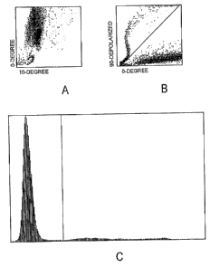
FIG. 1A is a scatter plot that illustrates 10 /0 scattering for removal of
depolarizing interference.
FIG. 1 B is a scatter plot that illustrates 0 /90 depolarized scattering for
removal of depolarizing interference.
FIG. 1 C is a 0 , 90 depolarized histogram that summarizes the results
of FIGS. 1A and 1 B.
FIG. 2A is a scatter plot that illustrates 10 /0 scattering for
differentiating white blood cells and nucleated red blood cells plus noise
(lysed red blood cells in particulate form, cell debris, and platelet clumps)
in
the absence of depolarizing interference.
5
CA 02673031 2009-06-16
WO 2008/079599 PCT/US2007/086217
FIG. 2B is a scatter plot that illustrates 10 /90 depolarized scattering
for differentiating white blood cells and nucleated red blood cells plus noise
(lysed red blood cells in particulate form, cell debris, and platelet clumps)
in
the absence of depolarizing interference.
FIG. 2C is a 0 histogram that summarizes the results of FIGS. 2A and
2B.
FIG. 3A is a scatter plot that illustrates 10 /0 scattering for
differentiating nucleated red blood cells and noise (lysed red blood cells in
particulate form, cell debris, and platelet clumps) in the absence of
depolarizing interference and white blood cells.
FIG. 3B is a scatter plot that illustrates 10 /90 depolarized scattering
for differentiating nucleated red blood cells and noise (lysed red blood cells
in
particulate form, cell debris, and platelet clumps) in the absence of
depolarizing interference and white blood cells.
FIG. 3C is a 10 histogram that summarizes the results of FIGS. 3A
2o and 3B.
FIG. 4A is a scatter plot that illustrates 0 /90 scattering for
differentiating possible platelet clumps and white blood cells.
FIG. 4B is a scatter plot that illustrates 10 /90 scattering for
differentiating possible platelet clumps and white blood cells.
FIG. 4C is 90 a histogram that summarizes the results of FIGS. 4A
and 4B.
FIG. 5A is a scatter plot that illustrates 10 /0 scattering for determining
platelet clumps, without ristocetin.
6
CA 02673031 2009-06-16
WO 2008/079599 PCT/US2007/086217
FIG. 5B is a scatter plot that illustrates 0 /90 scattering for determining
platelet clumps, without ristocetin.
FIG. 5C is a scatter plot that illustrates 10 /90 for determining platelet
clumps, without ristocetin.
FIG. 5D is a scatter plot that illustrates 10 /0 scattering for determining
platelet clumps, with ristocetin.
FIG. 5E is a scatter plot that illustrates 0 /90 scattering for determining
platelet clumps, with ristocetin.
FIG. 5F is a scatter plot that illustrates 10 /90 for determining platelet
clumps, with ristocetin.
FIG. 6A is a 90 histogram that summarizes the results of FIGS. 5A,
5B, and 5C. The presence of platelet clumps is not apparent.
FIG. 6B is a 90 histogram that summarizes the results of FIGS. 5D,
2o 5E, and 5F. The presence of platelet clumps is apparent.
FIG. 7 is a graph that compares the accuracy of the method described
herein against the nucleated red blood cell count of the CELL-DYN 4000
hematology analyzer.
FIG. 8A is a 10 /0 scatter plot that illustrates a low outlier, i.e., an
outlier at the right end of FIG. 7.
FIG. 8B is a 0 histogram that summarizes the results of FIG. 8A.
FIG. 9A is a 10 /0 scatter plot that illustrates a false positive or false
negative, i.e., results at the left end of FIG. 7.
FIG. 9B is a 0 histogram that summarizes the results of FIG. 9A.
7
CA 02673031 2009-06-16
WO 2008/079599 PCT/US2007/086217
FIGS. 10A, 10B, 10C, 10D, 10E, 10F, and 10G are 10 /0 scatter plots
that illustrate a range of abnormal examples, i.e., 1/100 to 162/100 nucleated
red blood cells per white blood cells. The boxes indicate where the algorithm
searches for the nucleated red blood cells.
FIG. 11 is a scatter plot that illustrates an abnormal example commonly
missed on account of sickle cell interference.
FIGS. 12A, 12B, and 12C are 10 /0 scatter plots that illustrate three
normal examples. The boxes indicate where the algorithm searches for the
nucleated red blood cells.
FIGS. 13A, 13B, and 13C are 10 /0 scatter plots that illustrate
examples having less than 1/100 nucleated red blood cells per white blood
cell. The boxes indicate where the algorithm searches for the nucleated red
blood cells.
DETAILED DESCRIPTION
As used herein, the expression "morphological assessment" means
assessment of the shape of a cell. The term "leukocyte" means white blood
cell. Unlike red blood cells, white blood cells occur in many different types.
Examples of leukocytes include granulocytes, neutrophils, eosinophils,
basophils, lymphocytes, and monocytes. The expression "reference method"
means a method of the prior art against which a test method is compared.
The term "sickle cell" means a red blood cell shaped like a sickle. A sickle
cell
is typically resistant to a lyse reagent. The term "thalassemic" relates to a
genetic blood disorder in which the bone marrow cannot form sufficient red
cells and red cell survival is also reduced. The term "lymphocyte" means a
white blood cell that matures in lymph nodes, the spleen, and other lymphoid
tissues, enters the blood, and circulates throughout the body. The expression
"nucleated red blood cell" means an immature red blood cell that still
contains
8
CA 02673031 2009-06-16
WO 2008/079599 PCT/US2007/086217
a nucleus. As used herein, the term "noise" includes, but is not limited to,
such substances as lysed red blood cells in particulate form, cell debris, and
platelet clumps.
As used herein, the term "event" means a particle of that is of a size
sufficient to trigger the 0 detector, whereby that detector signals the
analyzer
to collect 0 ,10 , 90 , and 90 depolarized measurements of that particle.
Particles include, but are not limited to, are white blood cells (WBC), red
blood
cells (RBC), platelets (PLT), RBC fragments, WBC fragments, lipids, platelet
(PLT) clumps.
As used herein, the expression "lyse reagent" means a lyse reagent of
the type described in U. S. Patent No. 5,958, 781, incorporated herein by
reference. As used herein, the term "diluent" means a diluent of the type
described in U. S. Patent No. 5,227,304, incorporated herein by reference.
The method of this invention can be performed with an automated
hematology analyzer having a laser light source with a multi-directional
detection system, a multi-angle depolarizing flow cytometer, such as, for
example, a cytometer of the type described in U. S. Patent No. 5,510,267,
incorporated herein by reference. A representative example of a
commercially available automated hematology analyzer suitable for use in the
method of this invention is a CELL-DYN 3000 Series analyzer using a red
laser light source, commercially available from Abbott Laboratories, Abbott
Park, IL. Detection of optical scattering at 0 and 10 are preferred for
detection and measurement of total white blood cell and nucleated red blood
cell counts. Prior to being analyzed by the flow cytometer, the blood sample
to be analyzed is subjected to a differential lyse reagent. A representative
example of a commercially available differential lyse reagent suitable for use
in the method of this invention is described in U. S. Patent No. 5,958,781,
previously incorporated herein by reference.
A schematic diagram of an automated hematology analyzer suitable for
use in this invention can be found in U. S. Patent No. 5,510,267, previously
incorporated herein by reference. U. S. Patent No. 5,510,267 also describes
in detail the principle of impedance cell counting and sizing. U. S. Patent
No.
5,510,267 further describes an optical transducer suitable for use with the
automated hematology analyzer that can be used to practice this invention.
9
CA 02673031 2009-06-16
WO 2008/079599 PCT/US2007/086217
The type of lyse reagent and the concentration thereof are important
for the method of this invention. The method of this invention calls for the
use
of a lysed sample. In one embodiment, the sample can be lysed by mixing a
lyse reagent (such as, for example, the lyse reagent described in U. S. Patent
No. 5,958,781) with a sample diluted with a diluent (such as, for example, the
diluent described in U. S. Patent No. 5,227,304). The ratio of the lyse
reagent
to the diluent can range from about 1:3 to about 1:8. The lysing reagent lyses
the whole blood sample to form a lysate. A portion of the lysate is
transferred
to a hemoglobin flow cell for hemoglobin measurement (as described in U. S.
Patent No. 5,958,781), and then the remaining portion of the lysate or the
lysate previously used is transferred to an automated analyzer for counting of
white blood cells and nucleated red blood cells. As used herein, the range
"from about 1:3 to about 1:8" means about one part lyse reagent to about
three to eight parts diluent.
In the preferred embodiment, the lyse reagent comprises an aqueous
solution of one or more quaternary ammonium salts (e.g., Br or CI) in an
amount ranging from about 15 to about 150 g/L and hydroxylamine salts (e.g.,
HCI) in an amount ranging from about 0.5 to about 50 g/L; sodium chloride in
an amount ranging from about 0 to about 10 g/L; having a pH of from about
2o 2.5 to about 6.0; and having an osmolality of in an amount ranging from
about
150 to about 700 mOsm/kg.
The method of this invention eliminates counting the substances that
interfere with the counting of nucleated red blood cells and white blood cells
in
all clinical blood samples and provides an accurate first pass result with all
clinical blood samples, unlike methods of the prior art. Interferences that
are
omitted using the multi-dimensional algorithm of this invention include:
(1) Lysed red blood cells in particulate form, which are omitted by
using data collected by 0 and 10 detectors.
(2) Lipids, which are omitted by using data collected by 90
Depolarized and 0 detectors.
(3) Platelet clumps, which are omitted by using data collected by
90 detector.
CA 02673031 2009-06-16
WO 2008/079599 PCT/US2007/086217
The method of this invention can be used with known and reliable apparatus,
such as, for example, automated hematology analyzers that are commercially
available or are currently being developed, and known and reliable reagents,
such as, for example, very low cost reagents for determining white blood cells
and hemoglobin.
In particular, the method of this invention can be used with the CELL-
DYN 3000 series of automated hematology analyzers with updated flowscript
and software. Furthermore, the method of this invention can be used with
CELL-DYN 4000/Sapphire series of automated hematology analyzers with
optical and software improvements.
Eliminating interferences provides:
(1) An increase in first run reportable white blood cells (less
invalidation);
(2) Sensitive enumeration of platelet clumps;
(3) Sensitive enumeration of lipid droplets.
In other methods in the art, blood specimens that contain hypotonically
resistant red blood cells or fragile white blood cells tend to be problematic.
2o Those methods attempt to produce a white cell differential at the same time
as white blood cell counts and nucleated red blood cell counts. The method
of this invention differentiates between nucleated red blood cells and white
blood cells, whereby hypotonically resistant red blood cells and fragile white
blood cells are not problematic. When used with a hematology analyzer, the
method of this invention will not require an additional reagent for analysis
because the reagent used is already used for determination of hemoglobin.
With a more accurate white blood cell count, the correction of determination
of
hemoglobin for white blood cell count can be improved as well.
The method of this invention enables accurate counting of total white
blood cells and nucleated red blood cells with a single blood sample and the
same reagent, without significant interference from the hypotonically
resistant
red blood cells, platelet clumps, reticulocytes, and cell debris that are
present.
The method of this invention does not significantly affect the white blood
cell
counts of fragile lymphocytes and other leukocytes in all clinical blood
11
CA 02673031 2009-06-16
WO 2008/079599 PCT/US2007/086217
samples. The method of this invention also detects and counts platelet
clumps in the same assay, and can be used with bone marrow and cord blood
samples without significant interference.
The following non-limiting examples illustrate the method of this
invention. In the following examples, the parameters that are measured are
described as follows:
(1) 0 light scatter: light scattered at from about 1 to about 3 with
respect to the laser beam.
(2) 10 light scatter: light scattered at from about 3 to about 10
with respect to the laser beam.
(3) 90 light scatter: light scattered orthogonally to the laser beam.
(4) 90 depolarized light scatter: light scattered orthogonally to the
laser beam, which by interaction with white cells is no longer
vertically polarized.
EXAMPLE 1
The current CELL-DYN 3000 Series analyzer and CELL-DYN Diff
Screen reagents, diluent, and lyse reagent, are used for this method. The
diluent was described in U. S. Patent No. 5,227,304, previously incorporated
herein by reference and the lyse reagent was described in U. S. Patent No.
5,958,781, previously incorporated herein by reference. The method involves
the step of mixing lyse reagent with a whole blood sample diluted by a
diluent,
wherein the ratio lyse reagent to diluent ranges from about 1 part lyse
reagent
to from about 3 parts to about 8 parts diluent. The lyse reagent lyses the
whole blood sample to form a lysate. The lysate is then transferred to the
analyzer for white blood cell and nucleated red blood cell counting. During
actual operation, 200 pL of lyse reagent diluted with 900 pL of diluent is
then
immediately mixed with 10 pL of blood/sample in the mixing chamber. After a
bubble mixing, the lysate is analyzed automatically in the analyzer.
12
CA 02673031 2009-06-16
WO 2008/079599 PCT/US2007/086217
EXAMPLE 2
Referring now to FIGS. 1A, 1 B, and 1C, with respect to the scatter plot
depicting 90 depolarized light scatter versus 0 light scatter for removing
depolarizing interference, it can be seen that depolarizing interference is
removed because there is a discernible separation between the events above
the diagonal line and the events below the diagonal line in FIG. 1 B. For each
of the scatter plots depicting 90 depolarized light scatter versus 0 light
scatter, an imaginary line is drawn to each event from the point x = 0, y = 0.
The angle between the imaginary line and the x-axis is determined for each
event. The x-axis of the 0 , 90 depolarizing histogram is divided between the
depolarizing events (e.g., lipid events) and the non-depolarizing events
(e.g.,
white blood cell events, nucleated red blood cell events, platelet clump
events, and other non-depolarizing substance events).
The 0 , 90 depolarized histogram for removing depolarizing
interference shows a high frequency of events to the left of the vertical line
and a measurable frequency of events to the right of the vertical line in FIG.
1 C. The events to the right of the vertical line in FIG. 1 C represent the
frequency of the lipid events; the events to the left of the vertical line in
FIG.
1 C represent the frequency of the remaining events in the samples (e.g.,
white blood cell events, nucleated red blood cell events, platelet clump
events, and other non-depolarizing substance events).
EXAMPLE 3
Referring now to FIGS. 2A, 2B, and 2C, with respect to the
differentiation between white blood cells and the combination of nucleated red
blood cell events plus noise, it can be seen that there is a high frequency of
nucleated red blood cell events plus noise at the bottom of the scatter plot,
below the horizontal line in FIG. 2A, depicting 0 light scatter versus 10
light
scatter, and a high frequency of white blood cell events at the top of the
scatter plot, above the horizontal line in FIG. 2A, depicting 0 light scatter
versus 10 light scatter. It can also be seen that there is a high frequency
of
nucleated red blood cell events plus noise at the left side of the scatter
plot, to
13
CA 02673031 2009-06-16
WO 2008/079599 PCT/US2007/086217
the left of the vertical line in FIG. 2B, depicting 90 depolarized light
scatter
versus 0 light scatter, and a high frequency of white blood cell events at
the
right side of the scatter plot, to the right of the vertical line in FIG. 2B,
depicting 90 depolarized light scatter versus 0 light scatter. The 0
histogram in FIG. 2C shows a high frequency of events to the right of the
vertical line and a measurable frequency of events to the left of the vertical
line in FIG. 2C. The events to the right of the vertical line in FIG. 2C
represent
the frequency of the white blood cell events; the events to the left of the
vertical line in FIG. 2C represent the frequency of the nucleated red blood
cell
events plus noise.
EXAMPLE 4
Referring now to FIGS. 3A, 3B, and 3C, with respect to the
differentiation between noise and nucleated red blood cells, it can be seen
that there is a high frequency of nucleated red blood cell events at the right
side of the scatter plot, to the right of the vertical line in FIG. 3A,
depicting 0
light scatter versus 10 light scatter, and a high frequency of noise at the
left
side of the scatter plot, to the left of the vertical line in FIG. 3A,
depicting 0
light scatter versus 10 light scatter. In FIG. 3B, it can be seen that there
is a
high frequency of nucleated red blood cell events at the bottom of the scatter
plot depicting 90 depolarized light scatter versus 10 light scatter. The 10
histogram shows a measurable frequency of events to the left of the vertical
line in FIG. 3C and a high frequency of events to the right of the vertical
line in
FIG. 3C. The events to the right of the vertical line in FIG. 3C represent the
frequency of nucleated red blood cell events; the events to the left of the
vertical line in FIG. 3C represent the frequency of noise.
EXAMPLE 5
Referring now to FIGS. 4A, 4B, and 4C, with respect to the
differentiation between white blood cells and platelet clumps, it can be seen
that there is a high frequency of white blood cell events at the top of the
scatter plot, above the horizontal line in FIG. 4A, depicting 90 light
scatter
14
CA 02673031 2009-06-16
WO 2008/079599 PCT/US2007/086217
versus 0 light scatter, and a low frequency of possible platelet clump events
at the bottom of the scatter plot, below the horizontal line in FIG. 4A,
depicting
90 light scatter versus 0 light scatter. It can also be seen that there is a
high
frequency of white blood cell events at the top of the scatter plot, above the
horizontal line in FIG. 4B, depicting 90 light scatter versus 10 light
scatter,
and a low frequency of possible platelet clump events at the bottom of the
scatter plot, below the horizontal line in FIG. 4B, depicting 90 light
scatter
versus 10 light scatter. The 90 histogram shows a non-measurable
frequency of events to the left of the vertical line in FIG. 4C and a high
frequency of events to the right of the vertical line in FIG. 4C. The events
to
the right of the vertical line in FIG. 4C represent the frequency of white
blood
cell events; the events to the left of the vertical line in FIG. 4C represent
the
possibility of platelet clump events.
From the foregoing examples, EXAMPLES 1 through 5, inclusive, it
can be seen that the method of this invention utilizes existing optical
equipment and existing reagents in a different way to improve the
enumeration of nucleated red blood cells and the enumeration of white blood
cells. The existing CELL-DYN instruments can be set up to focus on
particles having the size of nucleated red blood cells. The lyse reagent is
optimized to lyse the red blood cells and retain the differentiating
properties of
the nucleated red blood cells and the white blood cells, thereby improving the
sensitivity of existing methods for enumerating nucleated red blood cells and
enumerating white blood cells.
EXAMPLE 6
This example illustrates the differentiation between platelet clumps and
white blood cells. Ristocetin is a reagent that can be used to cause platelets
to aggregate (clump), whereby detection of the platelets is facilitated. FIG.
5A
depicts 0 light scatter versus 10 light scatter; FIG. 5B depicts 90 light
scatter versus 0 light scatter; FIG. 5C depicts 90 light scatter versus 10
light scatter. In FIGS. 5A, 513, and 5C, where ristocetin was not used, the
portions above the horizontal lines in FIGS. 5A, 513, and 5C show white blood
cell events. The portions below the horizontal lines in FIGS. 5A, 513, and 5C
CA 02673031 2009-06-16
WO 2008/079599 PCT/US2007/086217
show possible platelet clump events. FIG. 5D depicts 0 light scatter versus
light scatter; FIG. 5E depicts 90 light scatter versus 0 light scatter; FIG.
5F depicts 90 light scatter versus 10 light scatter. In FIGS. 5E and 5F,
where ristocetin was used, the portions above the horizontal lines show white
5 blood cell events. The portions below the horizontal lines show possible
platelet clump events. It can be seen that the employment of ristocetin
enables detection of platelet clumps.
The 90 histogram in FIG. 6A summarizes the results of FIGS. 5B and
5C. The 90 histogram in FIG. 6B summarizes the results of FIGS. 5E and
1o 5F. In FIG. 6A, it can be seen that there is a non-measurable frequency of
events to the left of the vertical line and a high frequency of events to the
right
of the vertical line. The events to the right of the vertical line in FIG. 6A
represent the frequency of white blood cell events; the events to the left of
the
vertical line in FIG. 6A represent the possibility of platelet clump events.
In
FIG. 6B, it can be seen that there is a measurable frequency, i.e., a
moderately high frequency, of events to the left of the vertical line and a
high
frequency of events to the right of the vertical line. The events to the right
of
the vertical line in FIG. 6B represent the frequency of white blood cell
events;
the events to the left of the vertical line in FIG. 6B represent the
possibility of
platelet clump events.
The 90 detector is used to measure the mismatch of refractive index
between the solution outside the cell and inside the cell. Because platelet
clumps only have one refractive index (no cell structure) the 90 measurement
is low compared with the white blood cell population.
EXAMPLE 7
FIG. 7 is a graph that shows the accuracy of the method described
herein against the CELL-DYN 4000 instrument. It can be seen that the
method described herein compares very well to the results of the CELL-DYN
4000 instrument with respect to the percentage of nucleated red blood cells.
While the CELL-DYN 4000 instrument is a very effective instrument, the cost
of running that instrument is high, and the CELL-DYN 4000 instrument is
complex. The CELL-DYN 4000 instrument uses an expensive dye, an
16
CA 02673031 2009-06-16
WO 2008/079599 PCT/US2007/086217
expensive complicated lyse reagent, and an expensive light source. These
costly components are important for determining white blood cell differential,
the white blood cell count, and the nucleated red blood cell count at the same
time. The method described herein does not attempt to provide a white blood
cell differential. The method described herein provides a more reportable
white blood cell result and nucleated red blood cell result, because the
method is capable of eliminating various sources of interferences, namely,
hypotonically resistant red blood cells, lipids, and platelets clumps. At the
same time, samples containing fragile white blood cells are counted
accurately. The method of this invention identifies nucleated red blood cells
at a concentration of less than 10 nucleated red blood cells per 100 white
blood cells, as can be seen from the outlier examples. The method described
herein is less costly to implement and perform on account of the lower cost of
the light source and reagents that can be used with the method. The CELL-
DYN 4000 instrument currently uses a relatively expensive blue Argon laser.
The reagents for the CELL-DYN 4000 instrument involve two lyse
components and propidium iodide dye, which leads to greatly complexity and
expense of manufacture, and, consequently, greater expense to the user.
In the bias plot shown in FIG. 7, the vertical axis represents the
2o difference between nucleated red blood cells determined by the algorithm
described herein and the nucleated red blood cells determined by the CELL-
DYN 4000 instrument. The data represent the number of nucleated red
blood cells per 100 white blood cells. The date involved 106 specimens
tested in duplicate, 26 in-house, 80 abnormal with 34 positive nucleated red
blood cell counts and 6 fragile lymphocyte counts. From the plot, it can be
seen that there is a low outlier. From the plot, it can also be seen that
there
are numerous false positive test results. The outlier and the false positive
test
results will be discussed in EXAMPLES 8 and 9.
EXAMPLE 8
This example involves a discussion of the low outlier shown in FIG. 7.
FIG. 8A is a scatter plot showing 0 scatter versus 10 scatter, the portion
enclosed by the rectangular box includes a significant number of nucleated
17
CA 02673031 2009-06-16
WO 2008/079599 PCT/US2007/086217
red blood cell events. The portion above the rectangular box in FIG. 8A
includes white blood cell events. In FIG. 8B, it can be seen that there is a
measurable frequency, i.e., a moderately high frequency, of events to the left
of the vertical line and a high frequency of events to the right of the
vertical
line. The events to the right of the vertical line in FIG. 8B represent the
frequency of white blood cell events; the events to the left of the vertical
line in
FIG. 8B represent the possibility of nucleated red blood cell events.
EXAMPLE 9
This example involves a discussion of the false positive outliers shown
in FIG. 7. One sample is discussed in this example. In FIG. 9A, in the scatter
plot showing 0 scatter versus 10 scatter, the portion enclosed by the
rectangular box includes a significant number of nucleated red blood cell
events. The portion above the rectangular box in FIG. 9A includes a
significant number of white blood cell events. In FIG. 9B, it can be seen that
there is a measurable frequency, i.e., a moderately high frequency, of events
to the left of the vertical line and a high frequency of events to the right
of the
vertical line. The events to the right of the vertical line in FIG. 9B
represent
the frequency of white blood cell events; the events to the left of the
vertical
line in FIG. 9B represent the frequency of nucleated red blood cell events.
FIGS. 9A and 9B show that these samples indeed contain nucleated red
blood cells.
EXAMPLE 10
This example shows a series of scatter plots that illustrate a range of
abnormal examples where nucleated red blood cells are present. The scatter
plots show 0 scatter versus 10 scatter. In FIGS. 10A through 10G,
inclusive, the blood samples exhibit a range from about 1 to about 162
nucleated red blood cells per 100 white blood cells.
18
CA 02673031 2009-06-16
WO 2008/079599 PCT/US2007/086217
EXAMPLE 11
This example shows a scatter plot that illustrates an abnormal example
commonly missed on account of sickle cell interference. The scatter plots
show 0 scatter versus 10 scatter. The example shows that the method of
this invention can detect nucleated red blood cells in the presence of
interfering cells.
EXAMPLE 12
This example shows a series of scatter plots that illustrate a range of
normal examples of true negatives of nucleated red blood cells. The scatter
plots show 0 scatter versus 10 scatter. The rectangular boxes in FIGS. 12A,
12B, and 12C show where the algorithm described herein would search for
nucleated red blood cells.
EXAMPLE 13
This example shows a series of scatter plots that illustrate a range of
samples of fragile white blood cells. Fragile white blood cells present a
challenge to current technology, because fragile white blood cell events
resemble nucleated red blood cell events occupying the same scatter space.
The scatter plots show 0 scatter versus 10 scatter. The rectangular boxes
in FIGS. 13A, 13B, and 13C show where the algorithm described herein
would search for nucleated red blood cells. In each blood sample, the events
enclosed by the rectangular boxes indicate that there is less than one
nucleated red blood cell per 100 white blood cells.
Various modifications and alterations of this invention will become
apparent to those skilled in the art without departing from the scope and
spirit
of this invention, and it should be understood that this invention is not to
be
unduly limited to the illustrative embodiments set forth herein.
19