Note: Descriptions are shown in the official language in which they were submitted.
CA 02673777 2009-06-25
WO 2008/082844 PCT/US2007/086599
DEVICES AND METHODS FOR PLACEMENT OF PARTITIONS
WITHIN A HOLLOW BODY ORGAN
Cross-Reference To Related Application
This application is a continuation-in-part of co-pending application Serial
No.
11/324,135 filed on December 29, 2005, which is a continuation-in-part of
application
Serial No. 10/797,303 filed on March 9, 2004, both of which are herein
incorporated by
reference.
Backaound of the Invention
1. Field of the Invention:
The present invention relates generally to medical devices and methods. More
particularly, it relates to devices and methods for creating a partition
within a hollow body
organ, particularly a stomach, intestinal tract, or other region of the
gastrointestinal tract,
and affixing the tissue.
2. General Background and State of the Art:
In cases of severe obesity, patients may currently undergo several types of
surgery
either to tie off or staple portions of the large or small intestine or
stomach, and/or to
bypass portions of the same to reduce the amount of food desired by the
patient, and the
amount absorbed by the gastrointestinal tract. The procedures currently
available include
laparoscopic banding, where a device is used to "tie off' or constrict a
portion of the
stomach, vertical banded gastroplasty (VBG), or a more invasive surgical
procedure
known as a Roux-En-Y gastric bypass to effect permanent surgical reduction of
the
stomach's volume and subsequent bypass of the intestine.
Typically, these stomach reduction procedures are performed surgically through
an
open incision and staples or sutures are applied externally to the stomach or
hollow body
organ. Such procedures can also be performed laparoscopically, through the use
of smaller
incisions, or ports, through trocars and other specialized devices. In the
case of
laparoscopic banding, an adjustable band is placed around the proximal section
of the
stomach reaching from the lesser curve of the stomach around to the greater
curve, thereby
creating a constriction or "waist" in a vertical manner between the esophagus
and the
pylorus. During a VBG, a small pouch (approximately 20 cc in volume) is
constructed by
forming a vertical partition from the gastroesophageal junction to midway down
the lesser
curvature of the stomach by externally applying staples, and optionally
dividing or
resecting a portion of the stomach, followed by creation of a stoma at the
outlet of the
CA 02673777 2009-06-25
WO 2008/082844 -2- PCT/US2007/086599
partition to prevent dilation of the outlet channel and restrict intake. In a
Roux-En-Y
gastric bypass, the stomach is surgically divided into a smaller upper pouch
connected to
the esophageal inflow, and a lower portion, detached from the upper pouch but
still
connected to the intestinal tract for purposes of secreting digestive juices.
A resected
portion of the small intestine is then anastomosed using an end-to-side
anastomosis to the
upper pouch, thereby bypassing the majority of the intestine and reducing
absorption of
caloric intake and causing rapid "dumping" of highly caloric or "junk foods."
Although the outcome of these stomach reduction surgeries leads to patient
weight
loss because patients are physically forced to eat less due to the reduced
size of their
stomach, several limitations exist due to the invasiveness of the procedures,
including
time, use of general anesthesia, time and pain associated with the healing of
the incisions,
and other complications attendant to major surgery. In addition, these
procedures are only
available to a small segment of the obese population (morbid obesity, Body
Mass Index >
40) due to their complications, leaving patients who are considered obese or
moderately
obese with few, if any, interventional options.
In addition to surgical procedures, certain tools exist for securing tissue
such as the
stapling devices used in the above-described surgical procedures and others
such as in the
treatment of gastroesophageal reflux disease (GERD). These devices include the
GIA
device (Gastrointestinal Anastomosis device manufactured by Ethicon
Endosurgery, Inc.
and a similar product by USSC), and certain clamping and stapling devices as
described in
U.S. Pat. Nos. 5,403,326; 5,571,116; 5,676,674; 5,897,562; 6,494,888; and
6,506,196 for
methods and devices for fundoplication of the stomach to the esophagus for the
treatment
of gastroesophageal reflux disease (GERD). In addition, certain tools, such as
those
described in U.S. Pat. Nos. 5,788,715 and 5,947,983, detail an endoscopic
suturing device
that is inserted through an endoscope and placed at the site where the
esophagus and the
stomach meet. Vacuum is then applied to acquire the adjacent tissue, and a
series of
stitches are placed to create a pleat in the sphincter to reduce the backflow
of acid from the
stomach up through the esophagus. These devices can also be used transorally
for the
endoscopic treatment of esophageal varices (dilated blood vessels within the
wall of the
esophagus).
There is a need for improved devices and procedures. In addition, because of
the
invasiveness of most of the surgeries used to treat obesity and other gastric
disorders such
as GERD, and the limited success of others, there remains a need for improved
devices and
methods for more effective, less invasive hollow organ restriction procedures.
CA 02673777 2009-06-25
WO 2008/082844 -3- PCT/US2007/086599
Summary Of The Invention
A device for tissue acquisition and fixation is described that may be utilized
for
creating a partition within a hollow body organ, such as the stomach,
esophageal junction,
and other portions of the gastrointestinal tract for performing a
gastroplasty. Generally,
the device may be advanced in a minimally invasive manner within a patient's
body, e.g.,
transorally, endoscopically, percutaneously, etc., to create one or several
divisions or
plications within the hollow body organ. Such divisions or plications can form
restrictive
barriers within the organ, or can be placed to form a pouch, or gastric lumen,
smaller than
the remaining stomach volume to essentially act as the active stomach such as
the pouch
resulting from a surgical Roux-En-Y gastric bypass procedure. Examples of
placing
and/or creating divisions or plications may be seen in further detail in U.S.
Pat. No.
6,558,400; U.S. Pat. App. Serial No. 10/188,547 filed July 2, 2002; and U.S.
Pat. App.
Serial No. 10/417,790 filed April 16, 2003, each of which is incorporated
herein by
reference in its entirety.
The device may be advanced within a body through a variety of methods, e.g.,
transorally, transanally, endoscopically, percutaneously, intraperitoneal
surgically (natural
orifice transvisceral endoscopic surgery), etc., to create one or several
divisions or
plications within a hollow body organ, e.g., to create a gastric lumen or
partition to reduce
the effective active area of the stomach (e.g., that which receives the
initial food volume),
performed from within the stomach cavity. The smaller gastric lumen created
may be
about 18 mm in diameter and about 70 cm in length with a volume of about 10 cc
to about
100 cc, for example about 10 cc to about 25 cc, and may be achieved in a
minimally
invasive procedure completely from within the stomach cavity. Moreover, the
device is
configured such that once acquisition of the tissue is accomplished,
manipulation of the
acquired tissue is unnecessary as the device is able to automatically
configure the acquired
tissue into a desired configuration.
The device may generally include a first acquisition member and a second
acquisition member in apposition to one another along a first longitudinal
axis, wherein
optionally, at least one of the acquisition members is adapted to adhere
tissue thereto such
that the tissue is positioned between the first and second acquisition
members, and
optionally wherein at least one of the acquisition members is movable relative
to the first
longitudinal axis between a delivery configuration and a deployment
configuration.
Moreover, the system may also include a septum, or a separator, removably
positioned
between the first and second acquisition members, wherein at least one of the
acquisition
CA 02673777 2009-06-25
WO 2008/082844 PCT/US2007/086599
members is movable relative to the septum between a delivery configuration and
a
deployment configuration.
A handle may be located at a proximal end of an elongate body or member and
used to manipulate the device advanced within the hollow body organ as well as
control
the opening and closing/clamping of the acquisition members onto the tissue.
The
elongate body may include a series of links, or be formed from an extrusion
fabricated
with one or more various lumens to accommodate the various control mechanisms
of the
acquisition device. Similarly, the control mechanisms may be grouped together
and
sheathed in a thin skin sheath, such as a heat shrink or spirally wound
adhesive backed
tape. A working lumen may extend entirely through the elongate member and may
be
sized to provide access to the distal end for various surgical tools, such as
an endoscope or
other visualization device, and/or therapeutic devices such as snares,
excisional tools,
biopsy tools, etc. once the distal end of the assembly is positioned within
the hollow body
organ. The acquisition members may be joined to the elongate body via a
passive or active
hinge member, adaptable to position the assembly. The acquisition members may
generally include a cartridge member placed longitudinally in apposition to an
anvil
member. The cartridge member may contain one or several fasteners, e.g.,
staples, clips,
anchors, etc., which may be actuated via controls located proximally on the
handle
assembly. Moreover, the septum or tissue barrier may be removably positioned
between
the cartridge member and anvil member and used to minimize or eliminate cross
acquisition of the tissue into the cartridge member and/or anvil member.
Methods of placing a partition from within a hollow body organ using the
device
disclosed herein generally includes positioning a first acquisition member and
a second
acquisition member adjacent to a region of tissue within the hollow body
organ, wherein
the first and second acquisition members are in apposition to one another
along a first
longitudinal axis, adhering tissue from the region to each of the first and
second
acquisition members, and securing the adhered tissue between the first and
second
acquisition members. Such a method may also involve pivoting at least one of
the
acquisition members about the longitudinal axis to an open or closed
configuration, and
still another method involves pivoting at least one of the members about the
transverse
axis. Another method may also include moving a septum or tissue barrier
relative to the
first and second acquisition member to control the length of the region of
tissue acquired
by the acquisition members, or removing the septum from between the first
acquisition
member and the second acquisition member.
CA 02673777 2009-06-25
WO 2008/082844 PCT/US2007/086599
Methods are also disclosed of placing a partition from within a first and a
second
organ. A tissue treatment device having first and second regions for
releasably adhering
tissue from the organs can be used to fasten the adhered tissue between the
first and second
regions of the tissue treatment device. In one embodiment, tissue from the
gastroesophageal junction ("GEJ"), including the lower esophageal sphincter,
along with
tissue from the stomach is acquired with the tissue treatment device. The
tissue treatment
device forms one or more plications beginning at the GEJ and ending in the
stomach cavity
to form a pouch or an extension of the esophagus into the stomach cavity.
While the device is in a delivery configuration, the components of the distal
working portion of the device (the cartridge member and anvil member) are
disposed such
that the cartridge and anvil are directly positioned into apposition about the
septum. Once
desirably positioned, one or both of the cartridge member and anvil member may
be
rotated about a pivot or translationally moved in parallel to one another.
Then, portions of
the stomach wall may be acquired by, or drawn within their respective
openings. The
configuration of the cartridge member and anvil member and the positioning of
the device
within the stomach are such that this tissue acquisition procedure also
enables the devices
to be self-adjusting with respect to the acquired tissue. Alternatively, the
cartridge and
anvil members may close to within a fixed distance, i.e. a fixed distance
clamp gap.
Moreover, the device is configured such that portions of the stomach wall are
automatically positioned for fixation upon being acquired and the tissue
becomes
automatically adjusted or tensioned around the perimeter of the distal working
portion of
the device in the stomach and within the distal working portion inner volume,
to achieve
the desired resulting geometry (e.g., small gastric pouch or restrictive
partition or baffle).
Because of the manner in which the tissue is acquired, the tissue intimately
surrounds the
cartridge member and anvil member to define or calibrate the subsequent volume
of the
resulting gastric lumen. Thus, the gastric volume may be somewhat controlled
by
adjusting the volume of the cartridge member and anvil member, or the use of
accessory
devices such as a scope or balloon. As a result, once the desired volume is
known and
incorporated in the device, the user can achieve a controlled acquisition and
without
intraprocedural adjustments or positioning requirements.
The septum may act effectively as a barrier between the openings to facilitate
the
acquisition of the tissue to their respective openings while minimizing or
eliminating cross
acquisition of the tissue into the cartridge member and/or anvil member. In
other
alternatives, the septum may be omitted from the device and acquisition of the
tissue may
CA 02673777 2009-06-25
WO 2008/082844 -6- PCT/US2007/086599
be accomplished by sequentially activating vacuum forces within the openings.
Once the
tissue has been acquired, the septum may be removed from between the cartridge
member
and anvil member by translating the septum distally or proximally of the
cartridge member
and anvil member or left within the stomach for later removal. Alternatively,
the septum
may rotate, collapse, be ejected up and out from between the jaws. In one
alternative, the
septum includes a sail that can be extended between the cartridge member and
anvil
member after being delivered to the stomach, and then collapsible between the
cartridge
member and anvil member for removal from the stomach.
The cartridge member of the tissue treatment device may be re-loadable with a
removable staple cartridge after forming a plication within the cavity. This
allows the
same tissue treatment device to form multiple plications within the cavity. A
method of
treating a stomach cavity may include forming a first plication within the
stomach cavity
using staples from a first removable staple cartridge, which is easily removed
from the
cartridge member. A second removable staple cartridge is then inserted into
the cartridge
member, and a second plication is formed within the stomach cavity using
staples from the
second removable staple cartridge.
Another method of forming a plication within the stomach cavity may include
clamping the cartridge member and the anvil member together and then
reevaluating the
folds of tissue acquired before firing staples into acquired tissue. This
method helps to
ensure that the desired tissue is acquired by the tissue treatment device, and
that there are
no folds or pleats present in the acquired tissue. After inserting the tissue
treatment device
transorally to the stomach cavity, stomach tissue is acquired at a target
region for treatment
with the tissue treatment device. The cartridge member and anvil member of the
tissue
treatment device are then clamped together grasping the acquired target
tissue, and the
stomach cavity can be insufflated to inspect the acquired target tissue with
an endoscope.
If the inspection reveals that the tissue acquired is not the targeted tissue
or that there are
folds or pleats within the acquired tissue, the acquired tissue can be
released from the
tissue treatment device and re-acquired. If the inspection reveals the
acquired tissue would
form a desired sleeve, then the stomach cavity can be desufflated once again,
and the
cartridge member and the anvil member may be unclamped into an open
configuration, to
fully acquire the tissue. The tissue treatment device may include a barrier
such as a
septum, in which case the barrier would be moveable between the cartridge and
anvil
member to control the length of tissue secured, or removed from between the
cartridge
member and the anvil member to fasten the full length of the cartridge and
anvil working
CA 02673777 2009-06-25
WO 2008/082844 -~- PCT/US2007/086599
surface. After which, the cartridge member and anvil member are clamped
together and
the stomach cavity is again insufflated to inspect the acquired tissue. If the
inspection
reveals a desired formation between the folds of acquired tissue, the
cartridge member and
anvil member are fully clamped together and the acquired tissue is plicated to
form a
gastric sleeve within the stomach cavity and the GEJ.
Another method involves lightly clamping the cartridge member and the anvil
member together and then reevaluating the folds of tissue acquired before
firing staples
into acquired tissue. After inserting the tissue treatment device transorally
to the stomach
cavity, stomach tissue is acquired at a target region for treatment with the
tissue treatment
device. The cartridge member and anvil member of the tissue treatment device
are then
lightly clamped together grasping the acquired target tissue, and the stomach
cavity can be
insufflated to inspect the acquired target tissue with an endoscope. If the
inspection
reveals that the tissue acquired is not the targeted tissue or that there are
folds or pleats
within the acquired tissue, the acquired tissue can be released from the
tissue treatment
device and re-acquired. If the inspection reveals the acquired tissue would
form a desired
sleeve, then the stomach cavity can be desufflated once again, and the
cartridge member
and the anvil member may be fully clamped and the acquired tissue is plicated
to form a
gastric sleeve at least partially within the stomach cavity. The tissue
treatment device may
include a barrier such as a septum, in which case the barrier would be
moveable between
the cartridge and anvil member to control the length of tissue secured, or
removed from
between the cartridge member and the anvil member to fasten the full length of
the
cartridge and anvil working surface.
A system for reducing the volume of the stomach cavity may include the tissue
treatment device for forming a plication at least partially within the stomach
cavity and a
restrictor device to further restrict the stomach cavity. When forming a
sleeve along the
lesser curve of the stomach with the tissue treatment device, the restrictor
device may be
used to acquire tissue between an anvil member and a cartridge member to form
a single
fold plication within the sleeve. The restrictor device may have a vacuum pod
located
between the anvil and cartridge member to acquire the desired tissue. A distal
outlet of the
sleeve can be reduced by placing a plication with the restrictor device near
the distal outlet.
Further, the tissue treatment device may form multiple continuous plications
within the
stomach cavity. In the event that unwanted stomas are form between the
multiple
plications, the restrictor device can be used to close those unwanted stomas.
CA 02673777 2009-06-25
WO 2008/082844 -8- PCT/US2007/086599
Brief Description of the Drawings
FIG. 1 shows a perspective view of one embodiment of a gastroplasty device.
FIG. 2 shows a perspective view of a link used to form an elongated tubular
member for the gastroplasy device.
FIG. 3 shows a cross-sectional view taken along line 3-3 of FIG. 2.
FIG. 4 shows multiple links used to form the elongated tubular member joined
together.
FIG. 5 shows a tissue treatment device of the gastroplasty device with jaws
opened
and a wire retractor and sail extended.
FIG. 6 shows a partial cross-sectional view of the tissue treatment device.
FIG. 7 shows a variation of a basket that may be disposed within a vacuum pod
of
the tissue treatment device.
FIG. 8 shows a variation of a septum including sail that may be positioned
between
the jaws of the tissue treatment device in a retracted or delivery
configuration.
FIG. 9 shows the septum and sail of FIG. 8 with the retractor wire extended
and the
sail raised.
FIG. 10 shows the extended retractor wire and sail of FIG. 9 with the septum
moved distally on a septum rail.
FIG. 11 shows the tissue treatment device of the gastroplasty device in a
delivery
configuration with an endoscope alongside.
FIG. 12 shows the tissue treatment device of FIG. 11 with the jaws opened, the
sail
and retractor wire extended, and the septum moved distally on the septum rail
such that the
septum is positioned within a slit of a split flexible tip attached to the
distal end of the
tissue treatment device.
FIG. 13 shows a backside perspective view of the tissue treatment device with
the
distal tip removed for clarity.
FIG. 14 shows a perspective view of a removable staple cartridge.
FIG. 15 shows a perspective view of the removable staple cartridge positioned
within a cartridge member of the tissue treatment device.
FIG. 16 shows a partial cross-sectional side elevational view of the removable
staple cartridge positioned within the cartridge member of the tissue
treatment device.
FIG. 16A shows partial cross-sectional side elevational view of staples being
fired
from the staple cartridge and crimped against an anvil.
FIG. 17 depicts a round wire staple.
CA 02673777 2009-06-25
WO 2008/082844 -9- PCT/US2007/086599
FIG. 18 shows a planar view of an end ring that is connected to a distal end
of the
flexible elongated member.
FIG. 19 shows a partial cross-sectional view of the tissue treatment device in
a
closed position.
FIG. 20 shows a partial cross-sectional view of the tissue treatment device in
an
open position.
FIGS. 21 through 24 show another embodiment of a tissue treatment device
having
a distal clamping wire passed around a distal vertical pulley.
FIG. 25A shows a partial cross-sectional view of the tissue treatment device
in a
closed position with the anvil member aligned with the cartridge member.
FIG. 25B shows a partial cross-sectional view of the tissue treatment device
in a
closed position with the anvil member off-set from the cartridge member.
FIG. 26 shows a partial cross-sectional view of an embodiment of the tissue
treatment device having a tapered anvil.
FIG. 27A shows another embodiment of the tissue treatment device having a bent
or twisted anvil member.
FIG. 27B shows a partial cross-sectional view of an embodiment of the tissue
treatment device having a wide stop and a pin at its proximal end to pinch the
distal end of
the device when it is closed.
FIGS. 28A through 28C depict an embodiment of a Kevlar rope connected to the
distal end of the tissue treatment device to assist in closing the jaws of the
tissue treatment
device.
FIGS. 29A through 29C depict an embodiment of a stainless steel cable
connected
to the distal end of the tissue treatment device to assist in closing the jaws
of the tissue
treatment device.
FIG. 30 shows the proximal end of the tissue treatment device having an
electrical
switch for detecting closure of the jaws.
FIGS. 31 and 32 show a partial cross-sectional view of the proximal end of the
tissue treatment device having a vacuum detection device for detecting closure
of the jaws.
FIGS. 33 through 35 show the proximal end of the tissue treatment device
having a
fiber optic sensor for detecting closure of the jaws.
FIG. 36 shows the proximal end of the tissue treatment device having a
proximity
sensor for detecting closure of the jaws.
CA 02673777 2009-06-25
WO 2008/082844 -10- PCT/US2007/086599
FIGS. 37 and 38 show the proximal end of the tissue treatment device having a
mechanical clamp for detecting closure of the jaws.
FIG. 39 shows a partial cross-sectional view of the proximal end of the tissue
treatment device having a positive pressure device for detecting closure of
the jaws.
FIG. 40 shows a perspective view of a handle assembly.
FIG. 41 shows a partial cross-sectional view of the handle assembly shown in
FIG.
40.
FIGS. 41A and 41B show a push/pull mechanism disposed within the handle
assembly.
FIGS. 42 through 49 show illustrative views and cross-sectional views of one
method of forming a gastric sleeve in a stomach cavity.
FIG. 50 shows a perspective view of a variation of a stapler restrictor.
FIG. 50A shows a stapler assembly of the stapler restrictor in a closed
configuration.
FIG. 50B shows the stapler assembly of the stapler restrictor in an open
configuration.
FIG. 50C shows the stapler assembly of the stapler restrictor with a rubber
tip and
removable staple cartridge removed.
FIG. 50D shows the stapler assembly of the stapler restrictor with an anvil
member
removed.
FIG. 50E shows the removable staple cartridge and rubber tip being loaded into
a
cartridge member of the stapler assembly.
FIGS. 51 through 53A show cross-sectional views of one method of forming a
single fold of tissue near a distal stoma of a gastric sleeve to narrow the
distal stoma.
FIG. 54 shows a cross-sectional view of a gastric sleeve formed with one dual
fold
plication and restricted with three single fold plications.
FIG. 55 shows an illustration of a stomach cavity with two dual fold
plications
forming a long sleeve and two single fold plications along the lesser curve to
narrow the
gastric sleeve.
FIG. 56 shows an illustration of a stomach cavity with three dual fold
plications
forming a long sleeve and three single fold plications along the lesser curve
to narrow the
gastric sleeve.
FIG. 57 shows an illustration of a stomach cavity with four dual fold
plications
forming a long sleeve from the gastroesophageal junction to the pylorus.
CA 02673777 2009-06-25
WO 2008/082844 -11- PCT/US2007/086599
Detailed Description of the Embodiments
A gastroplasty device for tissue acquisition and fixation, and methods of use
are
described. In general, the gastroplasty device described herein may be
utilized for creating
a partition within a hollow body organ or two hollow body organs, such as the
stomach,
esophageal junction, and/or other portions of the gastrointestinal tract. The
gastroplasty
device may be advanced within a body through a variety of methods, e.g.,
transorally,
transanally, endoscopically, percutaneously, etc., to create one or several
divisions or
plications within the hollow body organ, e.g., to create a gastric lumen
within the stomach.
Further, the gastroplasty device may be assisted through the use of
laparoscopic guidance,
in particular, visualization of the external surface of the hollow body organ
to assist in
placement of the device, or within the organ cavity to monitor the procedure.
Similarly,
the device of the present invention may be used in conjunction with other
laparoscopic
procedures, or may further be modified by an additional step or procedure to
enhance the
geometry of the partition. For example, upon placement of a partition of the
present
invention, it may be desirable to perform a secondary step either transorally,
or
laparoscopically, to achieve the desired gastroplasty geometry, such as the
placement of a
single fold or plication within the gastric lumen or pouch as described in
U.S. Pat. App.
Serial No. 10/188,547, which was filed 7/2/2002 and is incorporated by
reference herein in
its entirety, to further restrict the movement of food through the pouch, or
the laparoscopic
placement of a band, clip, ring or other hollow reinforcement member at the
outlet of the
gastric lumen such as is done in a VBG, or lap-band procedure to reinforce or
narrow the
outlet of the lumen.
The gastroplasty device described here, allows for the creation of a smaller
gastric
lumen to be achieved in a minimally invasive surgical procedure completely
from within
the stomach cavity and gastroesophageal junction. Moreover, the devices
described herein
are configured such that once acquisition of the tissue is accomplished, any
manipulation
of the acquired tissue may be unnecessary as the device is able to
automatically configure
the acquired tissue into a desired configuration whereby the geometry of the
devices
approximates the resulting tissue geometry at the time of acquisition. In
operation, the
perimeter of the device, and any openings therein, form the template or mold
cavity around
and into which tissue flows, thereby creating a tissue structure that
approximates the
geometry of the mold. That is, as the device is configured such that portions
of the
stomach wall are automatically positioned for fixation upon being acquired,
and the tissue
becomes automatically adjusted or tensioned around the perimeter of the distal
working
CA 02673777 2009-06-25
WO 2008/082844 -12- PCT/US2007/086599
portion of the device in the stomach and within the distal working portion
inner volume, to
achieve the desired resulting geometry (e.g., small gastric pouch, restrictive
partition or
baffle, or extension of esophagus). Because of the manner in which the tissue
is acquired,
the tissue intimately surrounds the cartridge member and anvil member to
define or
calibrate the subsequent volume of the resulting gastric lumen. Thus, the
gastric volume
may be predetermined by adjusting the volume of the cartridge member and anvil
member.
Subsequent manipulation of the tissue may be performed, if desired, to effect
certain
configurations or further restrict the pouch; however, this manipulation may
be omitted
entirely.
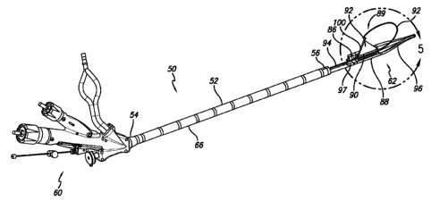
One embodiment of a gastroplasty assembly 50 is shown in FIG. 1. Assembly 50
includes an elongate tubular member 52 having a proximal end 54 and a distal
end 56 with
a lumen 58 defined within the elongate member. As shown in FIG. 1, a handle
assembly
60 is connected at the proximal end of the elongate member, and a tissue
treatment device
or working member 62 is attached at the distal end of the elongate member. The
tissue
treatment device is used to form either single or dual fold plications within
a stomach
cavity and the gastroesophageal junction.
The elongate tubular member 52 may have a circular or elliptical cross-
sectional
area. Alternatively, the cross-sectional area may take on any number of
different cross-
sectional configurations, e.g., hexagonal, octagonal, etc., provided that it
presents an
atraumatic surface to the tissue surfaces within the body. In the embodiment
shown, the
elongate member is a flexible shaft including a series of links 64 that
increase the
flexibility of the elongate member, and hence increase the ease in which the
device is
handled and operated. The lumen 58 may extend entirely through the tubular
member and
may be sized to provide access to the distal end 56 for various surgical tools
or therapies
once the distal end of assembly 50 is positioned within a hollow body organ,
and in
particular may be useful to place an endoscope or other visualization tool for
real time
visualization during the procedure. Alternatively, a fiberscope or other type
of
visualization tool may be integrated within the elongate member. Examples of
useful
scopes may be the Olympus GIF P140, the Fujinon EG 25PE, and the like. An
optional
separate thin walled oversheath or liner 66, may also be placed over the
acquisition device,
including the links of the elongate member, to assist in placement, or may be
placed over a
guide wire or obturator down the esophagus prior to placement of the
gastroplasty device
and removed with the gastroplasty device once the procedure is complete. The
oversheath
or liner may be made of a thin wall polymer such as polyolefin,
polytetrafluoroethylene
CA 02673777 2009-06-25
WO 2008/082844 -13- PCT/US2007/086599
(PTFE), expanded PTFE (ePTFE), silicone and the like, having a wall thickness
preferably
between about 0.001" and about 0.025". This liner can serve to guide the
gastroplasty
device, as well as help to limit trauma to the esophagus and other delicate
structures.
FIG. 2 shows a perspective view of a single link member 64 having a circular
body
68 with a first end 70 and a second end 72. The circular body defines at least
a portion of
the lumen 58. Alignment points are disposed on the circular body in order to
properly join
multiple links together to form the lumen. In one embodiment, the first end of
the link
includes at least two radial pins 74 and the second end includes at least two
radial holes 76
(shown in FIG. 3) that complement the radial pins from an adjacent link when
multiple
links are joined together. The radial pins (and radial holes) are shown to be
positioned less
than 90 from each other around the end of the link, however, the radial pins
(and radial
holes) can be positioned from near 0 to 180 from each other around the end
of the link.
The pins and complementary holes can be any shape, such as circular, oval or
any
polygonal shape. As shown in FIG. 2, there are a plurality of slots 77 cut or
formed within
the circular body of the link that allow the elongate tubular member 52 to
flex. In this
embodiment, one set of slots is positioned adjacent the first end and a second
set of slots is
positioned adjacent the second end, wherein the second set of slots are offset
from the
position of the first set of slots.
Referring now to FIG. 3, which is a cross-sectional view taken along line 3-3
of
FIG. 2, one embodiment of the internal cavity of the link 64 is shown to
include an inner
arch 78 and dividers 80 that divide the lumen 58 into an endoscope lumen 82
and a three
working lumens 84. In one embodiment, only an inner arch is present with no
dividers so
that there is only an endoscope lumen and one working lumen. It has also been
contemplated that the no divisions exist in the lumen of the link. The
endoscope lumen is
sized such that an endoscope may pass there through. In an exemplary
embodiment, the
inner arch and dividers are formed within every link, although the inner arch
and dividers
will only extend along a portion of the link's length, for example, only
between the first set
of slots 77a and the second set of slots 77b. Alternatively, the inner arch
and dividers are
only formed within every other sliding link in the chain that forms that
elongate member
52 to help increase its flexibility. The working lumens 84 provide passages
for various
cables for controlling the opening and closing of tissue treatment device 62
as well as
additional cables for actuating deployment of fasteners/staples from within
the staple
cartridge of the tissue treatment device. Moreover, the working lumens may be
used for
the passage of vacuum tubes connected to vacuum pods formed within the tissue
treatment
CA 02673777 2009-06-25
WO 2008/082844 -14- PCT/US2007/086599
device, and for the passage of retractor wires and septum wire (used to remove
septum
from the tissue treatment device). These passages provided by the working
lumens
prevent gross movements of the cables and coil pipes, and also keep the
endoscope free
from entanglement. The working lumens within the elongated member allow small
movements of the cables and coil pipes which may be important during bending
of the
shaft.
As shown in FIG. 4, three links 64 are shown joined together and bending or
flexing at the slots 77 formed in the circular body 68 of the links. The links
are attached or
coupled together by inserting the radial pins 74 of one link into the radial
holes 76 of an
adjoining link and the adjoined links are glued together across the flat
registration planes at
the end of the link members. It has been contemplated that the links could be
bonded
together mechanically using a dovetail joint to create attachment between each
link. The
design of the links allow them to transmit torque along the length of the
elongate member
52. Several links are attached to one another depending on the desired length
of the
elongate member. Typically, the length of the elongate member is determined by
the
anatomic length of a patient such that the distal end of the device reaches
into the patient's
stomach while the proximal end extends out of the patient's mouth for a length
sufficient
to enable the user to manipulate the controls of the device, approximately 30
cm - 110 cm
long, for example approximately 50 cm - 70 cm. Also, the diameter of the
elongate
member is less than about 60 Fr, and more preferable equal to or less than
about 54 Fr. In
one embodiment, the liner 66 is a polyethylene tape wrapped around the outer
surface of
the joined links to limit trauma to the esophagus and other delicate
structures, while still
allowing the elongate member to flex while limiting extensibility of the
shaft. Other
materials that may be used as coverings may include silicone, urethanes, or
other
polymers. In a further embodiment, such coatings may be sprayed on or applied
as a
coating or sheath, rather than wound as tape. However, it has also been
contemplated that
the elongated member may be formed using braided, molded, or slotted material,
such as
any metal or polymer. The elongated member may even be formed with a polymer
that
does not includes links.
Referring to FIGS. 1 and 5, the tissue treatment device 62 attached to the
distal end
56 of the elongate member 52 includes a working portion with a cartridge
member or jaw
86 placed longitudinally in apposition to anvil member or jaw 88. The length
of the jaws
is preferably about 70 mm, but may range between about 40 mm and about 100 mm.
Also,
the diameter of the jaws when in the closed configuration is about 16 mm, but
may be any
CA 02673777 2009-06-25
WO 2008/082844 -15- PCT/US2007/086599
diameter less than about 22 mm. When the tissue treatment device is in use,
the tissue of
the stomach wall (including, in some instances, the muscular tissue layers)
are adjusted or
tensioned around the perimeter of the distal working portion, and within the
distal working
portion inner volume, to achieve a desired resulting geometry (e.g., small
gastric pouch or
restrictive partition or baffle). Thus, the gastric pouch volume may be
predetermined by
adjusting the volume of the tissue treatment device, inner or outer profile.
Typically, the
volume of the pouch formed within the stomach is about 10 cc- 22 cc if one
plication is
used, or about 20 cc - 50 cc if two plications are used to form a longer pouch
within the
stomach. Cartridge member 86 may contain one or several fasteners, e.g.,
staples, clips,
anchors, etc., which may be actuated via controls located proximally on handle
assembly
60. A septum 89 may be removably positioned between the cartridge member and
the
anvil member while connecting member or pin 94 may connect the treatment
device to the
elongate tubular member. The septum acts as a tissue barrier between the jaws
of the
device in one embodiment includes a base 90 attached to a sail. An atraumatic
distal tip 96
can also be attached to the distal end of the tissue treatment device to limit
trauma to the
esophagus and stomach cavity. In this embodiment, the atraumatic distal tip is
a split
flexible tip that opens and closes with the jaws of the tissue treatment
device to protect the
stomach tissue from the septum when it is translated distally out from the
jaws. Further, it
is preferred that the distal tip is about 4 inches in length, however, a
length of between 2 to
5 inches is desirable to prevent the tip and the tissue treatment device from
becoming
caught in the folds of tissue found in the stomach. An atraumatic proximal
tail 97 is also
shown disposed on the proximal end of the anvil member. The cartridge member
may also
include an atraumatic proximal tail as well. The proximal tail is formed of a
soft plastic
and helps prevent trauma to the patient when the device is moved proximally
within a
newly formed pouch or completely removed from the stomach cavity. There is
also a
retractor wire 98 attached to a mast or sail arm 100 that extends the sail 92
above the base
90. It has also been contemplated that the sail can be raised by a spring
loaded sail arm
that extends when the jaws of the device are opened. Also, the sail may be
raised by a
pull-wire that is attached to the sail arm at one end and to handle assembly
at the other. To
raise the sail, the user would pull the pull-wire proximal to move the hinged
sail arm and
extend the sail. Another embodiment of the tissue treatment device including a
retractor
wire and sail is described in more detail with reference to FIGS. 76-80 of
U.S. Patent
Application Serial No. 11/282,320, which was filed on November 17, 2005, and
is hereby
incorporated by reference in its entirety.
CA 02673777 2009-06-25
WO 2008/082844 -16- PCT/US2007/086599
As shown in FIG. 6, which is a cross-sectional view of the cartridge and anvil
members 86 and 88 alone for clarity, both members of the tissue treatment
device 62
define openings or vacuum pods 102 and 104, respectively, along a portion of
the length or
the entire length of each of the members. One or both of these openings may be
connected
via tubing through elongate member 52 to vacuum ports located at the handle
assembly 60.
Alternatively, a central vacuum lumen may supply both ports, or may bifurcate
at the
proximal or distal end of elongate member. Targeted tissue can be sucked into
these
openings when a vacuum is applied. To prevent tissue from becoming caught or
snagged
onto the edges of the openings, baskets 106 with holes can be placed within
each opening.
Further, the baskets maintain a plenum within the vacuum openings allowing the
vacuum
to flow to all areas of the vacuum pod. One embodiment of the basket is shown
in FIG. 7
that has rows of three openings per side, and with each individual opening
measuring
about 0.070 inch in width and about 0.060 inch in height. However, baskets
with any
number of rows of openings per side and with a variety of sizes have also been
contemplated. Also, to prevent "snagging" of tissue, the outside of the tissue
treatment
device is designed to be as smooth as possible. This allows tissue to "flow"
around the
device and into the openings. It may also be desired to effect the outside
shape so that the
acquisition slows the flow of certain tissue to acquire more of a particular
target tissue, i.e.
slow the flow of mucosa layer into the pod to allow more of the serosa layer
to be
gathered.
Detailed views of one embodiment of the tissue treatment device 62 with the
septum 89 are shown in FIGS. 5 and 8 through 12. The cartridge member 86 and
anvil
member 88 may be both or singularly articulatable relative to one another or
relative to
elongate member 52. A hinge longitudinally positioned between cartridge member
and
anvil member may be configured to enable the device to be pivoted into an open
configuration for the acquisition of tissue and a closed or deployment
configuration for
delivery or advancement of the device into the hollow body organ. In one
embodiment,
the retractor wire 98 extends from the proximal end 54 of the elongate member
to the
tissue treatment device and through the hinge that pivots the members 86 and
88. In an
alternative embodiment, the wire retractor extends through a strap that is
attached to the
backside of the tissue treatment device.
FIGS. 8 through 10 show the operation of the sliding septum 89 including the
base
90 and the sai192. For ease of reference, these figures are shown without
members 86 and
88 of the tissue treatment device. The base 90 of the septum is slidably
positioned within a
CA 02673777 2009-06-25
WO 2008/082844 _ 1 7_ PCT/US2007/086599
septum rail 108 that is disposed between the cartridge member 86 and anvil
member 88 on
the hinge that joins these two members. Alternatively the sliding septum by be
attached to
a wire instead of a rail to translates the septum within and out of the tissue
treatment
device. A stop 110 disposed on the proximal end of the septum prevents the
base of the
septum from sliding completely off of the septum rail. The stop comes into
contact with a
proximal ridge 112 of the septum rail when the septum is fully positioned with
the septum
rail (see FIG. 8), and the stop comes into contact with a distal ridge 114 of
the septum rail
when the septum is pushed distally along the rail (see FIG. 10). A septum wire
116
attached to the proximal end of the base of the septum runs through the
elongate tubular
member 52 to the handle assembly 60 where a user can manipulate the wire to
move the
septum along the rail. One edge of the sail 92, which in this embodiment is
triangular
shaped, is attached to the base of the septum while another edge of the sail
is connected to
the sail arm or mast 100. The sail arm is composed of stainless steel or other
rigid material
such as a polymer, and a first end 120 is attached at to the base of the
septum with a rivet,
or other connector, and a second end 122 of the sail arm is connected to a
first end 126 of
the retractor wire 98 that is curved. As shown in FIG. 9, a sail wire 124 is
attached to the
remaining edge of the triangular sail, with one end tied to the distal end of
the septum and
the other end of the sail wire being tied to the sail arm near the second end
of the sail arm.
In another embodiment, the sail wire may not be used, or an alternative may be
used such
as a flexible tape.
With the first end 126 of the retractor wire 98 secured to the tissue
treatment device
62, the remaining portion of the retractor wire passes through the hinge and
the septum rail
108 and then through the elongate member 52, where a second end 128 of the
retractor
wire is positioned at the proximal end 54 of the device 50. When the tissue
treatment
device 62 is in the delivery position with the jaws 86 and 88 closed as shown
in FIG. 11,
the first end 126 of the wire retractor 98 rests inside a slit 132 formed in
the split flexible
tip 96 and the sai192 is in a collapsed position so that it is folded between
jaws 86 and 88.
FIG. 8 illustrates how the septum is positioned within the tissue treatment
device in the
delivery position. To extend the retractor wire as shown in FIG. 9, the user
manipulates
the second end of the retractor wire and pushes it distally so that a loop of
excess wire 130
extends from the tissue treatment device since the first end of the retractor
wire is attached
to the sail arm 100. The extended retractor wire manages tissue within the
stomach cavity
by blocking unwanted tissue away from the tissue treatment device. Typically
the
retractor wire is deployed to retract the greater curvature of the stomach,
but it can also
CA 02673777 2009-06-25
WO 2008/082844 -18- PCT/US2007/086599
assist in smooth out the mucosal tissue surface and assist in physically
positioning the
treatment device against the stomach wall. Any amount of excess wire can be
used since
the sail arm is rigid and prevents the sail from collapsing.
In some embodiments, the retractor wire 98 is a nitinol wire, although any
material,
including stainless steel or a comparatively stiff polymer, can be used to
form the wire
structure. It is preferred that the retractor has a diameter of 0.052 inch,
although different
diameters can be used, such as between about 0.045 inch to about 0.075 inch.
Extending
the retractor wire raises the sail 92 by moving the sail arm 100 away from the
tissue
treatment device 62 and the base 90 of the septum 89. Targeted tissue is drawn
into
vacuum pods 102 and 104 located in the cartridge member 86 and the anvil
member 88
when a vacuum is created, and the extended sail 92 acts as a barrier to
prevent tissue from
crossing over from one pod to the other. This helps to ensure that the
plication or staple
line formed in the stomach cavity is continuous without any stomas or holes.
For example,
in FIG. 45, the septum rail can be advanced a short distance distally, to
accommodate
tissue acquisition of the GEJ region of the stomach so that a complete
plication is
performed, and there is not communication between the remnant stomach and the
pouch or
lumen, at the level of the GEJ.
The sail 92 may be formed of any flexible material, for example polyethylene
tape
including polyethylene film with an acrylic adhesive, that is wrapped around
and secured
to the sail wire 124, which in one embodiment may be a Kevlar aramid line.
Other
materials that can be used to form the sail include any plastic or flexible
material, for
example the sail element may be cut from a sheet of material, or molded to a
particular
shape. Such other materials may include polyester (e.g., DACRON from E. I. du
Pont de
Nemours and Company, Wilmington, DE), polypropylene, polytetrafluoroethylene
(PTFE), expanded PTFE (ePTFE), nylon, or silicone.
As shown in FIGS. 5, 11, and 12 the split flexible tip 96 includes a
cylindrical body
with a proximal end 134 and a distal end 136. The split 132 is formed at the
proximal end
of the cylindrical body and is wide enough to house the first end 126 of the
retractor wire
98 when the device is in its delivery configuration (FIG. 11). The cylindrical
body
includes a progressive taper towards the distal end for insertion through the
esophagus.
Also, the cylindrical body may include a guide wire lumen 138 so the device
can track
along a guide wire that has been positioned within the stomach cavity. The
proximal end
of the split flexible tip can be attached to the distal end of the tissue
treatment device 62
with an adhesive and/or mechanically with pins or a post extending from the
tissue
CA 02673777 2009-06-25
WO 2008/082844 -19- PCT/US2007/086599
treatment device. If adhesive is used, the surface area at the cross section
of the proximal
end of the cylindrical body should be nearly as large as the surface area of
the ends of the
jaws 86 and 88. The split 132 allows the split flexible tip to open and close
with the tissue
treatment device, and provides a space for the retractor wire to extend
through. Further, as
shown in FIG. 12, when the septum 89 is advanced distally along the septum
rail 108 to
allow the jaws 86 and 88 to form a staple line within the acquired tissue, the
base 90 of the
septum stays within slit 132, and therefore, the flexible tip protects the
stomach tissue from
the rigid base when it is moved distally. Alternatively the septum itself may
be formed of
a flexible material to be atraumatic and facilitate bending. In order for the
split flexible tip
to open and close and be atraumatic to the tissue of the patient, it is formed
of a flexible
elastomeric material, such as silicone or urethane.
As best shown in FIG. 13, an endoscope shroud or sleeve 140 is attached to the
backside of the tissue treatment device 62. The shroud provides a passageway
for an
endoscope EN, and the passageway starts from the distal end 56 of the flexible
tubular
member 52 and ends along the backside of the tissue treatment device. It is
possible for
the shroud to extend any length along the tissue treatment device, and it may
even extend
past the distal end of the tissue treatment device. In one embodiment, the
tubular structure
of the shroud is formed by layers of tape, such as polyester tape including
polyester film
with an acrylic adhesive, although any flexible material may be used to form
the shroud.
Other materials include polyethylene tape including polyethylene film with an
acrylic
adhesive, or polyimide tubing. In some embodiments the shroud may be molded or
formed over a mandrel and then attached to the tissue treatment device. It is
preferable
that the shroud surface be smooth and flexible to be atraumatic to the
esophagus when
passed to the treatment area. A collar 142 is attached to an end ring 144
located at the
distal end of the tubular member, and the proximal end of the shroud is
attached to or
wrapped around the collar as shown in FIG. 13. In one embodiment, the collar
includes a
beveled end, which allows the treatment device to be more easily introduced
down the
patient's esophagus. The shroud is then attached to the tissue treatment
device by being
wrapped around a strap 146 attached to the backside of the tissue treatment
device that
may also provide a passageway for the retractor wire in some embodiments. In
other
embodiments that do not include the strap, the shroud can be adhesively
attached to the
tissue treatment device. In use, the shroud tube lumen directs the endoscope
EN around
the jaws of the tissue treatment device for real-time viewing of the
procedure. Also, the
shroud 140 cradles or contains the endoscope to prevent the scope from
torqueing or
CA 02673777 2009-06-25
WO 2008/082844 -20- PCT/US2007/086599
extending out of the insertion plane or extending into the lesser curve of the
stomach organ
which would affect the placement of the tissue treatment device, or in a
variety of
directions that may impact the resulting geometry of the gastroplasty or
pouch.
In one embodiment, the cartridge member 86 may contain a removable staple
cartridge 148 containing fasteners while the anvil member 88 may have an anvil
150 with
dimples, such that the position and number of dimples in the anvil correspond
to the
number and position of fasteners within the removable staple cartridge. The
removable
cartridge, which is shown in FIG. 14, is removable so that during a procedure,
more than
one staple line may be formed within the stomach cavity using the same
gastroplasty
assembly 50. Referring to FIG. 14, the staple cartridge includes a staple
housing 154 that
stores the staples, with a top end 156 and a bottom end 158. The top end
includes staple
apertures 160 where the staples are ejected from the cartridge and into the
acquired tissue.
To lock the removable cartridge into the cartridge member, the housing may
include a
locking pin 162 attached to a flexing beam 164 that is attached to the
housing. There is
also a lift shelf 166 disposed within the housing that provides an area to
grab and lift the
staple cartridge out of the cartridge member with a pair of forceps or other
tool.
Referring now to FIG. 15, the removable cartridge 148 is locked in position
within
the cartridge member 86. As shown, the cartridge member includes a lock hole
168 that
receives the flexible locking pin 162 of the removable cartridge. The
cartridge member
also includes a lift clearance 170 that provides access to the lift shelf 166
of the removable
cartridge. To remove a dispensed cartridge from the cartridge member, an
instrument can
be inserted through the lock hole to press or move the locking pin away from
and out of
the lock hole. At the same time, another instrument can be inserted into the
lift shelf to pry
the cartridge out of the cartridge member. A full cartridge may then be placed
into the
cartridge member so that the locking pin snaps into position within the lock
hole.
To deploy the staples housed within the cartridge 148, a wedge 172 may be
pulled
proximally through the cartridge member via a staple actuation wire 174. The
actuation
wire may be manipulated at the handle assembly 60, as will be described below,
when
staples are to be deployed into the tissue. In one embodiment, the wedge is a
double blade
wedge (see FIG. 25) and as the wedge is pulled proximally, the wedge engages a
staple
pusher 176 that is disposed over corresponding staples. As best shown by the
staple
apertures 160 in FIG. 14, in this embodiment the staple cartridge includes
three rows of
eleven staples for a total of thirty-three staples in each cartridge. The
outer rows of staples
are aligned with one another while the middle row of staples is staggered. A
single staple
CA 02673777 2009-06-25
WO 2008/082844 -21- PCT/US2007/086599
pusher is configured to engage multiple staples in adjacent rows, and in one
embodiment,
one staple pusher engages one row of three staples. Depending on the length of
the desired
staple line, more or less pushers may be employed. For example, staple
cartridges may
include 6 to 20 pushers. FIG. 16 shows the wedge coming in contact with the
most distal
staple pusher. Pushers may be designed so that staples contact the anvil at
different times
optimizing the required force to fire the staples. Alternatively, the spacing
may be derived
from tissue healing properties. Or a combination of the two may effect the
design of the
pushers. FIG. 16A shows in more detail the wedge engaging multiple staple
pushers to
fire the staples against the anvil 150. The wedge in this embodiment includes
a slope,
typically between 15 to 30 , for example between 20 to 23 , however this
angle may
vary. Alternatively, the wedge may contain multiple slopes, for example a
slope and then
a flattened portion. Also, the staple pusher includes a complementary sloped
surface 178
for slidingly engaging the sloped surface of the wedge. As the wedge engages
the sloped
surface of the staple pusher, the staple pusher is pushed towards the housed
staples as the
pusher is guided via one or more guides 180 to fire the staples.
Referring to FIG. 16, the wedge 172 is disposed within a wedge insert 182,
which
is near the distal end of the cartridge member 86. The wedge insert includes
guides 184
that the wedge follows when it is initially pulled proximally to fire the
staples. Before the
device is activated and the staples are fired into tissue, the wedge is stored
along the wedge
insert where it does not engage or begin to push the stapler pusher 176 toward
the staples.
While the wedge is in this starting position, it is also held in place during
handling by a
shear pin 186 that is molded into the staple cartridge 148. The shear pin
ensures that the
wedge does not begin to contact the staples in the cartridge. In one
embodiment, to move
the wedge proximally out of the wedge insert and past the shear pin,
sufficient force is
translated down the staple actuation wire 174 to the wedge to break the shear
pin away
from the staple cartridge so that the wedge is free to move proximally along
the cartridge
member. If after firing the staples of one staple cartridge and the tissue
treatment device
62 is to be reloaded with another staple cartridge, the staple cartridge is
removed as
described above and the wedge is manually pushed distally along the cartridge
member
until it is positioned back into its starting position within the wedge
insert. It is important
that the wedge is pushed back to the starting position, otherwise, when
another cartridge is
loaded into the device staples may be pre-fired due to the position of the
wedge. Then,
another staple cartridge is loaded into the cartridge member, and the shear
pin molded into
this staple cartridge will hold the wedge in its starting position during
handling.
CA 02673777 2009-06-25
WO 2008/082844 -22- PCT/US2007/086599
A staple 188 is shown in FIG. 17 and exemplifies one embodiment of a staple
that
is used in this embodiment of the removable staple cartridge 148. The staple
is formed of
a round wire and includes a base 190 with two legs 192 each having a chisel
point 194.
However, other types of wire may be used to form the staple, such as flat wire
or a wire
with any cross-sectional shape. The staple may include notches for
preferential bending
into a desired configuration with reduced force. It is desirable that the
staple is formed of
titanium, however, other rigid material may be used such as stainless steel.
In this
embodiment, the diameter of the wire used to form the staple is about 0.009
inch, but may
be smaller or larger, e.g., between about 0.007 inch to about 0.012 inch.
Further, the
length of the staple in this embodiment is about 5.3 mm, however, the length
of the staple
may range from about 3.5 mm to about 6.0 mm, or more preferably between 4.8 mm
and
5.8 mm. The width of the staple's base is between 2 mm and 4 mm, and more
preferably is
about 3 mm.
The tissue treatment device 62 is connected to the distal end 56 of the
elongate
member 52 by connecting member 94 that is attached to an end ring 196 disposed
at the
distal end of the elongate member. A cross-sectional view of the end ring is
shown in FIG.
18 and shows several apertures 198 disposed through the end ring to provide a
passageway
for the endoscope, vacuum tubes and various wires. In one embodiment, the
apertures are
designated as follows: aperture 198a is a passageway for the endoscope,
aperture 198b is a
passageway for the cartridge member vacuum tube, aperture 198c is a passageway
for the
anvil member vacuum tube, aperture 198d engages the connecting member 94 that
is
attached to the tissue treatment device, aperture 198e is a passageway for the
septum wire
116, aperture 198f is a passageway for the retractor wire 98, aperture 198g is
a passageway
for the opening cable (discussed below), aperture 198h is a passageway for the
outer
clamping cable (discussed below), aperture 198i is a passageway for the inner
clamping
cable (discussed below), and aperture 198j is a passageway for the staple
actuation wire
790. In other embodiments, the apertures may be rearranged in any design and
additional
apertures may be disposed through the end ring for additional wires.
Each of members 86, 88 may have openings to allow for the routing and passage
of
clamping cables through the device for enabling cartridge member 86 and anvil
member
88 to be clamped together and opened. FIGS. 19 and 20 show partial cross-
sectional views
taken along the tissue treatment device 62 with the vacuum tubing and cables
routed
through the device. As shown, cartridge vacuum tube 200 and anvil vacuum tube
202 may
be routed through elongate member 52 into a proximal end of each cartridge
member 86
CA 02673777 2009-06-25
WO 2008/082844 -23- PCT/US2007/086599
and anvil member 88 for fluid connection with respective openings 102, 104.
Outer
clamping cable 204 and inner clamping cable 206 may be passed through elongate
member
52 around vertical pulleys 208 in the cartridge member and across to the anvil
member
where the ends may be held in the anvil member with ball crimps. To clamp
cartridge
member and anvil member closed, the cables 204 and 206 are pulled proximately
using the
handle assembly 60. The closed configuration of the tissue treatment device is
shown in
FIG. 19. Opening cable 210 may be passed through elongate member 52 around the
horizontal pulley 212 in the cartridge member and around an open cam 214 to
the anvil
member, where the end is held in the anvil member with a ball crimp. Cartridge
and anvil
members may be opened with respect to one another by pulling or tensioning the
opening
cable proximately using the handle assembly. This open configuration is shown
in FIG.
20.
Referring now to FIGS. 21 through 24, another embodiment of a tissue treatment
device 62 is shown to include a distal clamping cable 216 to help close or
clamp the
cartridge and anvil members 86, 88 together. In the embodiment shown, a
proximal end of
the distal clamping cable is attached at the handle 60 and a distal end of the
distal clamping
cable is attached to the distal end of the tissue treatment device. As shown
in FIG. 21,
cartridge vacuum tube 200 and anvil vacuum tube 202 may be routed through
elongate
member 52 into a proximal end cap 218 of each cartridge member and anvil
member for
fluid connection with respective openings 102, 104. Proximal outer clamping
cable 204
and proximal inner clamping cable 206 may be passed through elongate member
and
around proximal vertical pulleys 208 in the cartridge member and across to the
anvil
member where the ends may be held in the anvil member with ball crimps. FIG.
23 shows
the proximal end of the tissue treatment device with the proximal end cap
removed
showing the proximal vertical pulleys, and the proximal inner and outer
clamping cables
204, 206 including coil pipes 220. In this embodiment, the distal clamping
cable is also
passed through the elongate member into the tissue treatment device and around
a distal
vertical pulley 222 housed within a distal end cap 224 of the cartridge member
and across
to the anvil member where the end may be held in the anvil member with a ball
crimp.
FIG. 22 shows the distal end of the tissue treatment device with the distal
end cap removed
to show the distal vertical pulley. To clamp the cartridge member and anvil
member
closed, cables 200, 202, and 216 are pulled proximately by the user at the
handle assembly.
While the proximal cables provide a clamping force at the proximal end of the
tissue
treatment device, the distal cable provides a clamping force at the distal end
of the tissue
CA 02673777 2009-06-25
WO 2008/082844 -24- PCT/US2007/086599
treatment device to provide a more even clamping force across the entire
length of the
tissue treatment device. In this embodiment, the proximal opening cable 210
may be
passed through elongate member around the proximal horizontal pulley 212 as
discussed
above.
In another embodiment, one of the proximal clamping cables 204 or 206 can be
re-
routed to the distal vertical pulley 222 to become the distal clamping cable
216. This
embodiment still provides a clamping force at the distal end of the tissue
treatment device
without having to add an additional cable to the system. Providing a clamping
force at the
distal end of the tissue treatment device 62 helps compensate for any
deflection of the jaws
caused by the acquisition of tissue.
A cross-section view of one embodiment of the tissue treatment device 62 is
shown
in FIG. 25A detailing the alignment of the dimples 152 of the anvil 150 with
the staple
apertures 160 of the staple cartridge 148. This figure also shows the double-
blade wedge
172, which preferably has a height of about 0.230 inch, and the staple pusher
176, which
preferably has a height of about 0.128 inch. The staple cartridge has a depth
of about
0.282 inch in one embodiment and about 0.272 inch in another embodiment. A
clamp gap
of the device, designated as line 226, which is the distance between the
staple apertures of
the staple cartridge and the contact edge of the anvil, is about 0.070 inch
and more
preferable about 0.090 inch. Also, a closed staple height is designated as
line 228 and is
the height of the staple after it has been fired from the cartridge and
crimped by the anvil.
In one embodiment the closed staple height is about 0.070 inch, and in another
more
defined embodiment, the closed staple height is 0.095 inch. The cross-section
of the
dimples are shown to have a cross-sectional shape with tapered side-walls and
a flat
bottom. However, it has been contemplated that the cross-sectional shape of
the dimples
may have any shape, including V or U shapes. Further, the depth or pocket of
the dimples
may vary between about 0.010 inch to about 0.030 inch, and it is preferred
that the pocket
of the dimple be about 0.020 inch.
In the embodiment shown in FIG. 25A, the dimples 152 are in-line with the
staple
apertures 160 when the jaws are fully closed so that when the staples are
ejected from the
staple cartridge, the chisel points or ends of the staple legs will hit the
center of the
dimples. However, it is possible that when tissue is acquired by the tissue
treatment
device, the anvil member 88 may be prevented by the tissue positioned within
the tissue
treatment device from fully clamping against the cartridge member 86. This may
cause a
misalignment so that the chisel points of the staple legs will not be centered
within the
CA 02673777 2009-06-25
WO 2008/082844 -25- PCT/US2007/086599
dimples of the anvil when the staples are fired into the acquired tissue. In
one embodiment
shown in FIG. 25B, the anvil member is offset about 0.010 inch outboard from
the
cartridge member to compensate for the incomplete jaw closure when tissue is
acquired.
Alternatively or in addition, the anvil member may be angled with respect to
the
longitudinal axis of the jaws to minimize deflect of the distal end of the
jaws. As shown
in FIG. 25B, with the jaws 86, 88 completely closed, the chisel points of the
staple legs hit
the sides of the dimples because the anvil member is offset in this figure.
However, as one
skilled in the art can recognize, when the jaw closure is incomplete because
of the acquired
tissue positioned between the cartridge and anvil members, the staples fired
into the tissue
will meet near the middle of the dimples due to the anvil being offset. It has
also been
contemplated that the anvil 150 can be repositioned on the anvil member to
compensate
for the incomplete jaw closure as well.
During a procedure, when the tissue treatment device 62 acquires tissue
between
the jaws or cartridge and anvil members 86 and 88, it is desired to achieve
parallel jaw
closure along the length of the jaws from the proximal end to the distal end
of the jaws to
help ensure a successful staple line is placed within the acquired tissue. One
method of
ensuring parallel jaw closure is to have both distal and proximal clamp cables
as described
with reference to FIGS. 21 through 24. In this embodiment, the distal clamp
cable 216
provides a clamping force at the distal end while the proximal clamping cables
204 and
206 provide a clamping force a the proximal end of the jaws.
In another embodiment where only proximal clamping cables are provided to
clamp the jaws or cartridge and anvil members 86 and 88, the distal end of the
anvil
member may deflect or twist in relation to the proximal end of the anvil
member when the
tissue treatment device 62 closes after acquiring tissue. If the distal end of
the anvil
member deflects away from the cartridge member, then during the firing of the
staples, the
staples at the distal end of the staple cartridge may not be fully crimped
against the anvil
150 of the anvil member. To compensate for the possible deflection of the
anvil member,
FIG. 26 depicts the anvil in one embodiment being tapered from a proximal end
230 to a
distal end 232 giving the anvil a wedge shape. In other words, the distal end
of the anvil
has a greater thickness than the proximal end, thereby keeping the anvil's
surface parallel
to the surface of the cartridge member even though the distal end of the anvil
member may
deflect or twist in relation to the proximal end when the jaws of the tissue
treatment device
are clamped together after acquiring tissue. As an example, the proximal end
of the anvil
CA 02673777 2009-06-25
WO 2008/082844 -26- PCT/US2007/086599
may have a thickness of about 0.030 inch, and the anvil may taper up to the
distal end of
the anvil having a thickness of about 0.048 inch.
In another embodiment, the anvil member 88 may be pre-bent or twisted to
compensate for any deflection caused by the clamping of tissue acquired within
the tissue
treatment device. As shown in FIG. 27A, the anvil member has been twisted so
that when
the tissue treatment device 62 is in its closed position, a distal end 234 of
the anvil member
is located closer to the cartridge member 86 than a proximal end 236 of the
anvil member.
This twist in the anvil member helps keep the anvil's surface parallel to the
surface of the
cartridge member when acquired tissue between the jaws of the tissue treatment
device
deflects the distal end of the anvil member away from the cartridge member.
In another embodiment, stops can be disposed on the proximal end of the tissue
treatment device 62 to more heavily bias the distal end of the anvil member 88
towards the
cartridge member. FIG. 26 shows one embodiment where an anvil stop 238 and a
cartridge stop 240 are disposed opposite one another at the proximal end of
the tissue
treatment device to prevent the anvil member from fully closing against the
cartridge
member. This is done to provide an optimal distance between the cartridge
member and
the anvil so that the staple is crimped into an ideal B-shape. If the stops
238 and 240 are
removed, the staple may become crimped too tightly and may not be able to hold
a fold of
tissue together without tearing out of the tissue. The outer clamping cables
(not shown in
FIG. 26) may pass through a lumen 245 of the stops. In another embodiment
shown in
FIG. 27B, a wide anvil stop 242 is disposed on the proximal end of the anvil
member and a
wide cartridge stop 244 is disposed on the proximal end of the cartridge
member opposite
the wide anvil stop. There is also an adjustable screw or a stationary pin 246
disposed
within the wide anvil stop, that extends partially past the surface of the
wide anvil stop.
When the jaws 86 and 88 are clamped together, the pin in the wide anvil stop
comes into
contact with the surface of the wider cartridge stop, and as the clamping
cables continue to
apply force to close the jaws, a lever action is created that biases the
distal end of the anvil
member to pinch against the distal end of the cartridge member as shown in
FIG. 27B.
This action created by the wide stops helps to compensate for deflection of
the distal end
of the jaws when tissue is acquired with the tissue treatment device, as
further described
below.
It has also been contemplated that to help compensate the deflection caused by
tissue between the cartridge and anvil members 86 and 88, the embodiment
including the
wide stops 242 and 244 (FIG. 27B) can be combined with the embodiment having
the
CA 02673777 2009-06-25
WO 2008/082844 -27- PCT/US2007/086599
twisted anvil member (FIG. 27A), and/or the embodiment with the tapered anvil
(FIG. 26).
Further, these embodiments or combination of embodiments may be used in
combination
with the embodiment of the tissue treatment device that includes distal
clamping cables
(FIGS. 21-24).
Another embodiment is shown in FIGS. 28A through 28C, where a wire is
connected to the distal end of the jaws 86 and 88 to assist the proximal
clamping cables
204 and 206 in closure of the tissue treatment device 62. Referring to FIG.
28A, a Kevlar
rope 248 is shown attached to a spring clip 249 on the back side of the sail
arm 100. The
Kevlar rope runs from the handle assembly 60, through the length of the
elongate member
52, through the cartridge member 86, around a pulley 250 disposed on the
distal end of the
cartridge member, through the spring clip, and into the distal end of the
anvil member
where it is attached with a knot or ball crimp 251. The Kevlar rope may be
formed of any
other flexible wire. In this embodiment, when loading the septum 89 in the
jaws initially,
slack is left in the Kevlar rope and the jaws are closed with the device in a
delivery
configuration. Then, when the device is in the desired position within the
stomach and the
jaws are opened, the septum is advanced along the septum rail 108 and the
sai192 is raised
by advancing the retractor wire 98. The initial slack in the Kevlar rope
allows these
movements without putting tension on the rope. In practice, the septum is then
pulled back
between the jaws as shown in FIG. 28A. As shown in FIG. 28B, after the
targeted tissue is
vacuum-acquired and the septum moved to the distal end of the septum rail, the
jaws are
closed using the proximal clamping cables as previously described. At this
point tension is
applied to the Kevlar rope by pulling an end of the Kevlar rope located at the
handle
assembly, which releases the rope from the spring clip as shown in FIG. 28C.
The septum
including the sail arm are distal to the Kevlar rope at this point. Further
tension is applied
to the Kevlar rope to assist the closure of the jaws at the distal end. After
completion of
stapling, tension on the Kevlar rope is released and the jaws are opened to
fully release the
tissue. The retractor wire is then pulled proximally to collapse the sail and
the septum is
then moved proximally along the septum rail so that it is positioned between
the jaws.
During this procedure, slack in the Kevlar rope prevents the sail arm from
rubbing on the
Kevlar rope. The jaws are then fully closed with the septum inside, and the
device is
withdrawn from the stomach cavity. With the Kevlar rope being activated after
the
proximal clamp cables, usually more than 50% of the total clamping force comes
from the
proximal end of the jaws. This embodiment assists in assuring a complete
closure or
CA 02673777 2009-06-25
WO 2008/082844 -28- PCT/US2007/086599
clamping of the device against folds of tissue that are acquired between the
jaws of the
device. The jaws may close to distal stops to prevent over-clamping at the
distal end.
Referring now to FIGS. 29A through 29C, another embodiment of a cable
connected to the distal end of the jaws 86 and 88 is shown to assist the
proximal clamping
cables 204 and 206 in completely closing the tissue treatment device 62. In
this
embodiment, a stainless steel cable 252 is routed from the handle assembly 60,
through the
elongate member 52 and the cartridge member 86, around a pulley 253 disposed
within the
distal end of the cartridge member, and through a slot 254 disposed within the
base 90 of
the septum 89 to the anvil member 88 where the cable is attached with a ball
crimp 255.
While the septum is in any position but fully extended distally past the jaws
of the device,
the slot is relatively close to the hinge between the jaws as shown in FIG.
29A, which
prevents the cable from being aligned horizontally between the pulley and the
ball crimp.
When the device is delivered to the stomach cavity, and before tissue is
clamped between
the jaws, there is no tension on the stainless steel cable. After the tissue
is acquired and
the septum is moved distally along the septum rail 108, the cable slides
through the slot of
the base as shown in FIG. 29B. When the septum is at the distal end of the
septum rail as
shown in FIG. 29C, the cable is positioned within a through hole 256 located
at a proximal
end of the slot within the base of the septum. The slot of the base is raised
at the proximal
end of the septum so that the through hole is aligned with the cable as shown
in FIG. 29C.
After applying at least 50% of the total intended force on the proximal end of
the device
with the proximal clamping cables 204 and 206, tension is applied on the
stainless steel
cable by pulling a proximal end of the cable proximally at the handle
assembly. With the
through hole of the proximal raised portion of the slot aligned with the
stainless steel
cable, no force is applied on the septum and the cable provides additional
force to fully
clamp the jaws against the acquired tissue. Upon completion of the stapling
process,
tension on the stainless steel cable is fully released and jaws are opened to
release the
acquired tissue. When pulling the septum back to its proximal position within
the septum
rail, the cable slides along the slot to the distal end of the base as shown
in FIG. 29A, and
any slack in the cable is collected under endoscopic view without applying
significant
force to the cable. The jaws are closed and the device may be removed from the
stomach
cavity. Using the steel cable to clamp the distal end of the jaws helps to
ensure the jaws
are fully closed before firing staples into the acquired tissue.
Several embodiments of the tissue treatment device 62 have also been
contemplated that detect complete closure of the jaws 86 and 88 at that handle
assembly
CA 02673777 2009-06-25
WO 2008/082844 -29- PCT/US2007/086599
60. These detection mechanisms, may be optical, electrical, mechanical,
tactile, pressure
measurements and the like. In one embodiment depicted in FIG. 30, an
electrical switch
260 is disposed at the proximal end of the tissue treatment device and trips
when the jaws
reach a certain distance between them or when they are fully closed. The
electrical switch
includes a push-button switch 262 disposed on one jaw, in this embodiment the
cartridge
member 86, and a pusher 264 disposed on the other jaw, in this embodiment the
anvil
member 88. The push-button switch is attached to a frame 266 that is attached
to the
cartridge member with a fastener, such as a screw 268. On the anvil member,
the pusher
includes a metal plate 269 attached to the anvil member with an inner screw
270. An outer
screw 272 that is adjustable is attached to the metal plate and the anvil
member. The
distance of tripping the push-button switch is adjustable with the outer screw
of the pusher,
and the pusher is designed to actuate the switch at a distance between about
0.000 inch to
about 0.040 inch. Also, the electric switch is designed to allow the jaws to
continue to
move towards each other while the push-button switch continues to be in the
electrically
"closed" position. Other types of switches other than a push-button type can
be used.
Once the push-button switch is tripped, electrical wires 274, which provide an
electric
current to the switch, send a signal back to the handle assembly 60 where an
indicator,
such as an LED light, indicates to the user that the jaws of the device are
closed. Further,
the electrical switch may also be disposed on the distal end of the tissue
treatment device,
or one electrical switch can be attached to the proximal end while a second
electrical
switch can be attached to the distal end of the tissue treatment device.
Referring now to FIGS. 31 and 32, a vacuum detection device 276 is shown
attached to the proximal end of the tissue treatment device 52. The vacuum
detection
device includes a valve 278 positioned within a pocket 280 formed within the
anvil
member 88. The valve is biased open by a spring 282 that engages the valve
within the
pocket. A piston 284 attached to the valve extends out of the pocket and comes
into
contact with the proximal end cap 218 attached to the proximal end of the
cartridge
member 86 when the device is closed. The pocket is placed in the anvil member
so that
the valve intersects the vacuum line provided by the anvil vacuum tube 202
that supplies a
vacuum to an anvil chamber 286 within the anvil member. During use, the vacuum
created at the chamber and through the anvil opening 104 is measured at the
handle
assembly 60. When the jaws 86 and 88 are close to being completely closed as
shown in
FIG. 32, the piston comes in contact with the cartridge member, which in turn
compresses
the spring and the valve begins to open. The more the valve opens, the bigger
the drop in
CA 02673777 2009-06-25
WO 2008/082844 -30- PCT/US2007/086599
the vacuum supplied by the anvil vacuum tube. The valve can provide about 5
inch of
mercury drop when the jaws are fully closed. This drop in vacuum can be
measured at the
proximal end where the vacuum is created. When the vacuum begins to drop from
the
valve opening, tissue has already been acquired and is being clamped between
the jaws,
therefore, vacuum is no longer necessary to hold the tissue within the jaws.
In another
embodiment, a second valve can be placed on the distal end of the anvil member
and/or be
connected with the vacuum chamber of the cartridge member 86.
In another embodiment shown in FIG. 33 through 35, a fiber optic sensor 288 is
mounted to the proximal end cap 218 and is directed towards the cartridge stop
2401ocated
on the cartridge member 88. An optic fiber 290 runs from the tissue treatment
device 62,
through the elongated member 52, and to the handle assembly 60. When jaws 86
and 88
are open as shown in FIGS. 33 and 34, light emitted from the sensor is
generally dispersed
with a negligible amount of light being reflected off of the surface of the
cartridge stop. A
reflective material such as a mirror 292 is mounted on the anvil member as
shown, so that
when the jaws are about 0.035 inch apart (see FIG. 34), the mirror begins to
intersect the
optical path of the emitted light and reflects the a portion of the light. At
this point, the
distance between the mirror and the sensor in this embodiment is about 0.43
inch. When
the jaws are fully closed as shown in FIG. 35, the distance between the mirror
and the
sensor is about 0.039 inch, a difference of more than 10% and producing a more
than 20%
difference in the projected optical path, which is enough to create a drop of
about 50% in a
photon counting electronic counter placed outside the device at the handle
assembly. This
reading taken at the handle assembly indicates to the user that the jaws are
closed. In
another embodiment, the optical sensor can be placed at the distal end of the
tissue
treatment device, or a second optical sensor can be placed at the distal end
of the device.
A proximity sensor 294 can also be used to detect closure of the jaws 86 and
88 as
shown in FIG 36. In this embodiment, the proximity sensor is mounted to the
cartridge
member 86 and detects the distance between itself and the proximal end of the
anvil
member 88. The sensor can be wired to work as a switch so that once the jaws
are fully
closed, the proximity sensor sends a signal along wires 296 back to the handle
assembly 60
where an audio or visual output indicates that the jaws are closed. The sensor
can also be
wired to work as a distance measuring unit with a read out at the handle
assembly to
indicate when the jaws are fully closed. In another embodiment, the sensor can
be
programmed to trigger when the anvil reaches a certain distance from the
sensor without
any mechanical adjustments.
CA 02673777 2009-06-25
WO 2008/082844 -31- PCT/US2007/086599
Referring now to FIGS. 37 and 38, a mechanical clamp 298 can be positioned
within the anvil stop 238 and may be spring loaded to bias the clamp in an
open position.
A groove 300 is formed within the anvil member 88 and is sized such that a
clamp wire
302 can be positioned within the groove with enough space to move or rotate.
The groove
and the clamp wire pass underneath the clamp, and when the clamp is in the
open position
because the jaws 86 and 88 of the tissue treatment device 62 are opened, the
clamp wire,
which runs along the elongate member 52 and out of the handle assembly 60, is
free to
rotate or be moved back and forth within the groove. When the jaws are fully
closed as
shown in FIG. 38, the cartridge stop 240 of the cartridge member 86 comes in
contact with
and pushes the mechanical clamp against the clamp wire in the groove. At this
point, the
user will notice an increase in resistance to rotate or move the clamp wire
within the
groove, indicating that the jaws are fully clamped.
In FIG. 39, a positive pressure device 304 is shown attached to the proximal
end
of the tissue treatment device 52. The positive pressure device includes a
valve 306
positioned within a pocket 308 formed within the anvil member 88. The valve is
biased
open by a spring 310 that engages the valve within the pocket. A piston 312
attached to
the valve extends out of the pocket and comes into contact with the proximal
end cap 218
attached to the proximal end of the cartridge member 86 when the device is
closed. There
is also a separate fluid line 314 that is in communication with the pocket.
During use, air
or other fluid is supplied into the fluid line, and when the jaws 86 and 88
are close to being
completely closed, the piston comes in contact with the cartridge member,
which in turn
compresses the spring and the valve begins to open. When the valve opens, the
pressure
drops, which can be detected at the handle assembly 60 by a pressure gauge
that is in
communication with the proximal end of the fluid line or at the source of the
pressure.
Referring now to FIGS. 40 and 41, the handle assembly 60 will be discussed in
detail. As shown in FIG. 40, the handle assembly includes a housing 324 having
a
transition knuckle 326 that attaches to the elongate tubular member 52. There
is a scope
tube and seal mount 328 which allows entry of an endoscope into the elongate
member,
and a vacuum tube adapter 330 which holds proximal ends of vacuum tubes 200,
202 and
an insufflation tube (not shown) so that the tubes may be connected to a
vacuum source.
In one embodiment, the distal portion of the handle assembly including the
transition
knuckle, the scope tube and seal mount, and the vacuum tube adapter is sealed
pressure
tight. A clamp/open knob 332 is located at the proximal end of the handle
assembly and is
in connection with clamping and opening cables 204, 206, and 210. Twisting the
CA 02673777 2009-06-25
WO 2008/082844 -32- PCT/US2007/086599
clamp/open knob in a counter-clockwise direction opens the cartridge and anvil
members
86 and 88 of the tissue treatment device 62, and twisting the clamp/open knob
in a
clockwise direction clamps the cartridge and anvil members of the tissue
treatment device
closed.
There is also a wedge fire knob 334 in this embodiment that is in connection
with
the staple actuation wire 174. Turning the wedge fire knob in a clockwise
directions
causes the wedge 172 to move proximally within the staple cartridge 148 to
fire the
staples 188. A wedge lock 336 is disposed on the housing 324 near the wedge
fire knob
and is used to lock the staple actuation wire in position to prevent the wedge
from being
moved prior to forming a staple line within the stomach. In other embodiments,
the wedge
fire knob may be replaced with a lever or other activation means to pull the
wedge within
the staple cartridge. The handle assembly 60 also includes a retractor seal
338 to allow
entry of the retractor wire 98 into the elongate member 52 while maintaining
the sealed
portion of the handle. A proximal end of the retractor wire extends out of the
retractor seal
and the user may push or pull the retractor wire during the procedure to
extend and retract
the retractor wire within the stomach cavity. A groove 340 is also disposed on
the housing
to track a septum push-off knob 342 that may be tightened to secure the septum
wire 116
attached to the septum 90. The septum push-off knob can be moved along the
groove to
push the septum wire distally, thereby moving the septum distally along the
septum rail
108 when needed, as will be described below.
In one embodiment as shown in FIG. 40, there are markings, such as, colored
bands and/or numbers disposed on the housing 324 of the handle assembly 60
adjacent the
clamp/open knob 332 to indicate the force being applied at the tissue
treatment device 712.
A most proximal line or first line 306 adjacent the clamp/open knob symbolizes
40 pounds
of force being applied to the opening cable 210. Therefore, when the bottom
edge of the
clamp/open knob is located at the first line, this is an indication that the
jaws 86 and 88 of
the tissue treatment device are open. A second line 308, distal to the first
line represents
neutral, meaning that there is no tension being applied to the opening cable
or the
clamping cables 204 and 206. Distally adjacent to the second line is a third
line 310 that is
dotted in this embodiment, and symbolized that approximately 50 pounds of
force is being
applied to the clamping cables for a light clamp. If the edge of the
clamp/open knob is at a
fourth line 312, then that is an indication that approximately 135 pounds of
force is being
applied to the clamping cables for a full clamp. The last or fifth line 314 is
usually thicker
than the other lines and is red in color to indicate danger because at this
point there is
CA 02673777 2009-06-25
WO 2008/082844 _33_ PCT/US2007/086599
approximately 150 pounds of force being applied to the clamping cables for an
over clamp
which is not desired because this force may exceed the strength of the cables.
Still referring to FIG. 40, there also are markings, such as, colored bands
and/or
numbers disposed on the housing 324 of the handle assembly 60 adjacent the
wedge fire
knob 334 to indicate the force being applied at the wedge 172 through staple
actuation
wire 174 that is connected to the wedge fire knob. A most proximal line or
first line 316
adjacent the wedge fire knob represents neutral, meaning that there is no
tension being
applied to the actuation cable and. Therefore, when the bottom edge of the
wedge fire
knob is located at the first line or proximal to the first line, this is an
indication that the
wedge has not been moved within the staple cartridge 148. A second line 318,
distal to the
first line represents 25 pounds of force being applied to the staple actuation
wire. This is
enough force to break the shear pin, allowing the wedge to move the wedge
proximally in
the staple cartridge to begin firing staples. Distally adjacent to the second
line is a third
line 320 that is usually thicker than the previous two lines and is red to
indicate danger,
because this indicates that 60 pounds of force is being applied to the staple
actuation wire,
which may exceed its strength.
Referring now to FIGS. 41 through 41B, a push/pull mechanism 348 that is in
connection with the clamp/open knob 332 and the cables 204, 206 and 210 will
be
described. FIG. 41A shows the push/pull mechanism in an opened configuration
when the
jaws 86 and 88 of the tissue treatment device 62 are opened. To open the jaws,
the
opening cable 210 is tensioned with respect to an open coil pipe 350.
Tensioning is
accomplished by turning knob 332 counterclockwise, which pushes a balance bar
pivot
352 against a slide link 354. This action pushes a cross pin 356 distally,
which moves the
open coil pipe distally and has the effect of tensioning the opening cable
210. When the
knob 332 is turned counterclockwise, a lead screw 358, which is fixed
rotationally,
translates with the balance bar pivot and is pushed distally. Once the balance
bar pivot
comes in contact with the slide link, an open thrust bearing 360 begins to
compress open
springs 362 in a spring holder 364.
FIG. 41B shows the push/pull mechanism 348 in a closed configuration when the
jaws 86 and 88 of the tissue treatment device 62 are clamped together. To
close or clamp
the jaws, the outer and inner clamping cables 204, 206 are tensioned with
respect to clamp
coil pipes 364. Tensioning is accomplished by turning the knob 332 clockwise,
which
pulls the balance bar pivot 352 and the clamping cables proximally, and
redirects the
cables through guide block 366. When the knob 332 is turned clockwise, the
lead screw
CA 02673777 2009-06-25
WO 2008/082844 -34- PCT/US2007/086599
358 is pulled proximally to tension the clamping cables. Once the clamp cables
begin to
tension, a clamp thrust bearing 368 begins to compress clamp springs 370. Once
the lead
screw is pulled proximally so that the balance bar comes out of contact with
the slide
link(s), the tension on the open cables is released. Lead screws can have
various thread
pitches to create different clamp/open profiles. For example, constant thread
pitch would
cause the jaws to close at a consistent rate, but a varying pitch (along the
lead screw) could
generate sections of the travel to act quicker or slower, i.e. open fast,
close slow, etc.
Referring back to FIG. 41, the wedge fire knob 334 is attached to a wedge fire
mechanism 372 that is attached to the staple actuation wire 174. To fire the
staples at the
tissue treatment device 62, the knob 334 is turned clockwise to pull a lead
screw 374
proximally into the knob. The lead screw is rotationally fixed, but it is free
to translate.
Attached to the lead screw is a wedge wire adaptor 376 that tensions the
staple actuation
wire and pulls the wedge 172 located within the staple cartridge 148
proximally. Before
firing the staples, the wedge lock 336 should first be loosened and removed
from a guide
pin located between the lead screw and the wedge wire adaptor.
In one embodiment, the gastroplasty device 50 may be transorally delivered
through the esophagus to create one or more plications within the stomach
cavity. With
reference to FIGS. 42-48A, one method of using the gastroplasy device will be
described
to form a sleeve or pouch within the stomach cavity and the gastroesophageal
junction.
In one embodiment, an endoscope EN my be introduced transorally to the stomach
cavity of the patient to inspect the upper gastrointestinal tract and the
stomach cavity.
During the procedure, the patient is placed in the supine position and a bite
block is placed
within the mouth of the patient for access to the patient's esophagus. During
this initial
inspection, it may be important to note the position of the Z-line. A guide
wire GW may
then be introduced through the endoscope to the stomach cavity and the
endoscope may be
removed from the patient. With the guide wire in position, a bougie dilator
(not shown) is
introduced over the guide wire to dilate the esophagus and the
gastroesophageal junction
("GEJ"). The bougie dilator is removed and the proximal end of the guide wire
is passed
through the guide wire lumen 138 of the distal tip 96 and the tissue treatment
device 62 is
introduced through the esophagus and into the stomach cavity until the tissue
treatment
device is about 5 cm beyond the Z-line location as shown in FIG. 42. In one
embodiment,
there may be markings along the jaws 86 and 88 to properly position the tissue
treatment
device. Next, the guide wire GW is removed and the endoscope EN is inserted
through the
scope tube seal mount 328 of the handle assembly 60, the elongate member 52,
and the
CA 02673777 2009-06-25
WO 2008/082844 -35- PCT/US2007/086599
shroud 140. As shown in FIG. 43, the endoscope EN exits the shroud and is
retroflexed to
view the tissue treatment device to verify that the proximal end of the jaws
86 and 88 are
in the GEJ. In some embodiments, the stomach may be insufflated using the
insufflation
tube for easier viewing with the endoscope.
With the endoscope EN still in position to provide real-time viewing of the
tissue
treatment device 62, the jaws 86 and 88 are opened by rotating the clamp/open
knob 332
counterclockwise until the knob approaches the first line 306. At this time
the backside of
the tissue treatment device, the side including the shroud 140, should be
positioned against
the lesser curve ("LC") of the stomach cavity and the septum 89 should be
positioned away
from the LC.
Next, the septum 89 is advanced distally until the stop 110 contacts the
distal ridge
114 of the septum rail 108 and the retractor wire 98 and sail 92 are extended
as shown in
FIG. 44. To advance the septum distally within the septum rail, the septum
push-off knob
342 is unlocked by rotating it counterclockwise, and then the knob is moved
proximally
along the groove 340 about 2 cm and is then tightened to capture the septum
wire 116 and
slide it distally within the groove, thereby pushing the septum wire distally
at the tissue
treatment device 62. This procedure can be repeated until the stop of the
septum abuts the
distal ridge of the septum rail. When the time comes to move the septum stop
to the
proximal ridge of the septum rail, this procedure is reversed. To advance the
retractor wire
98 and deploy the sail 92, the retractor wire is advanced distally by pushing
the proximal
end 128 of the retractor wire through retractor seal 338 to retract the
greater curvature
("GC") of the stomach with the excess loop of wire 130. Although not shown, a
thumb
screw may be attached to the proximal end of the retractor wire to more easily
push the
retractor wire distally. This action also deploys the sail 92 as shown in FIG.
44. The
excess loop of wire can be positioned within the stomach by rotating the
gastroplasty
device 50 to achieve optimal tissue retraction.
The endoscope EN should then be positioned behind the tissue treatment device
62
to make sure the hinge line of the jaws 86 and 88 is close to the lesser curve
LC, since
excessive deployment of the retractor wire can possibly move the tissue
treatment device
away from the targeted tissue. If this unwanted movement does occur, then the
retractor
wire should be pulled back (proximally) until the tissue treatment device is
positioned
against the lesser curve LC. The axial position of the tissue treatment device
should also
be re-checked with the endoscope ensuring that the proximal portion of the
vacuum pod
openings 102 and 104 on the cartridge member 86 and anvil member 88 are buried
in the
CA 02673777 2009-06-25
WO 2008/082844 -36- PCT/US2007/086599
lower esophageal sphincter ("LES"). Markings on the jaws of the tissue
treatment device
can be used to check this positioning.
Once the physician is satisfied with the positioning of the tissue treatment
device
62 within the stomach, the septum 89 is retracted or moved proximally along
the septum
rail 108 until the mast or sail arm 100 presses against the GEJ at the cardiac
notch as
shown in FIG. 45 to ensure that tissue of the GEJ is incorporated into the
staple line, so
that no unwanted communication exists between the newly created lumen or
pouch, and
the remnant stomach at the level of the GEJ. This movement is accomplished at
the handle
assembly 60 by pulling back (or moving proximally) on the septum knob. At this
stage,
the vacuum tubes 200 and 202 are clamped off and connected to a vacuum pump,
which is
then turned on. When the pump is stabilized at full vacuum, (between
approximately 25
inHg - 30 inHg, preferably 27 inHg - 29.5 inHg), the stomach is slowly
desufflated and the
endoscope should be used to check that the tissue treatment device is close to
the posterior
lesser curve and that stomach tissue is presented optimally next to the
openings 102 and
104 on the cartridge member and anvil member. The stomach is then completely
desufflated and then the vacuum tubes 200 and 202 are opened to acquire tissue
in each
opening 102 and 104 of the cartridge and anvil members 86 and 88. In some
embodiments, the vacuum tubes are opened simultaneously, however, the vacuum
tubes
may be opened at different times as well. The extended sail and retractor wire
help
organize the targeted tissue into the appropriate jaw and prevent the acquired
tissue from
crossing into both jaws of the tissue treatment device. FIG. 45A depicts a
cross-sectional
view taken along line 45A-45A of FIG. 45, with folds of tissue being acquired
in the
openings 102 and 104. After about one minute of acquiring tissue within the
tissue
treatment device, the septum 89 is advanced distally along the septum rail 108
until the
septum stop 110 on base 90 contacts the distal ridge 114 of the septum rail
using the
method described above.
Referring to FIGS. 46 and 46A, with the septum 89 moved distally, the jaws 86
and
88 are lightly clamped together by rotating the clamp/open knob 332 in a
clockwise
direction until the knob completely covers the third line 310 on the handle
assembly 60. It
may be desired to insufflate the stomach once again to view the acquired
tissue that is
being held with the jaws of the tissue treatment device 62. When viewing the
acquired
tissue with the endoscope EN, the axial location of the tissue treatment
device should also
be rechecked as described above. It is desirable that the jaws have acquired
tissue from
both the GEJ, including a portion of the LES, and the stomach cavity.
Acquiring tissue
CA 02673777 2009-06-25
WO 2008/082844 -37- PCT/US2007/086599
above the Z-line helps to ensure that no proximal stomas will be formed at the
proximal
end of the sleeve that is being created. The physician should also check to
make sure that
septum is fully advanced and away from the acquired tissue. It is desirable
that the
acquired tissue have no large folds or pleats so that a smooth double sleeve
can be formed
with the device.
If the acquired tissue and septum position are acceptable, the retractor wire
98 is
pulled proximally at the handle assembly 60 while holding the septum wire 116
in place to
collapse the retractor wire and sail 92 as shown in FIG. 47. The stomach may
then be
desufflated once again and the vacuum tubes 200 and 202 may be unclamped to re-
apply
vacuum that holds the tissue within the openings of the members 86 and 88 of
the tissue
treatment device 62. After desufflation, the jaws 86 and 88 of the tissue
treatment device
are fully clamped together by rotating the clamp/open knob 332 in a clockwise
direction
until the knob covers the fourth line 312 on the handle assembly. The folds
are now ready
to be plicated, so in one embodiment, the wedge lock 336 on the side of the
handle
assembly is removed to allow the wedge fire knob to move the wedge and fire
the staples.
With tissue clamped between the members of the tissue treatment device, the
dual folds of
the tissue are stapled together by rotating the wedge fire knob 334 clockwise,
which moves
the wedge 172 proximally within the staple cartridge 148 to fire the rows of
staples into
the acquired tissue. FIG. 46A depicts a cross-sectional view taken along line
46A-46A of
FIG. 46, and shows the two folds of tissue being stapled together with the
tissue treatment
device.
Once completed, the jaws of the tissue treatment device 62 are opened by
rotating
the clamp/open knob counterclockwise beyond the second line 308, and the
vacuum pump
connected to tubes 200 and 202 is turned off. In one embodiment, the jaws are
not opened
fully to prevent stretching the newly formed staple line in the stomach. Then,
to remove
the acquired tissue from the tissue treatment device, the acquired tissue may
be flushed
from the openings 102 and 104 of the members 86 and 88 with a sterile water
bolus
delivered via vacuum tubes with a syringe. The openings of the members may be
flushed
individually or simultaneously. The tissue treatment device may then be
rotated and
pushed back and forth until visual verification of pod-tissue disengagement
can be made.
As shown in FIG. 48, the device is advanced out of the distal end of the newly
formed
sleeve, and the septum wire 116 and retractor wire 98 are both pulled
proximally from the
handle assembly to move the septum 89 proximally along the septum rai1208
until the stop
110 of the base 90 contacts the proximal ridge 112 of the septum rail. The
jaws of the
CA 02673777 2009-06-25
WO 2008/082844 -38- PCT/US2007/086599
tissue treatment device are then closed around the retracted septum and sail.
The distal tip
of the device can be visually inspected to ensure that the septum is clamped
within the
jaws, and then the endoscope EN is straightened as shown in FIG. 48. The
endoscope is
removed from the gastroplasty device 50, and then the entire device is removed
from the
stomach cavity, leaving a sleeve 400 within the stomach as shown in FIGS. 49
and 49A.
The endoscope may be re-introduced to visualize the stapled sleeve. By
acquiring tissue
from the GEJ, the sleeve extends from the GEJ down into the body of the
stomach, which
may include extending towards the region of the pylorus. It is often desirable
that tissue
from the LES, in the vicinity of the Z-line, is incorporated in the sleeve.
Beginning the
staple line that forms the sleeve in the GEJ helps to minimize any
communication between
the remaining stomach and the newly formed sleeve or pouch. Therefore, no
unwanted
stomas or gaps should exist at the proximal end of the staple line.
If a long sleeve, two or more sleeves or staple lines, is to be disposed
within the
stomach cavity, the removable staple cartridge 148 can be replaced with a
fresh staple
cartridge, as described above, and the entire procedure can be repeated to
form another
sleeve acting as though the distal end of the first sleeve 400 were the GEJ.
It is desirable
that gaps or stomas are minimized between individual staple lines that form a
long sleeve,
and in one embodiment the staple lines of a long sleeve may be overlapped (end
to end) to
minimize gaps between staple lines. When forming a second staple line, it is
preferred that
the septum 89 be oriented to fit between the stapled folds of tissue of the
first sleeve, or as
close as possible. In one embodiment, the sail arm 100 of the septum is
translated along
the tissue treatment device to its proximal position such that it abuts the
distal end of the
first staple line. Placing the tissue treatment device in this position
ensures that the tissue
near the distal end of the first staple line will be acquired by the proximal
end of the tissue
treatment device and incorporated into the newly formed second staple line.
This provides
one continuous staple line with no unwanted stoma formed between the first and
second
staple line. In another embodiment, translating the sail arm of the septum
such that it is
just distal to its proximal position on the septum rail and then abutting the
distal end of the
first staple line with the staple arm allows the tissue treatment device to
gather at least a
portion of the first staple line to overlap the two staple lines. This helps
to ensure that no
unwanted stomas are formed. Translating the sail arm of the septum a greater
distance
distally along the sail rail before acquiring tissue allows for a greater
portion of the first
staple line to be acquired by the tissue treatment device when it is aligned
such that the
translated sail arm is pressed against the distal end of the first staple
line.
CA 02673777 2009-06-25
WO 2008/082844 _39_ PCT/US2007/086599
Also, it is possible to reload the tissue treatment device 62 of the
gastroplasty
device 50 and then turn the tissue treatment device towards the lesser curve
of the stomach
to form a dual fold staple line down the lesser curve of the stomach. This
procedure will
narrow the sleeve formed by the two dual fold staple lines. In yet another
embodiment, the
tissue treatment device can be used to form a single fold staple line within
the stomach
cavity. In this embodiment, after forming a dual fold staple line, the tissue
treatment
device may be turned toward the lesser curve of the stomach to form another
staple line.
However, in this embodiment, a vacuum is only created in one of the vacuum
openings
102 or 104 so that only one fold of tissue is acquired and then stapled to
form a single fold
staple line. This single fold staple line may be used to further restrict the
sleeve formed by
the initial dual fold staple line.
After forming plications within the stomach cavity using the gastroplasty
device
50, a restrictor stapler 900 can then be used to perform a secondary step in a
gastric sleeve
or pouch formation procedure. The restrictor stapler may be similar to the
endoscopic
stapler described in U.S. Patent Application Serial No. 11/107,382, which is
incorporated
by reference in its entirety. There are multiple uses for the restrictor
stapler, such as
forming additional plications in the distal end of a gastric sleeve, and in
some situations to
close a stoma or fistula created by a gastric sleeve.
Referring to FIG. 50, the restrictor stapler 900 includes a stapler assembly
902 or
cartridge assembly connected via a flexible shaft 904 or elongated body having
a proximal
end and a distal end to a stapler handle 906. The stapler assembly includes a
fixation
member having a staple cartridge member 908, within which one or more staples
are
housed, and an anvi1910 in apposition to stapler cartridge member used to
provide a staple
closure surface when tissue to be affixed is adequately positioned between
staple cartridge
member and anvil. An optional smooth rubber tip 912 with a guide wire channel
914 may
be attached to the distal end of the staple cartridge member. The atraumatic
tip 912
prevents injury to tissue when the device is advanced down the esophagus, and
the guide
wire channel allows the fixation assembly to track down a guide wire. The
staple
assembly also includes an acquisition member for acquiring tissue. In one
embodiment a
vacuum pod 916 is attached or integrated into the staple cartridge member 908,
and a
vacuum line or tubing 917 extends from the vacuum pod, along the shaft 904 and
to the
handle 906. It is desirable that the overall insertion diameter of the stapler
assembly and
flexible shaft plus endoscope is equal to or less than 54 Fr. In one
embodiment, with the
jaws 908, 910 closed, the maximum perimeter of the device is equivalent to
about 15.1
CA 02673777 2009-06-25
WO 2008/082844 -40- PCT/US2007/086599
mm diameter (0.595 inch or 45 Fr diameter). With the jaws 908, 910 opened, the
maximum perimeter of the device is equivalent to about 17 mm diameter (0.668
inch or 51
Fr diameter).
With stapler assembly 902 connected at the distal end of flexible shaft 904,
the
handle 906 is connected at the proximal end of shaft. The flexible shaft is
configured to be
curved, and in one embodiment can achieve a 4 inch bend radius with a low
application of
force. The handle 906 may include a housing and grip 920 in apposition to an
actuation
handle 922. In use, the handle 906 allows the surgeon or user to hold and
manipulate the
restrictor stapler 900 with grip 920 while articulating stapler assembly 902
between an
open and close configuration via the actuation handle 922. A lever or staple
deployment
actuator 924 is also disposed on the handle 906 and is used to deploy staples
from the
stapler assembly 902. Moreover, the configuration of the handle 906 allows the
surgeon or
user to articulate the stapler assembly 902.
In one embodiment, the anvi1910 may be pivotally connected via a pivot 926 to
the
end of flexible shaft 904 as shown in FIG. 50A. The staple cartridge member
908 may be
configured to remain stationary relative to the flexible shaft 904 while anvil
910 may be
manipulated into an open and closed configuration with respect to flexible
shaft and staple
cartridge member. However, in another embodiment, the staple cartridge member
may be
pivotally connected to the flexible shaft and the anvil remains stationary. In
yet another
embodiment, both the anvil and the staple cartridge member can pivot into an
open and
closed configuration relative to the flexible shaft. The goal of this parallel
jaw opening to
minimize stretch of the sleeve to provide an even plication. To manipulate the
anvi1910 to
an open and closed configuration, a circular or disk-shaped cam may be
pivotally attached
about rotational pivot located on the side of the proximal end of stapler
assembly 902, as
described with reference to FIG. 31 in U.S. Patent Application Serial No.
11/107382.
Actuation wires or cables may be wound about cam such that when cable is
pulled, cam is
urged to rotate clock-wise about rotational pivot. Actuation cables may be
manipulated
from their proximal ends by the user. As cam is rotated in a clock-wise
direction, a portion
of staple cartridge member 908 may be engaged by the cam thereby forcing the
anvil 910
to pivot into an open configuration. An additional cam may also be affixed on
the opposite
side of stapler assembly 902 such that dual cams are configured to open and
close
simultaneously in parallel. As shown in FIG. 50B, when dual cams are used and
the jaws
908, 910 are opened, the surfaces of the jaws 908, 910 remain parallel to one
another. This
allows for better tissue acquisition between the jaws 908, 910.
CA 02673777 2009-06-25
WO 2008/082844 -41- PCT/US2007/086599
In one embodiment as shown in FIG. 50C, the anvil 910 includes an anvil
extension 928 disposed at the distal end of the anvil. The anvil extension
contacts the
ceiling of a pocket 930 disposed in the proximal end of the rubber tip 912 as
shown in FIG.
50D. This keeps the distal end of the anvil in plane and forces the stapler
cartridge 908
and anvil 910 to open in parallel as shown in FIG. 50B. Referring to FIG. 50C,
the staple
cartridge member 908 includes a cartridge extension 932 that engages a cutout
934 (see
FIG. 50E) located at the proximal end of the rubber 912 to secure the rubber
tip to the
stapler assembly 902.
In one embodiment of the restrictor stapler 900, there are two rows of staple
apertures 936 defined over the surface of the staple cartridge member 908, as
best shown
in FIG. 50D. Staples are deployed through the apertures in a similar manner as
described
with reference to FIGS. 22A to 23C of U.S. Patent Application Serial No.
11/107,382, by
pulling a staple actuation wire that in turn moves a wedge in contact with a
staple pusher to
fire a staple. In this embodiment, the lever or staple deployment actuator 924
is depressed
to pull the actuation wire in order to fire the staples from the stapler
assembly. The jaws
908, 910 of the stapler assembly 902 are closed, as shown in FIG. 50A, using
the actuation
handle 922, and then the staples may be deployed into the acquired tissue.
Other
variations may utilize fewer or greater than two rows of staple apertures.
In one embodiment of the restrictor stapler 900, the staple cartridge member
908
can be re-loaded with a removable staple cartridge 938. As shown in FIG. 50E,
the
removable staple cartridge may be integrated with the rubber tip 912, so that
they are
loaded and unloaded from the staple cartridge member 908 together. The
removable staple
cartridge 938 includes a detent pin 940 located near its distal end, that when
placed into the
staple cartridge member 908, locks into a lock hole 942 located near the
distal end of the
staple cartridge member. To load the removable staple cartridge 938 into the
restrictor
stapler 900, the jaws 908,910 of the stapler assembly 902 are opened, and the
lever or
staple deployment actuator 924 is raised to push a staple firing wedge to the
distal end of
the device. If the wedge does not travel to the end of the device, the wedge
may be
manually pulled using forceps. In one embodiment, firing cable struts can be
disengaged
from the lever and the lever can be lowered without moving the wedge from the
distal
location. The proximal end of the removable staple cartridge 938 may be
inserted into the
staple cartridge member 908 and slid back, ensuring wedge does not pre-fire
the staples.
Once the removable staple cartridge 938 is about 90% in place, the jaws of the
stapler
assembly are slowly closed to allow the anvil extension 928 to nest in the
pocket 930 of
CA 02673777 2009-06-25
WO 2008/082844 -42- PCT/US2007/086599
the rubber tip. The removable staple cartridge 938 is then pushed down and
back to ensure
it is completely in the staple cartridge member 908, and the jaws 908, 910 are
completely
closed. As the rubber tip/removable staple cartridge are loaded onto the
stapler assembly
902, cartridge extension 932 will engage the cutout 934 of the rubber tip 912
and the anvil
extension 928 will fit into the pocket 930 of the rubber tip, and the detent
pin 940 will snap
into the lock hole 942. To unload or remove the rubber tip/removable staple
cartridge, the
jaws are opened and the detent pin 940 is pushed away from the lock hole 942
and the
rubber tip/removable staple cartridge is removed by the user. The removable
staple
cartridges 938 may include two rows of six staples, each having 4.8 mm leg
length with 3
mm backspan. The staple line length in this embodiment is about 25.0 mm (0.984
inch),
although the length of the staple line may range from about 12.5 mm (0.492
inch) to about
50.0 mm (1.97 inch).
Generally, as shown in FIG. 51 the restrictor stapler 900 is advanced over a
guide
wire through the guide wire channel 914 of the rubber tip 912 and down the
esophagus to
the stomach cavity, the stapler assembly 902 is preferably in a closed
configuration.
Further, the restrictor stapler may be compatible with the side-by-side
insertion of a 8.6
mm diameter flexible endoscope EN or similar scope. The device will be axially
located
by referencing external markings on the shaft of the device, or visually by
using markings
on the head of the stapler assembly 902 relative to the distal end of the
stapled sleeve. In
terms of radial location, the device will be rotated and placed while under
direct
visualization. Alternatively, the user may rely on markings on the handle to
rotationally
orient the device. Once the stapler assembly 902 is in the desired position
within the
stomach cavity for placing a plication along the stomach wall, the guide wire
is removed,
and the stapler assembly may be articulated into an open configuration as
shown in FIG.
50B. A vacuum may then be created at the vacuum pod 916 to acquire a fold of
tissue 918
between the staple cartridge member 908 and the anvil 910. It is desirable
that a vacuum
device be used to achieve a vacuum level of about 27 inHg to about 29.5 inHg.
After the
vacuum level has stabilized, the jaws of the stapler assembly 902 are then
clamped closed
over the tissue as shown in FIG. 52. The vacuum created at the vacuum pod 916
is shut-
off, and the stomach cavity is insufflated to inspect and verify that the
restrictor stapler is
in the desired position relative to the distal end of the sleeve. If the
position is acceptable,
the endoscope may be straightened and removed. The stomach cavity is
desufflated and
vacuum is again created at the vacuum pod. The jaws of the stapler assembly
902 are then
fully opened to complete the tissue acquisition and then held until the vacuum
stabilizes.
CA 02673777 2009-06-25
WO 2008/082844 -43- PCT/US2007/086599
Once stabilized, the jaws of the stapler assembly are then clamped closed over
the tissue.
In some embodiments, the user waits fifteen seconds and then the staples are
deployed into
the acquired tissue to form a single fold plication. Vacuum is then released
and the stapler
assembly 902 is opened and the device is slightly advanced and rotated to
allow for the
gastric tissue to pull free from the vacuum pod, or the vacuum chamber may be
flushed
with sterile water or other fluids to expel the tissue from the pod. Once the
tissue is free,
the device is withdrawn leaving the geometry shown in FIGS. 57 and 57A, which
is a
cross-sectional view taken along line 57a-57b of FIG. 57. FIG. 57 is an
illustrative view
showing the longitudinal plication or dual fold sleeve 846 and the single fold
of tissue 918
created by the restrictor stapler 900 to decrease the diameter of the distal
outlet DO. The
staple cartridge member may be reloaded with another removable staple
cartridge 940 for
another firing if necessary. Multiple plications may be positioned using the
restrictor
stapler 900 anywhere within the stomach cavity or newly created gastric sleeve
or pouch.
For example, three additional single folds of tissue 918 may be created near
the distal
outlet DO created by the dual fold sleeve 846 as shown in FIG. 54.
By placing a single fold plication at the distal outlet DO of the sleeve 400,
the
restrictor stapler 900 may reduce the diameter of the distal outlet from about
1.5 cm to
about 0.5 cm. For example, in a wet lab test using canine tissue, a 19 mm
diameter sleeve
(57 Fr or 0.75 inch diameter) was reduced to 11.7 mm diameter (35 Fr or 0.46
inch
diameter) by placing two restrictor plications adjacent to each other at the
distal end of the
19 mm diameter sleeve. Therefore, these two restrictor plications removed 7.3
mm (22 Fr
or 0.29 inch) from the sleeve diameter. The final size of the distal outlet
was smaller than
the perimeter of the stapler assembly. In a further example, the restrictor
stapler places a
plication that takes-up approximately 15 mm (0.59 inches) of circumferential
tissue. This
would yield approximately 4.8 mm diameter reduction (14.3 Fr or 0.188 inch
diameter).
The restrictor plication can have a variety of lengths, for example a length
between about
0.25 inch to about 2 inch.
The gastroplasty device 50 and the restrictor stapler 900 can be used together
to
form a variety of geometries, some of which are disclosed in U.S. Patent
Application
Serial No. 11/107,382, which has already been incorporated by reference. In
addition,
multiple dual fold sleeves 400 may be placed consecutively in the stomach
cavity to form a
longer gastric sleeve, and multiple single folds of tissue 918 can be
positioned within the
longer gastric sleeve to reduce the diameter of the stoma. For example, FIG.
55 depicts
two dual fold sleeves 400 placed consecutively in the stomach cavity, with two
single folds
CA 02673777 2009-06-25
WO 2008/082844 _44_ PCT/US2007/086599
of tissue 918 each placed near the distal end of the individual sleeves 400.
FIG. 56 depicts
three consecutively placed sleeves 400 and three single folds of tissue 918
each placed
near the dial end of the individual sleeves 400. Although not shown, the
single folds of
tissue 918 can be placed anywhere along the lesser curve LC or
circumferentially within
the formed pouch to further restrict the diameter of the pouch. In another
example as
shown in FIG. 57, multiple sleeves 400 have been created within the stomach
cavity from
the GEJ to the pylorus. No single folds of tissue are shown in this example,
however, it
may be desirable to place multiple single folds of tissue along the lesser
curve LC of the
stomach.
In other embodiments, the distal outlet DO of the gastric sleeve or pouch may
be
further restricted by cinching the distal stoma together as disclosed in U.S.
Patent
Application Serial No. 11/056,327, which is hereby incorporated by reference
in its
entirety. In this embodiment, anchors may be placed circumferentially around
the distal
stoma, and then cinched together using a wire attached to all of the anchors.
In another
embodiment, an intragastric band may be placed at the distal outlet to further
reduce its
diameter as described in U.S. Patent Application Serial No. 11/067,598, which
is hereby
incorporated by reference in its entirety. Here, the intragastric band is
attached
circumferentially around the distal outlet, and then the band itself cinches
the distal outlet.
It should be recognized that once the gastric sleeve or pouch is created, many
methods
may used to further restrict the diameter of the distal outlet created by the
sleeve or pouch.
In describing the system and its components, certain terms have been used for
understanding, brevity, and clarity. They are primarily used for descriptive
purposes and
are intended to be used broadly and construed in the same manner. Having now
described
the invention and its method of use, it should be appreciated that reasonable
mechanical
and operational equivalents would be apparent to those skilled in this art.
Those variations
are considered to be within the equivalence of the claims appended to the
specification.