Note: Descriptions are shown in the official language in which they were submitted.
CA 02673971 2014-11-20
CARDIAC PACING USING THE INFERIOR NODAL EXTENSION
Field
The present disclosure relates generally to cardiac pacing. More particularly,
the
embodiments of the present disclosure relate to cardiac pacing using the
inferior nodal extension
("1NE") in the right atrium ("RA").
Background
In a healthy heart, a heartbeat originates in the RA in the sinoatrial ("SA")
node.
Activation spreads quickly across the atria to the atrioventricular ("AV")
node, which then delays
the wave of excitation. The delay enables the atria to contract before the
ventricles contract.
After the activation is delayed by, and leaves, the AV node, it enters and
excites the bundle of
His. This excitation of the bundle of His spreads in a precise pattern to the
ventricles through the
ventricular conduction system composed of Purkinje fibers. Excitation
spreading through this
system activates each ventricular cell at a precise time to produce a
coordinated ventricular
contraction.
For various reasons, the AV node can be blocked (referred to as "AV block"),
thus
inhibiting or preventing utilization of the normal conduction system of the
heart. AV block can
also be therapeutically induced for rate control in patients with atrial
fibrillation.
Ventricular pacing has been used for treating heart rhythm disorders when the
normal
conduction system of the heart can not be utilized due to AV block. However,
ventricular pacing
does not provide a high degree of electrical synchrony in the ventricular
cells that is required for
optimal mechanical function of the heart. As has been recently discovered,
over long term, this
can result in an increased occurrence of congestive heart failure.
One specific type of ventricular pacing is pacing from the right ventricular
("RV") apex
of the heart. RV pacing has been used due to the stability of the type of lead
and the ease of lead
placement. Examples of venous pacing leads and electrodes for RV pacing are
described in U.S.
Patent No. 6,094,596. However, direct RV pacing can lead to suboptimal
ventricular
1
CA 02673971 2009-06-26
WO 2008/063498 PCT/US2007/023836
Attorney Docket No. 3765.04W001
performance, such as desynchronized contractions, negative inotropic effects,
histological and
ultrastructural changes in ventricular tissue, risks of congestive heart
failure complications, and
even death.
Due to these drawbacks of RV pacing, alternative pacing sites, such as the RV
outflow
tract ("RVOT") and various septal sites, have been explored to improve cardiac
hemodynamics
during pacing. Further, resynchronization therapy has been advanced by
utilizing multiple
ventricular pacing sites, such as biventricular pacing. However, the required
physiological
degree of synchrony may not be achieved using these alternative pacing
methods. In addition,
the clinical consequences of RVOT pacing are unknown.
Direct His bundle pacing has also been used in an attempt to achieve
synchronized
ventricular contraction in patients with an intact ventricular conduction
system. However there
can be limitations associated with His bundle pacing in humans. For example,
studies have
reported difficulty in pacing the relatively small area of the His bundle and
difficulty inserting a
pacing lead into the membranous septum. Further, higher pacing and lower
sensing thresholds
can be required for His pacing than for RV pacing due to the high fibrous
content of the His
region. Also, because His bundle pacing site is located close to aorta, there
are potential,
devastating consequences of damage of the aorta.
Accordingly there is a need for improved cardiac pacing devices and methods
overcoming the deficiencies with conventional cardiac pacing.
Brief Summary
Cardiac pacing methods and systems according to embodiments of the present
disclosure
exploit the coupling between the INE and the His bundle to achieve His bundle
excitation
without engaging the compact AV node. In an embodiment, this can be
accomplished by using
the 1NE located in the RA to excite the bundle of His directly and effectively
bypass the AV
node.
In an embodiment, a method of providing stimulation to an inferior nodal
extension of a
heart includes providing a lead including an electrode, positioning the
electrode proximate an
inferior nodal extension of a heart, and effecting at least one of activation,
deactivation, or
modulation of the electrode to provide stimulation to the inferior nodal
extension.
In another embodiment, a method of pacing a heart includes providing a lead
including
an electrode, positioning the electrode within an anatomically effective
distance to provide
2
CA 02673971 2009-06-26
WO 2008/063498 PCT/US2007/023836
Attorney Docket No. 3765.04W001
stimulation to an inferior nodal extension, such as within about 5-6 mm of the
tricuspid valve
within the triangle of Koch of the heart for humans, and effecting at least
one of activation,
deactivation, or modulation of the electrode to excite a bundle of His of the
heart to produce
synchronized ventricular contractions.
In a further embodiment, a method of providing stimulation to an inferior
nodal extension
of a heart includes providing a lead including an electrode and providing
instructions to effect
movement of the lead such that the electrode is positioned proximate an
inferior nodal extension
of a heart and effect at least one of activation, deactivation, or modulation
of the electrode to
provide stimulation to the inferior nodal extension.
A device for providing stimulation to an inferior nodal extension including a
lead having
a distal portion and a proximal portion, the distal portion having first and
second electrodes
presented therewith, and a screw portion presented at the distal portion, the
screw portion
extending from a tip of the lead at the distal portion and extending past the
first and second
electrodes towards the proximal portion.
Brief Description of the Drawings
Fig. 1 is cross-sectional view of a heart;
Fig. 2 is a close-up schematic view of a portion of the heart of Fig. 1,
including the
triangle of Koch;
Fig. 3 depicts an endocardial approach to the INE, wherein a pacing lead
according to a
first embodiment is depicted partially in phantom lines;
Fig. 4 is a close-up view of a tip of the lead of Fig. 3;
Fig. 5 depicts a venous approach to the INE, wherein a pacing lead according
to a second
embodiment is depicted partially in phantom lines;
Fig. 6 is a close-up view of a tip of the lead of Fig. 4;
Fig. 7 is an electrical diagram for an electrode according to a third
embodiment;
Fig. 8A is a schematic view of the triangle of Koch, with transitional cells
("TCs")
omitted from the schematic for clarity. Interatrial septum ("IAS") pacing was
delivered from the
location marked with a box [1]. The roaming electrode was moved throughout the
triangle of
Koch, to the locations marked with additional boxes. Locations of bipolar
electrodes are marked
with small circles, and approximate locations of entry to the fast pathway
("FP") and slow
pathway ("SP") are depicted;
3
CA 02673971 2009-06-26
WO 2008/063498 PCT/US2007/023836
Attorney Docket No. 3765.04W001
Fig. 8B is a bar graph illustrating results for when 0.5-ms unipolar pulses
increasing in
amplitude from 0.33 to 10 mA were applied from the roaming electrode of Fig.
8A; 2-ms bipolar
IAS pacing was constant at 2X threshold (-2 mA). Each ramp pulse was applied
45-60 ms
before an IAS pacing pulse;
Fig. 9 is a comparison of selected electrograms and optical action potentials
("OAPs")
recorded from the same site for atrial tissue, His, nodal, SP, ventricular,
and intermediate traces,
with the dotted line marking the time of the bipolar inferior His electrogram
("IHE") trace (each
trace being 300 ms long);
Fig. 10A is a superior His electrogram ("SHE") trace corresponding to SP
pacing from a
roaming electrode located on the SP (SP electrode) producing SP and His
excitation - A, H, V
representing atrial, His, and ventricular components of IHE, respectively;
Fig. 10B is a SHE trace corresponding to IAS pacing of the high LAS first
excited atrial
tissue - A, H, V representing atrial, His, and ventricular components of IHE,
respectively;
Fig. 11 is a summary plot of activation patterns and stimulation thresholds
throughout the
triangle of Koch from 8 superfused experiments, wherein the box marks optical
mapping field of
view;
Fig. 12 is an OAP recorded from the area proximate the IHE and electrograms
from IHE,
crista terminal ("CrT"), and IAS pacing electrodes, wherein ramp artifacts,
which differ due to
aliasing (0.5-ms pulses sampled at 1,500 samples/s). Before the ramp is above
threshold, the
preparation is paced from the IAS. When the ramp is above threshold, 2:1 AV
block occurs;
Fig. 13A is a graph illustrating intervals between stimulation and His
excitation (S-H
intervals) associated with atrial/FP activation;
Fig. 13B is a graph illustrating intervals between stimulation and His
excitation (S-H
intervals) associated with SP and His activation for all superfused
experiments are plotted vs. the
distance from the His electrode;
Fig. 13C illustrates electrograms recorded at the end of a stimulation ramp,
which excited
the SP, and the following beat, which was paced from the IAS. The unipolar
ramp generated a
much larger stimulus artifact in the electrograms than bipolar LAS pacing; and
Fig. 14 is a schematic of the propagation of a premature stimulus to the His
bundle
through the SP.
4
CA 02673971 2009-06-26
WO 2008/063498 PCT/US2007/023836
Attorney Docket No. 3765.04W001
Detailed Description of the Drawings
A human heart 10 is depicted in Fig. 1. Fig. 1 depicts the following
structures of heart
10: RA 12, left atrium ("LA") 14, RV 16, left ventricle ("LV") 18, superior
vena cava ("SVC")
20, inferior vena cava ("IVC") 21, aortic arch 22, and pulmonary artery 24.
Referring to Fig. 2, the following structures are depicted in a close-up view
of a portion
of heart 10: INE 26, tricuspid annulus 28, coronary sinus ("CS") 30, CS ostium
32, compact AV
node 34, interatrial septum 36, lower nodal bundle 38, His bundle 40, tendon
of Todaro 42, and
ventricular septum 44. Another structure depicted in phantom lines in the
heart 10 is AV nodal
vein 46 and an ostium 48 thereof, which can provide an approach to INE 26
through AV node 34
via CS 30.
Referring again to Fig. 2, AV node 34 has at least two inputs which connect AV
node 34
to the surrounding atrial myocardium, each with unique electrophysiological
properties: the FP
and the SP/INE. The FP input to AV node 34, which lies near the apex of the
triangle of Koch in
the RA 12, has a relatively fast conduction velocity and long refractory
period. The SP input, on
the other hand, is located near the tricuspid valve in the isthmus between the
tricuspid annulus 28
and the coronary sinus ostium 32 in the RA 12. The SP possesses a relatively
slow conduction
velocity and relatively short refractory period. The distinct functional
characteristics of the FP
and SP are clinically manifested as AV nodal reentrant tachycardia ("AVNRT").
The coupling of the INE 26 to the His bundle 40 enables the exploitation of
this
connection to achieve His bundle 40 excitation without engaging the compact AV
node 34. As
depicted in Fig. 2 and as discussed in further below with respect to Fig. 5,
an approach to the
INE exists through AV nodal vein 46 via the CS 30, thereby reducing
difficulties that can be
associated with electrode placement. In some cases, the AV nodal vein 46 opens
directly to the
triangle of Koch.
Cardiac Pacing Using INE
The INE can be electrically stimulated to produce synchronized ventricular
contractions
via the normal conduction system of the heart. Excitation produced by pacing
of the INE
bypasses the compact AV node of the heart via the connexin 43-positive lower
nodal bundle and
thus can be used in some patients with AV block. Specifically, by using the
INE as a site for
placement of a pacing electrode, restoration of AV conduction in patients with
various degrees of
AV block can be accomplished. In addition, use of the INE as a site for
placement of a pacing
5
CA 02673971 2009-06-26
WO 2008/063498 PCT/US2007/023836
Attorney Docket No. 3765.04W001
electrode enables normal synchronous excitation via the specialized conduction
system of the
heart.
In pacing the INE, advantages include that a pacing lead does not have to be
passed
through any valves in the heart, which can otherwise reduce the effectiveness
of the valve
functionality. Further, with respect to synchronized ventricular contractions,
positioning a
pacing lead on the left side of the heart is not required.
Also, pacing INEs can solve the problem of the electrical and mechanical
asynchrony
that can be associated with conventional RV and biventricular pacing. The
synchronous
ventricular contraction produced by INE pacing can be used to reduce the
potential for pacing-
induced heart failure in patients.
The existence of the INE/SP "bypass tract" in the RA enables using the INE for
long-
term, synchronized pacing. Locating the pacing site can be straightforward for
at least the
following reasons: (1) the INE/SP has a unique electrogram signature, which
can be used to
guide the electrophysiologist during lead implantation, (2) because the INE/SP
has been a
preferred target for ablation of AVNRT, electrophysiologists have already
developed the tools
necessary to locate it, and (3) INE/SP capture can have a higher threshold
than the surrounding
atrial tissue, which can be used to differentiate INE/SP capture from capture
of atrial tissue.
Also, pacing from the INE/SP can have several advantages over direct His
bundle pacing:
(1) a relatively large area of the RA can be paced to activate the INE/SP,
which can alleviate the
difficulty of pacing the small His region located close to aorta, (2) INE/SP
pacing can be used to
avoid RVOT pacing, (3) INE/SP pacing can have lower pacing thresholds than
that required for
His bundle pacing due to the fibrous tissue surrounding the His bundle, and
(4) INE/SP pacing
can leave the normal AV node conduction pathway untouched while achieving a
synchronized
ventricular contraction, thus avoiding potential tissue damage that direct His
bundle pacing can
entail, and (5) the venous approach to the INE/SP can provide a stable lead
placement site,
reducing the number or lead dislodgements seen with direct His bundle pacing.
As an example,
in patients with intermittent AV block, INE/SP pacing can be a therapeutic
solution, enabling the
natural pacemaking and conduction system to pace the heart when it can and
pacing the INE/SP
to achieve a synchronized ventricular contraction when needed.
As discussed in detail herein, such as with respect to Figs. 3 and 5, there
are several
approaches to the INE, such as an endocardial (Fig. 3) and venous (Fig. 5)
approaches. Various
venous and atrial pacing leads and electrodes that can be used with INE pacing
according to the
6
CA 02673971 2014-11-20
various embodiments are described in U.S. Patent Nos. 6,745,081, 6,094,596,
6,085,119,
6,070,081, 5,545,204, 4,136,703, and 3,729,008, and PCT Publication Nos. WO
2006/042295
and WO 96/10961.
Referring to Fig. 3, a first approach to the ENE is endocardial. A device 100
comprises a
lead 102 and a catheter 104. Lead 102 comprises a proximal portion 106 and a
distal portion
108, Catheter 104 and distal portion 108 of lead 102 are inserted into the RA
through the SVC.
A tip 110 of lead 102 can be inserted, such as by screwing, into atrial tissue
above INE 26. The
insertion site in the atrial tissue can be within an anatomically effective
distance to provide
stimulation to an inferior nodal extension, such as within 5 mm to about 6 mm
of the tricuspid
valve within the triangle of Koch of the heart for humans. In embodiments, the
insertion site in
the atrial tissue can be within about 3 mm of the INE 26 for humans. In other
embodiments, the
insertion site in the atrial tissue can be within about 5 mm of the INE 26 for
humans. Those
skilled in the art will recognize that insertion site in the atrial tissue can
be more than about 5
mm or less than about 3 mm of the INE. Once lead 102 is inserted into atrial
tissue above INE
26, catheter 104 can be withdrawn or remain with lead 102. The INE of the
human, as well as
the venous approach to the ENE, is discussed in further detail in Hucker et
al., "Connexin 43
Expression delineates two discrete pathways in the human atrioventricular
junction," Anatomical
Record 2007, attached as Appendix A hereto..
Referring to Fig. 4, lead tip 110 in this embodiment can include first,
second, and third
electrodes 112a, 112b, 112c and a screw portion 114 surrounding the distal-
most two electrodes
112a, 112b. Lead tip 110 can further include a point 116, which can be
inserted into the atrial
tissue for pacing and sensing the INE. Lead tip 110 can then be screwed into
atrial tissue above
the ENE 26 by effecting a screwing motion of lead tip 110 such that screw
portion 114 drives
lead tip 110 into atrial tissue. Third electrode 112c can be used for far
field sensing of
ventricular contractions.
To achieve long term pacing with this approach, screw portion 114 can be about
2.0 mm
to about 3.0 mm long and can surround pacing and optionally sensing
electrodes. The pacing
7
CA 02673971 2009-06-26
WO 2008/063498 PCT/US2007/023836
Attorney Docket No. 3765.04W001
electrodes (such as 112a, 112b) can be buried within the atrial tissue. Those
skilled in the art
will recognize that greater than or fewer than three electrodes can be
included on pacing lead
102. Those skilled in the art will also recognize that screw portion 114 can
be shorter than about
2.0 mm and longer than about 3.0 mm or that other alternatives for securing
lead tip 110 to INE
26 pacing site can be used.
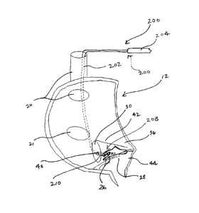
An AV nodal vein approach is depicted in Fig. 5. A device 200 comprises a lead
202 and
a catheter 204. Lead 202 comprises a proximal portion 206 and a distal portion
208.
Specifically referring again to Fig. 2, an approach to INE 26 exists through
AV nodal vein 46 via
the CS 30. In some cases, AV nodal vein 46 opens directly to the triangle of
Koch. Catheter 204
and distal portion 208 of lead 202 are inserted into the RA 12 through SVC 20,
into CS 30
through ostium 32 of CS 30, and into AV nodal vein 46 to INE 26. A tip 210 of
lead 202 in this
embodiment can comprise first, second, third, and fourth electrodes 212a,
212b, 212c, 212d.
There are catheter insertion methods and designs that enable navigating the CS
and
inserting the catheter and lead into the heart. Such insertion methods are
described in U.S.
Patent Nos. 6,745,081, 6,070,081, and 5,545,204, which are incorporated herein
by reference in
their entirety. Incorporation by reference is limited such that no subject
matter is incorporated
that is contrary to the explicit disclosure herein, no claims included in the
documents are
incorporated by reference herein, and any definitions provided in the
documents are not
incorporated by reference herein unless expressly included herein.
The catheter design of the embodiments can differ from conventional catheter
designs in
that it can be shorter than those used for epicardial pacing and can be
steerable enabling the
catheter tip to enter the AV nodal vein. For example, to facilitate entry into
the AV nodal vein,
the tip of the catheter can have a slight bend, such as between about 100 and
about 60 . Those
skilled in the art will recognize that in further embodiments the tip of the
catheter can have a
bend less than about 10 and greater than about 60 .
Referring again to Figs. 4 and 6, lead tip 110, 210 according to embodiments
can
comprise multiple electrodes (depicted as having three and four electrodes,
respectively), which
can be switched from sensing to pacing. Those skilled in the art will
recognize that in further
embodiments, greater than four or fewer than three electrodes can be included
on the lead tip.
Unipolar sensing on each lead can be used to determine which lead has the most
robust slow
pathway signal. The lead can then be switched to be the pacing lead and SP
pacing can be
8
CA 02673971 2014-11-20
accomplished with this lead. The non-pacing leads can then be used to monitor
ventricular rate
during pacing, which can be done in bipolar mode.
The circuit diagram in Fig. 7 illustrates an embodiment of how each lead
electrode of a
lead 300 can be switched from a pacing function to a sensing function. Two
leads electrodes
31 2a, 312b are depicted in the diagram for clarity. Circuit 300 comprises a
plurality of switches
302a, 302b, 302c, 302d, 302e, a first ground 304a and a second ground 304b, a
capacity 306, a
battery 308, electrocardiogram ("ECG") sensing circuitry 310, and a plurality
of electrodes 312a,
312b..
When switch 302a is closed, the capacitor can charge, and when switch 302a is
open,
capacitor 306 is disconnected from battery 308. With switches 302c and 302d
closed, a unipolar
ECG can be sensed from both lead 312a and lead 312b with the reference lead
for ECG sensing
circuitry 310 being the can of the device. ECG sensing circuitry 310 decides
which lead has the
best slow pathway ECG. Then switches 302c and 302d open and switch 302b or
302e, which are
controlled by ECG sensing circuitry 310, close to connect lead with the best
slow pathway
potential to capacitor 306 for unipolar pacing. Those skilled in the art
recognize that electrical
stimulation can be delivered in any number of ways electronically and in any
number of wave
shapes, frequency, voltage, and timing.
Synchronized ventricular pacing is described further in Hucker et al.,
entitled "Atrio-
Ventricular Conduction with and without AV Nodal Delay: Two Pathways to the
Bundle of His
in the Rabbit Heart," Am. J. Physiol. Heart Cir. Physiol. October 2007;
293(2):H1122-30.
9
CA 02673971 2009-06-26
WO 2008/063498 PCT/US2007/023836
Attorney Docket No. 3765.04W001
EXAMPLES
Setup
New Zealand White rabbits (n = 18, 2.5-3 months old, 2-3 kg) were anesthetized
with
100 mg/kg pentobarbital sodium and 1,000 IU heparin intravenously, after which
a midsternal
thoracotomy was performed and the heart removed. The heart was Langendorff
perfused with
oxygenated (95% 02-5% CO2) Tyrode at 37 C and received 50 1.t1 of 5 JIM Di-4-
ANEPPS
(Molecular Probes, Eugene, OR) over 5 minutes. The study was conducted in
superfused
isolated AV junctions (n = 8) and in Langendorff-perfused hearts (n = 10) with
the AV junction
exposed via right atriotomy. For superfused experiments, the AV junction was
dissected in cold
Tyrode (0 C) and the sinoatrial node was removed. The preparation was
superfused at 30
ml/min with Tyrode containing 15 mM of the excitation-contraction uncoupler
2,3-butanedione
monoxime (Sigma, St. Louis, MO) to inhibit motion artifacts. A 16 X 16
photodiode array was
used with an optical mapping system. Optical signals were sampled at 1.5 kHz,
averaged, and
low-pass filtered at 120 Hz. Optical activation maps displayed the optical
signal derivative,
which corresponds to wave fronts of excitation.
Electrogram Recordings
Electrodes were placed on the IAS and CrT, and a quadruple electrode on the
His bundle
recorded both the superior and inferior His electrogram (SHE and IHE,
respectively) to monitor
fast-His and slow-His excitation.
Changes in His excitation and His electrogram morphology can occur from
changes in
the AV node excitation pathway, because of the specific pacing protocol and
alternating
conduction pathways. If the AV node and His are excited by the SP (i.e., slow-
His excitation),
the 1HE has a larger amplitude than when the AV node is activated by the FP.
Conversely, when
the FP excites the AV node and His (i.e., fast-His excitation), the SHE has a
larger amplitude
than with slow-His excitation.
The location of each electrode is depicted in Fig. 8A. A fourth roaming
electrode was
used to record electrograms throughout the triangle of Koch. A schematic of
the triangle of
Koch is depicted in Fig. 8A. The reference lead was located 3 mm from the
electrode tip. The
TEFLON -coated 0.13-mm Pt/Ir wire tip had ¨0.07 mm stripped to mimic a
clinical
hemispheric tip. This electrode was mounted on a force transducer (FORT25;
World Precision
Instruments, Sarasota, FL) to control contact force, ensuring that contact
force was minimal and
CA 02673971 2009-06-26
WO 2008/063498 PCT/US2007/023836
Attorney Docket No. 3765.04W001
consistent between locations. The roaming electrode was moved in 1-mm
increments by a
motorized micromanipulator throughout the triangle of Koch (grid in Fig. 8A).
Electrograms
were recorded at 1.5 kHz (National Instruments, Austin, TX), and each
electrode location was
digitally photographed.
Stimulation Protocol
The preparation was paced from two electrodes: the IAS electrode and the
roaming
electrode (Fig. 8A). IAS pacing was constant at 2X threshold (-2 mA), with 2-
ms pulses and
300-ms cycle length (Fig. 8B). IAS pacing simulated sinus pacing and was used
to mask the AV
junctional rhythm, which originates in the SP, as well as to maintain tissue
excitability in a
consistent state as the roaming electrode was moved from location to location.
Stimulation
thresholds were determined with a ramp of unipolar pulses (0.5-ms pulses, 300-
ms cycle length,
amplitude ranging from 0.33 to 10 mA and increasing by 0.33 mA with each
pulse; Fig. 8B).
Each ramp pulse was delivered 45-60 ms before an LAS pacing pulse (Fig. 8B).
The pacing ramp
was delivered from the roaming electrode for anywhere from 14-24 locations
throughout the
triangle of Koch (grid in Fig. 8A), enabling quick determination of threshold
for each location.
Pacing threshold was defined as the amplitude of the ramp pulse that caused a
shift in the
activation pattern from IAS pacing to stimulation beginning from the roaming
electrode.
Results - Identifying the SP
The schematic in Fig. 8A indicates the approximate location of the inputs to
the SP and
FP. Although the anatomic substrate of the SP is often thought to be the INE,
the anatomic
substrate of the FP is less well defined and consists of TCs that overlay the
compact AV node.
Electrograms and OAPs were compared that were recorded during IAS pacing. Each
trace began at the moment of IAS pacing and was 300 ms long. Fig. 9 directly
compares
electrograms and OAPs recorded from the same site. During IAS pacing, the AV
node is
activated by the FP, and the SP acts as a dead-end pathway. Electrograms for
atrial tissue all
have a sharp signal immediately after the pacing artifact, which signifies
fast conduction through
the atrial myocardium. OAPs for atrial tissue have atrial action potential
morphology.
Electrograms for His contain both a fast signal directly after the pacing
artifact (similar in timing
to that seen in_traces for atrial tissue) and another sharp spike ¨80 ms
later, which reflects His
excitation. OAPs for His have two humps that are the summation of atrial and
His excitation.
11
CA 02673971 2009-06-26
WO 2008/063498 PCT/US2007/023836
Attorney Docket No. 3765.04W001
The first hump corresponds to atrial activation, whereas the second hump has
the distinct plateau
phase of His activation. Electrograms for nodal and SP contain several
components. The first
component matches in time the signals seen in the electrogram for atrial
tissue and is followed
by complex low-amplitude biphasic recordings that are consistent with slow
conduction during
the interval between atrial and His excitation. Electrograms for nodal are
located near the
anatomic location of the AV node, and OAPs for nodal have a nodal morphology.
Electrograms
and OAPs for SP are located along the SP. There is overlap in the
characteristics of the
electrograms and OAPs for nodal and SP.
Referring again to Fig. 9, the sharpest spikes in the electrograms correspond
to the
dF/dtmax of the fluorescent optical signals. In the nodal and SP traces, the
slow conduction
characteristics of the electrograms correlate well with the OAPs from the same
location. One
exception is the spike seen in the nodal electrogram, which occurs during the
plateau of the
OAP. On the basis of the timing of this spike relative to the bipolar IHE,
this spike most likely
reflects excitation of the nodal-His (NH) region of the node or the lower
nodal bundle (LNB),
where excitation accelerates into the His bundle.
Pacing stimuli applied within the triangle of Koch produced different
activation patterns,
depending on where the stimuli were delivered. The activation pattern caused
by IAS pacing
originated from the IAS electrode and spread rapidly across atrial tissue and
TCs that lie above
the conduction system in the triangle of Koch, which activated the FP of the
AV node. Because
activation maps display dF/dt, there is no conduction visible from ¨50 to 70
ms when there is
exclusively slow AV node conduction (which has a low amplitude dF/dt). After
this period, His
excitation occurred, which actually began in the LNB region. The interval
between 1AS pacing
and His excitation was ¨70 ms.
At many pacing locations within the triangle of Koch, pacing from the roaming
electrode
caused an atrial/FP activation pattern similar to that seen with IAS pacing.
When the roaming electrode was placed within ¨2 mm of the tricuspid valve
within the
triangle of Koch, it excited the SP. SP activation appears as a slow, narrow
activation pattern
that moved toward the apex of the triangle of Koch. After it reached the AV
node region,
excitation continued toward the His bundle without pause and also spread
retrogradely across the
IAS. In the SHE trace, the His electrogram morphology changed to a slow-His
morphology with
a smaller amplitude during SP pacing and returned to its original morphology
after the ramp
ended. The interval between stimulation and His excitation (the S-H interval)
is paradoxically
12
CA 02673971 2009-06-26
WO 2008/063498 PCT/US2007/023836
Attorney Docket No. 3765.04W001
longer for fast-His excitation than for slow-His excitation. Slow-His
electrograms were
observed in six of eight superfused experiments.
Similar optical activation patterns for SP and atrial/FP activation were
observed in the
AV junctions of Langendorff-perfused hearts. An IHE trace for activation maps
of SP pacing is
depicted in Fig. 10. SP pacing in the whole heart produced a slow narrow
activation pattern
followed without pause with His activation. In the intact heart, His
activation was followed by a
robust ventricular optical signal that could be seen through the overlying
atrial tissue. Atrial
activation preceded ventricular activation by ¨10-15 ms.
IAS pacing produced a fast wave of excitation that spread across the atrial
tissue and
TCs. After atrial activation, excitation spread from the AV node region in two
directions: the
wave front of His excitation spread toward the His electrode and a wave front
of decremental
conduction spread down the SP and died out. Ventricular activation followed
His excitation. A
small amplitude fast-His potential is seen in Fig. 10 for IHE with IAS pacing
(i.e., the IHE
deflection is larger with SP pacing than with IAS pacing).
Pacing within the triangle of Koch
Figs. 9-10 illustrate that the SP location can be identified with electrograms
and OAP
morphology, and SP vs. FP activation can be differentiated with activation
patterns and His
electrogram morphology in both the superfused and 'whole heart preparations.
Using both
activation patterns and His morphology, it was identified which activation
patterns occurred for
multiple pacing locations throughout the triangle of Koch.
Fig. 11 displays a summary of where the different activation patterns occurred
and the
pacing thresholds in the superfused experiments. Generally, pacing stimuli
applied within ¨2
mm of the tricuspid valve excited the SP or the His directly when pacing near
the apex of the
triangle of Koch. Direct His stimulation was defined as fast conduction
directly after
stimulation, which had an S-H interval of ¨10 ms and occurred in a small area
near the His
electrode. On average, the pacing threshold for SP/His pacing was 4.4 2.2
mA. Pacing stimuli
applied further away from the tricuspid valve generally excited atrial tissue,
producing FP
excitation of the AV node with an average stimulation threshold of 2.4 1.6
mA (P < 0.001
compared with SP/His pacing thresholds). Additionally, there was one area
between the
coronary sinus and the tricuspid valve that had very high pacing thresholds
(8.6 1.4 mA; P <
0.001 compared with atrial/FP thresholds). Pacing this region excited atrial
tissue and the FP of
13
CA 02673971 2009-06-26
WO 2008/063498 PCT/US2007/023836
Attorney Docket No. 3765.04W001
the AV node. OAPs from this region were quite noisy and very dissimilar from
the large-
amplitude atrial OAPs recorded from other areas of the triangle of Koch. High
pacing thresholds
and very low OAP amplitudes suggest that few excitable cells are located in
this region. There
were several instances where different activation patterns occurred at
different thresholds,
typically with atrial activation followed by SP or His excitation at higher
stimulus intensities.
Very similar pacing thresholds for each activation type were observed in the
Langendorff-
perfused hearts with the SP/His threshold statistically the same as the right
ventricular pacing
threshold.
Once SP activation of the His bundle has occurred, the SP would excite the His
bundle
throughout the duration of the stimulation ramp in a 1:1 fashion. However,
this was not the case
for all pacing locations that produced FP excitation. As depicted in Fig. 12,
pacing near the
superior border of the AV node often disrupted AV conduction by causing 2:1 AV
block or
prolonged AV conduction. In the example depicted in Fig. 12, IAS pacing
conducted 1:1 to the
His before the stimulation ramp crossed threshold, seen in both the
electrograms and the OAP
recorded above the His bundle. Once the stimulation ramp crossed threshold, it
excited atrial
tissue (seen in the shift in both the OAP and the CrT trace), but excitation
did not propagate to
the His. In the next beat, excitation propagates to the His (seen in the OAP
and the IHE), and
conduction continues in this 2:1 fashion. Therefore, AV conduction was
disrupted by pacing in
this location. Pacing stimuli delivered to this area of the triangle of Koch
caused 2:1 AV block
or prolonged AV conduction in five of eight experiments. Once AV conduction
was disrupted, it
did not fully recover in any of the five experiments, suggesting that the
effect of pacing in this
area was not neurologically mediated. The S-H interval was measured for each
pacing location.
The S-H interval usually stabilized three to five pulses after threshold was
reached, and
these stable values were measured. S-H intervals at high-stimulus intensities,
where a greater
amount of tissue was presumably depolarized by far-field stimulation, were
discarded because
the S-H intervals at the end of the ramp were generally different from the
stable S-H intervals.
Rarely, S-H intervals did not stabilize and linearly decreased throughout the
ramp, in which case
the range of S-H intervals was noted. Also, any S-H interval that was recorded
after AV
conduction was disrupted was discarded from the analyses, as depicted in Fig.
12. S-H intervals
were divided into two groups, those associated with atrial/FP activation and
those associated
with SP/His activation. When plotted against the horizontal distance from the
His electrode,
atrial S-H intervals illustrated no distance dependence (P = not significant;
Fig. 13A) but instead
14
CA 02673971 2009-06-26
WO 2008/063498 PCT/US2007/023836
Attorney Docket No. 3765.04W001
remained at a constant level of 81 19 ms. This constant interval between an
atrial stimulus and
His excitation is the AV delay. S-H intervals associated with SP and His
excitation exhibited a
strong correlation with distance from the His electrode (P <0.001; Fig. 13B).
If SP excitation
experienced the AV delay, one would expect a nonlinear jump from direct His
excitation at small
distances from the His electrode to values at or above the AV delay of 81 19
ms. Moreover,
the S-H intervals during SP pacing remained well below the AV delay measured
from atrial/FP
activation (81 19-ms delay for FP excitation and 53 25-ms delay for SP
excitation > 4 mm
from the His electrode; P < 0.001).
Fig. 13C depicts an example of S-H intervals measured during SP pacing and IAS
pacing
with the last ramp stimulus, which excited the SP, and the first IAS pacing
pulse after the ramp.
The S-H interval was 31 ms for the last ramp stimulus and increased to 64 ms
when the His
bundle was excited by IAS pacing. Paradoxically, the S-H interval for the SP
was shorter than
the S-H interval for the FP. The shifts from slow-His to fast-His potentials
are seen in both the
IHE and SHE traces. Similar S-H interval trends for both atrial and SP
excitation were observed
in Langendorff-perfused hearts, with SP S-H intervals shorter than FP S-H
intervals on average
Discussion
The results support previous findings that several layers of conduction exist
in the
triangle of Koch. Now, it was further demonstrated that these layers can be
differentially
engaged with varying stimulus strengths and pacing locations. It was found
that stimulation
thresholds for atrial excitation are significantly lower than for SP/His
activation, with the
exception of one area beneath the coronary sinus. Using the activation pattern
documented by
optical mapping, as well as slow-His electrograms, SP excitation was verified
and it was found
that not only do S-H intervals decrease linearly as the stimulus location
approaches the His
bundle for SP excitation, whereas S-H intervals associated with atrial/FP
activation remain
nearly constant, but also that paradoxically S-H intervals for the SP are
shorter than those
recorded for FP activation.
Because of the different modalities used to study the AV junction, there is a
"lack of
common terminology" for its components. For instance, many previous studies
have referred to
the AV node extension located in the isthmus between the coronary sinus and
the tricuspid valve
as the "posterior nodal extension." However, a naming task force has indicated
that the INE
more accurately describes its location when the heart is anatomically
oriented. Previous studies
CA 02673971 2009-06-26
WO 2008/063498 PCT/US2007/023836
Attorney Docket No. 3765.04W001
have described layers of atrial cells and TCs overlying the components of the
conduction system
in the triangle of Koch, such as the INE, AV node, and His bundle. Functional
studies have
provided evidence that the INE is the anatomic substrate of the SP. However,
there is debate
regarding whether the INE itself, the TCs overlying the INE, or a combination
is the true
substrate of the SP. The results can not distinguish which cell layer or
structure produced SP
excitation. However, optical mapping results confirm that this pathway can be
reliably excited
by pacing stimuli delivered within ¨2 mm of the tricuspid valve within the
triangle of Koch.
Plotting S-H intervals of atrial excitation against the distance from the His
electrode revealed no
correlation (Fig. 13A). The S-H interval of atrial activation is composed of
two intervals:
conduction time in the atrial layer and FP (S-AV node interval) and AV node
conduction to the
His bundle (the AV delay). Because conduction in the atrial layer is fast (-35
cm/s), small
changes in the distance between the stimulating electrode and the His
electrode change the S-AV
node interval minimally. Therefore, the main determinant of the S-H interval
is the AV delay,
which remains essentially constant.
On the other hand, plotting S-H intervals of SP excitation vs. distance from
the His
electrode illustrated a strong correlation (Fig. 13B). A greater dependence on
distance would be
expected for SP activation because, as the name implies, the SP conducts
slowly (-7 cm/s). ,
Therefore, one would expect the S-AV node interval to decrease slightly as the
distance between
the stimulus and the AV node decreased. However, once conduction time in the
SP is minimal,
the S-H interval should be determined by the AV delay, which is 81 19 ms
according to the
atrial activation data (Fig. 13A). As the His bundle is approached further,
the S-H interval
should then jump to a very small value when direct His activation occurs.
Instead, the raw data
and the pooled data in Fig. 13B depict S-H intervals almost entirely <81 ms
and a linear decrease
of the S-H interval as the His electrode is approached. Activation patterns
for SP pacing
illustrated SP excitation proceeding linearly to the His bundle without pause.
These data suggest
that there is no dependence of the SP/His S-H interval on the AV delay,
implying that SP
excitation avoids the AV delay and excites the His bundle directly.
Studies have investigated the rate-dependent properties of the SP vs. the FP
in the context
of premature stimuli, most commonly with pacing stimuli applied in the high
right atrium or to
the CrT. On the basis of these studies, the SP was given its name because
excitation took longer
to reach the His bundle than through the FP. However, it was found that SP
excitation reached
the His bundle faster than FP excitation. Despite this apparent paradox,
results are fully
16
CA 02673971 2009-06-26
WO 2008/063498 PCT/US2007/023836
Attorney Docket No. 3765.04W001
consistent with the large body of evidence concerning the SP for two reasons.
First, SP
excitation in previous studies traveled the full length of the SP, which data
indicates would have
an S-H interval similar to FP excitation (Fig. 13B, right). In the context of
a premature beat, this
activation would take even longer. In the absence of pacing stimuli applied
directly to the SP,
SP conduction to the His only occurs when a premature stimulus encounters a
refractory FP (Fig.
14). Because conduction traveling the full length of the SP takes the same
amount of time as the
AV delay (or longer with premature stimuli), it could be difficult to
recognize that SP conduction
avoided the AV delay. Only through pacing the SP incrementally along its
length could it
become apparent that SP excitation does not experience the same AV delay as FP
excitation.
Second, the SP was engaged directly, potentially avoiding any conduction delay
that may occur
at the interface of the atrial tissue with the SP (Fig. 14). This interface
may be responsible for an
additional delay in conduction, which would slow SP conduction even further.
Pacing was done at 300 ms within the triangle of Koch, close to the AV node
itself.
Therefore, one interpretation of the data is that because of the His bundle
proximity, pacing
stimuli excited the His bundle directly when stimuli were delivered within 2
mm of the tricuspid
annulus and the SP was not involved. Direct stimulation of the His bundle
would certainly result
in short S-H intervals. However, this possibility is unlikely for two reasons.
First, SP activation
was confirmed by being visualized with optical mapping. Second, pacing stimuli
applied much
closer to the His bundle itself produced atrial activation with an AV delay of
¨80 ms or longer
throughout the entire pacing ramp. Therefore, it seems that the tissue area
initially captured by
the pacing stimulus was rather small. Also, the S-H intervals that were
analyzed were measured
three to five beats after the pacing ramp reached threshold (i.e., <2X the
pacing threshold) which
also limits the extent of tissue that was excited at that point in the pacing
ramp.
There is a growing consensus in the literature that the His, LNB, and the INE
form a
continuous structure distinct from the compact AV node tissue on a
morphological, molecular,
and functional basis. The data suggests that, instead of a final common
pathway as classically
suggested, there are instead two pathways to the His bundle: one through the
compact node and
the other through the LNB. Similar arguments have been put forth suggesting
that the INE
connects to the His bundle through the LNB and that the FP passes through the
compact node.
17
CA 02673971 2009-06-26
WO 2008/063498 PCT/US2007/023836
Attorney Docket No. 3765.04W001
The concept of two pathways to the His is supported by changes in His
electrogram
morphologies (i.e., slow-His and fast-His electrograms). The distinct His
electrogram signatures
of FP or SP activation indicate that the activating pathway influences how the
His
bundle, or which part of the His bundle, is depolarized.
Based on this dual-pathway concept, the data suggests that SP excitation
begins in either
the inferior TCs or the [NE itself and travels via the INE to the His bundle
through the LNB with
a gradient of conduction velocities. As excitation approaches the His, the
level of connexin 43
increases and conduction velocity increases until the His bundle is reached.
FP activation from
the atrial tissue surrounding the AV node funnels into the compact node via
the TCs that overlie
the AV node. The AV node delay is due to the compact nodal tissue and its
connecting TCs,
after which excitation passes to the His. Interestingly, a small region was
identified below the
coronary sinus where stimulation thresholds were significantly higher than the
surrounding
myocardium. This area may serve as a localized area of block, contributing to
anisotropy in the
atrial myocardium and may play a role in atrial flutter and fibrillation,
similar to the block zone
in the intercaval region that can maintain typical atrial flutter.
The existence of an SP "bypass tract" in the right atrium expands the area
that can be
paced to achieve His bundle excitation with direct His bundle pacing
procedures, alleviating the
difficulty of pacing the small His region located close to aorta. Locating the
SP pacing location
could be guided by the slow conduction characteristics in SP electrograms,
such as depicted in
Fig. 9, which are used to guide SP ablations during AVNRT procedures. SP
pacing may very
well have lower pacing thresholds than those required for His bundle pacing
because of the
fibrous tissue surrounding the His bundle. Experiments in the rabbit revealed
that SP pacing and
direct His pacing had pacing thresholds that were not statistically different.
However, in the
rabbit, the endocardial side of the His bundle is only covered by a small
amount of connective
tissue, whereas, in the human, the His bundle is encased in the fibrous tissue
of the
central fibrous body. Therefore, it is possible that SP pacing in the human
will have a lower
threshold than His bundle pacing. Additionally, pacing thresholds for right
ventricular epicardial
pacing and SP/His pacing were not statistically different in the whole heart
experiments,
suggesting that clinical SP pacing thresholds may be close to pacing
thresholds for right
ventricular pacing. However, this study was conducted in normal rabbit AV
junction
preparations where no attempt was made to disrupt AV conduction. Conduction
curves are the
gold standard used to investigate AV node dual-pathway electrophysiology.
Because of the
18
CA 02673971 2014-11-20
number of pacing locations investigated, it was not possible to construct
conduction curves for
each. Although conduction curves are very useful clinical tools, optical
mapping in the
preparations confirmed the distinct activation patterns of FP vs. SP
activation, and optical
mapping of SP excitation corresponds very well to SP optical mapping performed
during
standard S1-S2 protocols. Additionally, slow-His potentials provided another
line of evidence
for FP vs. SP participation in conduction.
Further description is included in Hucker et al., "Atrioventricular Conduction
With and
Without AV Delay: Two Pathways to the Bundle of His in the Rabbit Heart," Am.
J. Physiol.
Heart Circ. Physiol., 2007 Aug.;293(2):H1122-30.
INE as Physiologic Pacemaker
The [NE can also be used for biological pacemaker therapy. Specifically, the
[NE is the
secondary physiological pacemaker of the heart and can be modified to become
the leading
pacemaker during failure of sinus nodal pacemaker. The inherent pacemaking
properties of the
INE can be enhanced to reach normal physiological heart rates. Specifically,
the pacemaking
properties of the INE can be enhanced by electric sympathetic. stimulation of
the elements of
sympathetic branch of cardiac autonomic nervous system surrounding the
myocytes of the ENE.
Delivery of sub-threshold high frequency (about 20 Hz to about 400 Hz) current
can be
used to stimulate the endogenous autonomic innervation surrounding the INE and
enable
acceleration of the physiological pacemaker in the [NE. Further, the
pacemaking properties of
the INE can be enhanced by electric stimulation of the elements of sympathetic
branch of cardiac
autonomic nervous system located within the myocardium of INE. Further
description is
included in Hucker et al.. "Automatic Control and Innervation of the
Atrioventricular Junctional
Pacemaker," Heart Rhythm, October 2007, 4(10), pages 1326-1335. The sub-
threshold approach'
can replace the need for traditional ventricular pacemaker leads. Sub-
threshold stimulation can be
applied using the device designs described above or can be applied with other
known electrode
designs.
19
CA 02673971 2009-06-26
WO 2008/063498 PCT/US2007/023836
Attorney Docket No. 3765.04W001
Electrical Modulation of INE
The INE can also be electrically modulated to treat several common conditions
in clinical
electrophysiology. For example, stimulation of the INE can be used for
treatment of several
cardiac rhythm disorders of supraventricular origin, including bradycardia and
tachycardia, and
also as a site for implantable device pacing treatment of brady arrhythmias
(slow heart rate).
Specifically, bradycardia can be treated by using the INE as a pacing site, as
the site of a
biological pacemaker that replaces the SA node by accelerating the intrinsic
rate of the INE, or
as the site of autonomic stimulation to increase the intrinsic pacemaking rate
of the INE.
INE as Cell Therapy Site
The INE can also be used as a site for cell therapy delivery for
reconstituting a biological
pacemaker, because of ease of access via the AV nodal vein, intrinsic
pacemaking properties,
and high degree of physiological autonomic control as compared with the atrial
and ventricular
myocardium. To deliver gene therapy or cell therapy to the INE, a catheter,
such as the device
designs described above, can possess a fluid eluting tip to deliver a saline
solution containing the
gene or cell therapy. The catheter can further possess a retractable needle,
which can extend
from the catheter tip to puncture tissue and a sensing electrode to locate the
INE. Cell therapy
can be delivered through both of the above-described approaches: the
endocardial approach or
the AV nodal vein approach.
INE as Gene Therapy Site
The INE can also be used as a site for gene therapy. Specifically, the
inherent
pacemaking properties of the INE can be increased by delivering genes via an
electroporating
catheter encoding the pacemaker channel isoforms HCN I, HCN2, HCN3, or HCN4 or
elements
of the autonomic nervous system.
Gene therapy can be delivered in the following manner. A fluid eluting
catheter can be
placed in the AV nodal vein and the site with the largest slow potential will
be located. A
second catheter will locate slow potentials on the endocardial surface of the
INE. Sub-threshold
alternating current (in the range of about 1.0 to about 50.0 microamps, for
example) can be
passed between the tip of each catheter to minimize the impedance between the
two leads and
localize the slow pathway. The fluid eluting needle of the venous catheter can
extend to
puncture the venous wall. Electroporating current can be passed between the
two catheters and
CA 02673971 2014-11-20
the saline solution containing the gene therapy will be released
simultaneously. Those skilled in
the art will recognize that in further embodiments the current of less than
about I microamp or
more than about 50 microamps can be used.
INE for Rate Control
The INE can further be used for rate control during atrial tachyarrhythmias,
including
atrial flutter and atrial fibrillation, and AVNRT. For example, a stimulation
pulse (or a series of
pulses) applied to the [NE during AVNRT can terminate an arrhythmia without
the need for
radiofrequency ablation and the potential complications (AV block) that can
occur with this
procedure.
Stimulation of the autonomic innervation of the INE can be used as an
effective treatment
for ventricular rate control during atrial fibrillation, such as because of
the short refractory period
of the 1NE. Parasympathetic stimulation of the INE can block excitation in the
slow pathway,
thus, filtering properties of the AV node can be enhanced because excitation
of the ventricles
will have to travel through the FP, which has a longer refractory period.
Pacing of the INE
provides conduction via the slow pathway and the lower nodal bundle which has
higher safety
factor as compared to conduction via the FP and compact AV node. As a result,
pacing of the
INE can provide safe rate control for atrial arrhythmias without the need of
AV nodal ablation in
patients with paroxysmal and chronic atrial tachyarrhythmias.
Common forms of AVNRT involve the INE as one pathway of the reentrant
arrhythmia,
and can be treated by radiofrequency ablation lesions created on or near the
INE. In another
embodiment, a stimulation pulse, or a series of pulses, applied to the INE
during AVNRT can be
used to terminate the arrhythmia without the need for radiofrequency ablation
and the potential
complications, such as, for example, AV nodal block that can occur with this
procedure. Each of
the tachyarrhythmia therapies can be accomplished with the endocardial or
venous approach to
the INE using the catheter designs described above.
The scope of the claims should not be limited by particular embodiments set
forth herein, but
should be construed in a manner consistent with the specification as a whole.
=
21
CA 02673971 2009-06-26
WO 2008/063498
PCT/US2007/023836
APPENDIX A
22
CA 02673971 2009-06-26
WO 2008/063498
PCT/US2007/023836
Hucker: Cx43 in the Human AV Junction
November 5, 2007
Connexin 43 Expression Delineates Two Discrete Pathways in the
Human Atrioventricular Junction
Running title: Cx43 in the human AV junction
William J. Hucker, Megan L. McCain, Jacob I. Laughner, Paul A. Iaizzo,* Igor
R.
Efimov
Washington University in Saint Louis, MO and *University of Minnesota,
Minneapolis,
MN
Address for correspondence: Igor R. Efimov, Department of Biomedical
Engineering,
Washington University in St. Louis, MO 63130.
This work was supported by:
American Heart Association grant in aid 0750031Z
23
CA 02673971 2009-06-26
WO 2008/063498
PCT/US2007/023836
Hucker: Cx43 in the Human AV Junction
Abstract
Gap junction expression has been studied in the atrioventricular junction
(AVJ) of many
species, however their distribution in the human AVJ is unknown. The AVJ
expression
of the gap junction protein connexin 43 (Cx43) is species dependent; therefore
we
investigated its distribution in the human AVJ. Methods: Using Masson
trichrome
histology, we reconstructed the AVJ of 3 normal human hearts and one with
dilated
cardiomyopathy in 3D. Cx43 was immunolabeled with vimentin and a-actinin to
determine the cellular origin of Cx43, and was quantified in the following
structures:
interatrial septum (IAS), His bundle, compact node (CN), lower nodal bundle
(LNB),
leftward and rightward nodal extensions (LE and RE), and inferior,
endocardial, and left-
sided transitional cells. Results: Histology revealed two nodal extensions in
3/4 hearts.
Cx43 was found in the myocytes, but not fibroblasts of the AVJ. LE and CN Cx43
was
lower than the IAS (P<0.05) and the RE, LNB, and His all expressed Cx43
similarly,
with approximately half of IAS expression (RE: 44 36%; LNB: 50 26%; His: 48
12%,
P=NS compared to LAS). Cx43 levels in transitional cells were similar to the
LAS
(P=NS). Conclusions: Cx43 was found in myocytes of the human AVJ, and its
expression pattern delineates two separate continuous structures: one consists
of the LE
and CN with little Cx43 and the other consists of the His, LNB and RE
expressing
approximately half the Cx43 of the LAS. The differential Cx43 expression may
provide
each structure with unique conduction properties, contributing to arrhythmias
arising
from the AVJ.
Keywords: Atrioventricular node, dual-pathway electrophysiology, connexin43,
slow
pathway, AVNRT
24
CA 02673971 2009-06-26
WO 2008/063498
PCT/US2007/023836
Hucker: Cx43 in the Human AV Junction
Introduction
Detailed investigations of the atrioventricular junction (AVJ) in many
animal models have demonstrated the enormous complexity of this structure in
terms of
cell and tissue morphology, functional characteristics, and protein
distribution. While
many histological studies have investigated the human AVJ, and clinical
electrophysiological studies have provided a substantial amount of information
regarding
AVJ function in vivo, the number of molecular investigations of the human AVJ
is very
limited because of the inherent difficulty of procuring human tissue in a
timely fashion
(Davis et al., 1995). Nevertheless, such studies are vitally necessary to the
basic
understanding of clinically relevant phenomena, such as atrioventricular nodal
reentrant
tachycardia (AVNRT) and AV block, and to increase the understanding of
variables such
as age which are difficult to address in animal models. In addition,
interspecies
differences in protein expression patterns can limit the extrapolation of
animal data to the
human (Coppen and Severs, 2002; Coppen et al., 2003). Therefore human
molecular
studies are crucial because expression patterns in the human AVJ should become
the
framework in which studies from animal models are interpreted.
Several tissue types are involved in atrial-His conduction, and the term AVJ
is used in this study to encompass them all (Billette, 2002). Proximally, the
specialized
conduction tissue of the AVJ consists of the inferior nodal extensions, which
extend from
near the coronary sinus (CS) ostium to the node itself (Inoue and Becker,
1998).
Previously, these extensions were termed "posterior" rather than "inferior"
(Inoue and
Becker, 1998; Dobrzynski et al., 2003), however when the heart is oriented
anatomically,
these extensions actually run inferior to the CN and therefore we use the term
"inferior
CA 02673971 2009-06-26
WO 2008/063498
PCT/US2007/023836
Hucker: Cx43 in the Human AV Junction
nodal extension" in this study (Cosio et al., 1999). The inferior nodal
extensions merge
to become the AV node (AVN), which then penetrates the central fibrous body to
become
the His bundle (Tawara, 1906). Connecting the AVN and nodal extensions to the
surrounding atrial tissue are transitional cells that can be divided into
three groups based
on their location relative to the AVN: endocardial transitional cells contact
the AVN on
its endocardial aspect, left sided transitional cells approach the AVN from
the left side of
the interatrial septum (IAS), and inferior transitional cells approach the AVN
near the
coronary sinus ostium (Anderson and Ho, 2002; Anderson and Ho, 2000).
To accommodate its complex physiological role, the AVJ has developed
very heterogeneous gap junction expression, with more types of gap junctional
proteins
expressed in the AVJ than anywhere else in the heart. Specifically, four
cardiac
connexins have been described to date (Cx43, Cx40, Cx45, and Cx30.2/31.9), and
each
of these proteins has been found in animal studies of the AVJ (Boyett et al.,
2006). Cx43
and Cx40 are both associated with rapidly conducting cardiac tissues (with
Cx40 having
a higher conductance than Cx43), and Cx45 and Cx30.2/31.9 are associated with
slowly
conducting tissues (Boyett et al., 2006). The expression patterns of these
connexins in
the AVJ are very species dependent: rat hearts do not express Cx43 or 40 in
the AVJ,
however rabbits express both (Coppen and Severs, 2002; Dobrzynski et al.,
2003). Only
one study has documented the expression of connexins in the human AVJ to our
knowledge (Davis et al., 1995), where they found that Cx43, Cx40, and Cx45
were all
present, yet this study did not survey where in the AVJ each connexin was
expressed.
Traditionally, connexins have been assumed to form gap junctions between
two adjacent myocytes. However, recent publications also suggest that
functional gap
26
CA 02673971 2009-06-26
WO 2008/063498
PCT/US2007/023836
Hucker: Cx43 in the Human AV Junction
junctions are formed between myocytes and fibroblasts (Camelliti et al.,
2005), possibly
via Cx43 coupling (Goldsmith et al., 2004). In this study, we investigated
Cx43
expression throughout the various tissues of the AVJ, as well as whether Cx43
forms gap
junctions between myocytes and fibroblasts. We correlated Cx43
immunofluorescence
with 3D reconstructions of the AVJ constructed from Masson trichrome
histology,
showing that Cx43 expression pattern delineates domains of the AVJ (not
apparent with
histology) that may have functional consequences.
27
CA 02673971 2009-06-26
WO 2008/063498
PCT/US2007/023836
Hucker: Cx43 in the Human AV Junction
Methods
The use of human hearts for research was approved by the Institutional Review
Boards at Washington University and the University of Minnesota. Three
specimens
were provided by the Upper Midwest Organ Procurement Organization (LifeSource,
St.
Paul, MN; they also provided pre-approval for this protocol); these hearts
were deemed
non-viable for transplantation. An additional specimen was a transplant
recipient's
explanted heart with idiopathic dilated cardiomyopathy (DCM). The clinical
data that is
known regarding each specimen is shown in Table I. These specimens were placed
in
frozen tissue embedding media (Histo PrepTM, Fisher Scientific, Fair Lawn, NJ)
and
stored in a -80 C freezer at the University of Minnesota, before being shipped
on dry ice
to Washington University. Subsequently, they were thawed at 4 C before
dissection.
The triangle of Koch was exposed, and an approximately 4x4cm2 area of tissue
at the
apex of the triangle of Koch was dissected (Figure 1A) and re-frozen at -80 C.
The
tissue blocks were cryosectioned at 161.tm, mounted on Superfrost Plus glass
slides
(Fisher Scientific), and stored at -80 C until use. The location of each
tissue section was
documented throughout the sectioning process.
3D Reconstruction
To create a 3D reconstruction of the AV junction, tissue sections
approximately
0.5-1.0 mm apart (Figure 1B) were stained with Masson trichrome. Histology
sections
were photographed with a 2x lens and a mosaic image of the tissue section was
created.
The image of each section was imported into Rhinoceros NURBS modeling for
Windows
version 3.0 (Robert McNeel & Associates) and outlined to separate areas of:
fat as well as
28
CA 02673971 2009-06-26
WO 2008/063498
PCT/US2007/023836
Hucker: Cx43 in the Human AV Junction
any imbedded strands of transitional cells, IAS/transitional cells,
ventricular septum,
connective tissue (central fibrous body, mitral and tricuspid valves),
conduction system
(His bundle, compact AV node, lower nodal bundle, rightward and leftward
inferior
nodal extensions), and major arteries and veins (Figure IC). Transitional
cells were
incorporated into the areas of the LAS or the fatty tissue surrounding the
conduction
system because the transitional cell boundary was difficult to define and
quite irregular,
which made 3D reconstruction of transitional cells confusing and unclear.
However in
Figure IC, arrows point to transitional cells that lie within the fatty
tissue, and
arrowheads point to transitional cells from the left atrium. The set of
derived outlines
from each section was rotated and aligned to those of the previous section.
The correct
3D placement of each section was determined by using distances recorded during
tissue
cryosectioning. Figure 1D shows the resultant correctly aligned and positioned
outlines
for the conduction system in the explanted heart from the patient with DCM.
For each
tissue type, the set of outlines was lofted to create a mesh approximating the
3D volume
(Figure 1E), which was then rendered to create a solid 3D volume (Figure 1F).
Immunohistochemistry
For immunohistochemistry, tissue sections were first fixed, permeabilized, and
blocked by immersion in 3.7% formaldehyde for 5 minutes, 0.15% Triton for 15
minutes,
and 10% normal horse serum for 60 minutes. Using immunohistochemistry, we
visualized three different proteins: the gap junctional protein Cx43, the
myocyte marker
a-actinin which is expressed in the sarcomeres of myocytes, and the
intermediate
filament protein vimentin, which is expressed in the cytoskeletal intermediate
filaments
29
CA 02673971 2009-06-26
WO 2008/063498
PCT/US2007/023836
Hucker: Cx43 in the Human AV Junction
of fibroblasts and endothelial cells (Camelliti et al., 2005). In cardiac
tissue, endothelial
cells are present in blood vessels, whereas fibroblasts are present throughout
the
myocardial tissue. Therefore, fibroblasts can be differentiated from
endothelial cells
based on location in the tissue and we used this marker to visualize
fibroblasts in the
myocardium. The following primary antibodies were applied overnight at 4 C:
rabbit
anti-Cx43 (Sigma, 1:1000), mouse anti-a-actinin (sarcomere specific, Sigma,
1:1600),
and guinea pig anti-vimentin (Progen, 1:800). The following secondary
antibodies were
applied for 2 hours at room temperature: Alexa Fluor 555 goat anti-rabbit IgG
(Molecular
Probes, 1:800), Alexa Fluor 488 goat anti-mouse IgGI (Molecular Probes,
1:800), and
Alexa Fluor 647 goat anti-guinea pig IgG (Molecular Probes, 1:800).
Immunofluorescent
studies in human cardiac tissue can be quite difficult due to autofluorescence
(Billinton
and Knight, 2001), therefore sections were immersed in a 1% Sudan Black
(Sigma)
solution for 10 minutes (Schnell et al., 1999) to reduce autofluorescence
originating from
lipofuscin particles found in human tissue. Tissue sections were subsequently
mounted
with ProLong Gold antifade reagent with DAPI (Invitrogen).
Connexin Quantification
Confocal immunohistochemical images were collected using a 40x lens and
individual images were pieced together to create a mosaic image at three
different planes
within the AVJ. The first plane was a section through the inferior nodal
extensions, the
second plane was through the compact AV node (CN), and the third plane was
through
the His bundle. In the first plane, Cx43 was quantified in the leftward
inferior nodal
extension, rightward extension, inferior transitional cells, and the IAS. In
the second
CA 02673971 2009-06-26
WO 2008/063498
PCT/US2007/023836
Hucker: Cx43 in the Human AV Junction
plane, Cx43 was quantified in the CN, lower nodal bundle (LNB), endocardial
transitional cells, left atrial transitional cells and the IAS. Finally in the
third plane, Cx43
was quantified in the His bundle and the interatrial septum (IAS). Connexin
densities
within the various regions of the tissue were determined using a custom
program
(MATLAB, Mathworks, Natick, MA). A full description of the algorithm has been
published previously (Hucker et al., 2007a) and is provided in the online data
supplement. Briefly, the mosaic image of the area of interest was thresholded
twice to
produce two black and white images of the Cx43 channel. The first threshold
selected
positive Cx43 staining in the image. The second threshold was much lower than
the first
and selected any tissue in the image. The area of each was corrected, and the
Cx43 area
was divided by the tissue area to give a percentage of the tissue area that
corresponded to
Cx43 staining (see online data supplement).
Colocalization
Colocalization plots were used to determine which cell types expressed Cx43.
In
a three channel, three dimensional confocal Z-stack, each voxel has three
intensity
values, one each for red, green, and blue staining. A colocalization plot,
generated with
Volocity (Improvision, Inc., Lexington, MA), displays two of these intensity
values as a
function of each other. By definition, two proteins are highly colocalized in
a particular
volume when fluorescence intensities corresponding to these two proteins are
high in the
voxel corresponding to this volume. Therefore, if two proteins are colocalized
in many
voxels, the colocalization plot will contain a significant diagonal
distribution. Voxels
with the highest degree of colocalization will be displayed in the upper-right
quadrant. In
31
=
CA 02673971 2009-06-26
WO 2008/063498
PCT/US2007/023836
Hucker: Cx43 in the Human AV Junction
contrast, if the two proteins are not colocalized, the colocalization plot
shows voxel
values near each axis, with no diagonal elements present.
Statistics
Cx43 quantification data are represented as mean standard deviation.
Cx43 levels were compared using the non-parametric Kruskal Wallis test
(MATLAB). A value of p<0.05 was considered statistically significant.
32
CA 02673971 2009-06-26
WO 2008/063498
PCT/US2007/023836
Hucker: Cx43 in the Human AV Junction
Results
Cx43 expression in the Conduction System
For one representative AVJ preparation, Masson trichrome histology, Cx43
immunofluorescence, and the 3D reconstruction of the preparation is shown in
Figures 2-
4. Inferiorly, the AVN begins as the inferior nodal extensions, which vary
both in length
and number (Inoue and Becker, 1998). In three of the four hearts in this
study, there were
two inferior nodal extensions, while the other had only one. The preparation
shown in
Figures 2-4 possessed both a leftward and rightward extension: the leftward
extension
begins near the left side of the IAS, whereas the rightward extension lies
adjacent to the
septal leaflet of the tricuspid valve (Figure 2A). While the rightward
extension expressed
Cx43 (Figure 2E and 2F), there was virtually no Cx43 present in the leftward
extension
(Figure 2D). In this preparation, the rightward extension was located in close
proximity
to layers of inferior transitional cells, which also expressed Cx43 (Figure 2E
and 2F).
Transitional cells were found closely related to the rightward extension in 3
of 4
preparations. Additionally, large veins were observed in close proximity to
the rightward
extension in each preparation, as seen in Figure 2A-C. Between the leftward
extension
and the rightward extension, there was a variable amount of Cx43 negative
tissue which
was continuous with the compact AVN superiorly (Figure 2C). The amount of this
tissue
varied between preparations. Throughout Figure 2 it is clear that fibroblasts
are
interspersed between the myocytes of the rightward and leftward extensions, as
shown
with vimentin staining (blue). The 3D reconstruction of this preparation is
shown in
Figure 2G, split open at the plane shown in Figure 2A-C to display the
rightward and
leftward extensions within the tissue.
33
CA 02673971 2009-06-26
WO 2008/063498
PCT/US2007/023836
Hucker: Cx43 in the Human AV Junction
Figure 3 illustrates the Masson trichrome, Cx43 expression, and 3D
reconstruction of the AVN in this preparation. The AVN is bordered b)the
central
fibrous body (CFB), a layer of fibrofatty tissue separating the AVN from
atrial tissue, and
by thin layers of endocardial transitional cells as seen in Figure 3A. On the
posterior
aspect of the AVN, transitional cells which connect the left side of the IAS
to the AVN
make contact with AVN tissue. Many strands of transitional cells also lie
within the
fibrofatty tissue layer, however these strands of tissue were not outlined
separately in the
reconstruction.
The AVN can be divided into the compact node (CN) and the lower nodal bundle
(LNB) based on functional and morphological characteristics (Anderson et al.,
1974;
Billette, 2002). The CN is composed of small densely packed irregularly shaped
cells,
whereas the LNB cells are larger and oriented parallel to each other. We
observed a
consistent heterogeneity in Cx43 expression between these two structures. The
LNB,
which occupies the anterior portion of the AVN, closest to the ventricle,
expressed more
Cx43 than the posterior CN region, closest to the atrium (Figure 3B, C, and
G).
Inferiorly, the CN was continuous with the leftward extension, and the LNB was
continuous with the rightward extension. Therefore the CN and leftward
extension were
outlined as one continuous structure in the reconstruction. Likewise, the
rightward
extension and LNB were reconstructed as a single volume (Figure 3H). Both sets
of
transitional cells that are visible in this section, the left sided and the
endocardial
transitional cells, expressed high levels of Cx43, similar to the LAS (Figure
3D and E).
As can be seen in Figure 3, an extensive network of fibroblasts was present
around the
myocytes of the conduction system in the entire AVN region (Figure 3B, F, and
G).
34
CA 02673971 2009-06-26
WO 2008/063498
PCT/US2007/023836
Hucker: Cx43 in the Human AV Junction
Figure 4 displays the His bundle of this preparation. The His bundle is
completely surrounded by the fibrous tissue of the central fibrous body (CFB),
as seen in
both the histology and the 3D reconstruction (Figure 4A and 4E). The proximal
end of
the His bundle was defined as the point where the AVN was completely
surrounded by
the CFB, which was the transition point suggested by Tawara between the AVN
and the
His bundle (Tawara, 1906). The His bundle was reconstructed from this point to
the
point where it joined the ventricular septum.
Cx43 was heterogeneously expressed throughout the His bundle, as seen in
Figure
4B and 4C. In this example, a higher level of Cx43 expression is seen on the
endocardial
and ventricular aspects of the His bundle. However this pattern was not
consistent; in
other preparations Cx43 was highly expressed on the atrial border of the His
bundle.
While Tawara's transition point between the His bundle and the AVN is
convenient
morphologically, we did not observe an abrupt change in Cx43 expression at the
beginning of the His bundle as defined by Tawara. Instead Cx43 expression
gradually
changed from the CN/LNB pattern seen in the AVN to the pattern shown in the
His
bundle. As can be seen in Figure 4B and 4D, a large number of fibroblasts are
also
present in the His bundle surrounding most of the His bundle myocytes.
Even though an extensive network of fibroblasts was present throughout the
AVJ,
as seen in Figures 2-4, we found that the overwhelming majority of Cx43 was
solely
expressed within the myocytes, with exceedingly few examples of Cx43 within
fibroblasts or between myocytes and fibroblasts. Figure 5 displays maximum
projection
images of two representative, high resolution 3D confocal image stacks where a-
actinin,
vimentin, and Cx43 were labeled. Using the method of colocalization plots, we
CA 02673971 2009-06-26
WO 2008/063498
PCT/US2007/023836
Hucker: Cx43 in the Human AV Junction
determined the cellular origin of Cx43 in each voxel. In panels A-C, data
recorded from
the LNB are displayed. The maximum projection image illustrates that
fibroblasts
surround the myocytes, however Cx43 is expressed solely within and between the
myocytes. In panel B, a colocalization plot illustrates the red and green
intensity values
of each voxel within the volume imaged. The voxels which had high green
intensities
(i.e. specific Cx43 staining) were spread across the values of the a-actinin
axis,
indicating that Cx43 staining was present in voxels which also expressed a-
actinin. In
panel C, a colocalization plot of the blue and green intensity values from the
same
volume is displayed. In this plot, the voxels of specific Cx43 staining are
clustered along
the Cx43 axis, indicating that specific staining of Cx43 only occurred in
voxels with no
blue intensity. In fact, of the 14,490 voxels in this particular volume that
were in the
upper half of Cx43 intensity, only 1 voxel was also in the upper half of
vimentin
intensity. Figure 5D-F illustrates the same pattern of Cx43 expression in the
CN. In the
CN, there is much less Cx43 than in the LNB (Figure 3), however the
colocalization plots
in panel E and F indicate that whatever Cx43 is expressed in the CN is within
or between
myocytes, with very few voxels of specific Cx43 staining having any blue
intensity. In
this volume, 34 of the 953 voxels in the upper half of Cx43 staining were also
in the
upper half of vimentin staining, however these voxels were not clustered
together in one
location. Instead, they appeared as one or two voxels that were independent of
each
other. Thus, we conclude that there very little evidence for gap junction
formation
between myocytes and fibroblasts in the human AVJ.
Cx43 Quantification
36
CA 02673971 2009-06-26
WO 2008/063498
PCT/US2007/023836
Hucker: Cx43 in the Human AV Junction
Cx43 was quantified throughout the various tissue types in the AVJ. For
comparison, Cx43 was also quantified in the IAS in each tissue section. Cx43
expression
was relatively constant between the three different groups of transitional
cells and the
LAS, as shown in Figure 6. When expressed as a percentage of LAS Cx43, Cx43 in
the
endocardial transitional cells was 112 32%, 78 26% in the left sided
transitional cells,
and 89 35% in the inferior transitional cells (P=NS for each).
When Cx43 was quantified in the conduction system of each preparation, a
distinct pattern emerged as shown in the bar graph in Figure 7. The rightward
extension,
LNB, and His bundle all expressed Cx43 similarly to each other (ratio to the
LAS was
44 36%, 50 26%, and 48 12% respectively). While the average Cx43 expression in
each of these structures was not statistically different from the IAS due to
the small
number of hearts in this study, it is clear that the Cx43 expression was
consistent between
them. However Cx43 expression in the leftward extension and the CN were both
statistically lower than in the IAS (5 4% and 12 11% of the IAS signal
respectively,
P<0.05 for each). Cx43 expression was also statistically lower in the leftward
extension
and the CN than in the LNB (P=0.03 and P=0.02, respectively).
Conduction System Reconstruction
The 3D reconstructions of the conduction system from each heart, consisting of
the leftward and rightward extensions, CN, LNB, and His bundle is shown in two
views
in Figure 8. Each panel shows the reconstruction of the conduction system of
one heart.
The left view of each panel demonstrates the orientation of the conduction
system within
the triangle of Koch, blood vessels located in close proximity to the
conduction system,
and the CFB, which encases the His bundle. The right view shows the conduction
system
37
CA 02673971 2009-06-26
WO 2008/063498
PCT/US2007/023836
Hucker: Cx43 in the Human AV Junction
by itself, and the three planes where Cx43 expression was quantified. The His
bundle of
each preparation is shown in green. The LNB and rightward extension are
depicted as a
continuous structure in yellow. The leftward extension and CN are shown as a
continuous structure in cyan. Finally layers of inferior transitional cells
which were
closely related to the rightward extension are shown in orange. These areas of
close
apposition between transitional cells and the rightward extension may be the
interface
between the atrial myocardium and the nodal extension. In two preparations
(Figure 8A
and B), the rightward extension was longer than the leftward extension. As
seen in
Figure 8C, one preparation had a rightward, but no leftward extension, and
finally in the
heart with DCM (panel D) the leftward extension was actually slightly longer
than the
rightward extension.
In the DCM heart (Figure 8D), the IAS was much thicker than in the other three
preparations, and in this heart the leftward extension protrudes prominently
towards the
left atrial side of the IAS (in the dorsal-ventral direction). Therefore, an
additional view
of the conduction system, which is rotated 900 from the endocardial view shown
in panels
A-C, is shown in panel D. The conduction system in the DCM heart was thicker
than
those of the normal hearts, as can be seen in Figure 8D. Animations of the
reconstruction
of the DCM heart and the normal heart in panel A are provided in the online
data
supplement.
38
CA 02673971 2009-06-26
WO 2008/063498
PCT/US2007/023836
Hucker: Cx43 in the Human AV Junction
Discussion
In this study, we have reconstructed the 3D anatomy of the conduction system
in
the human AVJ and mapped the distribution and cellular origin of Cx43. Our
results
indicate that Cx43 is distributed heterogeneously in the myocytes of the
region: the
rightward extension, LNB, and His bundle express similar levels of Cx43
(approximately
half the level of IAS expression), while the leftward extension and the CN
express very
little Cx43 as compared to the IAS.
Inoue and Becker described a series of 21 hearts where they found that 7/21
(33%) had only a rightward but no leftward extension, 13/21 (62%) had a
rightward
extension that was longer than the left, and 1/21 (5%) that had only a
leftward extension
(Inoue and Becker, 1998). Our sample of hearts closely mimics those described
by Inoue
and Becker: 2/4 (50%) had a longer rightward extension than leftward, and 1/4
(25%) had
only a rightward extension. Interestingly, in the DCM heart of our study the
leftward
extension was actually longer than the rightward extension, which is a variant
that Inoue
and Becker did not observe. Whether this variation is simply rare and did not
surface in
their investigations, or whether it is due to the pathologic state of this
sample can not be
determined from our study. It is certainly possible that the long leftward
extension is
related to the DCM because the overall morphology of the conduction system was
different in this heart (Figure IC), and it was larger than the other hearts
in this study
(Figure 8D). The thickness of the IAS was also much greater in this heart than
in the
other hearts (data supplement movie 2), and therefore the leftward extension
may have
elongated preferentially in this heart as the myocardium remodeled because it
was
oriented in the dorsal-ventral direction. We also found that the Cx43
expression in this
39
CA 02673971 2009-06-26
WO 2008/063498
PCT/US2007/023836
Hucker: Cx43 in the Human AV Junction
heart was higher than the normal hearts (Figures 6 and 7), however a previous
study
demonstrated that left ventricular Cx43 expression is reduced in patients with
DCM
(Dupont et al., 2001). Therefore future studies will be necessary to determine
the effect
of various cardiomyopathies on the AVJ.
In the study of Inoue and Becker (1998) which was entirely based upon
histological investigations, no mention of the LNB was made. In part, this may
be due to
the fact that the LNB of the human is diffcult to recognize with histology
alone. In this
study, we found that with theadditional use of the Cx43 marker, we could
consistently
visualize the LNB, which indicates that morphologically similar cells and
structures can
possess very different molecular characteristics. Future studies will be
necessary to
determine if other connexin isoforms delineate similar domains throughout the
human
AVJ.
The rabbit AVJ is often used as an experimental model of the human AVJ. In the
rabbit, there is only one nodal extension which lies below the right atrial
endocardium in
the myocardial isthmus between the coronary sinus and tricuspid valve, very
similar to
the position of the human rightward extension. In this study, Cx43
quantification
revealed two axes of Cx43 expression in the human AVJ: the rightward
extension, LNB,
and the His bundle all express Cx43 similarly to each other and at higher
levels than the
CN and leftward extension. A previous study reported that the rabbit also has
two
domains of Cx43 expression. Ko et al found that the His bundle, lower nodal
cells, and
the nodal extension all express Cx43 similarly and that the CN does not
express
detectable Cx43 (Ko et al., 2004). Therefore it is consistent between the
human and the
rabbit that the rightward extension/LNB is connected to the His bundle
differently than
CA 02673971 2009-06-26
WO 2008/063498
PCT/US2007/023836
Hucker: Cx43 in the Human AV Junction
the leftward extension/CN. The similarity between the data in this study and
rabbit
investigations provides further evidence that the rabbit is an appropriate
model of the
human cardiac conduction system (Rothenberg and Efimov, 2006).
Using the method of colocalization plots, we have concluded that the
interaction
between fibroblasts and myocytes via Cx43 is minimal if any in the human AVJ
(Figure
5). The colocalization technique is useful to tease out the location of
immunofluorescent
signals in 3D space. It is also quantitative: it displays the values of every
voxel within a
stack of images, which removes any selection bias from the results. However
this
technique assumes that if fibroblasts express Cx43, the signal would be
colocalized with
vimentin. This assumption may or may not be correct because vimentin is a
cytosolic
protein and Cx43, if expressed and functional, would be membrane bound.
However at
the optical resolution used in this study, the pixel resolution of our
confocal images is
240nm in X and Y (125 x125 p.m field of view with 512 x 512 pixels) and ¨500nm
in Z
which is quite large with respect to the separation of proteins in a cell. It
seems
reasonable that vimentin fibers would be expressed within 240nm of the cell
membrane,
therefore we feel that our assumption is valid. Also, the same restrictions on
the physical
overlap of a-actinin and Cx43 would apply because sarcomere specific a-actinin
would
be expressed in the cytosol of myocytes, yet the colocalization plot indicates
that there is
colocalization between a-actinin and Cx43.
Functional Implications
Dual pathway electrophysiology is one of the pathological hallmarks of the
human AVJ, providing the substrate for reentrant arrhythmias such as AV nodal
reentrant
tachycardia (AVNRT) (Moe et al., 1956). Typically the AVJ is described as
having two
41
CA 02673971 2009-06-26
WO 2008/063498
PCT/US2007/023836
Hucker: Cx43 in the Human AV Junction
functional pathways, a slow and a fast pathway, which give rise to AVNRT.
Anatomically, the substrate for the slow pathway involves the isthmus of
myocardium
between the coronary sinus and the tricuspid valve (Nikolski et al., 2003)
which is
ablated to treat AVNRT, and transitional cells act as the fast pathway. There
is evidence
that the rightward extension is the substrate of the slow pathway (Inoue et
al., 1999;
Medkour et al., 1998), however it is debated whether the rightward extension,
the inferior
transitional cells which overlay this extension, or a combination of both is
the true
substrate of the slow pathway (McGuire, 2000). If the rightward extension is
indeed a
substrate of the slow pathway, it is intriguing that our results indicate that
the rightward
extension contains a relatively large amount of Cx43 because Cx43 expression
would
imply fast conduction. However conduction velocity is not solely dependent
upon Cx43
expression; Cx40, Cx45, possibly Cx30.2/31.9 (Kreuzberg et al., 2006), as well
as ion
channel expression will certainly affect conduction velocity in the nodal
extensions. In
reality, it may not be conduction time within the inferior nodal extension
that is
responsible for the "slow" nature of the slow pathway, but it may be the
interface
between the inferior transitional cells and the inferior nodal extension that
is responsible
for slow conduction (Hucker et al., 2007b; Nikolski et al., 2003). However,
future
studies correlating functional properties of the nodal extensions with the
distribution of
Cx40, Cx45, Cx30.2/31.9 as well as ion channel distributions will be necessary
to fully
understand the role of the nodal extensions in AVNRT.
We found that the probable substrate of the fast pathway, the transitional
cell
layers around the CN, expressed Cx43 at similar levels to the atrial septum
which would
functionally support fast conduction within these cell layers. Both
structurally and
42
CA 02673971 2009-06-26
WO 2008/063498
PCT/US2007/023836
Hucker: Cx43 in the Human AV Junction
functionally, these cells are intermediate between the atrial myocardium and
the AVN in
terms of action potential characteristics and cell morphology (Anderson and
Ho, 2002;
Billette, 1987). However in terms of Cx43 expression, Figure 3C shows that
instead of a
smooth transition in Cx43 at the interface of the transitional cells and the
CN, there is a
dramatic decrease in Cx43 expression from the transitional cells to the CN,
which may
slow action potential propagation into the CN (Shaw and Rudy, 1997).
The role of the leftward extension/compact nodal structure is less clear.
Because it
expresses little Cx43, this structure would presumably conduct slowly and may
provide a
slowly conducting pathway in cases of AVNRT where more than one slow pathway
is
observed, or in intranodal reentry. Consistent with this hypothesis is the
correlation
between the fact that AVNRT involving multiple pathways is less common than
AVNRT
involving one slow pathway, and the leftward extension is inconsistently
expressed in
humans (Inoue and Becker, 1998). The leftward extension could also provide a
slowly
conducting pathway between the left atrial side of the IAS and the nodal
tissue (Katritsis
and Becker, 2007). Finally, inferior transitional cells come into close
proximity to the
leftward extension in some cases (Figure 4A-C) and therefore a reentry circuit
may
possibly be sustained between the two nodal extensions and the inferior
transitional cells.
The continuous expression of Cx43 from the rightward INE to the LNB and the
His bundle implies that these structures form one continuous structure and
that the
rightward extension is connected to the His bundle differently than the
leftward
extension. In the rabbit, functional studies in our lab and others have
indicated that
excitation spreading from the inferior nodal extension excites the His bundle
differently
than excitation spreading from the fast pathway (Hucker et al., 2007b; Zhang
et al.,
43
CA 02673971 2009-06-26
WO 2008/063498
PCT/US2007/023836
Hucker: Cx43 in the Human AV Junction
2001), and specifically that the AV delay can be avoided by pacing near the
inferior
nodal extension (Hucker et al., 2007b). Our data in this study suggest that
the same may
be true in the human with excitation in the rightward extension spreading via
the LNB to
a specific Cx43 positive domaird the His bundle, bypassing the compact AV
node.
The possibility of unique coupling between the rightward extension and the His
bundle opens the possibility of exploiting this connection to achieve His
bundle
excitation without engaging the compact AV node. Recent pacing strategies have
explored alternative pacing sites, such as direct His bundle pacing, to
achieve
synchronized ventricular contraction (Laske et al., 2006; Deshmukh and
Romanyshyn,
2004; Zanon et al., 2006). Attempting to pace the rightward extension rather
than the His
bundle itself would expand the area at the base of the right atrium where a
pacing lead
could be implanted and potentially lower pacing thresholds because the
rightward
extension is not encased in fibrous tissue like the His bundle.
Our 3D reconstruction of the human AVJ indicated that the tissue surrounding
the
conduction system is richly vascularized. The veins surrounding the AVJ are
large, with
diameters ranging from 0.29-0.56mm in the 4 AVJs we reconstructed. Because the
coronary sinus is located very close to the AVJ, the veins shown in the 3D
reconstructions of Figure 8 either join the coronary sinus very near its os,
or drain into the
right atrium directly. While CS os access can be challenging in some patients
(Hill et al.,
2006), it may soon be possible to navigate these veins with catheters for
ablations,
pacing, or localized pharmacologic delivery (e.g. to achieve AVN modulation
during
atrial fibrillation) without the tissue damage induced by an active lead
screwed into the
myocardium (Sigg et al., 2006).
44
CA 02673971 2009-06-26
WO 2008/063498
PCT/US2007/023836
Hucker: Cx43 in the Human AV Junction
Conclusions
Our study presents for the first time the mapping of Cx43 in the human
AVJ. We have found that the conduction system in the human AVJ expresses two
domains with respect to Cx43 density: the rightward extension, LNB, and His
bundle all
express Cx43 similarly to each other, while the leftward extension and the CN
possess
very little Cx43. These two separate domains may therefore possess unique
conduction
properties that contribute to heterogeneous conduction and supraventricular
arrhythmias
arising from the AVJ.
Limitations
Our study was limited to a single connexin isoform, Cx43. However, other
isoforms are known to be expressed in the AVJ of mammalian species, including
Cx45,
Cx40, and Cx31.9/Cx30.2. Unfortunately, we were unable to obtain antibodies to
these
connexins which would provide quantifiable signals in the human AVJ.
Additionally,
our study was based on immunohistochemistry, and therefore the detection of
Cx43 was
limited by the resolution of this technique. It is certainly possible that gap
junctions
composed of Cx43 were small enough to escape detection with
immunofluorescence.
Our study was conducted in human hearts which were not previously
characterized in the electrophysiology lab.
CA 02673971 2009-06-26
WO 2008/063498
PCT/US2007/023836
Hucker: Cx43 in the Human AV Junction
Reference List
Anderson RH, Ho SY. 2000. The Atrial Connections of the Specialized Axis
Responsible
for AV Conduction. In: Mazgalev TN, Tchou PJ, editors. Atrial-AV Nodal
Electrophysiology: A View from the Millenium. Armonk, NY: Futura Publishing
Company. p 3-24.
Anderson RH, Ho SY. 2002. The morphology of the specialized atrioventricular
junctional area: the evolution of understanding. Pacing Clin Electrophysiol
25:957-966.
ANDERSON RH, Durrer D, JANSE MJ, VAN CAPELLE FJL, BILLETE JACQ, Becker
AE, DURRER DIRK. 1974. A Combined Morphological and Electrophysiological Study
of the Atrioventricular Node of the Rabbit Heart. Circ Res 35:909-922.
Billette J. 1987. Atrioventricular nodal activation during periodic premature
stimulation
of the atrium. Am J Physiol 252:H163-H177.
Billette J. 2002. What is the atrioventricular node? Some clues in sorting out
its structure-
function relationship. J Cardiovasc Electrophysiol 13:515-518.
Billinton N, Knight AW. 2001. Seeing the Wood through the Trees: A Review of
Techniques for Distinguishing Green Fluorescent Protein from Endogenous
Autofluorescence. Analytical Biochemistry 291:175-197.
Boyett MR, lnada S, Yoo S, Li J, Liu J, Tellez J, Greener ID, Honjo H,
Billeter R, Lei M,
Zhang H, Efimov IR, Dobrzynski H. 2006. Connexins in the sinoatrial and
atrioventricular nodes. Adv Cardiol 42:175-197.
Camelliti P, Borg TK, Kohl P. 2005. Structural and functional characterisation
of cardiac
fibroblasts. Cardiovascular Research 65:40-51.
Coppen SR, Kaba RA, Halliday D, Dupont E, Skepper JN, Elneil S, Severs NJ.
2003.
Comparison of connexin expression patterns in the developing mouse heart and
human
foetal heart. Mol Cell Biochem 242:121-127.
Coppen SR, Severs NJ. 2002. Diversity of connexin expression patterns in the
atrioventricular node: vestigial consequence or functional specialization? J
Cardiovasc
Electrophysiol 13:625-626.
Cosio FG, Anderson RH, Kuck KH, Becker A, Borggrefe M, Campbell RW, Gaita F,
Guiraudon GM, Haissaguerre M, Rufilanchas JJ, Thiene G, Weliens HJ, Langberg
J,
Benditt DG, Bharati S, Klein G, Marchlinski F, Saksena S. 1999. Living anatomy
of the
atrioventricular junctions. A guide to electrophysiologic mapping. A Consensus
Statement from the Cardiac Nomenclature Study Group, Working Group of
Arrhythmias,
European Society of Cardiology, and the Task Force on Cardiac Nomenclature
from
NASPE. Circulation 100:e31-e37.
46
CA 02673971 2009-06-26
WO 2008/063498
PCT/US2007/023836
Hucker: Cx43 in the Human AV Junction
Davis LM, Rodefeld ME, Green K, Beyer EC, Saffitz JE. 1995. Gap junction
protein
phenotypes of the human heart and conduction system. J Cardiovasc
Electrophysiol
6:813-822.
Deshmukh PM, Romanyshyn M. 2004. Direct his-bundle pacing: Present and future.
Pace-Pacing and Clinical Electrophysiology 27:862-870.
Dobrzynski H, Nikolski VP, Sambelashvili AT, Greener ID, Yamamoto M, Boyett
MR,
Efimov IR. 2003. Site of origin and molecular substrate of atrioventricular
junctional
rhythm in the rabbit heart. Circ Res 93:1102-1110.
Dupont E, Matsushita T, Kaba RA, Vozzi C, Coppen SR, Khan N, Kaprielian R,
Yacoub
MH, Severs NJ. 2001. Altered Connexin Expression in Human Congestive Heart
Failure.
Journal of Molecular and Cellular Cardiology 33:359-371.
Goldsmith EC, Hoffman A, Morales MO, Potts JD, Price RL, McFadden A, Rice M,
Borg TK. 2004. Organization of fibroblasts in the heart. Dev Dyn 230:787-794.
Hill AJ, Ahlberg SE, Wilkoff BL, Iaizzo PA. 2006. Dynamic obstruction to
coronary
sinus access: The Thebesian valve. Heart Rhythm 3:1240-1241.
Hucker WJ, Nikolski VP, Efimov IR. 2007a. Autonomic control and innervation of
the
atrioventricular junctional pacemaker. Heart Rhythm 4:1326-1335.
Hucker WJ, Sharma V, Nikolski VP, Efimov IR. 2007b. Atrio-Ventricular
Conduction
with and without AV Nodal Delay: Two Pathways to the Bundle of His in the
Rabbit
Heart. Am J Physiol Heart Circ Physio100115.
Inoue S, Becker AE. 1998. Posterior extensions of the human compact
atrioventricular
node: a neglected anatomic feature of potential clinical significance.
Circulation 97:188-
193.
Inoue S, Becker AE, Riccardi R, Gaita F. 1999. Interruption of the inferior
extension of
the compact atrioventricular node underlies successful radio frequency
ablation of
atrioventricular nodal reentrant tachycardia. J Interv Card Electrophysiol
3:273-277.
Katritsis DG, Becker A. 2007. The atrioventricular nodal reentrant tachycardia
circuit: A
proposal. Heart Rhythm 4:1354-1360.
Ko YS, Yeh HI, Ko YL, Hsu YC, Chen CF, Wu S, Lee YS, Severs NJ. 2004. Three-
dimensional reconstruction of the rabbit atrioventricular conduction axis by
combining
histological, desmin, and connexin mapping data. Circulation 109:1172-1179.
Kreuzberg MM, Schrickel JW, Ghanem A, Kim JS, Degen J, Janssen-Bienhold U,
Lewalter T, Tiemann K, Willecke K. 2006. Connexin30.2 containing gap junction
channels decelerate impulse propagation through the atrioventricular node.
Proc Natl
Acad Sci U S A 103:5959-5964.
47
CA 02673971 2009-06-26
WO 2008/063498
PCT/US2007/023836
Hucker: Cx43 in the Human AV Junction
Laske TG, Skadsberg ND, Hill AJ, Klein GJ, Iaizzo PA. 2006. Excitation of the
intrinsic
conduction system through His and interventricular septa] pacing. Pace-Pacing
and
Clinical Electrophysiology 29:397-405.
McGuire M. 2000. What is the Slow AV Nodal Pathway? In: Mazgalev TN, Tchou PJ,
editors. Atrial-AV Nodal Electrophysiology: A View from the Millenium. Armonk:
Futura. p 183-197.
Medkour D, Becker AE, Khalife K, Billette J. 1998. Anatomic and Functional
Characteristics of a Slow Posterior AV Nodal Pathway: Role in Dual-Pathway
Physiology and Reentry. Circulation 98:164-174.
MOE GK, PRESTON JB, BURLINGTON H. 1956. Physiologic evidence for a dual A-V
transmission system. Circ Res 4:357-375.
Nikolski VP, Jones SA, Lancaster MK, Boyett MR, Efimov IR. 2003. Cx43 and the
Dual-Pathway Electrophysiology of the AV Node and AV Nodal Reentry. Circ Res
92:469-475.
Rothenberg F, Efimov IR. 2006. Three-dimensional anatomy of the conduction
system of
the early embryonic rabbit heart. Anat Rec A Discov Mol Cell Evol Biol 288:3-
7.
Schnell SA, Staines WA, Wessendorf MW. 1999. Reduction of Lipofuscin-like
Autofluorescence in Fluorescently Labeled Tissue. J Histochem Cytochem 47:719-
730.
Shaw RM, Rudy Y. 1997. Ionic Mechanisms of Propagation in Cardiac Tissue:
Roles of
the Sodium and L-type Calcium Currents During Reduced Excitability and
Decreased
Gap Junction Coupling. Circ Res 81:727-741.
Sigg DC, Hiniduma-Lokuge P, Coles JA, Jr., Falkner P, Rose R, Urban JF,
Ujhelyi MR.
2006. Focal Pharmacological Modulation of Atrioventricular Nodal Conduction
via
Implantable Catheter: A Novel Therapy for Atrial Fibrillation? Circulation
113:2383-
2390.
Tawara S. 1906. Das Reizleitungssystem des Saugetierherzens: Eine Anatomische-
Histologische Studie Uber Das Atrioventrikularbundel Und Die Purkinjeschen
Faden.
Jena: Verlag von Gustav Fischer.
Zanon F, Baracca E, Aggio S, Pastore G, Boaretto G, Cardano P, Marotta T,
Rigatelli G,
Galasso M, Carraro M, Zonzin P. 2006. A feasible approach for direct His-
bundle pacing
using a new steerable catheter to facilitate precise lead placement. Journal
of
Cardiovascular Electrophysiology 17:29-33.
Zhang Y, Bharati S, Mowrey KA, Zhuang S, Tchou PJ, Mazgalev TN. 2001. His
electrogram alternans reveal dual-wavefront inputs into and longitudinal
dissociation
within the bundle of His. Circulation 104:832-838.
48
CA 02673971 2009-06-26
WO 2008/063498
PCT/US2007/023836
Hucker: Cx43 in the Human AV Junction
Table and Figure Legends
Table 1: Patient characteristics for each sample used in this study
Figure 1: Dissection of human AV junction and creation of 3D reconstruction.
A: Dissected AV junctional preparation with anatomical landmarks labeled. B:
High
resolution image of the boxed area in A, with locations of sections stained
with Masson
trichrome marked. Also the triangle of Koch is outlined, which is bounded by
the tendon
of Todaro (TT), the coronary sinus (CS) and the septal leaflet of the
tricuspid valve (TV).
C: Masson trichrome staining of section marked by a red line in B with
different tissue
areas outlined for 3D reconstruction. D: Outlines of the conduction system of
all sections
aligned in three dimensions. Red outline corresponds to the AV node outlined
in C. E:
Outlines of the conduction system lofted to create a 3D mesh. F: 3D mesh in E
rendered
to approximate the 3D volume. AVN: AV node; CFB: central fibrous body; FO,
fossa
ovalis. IAS: intratrial septum; LE: leftward extension; RE: rightward
extension; VS:
ventricular septum; S,I,P,A: Superior, inferior, posterior, anterior
orientation.
Figure 2: Cx43 density in the nodal extensions. A: Masson trichrome stain of
the
nodal extensions. Outlined area surrounding the nodal extensions corresponds
to
immunohistochemistry shown in panels B and C. B: Immunohistochemistry of the
nodal
extensions showing a-actinin in red, vimentin in blue, and Cx43 in green. C:
Cx43
expression in the nodal extensions. D-F: Higher magnification of Cx43,
vimentin, and a-
actinin expression in leftward extension (D), the rightward extension, and the
inferior
transitional cells (E and F). G: 3D reconstruction of the AVJ split open at
the plane of
49
CA 02673971 2009-06-26
WO 2008/063498
PCT/US2007/023836
Hucker: Cx43 in the Human AV Junction
section shown in panels A-C. CFB: central fibrous body; LAS: interatrial
septum; LE:
leftward extension; RE: rightward extension; VS: ventricular septum; P,A:
Posterior-
anterior orientation.
Figure 3: Cx43 density in the AV node. A: Masson trichrome stain of the AVN.
Outlined area surrounding the AVN corresponds to immunohistochemistry shown in
panels B and C. B: Immunohistochemistry of the AVN showing a-actinin in red,
vimentin in blue, and Cx43 in green. C: Cx43 expression in the AVN. D-G:
Higher
magnification of Cx43, vimentin, and a-actinin expression in various areas of
the AVN
region. H: 3D reconstruction of the AVJ split open at the plane of section
shown in
panels A-C. CFB: central fibrous body; LAS: interatrial septum; LNB: lower
nodal
bundle; VS: ventricular septum; P,A: Posterior-anterior orientation.
Figure 4: Cx43 density in the His bundle. A: Masson trichrome stain of the His
bundle. Outlined area surrounding the His bundle corresponds to
immunohistochemistry
shown in panels B and C. B: Immunohistochemistry of the His bundle showing a-
actinin
in red, vimentin in blue, and Cx43 in green. C: Cx43 expression in the His
bundle. D:
Higher magnification of Cx43, vimentin, and a-actinin expression in the His
bundle. E:
3D reconstruction of the AVJ which was split open at the plane of section
shown in
panels A-C. CFB: central fibrous body; IAS: interatrial septum; VS:
ventricular septum;
P,A: Posterior-anterior orientation.
CA 02673971 2009-06-26
WO 2008/063498
PCT/US2007/023836
Hucker: Cx43 in the Human AV Junction
Figure 5: Cellular Expression of Cx43. A: maximum projection image of Cx43
(green)
a-actinin (red) and vimentin (blue) staining in the lower nodal bundle (LNB).
B:
Colocalization of Cx43 and a-actinin, showing that voxels of high Cx43
intensity also
have a high a-actinin signal. C: Colocalization of Cx43 and vimentin, showing
that
voxels of high Cx43 intensity have no significant vimentin signal. D-E: Data
similar to
A-C for the compact node (CN). See text for details.
Figure 6: Cx43 density in transitional cells in the AVJ. Cx43 density in the
endocardial (endo), left sided, and inferior transitional cells. All densities
are normalized
to the Cx43 density of the interatrial septum (IAS). DCM: dilated
cardiomyopathy.
Figure 7: Cx43 density in the conduction system of the AVJ. All densities are
normalized to the Cx43 density of the interatrial septum (IAS). CN: compact
AVN;
DCM: dilated cardiomyopathy;
LNB: lower nodal bundle; LE: leftward extension; RE: rightward extension.
Figure 8: 3D reconstruction of the AVJ conduction system. A-C: endocardial
view of
the conduction system of each normal heart. Left side of each panel displays
the
connective tissue and blood vessels surrounding the conduction system, as well
as the
location of the conduction system within the triangle of Koch of each
preparation. Right
side of each panel shows the conduction system and the three planes where Cx43
was
quantified. D: Conduction system reconstruction of the heart with dilated
cardiomyopathy. Left side of panel displays the same endocardial view as shown
in A-C.
51
CA 02673971 2009-06-26
WO 2008/063498
PCT/US2007/023836
Hucker: Cx43 in the Human AV Junction
Middle of panel shows the conduction system rotated 900 to more clearly show
leftward
extension. CFB: central fibrous body; IAS: interatrial septum; TT: tendon of
Todaro; VS:
ventricular septum. A-P, S-I, D-V: anterior-posterior, superior-inferior, and
dorsal-ventral
orientations.
52
CA 02673971 2009-06-26
WO 2008/063498
PCT/US2007/023836
Heart Sex Age Cause of Death
1 M 40 Brain Tumor
2 M 70 Intercerebral Hemorrhage
3 F 58 Intercerebral Hemorrhage
4 M 43 Explanted: Idiopathic Dilated
Cardiomyopathy
Patient characteristics for each sample used in this study
172x50mm (300 x 300 DPI)
53
CA 02673971 2009-06-26
WO 2008/063498 PCT/US2007/023836
,, ..
A ' '. \
4
,., , .. . , . '= -. = .,. , c,,_ _m' .= ,,Lc. --,-_-----
: --_
_-r-.:, -..-,=-_ -- r_.-_, ian____-____-g--:_:-_ -- ---. - -_7 - --
-,l-----e---.--_- - ,--. - ..-O.:---
,--_----;z--,-
_-f-._- ----I -- ---_K,--_-_-----
- -
- _-o-.--_--.-._--_c_----_--h
V0 ¨F -----It¨ ,-
"-
__..õ-1---___-------.-- ----,----7-------z-=õ,- ... .
µ - .. ,! -:--.--:_--:-.-_-_-___,_---------- _____-. ---_-____L---___
, -.-_-
' ' i i . ' ---' =P A CS
twrt ' 1
1
4
: bAot-`µ "ArbeglArkirtg , 7 \
,:k-,,,,r,,c;i1,...t.,t,00:0.,-..,..,: :-"4:1;=11-4= ,':', .14. 'g\
,,:t-A., = /... ''k lig .1r,V,:reif,,4,1, - '1 ',.fr.'"==4/,'C' . e ti
'.i=
k 11.4 -.....-1.T.I.V*-1.1-,.',:,"'":-.---i..".;, ' -------= 1 Y.- ¨ ÷ ' f
lik! \
Fat.;-?; It',
'r-- ,' ===.4¨=-:._
=-,..-.-,;?õ ,...,14,õ...õ,;it,ze;.:,:-.4,-,i,,,w,.... ¨ ..,, õ,.
...1%,,,,..
-
4,,zAk-xvtw,---;=z---,1=,,---Al= :-,-,'--- =-=;,..,-= = . A Ar ,
,11,--_-õt4tetw*õ<4-4,.-:.:!,,,,,....--...s. -----=4- ', i -- It 1
:4N= \-4t,--,,l':4,.;:n"-- ''`;'=::.!::;':%,":-, '1?. ., f'1".=-,"-,
tkv .
:::,,,=#,,,..ft,,Ivki. , :,::,.., =,..."';'; YA, i .4 ''', ..-4K-' ' =il,
I
s s
t
I 1111411µlba,.._ .1114.1111' I AV N
= is 1
...4..... ....- Plifp, '
..14=11Wwwn
.........,
,
Dissection of human AV junction and creation of 3D reconstruction. A:
Dissected AV
junctional preparation with anatomical landmarks labeled. B: High resolution
image of the
boxed area in A, with locations of sections stained with Masson trichrome
marked. Also
the triangle of Koch is outlined, which is bounded by the tendon of Todaro
(TT), the
coronary sinus (CS) and the septal leaflet of the tricuspid valve (TV). C:
Masson trichrome
staining of section marked by a red line in B with different tissue areas
outlined for 3D
reconstruction. D: Outlines of the conduction system of all sections aligned
in three
dimensions. Red outline corresponds to the AV node outlined in C. E: Outlines
of the
conduction system lofted to create a 3D mesh. F: 3D mesh in E rendered to
approximate
the 3D volume. AVN: AV node; CFB: central fibrous body; FO, fossa ovalis. IAS:
intratrial
septum; LE: leftward extension; RE: rightward extension; VS: ventricular
septum; S,I,P,A:
Superior, inferior, posterior, anterior orientation.
54
CA 02673971 2009-06-26
WO 2008/063498
PCT/US2007/023836
137x193mm (300 x 300 DPI)
CA 02673971 2009-06-26
WO 2008/063498 PCT/US2007/023836
_ _____________________________________
'A!!"?'4,',,CTr = B A.......,k- /V LE C LE
..."
:.', =tV*=.....LE = = i 4T
..4,, - õµ,410
-;D Inferior
.,t -1. =to , \õ, ,4. ,,.;:i = ; f
TCs sl ,=1 4 Cx43 = .3i,
Orq:/, , s'e' -:,,=µ1 i =:.- i u.-actinin
' -,v-,
4 >,' vimentin
ql.,,,i.x,:,/,44..).1 =!,,,, V , 1
p\
RE,\,
)
N.k.i:A.
,, 'As ',:ikl.=,, iN1 ,,= 'µµ EN
/4,4-4=: 4.. Lit = '..,,Ck It P I',I, P'::'; I -"=1111k A:,:tb,.
9-.,:,., )$,Tt . i tt, `µ,4: E
500 [im F\\ 500 um
44 _ ,.. 1 1 '`=1', % d 1õMil Ilm , µ0' k .
= '='' i-,=
=
,,.==:µ 1 , = .., .,...,1 = / ,,,
4_,.;
D . = = LE ' -, = E - = ,-,µ c0 i =.' F;;-,) At4 =; kITCi
rt ss, -1, ,,I$ ''. ';( -..Y
",,,,=õ;,,,-;:;=kT,....-r.; , õr, u.., .1' ,,
'''.,'k '' ,- t ' . -,..". ,* = , ; A=\ .ikt 'Al3t\
( 1,,,t = . ". :^.= ,
, =- = 1. ',/' ,: ., . ' \ =1'Nk µ\', \ 1. , Vi,
.. 1. ' : r
' - ' ' * ) = RE''...,\\'`,..V. - ... =
':''', = :',--'
,= .. ,,, iõ ,= ,i,.!
:, - - - , .= , , -%,/, = -, :.µ,, , v=
,, 1 uht,
,:,. = *. , ' ' -= .0-, rs):- .. = µ 1.4/ ,\,,,.
100 urn' -, = : 100 HMI ,ss \:" . -%,
1D0 limo*"
== .........1,,,, , i ,t,, mwm,=õ = . -4.s, ,
0.111111==1/0/./NNON/MiNab
G-----
,/
,., --, ifi = 1,..,}
1
1 ' S..7.= /
\
'1,/ \ ,
1 /
/
\
N,
,
.. _
1E Ventricle 1l Atrium H Connective Tissue
El Fat ORE 0 LE ElArtery I I Vein
Cx43 density in the nodal extensions. A: Masson trichrome stain of the nodal
extensions.
Outlined area surrounding the nodal extensions corresponds to
immunohistochemistry
shown in panels B and C. B: Immunohistochemistry of the nodal extensions
showing a -
actinin in red, vimentin in blue, and Cx43 in green. C: Cx43 expression in the
nodal
extensions. D-F: Higher magnification of Cx43, vimentin, and a-actinin
expression in
leftward extension (D), the rightward extension, and the inferior transitional
cells (E and
F). G: 3D reconstruction of the AVJ split open at the plane of section shown
in panels A-C.
CFB: central fibrous body; IAS: interatrial septum; LE: leftward extension;
RE: rightward
extension; VS: ventricular septum; P,A: Posterior-anterior orientation.
124x182mm (350 x 350 DPI)
56
CA 02673971 2009-06-26
WO 2008/063498 PCT/US2007/023836
,A,.' 0 ..,..`,..,=41' p.,,, ..,,õ*.... g rril: . _______ en ci..:".=
.!
1
, iw,,conetpact ,
, , = ?itty,f1Tzi ),.i .......;.. ,\ = AN `= . , '-',õ ::
'
't-,..= I. - .1-4¨' i:, . =:!=,- ' : 1.). = i
'tt = ' , = ` = 1¨ iet7 = ar
il .. ' ,Th,. = k
',;,=;., ,,,,, , -f = ,d,l,a1 : = , ,, . \ \ Cx43
,
. =-... ., = =.,:.: ' - ,t4 TCs = ' 'Th '''' 6.t.r, Cx43
' `e i , 0 = 'it: i ' . t , +, ' 0 ÷,-actinin , ,..=
,;,1 r's, , ' , = SI =Nvimentin . ',=:',
,\),
'..:4`,44 , .,õ, , ' , s : ;; .1-
µSC, -2.1 , , = - , . -,,, , .4 = . .. .
=,
.':.1'.9<=:A= 044. l' ' : . %.,';',;., E I LNB
;i 6,4t, ' I i: '=":. ..''',:' .z. ' -'
'Itit,ix ,,:i I , ='4,;=:;,,;.= . -1;1
,
'tµl,
v't,10, ';,,,,F,,,,ii, 161 mill =:; 1µ..,.,,,i4
vA.\.-
-,,,,Ns 4A1A/4%qt'...;1,:. t, 3 Cia-I M
=.µ; i'!0.41 5:tti.Ltiat=;,t1 . I 'I- 0P)
i
..... , , - x %=== ,,
D L qf t..7 Cs E;,10,V,E.9 d,c) Foorppqp1 p .A;
i),* 4..= \ - : il-Ve', .õ.:?AvNµ,.,'-;
II...k. ',.'').E...q. 4 . ',, = ... 0-,..-y., ....
µ'.-, -,-..-:' 2-- = ..,` 1 'II k, , ; ,I , A `s..÷.xl $=A
''' " 11,
0! ,- . ^-"'... ..::-- e:: ', ' = lyiµ
.. \ )
,-,'N'.' ...,,,IA.' .":147,^ 3 Z,-,.
ti$,:t.''',,,.?'1,'..
_ ..- = 4 1 41- dc;\ ii .i=f4.4"
N.i..=
0 10 u li rh CIN1; . = I hi yr,i,..-o-
t'''..4\1\,µ, 4'=t":' ';:', 4. ' .4.1S 1
. 7P;"1,4; .i).='.....::./:i
rõ....,
,
. , ,
,
,
r s
i ( ....._,
,
, \
, \
,
,
,
i \
:s- / k
s , )
,¨ ¨ 1 ,- ¨ ¨ ¨
El Ventricle El Atrium "'Connective Tissue
= Fat = LNB = Compact AVN = Artery L Vein
Cx43 density in the AV node. A: Masson trichrome stain of the AVN. Outlined
area
surrounding the AVN corresponds to immunohistochemistry shown in panels B and
C. B:
Immunohistochemistry of the AVN showing a -actinin in red, vimentin in blue,
and Cx43
in green. C: Cx43 expression in the AVN. D-G: Higher magnification of Cx43,
vimentin, and
a-actinin expression in various areas of the AVN region. H: 3D reconstruction
of the AV.)
split open at the plane of section shown in panels A-C. CFB: central fibrous
body; IAS:
interatrial septum; LNB: lower nodal bundle; VS: ventricular septum; P,A:
Posterior-
anterior orientation.
103x146mm (350 x 350 DPI)
57
CA 02673971 2009-06-26
WO 2008/063498 PCT/US2007/023836
, ,,,,/,.,
B .
' =
Cx43
C -
' e-, Cx43
-. 4, =,1 , .',.s. 1 =:- ,
,s,fp . a
= 1,4,a-actinin
-*;,-."-, µ. P,,, ' vi
11 =
::,, ,,-= ky , mentin . 0 ..)
His His ,
\ .. ' = CF ,tfili =-= ,-õ,. == '-=
.''''',',
.:..,13', , 'of, , õ õ..,, ...õ \
.. , .,
P= '== .1.0, ,. ¨ ¨ = .1 ,..... ,
',.,....ii:',...\,,,:,.1,,, . - ,. =
' ..:`iz,...' . ,:k1',=iii = ...
, ,tµ 4 ',It;-, , õ .,,- J.:,=,:x. .
=.
µ ...s.. .,
. 0....k.=. . = .....,µ,V
A ' , AIX,' =:,Vit 300 PM:
1, Vµ 300 um :'..,- .
, ft=voi.p'......;. v . mod= ;;::,=,,,:,.;;-,-;-).µ .
1 m 11,y4.44, ,1,.!,,,,v4,,,,\\4 ,1 ,.:-.,=,:s..::1:
:::, , - ,...7..... -
= ,
:.f.it.i.k=i:,,:r.. -4.,
r.., ,:,,, ,=-_-: ,.,:.:4..^ )
=
,
r
,. ...,.......,..,
õ!..,4,.....t.,,,).....,...,..;õ,.....,
..,= _..õ õ.1......, \,
.4,........,..,,\,:AN* =,*; :1/4'
v,õ ,,,,,,,õ =.-14 :===.-,;\ = i,'; \ $
t. ,.'.. =,..,:**`.-,. ''','..\4:".=,: i
- ..-',µ': = .'..`:tõ, '''. = ====..,-,,' /
\
, ,
., ,,,, - ,. = ",' '...:: =;'..' - ' 1
i \
_.... :- , . = ,.1 "..,µ,, = = ,,,; ' i
, 1 ,
z
, . ,q.,=,,,,,',. ! . :',-",. '-':..o, ',- , \
, , /
, /
1
Atrium [I Ventricle III Connective Tissue
= '' =
100 Pill .. ' ..-=:-, ,,,.. , 4
...ii.
"¨ - .''"',- ' '`. El His Bundle C3Fat IEVein I:Artery
Cx43 density in the His bundle. A: Masson trichrome stain of the His bundle.
Outlined area
surrounding the His bundle corresponds to immunohistochemistry shown in panels
B and
C. B: Immunohistochemistry of the His bundle showing a-actinin in red,
vimentin in blue,
and Cx43 in green. C: Cx43 expression in the His bundle. D: Higher
magnification of Cx43,
vimentin, and a-actinin expression in the His bundle. E: 3D reconstruction of
the AV./
which was split open at the plane of section shown in panels A-C. CFB: central
fibrous
body; IAS: interatrial septum; VS: ventricular septum; P,A: Posterior-anterior
orientation.
141x127mm (350 x 350 DPI)
58
CA 02673971 2009-06-26
WO 2008/063498
PCT/US2007/023836
1JNLNB B LNB
Cx43 = =
41-actinin
s.
c
µk, 4- 46
') 9 -,A E
<,
= "
,s ''`(=5Oum= .
=4 Cx43 Cx43
=
D 'At CN E CN
- . c =
4:D
= = t.:7,> a
= v.' : , =
' rs4L- 4 =
¨.----rs 50 -m'
Cx43 = - Cx43
Cellular Expression of Cx43. A: maximum projection image of Cx43 (green) a -
actinin
(red) and vimentin (blue) staining in the lower nodal bundle (LNB). B:
Colocalization of
Cx43 and a -actinin, showing that voxels of high Cx43 intensity also have a
high a -
actinin signal. C: Colocalization of Cx43 and vimentin, showing that voxels of
high Cx43
intensity have no significant vimentin signal. D-E: Data similar to A-C for
the compact
node (CN). See text for details.
217x144mm (300 x 300 DPI)
59
CA 02673971 2009-06-26
WO 2008/063498 PCT/US2007/023836
Cx43 Quantification in Transitional Cells
150- -
2125
:E IIIII
E
'W 100
I
1111
< 75
. .. .,
o
e
e . = , '
0 25
- 4
Endo Left Inferior
Cx43 Staining (%)
Heart IAS Endo Left IAS Inferior
1 3.92 3.79 2.21 3.42 2.11
2 6.42 4.77 4.91 8.38 11.40
3 4.83 6.85 3.03 8.96 5.78
4 (DCM) 7.85 10.73 9.01 14.28 13.63
Average 5.76 6.53 4.79 8.76 8.23
Ratio*100 112 32% 78 26% 89 35%
Cx43 density in transitional cells in the AV). Cx43 density in the endocardial
(endo), left
sided, and inferior transitional cells. All densities are normalized to the
Cx43 density of
the interatrial septum (IAS). DCM: dilated cardiomyopathy.
107x110mm (600 x 600 DPI)
CA 02 673 971 2 0 0 9-0 6-2 6
WO 2008/063498
PCT/US2007/023836
=
Cx43 Quantification
80-
in the Conduction System,
-
a)
c
-
c
-
03
(14)
-
U) IMMI
....A.
41 40.
46
I
c *
a
I
2 2I:>
(9
0. *
His CN LNB - LE RE
Cx43 Staining (%)
Heart IAS His IAS CN LNB IAS LE RE
1 3.42 1.68 3.92 0.153 1.44 3.42 0.209 0.97
2 4.15 2.35 6.42 0.505 5.46 8.38 0.086 3.76
3 6.59 3.61 4.83 1.377 2.61 8.96 0.91
4 (DCM) 13.47 4.08 7.85 0.609 1.91 14.28 1.124
13.42
Average 6.91 2.93 5.76 0.661 2.86 8.76 0.473 4.76
Ratio*100 48 12% 12 11% 50 26% 5 4% 44 36%
Cx43 density in the conduction system of the AVJ. All densities are normalized
to the
Cx43 density of the interatrial septum (IAS). CN: compact AVN; DCM: dilated
cardiomyopathy; LNB: lower nodal bundle; LE: leftward extension; RE: rightward
extension.
83x60mm (600 x 600 DPI)
=
=
61 =
CA 02673971 2009-06-26
WO 2008/063498
PCT/US2007/023836
A 40 Year Old I g 70 Year Old (_. s C 58 Year ad
= " (
Male , Male µ..
P+A .. 0 Female ,4
IASI 1 , 1 3
.. ----\ , { ..= ., 4*. , c; 14 : + A
..
= 3: ---- k.: '1.11 '-' \ µ' 1
ID ii , - \ ' i==16. /
= 1 ¨
i 3,1_2_ I ( x p+A l 3 ._,.-..--- - 7-7---"'%7 i
3mm
CD 1 10) ICE)
D 43 Year Old r .
. Ilk INConnective
. Tissue
Male Dilated
.- 1:1141s
. CarChomyopathy s s
OCompact AVW
i +A :0+VI Leftward Extension
. . s OLNBIRightward
, .0tir 4 Extension
' rri.j, ' , 4"4 A . , ,
. t =Sµii ti = ['inferior
Transitional
... = 7 Cells
ClArtery
CD = CIVem
3D reconstruction of the AV.) conduction system. A-C: endocardial view of the
conduction
system of each normal heart. Left side of each panel displays the connective
tissue and
blood vessels surrounding the conduction system, as well as the location of
the
conduction system within the triangle of Koch of each preparation. Right side
of each
panel shows the conduction system and the three planes where Cx43 was
quantified. D:
Conduction system reconstruction of the heart with dilated cardiomyopathy.
Left side of
panel displays the same endocardial view as shown in A-C. Middle of panel
shows the
conduction system rotated 900 to more clearly show leftward extension. CFB:
central
fibrous body; IAS: interatrial septum; Tr: tendon of Todaro; VS: ventricular
septum. A-P,
S-I, D-V: anterior-posterior, superior-inferior, and dorsal-ventral
orientations.
89x43mm (600 x 600 DPI)
62
CA 02673971 2009-06-26
WO 2008/063498
PCT/US2007/023836
Cx43 Quantification
Tissue sections were taken from three different locations in the triangle of
Koch:
the His bundle, the compact AV nodal region, and the inferior nodal
extensions. A
mosaic image was created of the interatrial septum (IAS) and the tissue of the
conduction
system. A representative image of Cx43 staining is shown in data supplement
Figure IA.
For each region of interest, a three step algorithm determined the area of
each image
corresponding to Cx43 staining. First, the image was thresholded (data
supplement
Figure I B). The threshold at which the number of pixels fell below
approximately 0.2%
of the total number of pixels in the image was determined from the histogram
of the
image intensity values. This threshold was empirically determined to
reproducibly select
areas of Cx43 staining. Once the mosaic image was thresholded, holes within
areas above
threshold were filled and any area above threshold consisting of less than 3
m2 was
discarded as noise (Figure 1D). The amount of total tissue within each image
was
determined by thresholding the image at an intensity value of 10 for 8 bit
images and then
filling small holes (Figure IC). Connexin density was calculated for each
image as the
Cx43 area divided by the tissue area. Connexin density of the conduction
system was
compared to the density in the interatrial septum. All images from the same
preparation
were photographed using identical settings and the same thresholds were used
for atrial
images and conduction system images from the same preparations.
63
CA 02673971 2009-06-26
WO 2008/063498
PCT/US2007/023836
Figure and Movie Legends
Data Supplement Figure 1: Cx43 Quantification. A: Photograph of Cx43
staining. B: Image thresholded to select Cx43 staining. C: The same image
thresholded to select any tissue within the image. D: The thresholded image in
B
with the small areas above threshold removed and any black holes completely
surrounded by white pixels filled. Connexin density was computed as:
density=[(Cx43 area)/(tissue area)]*100.
Data Supplement Movie 1: The 3D reconstruction of a normal heart, which begins
with a right endocardial view. Ventricular septum is shown in red, connective
tissue in blue, atrial tissue is pink, and fat is white. As the movie plays,
the tissue
components surrounding the conduction system are removed to reveal the
conduction system. The His bundle is shown in green, the compact AVN and
leftward extension are cyan, and the lower nodal bundle and rightward
extension
are yellow. Veins closely associated with the conduction system are shown in
purple and the AVN artery is shown in maroon.
Data Supplement Movie 2: The 3D reconstruction of the DCM heart, which begins
with a right endocardial view. Ventricular septum is shown in red, connective
tissue in blue, atrial tissue is pink, and fat is white. As the movie plays,
the tissue
components surrounding the conduction system are removed to reveal the
conduction system. The His bundle is shown in green, the compact AVN and
leftward extension are cyan, and the lower nodal bundle and rightward
extension
64
CA 02673971 2009-06-26
WO 2008/063498
PCT/US2007/023836
are yellow. Inferior transitional cells closely related to the rightward
extension are
orange. Veins closely associated with the conduction system are shown in
purple
and the AVN artery is shown in maroon. Notice that the leftward extension of
this
heart approaches the left side of the interatrial septum.
CA 02673971 2009-06-26
WO 2008/063498 PCT/US2007/023836
, .
A .1 . ,.
t . Cx4õ3 b ' B o ''' v .
,
tõ
=' 4 ' "... '' : 4
11 % L
.
= i v% ; 114
, `! , =r= ,
= ' 'It ' i = i n .:P. ' 14 0 .
*
s t It ' 't ili
!i, = - It II. =
= .14,1 : .4 . tg, ' .
4 =:1/ 6,, .
=
l'
= - '
. f = r µ ..... , ,,,, It
,õ,.. :
f . f
lt,=, .,
vvt ,
1O011-11 .,1 µ ; ' = , 11
i
, , , ill =Ix Pi I eloµ.:
i = -
, µ - = k '
1,7
Inint .
= ' : µ=%+'-`I'' -,= %
C' ...7:,..; D
' ..,,,_,, ' A ,õ f:,:r.,411,-.1 , , '., 4a.
= i c.
-. - ,, ¨ ' '11:. ".= '
, 4 =)%
dS,
=,.. '4" .1- .
=10'.' ..,11,, se I
,
i 4
,
0.-, , '4 v: , !. = 4 A e . =
. .,
',..=,, I f i "0 ... t .
:ii
- '. ' . :/''' '
i
1 s µ
.. = i
White Area=Tissue Area White Area=Cx43 Area
cCx43 rea Density¨ a *100 4)
Tissue area
Cx43 Quantification. A: Photograph of Cx43 staining. B: Image thresholded to
select Cx43
staining. C: The same image thresholded to select any tissue within the image.
D: The
thresholded image in B with the small areas above threshold removed and any
black
holes completely surrounded by white pixels filled. Connexin density was
computed as:
density=[(Cx43 area)/(tissue area)]*100.
188x219mm (300 x 300 DPI)
66