Note: Descriptions are shown in the official language in which they were submitted.
CA 02674456 2014-09-10
TENDON OR LIGAMENT BIOPROSTHESES
AND METHODS OF MAKING SAME
FIELD OF THE INVENTION
[0002] The invention relates to implantable prostheses.
BACKGROUND OF THE INVENTION
[0003] It is believed that the linear organization of natural collagen
fibers in
tendons results in optimal stiffness and strength at low strains under tensile
loads. However,
this organization makes repairing ruptured or lacerated tendons difficult.
Current suturing
techniques to join split ends of tendons, while providing sufficient
mechanical strength to
prevent gapping, are often inadequate to carry normal loads and may not ever
allow the
tendon to regain original mechanical properties or mobility. Immobilization
protocols used to
restore tendon congruity may result in scar formation at the repair site and
peripheral
adhesions that can limit excursions. One or more similar issues may be
associated with
conventional ligament repair techniques.
SUMMARY OF EMBODIMENTS OF THE INVENTION
[0004] Embodiments of the present invention are directed to
implantable
biocompatible prostheses that provide new and alternative surgical treatments
of tissue.
1
CA 02674456 2009-07-03
WO 2008/085493 PCT/US2007/026365
[0005] In some embodiments, the implantable bioprosthesis have a construct
with a multi-fiber bundle or array, e.g., a plurality of fibers held together.
[0006] In some embodiments, the fibers can comprise nordihydroguaiaretic
acid (NDGA) NDGA-treated polymer fibers. The construct may have a
substantially
flat configuration sized and configured to define a ligament bioprosthesis. In
other
embodiments, the construct may have a substantially circular cross-sectional
shape
sized and configured to define a tendon bioprosthesis.
[0007] Some embodiments are directed to implantable ligament or tendon
bioprosthesis that include: (a) a flexible implantable biocompatible construct
comprising polymerized collagen fibers having opposing first and second end
portions; and (b) a first and second suture wrapped laterally at least once
about a
perimeter of a respective one of the first and second end portions of the
construct and
tied with a square knot, the suture having suture legs extending away from the
square
knot for affixing the construct to local bone or tissue.
[0008] In some embodiments, the fibers can be formed or arranged as an
array of substantially parallel polymerized fibers. In some embodiments, the
suture
may be wrapped a second time around the respective end portion of the flexible
construct and further tied with a pair of half-hitch knots that reside
substantially
symmetrically across from each other on opposing lateral sides of the
construct so that
pairs of the suture legs both extend axially away from the construct from an
exit
location that is laterally outside the bounds of the construct to attach to
local tendon or
ligament structure.
[0009] In particular embodiments, at least one of the sutures is wrapped a
second and third time with respective second and third loops of the suture,
and
wherein each of the second and third loops ending with a two half hitch knots
oriented
to provide a desired exit configuration of the suture legs.
[0010] In some embodiments, the array of substantially parallel fibers
comprise between about 15-57 elongate fibers compressed together so that
adjacent
fibers snugly contact each other to define the construct.
[0011] Some embodiments are directed to implantable ligament or tendon
bioprostheses that include a flexible implantable biocompatible construct
having a
primary body comprising an array of substantially parallel polymerized
collagen
fibers having opposing first and second end portions, with at least one of the
end
portions merging into a braided segment of bundles of fibers defining the
array.
2
CA 02674456 2014-09-10
[0012] Still other embodiments are directed to a medical kit for a
tendon or
ligament repair, augmentation or replacement. The kits include: (a) an
implantable
bioprosthesis construct having a primary body defined by an array of
substantially parallel
NDGA collagen fibers and having at least one suture attached to at least one
end portion
thereof; and (b) a sterile package sealably enclosing the collagen fiber
construct with the at
least one suture attached therein.
[0013] Still other embodiments are directed to methods of making a
medical
device. The methods include: (a) arranging a plurality of NDGA treated
collagen fibers into a
tendon or ligament prosthesis; (b) attaching a suture to at least one end
portion of the tendon
or ligament prosthesis; and (c) enclosing the prosthesis in a sterile package.
[0013a] According to another aspect, there is provided an implantable ligament
or
tendon bioprosthesis, comprising:
a flexible implantable biocompatible construct comprising polymerized collagen
fibers having opposing first and second end portions, wherein the construct is
configured as
an array of substantially parallel polymerized synthetic collagen fibers;
a first suture wrapped laterally at least once about a perimeter of the first
end portion
of the construct and tied with a first square knot; and
a second suture wrapped laterally at least once about a perimeter of the
second end
portion of the construct and tied with a second square knot;
wherein each suture has a pair of suture legs that extend away from the
respective
square knot for affixing the construct to local bone or tissue, and wherein
the sutures are
resorbable and adapted to anchor to local tendon or ligament structure,
wherein each of the first and second sutures are further wrapped around the
respective
end portion of the flexible construct and further tied with a respective pair
of half-hitch such
that each respective pair resides substantially symmetrically across from each
other on
opposing lateral sides of the construct so that the pairs of the suture legs
both extend axially
away from the construct from a respective exit location that is laterally
outside the bounds of
construct, and wherein one leg in each pair is transversely spaced apart from
the other so that
a first leg in each pair extends axially away from the construct adjacent a
first long side of the
construct and a second leg in each pair extends axially away from the
construct adjacent an
opposing second long side to attach to local tendon or ligament structure.
10013b1 According to another aspect, there is provided an implantable ligament
or
tendon bioprosthesis, comprising:
3
CA 02674456 2015-06-22
a flexible implantable biocompatible construct having a primary body
comprising an
array of substantially parallel polymerized collagen fibers having opposing
first and second
end portions, with at least one of the end portions merging into a braided
segment of bundles
of fibers defining the array; and
a first suture pre-attached to the first end portion of the construct and a
second suture
pre-attached to the second end portion of the construct, wherein each of the
first and second
sutures are attached to the construct with a respective square knot and at
least one respective
pair of half-hitch knots,
wherein each suture has a pair of suture legs that extend away from the
respective
square knot, wherein the half-hitch knots of each respective pair reside
substantially
symmetrically across from each other on opposing lateral sides of the
construct so that the
pairs of suture legs both extend axially away from the construct from a
respective exit
location that is laterally outside the bounds of the construct, and wherein
one leg in each pair
is transversely spaced apart from the other so that a first leg in each pair
extends axially away
from the construct adjacent a first long side of the construct and a second
leg in each pair
extends axially away from the construct adjacent an opposing second long side
to attach to
local tendon or ligament structure.
10013c1 According to another aspect, there is provided a medical kit for a
tendon or
ligament repair, augmentation or replacement, comprising:
an implantable bioprosthesis construct having a primary body defined by an
array of
substantially parallel NDGA collagen fibers and having at least one suture
attached to at least
one end portion thereof, wherein the at least one suture comprises a first
suture that is
attached to an outer surface of the construct with a square knot, the square
knot merging into
a pair of longitudinally extending free legs, wherein the free legs proximate
the square knot
are transversely spaced apart from each other; and
a sterile package sealably enclosing the collagen fiber construct with the at
least one
suture attached therein,
wherein the first suture is wrapped around the at least one end portion of the
construct
and further tied with a pair of half-hitch knots that reside substantially
symmetrically across
from each other on opposing lateral sides of the construct so that the pair of
free legs extends
axially away from the construct from a respective exit location that is
laterally outside the
bounds of the construct, and wherein one leg in the pair is transversely
spaced apart from the
other so that a first leg in the pair extends axially away from the construct
adjacent a first
long side of the construct and a second leg in the pair extends axially away
from the construct
adjacent an opposing second long side to attach to local tendon or ligament
structure.
3a
CA 02674456 2015-06-22
[0013d] According to another aspect, there is provided a medical kit with an
implantable ligament or tendon bioprosthesis, comprising:
a sterile package;
a flexible elongate implantable biocompatible construct in the package, the
construct
comprising NDGA polymerized collagen fibers having opposing first and second
end
portions; and
a first suture pre-attached to the first end portion of the construct and a
second suture
pre-attached to the second end portion of the construct, wherein each of the
first and second
sutures are attached to the construct with a respective square knot and at
least one respective
pair of half-hitch knots,
wherein each suture has a pair of suture legs that extend away from the
respective
square knot, wherein the half-hitch knots of each respective pair resides
substantially
symmetrically across from each other on opposing lateral sides of the
construct so that the
pairs of suture legs both extend axially away from the construct from a
respective exit
location that is laterally outside the bounds of construct, and wherein one
leg in each pair is
transversely spaced apart from the other so that a first leg in each pair
extends axially away
from the construct adjacent a first long side of the construct and a second
leg in each pair
extends axially away from the construct adjacent an opposing second long side
to attach to
local tendon or ligament structure.
[0013e] According to another aspect, there is provided an implantable
ligament or
tendon bioprosthesis, comprising:
a flexible implantable biocompatible construct comprising NDGA polymerized
collagen fibers having opposing first and second end portions,
a first suture wrapped laterally at least once about a perimeter of the first
end portion
of the construct and tied with a square knot; and
a second suture wrapped laterally at least once about a perimeter of the
second end
portion of the construct and tied with a square knot, wherein each suture has
suture legs that
extend away from the square knot for affixing the construct to local bone or
tissue,
wherein the construct is configured as an an-ay of substantially parallel
polymerized
collagen fibers, and wherein the sutures are resorbable and adapted to anchor
to local tendon
or ligament structure.
[0013f[ According to another aspect, there is provided a method of
making a
medical device, comprising:
arranging a plurality of NDGA treated collagen fibers into a tendon or
ligament
prosthesis; and
3b
CA 02674456 2014-09-10
attaching a suture to at least one end portion of the tendon or ligament
prosthesis
wherein the suture is tied with a square knot and having a pair of suture legs
extending
axially away from the prosthesis.
[0013g] According to another aspect, there is provided a method of making a
medical kit, comprising:
arranging a plurality of NDGA treated collagen fibers into a tendon or
ligament
prosthesis;
attaching a suture to at least one end portion of the tendon or ligament
prosthesis
wherein the suture is tied with a square knot and having a pair of suture legs
extending
axially away from the prosthesis; and
enclosing the prosthesis in a sterile package.
[0014] Further features, advantages and details of the present
invention will be
appreciated by those of ordinary skill in the art from a reading of the
figures and the detailed
description of the embodiments that follow, such description being merely
illustrative of the
present invention.
BRIEF DESCRIPTION OF THE DRAWINGS
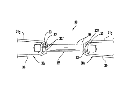
[0015] Figure 1A is a top view of an array or bundle of fibers used to
form an
implantable biocompatible construct according to embodiments of the present
invention.
[0016] Figure 1B is a digital photograph of a prototype of a
biocompatible
construct according to embodiments of the present invention.
[0017] Figure 1C is a schematic illustration of a target repair site.
[0018] Figure 1D is a schematic illustration of an implantable
construct placed at
the target repair site according to embodiments of the present invention.
100191 Figures 2A-2C are cross- and longitudinal-views of the implant
shown in
Figure 1A illustrating exemplary configurations according to embodiments of
the present
invention.
[0020] Figure 3A is a schematic illustration of a medical kit
according to
embodiments of the present invention.
[0021] Figure 3B is a schematic illustration of a medical kit with a
substrate
configured to hold the construct with suture(s) according to embodiments of
the present
invention.
3c
CA 02674456 2009-07-03
WO 2008/085493 PCT/US2007/026365
=
[0022] Figure 4 is a flow chart of operations that can be used to carry out
embodiments of the invention.
[0023] Figure 5A is a graph of tensile strength of NDGA fibers of different
fibers showing strength (MPa) as a function of test rate in mm/sec.
[0024] Figure 5B is a graph of stiffness of NDGA fibers of different fibers
showing modulus as a function of test rate in mm/sec.
[0025] Figure 5C is a graph of strain at failure of NDGA fibers of different
fibers showing strain as a function of test rate in mm/sec.
[0026] Figure 6A is an enlarged digital photograph of an implanted construct
illustrating the construct and suture with neo-tendon growth based on in vivo
trials.
[0027] Figure 6B is a greatly enlarged digital photograph of the implanted
construct shown in Figure 6A illustrating a sectional view of fibers and neo-
tendon
growth.
[0028] Figure 7A is a schematic illustration of a tendon bioprosthesis
illustrating tensile testing thereof with a failure site outside the bounds of
the
implanted construct.
[0029] Figure 7B is a graph of tensile strength (Newtons) at 3 weeks post-
implantation and at various locations for the construct shown in Figure 7A.
[0030] Figure 7C is a graph of tensile strength (Newtons) at 6 weeks post-
implantation and at various locations for the construct shown in Figure 7A.
[0031] Figure 713 is a graph of tensile strength (Newtons) from 3 weeks to
48 weeks post-implantation with the construct of Figure 7A and a contralateral
control.
DETAILED DESCRIPTION
[0032] The present invention now is described more fully hereinafter with
reference to the accompanying drawings, in which embodiments of the invention
are
shown. This invention may, however, be embodied in many different forms and
should not be construed as limited to the embodiments set forth herein;
rather, these
embodiments are provided so that this disclosure will be thorough and
complete, and
will fully convey the scope of the invention to those skilled in the art.
[0033] Like numbers refer to like elements throughout. In the figures, the
thickness of certain lines, layers, components, elements or features may be
4
CA 02674456 2009-07-03
, =
WO 2008/085493 PCT/US2007/026365
=
exaggerated for clarity. Broken lines illustrate optional features or
operations unless
specified otherwise.
10034] The terminology used herein is for the purpose of describing
particular embodiments only and is not intended to be limiting of the
invention. As
used herein, the singular forms "a", "an" and "the" are intended to include
the plural
forms as well, unless the context clearly indicates otherwise. It will be
further
understood that the terms "comprises" and/or "comprising," when used in this
specification, specify the presence of stated features, integers, steps,
operations,
elements, and/or components, but do not preclude the presence or addition of
one or
more other features, integers, steps, operations, elements, components, and/or
groups
thereof. As used herein, the term "and/or" includes any and all combinations
of one
or more of the associated listed items. As used herein, phrases such as
"between X
and Y" and "between about X and Y" should be interpreted to include X and Y.
As
used herein, phrases such as "between about X and Y" mean "between about X and
about Y." As used herein, phrases such as "from about X to Y" mean "from about
X
to about Y."
[0035] Unless otherwise defined, all terms (including technical and scientific
terms) used herein have the same meaning as commonly understood by one of
ordinary skill in the art to which this invention belongs. It will be further
understood
that terms, such as those defined in commonly used dictionaries, should be
interpreted
as having a meaning that is consistent with their meaning in the context of
the
specification and relevant art and should not be interpreted in an idealized
or overly
formal sense unless expressly so defined herein. Well-known functions or
constructions may not be described in detail for brevity and/or clarity.
[0036] It will be understood that when an element is referred to as being
"on", "attached" to, "connected" to, "coupled" with, "contacting", etc.,
another
element, it can be directly on, attached to, connected to, coupled with or
contacting
the other element or intervening elements may also be present. In contrast,
when an
element is referred to as being, for example, "directly on", "directly
attached" to,
"directly connected" to, "directly coupled" with or "directly contacting"
another
element, there are no intervening elements present. It will also be
appreciated by
those of skill in the art that references to a structure or feature that is
disposed
"adjacent" another feature may have portions that overlap or underlie the
adjacent
feature.
CA 02674456 2009-07-03
WO 2008/085493 PCIMS2007/026365
[0037] It will be understood that, although the terms first, second, etc. may
be used herein to describe various elements, components, regions, layers
and/or
sections, these elements, components, regions, layers and/or sections should
not be
limited by these terms. These terms are only used to distinguish one element,
component, region, layer or section from another region, layer or section.
Thus, a
first element, component, region, layer or section discussed below could be
termed a
second element, component, region, layer or section without departing from the
- teachings of the present invention. The sequence of operations (or steps) is
not
limited to the order presented in the claims or figures unless specifically
indicated
otherwise.
[0038] The terms "implant" and "prosthesis" are used interchangeably
herein to designate a product configured to repair or replace (at least a
portion of) a
natural tendon, ligament or other tissue of a mammalian subject (for
veterinary or
medical (human) applications). The term "implantable" means the device can be
inserted, embedded, grafted or otherwise chronically attached or placed on or
in a
patient. The term "tissue" means skin, muscle, bone or other group of cells.
[0039] The term "array" means an arrangement of fibers in rows and/or
columns that are held together as in a matrix.
[0040] Collagen "microfibrils," "fibrils," "fibers," and "natural fibers"
refer to
naturally-occurring structures found in a tendon. Microfibrils are about 3.5
to 50 nm
in diameter. Fibrils are about 50 nm to 50 p.m in diameter. Natural fibers are
above
50 pin in diameter. A "synthetic fiber" refers to any fiber-like material that
has been
formed and/or chemically or physically created or altered from its naturally-
occurring
state. For example, an extruded fiber of fibrils formed from a digested tendon
is a
synthetic fiber but a tendon fiber newly harvested from a mammal is a natural
fiber.
Of course, synthetic collagen fibers can include non-collagenous components,
such as
particulates, hydroxyapatite and other mineral phases, or drugs that
facilitate tissue
growth. For example, the fibers and/or constructs formed from the fibers can
include
compositions can contain carbon nano-tubes, zinc nano-wires, nano-crystalline
diamond, or other nano-scale particulates; larger crystalline and non-
crystalline
particulates such as calcium phosphate, calcium sulfate, and apatite minerals.
For
example, the compositions can contain therapeutic agents such as
bisphosphonates,
anti-inflammatory steroids, growth factors such as basic fibroblast growth
factor,
tumor growth factor beta, bone morphogenic proteins, platelet-derived growth
factor,
6
CA 02674456 2014-09-10
and insulin-like growth factors; chemotactic factors such fibronectin and
hyaluronan; and
extracellular matrix molecules such as aggrecan, biglycan, and decorin. See,
e.g., US Patent
6,821,530. In some embodiments, the constructs can contain cells, engineered
cells, stem
cells, and the like. Combinations of the above or other materials can be
embedded, coated
and/or otherwise attached to the construct.
[0041] The term "suture" refers to a flexible elongate material that
is used to
attach the bioprosthesis to a target anatomical structure to help hold the
bioprosthesis in
location in the body. The suture may be resorbable or non-resorbable,
synthetic or natural.
The suture can be configured to hold the implant in location for at least an
initial post-
implantation period of at least about 1 week, but may reside permanently in
the body or, as
noted above, may be substantially resorbable over time. The suture can be a
single filament
or multi-filament thread, floss, gut or wire, or combinations thereof that can
be used to hold a
portion of an implant against or attached to target structures, typically to
bone and/or tissue.
The suture may comprise a resorbable or non-resorbable biocompatible_material.
Examples of
suture materials include elastomeric materials, such as, for example,
polymers, copolymers
and/or derivatives thereof, including Vicryle, as well as other materials
including, for
example, NITINOL, and combinations thereof. The suture may be used with a
suture anchor
(bone or tissue anchor), staple, screw, plate or other biocompatible fixation
member to affix
the implant in the desired location and/or orientation.
[0042] The term "atraumatic" with respect to suture needles with
thread refers to
an atraumatic or eyeless needle attached to a specific length of suture
material (thread or
filament). The suture and needle are preformed and purchased as a unit, as the
suture needle
manufacturer swages or binds the suture thread to the eyeless atraumatic
needle at the
factory. In a conventional traumatic needle with suture, the thread comes out
of the needle's
hole or eye on both sides. When passing through the tissues, this type of
suture may rip
tissue, at least to a certain extent. In contrast to the conventional "trauma"-
type needle with
suture, the atraumatic needle with suture does not cause trauma (hence the
name
"atraumatic"). Because of these advantages, atraumatic needles with sutures
are today very
widely used.
100431 As with conventional sutures, the sutures of atraumatic needles
can be
absorable or non-absorable. As is well known, there are several shapes of
atraumatic needles,
including straight, half curved, one-third curved and others. The body of the
7
CA 02674456 2014-09-10
needle is available also in different makes, like circular, with edge on the
outer side, with
edge on the inner side, and others.
100441 The term "flexible" means that the so-called member can be
flexed or bent.
100451 The array of fibers can be held together in any suitable manner
including
by their natural affinity to stick together upon compression or extrusion, by
using a sticky
coating or adhesive, such as a gelatinous coating, or by otherwise attaching
the fibers to form
the array. In some embodiments, the fibers can comprise polyglycolic acid,
polylactice acid,
or combinations of these, as discussed below, to help hold the fibers together
for the
bioprosthesis, such as, for example, an Achilles Tendon implant. The fibers
may also
optionally comprise braided segments. The term "braided" and derivatives
thereof mean to
(inter)weave and/or interlock in any manner, three or more fibers or bundles
of fibers
together, including knitting and knotting and combinations of these or other
interlocking
constructions.
[00461. Figure 1A is a schematic illustration of an implantable
construct 20 with
multiple fibers 10 that can be held together to form an array of fibers. As
shown in Figure
1A, the multiple fibers 10 can be axially arranged so that at least a majority
of the fibers are
substantially parallel to each other over at least a major portion of the
length of the construct
20, typically over substantially the entire length of the construct 20. The
construct 20 and/or
fibers 10 can incorporate anti-inflammatory agents or other pharmaceutically
suitable agents.
The construct 20 and/or fibers 10 can be coated or impregnated with a thin
film of polylactic
acid (PLA) or other suitable substance to promote strength and/or ease of
handling. For
example, the construct 20 can be dipped, painted or sprayed with a 3% solution
of PLA in
chloroform or other suitable solution.
100471 Figure 1A also illustrates that an attachment member 30, such
as a suture
30s, can be attached to each end portion of the construct 20 and used to affix
the construct 20
to local tissue. In the embodiment shown in Figures 1A and 1B, the suture 30s
is tied to the
construct 20 so that opposing legs 311, 312 that extend from a looped portion
32 of the suture
have one or more loops 321 encasing the construct 20 that is tied to form one
or more knots
33. The knot 33 can be configured to provide a secure attachment to the array
or fiber bundle
20a and organize the parallel array of fibers into a desired cross-sectional
configuration (see,
e.g., Figures 2A, 2B). The knot configuration can position the suture 30s to
reach out into
adjacent tissue for
8
CA 02674456 2014-09-10
anchorage at about 180 to about 360 degrees from each other. The suture legs
31, 312 can
extend substantially parallel to each other from opposing outer lateral edges
of the construct
20 in the direction of the target anchoring-site. In the embodiment shown, the
sutures 30s are
oriented to exit the construct body outside the bounds of the construct itself
at opposing side
locations and extend substantially parallel to the anchoring site. The looped
portion 32 and
the knot(s) 33 are configured to improve tensile/compression force
distribution and/or cancel
unwanted torque. In some embodiments, the attachment member 30 (suture(s)) can
be placed,
e.g., tied to the implant/construct when the fibers are dry and the suture(s)
can hydraulically
fix in place when the bioprosthesis hydrates after placement in vivo. See,
e.g., co-pending
U.S. Application Publication No. 2008/0200992.
[0048] In the embodiment shown in Figure 1A, a multi-fiber bundle 20a
has a
knot configuration that is formed by a loop 32 around the bundle 20a secured
by a square
knot followed by additional loops each ending in two half-hitches. The number
of additional
loops 32 can be adjusted in accordance with the diameters of the suture
material and the size
of the fiber bundle 20a to facilitate the correct positioning of the exit legs
or strands 311, 312.
Other knot configurations may also be used. The number of fibers 10 used in
the embodiment
shown in Figure IA is sixteen (16), but greater or lesser numbers may also be
used, typically
depending on the target repair site.
[0049] In some embodiments, other initial knot configurations may be
used in lieu
of or with the square knot, although typically the first knot is tied to be
substantially flat so as
to not to unduly project and irritate local structure when implanted.
Similarly, instead of or in
combination with half-hitches, other knots can be used with the loops, and
different loops
may have different knot configurations or may even knot use a knot on a
particular loop.
[0050] One intended use of the construct as a bioprosthesis is to
bridge gaps in
tendon and ligaments by providing the construct in a matching length and
suturing into the
patient's own remaining tendon or ligament end portions using a suitable
surgical tying
technique, such as, for example, but not limited to, a double Kessler
technique or similar
methodology.
[0051] Figure 1C illustrates an example of a target repair site 111r
with two
separated tendon ends 111t, 110t that can be treated with the construct 20
implanted
9
CA 02674456 2009-07-03
WO 2008/085493 PCT/US2007/026365
in the subject according to embodiments of the invention. As shown, the
construct 20
is for an Achilles tendon repair. As shown in Figure 1D, one end portion of
the
construct 20a is attached to the first separated portion of the tendon 110t
undergoing
repair and/or treatment and the other end portion 20b is attached to the
spaced apart
portion of the tendon 111t. As is also shown, the first end portion 20a is
attached via
a suture 30s and the second end portion 20b is attached using a suture 30s.
Other
anchoring or attachment means may be used. The sutures 30s may be resorbable
or
non-resorbable. Adhesive 22 may be used to help secure one or both of the end
portions 20a, 20b during an initial healing phase for additional
stabilization.
[0052] The construct 20 can be preformed in different lengths for selection
by a clinician during a surgical procedure or can be cut to length in situ by
a clinician.
The construct 20 can be preformed with the suture(s) 30 attached to the
construct and
provided in a medical kit to reduce onsite preparation time. This embodiment
may be
particularly suitable where the construct 20 is provided in predetermined
lengths. The
construct 20 can be configured to have a strength and stiffness similar to
natural
tendon or ligament and can provide an effective scaffold for neo-tendon and
ligament
to grow into and further enhance the repair.
[0053] In some embodiments, the plurality of fibers 10 in a respective
construct 20 can be between about six to about fifty, typically between about
ten to
about twenty-seven. Lesser and greater numbers of fibers may be used depending
on
the desired strength or other mechanical parameter of the target implant site.
[0054] Figures 2A and 2B illustrate that the construct 20 can have different
cross-sectional shapes. Figure 2A illustrates that the construct can have a
substantially tubular shape 20r, with a circular or oval cross-sectional
shape, while
Figure 2B illustrates that the construct has a substantially flat
configuration 20f.
Figure 2C illustrates that a portion of the construct 20 can include a braided
segment
20b. As shown, the braided segment 20b is formed by bundles of the fibers in
the
array and may be used to provide a stronger attachment segment for a suture
30s or
other attachment/fixation member 30. Combinations of these and other shapes
over
different portions of the body of the construct 20 may also be used. The
construct 20f
shown in Figure 2B may be particularly suitable as a ligament prosthesis, such
as for
an ACL repair or replacement. The construct 20r shown in Figure 2A may be
particularly suitable as a tendon-prosthesis, such as, for example, the flexor
tendon.
CA 02674456 2014-09-10
Other configurations may also be used as suitable for the target treatment
site/prosthesis.
100551 Typically, the construct 20 is configured to have substantially
the same
physical thickness and/or configuration as the replaced or repaired tissue so
as to not cause
discomfort or physical abnormalities in structure.
[0056] The array can be a relatively tightly compressed array of
fibers or a
relatively loosely compressed or attached arrangement having voids between
some adjacent
fibers depending on the target location and the desired mechanical properties
and
configuration and to allow for neo tissue in-growth.
[0057] In some embodiments, the construct 20 is between about 0.5-50
cm long,
typically between about 1-25 cm, and in some embodiments between about 1 cm to
about 10
cm long. The construct 20 may have a width that is between about 0.05 to 8 cm,
and is
typically between about 1 cm - 3 cm. The constructs 20 may have a cross-
sectional thickness
of about 0.01 to about 30 mm. For the flat construct 20f, the thickness may be
more typically
between about 0.1 to about 10 mm, while the tubular construct 20r may have a
thicker cross-
section, such as between about 5-30 mm.
[0058] Figure 3A illustrates a medical kit 200 that includes the
braided construct
20 and may optionally include at least one suture 30s, which, as shown, may be
pre-attached.
The suture 30s may be provided in the form of an atraumatic needle with suture
(not shown).
The suture 21 can be a bone anchor suture and/or be configured to cooperate
with a bone
tunnel as is well-known. The kit 200 may include other components, such as,
for example, a
container of surgical adhesive and the like. The construct 20 may be held
hydrated in a sterile
flexible sealed package 21 of sterile liquid 22. The kit 200 may have a
package 200p that can
include more than one size (length and/or thickness) construct 20, shown as
provided in two
lengths, L1, L2. The kit 200 may include a temperature warning so that the
construct 20 is not
exposed to unduly hot temperatures that may degrade the implant. A temperature
sensor may
optionally be included on the package of the kit (not shown) to alert the
clinician as to any
excessive or undue temperature exposure prior to implantation. Figure 3B
illustrates the kit
200 can include a substrate 121 that holds the construct 20 with a notch or
well region 122 to
hold the loop/knot 32, 33 to maintain a desired orientation for easy-access to
the construct 20
and suture(s) 30 at a point of use. The
11
CA 02674456 2014-09-10
package 200 may also include a mating top or "lid" to trap the construct in
position and/or
protect it during shipment (not shown).
[0059] Figure 4 illustrates some operations that can be used to carry
out
embodiments of the invention. As shown, a plurality of biocompatible fibers
are provided
(block 100). The fibers are attached as a multi-fiber array to form a
biocompatible
implantable bioprosthesis construct (block 105).
[0060] The fibers may comprise NDGA polymerized collagen fibers (block
102).
The construct can have a flat shape and may be used for a ligament repair or
replacement
(block 112). The construct can have a substantially solid core tubular
configuration or
substantially circular cross-section and can be used for a tendon repair or
replacement (block
114).
[0061] Optionally, the construct can be implanted in a patient using
one or more
of a suture, suture anchor, bone anchor, bone tunnel and the like (block 115).
The suture can
be a suture with an atraumatic needle and may be pre-applied to the construct
and packaged
in a medical kit for subsequent use.
[0062] Also, the construct can optionally include, e.g., be coated,
impregnated
and/or amalgamated with a gel or other material (block 116). The coating may
be to promote
fibroblasts, and/or comprise one or more of an antiinflammatory agent, an
antibiotic or other
therapeutic agent.
[0063] The construct 20 is biocompatible and may be absorbed, resorbed
and/or
biodegradable over time.
[0064] The constructs 20 can be configured to have at least about 60%
of the
tensile strength of natural tendon, and may have tensile strength, and/or
dynamic flexibility
stiffness of similar to or even greater than these properties in corresponding
natural tissue,
e.g., natural ligament or tendon fibers. Embodiments of the invention may be
particularly
suitable for augmenting, repairing or replacing tendons and ligaments.
[0065] In some embodiments, the fibers comprise any collagen fibers
formed in
any suitable manner to be acceptable as a biomedical implant/construct.
[0066] In particular embodiments, the fibers can comprise NDGA-treated
collagen. Suitable ways of forming NDGA polymerized and/or treated fibers are
described in
U.S. Patent Nos. 6,565,960 and 6,821,530. Generally stated, bulk collagen can
be solubilized
by digestion with a protease, then extruded into a
12
CA 02674456 2014-09-10
synthetic fiber. Properly processed NDGA polymerized fibers are biocompatible.
After the
polymerization process, the fibers can be washed in ethanol and phosphate
buffered saline to
remove cytotoxins due to leachable reaction products.
[0067] Testing has been demonstrated that NDGA-treated collagen fibers
are
biocompatible and have desirable mechanical properties. Figures 5A-5C
illustrate exemplary
strain rates of NDGA treated collagen fibers. The fibers were mounted in
clamps with 2 cm
nominal tested length. Fibers were deformed to failure. As shown, the fibers
are nearly elastic
in tension; i.e., strain rate independent. The linear portion of the
stress/strain curve was used
to calculate the elastic modulus (stiffness) and the force at which the fibers
failed was
normalized to cross sectional area yielding tensile strength. Values shown are
means +/- S.D.
for six specimens. For additional discussion of the NDGA polymerized fibers,
see, Thomas J.
Koob, Biomimetic approaches to Tendon Repair, Comparative Biochemistry and
Physiology
Part A 133 (2002) 1171-1192. See also, co-pending U.S. Application Publication
No.
2008/0161917 to Koob et al., entitled, Methods of Making High Strength NDGA
Polymerized
Collagen Fibers and Related Collagen-Prep Methods, Medical Devices and
Constructs.
[0068] In some embodiments, the NDGA collagen fibers may, in some
embodiments, be high-strength. The term "high-strength" refers to fibers
having an average
tensile strength of at least about 150 MPa, such as between about 180 MPa and
350 MPa, and
typically, for bovine, porcine or caprine based "donor" collagen, between
about 180 MPa and
280 MPa, such as between about 240-279 MPa (measured on average). The fibers
may also
have suitable stiffness and strain yield. In general, the fibers can have a
stiffness of at least
about 200 MPa (e.g., at least about 300, 400, 500, or 600 MPa), and a strain
at failure of less
than about 20% (e.g., less than about 15 or 10%). The fibers may be formed
with a relatively
thin diameter, such as, for example about a .08 mm dry diameter (on average)
and about a
0.13 mm wet diameter (on average).
100691 To make the collagen fibers, preparatory donor collagen material can be
pepsin-derived or solubilized collagen that is processed/purified. The
purified collagen
preparatory material is dialyzed a plurality of times in a selected liquid for
a desired period of
time. The dialyzing is typically repeated three times. The dialyzing can be
carried out against
dionized (DI) water in a volume ratio of between about 30:1
13
CA 02674456 2014-09-10
to about 100:1, typically about 60 to 1, for between about 30-90 minutes,
typically about 40
minutes. The dialyzing can form a substantially clear gel of collagen fibrils
indicating good
organization (substantially parallel fibrils), where opacity indicates less
organization. The
organization can help improve tensile strength of subsequently cross-linked
fibers.
100701 The dialyzed collagen material can be incubated for a desired
time before
placing in a fiber-forming buffer. The dialyzed gel can be cross-linked to
provide collagen
fibers for medical constructs. The polymerization (e.g., cross-linking) can be
carried out
using NDGA and the resultant NDGA treated collagen fibers can be relatively
thin, such as,
for example, about 0.08 mm dry diameter (on average).
100711 The incubation may be for at least about 24 hours, typically 24-
48 hours,
and may be at room temperature of between about 15-30 C, typically about 25
C. The
dialysis process can be used before cross-linking for subsequent use with any
suitable cross-
linking materials, to promote collagen organization, such as, for example, and
the process is
not limited to NDGA, but may be useful with other materials, including, for
example,
glutaraldehyde.
[0072] For additional discussion of methods used to form high-strength
NDGA
treated collagen fibers, see,U U.S. Application Publication No. 2008/0161917.
100731 The array or bundle 20 can be formed with fibers having widths
in any
suitable range, typically in the range of between about 0.01-10 mm. One or
more of the fibers
may be continuous or discontinuous over the length of the construct 20.
100741 The present invention is explained in greater detail in the
following non-
limiting Examples.
EXAMPLES
100751 Tendon replacement was performed in rabbit Achilles tendon
using a
parallel array of fibers tied together with a knot made of suture material.
The fibers were
made from NDGA cross-linked collagen as described above and in U.S. patent
#6,565,960.
In-vivo studies were conducted using the rabbit model, wherein a 1 cm gap in
the
gastrocnemius tendon was replaced with a 16 fiber 1 cm long bioprosthesis
(Vicryl 4-0
sutures). The results showed excellent biocompatibility, abundant
14
CA 02674456 2009-07-03
WO 2008/085493 PCT/US2007/026365
formation of neo-tendon (Figures 6A, 6B) and biomechanical properties reaching
60% of the contralateral normal tendon with 6 weeks (Figure 7C).
[0076] The Vicryl suture used a knot that provided a secure attachment to
the bundle and organized the parallel array of fibers into a round cross
section. Also,
the knot positioned the sutures that reach out into the adjacent tissue
anchorage at 180
degree to each other in order to cancel out unwanted torque. The knot
configuration
had a loop around the bundle secured by a square knot followed by additional
loops
each ending in two half hitches.
[0077] Figure 6A is an enlarged digital photograph of an implanted construct
illustrating the construct and suture with neo-tendon growth based on the in
vivo
rabbit trials. Figure 6B is a greatly enlarged digital photograph of the
implanted
construct shown in Figure 6A illustrating a sectional view of fibers and neo-
tendon
growth.
[0078] Figure 7A is a schematic illustration of a tendon bioprosthesis
illustrating tensile testing thereof with a failure site outside the bounds of
the
implanted construct. Figure 7B is a graph of tensile strength (Newtons) at 3
weeks
post-implantation and at various locations for the construct shown in Figure
7A.
Figure 7C is a graph of tensile strength (Newtons) at 6 weeks post-
implantation and
at various locations for the construct shown in Figure 7A. Figure 71)
illustrates
additional tensile strength data on ex vivo mechanical tests out to 48 weeks
(of the
bioprosthesis repair using a construct shown in Figure 7A and a contralateral
control).
[0079] This bioprosthesis offers the advantages of having strength and
stiffness similar to natural tendon or ligament, excellent biocompatibility,
and
provides an effective scaffold for neo-tendon and ligament to grow in and
further
enhance the repair.
[0080] The foregoing is illustrative of the present invention and is not to be
construed as limiting thereof Although a few exemplary embodiments of this
invention have been described, those skilled in the art will readily,
appreciate that
many modifications are possible in the exemplary embodiments without
materially
departing from the novel teachings and advantages of this invention.
Accordingly, all
such modifications are intended to be included within the scope of this
invention as
defined in the claims. The invention is defined by the following claims, with
equivalents of the claims to be included therein.