Note: Descriptions are shown in the official language in which they were submitted.
CA 02675521 2014-11-05
HUMAN CANCER STEM CELLS
[0001] This application claims priority to provisional applications
60/881,497, filed January 22,
2007, 60/907,180, filed March 23, 2007, 60/924,247, filed May 4,2007,
60/950,714, filed July
19, 2007, 60/972,613, filed September 14, 2007.
Technical Field
[0002] The invention relates generally to human cancer stem cells and methods
for their
isolation, characterization and use. More specifically, the invention also
relates to methods of
identifying human cancer stem cells, their uses in therapeutics, target/drug
discovery, anti-tumor
vaccines and cancer diagnosis and treatment.
Background Art
[0003] Eliminating cancer from a patienfs body is challenging because,
although cancerous cells
proliferate in an uncontrolled manner, the cells do not necessarily appear to
be "foreign" to the
body and are therefore difficult to target. Existing cancer treatments tend to
be insufficiently
targeted to the cancer cells and are destructive to a patient's healthy
tissue. Such treatments
typically include X-rays, chemotherapy, proton therapy, and surgery.
Treatments that incite the
body's immune system to exhibit a positive immune response against these
cancer cells would be
preferred.
[0004] Although cancer cells express cancer-associated antigens, they are
often able to evade an
immune response because of their ability to hide the cancer antigens from the
immune system
and/or because the exposed antigens are normal, non-mutated differentiation
molecules or
proteins that the human immune system normally recognizes or tolerates. Cancer
stem cells also
have been reported that are resistant to current therapies of chemotherapy and
radiation. (See,
e.g., Targeted therapy for cancer stem cells: the patched pathway and ABC
transporters.
Oncogene, (2007) 26(9):1357-60.; Radiation resistance and stem-like cells in
brain tumors.
Cancer Cell (2006); 10(6):454-6.; WNT/beta-catenin mediates radiation
resistance of mouse
mammary progenitor cells. Proc Nail Acad Sci USA (2007) 104(2):618-23. Epub
2007 Jan 3.)
1
CA 02675521 2009-07-14
WO 2008/091908 PCT/US2008/051730
[0005] To effectively use immunotherapy to treat a cancer, a patient must
have, or be provided
with, a sufficient number of cancer-reactive lymphocytes that can both reach
the cancer site and
have effector mechanisms to destroy the cancer cells.
[0006] Some therapies under investigation are aimed at heightening the immune
response in
general, and include for example administration of chemical messengers such as
cytokines (e.g.
IL-2 and/or IL-12), lymphocytes specific for telomerase, bacterial extracts or
drugs that boost the
immune system. In an attempt to make the immune response more specific for the
tumor cells,
some treatments administer autologous tumor cells either combined with
cytokines¨e.g. GM-
CSF, gamma interferon or IL-2, individually or in combination--or transfected
with the genes
that encode these cytokines. Some success has been observed in cell-transfer
therapies where
autologous lymphocytes are sensitized to cancer cells ex vivo and then infused
back into the
patient. A similar approach utilizes tumor cell lines instead of autologous
tumor cells.
[0007] Adjuvants are commonly used with cancer vaccine immunotherapy. One
approach uses
dendritic cells (DCs) that are highly potent antigen-presenting cells to
provoke a positive anti-
cancer immune response in patients. Dendritic cells express MHC class I and
MHC class II
molecules, co-stimulatory molecules and adhesion molecules that provide
signals for the
stimulation of naive T cells, CD4+ T-helper cells, CD8+ cytotoxic T
lymphocytes (CTLs),
natural killer (NK) and thymic derived NK cells (NKT) cells. DCs have the
capacity to take up
various types of molecules. Consequently, DCs can be loaded with tumor-
associated antigens
(TAAs) in various forms and administered as vaccines.
[0008] One DC-based approach uses DC-cancer cell hybrids generated by fusion
of cancer cells
with dendritic cells to combine sustained cancer antigen expression with the
antigen-presenting
and immune stimulatory capabilities of the DC. In animal models, immunization
with DC-
cancer cell hybrids can provide some form of anti-cancer protection or
eradicate established
disease. Hybrids of autologous DCs comprised of cancer cell lines or primary
human cancer cells
(including breast carcinoma cells) have been shown to induce CTL responses
against autologous
cancer cell types in vitro. Clinical studies of the treatment of renal cell
carcinoma and glioma
have demonstrated that vaccination with DC-cancer cell hybrids can safely
induce anti-cancer
immune responses in patients.
[0009] One hypothesis to explain how tumors grow and metastasize is the cancer
stem cell
hypothesis, which states that there is a small, distinct subset of cells
within each tumor that is
2
CA 02675521 2009-07-14
WO 2008/091908 PCT/US2008/051730
capable of indefinite self-renewal and of developing into the more adult tumor
cell(s), which are
relatively limited in replication capacity. It has been hypothesized that
these cancer stem cells
(CSC) might be more resistant to chemotherapeutic agents, radiation or other
toxic conditions,
and thus, persist after clinical therapies and later grow into secondary
tumors, metastases or be
responsible for relapse. It has been suggested that CSCs can arise either from
the tissue stem
cells or from a more differentiated tissue progenitor cell(s). While
supporting data for this is
strong for hematopoietic stem and progenitor cells and hematopoietic tumors,
less is known
about solid tumors and their respective CSCs.
[0010] Solid tumors are thought to arise in organs that contain stem cell
populations. The
tumors in these tissues consist of heterogeneous populations of cancer cells
that differ markedly
in their ability to proliferate and form new tumors; this difference in tumor-
forming ability has
been reported for example with breast cancer cells and with central nervous
system tumors.
While the majority of the cancer cells have a limited ability to divide,
recent literature suggests
that a population of cancer cells, termed cancer stem cells, has the exclusive
ability to
extensively self-renew and form new tumors. Growing evidence suggests that
pathways that
regulate the self-renewal of normal stem cells are deregulated or altered in
cancer stem cells,
resulting in the continuous expansion of self-renewing cancer cells and tumor
formation.
[0011] It has been suggested that cancer patient prognosis is associated with
stem cell
phenotype/biology. (See e.g., Molecular profiling identifies prognostic
subgroups of pediatric
glioblastoma and shows increased YB-1 expression in tumors. J Clin Oncol.
(2007) 25(10):1196-
208; Cancer stem cells are central to metastasis, which accounts for 90% of
the lethality of
cancer. Cell Res. (2007) 17:3-14.) It has also been observed that patients
with autoimmune
reactions to self-stem cells demonstrate decreased cancer progression. (See,
e.g. "immunity to
cancer stem cells may help protect people with a precancerous condition from
developing the
full-blown disease" J Exp Med (2007) 204(4):831-40.)
[0012] Tissue stem cells exist in specific niches or microenvironments that
are critical for
maintaining them in the appropriate developmental and metabolic state. These
microenvironments are not completely understood, but their disruption by
genetically knocking
out an important factor can result in the disregulation of stem cell
homeostasis both during
development and in the adult. In trying to understand the microenvironments
that support tissue
stem/progenitor cells, many researchers have taken the approach of deriving
serum free culture
3
CA 02675521 2009-07-14
WO 2008/091908 PCT/US2008/051730
conditions where the medium, substrate and physical environment produce an
optimized
environment for maintaining specific fetal and neonatal tissue stem/progenitor
cells (SPC) in a
defined state in which the SPC can replicate, but not differentiate (see for
example U.S. Patent
Nos. 6,436,704 and 6,416,999). The optimized media and culture conditions that
are distinct for
different types of SPC can be seen as recreating the stem cell niche that
these cells occupy in
vivo. These media and optimized conditions are specifically tailored to the
SPCs in that they
specifically select out the SPCs and cannot support the survival and/or growth
of any other cell
types in a tissue. Consequently, non-SPC cells will not survive and replicate
under the SPC-
preferred conditions and are lost during culture and passage, leaving only a
pure SPC population,
even when the SPC represents only a very small percentage of the starting
culture. The
conditions must also remove any signals for further differentiation of the SPC
to allow its
maintenance in culture over an extended period of time.
[0013] One hypothesis about how tumors originate is that tumors arise from the
tissue SPC by a
series of mutational events. Data exists to suggest that some tumor cells will
"home in" to
specific SPC niches when they move about the body during the process of
metastasis. Media
and optimized culture conditions derived to support the survival and growth of
SPC might also
be able to preferentially support the growth and survival of CSCs, thus
selecting for this type of
cell when the dispersed tumors are put into culture. Such media and optimized
culture
conditions may allow for the establishment of pure CSC cultures that would be
capable of long-
term or extensive growth and maintenance of the characteristics of the rare
CSC phenotype.
[0014] The tumor stem cell has been hypothesized, but there does not yet exist
a reliable way of
identifying these cells, nor does consensus exist on all their
characteristics. Some researchers
have proposed that cancer stem cells can be identified based on marker
expression (see e.g., Al-
Hajj et al. (2003) Proc Natl Acad Sci USA 100:3983-3988; O'Brien et al. (2007)
Nature
445:106-110; and Clarke et al. (2006) Cell 124: 1111-5). CD133 has been
proposed to be a
marker found in cancer stem cells in brain tumors and in human prostatic
epithelial stem cells.
CD44 expression accompanied by no or low CD24 expression was hypothesized to
be expressed
by some solid cancer stem cells (e.g., breast cancer). CD34 is a marker
present on the surface of
blood vessels and immature blood cells that has also been associated with
hematopoietic stem
cells.
4
CA 02675521 2009-07-14
WO 2008/091908 PCT/US2008/051730
[0015] The establishment of pure CSC cultures would be a great advantage in
studying and the
understanding of the regulation of tumorgenesis and metastasis and in the
discovery and
development of CSC-directed therapies for cancer. Accordingly, there exists a
need for methods
to identify, isolate, culture and characterize cancer stem cells.
[0016] The invention described herein overcomes many of the unmet needs and
shortcomings
mentioned above and provides for methods of isolation, maintenance and growth
of human
cancer stem cells. The invention also describes a constellation of
characteristics of cancer stem
cells, and specifically the characteristics of a novel population of cells
from colon, prostate, lung,
pancreas, breast, mantle cell lymphoma, and Merkel's tumors with the
biological characteristics
of CSCs.
[0017] The present invention also provides a simple, effective and efficient
method for treating
cancer, preventing cancer, delaying the onset of cancer or delaying the
progression of cancer via
administration of the CSC-based vaccines and treatments described herein.
Figures
[0018] Figure 1 shows selective growth of CSC colonies from prostate carcinoma
tumor
(PRCA) using a defined cell culture medium.
[0019] Figure 2 shows a set of CSC colonies derived from basal cell carcinoma
(BCCA1)
cultured in the defined medium selective for this cancer stem cell type.
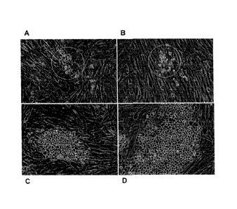
[0020] Figure 3 shows CSC colonies with a similar distinctive morphology
growing from colon,
(panels A-C, F), Merkel Cell (panel D) and a prostate (panel E) tumor. Panels
A and B show
cultures derived from a colon tumor: primary culture on day 42 of culture (A)
and the same
culture area 3 days later (B). Note the small round cells that are apparent in
A (arrow and insert)
are still dividing, while other cells in the culture die (B). Figure 3C shows
a colon CSC after one
passage which has now become almost entirely stem cells. Panels A, B, and D-F
show primary
cultures where the CSC are beginning to become apparent and the non-stem
component of the
tumor are either static, dying, or dead. These cultures, with continued
passage, have all become
CSC lines of this invention as indicated: CRCA1115 (A, B), RECA0515 (C), MCC
(D),
PRCA0611(E), and CRCA0404 (F).
[0021] Figure 4 shows photos of tumors formed under the kidney capsule of SCID
mice from
various cell lines. On the left are tumors formed from 100 cells each of 2
colon CRC grown for
CA 02675521 2009-07-14
WO 2008/091908 PCT/US2008/051730
7 (A ¨ RECA1208) or 8 (B ¨ RECA0515) weeks. Substantial tumors are seen in
both cases.
The middle panels show the tumor formation from animals implanted with 5x10⁴
cells of
CRCA1115 colon CSC (C) after 4.5 weeks or 10 fold (5x10⁵) more of the non-CSC
RECA0705
(D) after 8 weeks. The white fibrous material in D is from the collagen in the
implant; the highly
refractive material is fat. The human DNA out/in ratio for this tumor is <1Ø
The last 2 panels
are of kidneys after implantation of 5x10⁵ prostate tumor cells from a CSC.
The CSC (E ¨
PRCA0425) cell-derived tumor is shown after 8 weeks. These cells clearly grow
more slowly
than the colon CSC seen in panel C at 4.5 weeks but do form tumors from 100
cells. Finally
panel F is a kidney after implantation of 2.5x10⁵ PRCA0312-435TR cells for 5
weeks. No
tumor is visible. Thus the non-CSC lines are clearly distinguished from the
CSC lines using this
SRC xenograft model
[0022] Figure 5 shows that the antibody known as KID24 decreases growth of
tumors in cancer
stem cell metastatic models established in the subrenal capsule of mice, using
the colon
(CRCA0404) & Merkel cancer stem cell lines of this invention.
[0023] Figure 6 shows the effect of the antibody known as KID24 on metastases,
specifically on
Merkel Cell cancer that metastasizes to multiple organs including the pancreas
from tumors
established in the subrenal capsule. Mets are quantified using the QPCR for
huDNA described
in the Examples.
Disclosure Of The Invention
[0024] This invention relates to the field of tumor biology and cell biology.
In one aspect, the
invention relates to an isolated population of cancer stem cells cultured from
tumor tissue. The
cells may be cultured in serum-free nutrient medium, and may have cell
surfaces that are
substantially free of serum biomolecules. The cells may be maintained in
culture without
senescence or loss of growth capacity for an extensive period of time.
[0025] The cancer stem cells of this invention are useful in drug screening,
as tools for genomic
and proteomic profiling (in vitro, in vivo and metastatic status) of diseased
tissues, for
multivalent cancer stem cell vaccines, for diagnostics and imaging of cancer-
related antigens,
and for the identification of therapeutic agents such as antibodies and drugs.
[0026] Another aspect of this invention relates to methods of isolating such a
population of
substantially pure human cancer stem cells that can be maintained in culture
without senescence
6
CA 02675521 2009-07-14
WO 2008/091908 PCT/US2008/051730
or loss of growth capacity (ability to self-renew). These methods are based on
culturing the
human cancer stem cells in an environment that has been optimized for their
growth. The
nutrient media for culturing human cancer stem cells is based on media
formulations that have
been optimized for supporting the growth of corresponding fetal
progenitor/stem cells.
[0027] Another aspect of this invention relates to methods of characterizing
such a population of
substantially pure human cancer stem cells through functional assays. Such
methods are known
in the art and are suitable for the characterization of the human cancer stem
cells of this
invention. Generally such methods are xenograft models for tumor formation in
an immune-
compromised host animals from the implantation of a small number of human
cancer stem cells.
[0028] Another aspect of this invention provides methods for characterization
of tumor (cancer)
stem cell cultures. Such characterization can include the expression of
certain markers,
including but not limited to CD34, a marker that has been previously
associated with
hematopoietic stem cells.
[0029] In yet another aspect of this invention, this invention relates to
methods of generating
human tumor xenograft models by introducing a population of human cancer stem
cells into a
non-human, mammalian recipient.
[0030] In another aspect of this invention, the invention relates to methods
using a human cancer
stem cell or fragment thereof as an immunogen and for suitably providing an
isolated cancer
stem cell to serve as an immunogen. These methods are useful for the creation
of anti-cancer
stem cell therapeutic and diagnostic agents, and as vaccine to boost an
individual's immune
response. The vaccine may be used therapeutically or preventatively. A
therapeutic vaccine is
administered to a subject having cancer to treat the cancer. In a subject
having cancer, the
vaccine may be made from the subject's own cancer cells, or from allogenic
cancer cells or tumor
cell lines. A preventative vaccine is administered to a subject without cancer
to reduce the risk
of the subject developing cancer.
[0031] Another aspect of this invention provides methods of providing a source
of human cancer
stem cells as biological components for developing pharmaceutical drugs
wherein human cancer
stem cell cultures are used as a source of cancer stem cell biological
components in which one or
more of these human cancer stem cell biological components are the targets of
the drugs that are
being developed.
7
CA 02675521 2014-11-05
[0032] In another aspect of this invention, the invention relates to methods
of providing human
cancer stem cell cultures for use in bioassay development. Human cancer stem
cell cultures can
be used in bioassays to identify factors, agents, or compounds that can affect
the growth and/or
survival of the human cancer stem cells. Such effects can include the growth
promotion, growth
arrest, stasis, death, apoptosis, changes in metabolism, changes to gene
expression, changes in
protein expression or alteration of growth/metabolic pathways. Cancer stem
cells can be used in
bioassays and/or chug discovery to understand molecular pathways of
tumorgenesis, tumor
establishment, tumor growth and metastasis.
Detailed Description Of The Invention
[0033] The following detailed description of the invention is provided to aid
those skilled in the
art in practicing the present invention. This detailed description should not
be construed to limit
the present invention, as those of ordinary skill in the art may make
modifications of the
embodiments disclosed herein
[0034] The practice of the present invention will employ, unless otherwise
indicated,
conventional techniques of immunology, molecular biology, microbiology, cell
biology and
recombinant DNA, which are within the skill of the art. See, e.g., Molecular
Cloning: a
laboratoy manual, 2nd edition Sambrook, et al. (1989); Current Protocols In
Molecular Biology
F. M. Ausubel, et al. eds., (1987); the series Methods In Enzymology, Academic
Press, Inc.; PCR
2: A Practical Approach, M.J. MacPherson, B.D. Hames and G.R. Taylor, eds.
(1995),
Antibodies, A Laboratoty Manual, Harlow and Lane, eds. (1988), Adult and
Pediatric Urology,
J. Gillenwater at al., eds. (2002), and Animal Cell Culture, R.I. Freshney,
ed. (1987).
[0035] As used in the specification and claims, the singular form "a", "an",
and "the" include
plural references unless the context clearly dictate otherwise. For example,
the term "a cell"
includes a plurality of cell, including mixtures thereof.
[0036] As used in the specification and claims, the terms "cancer stem
cell(s)" and "CSC" are
interchangeable and refer to solid cancer stem cells. CSCs are mammalian, and
in preferred
8
CA 02675521 2009-07-14
WO 2008/091908 PCT/US2008/051730
embodiments, these CSC are of human origin, but they are not intended to be
limited thereto. As
used herein "tumor stem cells" typically refers to cells isolated and cultured
from solid human
tumors, and are used interchangeably with cancer stem cells.
[0037] Cancer stem cells are defined and functionally characterized as a small
subset of cells
from a tumor that can grow indefinitely in vitro under appropriate conditions
(ability for self-
renewal), are able to form tumors in vivo using only a small number of cells.
Other common
approaches to characterize CSCs involve morphology and examination of cell
surface markers,
transcriptional profile, and drug response.
[0038] In embodiments of the present invention, multiple CSC lines have been
established from
multiple tumor types. These CSCs share some characteristic cell surface
antigens and others are
distinct. Some embodiments of the CSC lines of this invention can grow
indefinitely and form
tumors from < 20 cells in vivo. Some CSCs are spontaneously metastatic in
subrenal capsule
animal models or orthotopic xenografts. The CSC lines of this invention and
cell lines derived
from CSC metastases have characteristic changes in cell surface markers
expression, such as
CD34 and CD44 expression. Marker expression may change with culture conditions
and with
cell line passage in an animal.
[0039] Cancer stem cell lines described herein have been examined for
differential display of
cell surface antigens; patterns of cell surface antigen display also differ
after passage of cell lines
in an animal, in different culture conditions (with or without animal-derived
products), and from
primary to metastatic tumors, but are stable, over numerous passages in vitro,
when the cells are
maintained in the preferred media as described. This pattern of cell surface
antigen and cell
marker changes are useful to identify and characterize the cancer stem cells
of this invention.
[0040] An "antibody" is an immunoglobulin molecule capable of binding an
antigen. As used
herein, the term encompasses not only intact immunoglobulin molecules, but
also anti-idiotypic
antibodies, mutants, fragments, fusion proteins, humanized proteins and
modifications of the
immunoglobulin molecule that comprise an antigen recognition site of the
required specificity.
[0041] A "monoclonal antibody" refers to a homogeneous antibody population
wherein the
monoclonal antibody is comprised of amino acids (naturally occurring and non-
naturally
occurring) that are involved in the selective binding of an antigen.
Monoclonal antibodies are
highly specific, being directed against a single antigenic site. The term
"monoclonal antibody"
encompasses not only intact monoclonal antibodies and full-length monoclonal
antibodies, but
9
CA 02675521 2009-07-14
WO 2008/091908 PCT/US2008/051730
also fragments thereof (such as Fab, Fab', F(ab')2, Fv), single chain (ScFv),
mutants thereof,
fusion proteins comprising an antibody portion, humanized monoclonal
antibodies, chimeric
monoclonal antibodies, and any other modified configuration of the
immunoglobulin molecule
that comprises an antigen recognition site of the required specificity and the
ability to bind to an
antigen. It is not intended to be limited as regards to the source of the
antibody or the manner in
which it is made (e.g., by hybridoma, phage selection, recombinant expression,
transgenic
animals, etc.).
[0042] "Humanized" antibodies refer to a molecule having an antigen-binding
site derived from
an immunoglobulin from a non-human species and the remaining immunoglobulin
structure of
the molecule based upon the structure and/or sequence of a human
immunoglobulin. The
antigen-binding site may comprise either complete variable domains fused onto
constant
domains or only the complementarity determining regions (CDRs) grafted onto
appropriate
framework regions in the variable domains.
[0043] The term "antigen" is a molecule which can include one or a plurality
of antigenic
determinants or epitopes to which an antibody can bind. An antigen is a
substance that can have
immunogenic properties, i.e., induce an immune response. Antigens are
considered to be a type
of immunogen. As used herein, the term "antigen" is intended to mean full-
length proteins as
well as peptide fragments thereof containing or comprising one or a plurality
of epitopes.
Antigens may also comprise one or more antigenic determinant sites, or
comprise one or more
fragments of such sites, variants of such sites, or peptidomimetics of such
sites. Antigens may
be protein, partly protein, or non-proteinaceous. These compounds may be
glycosylated.
[0044] The terms "surface antigens" and "cell surface antigen" are used
interchangeably herein
and refer to the plasma membrane components of a cell. These components
include, but are not
limited to, integral and peripheral membrane proteins, glycoproteins,
polysaccharides, lipids, and
glycosylphosphatidylinositol (GPI)-linked proteins. An "integral membrane
protein" is a
transmembrane protein that extends across the lipid bilayer of the plasma
membrane of a cell. A
typical integral membrane protein consists of at least one membrane-spanning
segment that
generally is comprised of hydrophobic amino acid residues. Peripheral membrane
proteins do
not extend into the hydrophobic interior of the lipid bilayer and they are
bound to the membrane
surface by noncovalent interaction with other membrane proteins. GPI-linked
proteins are
proteins that are held on the cell surface by a lipid tail that is inserted
into the lipid bilayer.
CA 02675521 2009-07-14
WO 2008/091908 PCT/US2008/051730
[0045] "Immunogen" refers to any substance that induces an immune response. A
substance that
is an immunogen is described as being "immunogenic". Induction of immune
response includes
but is not limited to activation of humoral responses (e.g., producing
antibodies) or cellular
responses (e.g., priming cytotoxic T cells), inflammatory responses (e.g.,
recruitment of
leukocytes), and secretion of cytokines and lymphokines.
[0046] The term "heterologous" as applied to a cell used for immunization or
transplantation
means that the cell is derived from a genotypically distinct entity from the
recipient. For
example, a heterologous cell may be derived from a different species or a
different individual
from the same species as the recipient. An embryonic cell derived from an
individual of one
species is heterologous to an adult of the same species. "Heterologous" as
applied to a recipient
means that the recipient is a genotypically distinct entity from the source of
the cells that are
being introduced into the recipient.
[0047] A cell surface is "substantially free of serum biomolecules" when at
least about 50% of
the human cancer stem cell surfaces, more preferably at least about 75% of the
human cancer
stem cell surfaces, even more preferably at least about 90% of the human
cancer stem cell
surfaces, and most preferably at least about 95% of the human cancer stem cell
surfaces do not
have serum biomolecules derived from serum binding to the cell surface such
that antigenic sites
or antibody binding sites are bound or are unavailable for antigenic
recognition by an antibody or
a portion of an antibody. Cell surface can determined by measuring the cell
size, either by
microscopy or flow cytometry. For example, synthetic beads of various known
sizes are
commonly used for calibration in flow cytometry. A small quantity of
calibrated beads may be
mixed with cancer stem cells and the resultant population is analyzed by flow
cytometry.
Human cancer stem cells can then be compared with the size of the calibrated
beads.
Calculations of cell surface amount can be accomplished since the sizes of the
beads are known.
[0048] "Senescence" as used herein refers to the phenomenon where cells lose
the ability to
divide.
[0049] As used herein, a "substantially pure" population of cells is a
population of cells that is
comprised at least about 85% of the cells of interest, preferably at least
about 90%, and even
more preferably about 95% or more.
[0050] "Serum," as used herein, refers to the fluid phase of mammalian blood
that remains after
blood is allowed to clot.
11
CA 02675521 2009-07-14
WO 2008/091908 PCT/US2008/051730
[0051] "Serum biomolecules", as used herein, refers to biological compositions
found in serum.
Examples include, but are not limited to, albumin, al-globulin, a2-globulin, b-
globulin, and g-
globulin. Serum biomolecules include biological compositions, whole or
partial, which are
either naturally found in serum or derived from processing and handling of
serum.
[0052] The terms "mammals" or "mammalian" refer to warm blooded vertebrates
which include
but are not limited to humans, mice, rats, rabbits, simians, sport animals,
and pets.
Cancer Stem Cell Antigens
[0053] In embodiments of the present invention, certain antigens have been
detected on the
surface of the cancer stem cells disclosed herein. These include known
antigens, novel antigens,
and antigens not previously associated with tissue or cancer stem cells. By
way of example and
not of limitation, these antigens include B7H3 (various epitopes), CD46,
transferrin receptor,
CD112 (polio virus receptor related protein 2), ephA2 receptor, EGFR, ALCAM
(CD166),
alpha-V-beta-5, JAM3, priopionyl-Coenzyme A carboxylase alpha,
carboxypeptidase M,
carboxypeptidase C, LDL-receptor, desmoglein2, ADAM9, CEA CD66e, oncostatin M
receptor
beta, alpha 2 integrin, and prostatin. Cell lines that express these antigens
are particularly
preferred for use in the methods of these inventions.
[0054] Methods for isolating and obtaining these cancer stem cell antigens are
common.
Preferred methods use antibodies directed against these antigens. Examples of
antibodies
directed to some of these antigens are provided in the following disclosures:
B7H3 (PCT WO
2004/001381 and US 60/733,041, particularly antibody TES7, PTA-7093), CD46 (US
7,148,038,
particularly antibody PA7, PTA-3706), transferrin receptor (PCT WO 05/121179,
particularly
antibody LUCA31, PTA-6055), ephA2 receptor (PCT WO 06/084226, particularly
antibody
SPL1, PTA-6059), JAM3 (PCT WO 06/084078, particularly antibody PACA4, PTA-
6510),
carboxypeptidase M (PCT WO 06/076584, particularly antibody KID31, PTA-6516),
ADAM9
(PCT WO 06/084075, particularly antibody KID24, PTA-5174), and oncostatin M
receptor beta
(PCT WO 06/084092, particularly antibody LUCA38, PTA-6511). These antibodies
are
particularly useful as cancer stem cell markers according to the teachings
herein.
[0055] These antigens are desirable markers for use in identifying cancer stem
cells generally,
discovering new cancer stem cells, or for identifying selected subsets of
cancer stem cells, such
as those that are tissue-specific or developmental-stage specific. The present
invention discloses
12
CA 02675521 2009-07-14
WO 2008/091908 PCT/US2008/051730
panels of antigens not heretofore appreciated as cancer stem cell specific,
thus enabling a means
for profiling cells and identifying their stem-cellness. Cells may be selected
from a population,
using flow analysis and other commonly known means, based on the presence of
some or all of
the stem cell markers disclosed herein, alone or in combination with other
known markers.
Routine experimentation is used to determine the presence on a cell surface of
the antigens
disclosed herein, to permit collection of a unique, cancer stem cell antigen
"fingerprint" for a
particular tissue, cell or cell culture.
[0056] The inventors have identified a set of markers that are present on a
predominant number
of cancer stem cells from solid tumors. These sets include some or all of the
antigens identified
above. They are not exclusively present only on cancer stem cells; using the
methods taught
herein these markers will bind (to some extent) to normal tissue stem cells,
normal tissues, tumor
tissues, or daughter cells. Many of these antigens are present on both normal
and cancer stem
cells. Some are present on at least five of the cancer stem cell lines
disclosed herein, such as
B7H3 (all epitopes). It is expected that the marker profiles of solid tumor-
derived cancer stem
cells will differ from those derived from hematopoietic cells.
[0057] These antigens are also useful for assessing the surface of cells over
time. In data not
shown, a comparison and differential binding assay was performed using a panel
of antibodies
and three pairs of cancer stem cell lines: the breast cancer stem cell line
BRCA1103 was
assessed at passages 11 and 12 to show reproducibility of the assay; rectal
carcinoma cancer
stem cell line RECA0515 was assessed at passages 10 and 16 to show stability
of the cell lines,
and colorectal carcinoma cancer stem cell line CRCA0404 was used to compare
the parental line
and a clone from this line. The antigen profile of the RECA0515 p10 vs p16 was
13% different,
the BRCA1103 pl 1 vs p12 was 2% different, and the CRACA0404 vs clone antigen
profile was
6% different.
[0058] As disclosed herein, sets of cancer stem cell markers are selected to
optimize selection of
cells with desired characteristics; for example, the marker profile of
daughter cells, cells from
metastatic deposits, or cells that have been passaged in vivo will be
different from the profile of
cancer stem cells from tissues or primary cell culture.
[0059] Certain antigens will not be expressed on all cells of a tumor tissue
yet will be present on
the cancer stem cells from that tumor; methods are commonly known in the field
for
determining this kind of differential expression.
13
CA 02675521 2009-07-14
WO 2008/091908 PCT/US2008/051730
[0060] The methods disclosed herein for use of these cancer stem cell antigens
as markers are
usefully combined with other methods for identifying cancer stem cells, such
as selection using
cell culture methods, assessment of phenotype or morphology.
[0061] These particularly preferred antigens are desirable vaccine components,
presented in a
polyvalent vaccine, combination therapeutic composition, or as individual
therapeutic
compositions for prevention, treatment, or diagnosis of disease. Agents that
bind to these cancer
stem cell antigens are useful for targeting therapeutic or diagnostic moieties
to the cancer stem
cells.
[0062] Aspects of this invention include isolated cancer stem cells with
cancer stem cell
morphology that bind to one of more of the antibodies TES7, PA7, LUCA31, SPL1,
PACA4,
KID31, KID24, and LUCA38.
[0063] The cancer stem cells of this invention are also used to discover and
screen for antigens
that, when bound by ligand, modulate the production of cytokines such as
angiogenic and growth
factors. For example, the cancer stem cell-binding antibody referred to herein
as TES7, directed
to an isoform of B7-H3, decreases the secretion of angiogenic factors VEGF and
MIP- 1 alpha
(CCL3) by both stroma cells and cancer stem cells. Both TES7 and the antibody
referred to
herein as KID24 have been shown to have the ability to modulate cytokine
pathways and
cytokine signaling. This provides new insight into signaling mechanisms that
are capable of
driving tumor growth, and cancer stem cells of this invention provide the
ability to identify
growth modulatory antibodies that would be missed in standard growth assays.
According to the
teachings of this invention, antibodies raised against antigens present on the
cancer stem cells of
this invention are used in cytokine assays to determine if the antibody /
antigen modulates
cytokine signaling. A large variety of cytokine assays suitable for use in the
practice of this
invention are well known in the art.
Isolation and maintenance of solid cancer stem cells.
[0064] In preferred embodiments, the human cancer stem cells of this invention
are isolated from
solid human tumor tissue. The following methods are illustrative rather than
limiting; other
commonly known methods are acceptable in the practice of this invention.
14
CA 02675521 2009-07-14
WO 2008/091908 PCT/US2008/051730
[0065] A solid human tumor tissue is rinsed with phosphate buffered saline
(PBS), preferably
several times. The PBS may contain antibiotic and/or anti-fungal agents such
as, but not limited
to gentamycin. The solid human tumor tissue is minced into cubes of
approximately 1 mm,
suspended in dissociation media. The dissociation media is basal media
supplemented with a
cell dissociation agent. A wide variety of dissociation agents can be used
including but not
limited to EDTA, EGTA, trypsin and collagenase-dispase. A preferred
dissociation agent is
collagenase dispase used at a concentration that will allow for the partial
dissociation of cells
from the minced tumor tissue. A preferred concentration is 10% weight by
volume in PBS. The
use of a trypsin inhibitor may also be included in the dissociation media. A
preferred trypsin
inhibitor is soybean trypsin inhibitor (STI) used at a suitable concentration.
As a non-limiting
example, one typical suitable concentration of STI is 10%(v/v).
[0066] The cells are incubated in the dissociation media at 37 C. At five-
minute intervals, the
suspension are pipetted to loosen the cell aggregates. The enzymatic activity
is stopped when the
aggregates are 10-20 cells in size. The cells are pelleted by centrifugation
and washed with basal
medium and pelleted by centrifugation. The supernatant is removed, the tissue
is resuspended in
basal medium, then transferred to a culture dish.
[0067] A wide variety of basal media are used to keep the pH of the liquid in
a range that
promotes survival of human solid cancer stem cells. Non-limiting examples
include
F12/DMEM, Ham's F10 (Sigma), CMRL-1066, Minimal essential medium (MEM, Sigma),
RPMI-1640 (Sigma), Dulbecco's Modified Eagle's Medium (DMEM, Sigma), OPTI-MEMO
(GIBCO BRL)and Iscove's Modified Eagle's Medium (IMEM). In addition, any of
the basal
nutrient media described in Ham and Wallace (1979) Meth. Enz., 58:44, Barnes
and Sato (1980)
Anal. Biochem., 102:255, or Mather, J.P. and Roberts, P.E. (1998)
"Introduction to Cell and
Tissue Culture", Plenum Press, New York can also be used. In some instances
the basal media
may use fructose as a sugar source, such as in the media described in patent
application
publication WO 2005/028626.
[0068] Basal medium is added to the culture dish and the tissue is incubated
at 37 C in a
humidified atmosphere. In preferred embodiments that promote cancer stem cell
survival and
growth, a variety of nutrients are added to supplement the basal media, thus
creating a "nutrient
media". Human cancer stem cell aggregates are placed in this media, and, in a
preferred
embodiment, the CSCs migrate out of the cell aggregates into the media and
anchor to the
CA 02675521 2009-07-14
WO 2008/091908 PCT/US2008/051730
culture dish or other supplied anchor material. The remnant of the minced
tissues that do not
attach to the culture dish or anchor will flow in the medium and will be
removed by medium
change after a short time in culture, e.g., 1-2 weeks.
[0069] In another preferred embodiment, cells from human cancer cell
aggregates placed in
nutrient media all attach to the culture dish and the human cancer stem cells
slowly establish and
grow amongst the other cell types from the human tumor. Eventually, the human
cancer stem
cells will form a substantially pure population of cells and the other
contaminating cell types will
no longer be in the culture. The culture process and environment will not
support the replication
and/or survival of contaminating cell types and will promote the survival and
growth of the
human cancer stem cells so as to generate a substantially pure population of
human cancer stem
cells. The population of human cancer stem cells is capable of long-term
growth in culture and
is capable of extensive proliferation and growth without senescence.
[0070] The human cancer stem cells can be grown in tissue culture containers
(e.g., flasks,
plates, etc.) that are either uncoated or coated with different substrates.
Non-limiting examples
of substrates that may be used include fibronectin, laminin, collagen,
polylysine, nitrocellulose,
nylon, and polytetrafluoroethylene. The size of the tissue culture containers
is proportional to
the amount of human tumor tissue being placed within the containers. A skilled
artisan may
determine the correct size of the tissue culture containers by a stepwise
increment of tumor tissue
placed within the tissue culture containers. When the human tumor tissue is
first placed within
the tissue culture containers, the media is generally clear in overall
turbidity. As cells migrate
out and away from the tumor tissue pieces, the media will become more opaque
and more turbid.
At the point where the media is highly turbid, more nutrient media is placed
in the tissue culture
containers to replenish the nutrients consumed by the human tumor cells by
adding more fresh
medium or changing medium completely. Additionally or in the alternative, when
the media
becomes turbid, a small amount of cells may be removed from the tissue culture
containers and
checked for cell viability, for example, with trypan blue staining. Tissue
culture containers that
have been overrun with too many cells will begin to show decreased cell
viability.
[0071] Continued culture of the human cancer stem cells generally involves
transfer of the cells
to one or more new culture containers. Preferably, such transfer is done
before the culture
container is overrun with cells (e.g., as demonstrated by reduced cell
viability. The cells may be
transferred to other containers of a larger size (e.g., greater cubic volume)
to accommodate the
16
CA 02675521 2009-07-14
WO 2008/091908 PCT/US2008/051730
increasing amount of cells. Alternately, the cells may by 'split' into several
separate tissue
culture containers with fresh nutrient media (also known as "subculturing").
In this manner, a
substantially pure population of human cancer stem cells can be obtained and
propagated.
[0072] Removal of cells from a tissue culture container is preferably
accomplished by enzymatic
treatment to detach the cells from the surface(s) of the plastic tissue
culture containers and/or the
substrate used (e.g., fibronectin, laminin, etc.). In a more preferred
embodiment, an enzyme such
as collagenase-dispase is used in an effective amount to dissociate human
cancer stem cells from
the sides of the tissue culture flask. An effective amount is at least about
10%, more preferably
at least about 1%, and most preferably at least about 0.1% collagenase-dispase
by volume. After
detachment of cells from the surface(s) of the tissue culture container, the
enzyme is washed
away with a basal media, preferably the nutrient media disclosed herein, and
the cells are placed
in new culture containers with a nutrient media, preferably the nutrient media
disclosed herein.
The nutrient media can include growth factors and compounds that are found in
the nutrient
media optimized for fetal stem/progenitor cells of the same tissue origin as
the human cancer
stem cells.
[0073] The frequency of feeding human cancer stem cells is dependent on the
rate of nutrient
metabolism of the cells and the stability of the added hormones and growth
factors. The higher
rate of nutrient metabolism, the more frequent the cells need to be fed.
Generally, media acidity
will increase as cells metabolize nutrients in the media. Some nutrient media
(e.g., RPMI-1640,
DMEM, EMEM, etc.) contain pH-sensitive dyes that indicate the acidity such
that media
changes color when it becomes acidic. Nutrient media can then be added to
bring acidity of the
existing media to an acidity that will sustain life and promote growth of the
cells. Alternatively,
a small portion of the cells may be removed from the tissue culture container
and assessed for
cell viability, for example, with trypan blue staining. If the nutrient media
has been metabolized,
cell viability will be poor (e.g., less than 50%). A frequency of feeding that
is preferable for
promoting the survival and growth of human cancer stem cells in serum-free
defined medium is
about twice a week. The human cancer stem cells of this invention are capable
of long-term
growth in culture without senescence.
17
CA 02675521 2009-07-14
WO 2008/091908 PCT/US2008/051730
Human Colorectal Carcinoma Stem Cells (CRCA)
[0074] Human colorectal carcinoma stem cells are isolated from human
colorectal carcinoma
tissue. Once the tumor tissue is cleaned, minced and dissociated, it is placed
in a colorectal
carcinoma stem cell sustaining nutrient media and the CSCs allowed to grow.
The nutrient
media is a suitable basal media that includes nutrients optimized for the
growth and propagation
of human colorectal carcinoma stem cells. A preferred embodiment uses F12/DMEM
(50:50)
basal medium. Examples of supplemental nutrients include, but not limited to
insulin,
transferrin, epidermal growth factor, selenium, triiodothyronine (T3),
ethanolamine,
phosphoethanolamine, hydrocortisone, and I-tocopherol (vitamine E). In a
preferred
embodiment, the following amounts of nutrients are used to promote human
colorectal
carcinoma stem cell survival and growth: at least about 10 ng/ml insulin and
not more than
about 1 mg/ml insulin, more preferably about 10 pg/m1 insulin; at least about
1 pg/m1transferrin
and not more than about 100 pg/m1transferrin, more preferably about 10
pg/m1transferrin; at
least about 500 pg/ml epidermal growth factor (EGF) and not more than 5
pg/m1EGF, more
preferably 5 ng/ml EGF; at least 1x1 0' M selenium and not more than 1x1 06M
selenium, more
preferably 2.5x10⁻⁸M selenium; at least 1x10-14M tiiodothyronine (T3) and not
more than 1x10-19
M T3, more preferably 1x10-12M T3; at least 1x10⁻⁸M ethanolamine and not more
1x10⁻⁴M
ethanolamine, more preferably 1x10⁻⁶M ethanolamine; at least 1x10⁻⁸M
phosphoethanolamine
and not more than 1x10⁻⁴M phosphoethanolamine, more preferably 1x10⁻⁶M
phosphoethanolamine; at least 1x10-11M hydrocortisone and not more than 1x10-
7M
hydrocortisone, more preferably 1x10⁻⁹M; at least 10Ong/m1 vitamin E and not
more than 100
g/ml vitamin E, more preferably 5 g/ml vitamin E. Antibiotic and/or
antifungal agents, such
as gentamycin, penicillin, and/or streptomycin may also be added to the
medium, but it is
preferred that antibiotics/antifungal agents only be added during the initial
stages of culture (e.g.,
the first 2 to 5 days).
[0075] The cells can be grown and passaged in a variety of culture vessels
that are well known in
the art. A preferred embodiment is that the human colorectal carcinoma stem
cells are cultured
in culture dishes that have been coated with a substrate. There are a variety
of culture substrates
that are known in the art. Examples of such substrates include, but are not
limited to, collagen,
fibronectin, laminin, vitronectin, Matrigel and etc. A particularly preferred
embodiment is that
18
CA 02675521 2009-07-14
WO 2008/091908 PCT/US2008/051730
the human colorectal carcinoma stem cells are grown and passaged on culture
dished coated with
either fibronectin or laminin.
Human Rectal Carcinoma Stem Cells (RECAs)
[0076] Human rectal carcinoma stem cells are isolated from human rectal
carcinoma tissue.
Once the tumor tissue is cleaned, minced and dissociated, it is placed in a
rectal carcinoma stem
cell-sustaining nutrient media and the CSCs permitted to grow. The nutrient
media is a suitable
basal media that includes nutrients optimized for the growth and propagation
of human rectal
carcinoma stem cells. A preferred embodiment uses F12/DMEM (50:50) basal
medium.
Examples of supplemental nutrients include, but not limited to insulin,
transferrin, EGF,
selenium, T3, ethanolamine, phosphoethanolamine, hydrocortisone, vitamin E and
porcine
pituitary extract (PPE). In a preferred embodiment, the following amounts of
nutrients are used
to promote human rectal carcinoma stem cell survival and growth: at least
about 10 ng/ml
insulin and not more than about 1 mg/ml insulin, more preferably about 10
pg/m1 insulin; at least
about 1 pg/m1 transferrin and not more than about 100 pg/m1 transferrin, more
preferably about
pg/m1transferrin; at least about 500 pg/ml epidermal growth factor (EGF) and
not more than
5 g/ml EGF, more preferably 5 ng/ml EGF; at least 1x1 0' M selenium and not
more than 1x10
6M selenium, more preferably 2.5x10⁻⁸M; at least 1x1 0'4M tiiodothyronine (T3)
and not more
than 1x1 0' M T3, more preferably 1x10-12M T3; at least 1x10⁻⁸M ethanolamine
and not more
1x10⁻⁴M ethanolamine, more preferably 1x10⁻⁶M ethanolamine; at least 1x10-
8M
phosphoethanolamine and not more than 1x10⁻⁴M phosphoethanolamine, more
preferably 1x10
6M phosphoethanolamine; at least 1x10-11M hydrocortisone and not more than
1x10⁻⁷M
hydrocortisone, more preferably 1x10⁻⁹M; and at least 10Ong/m1 vitamin E and
not more than
100 g/ml vitamin E, more preferably 5 g/ml vitamin E.
[0077] Other growth factors may be added to the nutrient media to promote the
growth and
survival of human rectal carcinoma stem cells. Such growth factors can
include, but not be
limited to, pituitary extract from animal pituitary. There are many animal
pituitary extracts that
are known in the art. Examples of preferable pituitary extracts include, but
are not limited to
human pituitary extract (HPE), bovine pituitary extract (BPE) and porcine
pituitary extract
(PPE). Preparation of pituitary extracts is well known in the art and can be
suitable for the
isolation and growth of the human cancer stem cells of this invention.
19
CA 02675521 2009-07-14
WO 2008/091908 PCT/US2008/051730
[0078] One preferable method of preparation of porcine pituitary extract
includes using 100
grams of porcine pituitaries and adding 250 ml 0.15M NaCl. The pituitary and
NaC1 mixture is
pulsed in a chilled food processor a couple of a times and then pureed in the
food processor for
approximately 10 or until the desired consistency is achieved. The mixture is
then transferred to
a beaker and stirred on a magnetic stirrer for approximately 90 minutes. The
mixture is then
transferred into an appropriate tube and centrifuged for 45 minutes at 18,000
rpm at 4 C. Decant
the supernatant and centrifuge the supernatant at 20,000 rpm for 45 minutes at
4 C. Filter the
supernatant through a 0.8pm filter and then through a 0.45 pm filter and
finally through a 0.22
pm filter. The concentration of total proteins/ml of PPE can be determined
using standard
methods known in the art. Preferably, the protein concentration should be
approximately
15mg/m1 of PPE. The resulting porcine pituitary extract can be aliquoted and
frozen until
needed.
[0079] Porcine pituitary extract prepared in the above-described method can be
added to the
nutrient media to promote the survival and growth of the human rectal
carcinoma stem cells of
this invention. In a preferred embodiment, at least 7 g total protein of
PPE/ml of nutrient media
and not more than 7mg total protein of PPE/ml of nutrient media, more
preferably,
approximately 75 g total protein of PPE/ml of nutrient media is added for the
survival and
growth of the human rectal carcinoma stem cells of this invention.
[0080] Antibiotic and/or antifungal agents, such as gentamycin, penicillin,
and/or streptomycin
may also be added to the medium, but it is preferred that
antibiotics/antifungal agents only be
added during the initial stages of culture (e.g., the first 2 to 5 days).
[0081] The cells can be grown and passaged in a variety of culture vessels
that are well known in
the art. A preferred embodiment is that the human rectal carcinoma stem cells
are cultured in
culture dishes that have been coated with a substrate. There are a variety of
culture substrates
that are known in the art. A particularly preferred embodiment is that the
human rectal
carcinoma stem cells are grown and passaged on culture dished coated with
fibronectin, laminin
or a mixture of fibronectin and laminin.
Human Lung Carcinoma Stem Cells
[0082] Human lung carcinoma stem cells are isolated from human lung carcinoma
tissue. Once
the tumor tissue is cleaned, minced and dissociated, the tissue is placed in a
lung carcinoma stem
CA 02675521 2009-07-14
WO 2008/091908 PCT/US2008/051730
cell-sustaining nutrient media and the CSCs are allowed to grow. The nutrient
media is a
suitable basal media that includes nutrients optimized for the growth and
propagation of human
lung carcinoma stem cells. A preferred embodiment uses F12/DMEM (50:50) basal
medium.
Examples of supplemental nutrients include, but not limited to insulin,
transferrin, EGF,
selenium, and porcine pituitary extract (PPE). In a preferred embodiment, the
following amounts
of nutrients are used to promote human lung carcinoma stem cell survival and
growth: at least
about 10 ng/ml insulin and not more than about 1 mg/ml insulin, more
preferably about 10 pg/m1
insulin; at least about 1pg/m1 transferrin and not more than about 100 g/m1
transferrin, more
preferably about 10 pg/m1transferrin; at least about 500 pg/ml epidermal
growth factor (EGF)
and not more than 5 g/m1 EGF, more preferably 5 ng/ml EGF; at least about 1x10-
16M selenium
and not more than 1x10⁻⁶M selenium, more preferably 2.5x10⁻⁸M selenium; and at
least 71.1g total
protein of PPE/ml and not more than 7mg total protein of PPE/ml, more
preferably 75 jig total
protein of PPE/ml. Antibiotic and/or antifungal agents, such as gentamycin,
penicillin, and/or
streptomycin may also be added to the medium, but it is preferred that
antibiotics/antifungal
agents only be added during the initial stages of culture (e.g., the first 2
to 5 days).
[0083] The cells can be grown and passaged in a variety of culture vessels
that are well known in
the art. A preferred embodiment is that the human lung carcinoma stem cells
are cultured in
culture dishes that have been coated with a substrate. There are a variety of
culture substrates
that are known in the art. Examples of such substrates include, but are not
limited to, collagen,
fibronectin, laminin, vitronectin, Matrigel and etc. A particularly preferred
embodiment is that
the human lung carcinoma stem cells are grown and passaged on culture dished
coated with
fibronectin.
Human Pancreatic Carcinoma Stem Cells
[0084] Human pancreatic carcinoma stem cells are isolated from human
pancreatic carcinoma
tissue. Once the tumor tissue is cleaned, minced and dissociated, it is placed
in a pancreatic
carcinoma stem cell-sustaining nutrient media and the CSCs are permitted to
grow. The nutrient
media is a suitable basal media that includes nutrients optimized for the
growth and propagation
of human pancreatic carcinoma stem cells. A preferred embodiment uses F12/DMEM
(50:50)
basal medium. Examples of supplemental nutrients include, but not limited to
insulin,
transferrin, EGF, selenium, T3, ethanolamine, phosphoethanolamine,
hydrocortisone,
21
CA 02675521 2009-07-14
WO 2008/091908 PCT/US2008/051730
progesterone, forskolin, heregulin and aprotinin. In a preferred embodiment,
the following
amounts of nutrients are used to promote human pancreatic carcinoma stem cell
survival and
growth: at least about 10 ng/ml insulin and not more than about 1 mg/ml
insulin, more
preferably about 10 pg/ml insulin; at least about 1 pg/ml transferrin and not
more than about 100
g/ml transferrin, more preferably about 10 g/ml transferrin; at least about
500 pg/ml epidermal
growth factor (EGF) and not more than 5 g/ml EGF, more preferably 5 ng/ml
EGF; at least
1x10⁻¹ M selenium and not more than 1x1 06M selenium, more preferably 2.5x10-
8M selenium;
at least 1x10-14M tiiodothyronine (T3) and not more than 1x1 0' M T3, more
preferably 1x1 0-
12M T3; at least 1x10⁻⁸M ethanolamine and not more 1x10⁻⁴M ethanolamine, more
preferably
1x10⁻⁶M ethanolamine; at least 1x10⁻⁸M phosphoethanolamine and not more than
1x10⁻⁴M
phosphoethanolamine, more preferably 1x10⁻⁶M phosphoethanolamine; at least
1x10-11M
hydrocortisone and not more than 1x1 07M hydrocortisone, more preferably 1x1
09M; at least
1x10⁻¹ M progesterone but no more than 1x10⁻⁶M progesterone, more preferably
1x10⁻⁸M
progesterone; at least lOnM forskolin but no more than 100pM forskolin, more
preferably about
pM forskolin; at least lOpM heregulin (HRG) but no more than 100nM heregulin,
and more
preferably 1-3nM heregulin; and at least 500 ng/ml aprotinin but no more than
500 g/ml
aprotinin, and more preferably 25 pg/ml aprotinin. Antibiotic and/or
antifungal agents, such as
gentamycin, penicillin, and/or streptomycin may also be added to the medium,
but it is preferred
that antibiotics/antifungal agents only be added during the initial stages of
culture (e.g., the first 2
to 5 days).
[0085] The cells can be grown and passaged in a variety of culture vessels
that are well known in
the art. A preferred embodiment is that the human pancreatic carcinoma stem
cells are cultured
in culture dishes that have been coated with a substrate. There are a variety
of culture substrates
that are known in the art. In a particularly preferred embodiment, the human
pancreatic
carcinoma stem cells are grown and passaged on culture dishes coated with
fibronectin. The
human pancreatic carcinoma stem cell cultures can be monitored daily. The
culture medium can
be collected to supplement the nutrient media of subsequent cultures. In a
preferred
embodiment, the culture medium of human pancreatic carcinoma stem cells are
collected every
third day and filtered with a 0.22 pm filter. This conditioned media can be
added to subsequent
culture at a concentration of at least 1% (vol/vol) but no more than 80%
(vol/vol) and more
preferably, 20% vol/vol.
22
CA 02675521 2009-07-14
WO 2008/091908 PCT/US2008/051730
[0086] Human pancreatic carcinoma stem cells form epithelial-like colonies
within 7-10 days of
initial plating and these epithelial-like colonies will spread among the non-
dividing stromal-like
cells. The human pancreatic carcinoma stem can be passaged and sub-cultured. A
skilled artisan
can determine if the human pancreatic stem cells are ready for sub-culturing.
In a preferred
embodiment, the human pancreatic carcinoma stem cells can be sub-cultured at
least 14 days
after the initial plating and no more than 40 days after the initial plating,
more preferably 21-24
days after the initial plating. When sub-culturing the human pancreatic
carcinoma stem cells,
they can be subcultured at least at a 1:2 ratio but no more than a 1:25 ratio
and more preferably at
a 1:3 ratio onto fibronectin-coated dishes. Aprotinin can be omitted from the
culture when no
further growth stimulation is observed from the presence of this growth
factor.
Human Merkel Cell Carcinoma Stem Cells
[0087] Human Merkel cell carcinoma stem cells are isolated from human Merkel
cell carcinoma
tissue. Once the tumor tissue is cleaned, minced and dissociated, it is placed
in a Merkel cell
carcinoma stem cell sustaining nutrient media and the CSCs are permitted to
grow. The nutrient
media is a suitable basal media that includes nutrients optimized for the
growth and propagation
of human Merkel cell carcinoma stem cells. A preferred embodiment uses
F12/DMEM (50:50)
basal medium. Examples of supplemental nutrients include, but not limited to
insulin,
transferrin, EGF, selenium, T3, ethanolamine, phosphoethanolamine,
hydrocortisone, forskolin,
progesterone and porcine pituitary extract (PPE). Optionally, nerve growth
factor 13 (NGF-13)
may be added as a supplemental nutrient to the basal media. In a preferred
embodiment, the
following amounts of nutrients are used to promote human Merkel carcinoma stem
cell survival
and growth: at least about 10 ng/ml insulin and not more than about 1 mg/ml
insulin, more
preferably about 10 pg/m1 insulin; at least about 1 pg/m1transferrin and not
more than about 100
pg/m1transferrin, more preferably about 10 g/ml transferrin; at least about
500 pg/ml epidermal
growth factor (EGF) and not more than 5 g/ml EGF, more preferably 5 ng/ml
EGF; at least
1x10⁻¹ M selenium and not more than 1x1 06M selenium, more preferably 2.5x10-
8M selenium;
at least 1x10-14M tiiodothyronine (T3) and not more than 1x10⁻¹ M T3, more
preferably 1x10
'2M T3; at least 1x10⁻⁸M ethanolamine and not more 1x10⁻⁴M ethanolamine, more
preferably
1x10⁻⁶M ethanolamine; at least lx10-8Mphosphoethanolamine and not more than
1x10⁻⁴M
phosphoethanolamine, more preferably 1x10⁻⁶M phosphoethanolamine; at least
1x10-' ' M
23
CA 02675521 2009-07-14
WO 2008/091908 PCT/US2008/051730
hydrocortisone and not more than 1x1 07M hydrocortisone, more preferably 5x10-
6M; at least 10
nM forskolin and not more than 500 M forskolin, more preferably 1-5 M
forskolin; at least
1x10-16M progesterone and not more than 1x10⁻⁶M progesterone, more preferably
10x1 0-8M
progesterone; and at least 71.1g total protein of PPE/ml and not more than
750pg total protein of
PPE/ml, more preferably about 7514 total protein of PPE/ml. PPE can be omitted
from the
culture when no further growth stimulation is observed from the presence of
this growth factor.
[0088] In some cases, nerve growth factor 13 (NGF-13) may be added to the
nutrient media to
promote the growth of the Merkel cell carcinoma stem cells. When using NGF13,
use at least 100
pg/ml NGF-13 and not more than 1pg/m1 NGF-13, more preferably 10 ng/ml NGF-13.
NGF-13 can
be omitted from the culture when no further growth stimulation is observed
from the presence of
this growth factor. Antibiotic and/or antifungal agents, such as gentamycin,
penicillin, and/or
streptomycin may also be added to the medium, but it is preferred that
antibiotics/antifungal
agents only be added during the initial stages of culture (e.g., the first 2
to 5 days).
[0089] The cells can be grown and passaged in a variety of culture vessels
that are well known in
the art. A preferred embodiment is that the human Merkel cell carcinoma stem
cells are cultured
in culture dishes that have been coated with a substrate. There are a variety
of culture substrates
that are known in the art. In a particularly preferred embodiment, the human
Merkel cell
carcinoma stem cells are grown and passaged on culture dishes coated with
fibronectin.
Human Prostate Carcinoma Stem Cells (PRCA)
[0090] Human prostate carcinoma stem cells are isolated from human prostate
carcinoma tissue.
Once the tumor tissue is cleaned, minced and dissociated, it is placed into a
prostate carcinoma
stem cell sustaining nutrient media and the CSCs are permitted to grow. The
nutrient media is a
suitable basal media that includes nutrients optimized for the growth and
propagation of human
prostate carcinoma stem cells. A preferred embodiment uses F12/DMEM (50:50)
basal medium
with no added calcium. Examples of supplemental nutrients include, but not
limited to calcium,
insulin, transferrin, EGF, selenium, T3, ethanolamine, phosphoethanolamine,
hydrocortisone,
testosterone and porcine pituitary extract (PPE).). In a preferred embodiment,
the following
amounts of nutrients are used to promote human prostate carcinoma stem cell
survival and
growth: at least about 10 ng/ml insulin and not more than about 1 mg/ml
insulin, more preferably
about 10 jig/ml insulin; at least about 1 pg/m1transferrin and not more than
about 100 jig/ml
24
CA 02675521 2009-07-14
WO 2008/091908 PCT/US2008/051730
transferrin, more preferably about 10 pg/m1transferrin; at least about 500
pg/ml epidermal
growth factor (EGF) and not more than 5 g/ml EGF, more preferably 5 ng/ml
EGF; at least
1x10⁻¹ M selenium and not more than 1x1 06M selenium, more preferably 2.5x10-
8M selenium;
at least 1x10-14M tiiodothyronine (T3) and not more than 1x1 0' M T3, more
preferably 1x1 0-
12M T3; at least 1x10⁻⁸M ethanolamine and not more 1x10⁻⁴M ethanolamine, more
preferably
1x10⁻⁶M ethanolamine; at least 1x10⁻⁸M phosphoethanolamine and not more than
1x10⁻⁴M
phosphoethanolamine, more preferably 1x10⁻⁶M phosphoethanolamine; at least
1x10-11M
hydrocortisone and not more than 1x1 07M hydrocortisone, more preferably 1-
5x10⁻⁹M; at least
50 pg/ml testosterone and not more than 5 g/ml testosterone, more preferably
50 ng/ml
testosterone; and at 15Ong total protein of PPE/ml and not more than 150 jig
total protein of
PPE/ml, more preferably about 15 jig total protein of PPE/ml. In another
preferred embodiment
no testosterone is added to the nutrient media. A skilled artisan can
determine if the addition of
testosterone is advantageous to the growth of the human prostate carcinoma
stem cells. Calcium
levels can also be varied in the establishment and maintenance of human
prostate carcinoma
stem cells. In some cases, no added calcium in the nutrient media is
advantageous for the
establishment of human prostate carcinoma stem cells. In other cases, low
levels of added
calcium is advantageous for the establishment of human prostate carcinoma stem
cells. When
using low levels of added calcium in the nutrient media, use at least 1nM
calcium and not more
than 100mM calcium, more preferably 0.1mM calcium. One skilled in the art
would be able to
determine if the use of calcium is advantageous for the isolation and/or
growth of human prostate
carcinoma stem cells.
[0091] Antibiotic and/or antifungal agents, such as gentamycin, penicillin,
and/or streptomycin
may also be added to the medium, but it is preferred that
antibiotics/antifungal agents only be
added during the initial stages of culture (e.g., the first 2 to 5 days).
[0092] The cells can be grown and passaged in a variety of culture vessels
that are well known in
the art. A preferred embodiment is that the human prostate carcinoma stem
cells are cultured in
culture dishes that have been coated with a substrate. There are a variety of
culture substrates
that are known in the art. In a particularly preferred embodiment, the human
prostate carcinoma
stem cells are grown and passaged on culture dishes coated with laminin.
CA 02675521 2009-07-14
WO 2008/091908 PCT/US2008/051730
Human Breast Carcinoma Stem Cells (BRCA)
[0093] Human breast carcinoma stem cells are isolated from human breast
carcinoma tissue.
Once the tumor tissue is cleaned, minced and dissociated, it is placed in a
breast carcinoma stem
cell sustaining nutrient media. The nutrient media is a suitable basal media
that includes
nutrients optimized for the growth and propagation of human breast carcinoma
stem cells. A
preferred embodiment uses F12/DMEM (50:50) basal medium. Examples of
supplemental
nutrients include, but not limited to insulin, transferrin, EGF, selenium, T3,
ethanolamine,
phosphoethanolamine, hydrocortisone, prostaglandin El and porcine pituitary
extract (PPE). In
a preferred embodiment, the following amounts of nutrients are used to promote
human breast
carcinoma stem cell survival and growth: at least about 10 ng/ml insulin and
not more than
about 1 mg/ml insulin, more preferably about 10 pg/m1 insulin; at least about
1 pg/m1transferrin
and not more than about 100 pg/m1transferrin, more preferably about 10
pg/m1transferrin; at
least about 500 pg/ml epidermal growth factor (EGF) and not more than 5
pg/m1EGF, more
preferably 5 ng/ml EGF; at least 1x1 0' M selenium and not more than lx1 0-6M
selenium, more
preferably 2.5x10⁻⁸M selenium; at least 1x1 0'4M tiiodothyronine (T3) and not
more than 1x10'
M T3, more preferably 1x10-12M T3; at least lx10-8M ethanolamine and not more
lx10-4M
ethanolamine, more preferably lx10-6M ethanolamine; at least lx10-8M
phosphoethanolamine
and not more than lx10-4M phosphoethanolamine, more preferably lx10-6M
phosphoethanolamine; at least 1x1 0' M hydrocortisone and not more than lx1 0-
6M
hydrocortisone, more preferably 1-5x10⁻⁸M; at least 10 pg/ml prostaglandin El
(PGE1) and no
more than 100 g/ml PGE1, more preferably 100 ng/ml PGE1; and at least 150 ng
total protein
of PPE/ml and more than 150 jig total protein of PPE/ml, more preferably about
15 jig total
protein of PPE/ml. . Antibiotic and/or antifungal agents, such as gentamycin,
penicillin, and/or
streptomycin may also be added to the medium, but it is preferred that
antibiotics/antifungal
agents only be added during the initial stages of culture (e.g., the first 2
to 5 days).
[0094] The cells can be grown and passaged in a variety of culture vessels
that are well known in
the art. A preferred embodiment is that the human breast carcinoma stem cells
are cultured in
culture dishes that have been coated with a substrate. There are a variety of
culture substrates
that are known in the art. In a particularly preferred embodiment, the human
breast carcinoma
stem cells are grown and passaged on culture dishes coated with fibronectin.
26
CA 02675521 2009-07-14
WO 2008/091908 PCT/US2008/051730
[0095] After establishing the human breast carcinoma stem cells in culture,
the cells may be
frozen down using routine methods known in the art. When thawing frozen human
breast
carcinoma stem cells for culture, it may be advantageous to the establishment
of culture to add
fetal bovine serum (FBS) during the initial stage of culture (e.g., the first
1 to 5 days). One
skilled in the art will be able to determine if the addition of fetal bovine
serum during the initial
stage of culture after thawing the human breast carcinoma would be
advantageous. In a
preferred embodiment, 2% FBS (v/v) is added to the nutrient medium during the
first stage of
culture after thawing the human breast carcinoma stem cells. After 2-5 days,
the nutrient
medium is changed and the FBS is omitted. Fetal bovine serum is not necessary
for the growth
of the human breast carcinoma stem cells after the initial thaw.
Human Basal Cell Carcinoma Stem Cells (BCCA)
[0096] Human basal cell carcinoma stem cells are isolated from human basal
cell carcinoma
tissue. Once the tumor tissue is cleaned, minced and dissociated, it is placed
in a basal
carcinoma stem cell sustaining nutrient media. The nutrient media is a
suitable basal media that
includes nutrients optimized for the growth and propagation of human basal
cell carcinoma stem
cells. A preferred embodiment uses F12/DMEM (50:50) basal medium. Examples of
supplemental nutrients include, but not limited to insulin, transferrin, EGF,
selenium, T3,
ethanolamine, phosphoethanolamine, hydrocortisone and porcine pituitary
extract (PPE). In a
preferred embodiment, the following amounts of nutrients are used to promote
human breast
carcinoma stem cell survival and growth: at least about 10 ng/ml insulin and
not more than
about 1 mg/ml insulin, more preferably about 10 pg/m1 insulin; at least about
1 pg/m1transferrin
and not more than about 100 pg/m1transferrin, more preferably about 10 g/ml
transferrin; at
least about 500 pg/ml epidermal growth factor (EGF) and not more than 5 g/ml
EGF, more
preferably 5 ng/ml EGF; at least lx1 0-16M selenium and not more than lx1 0-6M
selenium, more
preferably 2.5x10⁻⁸M selenium; at least lx1 0-14M tiiodothyronine (T3) and not
more than lx1 0-
10M T3, more preferably lx10-12M T3; at least lx10-8M ethanolamine and not
more lx10-4M
ethanolamine, more preferably lx10-6M ethanolamine; at least lx10-8M
phosphoethanolamine
and not more than lx10-4M phosphoethanolamine, more preferably lx10-6M
phosphoethanolamine; at least lx1 0-16M hydrocortisone and not more than lx1 0-
6M
27
CA 02675521 2009-07-14
WO 2008/091908 PCT/US2008/051730
hydrocortisone, more preferably 1x1 0-8M; and at least 7 jig total protein of
PPE/ml and more
than 750 jig total protein of PPE/ml, more preferably about 75 jig total
protein of PPE/ml.
[0097] Antibiotic and/or antifungal agents, such as gentamycin, penicillin,
and/or streptomycin
may also be added to the medium, but it is preferred that
antibiotics/antifungal agents only be
added during the initial stages of culture (e.g., the first 2 to 5 days).
[0098] The cells can be grown and passaged in a variety of culture vessels
that are well known in
the art. A preferred embodiment is that the human basal cell carcinoma stem
cells are cultured in
culture dishes that have been coated with a substrate. There are a variety of
culture substrates
that are known in the art. In a particularly preferred embodiment, the human
breast carcinoma
stem cells are grown and passaged on culture dishes coated with fibronectin.
Characterization of human cancer stem cells
[0099] Human cancer stem cells of this invention isolated in the manner
disclosed within have
several defining characteristics. First, the human cancer stem cells can be
characterized by their
growth potential (ability to self-renew). The human cancer stem cells are
suitable for long-term
growth in cell culture without losing their proliferation capacity.
[00100] Human cancer stem cells of this invention can be maintained without
losing their
proliferation capacity in basal nutrient media formulated to be optimized for
their growth. The
preferred nutrient media disclosed herein may be used to culture the human
cancer stem cells in
vitro. Different types of substrates or tissue culture plates can be used to
enhance the growth of
some of the human cancer stem cells.
[00101] As is well known to those of ordinary skill in the art, serum is
commonly added to
nutrient media to further enhance cell growth. Serum and other animal-derived
proteins or
media additives contain serum biomolecules, and it is particularly preferred
that the human
cancer stem cells of this invention be grown with none or minimal added serum
biomolecules.
The serum-free defined medium provided herein is optimized to select for CSCs.
The medium
provides necessary nutrients and growth factors for growth of the CSCs, while
removing any
different signal that may be present in serum.
[00102] Human cancer stem cells of this invention can be maintained in long-
term culture and
sub-cultured. At any selected time and after any passage in culture, the human
cancer stem cells
28
CA 02675521 2009-07-14
WO 2008/091908 PCT/US2008/051730
may be used as an immunogen, for bioassays, to establish human tumor models in
xenograft
models, or for drug discovery and/or development as disclosed herein.
Characterization of human cancer stem cells using markers
[00103] Another characteristic of the human cancer stem cells of this
invention is their
expression of specific markers. A wide variety of markers have been reported
to be expressed in
human cancer stem cells, including CD24, CD34, CD133 and CD44. In a preferred
embodiment, human cancer stem cells of this invention express CD34. CD34 is a
marker that
has been reported to be expressed on hematopoietic cancer stem cells. Solid
human cancer stem
cells in culture have not previously been observed to express CD34. It is a
feature of the present
invention that the expression of CD34 in solid tumor cells serves as a marker
for identification of
solid cancer stem cells. The preferred human cancer stem cells of this
invention express
detectable levels of CD34.
Characterization of Human Cancer Stem Cells by Morphology
[00104] Morphological features can identify human cancer stem cells of this
invention
isolated in the manner disclosed herein. As disclosed herein, human cancer
stem cells grow in
vitro as either attached cells or in a suspension culture depending on the
origin of the tumor from
which the human cancer stem cells are isolated. Morphology of human cancer
stem cells is
small, approximately 8-15 pm in size. When isolating human cancer stem cells
in the manner
disclosed herein, the human cancer stem cells can be distinguished from the
other cell types by
their small size. Human cancer stem cells can grow in clusters or islands,
eventually forming a
monolayer culture. When grown as attached cells, human cancer stem cells can
have a cuboidal
appearance, although the appearance of the human cancer stem cell can vary
depending on the
origin of the tumor from which the human cancer stem cells are isolated and
the culture
conditions. When grown as a suspension culture, human cancer stem cells can be
in a rounded
and cyst-like cluster and individual cells may have projectile(s).
Functional characterization of human cancer stem cells
[00105] Another property of human cancer stem cells is their ability to form
tumors in vivo
from small number of cells in a xenograft. Several xenograft models are
suitable for functional
29
CA 02675521 2009-07-14
WO 2008/091908 PCT/US2008/051730
characterization of cancer stem cells and are well known in the art. Two
preferred xenograft
models are subcutaneous implantation of cells in immune-compromised mice and
implantation
of cells underneath the kidney capsule (or renal capsule) of immune-
compromised mice.
[00106] When implanting human cancer stem cells in a xenograft model, a
substrate can be
used for ease of handling the small number of cells. Examples of suitable
substrates include, but
are not limited to, collagen, laminin, fibronectin, vitronectin, and Matrigel.
The human cancer
stem cells can be encapsulated in the substrate and the resulting "plug" can
be implanted directly
into the host animal.
[00107] Once implanted into a host animal, the human cancer stem cells are
allowed to grow
for a suitable amount of time. If a subcutaneous xenograft model is used, the
resulting tumor
formation can be visually seen in the animal or palpated after a suitable
amount of time. If a sub-
renal capsule xenograft model is used, the host animal will be sacrificed
after a suitable amount
of time and the kidney(s) can be visually inspected for the presence of tumor
formation. Other
methods of detecting tumor formation are also known in the art and can be
suitable. For
example, quantitative PCR (QPCR) can be performed on the excised kidney(s)
using human
specific primers. The amount of human DNA can be quantified to determine the
extent of tumor
formation.
[00108] Another method for detecting tumor formation is to excise the tumor
and
histologically determine if the tumor formed is of human cellular origin. This
method is
particularly preferred if additional information about the tumor phenotype is
important.
Uses of human cancer stem cells
Immunogen
[00109] A use for human cancer stem cells is as an immunogen. As disclosed in
this
invention, the unique serum-free culturing conditions allow the cell surfaces
of the human cancer
stem cells to remain free of serum proteins or serum biomolecules that may
bind to the surface.
Using the disclosed serum-free isolation and culturing techniques avoid a
potential problem of
antigenic sites that may be "masked" with binding by serum biomolecules.
Accordingly, a panel
of antibodies may be generated to newly available antigens that were "masked"
when using
culture conditions containing serum.
CA 02675521 2009-07-14
WO 2008/091908 PCT/US2008/051730
[00110] Human cancer stem cells isolated and cultured with the methods
disclosed herein can
be used as an immunogen that is administered to a heterologous recipient.
Methods of
administrating human cancer stem cells as immunogens to a heterologous
recipient include but
are not limited to: immunization, administration to a membrane by direct
contact such as
swabbing or scratch apparatus, administration to mucous membrane by aerosol,
and oral
administration. As is well known in the art, immunization can be either
passive or active
immunization. Methods of immunization can occur via different routes that
include but are not
limited to intraperitoneal injection, intradermal injection, footpad
injection, and local injection.
The subjects of immunization may include mammals such as mice. The route and
schedule of
immunization are generally in keeping with established and conventional
techniques for antibody
stimulation and production. While mice are employed in this embodiment, any
mammalian
subject including humans or antibody producing cells therefrom can be
manipulated according to
the processes of this invention to serve as the basis for production of
mammalian hybridoma cell
lines. Typically, mice are inoculated intraperitoneally with an immunogenic
amount of the
human cancer stem cells and then boosted with similar amounts of the
immunogen. In a
preferred embodiment, mice are inoculated via footpad injection with an
immunogenic amount
of the human cancer stem cells, with or without adjuvant, and then boosted
with similar amounts
of the immunogen. In an alternative, cells grown on non-biological membrane
matrix, are
surgically implanted intraperitoneally into the host mammal. Lymphoid cells,
preferably spleen
lymphoid cells from the mice, are collected a few days after the final boost
and a cell suspension
is prepared therefrom for use in the fusion.
[00111] Hybridomas are prepared from the lymphocytes and immortalized myeloma
cells
using the general somatic cell hybridization technique of Kohler, B. and
Milstein, C. (1975)
Nature 256:495-497 as modified by Buck, D. W., et al., (1982) In Vitro, 18:377-
381. Available
myeloma lines, including but not limited to X63-Ag8.653 and those from the
Salk Institute, Cell
Distribution Center, San Diego, Calif., USA, may be used in the hybridization.
The technique
involves fusing the myeloma cells and lymphoid cells using a fusogen such as
polyethylene
glycol, or by electrical means well known to those skilled in the art. After
the fusion, the cells
are separated from the fusion medium and grown in a selective growth medium,
such as HAT
medium, to eliminate unhybridized parent cells. Any of the media described
herein can be used
for culturing hybridomas that secrete monoclonal antibodies. As another
alternative to the cell
31
CA 02675521 2009-07-14
WO 2008/091908 PCT/US2008/051730
fusion technique, EBV immortalized B cells are used to produce the monoclonal
antibodies of
the subject invention. The hybridomas are expanded and subcloned, if desired,
and supernatants
are assayed for anti-immunogen activity by conventional immunoassay procedures
(e.g.,
radioimmunoassay, enzyme immunoassay, or fluorescence immunoassay).
[00112] Hybridomas that produce such antibodies may be grown in vitro or in
vivo using
known procedures. The monoclonal antibodies may be isolated from the culture
media or body
fluids by conventional immunoglobulin purification procedures such as ammonium
sulfate
precipitation, gel electrophoresis, dialysis, chromatography, and
ultrafiltration, if desired.
Undesired activity if present, can be removed, for example, by running the
preparation over
adsorbents made of the immunogen attached to a solid phase and eluting or
releasing the desired
antibodies off the immunogen.
[00113] Monoclonal antibodies may also be made by recombinant DNA methods,
such as
those described in U.S. Patent No. 4,816,567. DNA encoding the monoclonal
antibodies of the
invention can be readily isolated and sequenced using conventional procedures
(e.g., by using
oligonucleotide probes that are capable of binding specifically to genes
encoding the heavy and
light chains of murine antibodies). Once isolated, the DNA may be placed into
expression
vectors, which are then transfected into host cells such as simian COS cells,
Chinese hamster
ovary (CHO) cells, or myeloma cells that do not otherwise produce
immunoglobulin protein, to
obtain the synthesis of monoclonal antibodies in the recombinant host cells.
Prokaryotic hosts,
such as E. coli are also suitable for the recombinant production of antibodies
herein. The DNA
also may be modified, for example, by substituting the coding sequence for
human heavy and
light chain constant domains in place of the homologous murine sequences (U.S.
Patent No.
4,816,567) or by covalently joining to the immunoglobulin coding sequence all
or part of the
coding sequence for a non-immunoglobulin polypeptide. Such a non-
immunoglobulin
polypeptide can be substituted for the constant domains of an antibody
produced from using the
cells of the invention as an immunogen, or can be substituted for the variable
domains of one
antigen-combining site of an antibody produced from using the cells of the
invention to create a
chimeric bivalent antibody.
[00114] In this manner, a panel of novel antibodies to cell surface antigen
specific to human
cancer stem cells can be generated using the human cancer stem cells of this
invention. Once
32
CA 02675521 2009-07-14
WO 2008/091908 PCT/US2008/051730
monoclonal antibodies to cell surface antigens on human cancer stem cells are
made by the
method disclosed herein, the antibodies have several uses.
[00115] The antibodies may be sequenced and cloned for purposes of generating
recombinant
antibodies or humanized antibodies. Other uses of human cancer stem cell-
specific antibodies
include, but are not limited to, biological testing and purification (e.g.,
isolating human cancer
stem cells by flow cytometry or panning), therapeutic uses (e.g., promoting or
arresting cell
growth by binding of antibody to target cell or promoting or arresting growth
of a cell mass by
binding of antibody to target cell), biological markers (e.g., identification
of other human cancer
stem cells), and clinical diagnosis (e.g., identification of human cancer stem
cells).
Vaccines
[00116] The present invention contemplates the disclosed cancer cell
vaccines for use in both
active and passive immunization. Immunogenic compositions, useful as vaccines,
may be
prepared most readily from immunogenic peptides and a select cancer cell. The
cancer cell will
be from host cancer cells or from the same type of cancer cells, which may be
obtained from
appropriate cell lines or from non-autologous tumor cells.
[00117] The subject tumor cells are useful for generating cellular
vaccines. A variety of
methods for generating cellular vaccines known in the art are applicable (see,
e.g., US
Application Nos. 20050136066, 20050106130, and 20050260208). In particular,
one type of
antigen presenting cell, dendritic cells, has recently become of interest in
the area of cancer
immunotherapy. Dendritic cells are bone marrow-derived cells that can
internalize antigen and
process the antigen such that it is presented in the context of both the MHC
class I complex and
the MHC class II complex. In some aspects, a dendritic cell used in the
subject invention is able
to activate both CD8+ T cells (which are primarily cytotoxic T lymphocytes)
and CD4+ T cells
(which are primarily helper T cells). It should be understood that any cell
capable of presenting
a peptide derived from an internalized antigen on both class I and class II
MHC is a dendritic cell
of the invention (Steinman, Annu. Rev. Immunol. 9: 271-296 (1991)). In this
capacity, dendritic
cells can be used to present an antigen of interest to T cells. Several
approaches have been
adopted to directly load tumor antigens onto dendritic cells, including the
pulsing of tumor
peptides onto mature dendritic cells (Avigan, Blood Reviews 13: 51-64 (1999)).
Isolated
dendritic cells loaded with tumor antigen ex vivo and administered as a
cellular vaccine have
33
CA 02675521 2009-07-14
WO 2008/091908 PCT/US2008/051730
been found to induce protective and therapeutic anti-tumor immunity in
experimental animals
(Timmerman et al., Annu. Rev. Med. 50:507-529 (1999).
[00118] In one aspect, a CSC is allowed to contact in vivo or ex vivo an
antigen presenting
cell, such as a dendritic cell. Upon uptake of the antigens of the CSC by the
dendritic cell,
cellular vaccines are expected to be generated by other lymphocytic cells to
which the antigen is
presented. In addition to dendritic cells, other cells useful for presenting
agents include but are
not limited to macrophages, B cells, and other cells fused with a CSC of the
present invention.
[00119] In other embodiments, the therapeutic composition used for vaccination
or with the
other methods of this invention is not an intact CSC, instead it is a purified
cell membrane
preparation that serves as a vaccine to induce or augment the endogenous
response to the tumor
in a subject. The immunogen in these embodiments consists of plasma membrane
fragments or
vesicles, preferably oriented right side out and administered to a patient in
such a way as to
induce an immune response in a patient with cancer to epitopes shared between
the cells that are
the source of the immunogen and the patient's own cancer cells.
[00120] The cells originating the plasma membrane vesicles (PMV) comprise CSCs
of the
present invention that expresses a variety of oncofetal antigens commonly
expressed by tumors
of the originating type. Alternatively they could be fetal tissue stem cells,
not cancer stem cells.
[00121] The cancer cell vaccines will typically be prepared as injectables,
in the form of
suspensions. The cell suspensions may be mixed with excipients that are
pharmaceutically
acceptable and compatible with the cells. Suitable excipients include, for
example, water, saline,
dextrose, glycerol, ethanol or the like and combinations thereof. In addition,
if desired, the
vaccine may contain minor amounts of auxiliary substances such as wetting or
emulsifying
agents, pH buffering agents, or adjuvants that enhance the effectiveness of
the vaccine.
[00122] Vaccines are conventionally administered parenterally, by injection,
for example,
either subcutaneously or intramuscularly and are administered in a manner
compatible with the
dosage formulation, and in such amount as will be therapeutically effective
and immunogenic.
Alternatively, intradermal injection of the vaccine may be preferable. The
quantity to be
administered depends on the subject to be treated, including, e.g., the
capacity of the host's
immune system to synthesize antibodies, and the degree of protection desired.
Precise amounts
of the transformed cells required will depend to some extent on the judgment
of the practitioner
and the age, health, sex, etc., of the host. However, suitable dose ranges may
be determined from
34
CA 02675521 2009-07-14
WO 2008/091908
PCT/US2008/051730
animal models and initial clinical studies. Generally, it is contemplated that
on the order of
106 transformed cells will be required.
[00123] Adjuvants may be preferred in cases where the host immune system is
weakened or
compromised. Adjuvants commonly used include agents such as aluminum hydroxide
or
phosphate (alum), admixture with synthetic polymers of sugars (Carbopol RTM),
aggregation of
protein in the vaccine by heat treatment (e.g. 70-101 degrees C.) Aggregation
by reactivating
with pepsin treated (Fab) antibodies to albumin, mixture with bacterial cells
such as C. parvum
or endotoxins or lipopolysaccharide components of Gram-negative bacteria,
emulsion in
physiologically acceptable vegetable oils vehicles such as mannide mono-oleate
(Aracel A) or
emulsion with a 20% solution of perfluorocarbon (Fluosol-DA.RTM) used as a
block substitute
may also be employed. Other adjuvants are well known in the field.
[00124] In
certain instances, it will be desirable to administer multiple doses of the
vaccine,
usually not exceeding ten vaccinations, more usually not exceeding four and
preferably one or
more, usually two or three. The vaccinations will normally be at from two to
twelve week
intervals, more usually from three to five weeks. Periodic boosters at
intervals of 1-5 years,
usually three years, may be required to maintain a protective level of
antibodies and memory T
cells.
Pharmaceutical Vaccine Compositions
[00125] Pharmaceutical compositions containing the cancer cell vaccine are
preferably
administered parenterally, intraperitoneally, intradermally or
intramuscularly. Pharmaceutical
forms suitable for injection include sterile aqueous solutions or dispersions
for extemporaneous
preparation of the solutions or dispersions. In all cases the form must be
sterile and must be fluid
to the extent that easy syringability exists. It must be stable under the
conditions of manufacture
and storage and must be preserved against the contaminating action of
microorganisms, such as
bacteria and fungi. The carrier can be a solvent or dispersion medium
containing, for example,
water, ethanol, polyol (for example, glycerol, propylene glycol and liquid
polyethylene glycol,
and the like), suitable mixtures thereof, and vegetable oils. The proper
fluidity can be maintained
by the use of a coating such as lecithin, by the maintenance of the required
particle size in case of
a dispersion and by the use of surfactants. The prevention of the action of
microorganisms can be
effected by various antibacterial and antifungal agents such as parabens,
chlorobutanol, phenol,
CA 02675521 2009-07-14
WO 2008/091908 PCT/US2008/051730
sorbic acid, thimerosal and the like. In many cases, isotonic agents may be
included, for
example, sugars or sodium chloride. Prolonged absorption of the injectable
compositions can be
brought about by the use in the compositions of agents delaying absorption,
for example,
aluminum monostearate and gelatin.
[00126] Sterile injectable solutions are prepared by incorporating the
active compounds in the
required amount in the appropriate solvent with various of the other
ingredients enumerated
above, as required, followed by filtered sterilization. Generally, dispersions
are prepared by
incorporating the various sterilized active ingredients into a sterile vehicle
which contains the
basic dispersion medium and the required other ingredients from those
enumerated above. In the
case of sterile powders for the preparation of sterile injectable solutions,
the preferred methods of
preparation are vacuum-drying and freeze-drying techniques which yield a
powder of the active
ingredient plus any additional desired ingredient from a previously sterile-
filtered solution
thereof.
[00127] As used herein, "pharmaceutically acceptable carrier" includes any and
all solvents,
dispersion media, coatings, antibacterial and antifungal agents, isotonic and
absorption delaying
agents and the like. The use of such media and agents for pharmaceutically
active substances is
well known in the art. Except insofar as any conventional media or agent is
incompatible with
the active ingredient, its use in the therapeutic compositions is
contemplated. Supplementary
active ingredients can also be incorporated into the compositions.
[00128] The phrase "pharmaceutically acceptable" refers to molecular
entities and
compositions that do not produce an allergic or similar untoward reaction when
administered to a
human. The preparation of an aqueous composition that contains a protein as an
active ingredient
is well understood in the art. Typically, such compositions are prepared as
injectables, either as
liquid solutions or suspensions; solid forms suitable for solution in, or
suspension in, liquid prior
to injection can also be prepared. The preparation can also be emulsified.
[00129] Upon formulation, solutions will be administered in a manner
compatible with the
dosage formulation and in such amount as is therapeutically effective. The
formulations are
easily administered in a variety of dosage forms preferably as injectable
solutions.
[00130] For parenteral administration in an aqueous solution, for example, the
solution should
be suitably buffered if necessary and the liquid diluent first rendered
isotonic with sufficient
saline or glucose. These particular aqueous solutions are especially suitable
for intravenous,
36
CA 02675521 2009-07-14
WO 2008/091908 PCT/US2008/051730
intramuscular, subcutaneous, intradermal and intraperitoneal administration.
In this connection,
sterile aqueous media that can be employed will be known to those of skill in
the art in light of
the present disclosure. For example, one dosage could be dissolved in 1 ml of
isotonic NaC1
solution and either added to 1000 ml of hypodermoclysis fluid or injected at
the proposed site of
infusion, (see for example, "Remington's Pharmaceutical Sciences" 15th
Edition, pages 1035-
1038 and 1570-1580). Some variation in dosage will necessarily occur depending
on the
condition of the subject being treated. The person responsible for
administration will, in any
event, determine the appropriate dose for the individual subject. Moreover,
for human
administration, preparations should meet sterility, pyrogenicity, general
safety and purity
standards as required by FDA Office of Biologics standards.
Drug Discovery
[00131] Another use of human cancer stem cells is related to drug discovery.
Because the
isolated human cancer stem cell populations disclosed herein and those
obtained following the
teaching of this invention have not been previously isolated or cultured in
the disclosed manner,
they are novel and may display on their surface, express or secrete proteins
that have not been
heretofore discovered or characterized. Previous culturing techniques using
serum may inhibit
the display of proteins on the cell surface and/or the secretion of proteins.
Additionally, serum
proteins or serum biomolecules may cause a change in the CSC phenotype, and
bind to the
cancer stem cell surface and interfere with raising monoclonal antibodies to
endogenous CSC
antigens. Alternatively, proteins may change in function, conformation, or
activity as they are
being secreted and interacting with serum biomolecules. Proteins displayed or
secreted by
human cancer stem cells grown in a defined serum-free media have minimal
interference from
serum biomolecules and thus, may be more physiologically and topologically
accurate.
Therefore, proteins expressed, secreted by or displayed on the surface of a
human cancer stem
cell are desirable targets for drug development. In one embodiment, drugs are
made that target
specific proteins on human cancer stem cells and/or cells treated or
differentiated therefrom in
vivo. Binding of the drug may alter the growth capacity of the human cancer
stem cells. In yet
another embodiment, human cancer stem cells are used to develop or discover
small molecules
or other therapeutic agents that interact with human tumor cells. These small
molecules may be
synthetic or natural and can be used to stop, inhibit or promote the growth of
human tumor cells.
37
CA 02675521 2009-07-14
WO 2008/091908 PCT/US2008/051730
Methods of Treatment
[00132] Another aspect of the invention involves treating a disease condition
with a cancer
stem cell (entire or partial), or a modulator (including but not limited to
antibodies) of a target
antigen that is present on the surface of the tumor stem cells of this
invention.
[00133] In another aspect, the present invention provides a method comprising
administering
to a subject diagnosed with a disease an amount of a radiotherapeutic agent,
and a CSC or a
modulator of a CSC cell surface antigen. In preferred embodiments, the
radiotherapeutic agent is
selected from the group consisting of radioactive isotopes (e.g. At-211, 1-
131, 1-125, y-90, Re-186, Re
188 sm-153, Bi-212, r1,-32
and radioactive isotopes of Lu), although in other embodiments other
agents are administered, such as chemotherapeutic agents, toxins such as small
molecule toxins
or enzymatically active toxins of bacterial, fungal, plant or animal origin,
including fragments
and/or variants thereof, free radicals, electric charge, ischemia, oxidant
injury, heat shock,
cardiac hypertrophy, fever, inflammation, metabolic diseases, infection,
cytokines, growth
factors, hormones, pathogens, (e.g., bacteria, parasites, intracellular
parasites, fungi, viruses,
prions, and viroids), cell-cell interactions, soluble factors, and cell and
tissue damage of other
causes.
[00134] The subject treatment methods can employ a variety of therapeutic
compositions
disclosed herein, or in combination with any known treatment in the art. In
one embodiment, the
antibody populations and/or the hybridomas producing such antibodies are used
for the treatment
of a disease condition.
[00135] In another embodiment, the therapeutic compositions of this invention
are effective in
making the diseased or damaged cells more susceptible to complement dependent
cytotoxicity
(CDC), antibody dependent cell mediated cytotoxicity (ADCC) or other host
surveillance
immunity.
[00136] In another embodiment, therapeutic compositions of this invention are
used in
combination with other agents of the same or different classification. The
combination
treatments can occur in concert or one treatment may precede the other.
Subjects in need of
thereof can cycle to different therapeutic compositions at different time
points of their treatment.
[00137] Another aspect of the invention involves personalized treatment with
the therapeutic
compositions of the invention, in which the antigen(s) expressed on the
diseased cells of a
subject are determined, and then the therapeutic composition of this invention
that modulates a
38
CA 02675521 2009-07-14
WO 2008/091908 PCT/US2008/051730
particular subject antigen is then administered to the subject according to
the methods described
herein. In one embodiment, the therapeutic compositions of the present
invention are used for
the personalized treatment.
Treatment of Neoplastic conditions
[00138] Neoplastic conditions include benign or malignant tumors (e.g., renal,
liver, kidney,
bladder, breast, gastric, ovarian, colorectal, prostate, pancreatic, lung,
vulval, thyroid, hepatic
carcinomas; sarcomas; glioblastomas; and various head and neck tumors);
leukemias and
lymphoid malignancies; other disorders such as neuronal, glial, astrocytal,
hypothalamic and
other glandular, macrophagal, epithelial, stromal and blastocoelic disorders;
and inflammatory,
angiogenic, immunologic disorders and disorders caused by pathogens.
Particularly preferred
targets for treatment with therapeutic compositions and methods of the present
invention are
neoplastic conditions.
[00139] The invention provides methods to treat several specific neoplastic
conditions. In
certain embodiments, the neoplastic conditions are selected from the group
including but not
limited to adrenal gland tumors, AIDS-associated cancers, alveolar soft part
sarcoma, astrocytic
tumors, bladder cancer (squamous cell carcinoma and transitional cell
carcinoma), bone cancer
(adamantinoma, aneurismal bone cysts, osteochondroma, osteosarcoma), brain and
spinal cord
cancers, metastatic brain tumors, breast cancer, carotid body tumors, cervical
cancer,
chondrosarcoma, chordoma, chromophobe renal cell carcinoma, clear cell
carcinoma, colon
cancer, colorectal cancer, cutaneous benign fibrous histiocytomas,
desmoplastic small round cell
tumors, ependymomas, Ewing's tumors, extraskeletal myxoid chondrosarcoma,
fibrogenesis
imperfecta ossium, fibrous dysplasia of the bone, gallbladder and bile duct
cancers, gestational
trophoblastic disease, germ cell tumors, head and neck cancers, islet cell
tumors, Kaposi's
Sarcoma, kidney cancer (nephroblastoma, papillary renal cell carcinoma),
leukemias,
lipoma/benign lipomatous tumors, liposarcoma/malignant lipomatous tumors,
liver cancer
(hepatoblastoma, hepatocellular carcinoma), lymphomas, lung cancers (small
cell carcinoma,
adenocarcinoma, squamous cell carcinoma, large cell carcinoma etc.),
medulloblastoma,
melanoma, meningiomas, multiple endocrine neoplasia, multiple myeloma,
myelodysplastic
syndrome, neuroblastoma, neuroendocrine tumors, ovarian cancer, pancreatic
cancers, papillary
thyroid carcinomas, parathyroid tumors, pediatric cancers, peripheral nerve
sheath tumors,
39
CA 02675521 2009-07-14
WO 2008/091908 PCT/US2008/051730
phaeochromocytoma, pituitary tumors, prostate cancer, posterious unveal
melanoma, rare
hematologic disorders, renal metastatic cancer, rhabdoid tumor,
rhabdomysarcoma, sarcomas,
skin cancer, soft-tissue sarcomas, squamous cell cancer, stomach cancer,
synovial sarcoma,
testicular cancer, thymic carcinoma, thymoma, thyroid metastatic cancer, and
uterine cancers
(carcinoma of the cervix, endometrial carcinoma, and leiomyoma). In certain
preferred
embodiments, the cancerous cells are selected from the group of solid tumors
including but not
limited to breast cancer, colon cancer, prostate cancer, lung cancer, sarcoma,
renal metastatic
cancer, thyroid metastatic cancer, and clear cell carcinoma.
[00140] Carcinoma of the thyroid gland is the most common malignancy of the
endocrine
system. Carcinoma of the thyroid gland includes differentiated tumors
(papillary or follicular)
and poorly differentiated tumors (medullary or anaplastic). Carcinomas of the
vagina include
squamous cell carcinoma, adenocarcinoma, melanoma and sarcoma. Testicular
cancer is broadly
divided into seminoma and nonseminoma types.
[00141] Thymomas are epithelial tumors of the thymus, which may or may not be
extensively
infiltrated by nonneoplastic lymphocytes. The term thymoma is customarily used
to describe
neoplasms that show no overt atypia of the epithelial component. A thymic
epithelial tumor that
exhibits clear-cut cytologic atypia and histologic features no longer specific
to the thymus is
known as a thymic carcinoma (also known as type C thymoma).
[00142] In one preferred embodiment, the invention provides a method of
treating breast
cancer such as a ductal carcinoma in duct tissue in a mammary gland, medullary
carcinomas,
colloid carcinomas, tubular carcinomas, and inflammatory breast cancer.
Existing treatments
available for these breast cancers patients are surgery, immunotherapy,
radiation therapy,
chemotherapy, endocrine therapy, or a combination thereof. The subject
therapeutic
compositions can be administered after the subject has been treated with any
of these treatments
or a combination thereof. In certain embodiments, the subject therapeutic
compositions can be
administered after the subject has been treated with one or more
chemotherapeutic regimens such
as doxorubicin, cyclophosphamide, methotrexate, paclitaxel, thiotepa,
mitoxantrone, vincristine,
or combinations thereof. In other embodiments, the subject therapeutic
compositions can be
administered after the subject has been treated with one or more agents for
endocrine therapy
such as tamoxifen, megestrol acetate, aminoglutethimide, fluoxymesterone,
leuprolide, goserelin,
and prednisone.
CA 02675521 2009-07-14
WO 2008/091908 PCT/US2008/051730
[00143] In another embodiment, the present invention provides a treatment for
ovarian cancer,
including epithelial ovarian tumors such as adenocarcinoma in the ovary and an
adenocarcinoma
that has migrated from the ovary into the abdominal cavity. The subject
therapeutic
compositions can be administered after the subject has been treated with any
of the existing
treatments including immunotherapy, radiation therapy, chemotherapy, endocrine
therapy or a
combination thereof. In certain embodiments, the therapeutic compositions can
be administered
after the subject has been treated with one or more chemotherapeutic regimens
such as
cyclophosphamide, etoposide, altretamine, and ifosfamide. In other
embodiments, the subject
therapeutic compositions can be administered after the subject has been
treated with one or more
hormone therapy agents such as tamoxifen.
[00144] Additionally, the invention provides a method of treating cervical
cancer such as
adenocarcinoma in the cervix epithelial including squamous cell carcinoma and
adenocarcinomas. The chief treatments available for cervical cancer are
surgery (cryosurgery, a
hysterectomy, and a radical hysterectomy), immunotherapy, radiation therapy
(external beam
radiation therapy or brachytherapy) and chemotherapy. In certain embodiments,
the subject
therapeutic compositions can be administered after the subject has been
treated with one or more
chemotherapeutic regimens such as cisplatin, carboplatin, hydroxyurea,
irinotecan, bleomycin,
vincrinstine, mitomycin, ifosfamide, fluorouracil, etoposide, methotrexate, or
a combination
thereof.
[00145] The invention also provides a treatment for prostate cancer, such
as a prostate cancer
selected from the following: an adenocarcinoma or an adenocarcinoma that has
migrated to the
bone. Surgery, immunotherapy, radiation therapy, cryosurgery, hormone therapy,
and
chemotherapy are some treatments available for prostate cancer patients. Some
radiation therapy
options are external beam radiation, including three dimensional conformal
radiation therapy,
intensity modulated radiation therapy, and conformal proton beam radiation
therapy.
Brachytherapy and cryosurgery are other possible methods used to treat
prostate cancer. The
subject therapeutic compositions can be administered after the subject has
been treated with any
of these treatments or a combination thereof. In certain embodiments, the
subject therapeutic
compositions can be administered after the subject has been treated with one
or more hormone
therapy agents including luteinizing hormone-releasing hormone (LHRH) analogs
such as
leuprolide, goserelin, triptorelin, and histrelin, and LHRH antagonist such as
abarelix. In other
41
CA 02675521 2009-07-14
WO 2008/091908 PCT/US2008/051730
embodiments, the subject therapeutic compositions can be administered after
the subject has
been subjected to androgen deprivation therapy or androgen suppression therapy
including
orchiectomy. In other embodiments, the subject therapeutic compositions can be
administered
after the subject has been treated with an anti-androgen agent such as
flutamide, bicalutamide,
and nilutamide. In other embodiments, the subject therapeutic compositions can
be administered
after the subject has been treated with one or more chemotherapeutic agents
such as doxorubicin,
estramustine, etoposide, mitoxantrone, vinblastine, paclitaxel, docetaxel,
carboplatin, prednisone
or a combination thereof.
[00146] The present invention further provides methods of treating pancreatic
cancer such as
epithelioid carcinoma in the pancreatic duct tissue and an adenocarcinoma in a
pancreatic duct.
The subject therapeutic compositions can be administered after the subject has
been treated with
any of the existing treatments including chemotherapy and radiation, or a
combination thereof.
The subject therapeutic compositions can be administered after the subject has
been treated with
one or more chemotherapeutic agents such as 5-fluorouracil (5-FU), mitomycin,
ifosfamide,
doxorubicin, steptozocin, chlorozotocin, or combinations thereof.
[00147] The present invention provides additional methods of treating bladder
cancer such as
a transitional cell carcinoma in urinary bladder, urothelial carcinomas
(transitional cell
carcinomas), tumors in the urothelial cells that line the bladder, squamous
cell carcinomas,
adenocarcinomas, and small cell cancers. In certain embodiments, the subject
therapeutic
compositions can be administered after the subject has been treated with one
or more
immunotherapy agents such as Bacillus Calmete-Guerin (BCG), interferons, and
glycoproteins.
In other embodiments, the subject therapeutic compositions can be administered
after the subject
has been treated with one or more chemotherapeutic agents such as thitepa,
methotrexate,
vinblastine, doxorubicin, cyclophosphamide, paclitaxel, carboplatin,
cisplatin, ifosfamide,
gemcitabine, or combinations thereof.
[00148] Moreover, the present invention provides a treatment for lung cancer
such as non-
small cell lung cancer (NSCLC), which is divided into squamous cell
carcinomas,
adenocarcinomas, and large cell undifferentiated carcinomas, and small cell
lung cancer.
Treatment options for lung cancer include surgery, immunotherapy, radiation
therapy,
chemotherapy, photodynamic therapy, or a combination thereof. Some possible
surgical options
for treatment of lung cancer are a segmental or wedge resection, a lobectomy,
or a
42
CA 02675521 2009-07-14
WO 2008/091908 PCT/US2008/051730
pneumonectomy. Radiation therapy may be external beam radiation therapy or
brachytherapy.
In certain embodiments, the subject therapeutic compositions can be
administered before, after,
or concurrently with treatment of a subject with one or more chemotherapeutic
agents such as
cisplatin, carboplatin, paclitaxel, docetaxel, gemcitabine, vinorelbine,
irinotecan, etoposide,
vinblastine, gefitinib, ifosfamide, methotrexate, or a combination thereof.
[00149] The subject treatment methods can be particularly effective in
inhibiting growth of a
neoplastic cell, especially for those neoplastic cells that have been
previously exposed to an anti-
cancer agent. The subject methods can also be effective in maintaining or
increasing cell
susceptibility to an anti-cancer therapeutic agent.
[00150] The invention provides methods to treat several specific natural and
induced immune
deficiency states. For example, natural and induced immune deficiency states
include B cell
(antibody) deficiencies, combined T cell and B cell (antibody) deficiencies, T
cell deficiencies,
defective phagocytes, complement deficiencies and deficiencies due to the
administration of
immuno suppressants.
Methods of Administration
[00151] The therapeutic compositions of the present invention are administered
to a mammal,
preferably a human in accord with known methods, such as intravenous
administration as a bolus
or by continuous infusion over a period of time, by intramuscular,
intraperitoneal, transmucosal,
intracerebrospinal, subcutaneous, intra-articular, intrasynovial, intrathecal,
oral, topical, or
inhalation routes. Intravenous administration of antibodies is preferred. The
therapeutic
compositions of the present invention can be administered as described above
in combination
with a therapeutic treatment, e.g., chemotherapeutic agent. The therapeutic
agent can be
administered by the same of different routes of administration.
[00152] The therapeutic treatment may precede or follow the treatment with the
antigen
modulator, or may occur simultaneously. The effective amount of a therapeutic
agent can be
determined by routine testing. Co-administration includes simultaneous
administration, or
consecutive administration of the two agents in either order.
[00153] Administration in combination can include simultaneous administration
of two or
more agents in the same dosage form, simultaneous administration in separate
dosage forms, and
separate administration. That is, the subject therapeutic composition and
another therapeutic
43
CA 02675521 2009-07-14
WO 2008/091908 PCT/US2008/051730
agent can be formulated together in the same dosage form and administered
simultaneously.
Alternatively, subject therapeutic composition and another therapeutic agent
can be
simultaneously administered, wherein both the agents are present in separate
formulations. In
another alternative, the therapeutic agent can be administered just followed
by the other
therapeutic agent or vice versa. In the separate administration protocol, the
subject therapeutic
composition and another therapeutic agent may be administered a few minutes
apart, or a few
hours apart, or a few days apart.
[00154] Regarding neoplastic condition treatment, depending on the stage of
the neoplastic
condition, neoplastic condition treatment involves one or a combination of the
following
therapies: surgery to remove the neoplastic tissue, radiation therapy, and
chemotherapy. The
treatment methods of the present invention improve the therapeutic index of
therapeutic agents,
such as chemotherapy, and thereby allow the reduction of the effective dose to
be administered.
Accordingly, the therapeutic methods herein are especially useful in the
treatment of elderly
patients and others who do not tolerate well the toxicity and side effects of
chemotherapy and in
metastatic disease where radiation therapy has limited usefulness.
[00155] Other therapeutic regimens may be combined with the administration of
the anti-
cancer agents, e.g., therapeutic compositions and chemotherapeutic agents. For
example, the
patient to be treated with such anti-cancer agents may also receive radiation
therapy and/or may
undergo surgery.
[00156] For the prevention or treatment of disease, the appropriate dosage of
an therapeutic
composition, e.g., an antibody herein will depend on the type of disease to be
treated, as defined
above, the severity and course of the disease, whether the agent is
administered for preventive or
therapeutic purposes, previous therapy, the patient's clinical history and
response to the agent,
and the discretion of the attending physician. The agent is suitably
administered to the patient at
one time or over a series of treatments.
Formulations
[00157] Various formulations of the therapeutic compositions of this invention
may be used
for administration. In some embodiments, therapeutic compositions may be
administered neat.
44
CA 02675521 2009-07-14
WO 2008/091908 PCT/US2008/051730
In addition to the pharmacologically active agent, the compositions of the
present invention may
contain suitable pharmaceutically acceptable carriers comprising excipients
and auxiliaries that
are well known in the art and are relatively inert substances that facilitate
administration of a
pharmacologically effective substance or which facilitate processing of the
active compounds
into preparations that can be used pharmaceutically for delivery to the site
of action. For
example, an excipient can give form or consistency, or act as a diluent.
Suitable excipients
include but are not limited to stabilizing agents, wetting and emulsifying
agents, salts for varying
osmolarity, encapsulating agents, buffers, and skin penetration enhancers.
[00158] Suitable formulations for parenteral administration include aqueous
solutions of the
active compounds in water-soluble form, for example, water-soluble salts. In
addition,
suspensions of the active compounds as appropriate for oily injection
suspensions may be
administered. Suitable lipophilic solvents or vehicles include fatty oils, for
example, sesame oil,
or synthetic fatty acid esters, for example, ethyl oleate or triglycerides.
Aqueous injection
suspensions may contain substances that increase the viscosity of the
suspension and include, for
example, sodium carboxymethyl cellulose, sorbitol, and/or dextran. Optionally,
the suspension
may also contain stabilizers. Liposomes can also be used to encapsulate the
agent for delivery
into the cell.
[00159] The pharmaceutical formulation for systemic administration according
to the
invention may be formulated for enteral, parenteral or topical administration.
Indeed, all three
types of formulation may be used simultaneously to achieve systemic
administration of the
active ingredient. Excipients as well as formulations for parenteral and
nonparenteral drug
delivery are set forth in Remington, The Science and Practice of Pharmacy 20th
Ed. Mack
Publishing (2000).
[00160] Suitable formulations for oral administration include hard or soft
gelatin capsules,
pills, tablets, including coated tablets, elixirs, suspensions, syrups or
inhalations and controlled
release forms thereof.
[00161] Generally, these agents are formulated for administration by
injection (e.g.,
intraperitoneally, intravenously, subcutaneously, intramuscularly, etc.),
although other forms of
administration (e.g., oral, mucosal, etc) can be also used. Accordingly,
therapeutic compositions
are preferably combined with pharmaceutically acceptable vehicles such as
saline, Ringer's
solution, dextrose solution, and the like.
CA 02675521 2009-07-14
WO 2008/091908 PCT/US2008/051730
[00162] The particular dosage regimen, i.e., dose, timing and repetition,
will depend on the
particular individual and that individual's medical history. Generally
regarding antibodies, a
dose of at least about 100 ug/kg body weight, more preferably at least about
250 ug/kg body
weight, even more preferably at least about 750 ug/kg body weight, even more
preferably at least
about 3 mg /kg body weight, even more preferably at least about 5 mg /kg body
weight, even
more preferably at least about 10 mg/kg body weight is administered.
[00163] Empirical considerations, such as the half-life, generally will
contribute to the
determination of the dosage. Regarding antibodies, which are compatible with
the human
immune system, such as humanized antibodies or fully human antibodies, may be
used to
prolong half-life of the antibody and to prevent the antibody being attacked
by the host's
immune system. Frequency of administration may be determined and adjusted over
the course
of therapy, and is based on reducing the number of neoplastic cells,
maintaining the reduction of
neoplastic cells, reducing the proliferation of neoplastic cells, or delaying
the development of
metastasis. Alternatively, sustained continuous release formulations of a
subject therapeutic
composition may be appropriate. Various formulations and devices for achieving
sustained
release are known in the art.
[00164] In one embodiment, dosages for therapeutic compositions may be
determined
empirically in individuals who have been given one or more administration(s).
Individuals are
given incremental dosages of a therapeutic composition produced as described
herein. To assess
efficacy antibodies, a marker of the specific disease, disorder or condition
can be followed. In
embodiments where the individual has cancer, these include direct measurements
of tumor size
via palpation or visual observation, indirect measurement of tumor size by x-
ray or other
imaging techniques; an improvement as assessed by direct tumor biopsy and
microscopic
examination of the tumor sample; the measurement of an indirect tumor marker
(e.g., PSA for
prostate cancer) or an antigen identified according to the methods described
herein, a decrease in
pain or paralysis; improved speech, vision, breathing or other disability
associated with the
tumor; increased appetite; or an increase in quality of life as measured by
accepted tests or
prolongation of survival. It will be apparent to one of skill in the art that
the dosage will vary
depending on the individual, the type of neoplastic condition, the stage of
neoplastic condition,
whether the neoplastic condition has begun to metastasize to other location in
the individual, and
the past and concurrent treatments being used.
46
CA 02675521 2009-07-14
WO 2008/091908 PCT/US2008/051730
[00165] Other formulations include suitable delivery forms known in the art
including, but not
limited to, carriers such as liposomes. Liposomal preparations include, but
are not limited to,
cytofectins, multilamellar vesicles and unilamellar vesicles.
[00166] In some embodiments, more than one therapeutic composition may be
present. Such
compositions may contain at least one, at least two, at least three, at least
four, at least five
different therapeutic composition .
Articles of Manufacture
[00167] In another embodiment of the invention, an article of manufacture
containing the
therapeutic composition herein, alone or in combination with a therapeutic or
conditioning agent,
are provided. The article of manufacture comprises a container and a label.
Suitable containers
include, for example, bottles, vials, syringes, and test tubes. The containers
may be formed from
a variety of materials such as glass or plastic. The container holds a
composition that is effective
for treating a disease condition targeted and may have a sterile access port
(for example the
container may be an intravenous solution bag or a vial having a stopper
pierceable by a
hypodermic injection needle). The active agent in the composition is an
antigen modulator,
preferably an antibody. The label on, or associated with, the container
indicates that the
composition is used for diagnosing or treating the disease condition of
choice. The article of
manufacture will further comprise, within the same or a separate container, a
therapeutic agent
and optionally a pharmaceutically acceptable buffer, such as phosphate-
buffered saline, Ringer's
solution and dextrose solution. It may further include other materials
desirable from a
commercial and user standpoint, including other buffers, diluents, filters,
needles, syringes, and
package inserts with instructions for use.
Xenograft Models
[00168] Another use for human cancer stem cells is to create human tissue
models in non-
human mammals. In some embodiments, human cancer stem cells are placed under
the kidney
capsule of a xenograft recipient and allowed to grow. In another embodiment,
human cancer
stem cells are placed subcutaneously in a recipient animal and allowed to
grow. A skilled artisan
can determine the optimal combination in a stepwise fashion, by first
isolating human cancer
47
CA 02675521 2009-07-14
WO 2008/091908 PCT/US2008/051730
stem cells using the methods disclosed herein and then implanting them in the
desired xenograft
site.
[00169] In another embodiment, the human cancer stem cells are placed in an
orthotopic site
in a recipient animal. A skilled artisan can determine the optimal combination
in a stepwise
fashion, by first isolating human cancer stem cells using the methods
disclosed herein and then
implanting them in the desired orthopic site. Examples of this procedure are
well known in the
art including by way of a non-limiting example, placing breast cancer stem
cells in a mammary
fat pad.
[00170] Human cancer stem cells used for xenografts can be first combined with
a substrate.
Many suitable substrates are well known in the art and include, but not
limited to fibronectin,
Matrigel, collagen and laminin. Suitable recipient mammals include but are not
limited to mice
and rats. Typically in graft situations, donor tissue is vulnerable to attack
by the recipient's
immune system. To alleviate graft rejection, several techniques are used. One
method is to
irradiate the recipient with a sub-lethal dose of radiation to destroy immune
cells that may attack
the graft. Another method is to give the recipient cyclosporin or other T cell
immunosuppressive
drugs. With the use of mice as recipient mammals, a wider variety of methods
are possible for
alleviating graft rejection. One such method is the use of an immunodeficient
mouse (nude or
severe combined immunodeficiency or SCID). Human cancer stem cell xenografts
can be used
to study tumor formation and/or growth in vivo or used in drug discovery or
development.
Bioassays
[00171] The human cancer stem cells disclosed herein can be used in various
bioassays. In
one embodiment, the human cancer stem cells are used to determine which
biological factors
impact the growth capacity of the human cancer stem cells. By evaluating
cancer stem cells in a
stepwise fashion in combination with different biological compounds (such as
hormones,
specific growth factors, etc.), one or more specific biological compounds can
be found that
induce an alteration or change in the growth capacity or potential of the
human cancer stem cell.
[00172] Other uses in a bioassay for cancer stem cells are differential
display (e.g., mRNA
differential display) and protein-protein interactions using secreted proteins
from cancer stem
cells. Protein-protein interactions can be determined with techniques such as
yeast two-hybrid
system. Proteins from human cancer stem cells can be used to identify other
unknown proteins
48
CA 02675521 2009-07-14
WO 2008/091908
PCT/US2008/051730
or other cell types that interact with cancer stem cells. These unknown
proteins may be one or
more of the following: growth factors, hormones, enzymes, transcription
factors, translational
factors, and tumor suppressors. Bioassays involving human cancer stem cells
and the protein-
protein interaction these cells form and the effects of protein-protein or
even cell-cell contact
may be used to determine how these cells contribute to the establishment,
growth and formation
of tumors in vivo. The following examples provide a detailed description of
the isolation,
characterization, and use of human cancer stem cells. These examples are not
intended to limit
the invention in any way.
EXAMPLES
Example 1.
Isolation and culturing of colorectal carcinoma cells (CRCA)
[00173] Tissue from a colorectal carcinoma was briefly rinsed in sterile
phosphate buffered
saline (PBS) containing 100 ug/mL gentamycin, placed in a 100 mm tissue
culture dish, and
minced into small (<1 mm) pieces. The minced tissue was suspended in 5 mL of
dissociation
media (F12/DMEM containing 100 ug/mL gentamycin and 200 0_, collagenase-
dispase (10%
wt/vol in PBS) with soybean trypsin inhibitor (STI, 10% (v/v)) and incubated
at 37 C. At 5-
minute intervals, the suspension was pipetted to loosen cell aggregates.
Enzymatic activity was
stopped when aggregates of 10-20 cells appeared dissociated from the tissue.
[00174] The suspension was washed with F12/DMEM by centrifugation (4 minutes
at 900
rpm) and the resulting cell pellet was resuspended in culture medium (serum
free F12/DMEM
supplemented with insulin (10 g/mL), transferrin (10 jig/mL), epidermal
growth factor (EGF)
(5 ng/mL), selenium (2.5x10⁻⁸M), triiodothyronine (T3) (1x10-12M),
ethanolamine (1x1 0-6M),
phosphoethanolamine (1x10⁻⁶ M), hydrocortisone (5x10⁻⁸ M), vitamin E (5
jig/mL), and
optionally gentamycin (50 g/mL)).
[00175] The cell suspension was transferred and divided into five 100 mm
laminin or
fibronectin-coated dishes. Culture medium was added for a final volume of 10
mL per dish and
the dishes were incubated at standard incubation conditions.
[00176] The colorectal carcinoma stem cells grew in loose clusters and have
an epithelial
cellular morphology. As compared to the other cells in culture, the colorectal
carcinoma stem
49
CA 02675521 2009-07-14
WO 2008/091908 PCT/US2008/051730
cells were much smaller in size and therefore were further distinguished using
this
morphological feature. When initially placed into culture, the cells from the
colorectal
carcinoma tissue attached quickly (8-24 hours) to the culture dish. Medium was
changed
regularly (preferably every 3 days) and the cultures observed under a
microscope until the small
densely packed stem cell colonies could be seen. After several weeks, small,
loose clusters of
colorectal carcinoma stem cells were visible. The colorectal carcinoma stem
cells immediately
began to grow at an exponential rate and after about an additional 2 weeks in
culture, the cells
could be subcultured in the same medium to obtain a substantially purified
culture of colorectal
carcinoma stem cells which can be continuously subcultured in the same medium
as an
established cell line. At this point, the colorectal carcinoma stem cells were
split and passaged
using standard techniques known in the art using collagenase-dispase to lift
the cells from the
culture dish, without digesting to the point of single cells. One colorectal
carcinoma stem cell
culture has been passaged over 30 times without signs of senescence.
Example 2.
Isolation and culturing of rectal carcinoma cells (RECA)
[00177] Tissue from a rectal carcinoma was briefly rinsed in sterile
phosphate buffered saline
(PBS) containing 100 pg/mL gentamycin, placed in a 100 mm tissue culture dish,
and minced
into small (<1 mm) pieces. The minced tissue was suspended in 5 mL of
dissociation media
(F12/DMEM containing 100 g/mL gentamycin and 200 0_, collagenase-dispase (10%
wt/vol in
PBS) with soybean trypsin inhibitor (STI, 10%(v/v)) and incubated at 37 C. At
5-minute
intervals, the suspension was pipetted to loosen cell aggregates. Enzymatic
activity was stopped
when aggregates of 10-20 cells appeared dissociated from the tissue.
[00178] The suspension was washed with F12/DMEM by centrifugation (4 minutes
at 900
rpm) and the resulting cell pellet was resuspended in culture medium (serum
free F12/DMEM
supplemented with insulin (10 pg/mL), transferrin (10 g/mL), EGF (5 ng/mL),
selenium
(2.5x10⁻⁸M), T3 (1x10⁻⁹M), ethanolamine (1x10⁻⁶ M), phosphoethanolamine (1x10-
6 M),
hydrocortisone (5x10⁻⁸ M), vitamin E (5 jig/mL), and porcine pituitary extract
(PPE) (75 jig total
protein of PPE/mL).
[00179] The cell suspension was transferred and divided into five 100 mm
laminin/fibronectin
(50:50)-coated dishes. Culture medium was added for a final volume of 10 mL
per dish and the
CA 02675521 2009-07-14
WO 2008/091908 PCT/US2008/051730
dishes were incubated at standard incubation conditions described above for
the colon cancer
cultures. After the stem cells had arisen and been selected by subculture in
the described
medium the line can be carried as follows. The growth media was changed every
2-3 days until
the cells were 80-95% confluent and was ready to be subcultured. The human
rectal carcinoma
stem cells were passaged using standard conditions known in the art using
collagenase-dispase to
lift the cells from the culture dishes. One rectal carcinoma stem cell line
has been passaged over
50 times without signs of senescence.
Example 3.
Isolation and culturing of lung carcinoma stem cells
[00180] Tissue from a lung adenocarcinoma was briefly rinsed in sterile
phosphate buffered
saline (PBS) containing 100 pg/mL gentamycin, placed in a 100 mm tissue
culture dish, and
minced into small (<1 mm) pieces. The minced tissue was suspended in 5 mL of
dissociation
media (F12/DMEM containing 100 pg/mL gentamycin and 200 0_, collagenase-
dispase (10%
wt/vol in PBS) with soybean trypsin inhibitor (STI, 10%(v/v)) and incubated at
37 C. At 5-
minute intervals, the suspension was pipetted to loosen cell aggregates.
Enzymatic activity was
stopped when aggregates of 10-20 cells appeared dissociated from the tissue.
[00181] The suspension was washed with F12/DMEM by centrifugation (4 minutes
at 900
rpm) and the resulting cell pellet was resuspended in culture medium (serum
free F12/DMEM
supplemented with insulin (10 pg/mL), EGF (5 ng/mL), selenium (2.5x10⁻⁸ M),
bovine pituitary
extract (BPE) or PPE (75 jig total protein of PPE/mL).
[00182] The cell suspension was transferred and divided into five 100 mm
fibronectin-coated
dishes. Culture medium was added for a final volume of 10 mL per dish and the
dishes were
incubated at standard incubation conditions.
[00183] The growth media was changed every 2-3 days until the cells were 80-
95% confluent
and was ready to be subcultured. The human lung carcinoma stem cells were
passaged using
standard conditions known in the art using collagenase-dispase to lift the
cells from the culture
dishes. One human lung carcinoma stem cell culture has been passaged over 25
times without
signs of senescence.
Example 4.
51
CA 02675521 2009-07-14
WO 2008/091908 PCT/US2008/051730
Isolation and culturing of pancreatic ductal carcinoma stem cells
[00184] Tissue from a pancreatic ductal carcinoma was briefly rinsed in
sterile phosphate
buffered saline (PBS), placed in a 100 mm tissue culture dish, and minced into
small (< 1 mm)
pieces. The minced tissue was suspended in 5 mL of dissociation media
(F12/DMEM containing
100 pg/mL gentamycin and 200 0_, collagenase-dispase (10% wt/vol in PBS) with
soybean
trypsin inhibitor (STI, 10%(v/v)) and incubated at 37 C. At 5-minute
intervals, the suspension
was pipetted to loosen cell aggregates. Enzymatic activity was stopped when
aggregates of 10-
20 cells appeared dissociated from the tissue.
[00185] The suspension was washed with F12/DMEM by centrifugation (4 minutes
at 900
rpm) and the resulting cell pellet was resuspended in culture medium (serum
free F12/DMEM
supplemented with insulin (10 pg/mL), transferrin (10 g/mL), EGF (5 ng/mL),
selenium
(2.5x10⁻⁸M), T3 (1x10-12M), ethanolamine (1x10⁻⁶M), phosphoethanolamine (1 x10-
6 M),
forskolin (1-5 M), hydrocortisone (1x1 0-9M), progesterone (1x10⁻⁸M),
heregulin (HRG) (1-3
nM), and aprotinin (25 g/mL)).
[00186] The cell suspension was transferred and divided into three 100 mm
fibronectin-coated
(5 g/mL) dishes. Culture medium was added for a final volume of 10 mL per
dish and the
dishes were incubated at standard incubation conditions. Every 3 days, the
spent medium was
collected, filtered with a 0.22 pm filter, and added (20% vol/vol) to
reconstitute the cells.
Within 7-10 days of initial plating, only a few epithelial-like colonies
formed and these colonies
began to spread among the non-dividing stromal-like cells. Within another 14
days, the cultures
were subcultured at subconfluence and split 1:3 in fibronectin-coated 100 mm
dishes using
standard methods known in the art. After the pancreatic ductal carcinoma stem
cells were
established in culture (after the second or third passage), the use of
conditioned media from
previous cultures were no longer necessary. One skilled in the art would be
able to determine
when the use of conditioned medium would no longer be required based on
observing the growth
and appearance of the cells. Aprotinin was no longer added to the culture
medium when no
further growth stimulation was observed in the presence of this factor.
Subsequent growth
studies indicated a doubling time of 26 hours. One pancreatic ductal carcinoma
stem cell culture
has been passaged over 60 times without signs of senescence.
Example 5.
52
CA 02675521 2009-07-14
WO 2008/091908 PCT/US2008/051730
Isolation and culturing of Merkel cell carcinoma stem cells
[00187] Tissue from a Merkel cell carcinoma was briefly rinsed in sterile
phosphate buffered
saline (PBS) containing 100 pg/mL gentamycin, placed in a 100 mm tissue
culture dish, and
minced into small (<1 mm) pieces. The minced tissue was suspended in 5 mL of
dissociation
media (F12/DMEM containing 100 pg/mL gentamycin and 200 0_, collagenase-
dispase (10%
wt/vol in PBS) with soybean trypsin inhibitor (STI, 10%(v/v)) and incubated at
37 C. At 5-
minute intervals, the suspension was pipetted to loosen cell aggregates.
Enzymatic activity was
stopped when aggregates of 10-20 cells appeared dissociated from the tissue.
[00188] The suspension was washed with F12/DMEM by centrifugation (4 minutes
at 900
rpm) and the resulting cell pellet was resuspended in culture medium (serum
free F12/DMEM
supplemented with insulin (10 pg/mL), transferrin (10 g/mL), EGF (5 ng/mL),
selenium
(2.5x10⁸ M), T3 (1x10'2 M), ethanolamine (1x10⁶ M), phosphoethanolamine (1x10⁶
M),
forskolin (5 M), hydrocortisone (1x10⁻⁹M), progesterone (1x1 0-8M), PPE (15
jig total protein
of PPE).. Some cultures of Merkel cell carcinoma stem cells do not require PPE
for growth after
the culture has been established. This can be tested after the third or fourth
passage of the
Merkel cell carcinoma stem cell culture. In some cases, the addition of nerve
growth factor 13
(NGF-13) may be advantageous to the growth of the Merkel cell carcinoma stem
cell culture.
NGF-13 may be used at 10 ng/ml concentration after the culture has been
established.
[00189] The cell suspension was transferred and divided into five fibronectin-
coated 100 mm
dishes. Culture medium was added for a final volume of 10 mL per dish and the
dishes were
incubated at standard incubation conditions with a medium change every 3 days
until the stem
cell colonies become apparent (see Figures 1-3). At this point most of the non-
stem cells have
died and the stem cell colonies can be grown up to a sufficient cell density
(@ 50%) to
subculture at a density of 1:3.
[00190] Once the Merkel cell carcinoma stem cell culture was about 85-95%
confluent, the
culture was sub-cultured using TrypLE Express (Invitrogen) to lift the cells
from the culture
dish. The cells were split at a 1:5 to a 1:20 ratio, depending on use. In
between passages, the
growth medium was changed every 2-3 days. One Merkel cell carcinoma stem cell
culture has
been passaged over 30 times without signs of senescence.
Example 6.
53
CA 02675521 2009-07-14
WO 2008/091908
PCT/US2008/051730
Isolation and culture of prostate carcinoma stem cells
[00191] Tissue from a prostate carcinoma was briefly rinsed in sterile
phosphate buffered
saline (PBS) containing 100 pg/mL gentamycin, placed in a 100 mm tissue
culture dish, and
minced into small (<1 mm) pieces. The minced tissue was suspended in 5 mL of
dissociation
media (F12/DMEM containing 100 pg/mL gentamycin and 200 1_, collagenase-
dispase (10%
wt/vol in PBS) with soybean trypsin inhibitor (STI, 10%(v/v)) and incubated at
37 C. At 5-
minute intervals, the suspension was pipetted to loosen cell aggregates.
Enzymatic activity was
stopped when aggregates of 10-20 cells appeared dissociated from the tissue.
[00192] The suspension was washed with F12/DMEM by centrifugation (4 minutes
at 900
rpm) and the resulting cell pellet was resuspended in culture medium (serum
free, calcium free
F12/DMEM supplemented with calcium (0.1 mM), insulin (10 g/mL), transferrin
(10 g/mL),
EGF (5 ng/mL), selenium (2.5x10⁻⁸M), T3 (1x10-12M), ethanolamine (1x10⁻⁶M),
phosphoethanolamine (1x10⁶ M), hydrocortisone (1x10⁸ M), testosterone (50
ng/mL), and PPE
(about 15 g total protein of PPE/mL)).
[00193] The cell suspension was transferred and divided into five laminin-
coated 100 mm
dishes. Culture medium was added for a final volume of 10 mL per dish and the
dishes were
incubated at standard incubation conditions.
[00194] The initial culture of the prostate carcinoma cells did not attach
very well after three
days in culture. When changing the media for these cells, the spent culture
medium was
collected and any attached cells were removed using collagenase-dispase. The
spent culture
medium and cells were centrifuged and the resulting cell pellet was
resuspended in 10 ml of
fresh growth media and plated into in a new laminin-coated culture dish. 10 ml
of fresh growth
media was also placed in the original dish plate and this dish (and subsequent
used dishes) were
carried with media changes every 2-3 days until an established culture of
prostate carcinoma
stem cells was generated. The average time for colonies of prostate carcinoma
stem cells to
appear in primary prostate cancer cultures was between 4-6 weeks.
[00195] Figure lA shows a small colony of prostate carcinoma stem cells
growing out from
the other cell types in culture after about 5 weeks in culture. The culture
media was changed
every 2-3 days but the cells were not passaged or split. As highlighted in the
white circle, the
prostate carcinoma stem cells can be distinguished from the other cells in
culture by their
morphology. They preferentially grew in tight colonies and had an epithelial
cellular
54
CA 02675521 2009-07-14
WO 2008/091908 PCT/US2008/051730
morphology. As compared to the other cells in culture, the prostate carcinoma
stem cells were
much smaller in size and therefore were further distinguished using this
morphological feature.
[00196] After the establishment of small colonies, it is common that the
prostate carcinoma
stem cells will then enter exponential growth phase in the optimized growth
media. Figure 1B
shows a small colony of prostate carcinoma stem cells starting exponential
growth phase. Figure
1C shows the same colony of prostate carcinoma stem cells after three days in
exponential
growth phase. Figure 1D shows the same colony of prostate carcinoma stem cells
after 6 days in
exponential growth phase. Eventually, the prostate carcinoma stem cells formed
an isolated,
substantially pure population of cells and the other cell types were not
present in the culture.
[00197] Once a substantially pure population of prostate carcinoma stem cells
was
established, the cells were passaged using standard techniques known in the
art using
collagenase-dispase to lift the cells off of the culture dish. Medium was
changed every 3 days
between passages. One prostate carcinoma stem cell line has been passaged over
30 times
without signs of senescence.
Example 7.
Isolation and culture of basal cell carcinoma stem cells.
[00198] Tissue from a basal cell carcinoma was briefly rinsed in sterile
phosphate buffered
saline (PBS) containing 100 pg/mL gentamycin, placed in a 100 mm tissue
culture dish, and
minced into small (<1 mm) pieces. The minced tissue was suspended in 5 mL of
dissociation
media (F12/DMEM containing 100 pg/mL gentamycin and 200 0_, collagenase-
dispase (10%
wt/vol in PBS) with soybean trypsin inhibitor (STI, 10% (v/v)) and incubated
at 37 C. At 5-
minute intervals, the suspension was pipetted to loosen cell aggregates.
Enzymatic activity was
stopped when aggregates of 10-20 cells appeared dissociated from the tissue.
[00199] The suspension was washed with F12/DMEM by centrifugation (4 minutes
at 900
rpm) and the resulting cell pellet was resuspended in culture medium (serum
free F12/DMEM
supplemented with insulin (10 pg/mL), transferrin (10 g/mL), EGF (5 ng/mL),
selenium
(2.5x10⁸ M), T3 (1x10'2 M), ethanolamine (1x10⁶ M), phosphoethanolamine (1x10⁶
M),
triiodothyronine (T3) (lx 10-12M), hydrocortisone (1x10⁻⁸M), and PPE (about 75
jig total protein
of PPE/mL)).
CA 02675521 2009-07-14
WO 2008/091908 PCT/US2008/051730
[00200] The cell suspension was transferred and divided into five fibronectin-
coated 100 mm
dishes. Culture medium was added for a final volume of 10 mL per dish and the
dishes were
incubated at standard incubation conditions. The medium is changed every 3
days until the stem
cells with a small tightly packed morphology (see Figures 1-3) become
apparent. At this point,
as with the other cancer types, the stem cells begin to grow more rapidly.
They can then be
subcultured and after a few subcultures will be the only cell type remaining
in the culture.
[00201] Once the basal cell carcinoma stem cell culture was about 85-95%
confluent, the
culture was sub-cultured using TrypLE Express (Invitrogen) to lift the cells
from the culture
dish. Alternatively, trypsin or collagenase-dispase can also be used. The
cells were split at a 1:3
to a 1:5 ratio, depending on use. In between passages, the growth medium was
changed every 3
days. One basal cell carcinoma stem cell culture has been passaged over 15
times without signs
of senescence.
Example 8.
Isolation and culturing of breast carcinoma stem cells
[00202] Tissue from a breast carcinoma was briefly rinsed in sterile phosphate
buffered saline
(PBS) containing 100 g/mL gentamycin, placed in a 100 mm tissue culture dish,
and minced
into small (<1 mm) pieces. The minced tissue was suspended in 5 mL of
dissociation media
(F12/DMEM containing 100 g/mL gentamycin and 200 0_, collagenase-dispase (10%
wt/vol in
PBS) with soybean trypsin inhibitor (STI, 10%(v/v)) and incubated at 37 C. At
5-minute
intervals, the suspension was pipetted to loosen cell aggregates. Enzymatic
activity was stopped
when aggregates of 10-20 cells appeared dissociated from the tissue.
[00203] The suspension was washed with F12/DMEM by centrifugation (4 minutes
at 900
rpm) and the resulting cell pellet was resuspended in culture medium (serum
free F12/DMEM
supplemented with insulin (10 g/mL), transferrin (10 g/mL), EGF (5 ng/mL),
selenium
(2.5x10⁸ M), T3 (1x10'2 M), ethanolamine (1x10⁶ M), phosphoethanolamine (1x10⁶
M),
hydrocortisone (1x10⁸ M), prostaglandin El (PGE1) (100 ng/mL), and PPE (about
15 jig total
protein of PPE/mL)).
[00204] The cell suspension was transferred and divided into five fibronectin-
coated 100 mm
dishes. Culture medium was added for a final volume of 10 mL per dish and the
dishes were
incubated at standard incubation conditions. Human breast carcinoma stem cells
can be frozen
56
CA 02675521 2009-07-14
WO 2008/091908 PCT/US2008/051730
using standard methods once the culture is established. When thawing the cells
and putting them
back in culture, a small about of fetal bovine serum (about 2% (v/v)) may be
beneficial to ensure
the survival of the human breast carcinoma stem cells. After the initial thaw
and culture (e.g., 1-
2 days), the culture medium with fetal bovine serum can be removed and
replaced with serum-
free medium. Fetal bovine serum is not required or preferred for the continued
culture of the
human breast carcinoma stem cells. Cultures conditions containing greater than
2% FBS have
been shown to be inhibitory to the growth of human breast carcinoma stem
cells.
[00205] Human breast carcinoma stem cells can be subcultured. The spent medium
was
aspirated off and 5 ml of DME/F12 medium was used to wash the cells. The wash
medium was
aspirated off and 1 ml of trypsin was added to the cells and the cells were
incubated at 37 C
until the cells detached from the plate (approximately 5 minutes). One
milliliter soybean trypsin
inhibitor (STI) was added to neutralize the trypsin and the cells were
collected and centrifuged to
pellet. The cell pellet was resuspended in fresh growth medium and split at a
ratio of 1:3 to 1:5
depending on use into fibronectin coated dishes. The growth medium was changed
every 2-3
days and the cells were passaged or sub-cultured when they were 80-90%
confluent. One breast
carcinoma stem cell culture has been passaged over 25 times without signs of
senescence.
Example 9.
Characterization of human cancer stem cells
[00206] Experiments to characterize human cancer stem cells were performed,
looking for cell
surface expression of markers reported to be expressed on some cancer cells.
CD24, CD34,
CD44 and CD133 expression analyses of cells identified morphologically as
cancer stem cells
were performed using flow cytometry.
[00207] The cells were lifted from the flask using 2m1 of 0.2%
Collagenase/dipase for 5
minutes or until cells dissociate or release from the flask. The cells were
triturated using a 5 ml
pipette to eliminate any cell clumps and then transferred to a 15 ml conical
tube and spun down
for 5 minutes at 1200 rpm. The supernatant was removed and the cells are
resuspended in 1 ml/
T75 flask or 5 ml/T175 flask of Analysis Buffer (Hank's Balanced Salt Solution
with 1% BSA).
Cells were counted using a hemacytometer.
[00208] 50,000 to 100,000 cells were mixed with primary antibody and controls
at the
concentration listed below:
57
CA 02675521 2009-07-14
WO 2008/091908 PCT/US2008/051730
1. anti-CD24/PE at 100 1/108cells (Miltenyi)
2. anti-CD44/PE at 100 1/108 cells (Miltenyi)
3. anti-CD34/PE at 10 1/105 cells(Miltenyi)
4. anti-CD133/PE at 2 1/5x104 cells (Miltenyi)
5. Isotype control
6. FC Block at 100 1/108 cells (Miltenyi)
The cells were then incubated in the dark at 4 degrees for 20 minutes.
[00209] After incubation, the volume was adjusted to 200111 with Analysis
Buffer and spun
down for 5 seconds at 1200 rpm. The supernatant was removed and the cells were
resuspended
in 200 ill of fresh Analysis Buffer. 5 ill of 7-aminoactinomycin-D (7-AAD) or
propidium iodide
was added to the cells just prior to analysis for live/dead gating. The cells
were analyzed using a
FACSCalibur or Guava machine.
[00210] The results of the CD24, CD34, CD44 and CD133 expression on the cell
lines are
summarized in Table 1 and Table 2 below. In these tables "+" denotes a one log
or higher shift
in fluorescence intensity, "med" denotes a 0.5 to 1 log shift in fluorescence
intensity, "low"
denotes up to 0.5 log shift in fluorescence intensity and "-" denotes no shift
in fluorescence
intensity.
[00211] As shown in Table 1, a majority of the cancer stem cells expressed
CD34, a known
hematopoietic stem cell marker that has not previously been associated with
solid cancer stem
cells. Because CD34 is also known to be expressed on endothelial cells, as a
control the
expression of another endothelial cell marker (CD141) was examined. None of
these cancer
stem cells expressed CD141, thus, they are unlikely to be endothelial cells or
hematopoietic
cells.
[00212] To study the effects of metastasis on cancer stem cell markers, CD24,
CD34 and
CD44 expression analyses were performed on cancer stem cells that metastasized
in animal
xenograft experiments. Merkel cell carcinoma stem cells and 9926 (pancreatic
carcinoma stem
cells) were implanted in the sub-renal capsule of an immune-compromised mouse
host. After
about 6-8 weeks the animals were sacrificed and inspected for tumor formation.
In addition to
the primary tumor that formed on the kidney of the host, with both the Merkel
and the 9926 cells,
spontaneous metastases were observed in the body cavity of the host. These
metastases were
removed and cultured in the original growth medium. After the cells had grown
up in culture,
58
CA 02675521 2009-07-14
WO 2008/091908 PCT/US2008/051730
the cells were dispersed and analyzed for CD24, CD34, and CD44 expression
using flow
cytometry according to the methods described above. The results of these
experiments are
summarized below in Table 2.
[00213] With both Merkel and 9926 cell types, the parent cells showed a
pattern of cellular
surface marker expression that was different from the marker patterns on cells
from metastatic
deposits. The Merkel cell carcinoma lines derived from metastases showed high
expression of
CD44, low expression of CD24 and no expression of CD34. By comparison, the
Merkel parent
cells (that had not been animal passaged) had no expression of CD44, no
expression of CD24
and high expression of CD34 (see Table 1). Similar results were also seen with
the 9926
pancreatic carcinoma metastases, where the CD44 expression was high and CD34
expression
was low, in comparison to the parent 9926 pancreatic carcinoma stem cells that
had no CD44
expression and high CD34 expression.
[00214] To study the effects of animal passage on cancer stem cell markers,
CD24, CD34 and
CD44 expression analyses were also performed on cancer stem cells that were
passaged through
xenografts in an animal host. Three human cancer stem cell cultures (Merkel
cell carcinoma,
CRCA0404 and PRCA629a) were each placed in a mouse host as a xenograft (either
in the
subrenal capsule or subcutaneous), tumors were allowed to form, and then the
primary tumors
were removed and cultured in the appropriate medium for each cell type. After
growing in
culture, the cells were dispersed, and their level of CD24, CD34 and CD44
expression was
analyzed using flow cytometry. The results are summarized below in Table 2.
[00215] Changes in the expression of cell surface markers were observed in the
new primary
tumors when compared to their parents. Cells from primary tumors from the
Merkel cell
carcinoma and also from the CRCA0404 colorectal carcinoma showed a loss of
CD34
expression (as compared to the parental cancer stem cells) and a gain in CD44
expression. The
cells from the PRCA629a tumor, which is very slow growing in vivo, retained
high CD34
expression and had low CD44 expression. The results of these experiments
suggest that passage
through an animal may change the cell surface expression of markers that had
been associated
with their corresponding cancer stem cells.
[00216] In additional data not shown, changing cell culture conditions also
may have an effect
on cell surface markers; in the presence of forskolin, breast carcinoma cell
line BRCA1103
59
CA 02675521 2009-07-14
WO 2008/091908 PCT/US2008/051730
shows a reduction in both CD44 and CD24 cell surface markers compared to their
levels without
forskolin.
Table 1. CD24, CD34, CD44 and CD133 expression in human cancer stem cell
cultures
Cell CD34 CD44 CD24 CD133
CRCA0404 + Med- -
CRCA1115- - + -
RECA0515- - + -
RECA1208- - + -
Lung carcinoma
+ _
_
_
(CA130T308)
Pancreatic carcinoma
+ _
_
_
(9926c5)
Merkel cell carcinoma +- - -
PRCA1004 +- - -
PRCA629a +- - ND
PRCA0312-58 +- - ND
PRCA0425-72 +- - ND
BRCA1103- med + -
CA 02675521 2009-07-14
WO 2008/091908 PCT/US2008/051730
Table 2. CD24, CD34 and CD44 expression on human cancer stem cells after
animal
passage (metastases and new primary tumors)
Cell CD34 CD44 CD24
Merkel met - + Low
9926 met low + -
Merkel primary
tumor - + -
CRCA0404 primary
tumor - + -
PRCA629a primary
tumor + low -
Example 10.
Human tumor xenograft model: Tumorigenic potential of human cancer stem cells
[00217] Cancer stem cells are defined by being a small subset of tumor
cells (initial selection
by culture conditions) that are capable of self-renewal and that have the
ability of forming
tumors in vivo from a small number of cells. The tumor forming potential of
the cancer stem
cells were tested.
[00218] For these experiments, a range of cell number from 20 cells to 5x10⁴
cells /collagen
button were implanted into immune-deficient mice. The collagen button was
prepared using
type I rat-tail collagen. Preparation of type I rat-tail collagen is well
known in the art. Briefly,
tails from mature breeding rats were removed and the tendons were isolated and
weighed. One
gram of tendon produces 100m1 collagen solution, and each tail yields
approximately 1 to 1.5
grams of tendon. To extract the collagen, the tendons were placed in a dilute
acetic acid solution
(200 ial glacial acetic per gram of tendon in 100 ml water) containing
penicillin, streptomycin
and fungizone and stirred gently at 4 degrees Celsius for at least one week.
The solution was
then centrifuged and the collagen supernatant was stored at 4 degrees Celsius
until use.
[00219] For this study, 50 ial collagen buttons were prepared by
polymerizing the rat-tail
collagen in a setting solution containing Earle's Balanced Salt Solution
(EBSS), NaOH and
NaHCO3. Following polymerization, varying cell numbers (from 20 to 200) were
added per
61
CA 02675521 2009-07-14
WO 2008/091908
PCT/US2008/051730
collagen button. The cells were incubated in collagen overnight at 37 degrees
Celsius prior to
implantation.
[00220] For implantation of the cells under the kidney capsule, mice were
fully anesthetized
with tribromoethanol. A pocket was made in the kidney capsule to allow for the
placement of
cells, which was made through a paralumbar surgical approach to the right
and/or left kidney.
Following surgery, the animals were allowed to recover on a heated surface and
observed until
fully recovered from the anesthesia. Wound clips were removed ten days post
surgery. After 6-
12 weeks in the animal, the kidneys of the animals were removed and visually
inspected for
tumor formation. Quantitative PCR (QPCR) was also performed on the kidneys
using human
Alu specific sequence primers to confirm and quantify tumor formation. The
results are
summarized in Table 3 below.
[00221] All of the cancer stem cells were capable of forming tumors in immune-
deficient
mice at innocula of about 200 cells. These tumors were usually visibly
perceptible and were
confirmed using QPCR. To date, all cancer stem cells tested can form tumors
from innocula of
20 cells. (see Table 3). In all experiments, tumor formation was observed in
100% of the
animals implanted with human cancer stem cells. Generally, at least three
animals per condition
were used in every experiment. These results are consistent with the
characterization of these
cells being cancer stem cells because of their ability to form tumors in vivo
from a very small
number of cells.
[00222] In contrast 2 cancer-derived cultures did not form tumors (see
Figure 4). If the
dispersed prostate tumor cells were grown in serum (10%) containing medium for
2-4 weeks
instead of selective medium, as described, the resulting culture would not for
tumors when
implanted at up to 250,000 cells/ collagen button in the SRC. One of the colon
cultures gave an
anomalous cell line (CRCA0705) with a different morphology, growth
characteristics, and cell
surface protein binding characteristics. These cells will not form tumors at
any inoculum of
<200 to 500,000 cells/ collagen button (see Figure 4).
62
CA 02675521 2009-07-14
WO 2008/091908 PCT/US2008/051730
Table 3. Tumor formation in vivo using human cancer stem cells
Cell type # of cells Site of Time in vivo Tumor
inoculated inoculation formation?
Merkel cell 200 Sub-renal 6 weeks Yes
carcinoma 20 capsule 7 weeks Yes
Colorectal 200 Sub-renal 6-8 weeks Yes
carcinoma capsule
20 Yes
Rectal carcinoma < 200 Sub-renal 6-8 weeks Yes
20 capsule Yes
Lung carcinoma 200 Sub-renal 8 weeks Yes
20 capsule 7 weeks Yes
Prostate carcinoma < 200 Sub-renal 8 weeks Yes
20 capsule 7 weeks Yes
Breast carcinoma < 200 Sub-renal 6-8 weeks Yes
capsule
Pancreatic 200 Sub-renal 6-8 weeks Yes
carcinoma capsule
[00223] Figure 4 shows photos of tumors formed under the kidney capsule of
SCID mice from
various cell lines. As shown in that figure, the cancer stem cell lines are
clearly distinguishable
from the non-cancer stem cell lines using this subrenal cancer xenograft
model.
Example 11.
Human tumor xenograft model: Metastatic potential of human cancer stem cells.
[00224] This study was designed to use Merkel cell carcinoma stem cells
cultured from a
Merkel cell carcinoma (neuroendocrine cancer of the skin) in a human tumor
xenograft model.
The MCC cells when implanted in the subrenal capsule will form a tumor that
will
63
CA 02675521 2009-07-14
WO 2008/091908 PCT/US2008/051730
spontaneously metastasize to multiple organs in the peritoneal and chest
cavities. The metastases
can be seen visually, or quantified using the QPCR for human DNA.
[00225] Type I rat-tail collagen was prepared by the method described above.
For this study,
50 ill collagen buttons were prepared by polymerizing the rat-tail collagen in
a setting solution
containing Earle's Balanced Salt Solution (EBSS), NaOH and NaHCO3. Following
polymerization, 5 x 10⁵ Merkel cells were added per collagen button. The cells
were incubated
in collagen overnight at 37 C prior to implantation.
[00226] For implantation of the cells under the kidney capsule, mice were
fully anesthetized
with tribromoethanol. A pocket was made in the kidney capsule to allow for the
placement of
cells, which was made through a paralumbar surgical approach to the right
and/or left kidney. In
some studies, both kidneys received xenografts. Following surgery, the animals
were allowed to
recover on a heated surface and observed until fully recovered from the
anesthesia. Wound clips
were removed ten days post-surgery.
[00227] The tumors were allowed to grow for approximately 5-8 weeks. At the
end of the
study, the mice were sacrificed and the tumors removed. A significant number
of metastases
were found in the mice. Metastases could be found in the omentum, diaphragm,
spleen, ovary
and lungs of the mice with the implanted Merkel cell carcinoma cell cultures.
Generally, the
metastases were large in size and were easily perceptible to the naked eye.
[00228] Similar experiments were performed using RECA0515 cells and CRCA1115
cells.
Both cell types will metastasize to multiple sites from the sub-renal capsule.
Experiments using
the 9926 pancreatic cancer stem cells resulted in metastasis from the sub-
renal capsule to other
sites in the mouse. Also, 9926 pancreatic cancer stem cells metastasized at a
greater frequency
when implanted in the mouse prostate as compared to implantation under the
renal capsule.
Take together as a whole, these results show that human cancer stem cell
cultures can be used in
a successful xenograft model to understand the establishment, growth and
metastasis of human
tumors in vivo.
[00229] Interestingly, in similar experiments, 4 of the PRCA lines will also
metastasize
spontaneously from the SRC after 4 to 12 weeks in the animal, even when (in
some cases) a very
few (<500) cells were implanted.
[00230] It has been predicted that cancer stem cells are the cell type in the
tumor that
metastasizes to distant sites [see Li, F. Tiede, B., Massague, J. & Kang, Y.
(2007) Cell Res 17, 3-
64
CA 02675521 2009-07-14
WO 2008/091908 PCT/US2008/051730
14]. With this in mind it is interesting to note that four of the prostate CSC
described herein will
spontaneously metastasize to multiple organs from tumors grown from cells
implanted in the
SRC. In addition, the CRCA1115 colon tumor-derived line, the Merkel-derived
line, and the
CTLY (cutaneous T cell lymphoma-derived line) all metastasize from SRC tumors.
The PACA
pancreatic derived line will metastasize from tumors orthotopically implanted
in the pancreas.
Thus these CSC lines frequently exhibit the property of spontaneous metastasis
from a primary
solid tumor xenograft, a characteristic rarely seen in ATCC tumor derived cell
lines and most
other lines. A partial list of tumor-derived cell lines, with their tissue and
cell line phenotype,
obtained according to the teachings of this invention, are shown in Table 4.
¨ ¨
Cell Line Tissue & cell line phenotype Passage In-vivo growth
Designation
PRCA629* Prostate Stem cell-like >20 Ai
PRCA1004* Prostate Stem cell-like >30 Ai
PRCA0312-58* Prostate Stem cell-like 20 Ai
PRCA0312-43e* Prostate Stem cell-like 6 Ai metastatic
PRCA0425* Prostate Stem cell-like 17 Ai metastatic
PRCA0611* Prostate Stem cell-like 10 Ai
PRCA 0806 Prostate Stem cell-like 3 Ai metastatic
PRCA0702 Prostate Stem cell-like 7 Ai metastatic
PRCA0312- Prostate Stromal- mixed 2 NO
435TR* culture
CRCA0404* RA Colon Stem cell-like 22 Ai
CRCA1115* Colon Stem cell-like >30 Ai metastatic
RECA0515* Rectal Stem cell-like 21 Ai
RECA1208* Rectal Stem cell-like 9 Ai
RECA0705* Rectal 5 NO
PA9926* Pancreatic ductal Stem cell-like >50 Ai metastatic
CA130* Lung adenocarcinoma Stem cell-like >50 Ai
LUCA9979 Lung adenocarcinoma Stem cell-like >20 Ai
MCLY* Mantle cell lymphoma lymphoma >20 Ai
CA 02675521 2014-11-05
CTLY Cutaneous T cell lymphoma 4 I metastatic
BRCA1103 Breast ductal carcinoma Stem cell-like -- >30
BCC Basal cell carcinoma Stem cell-like 4
BCC2 Basal cell carcinoma Stem cell-like 4
BCCA0517 Basal cell carcinoma Stem cell-like 2
ABcc/MEL Melanoma Stem cell-like 12
MCC* (MRKL) Merkel cell Stem cell-like >30 metastatic
* data shown using these cell lines
[00231] Experiments were also performed to determine the effects of antibodies
on the growth
of cancer cell metastases. As shown in Figures 5 and 6, the antibody KID24
(PTA-5174),
known to bind to the surface of certain cancers, decreased the growth of tumor
metastases from
cancer stem cell line cancers established in subrenal capsule models.
[00232] It is understood that the examples and embodiments described herein
are for
illustrative purposes only and that various modifications or changes in light
thereof will be
suggested to persons skilled in the art.
66