Note: Descriptions are shown in the official language in which they were submitted.
CA 02675997 2009-07-17
WO 2008/097491 PCT/US2008/001417
Biomarkers of Ionizing Radiation Response
This application claims the priority benefit of U.S. provisional patent
applications, Serial Nos. 60/888,198, filed February 5, 2007, and 60/899,715,
filed
February 6, 2007, the entirety of which are herein incorporated by reference.
Field of the Invention
This invention provides reagents and methods for assessing cellular response
to ionizing radiation, a principal modality in cancer therapy. In particular,
the
invention provides methods using metabolomics for detecting and assessing the
presence of small molecules in irradiated cell populations, in comparison to
the
presence or absence of said molecules in nonirradiated cells. Specific
biomarkers for
radiation response identified herein are also provided. Such biomarkers are
useful for
diagnostic and prognostic indicators of cancer, cancer treatment, tumor
response to
radiation therapy, and exposure to radiation.
Background of the Invention
Approximately one-half of all cancer patients receive radiation therapy
(www.cancer.gov/cancertopics/factsheet/Therapy/radiation, Jan. 28, 2006). In
radiation therapy (also called radiotherapy, x-ray therapy, or irradiation),
certain
types of ionizing radiation (IR) are used to kill cancer cells and reduce or
eliminate
tumors. Id. Radiation therapy may be used alone or in combination with other
cancer
treatments, such as chemotherapy or surgery. In some cases, a patient may
receive
more than one type of radiation therapy. Id. Radiation therapy may be used to
treat
almost every type of solid primary or metastatic tumor, including cancers of
the brain,
breast, cervix, larynx, lung, pancreas, prostate, skin, spine, stomach,
uterus, or soft
tissue sarcomas. Id. Radiation dose to each site depends on a number of
factors,
including the type of cancer and whether there are tissues and organs nearby
that may
be damaged by radiation.
For malignant brain tumors, such as gliomas, radiation therapy is a standard-
of-care for intervention primarily because of difficulties associated with
delivery of
chemotherapeutic agents to the brain. There are an estimated 20,500 cases of
newly
diagnosed primary brain malignancies per year; glioblastoma multiforme (GBM),
the
most aggressive form, is also the most common (American Cancer Society, 2007,
1
CA 02675997 2009-07-17
WO 2008/097491 PCT/US2008/001417
Cancer Facts & Figures 2007, Atlanta: American Cancer Society). GBM is
typically
treated using a combined modality approach, consisting of surgery, radiation,
and
chemotherapy (Peacock & Lesser, 2006, Curr Treat Options Oncol. 7(6):479-89).
Radiation therapy, however, remains the primary treatment modality for
malignant
glioma. Unfortunately, several decades of effort towards improving clinical
outcomes
of GBM have been largely unsuccessful, with an overwhelming majority of
patients
recurring locally and rapidly succumbing to uncontrolled disease progression
despite
delivery of high doses of radiation to tumor. A clinically meaningful survival
benefit
in GBM has only recently been established by the European Organization for
Research and Treatment of Cancer (EORTC) (Stupp et al., 2005, N Engl J Med
352:
987-996), with the addition of the alkylating agent temozolomide to radiation
therapy.
Despite this progress, local control and long term survival remains frankly
dismal and
mechanistic understanding of the intrinsic radiation resistance of GBM
requires
further investigation.
Glial cells are responsible for the support of neurons and have high metabolic
activity. Certain small molecule metabolites, measured by non-metabolomic
approaches, have been associated with gliomas and thus are markers for
malignant
glial cells. These include polyunsaturated fatty acids, nucleotides, alanine,
glutamate,
N-acetylaspartate and choline-containing metabolites (Griffin & Shockcor,
2004, Nat
Rev Cancer 4:551-61; McKnight, 2004, Semin Oncol. 31:605-17). Unfortunately
the
fundamental processes underlying radiation response in malignant gliomas and
their
intrinsic radiation resistance have not been fully elucidated. This lack of
understanding is largely based on the exceedingly complex nature of the
radiation
response, which consists of the convergence of hundreds of signaling pathways
determined by fundamental cellular events. Recent experiments using
transcriptomic
and proteomic approaches, offer important insights into the complex nature of
these
interactions by testing tens of thousands of cellular processes in a single
experiment
(Szkanderova et al., 2005, Radiation Res 163: 307-15; Camphausen et al., 2005,
Cancer Res 65: 10389-10393; Khodarev et al., Proc Natl Acad Sci USA 98: 12665-
12670).
Although these "omics" platforms are likely to provide valuable insight into
the genetic basis of disease process, they are limited by the fact that they
usually
require tissue for analysis. Characterizing how a tumor responds to a
particular
cytotoxic insult (i.e. radiation) during therapy requires multiple repeat
biopsies and is
2
CA 02675997 2009-07-17
WO 2008/097491 PCT/US2008/001417
not clinically feasible. Non-invasive, real-time assessment of tumor response
is being
actively investigated using imaging, including PET and MR mass-spectroscopy,
but
these are still exploratory, costly, relatively non-specific, and have limited
insight into
specific cellular pathways contributing towards resistance (Ott et al., 2006,
J Clin
Oncol 24: 4692-8; Bezabeh et al., 2005, Am J Neuroradiol 26: 2108-2113;
Gillies et
al., 2000, Neoplasia 2: 139-151; Evelhoch et al., 2005, Cancer Res 65: 7041-
7044;
Griffin & Shockcor, 2004, Nat Rev Cancer 4: 551-61).
Metabolomics is the systematic and quantitative analysis of the diverse set of
metabolites created through biologically catalyzed reactions. When applied to
study
pathophysiological changes caused by genetic or noxious agents this holistic
examination of metabolic changes becomes a powerful tool to identify
biochemical
pathways effected by the agent of interest (Nicholson et al., 1999,
Xenobiotica 29:
1181-9; Nicholson et al., 2002, Nat Rev Drug Discov 1: 153-61; Fiehn, 2002,
Plant
Molecular Biology 48: 155-171.). Metabolite biomarkers have benefits over
traditional mRNA or protein markers because metabolites are created through
the
enzymatic action of functional proteins. These functional proteins are a
product of
mRNA that has been translated into proteins with proper post-translation
modifications and the co-factors necessary for in vivo biological activity.
Because
metabolites are the product of functioning and active biochemical pathways, as
biomarkers, they permit the assay of changes in actual active biological
processes,
processes that may only be predicted in transcriptomic and proteomic studies.
Transcriptomic and proteomic studies fail to measure functional biochemical
pathways as an endpoint. Metabolomics measures metabolites that are the
phenotypic
output of functional aspects of many different cellular and organismal
processes
present in for example, the genome, epigenome, transcriptome, proteome,
interactome and signal transduction. One of the most promising aspects of
metabolomic studies is that it permits the identification of changes in
functional
pathways.
Metabolomics may be performed using liquid or gas chromatography coupled
to mass spectrometry that permit separation, identification, and
quantification of
metabolites. This technology can be used for profiling the dynamic set(s) of
metabolites present in chemically complex samples such as biofluids, tissues,
and
media from cancer cell cultures. Metabolites that are altered in reproducible
and
robust manners in response to pathological or chemical insults in these
biological
3
CA 02675997 2009-07-17
WO 2008/097491 PCT/US2008/001417
matrices can serve as biomarkers of disease or toxic response (Cezar et al.,
2007,
Stem Cells and Development 16: 1-14; Griffin & Bollard, 2004, Curr Drug Metab
5:
389-398; van Ravenzwaay et al., 2007, Toxicol Lett 172: 21-8). Thus,
metabolite
profiling creates functional insight into the biochemical response of cancer
to therapy.
Differentially affected metabolites can be translated as biomarkers into the
clinical
setting and assayed in patient biofluids, such as serum, plasma, cerebrospinal
fluid,
urine, lymph, or saliva to test response to therapy or measure cancer
severity.
The analysis of glioma cell lines and cancer cells derived from gliomas has
revealed that specific metabolic pathways are involved in the radiation
response. This
suggests that specific cellular pathways are involved in the susceptibility or
refractoriness of these cells to IR. Hence there exists a need in the art to
define how
tumors respond to radiation and more specifically, why gliomas are resistant
to IR
therapy. There also exists a need in the art to identify small molecule
markers for
gliomas that are resistant or sensitive to radiation for diagnosis, prognosis
and course-
of-treatment monitoring.
Thus, there is a need in the art to identify said biomarkers for improving IR
treatment of cancer patients and to serve as a basis for personalized
medicine, to
increase the efficacy and safety of cancer care according to individual
biomarker
profiles.
Summary of the Invention
This invention provides novel biomarkers specific for ionizing radiation (IR)
response and methods for identifying said markers.
In a first aspect, the invention provides methods for identifying biomarkers
for
IR response in gliomas. In certain embodiments, the biomarkers are identified
by
metabolomics methods using a glioma cell line, U373 (available from the
American
Type Culture Collection (ATCC), Manassas, VA under Accession No. HTB 17). In
further embodiments, biomarkers are identified in a plurality of glioma cell
lines,
including but not limited to U373 (ATCC Accession No. HTB 17), T98G (ATCC
Accession No. CRL-1690), and U251 (provided by Paul Harari, University of
Wisconsin-Madison). Biomarkers provided in this aspect are small molecule
metabolites produced by said glioma cells in response to IR.
In certain embodiments of this aspect of the invention, these small molecule
metabolites are used for clinical monitoring and establishing a prognosis for
4
CA 02675997 2009-07-17
WO 2008/097491 PCT/US2008/001417
radiotherapy response. In particular, the prevalence of these candidate
biomarkers in
patient biofluids (in non-limiting examples, blood and fluid components
thereof such
as plasma and serum urine, lymph, cerebrospinal fluid, and saliva) prior to
and during
radiation therapy can inform physicians on the expected outcomes of radiation
therapy in individuals, e.g. personalized medicine.
This invention provides methods for measuring cellular response to IR, and for
reliably determining the cellular and biochemical effects of ionizing
radiation
exposure. In this aspect, the invention provides profiles comprising a
plurality of
small molecule biomarkers specific for irradiated cells, including such
profiles that
are specific to particular tumor cell types as well as profiles in common
between two
or a plurality of cell types. In certain aspects, said profiles are provided
wherein
metabolic profiles are altered in irradiated cells. In particular embodiments,
the
invention provides a profile of biomarkers from different active metabolic
reactions,
pathways, and networks whose response is altered by exposure of the cells to
ionizing
radiation.
In additional aspects, the invention provides methods for metabolomic
evaluation of cells exposed to ionizing radiation. In these methods, cells,
including
malignant cells and particularly glioma cells, are exposed to ionizing
radiation,
preferably at conventional clinical levels. Following IR treatment, cellular
metabolic
products are identified in IR-exposed cells and small molecule metabolites
identified.
In particular, biomarkers are identified in said cells in comparison with
nonirradiated
cells, wherein metabolic changes consequent to IR treatment are identified. In
certain
aspects, said comparisons are used to identify metabolic pathways activated or
inhibited by IR treatment.
The invention thus provides methods for identifying predictive biomarkers of
ionizing radiation response. In certain embodiments of this aspect, a dynamic
set
representative of a plurality of small molecules present in cells is
determined and
correlated with health and disease or IR-treatment. Small molecules such as
sugars,
organic acids, amino acids, fatty acids, and signaling low molecular weight
compounds participate in and reveal functional mechanisms of cellular response
to
pathological or radiation insult, thus serving as biomarkers of disease or
ionizing
radiation response. In certain embodiments, these small molecules can be
detected in
biological fluids including but not limited to serum, plasma, lymph, or
saliva. In a
particularly preferred embodiment, these biomarkers are useful for identifying
active
5
CA 02675997 2009-07-17
WO 2008/097491 PCT/US2008/001417
(or activated) metabolic pathways following molecular changes predicted by
other
methods.
The methods of the invention are advantageously used to identify biomarkers
for ionizing radiation by functional screening of irradiated cells, including
malignant
cells. These biomarkers are informative for metabolic and cellular pathways
and
mechanisms of ionizing radiation response. Importantly, these biomarkers can
be
used to assist in the evaluation of ionizing radiation response of tumorigenic
cells and
non-tumorigenic cell types.
Thus, the invention in a further aspect provides cellular products,
particularly
metabolic products, identified by methods of the invention. These products
include
preferably products associated with ionizing radiation response and
alterations in
associated metabolic pathways. Non-limiting examples of metabolic products
provided by the invention include phenyl acetate, phenylacetylglycine, 2-
phenylacetamide, alpha-N-phenylacetyl-L-glutamine, phenylacetic acid and other
metabolites in the phenylalanine pathway, salsolinol, serotonin,
butyrylcarnitine, L-
Threonine, glucosylgalactosyl hydroxylysine, 1-(9Z,12Z-octadecadienoyl)-rac-
glycerol, 7a-12a-Dihydroxy-3-oxo-4-cholenic acid, or 25:0 N-acyl taurine.
In additional embodiments of this aspect of the invention, these cellular
products can be utilized as biomarkers for ionizing radiation exposure.
The invention provides advantageous alternatives to conventional methods for
determining tumor response to IR treatment. Current methods require tissue
biopsy
and immunohistochemical analysis of a patient's tumor. However, repeated
biopsy to
assess patient response to cancer treatment causes patient discomfort, is
costly, and
cannot always be performed immediately following IR therapy. The inventive
methods using metabolomics, and the biomarkers identified thereby, provide a
significant improvement over current methods of tumor analysis. Instead of
analyzing
a solid tissue sample, cellular products are identified in patient biofluid or
serum
samples. This type of testing could reduce patient discomfort, permit repeated
measurement, and allow more timely assessments.
Specific preferred embodiments of the present invention will be better
understood from the following more detailed description of certain preferred
embodiments and the claims.
6
CA 02675997 2009-07-17
WO 2008/097491 PCT/US2008/001417
Brief Description of the Drawings
These and other objects and features of this invention will be better
understood
from the following detailed description taken in conjunction with the drawings
wherein:
Figure 1 is a depiction of hierarchal clustering of the fold change
differences
in 13,041 unique masses detected in supernatant or extracellular media from
U373
glioma cells and uncultured media (negative control for media background)
either
treated with 3 Gy of gamma radiation or left untreated. Medium was sampled at
three
different time points after radiation: one hour, 24 hours, and 48 hours.
Samples were
measured in triplicates (technical replicates) by liquid
chromatography/electrospray
ionization mass spectrometry (LC-ESI-TOF-MS). Refer to the figure legend for
positive fold changes and negative fold changes. Missing data is solid gray.
Figure 2 is a color depiction of the hierarchal clustering represented in
Figure
1. Positive fold changes are red, negative fold changes are green, and missing
data is
grey.
Figure 3 is a depiction of hierarchal clustering of the fold change
differences
detected from metabolites of the phenylalanine biochemical pathway detected as
described for Figure 1. Refer to the figure legend for positive fold changes
and
negative fold changes. Missing data is white.
Figure 4 is a color depiction of the hierarchal clustering represented in
Figure
3. Positive fold changes are red, negative fold changes are green, and missing
data is
grey.
Figure 5 is a schematic diagram of the phenylalanine metabolic pathway in
human cells, wherein several metabolites are upregulated as early as one hour
following ionizing radiation. Open arrows mark reactions leading to a
metabolite
with a statistically significant difference at one time point, Horizontally
striped dots
indicate a metabolite measured in this experiment.
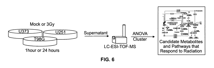
Figure 6 is a schematic diagram of the experimental design used to measure
the metabolic response of glioma cell lines to ionizing radiation. Three
different cell
lines, U373, U251, and T98G, were treated with 3Gy of ionizing radiation or
mock
treatment at two different time points. The cell supernatant was harvested and
examined for small molecule metabolites.
Figures 7A through 7H are chromatograms from 3Gy-treated and untreated
U373, U251, and T98G cell lines and media only controls. An overlay of
7
CA 02675997 2009-07-17
WO 2008/097491 PCT/US2008/001417
chromatograms from all experimental groups demonstrated high reproducibility
of
LC-ESI-TOF-MS.
Figure 8 is a Venn diagram of mass features exclusive to glioma cell lines
(absent in media). The box represents features that are common to glioma cell
lines
and the media. Eighty three features were detected at least once in each cell
line in the
absence of the media while 1428 features were detected in each cell line and
each
media sample at both time points.
Figures 9A through 9F are plots of normalized data of annotated statistically
significant molecules from Table 2 showing differences between treatment,
control,
and media. The bars represent standard error of the mean and n is the number
of
features measured per factor. Figure 9A: butyrylcamitine, 24 hours post IR;
Figure
9B: L-Threonine, 1 hour post IR; Figure 9C: Glucosylgalactosyl hydroxylysine,
24
hours post IR; Figure 9D: 1-(9Z,12Z-octadecadienoyl)-rac-glycerol, 24 hours
post
IR; Figure 9E: 7a, 12a-Dihydroxy-3-oxo-4-cholenic acid, 24 hours post IR;
Figure
9F: 25:0 N-acyl taurine, 24 hours post IR.
Figures l0A through 10C are principal component analysis (PCA) loading
plots that display the separation of samples into groups corresponding to cell
culture
supernatant and media by cell line. The normalized data from masses present in
each
cell line and condition were used as the input matrix. Figure l0A is a plot of
PCA
analysis performed on all secreted molecules. Figure lOB is a plot of data
from the 1
hour time point. Figure 10C is a plot of data from the 24 hour time point.
Open
squares correspond to untreated U251 cells, solid squares correspond to 3Gy
treated
U251 cells. Open triangles correspond to untreated T98G cells, solid triangles
correspond to 3Gy treated T98G cells. Open circles correspond to untreated
U373
cells, solid circles correspond to 3Gy treated U373 cells, open stars
represent
untreated media, and closed stars represent 3Gy treated media.
Figures 11 A and 11 B are depictions of hierarchal clustering of the fold
change
differences between irradiated and untreated cell culture supematant by cell
line and
time. Figure 11 A displays large differences between cell lines and Figure 11
B
displays hierarchical clustering of the fold changes between irradiated and
untreated
cell using cell lines as replicates. Refer to the figure legend for positive
fold changes
and negative fold changes. Missing data is grey.
8
CA 02675997 2009-07-17
WO 2008/097491 PCT/US2008/001417
Figures 12A and 12B are color depictions of the hierarchal clustering
represented in Figures l1A and I1B. Positive fold changes are red, negative
fold
changes are green, and missing data is grey.
Figures 13A through 13C are Venn Diagrams of secreted mass features having
a 2 fold or greater response to IR at 1 hour after treatment (Figure 13A), 24
hours
after treatment (Figure 13B), and both time points combined (Figure 13C).
These
diagrams show the number of secreted features with a common response to IR
within
and between cell lines.
Detailed Description of Preferred Embodiments
The present invention is more particularly described below and particularly in
the Examples set forth herein that are intended as illustrative only since
numerous
modifications and variations therein will be apparent to those skilled in the
art.
As used in the description herein and throughout the claims that follow, the
meaning of "a", "an", and "the" includes plural reference unless the context
clearly
dictates otherwise. The terms used in the specification generally have their
ordinary
meanings in the art, within the context of the invention, and in the specific
context
where each term is used. Some terms have been more specifically defined below
to
provide additional guidance to the practioner regarding the description of the
invention.
This invention provides reagents and methods for determining the cellular
and/or biochemical effects of ionizing radiation. The term "ionizing
radiation" as
used herein is intended to encompass high-energy radiation and electromagnetic
radiation and includes but is not limited to radiotherapy, x-ray therapy,
irradiation,
exposure to gamma rays, protons, alpha-particle or beta-particle irradiation,
fast
neutrons, and ultraviolet. In a preferred embodiment, the result of ionizing
radiation
administration on cell populations is determined by metabolomics (see
Metabolomics,
Methods & Protocols, (Wolfram Weckwerth ed., Humana Press 2007).
The term "cellular metabolite" or the plural form, "cellular metabolites," as
used herein refers to any small molecule or mass feature secreted by a cell.
In general
the size of said metabolites is in the range of about 55 to about 3000
Daltons. A
cellular metabolite may include but is not limited to the following: sugars,
organic
acids, amino acids, fatty acids, and/or hormones. In a preferred embodiment,
the
9
CA 02675997 2009-07-17
WO 2008/097491 PCT/US2008/001417
cellular metabolite is secreted from cancer cells, particularly glioma cells
or
melanoma cells.
The phrase "identifying cellular metabolites that are differentially produced"
as used herein includes but is not limited to comparisons of cells exposed to
ionizing
radiation to untreated (control) cells. Detection or measurement of variations
in small
molecule populations or mass features secreted by a cell, between treated and
untreated cells is included in this definition. In a preferred embodiment,
alterations in
cells or cell activity is measured by determining a profile of changes in
small
molecules in a treated versus untreated cells. Also included are comparisons
between
cells treated with different amounts, types or intensities of IR.
Alterations in small molecules such as sugars, organic acids, amino acids,
fatty
acids, and low molecular weight compounds are measured and used to assess the
effects of ionizing radiation on biochemical pathways. The screened small
molecules
can be involved in a wide range of biological activities including, but not
limited to
inflammation, anti-inflammation, vasodilation, neuroprotection, fatty acid
metabolism, products of collagen matrix degradation, oxidative stress,
antioxidant
activity, DNA replication and cell cycle control, methylation, biosynthesis of
nucleotides, carbohydrates, amino acids and lipids, among others. Small
molecule
metabolites are precursors, intermediates and/or end products of biochemical
reactions in vivo. Alterations in specific subsets of molecules can correspond
to a
particular biochemical pathway and thus reveal the biochemical effects of
ionizing
radiation. In a particularly preferred embodiment, metabolomics is used to
examine
the effects of IR on cancer cells.
Glioma cells are generally derived from glial cell tumors and in particular
brain tumors. However, gliomas may develop in the spinal cord or any other
part of
the central nervous system. In a preferred embodiment, the methods described
herein
specify "glioma," but methods are not to be limited solely to glioma tumors.
In
additional embodiments, disclosed methods include "glioblastoma multiforme"
(GBM) brain tumors, the most common type of brain tumor, as well as non-CNS
tumors including melanomas as one example.
In preferred embodiments the methods of the present invention are used to
assess differential cellular metabolite content and production from malignant
or
tumorigenic tissue. The term "tumor" or "malignant" includes cancerous tissue
at any
of the conventional four cancer stages (I-IV) as well as precancerous tissue.
In
CA 02675997 2009-07-17
WO 2008/097491 PCT/US2008/001417
alternative embodiments, the methods of the invention may examine precancerous
tissue. The term "precancerous" includes a stage of abnormal tissue growth
that is
likely or predisposed to develop into a malignant tumor.
The term "physical separation method" as used herein refers to any method
known to those with skill in the art sufficient to produce a profile of
changes and
differences in small molecules produced by cells, including tumor cells,
exposed to
ionizing radiation according to the methods of this invention. In a preferred
embodiment, physical separation methods permit detection of small molecules
including but not limited to sugars, organic acids, amino acids, fatty acids
and low
molecular weight compounds. Advantageous methods for separation comprise
chromatography, most preferably liquid chromatography (LC), and identification
methods comprise mass spectrometry techniques. In particular embodiments, this
analysis is performed by liquid chromatography/electrospray ionization mass
spectrometry (LC-ESI-TOF-MS), however it will be understood that small
molecules
as set forth herein can be detected using alternative spectrometry methods or
other
methods known in the art. Similar analyses have been applied to other
biological
systems in the art (Want et al, 2005, Chem Bio Chem. 6:1941-51), providing
biomarkers of disease or toxic responses that can be detected in biological
fluids
(Sabatine et al, 2005, Circulation 112:3868-875).
The term "biomarker" as used herein refers, inter alia to small molecules that
exhibit significant alterations between treated and untreated controls,
particularly with
regard to IR treatment. In preferred embodiments, biomarkers are identified as
set
forth above, by methods including LC-ESI-TOF-MS.
In preferred embodiments, the following small molecules are provided herein,
taken alone or in any informative combination, as biomarkers of cancer cell
response
to ionizing radiation: phenylacetate, phenylacetylglycine, 2-phenylacetamide,
alpha-
N-phenylacetyl-L-glutamine, phenylacetic acid and other metabolites in the
phenylalanine pathway, salsolinol, serotonin, butyrylcarnitine, L-threonine,
glucosylgalactosyl hydroxylysine, 1-(9Z,12Z-octadecadienoyl)-rac-glycerol, 7a-
12a-
dihydroxy-3-oxo-4-cholenic acid, or 25:0 N-acyl taurine.
The measurement of these biomarkers in patient blood, plasma, sera, lymph,
saliva, urine, or other patient specimen can provide a diagnostic or
prognostic
assessment of a patient's response to IR.
11
CA 02675997 2009-07-17
WO 2008/097491 PCT/US2008/001417
The term "biomarker profile" as used herein refers to a plurality of
biomarkers
identified by the inventive methods. Biomarker profiles according to the
invention
can provide a molecular "fingerprint" of the effects of ionizing radiation and
identify
small molecules significantly altered following ionizing radiation exposure.
In
preferred embodiments, biomarker profiles can be used to diagnose radiation
exposure or cellular response to radiation treatment.
In a further embodiment, the diagnosis of radiation exposure is not limited to
medical exposure, and may further include, but is not limited to the following
examples: accidental radiation exposure, war-related, or bioterror radiation
exposure.
The phrase "outside of medical treatment" includes the above-mentioned non-
limiting
examples.
A "biological sample" includes but is not limited to cells cultured in vitro,
a
patient sample, or biopsied cells dispersed and cultured in vitro. A "patient"
may be a
human or animal. A "patient sample" includes but is not limited to blood,
plasma,
serum, lymph, urine, cerebrospinal fluid, saliva or any other biofluid or
waste.
Examples
The Examples which follow are illustrative of specific embodiments of the
invention, and various uses thereof. They set forth for explanatory purposes
only, and
are not to be taken as limiting the invention.
All references cited herein are incorporated by reference. U.S. Publication
No. 20070248947A1 of October 25, 2007 and PCT Publication No. WO 2007/120699
of October 25, 2007 are explicitly herein incorporated by reference.
Example 1
Metabolomic Analysis of U373 Glioma Cells Treated with Ionizing Radiation
U373 glioma cells were exposed to a conventional dose of ionizing radiation
to demonstrate that metabolomics was useful for examining cellular response to
IR
and to identify biomarkers for response. The treated cells were analyzed as
set forth
below to determine changes in a total dynamic set of small molecules present
in cells
according to health and disease or insult states. Small molecules such as
sugars,
organic acids, amino acids, fatty acids and signaling low molecular weight
compounds were understood to participate in and reveal functional mechanisms
of
cellular response to pathological or radiation insult. These analyses were
also used to
12
CA 02675997 2009-07-17
WO 2008/097491 PCT/US2008/001417
identify active pathways following molecular changes implicated by other
methods
including for example transcriptomics and proteomics.
U373 glioma cells and uncultured media (negative control for media
background) were either treated with 3 Gy of gamma radiation or left
untreated. This
dose of IR represents the standard daily dose delivered in the treatment of
glioblastoma multiforme (GBM) during conventional fractionated treatment. The
media were sampled at three different time points following radiation
exposure: 1
(one) hour, 24 hours, and 48 hours. Medium collected from radiation exposed
cells
and control ("no-treatment") cells was subjected to liquid chromatography and
electrospray ionization mass spectrometry (LC-ESI-TOF-MS) to assess changes
and
differences in the metabolome produced by the cells in the presence and
absence of
ionizing radiation exposure. Samples were measured in triplicate (technical
replicates)
by LC-ESI-TOF-MS. Analysis of the standard deviation of the retention times
revealed that the majority of masses were comparable across LC-ESI-TOF-MS runs
with the majority of replicate measurements detected within 20 seconds of each
other.
This finding demonstrates the reproducibility of the detection method used.
Each sample had three replicates injected into a 2.1 x 200mm HPLC C18
column run on a 120 minute gradient from 5% acetonitrile, 95% water, 0.1%
formic
acid to 100% acetonitrile, 0.1% formic acid at a flow rate of 40 L/min. The
flow
through was introduced into an Agilent 1100 series LC-ESI-TOF-MS. Data was
collected from 0-1500 m/z range throughout the run. The raw data was loaded
into
the Analyst QS program (Agilent) to visualize retention time and mass features
prior
to data analysis. Mass Hunter MF (Agilent) software was used to deconvolute
the
data and determine the abundance of each mass. Masses within the range of 80-
1500
m/z, a charge of +1, and at least 2 ions were included in this analysis. A
mass was
considered to be the same across LC-ESI-TOF-MS runs using a simple binning
algorithm based on mass and retention time. Bins were created when masses
differed
by 10 ppm or if the same mass had a retention time difference greater than one
minute. Significance tests were determined by performing ANOVAs on the log
base 2
transformed abundance values. A complete randomized design was used with the
following formula:
{ Log2(abundancelb) = treatment, + glioma lineb + errorlb }
on each bin to determine significance. Imputation was not performed and
missing
data was omitted from the data analysis affecting the degrees of freedom for
each test
13
CA 02675997 2009-07-17
WO 2008/097491 PCT/US2008/001417
of significance. Clustering of the fold changes was performed using Cluster
3.0
software (De Hoon et al., 2004, Bioinformatics 20(Suppl. 1): i 101-i 108).
Principal
component analysis was performed using the open source statistical package R
and
the pcaMethods library. Significant differences in secreted metabolites were
detected
between irradiated and untreated glioma cell lines. Statistically significant
differential
secreted metabolites were determined using an ANOVA model. The distribution of
abundances appears to follow a normal distribution. Molecules with a p-value
less
than 0.05 and more than 3 degrees of freedom were considered significant when
comparing individual cell lines.
The plurality of small molecules identified using these methods was then
annotated by comparison with exact neutral masses of chemicals catalogued in
public
databases, e.g., METLIN Metabolite Database, Human Metabolome Database
(HMDB), Kyoto Encyclopedia of Genes and Genomes (KEGG), and the Biological
Magnetic Resonance Data Bank (BMRB). Mass spectrometry analysis also included
predicted chemical structures of small molecules based upon exact mass,
although
currently-available public databases do not in every instance include matching
small
molecules due to the lack of complete databases with the full spectrum of
human
metabolites.
A total of 161,923 mass signatures were detected by LC-ESI-TOF-MS
representing 48,608 unique neutral masses. Standard adducts from sodium and
potassium were removed from the spectra, which was also subject to
deisotoping.
This large number of neutral masses were measured because the data contained
signals that were measured one or two times across the entire experiment.
Masses
measured 2 or fewer times were considered to be spurious and removed from the
data
set. The final data set used for analysis contained 13,041 masses (-27% of the
unique
masses, Figure 1 and Figure 2). 3,356 (26%) of these masses corresponded to
small
molecules detected only in glioma samples. 471 of these masses were present
only in
the irradiated cells and 202 masses were measured only in the untreated glioma
samples.
Example 2
Phenylalanine and other Metabolites:
Biomarkers of Ionizing Radiation in U373 Glioma Cells
14
CA 02675997 2009-07-17
WO 2008/097491 PCT/US2008/001417
The results of the biomarker identification experiments disclosed in Example 1
were analyzed to identify particular metabolites and metabolic pathways
showing
differential activity in irradiated and control (nonirradiated) samples. One
biochemical pathway, the phenylalanine pathway, was particularly significant
at one
hour after irradiation. Several metabolites present in phenylalanine
metabolism had
significant changes after IR. These changes are shown in Figure 3 and Figure
4,
which illustrates the fold-change differences detected from metabolites of the
phenylalanine biochemical pathway. Refer to the legend for designations
representing positive and negative fold changes. Missing data is solid white.
A
schematic diagram of the phenylalanine metabolic pathway is shown in Figure 5.
Open arrows mark reactions leading to a metabolite with a statistically
significant
difference at one time point, dots indicate a metabolite measured in this
experiment,
striped boxes represent a metabolite that was not measured in this experiment.
One
putative metabolite in the phenylalanine metabolism pathway, phenylacetate,
was 3.5
fold more prevalent 1 hour after ionizing radiation (p= 0.002) than in non-
irradiated
samples.
Phenylacetate (PA) is a naturally occurring metabolite present in the
phenylalanine metabolic pathway that is typically detected in serum. (see
Figure 5).
Previous research has demonstrated that PA can inhibit the growth of tumor
cells in
vitro and in vivo (Samid et al., 1994, Cancer Res. 54:891-5). It has been
suggested
that PA may actually potentiate the response of tumor cells to IR (Miller et
al., 1997,
Int J Radiat Biol. 72:211-8.). Further investigations found that the amount of
PA
required to affect tumor growth is cell line dependent and that brain tumors
are more
sensitive to its effects than other tumor lines, but these results also called
into question
radiopotentiation of PA (Ozawa et al., 1999, Cancer Lett. 142:139-46). A phase
II
clinical study of PA did not find a significant response in patients with GBM
(Chang
et al., 1999, J Clin Oncol. 17:984-90). Interestingly, metabolites in the
phenylalanine
pathway feed into the production of DOPAchrome that leads to the production of
DHICA which is known to increase radioresistance in skin cancer (Figure 5).
The discovery of this pathway using unbiased methods demonstrates the
power of metabolomics to identify metabolic pathways that respond to IR. Other
metabolites in this pathway, which are included as biomarkers in this
invention and
were detected following exposure of two additional glioma cell lines (T98G and
U251) to 3Gy of ionizing radiation are: phenylpyruvate, phenylacetylglycine, 1-
CA 02675997 2009-07-17
WO 2008/097491 PCT/US2008/001417
phenylacetamide, alpha-N-phenylacetyl-L-glutamine and phenylacetic acid, which
where all significantly upregulated in response to IR. In addition, enol-
phenylpyruvate, phenylacetaldehyde, L-adrenaline, L-noradrenaline, and 3,4-
dihydroxymandelate were altered following IR. Altogether, these findings
converge
towards two main metabolic pathways: phenylalanine and tyrosine, and indicate
that
changes in these metabolites comprise biomarkers for IR.
Other molecules that can serve as biomarkers of IR in cancer are: salsolinol
(60% decrease at 24 hours, p < 0.000 1) and serotonin (7-fold increase at 1
hours, p <
0.0001; 60% decrease at 24 hours, p < 0.018; 2.5 increase at 48 hours, p =
0.005).
Interestingly salsolinol, a derivate of dopamine, is a neurotoxin that induces
apoptosis
in dopaminergic neurons (Mravec, 2006, Physiol Res. 55:353-64). Salsolinol is
significantly decreased at 24 hours after irradiation. Serotonin accumulation
was
significantly increased at 1 hour and 48 hours, but decreased at 24 hours
after
irradiation. Serotonin has been shown to cause an increase in IL-6 release in
glioma
cell lines (Lieb et al., 2005, JNeurochem. 93:549-59).
In addition, the following metabolites were also altered at statistically
significantly levels in response to IR: 2,7-Anhydro-alpha-N-acetylneuraminic
acid or
2-Deoxy-2,3-dehydro-N-acetylneuraminic acid, which was 2.5-fold upregulated at
48
hours, p=0.023; and 10-fold upregulated at 1 hour, p=0.008; N-
Acetylneuraminate
which was 1.4 fold downregulated at 48 hours, p=0.005; N-Acetyl-O-
acetylneuraminate, increased 1.2-fold at 48 hours, p=.016; Indoxyl sulfate or
4-
Phospho-L-aspartate, 3.4-fold downregulated at 24 hours, p=0.03; 5.9-fold
downregulated at 1 hour, p=0.007; N-Acetyl-L-histidine 1.28-fold downregulated
at 1
hour, p=0.003; 1.4-fold upregulated at 48 hours, p=0.0001; Isopentenyladenine
or L-
Acetylcarnitine up 4.8-fold at 1 hour, p=0.01; increased 2.9-fold at 48 hours,
p=0.0068; and reticuline, which was upregulated 4.1-fold at 1 hour, p=0.007.
Overall,
these results reflect that in vitro metabolomics of glioma cells is a robust
alternative
for the detection of small molecules, which can serve as translational
biomarkers of
ionizing radiation response.
Example 3
Metabolomic Analysis of Multiple Glioma Cell Lines Exposed to Ionizing
Radiation
16
CA 02675997 2009-07-17
WO 2008/097491 PCT/US2008/001417
Secreted or excreted small molecule metabolites from malignant glioma cell
lines in response to IR were evaluated using metabolomics. Glioma cell lines
were
treated with 3 Gy of IR and response was analyzed by metabolite profiling of
secreted
small molecules using liquid chromatography electrospray ionization time of
flight
mass spectroscopy (LC-ESI-TOF-MS). Statistically significant differences in
the
abundance of putative secreted metabolites were detected between irradiated
and
untreated cell lines.
U373, T98G, and U251 GBM cell lines were exposed to a conventional dose
(3 Gy) of ionizing radiation to identify biomarkers for cellular response to
IR. The
treated cells were analyzed as set forth below to determine changes in a total
dynamic
set of small molecules present in cells according to health and disease or
insult states.
These experiments were performed generally as set forth in Example 1, however
in
the present Example, three glioblastoma cell lines were examined in an effort
to
provide a more robust analysis and to identify common metabolites among glioma
cell lines. Because metabolic changes are inherent to cancer pathogenesis
(Griffin &
Shockcor, 2004, Nat Rev Cancer 4: 551-61; Jensen, 2006, Neurosurg Focus 20:
E24;
Brown & Wilson, 2004, Nat Rev Cancer 4: 437-47; Yetkin et al., 2002,
Neuroimaging
Clin N Am 12: 537-52) they can also be directly involved in tumor response to
ionizing radiation. This study examined whether there were common metabolic
changes to different glioma cell lines in response to IR.
The GBM cells lines U373 (ATCC# HTB 17), T98G (ATCC #CRL-1690),
and U251 (provided by Paul Harari, University of Wisconsin-Madison) were
cultured
under standard conditions to 50-70% confluence and then exposed to 3 Gy of
ionizing
radiation (IR) or placed in the irradiator but not exposed to the source (mock
treatment) (Figure 6). This dose of IR is comparable to the daily dose
delivered in the
treatment of GBM during fractionated radiotherapy. The cell cultures were
sampled at
one and 24 hours following IR. Media without cells were treated in the same
manner
as cell cultures and used as a reference to detect cell specific metabolites
except for
line U373 where no irradiated media was collected. Only one medium sample was
used as the untreated control for U251 and T98G. These uncultured, untreated
medium samples were duplicated in the data analysis and represented both 1 and
24
hour time points for the untreated media measurements.
Cell culture media supernatant from the irradiated and untreated glioma cell
lines was collected at one and 24 hours post-IR and stored at -80 C. The
samples
17
CA 02675997 2009-07-17
WO 2008/097491 PCT/US2008/001417
were simultaneously thawed and 125 L was processed for liquid chromatography
using Millipore 3kDa Centricon regenerated cellulose columns (Millipore) to
remove
proteins and large molecular weight biomolecules. The flowthrough was retained
for
analysis as it contains small molecules free of high molecular weight
compounds. The
flowthrough was lyophilized and suspended in 50 L 0.1 % formic acid.
5 L of each sample was injected in triplicate into a 2.1 x 30mm Zorbax C18-
SB column run on a 30 minute gradient from 5% acetonitrile, 95% water, 0.1%
formic acid to 100% acetonitrile, 0.1% formic acid at a flow rate of 200
L/min. The
flowthrough was introduced into an Agilent 1100 series LC-ESI-TOF-MS. Data
were
collected from 50-1500 m/z range throughout the run. The settings for the ion
source
were: gas temperature 350C, drying gas 9.5 L/min, nebulizer 30 psig, capillary
4000
V. The settings for the TOF were: fragmentor 185 V, Skimmer 60 V, OCT RF 250
V.
The chromatograms were inspected after each LC-MS run and any samples with
abnormal chromatography were repeated. Data were extracted from the
chromatographs using all information from 0-27 minutes (Figure 7).
The Mass Hunter MFE version 44 software (Agilent) was used to deconvolute
the data, which consists of removing isotopes and adducts, and establishing
the
abundance of each mass feature. The abundance was calculated as the sum of the
isotopic and adducts peaks folded into a single mass feature. Masses measured
within
the range of 50-1500 m/z, m/z charge of +1, a minimum abundance greater than
0.001 %, a signal to noise value greater than or equal to 5. After data
deconvolution
mass features with at least two ions and an abundance value greater than 0.05
quantile
were included in the data set used for binning. A set of mass features was
considered
to be the same across LC-ESI-MS-TOF runs using a simple binning algorithm
based
on mass and retention time. Mass features under 175 Da were binned by 0.00001
x
mass, while those from 176Da-300Da were binned by 0.000007 x mass and 0.000005
x mass when over 300Da with a retention time difference of less than seven
seconds.
The binning process was used to create unique compound identities (cpdID)
representing a single small molecule.
These binned data were separated into two distinct sets serving different
purposes. One set was used for qualitative analysis and the other data set was
used
for statistical analysis. The data set used for qualitative analysis contained
all mass
bins that contained at least 3 masses in order to remove compound IDs that may
be
due to experimental artifacts such as rare fragments or spurious integration
of
18
CA 02675997 2009-07-17
WO 2008/097491 PCT/US2008/001417
background peaks. The second data set was used for statistical analysis and
contained
mass bins that were detected in each cell line and treatment at a given time
point. The
mass feature bins (cpdIDs) used for statistical analysis were also filtered
against the
media and cpdIDs with an average abundance less than or equal to the media
were
removed from interpretation because they may not represent secreted/excreted
metabolites.
Prior to statistical analysis the data was divided into two subsets and
statistical
analysis was performed on the putative secreted mass feature subset.
Significance
tests were determined by performing ANOVAs on the log base 2 transformed row
and
column median normalized abundance values. A randomized complete block design
was used with the following formula Log2(abundancetb) = treatment, + glioma
lineb +
errortb on each mass feature bin to determine significance. This model was
only used
on mass features that were measured in each cell line and treatment.
Imputation was
not performed and missing data were omitted from the data analysis affecting
the
degrees of freedom for each test of significance. Principal component analysis
(PCA)
was performed using the open source statistical package R and the pcaMethods
library.
The average neutral exact mass of each mass feature bin was queried against
the public searchable databases METLIN (http://metlin.scripps.edu), The Human
Metabolome Database (http://www.hmdb.ca), Kyoto Encyclopedia of Genes and
Genomes (www.genome.jp/kegg/), and the Biological Magnetic Resonance Bank
(http://www.bmrb.wisc.edu/metabolomics/) for candidate identities. Measured
mass
features were considered to match a small molecule present in the databases if
their
exact masses were within 10 parts per million of the annotated database
molecule
(0.00001 x mass).
The small molecules altered in response to IR were a diverse group of
metabolites involved in fatty acid metabolism, products of collagen matrix
degradation, and other cellular processes, as set forth in greater detail in
Example 4
below. As radiotherapy remains the primary treatment modality for malignant
glioma
and prognosis remains poor, defining the metabolic or biochemical response of
gliomas to IR provides insight into cellular processes contributing towards
their
intrinsic radiation resistance. In addition, these molecules could also serve
as
candidate biomarkers to predict the response and/or resistance of IR in
malignant
gliomas.
19
CA 02675997 2009-07-17
WO 2008/097491 PCT/US2008/001417
Example 4
Biomarkers of Ionizing Radiation in Glioma Cell Lines
The results of the metabolite identification experiments disclosed in Example
3 were used to identify particular metabolites and metabolic pathways showing
differential activity in irradiated and control (nonirradiated) samples. The
data set
used for qualitative analysis contained all mass bins with at least three
masses in order
to remove compound IDs that may be due to experimental artifacts such as rare
fragments or spurious integration of background peaks. This data set contained
13,308
mass feature bins and was used for metabolite annotation and analysis of
compounds
that were present only in one condition or specific cell line. The second data
set,
subject to statistical analysis, contained mass bins that were detected in
each cell line
and treatment and detected above the media background abundance at a given
time
point. This second data set was used to determine statistical significance of
differentially accumulated small molecules in response to IR. Using these
criteria,
statistical analysis was performed on a total of 1339 mass feature bins.
Analysis of the "metabolic profiles" of each glioma cell line was performed
using the qualitative data set (Figure 8). The data were examined for
metabolites or
mass features not found in the medium but unique to one cell line or common
metabolites among cell lines. 206 metabolites or mass features were
exclusively
detected in the supernatant of cell cultures and absent in cell culture media.
U373
(18%) had the most unique secreted metabolites or mass features followed by
U251
(10%) and T98G (5%). 40% of uniquely secreted or excreted metabolites or mass
features were common to all three cell lines (Figure 8), thus when the effect
of the
media is removed from the analysis, it is likely that they share a similar
signature or
complement of secreted metabolites and mass features.
Ionizing radiation induces statistically significant changes to small molecule
metabolites in glioblastoma cell lines. Statistically significant differences
in the
abundance of secreted small molecule metabolites were detected between
irradiated
and untreated glioma cell lines. These differences were determined by an ANOVA
model that used the different cell lines as biological replicates. In the
ANOVA
model, a p-value <0.025 was used to determine significance. Among the secreted
features, 125 small molecules (9%) were significantly different; with 4 (0.2%)
common to both time points and 55 (4.1%) and 73 (5.5%) small molecules
detected at
CA 02675997 2009-07-17
WO 2008/097491 PCT/US2008/001417
one and 24 hour time points, respectively. The majority of differentially-
secreted
mass features (77%) did not have putative metabolite annotations in public
small
molecule metabolite databases. (Table 1) This finding is a reflection of the
relatively
early status of metabolomics in comparison to other "omics" and it also
highlights a
heightened demand for population of inetabolomic databases.
Table 1: Statistically significant differentially secreted mass features and
putative
metabolites from three GBM cell lines after 3GY IR treatment.
Monoisotopic Retention . Fold P Value Putative Human T~me ~,~Ip Mass Titn4
Change alphl~ 0.0?~ Metabolite
577 92.0595 1.433283 lh 1.54 0.0016 no hit
...... ......... ......_ ............ 687 97.0894 4.234614 lh -1.99 0.0078 no
hit
1181 113.084 4.814683 lh -2.74 0.0093 no hit
....... ......... ........ ........ ........
........................................... .. .. .
1323 115.0628 10.21004 24h 2.82 0.0013 Proline
.......................................... ................
............................................................
........................
...................................:....................................
....................................................,..........................
... .... .............
.... .................................................. ..........
1852 129.1519 4.810348 24h 1.95 0.0056 no hit
.... . ........ .... .. . . . .. ........
2293 143.0943 3.493017 1h -1.70 0.0073 Proline betaine
................ ....................... _.....__.._ ...........
...._.......................... .........................
_...............;
148.049 1.436515 24h 1.25 0.0185 Aspartylhydroxa
mate (oxidation
?515........... . product)
.................... .................................. ..._
.................. :............................ ._.........................
.................. ............... .................... ...................
...........,........................................................ .
_......:.......................................................................
.............,.
2628 150.9764 9.751492 24h 2.81 0.0228 no hit
.........._...................:................... .......... .. ..... .......
........... .......... ................ .......:.....................
2629 150.9764
10.21161 24h 1.43 0.0100 no hit
:.... .
..................................................__...................._.;....
._.._...............__............_.........;......................__..........
...............
_ ............. .....:..._ __ _. ...
3129 162.0666 23.84309 24h -1.94 0.0170 no hit
3143 162.1041 14.83821 lh 1.98 0.0064 no hit
. ........ ,
3788 177.9856 0.708176 lh 1.46 0.0000 no hit
....... ............... ........ ........... ..........
3856 179.0897 1.623656 24h -1.66 0.0005 no hit
................ ,_._............................. ..........................
................. ......... ...................................... _
.,....................................
............................................... .........................
...._ ......................... ..................... 3955 1 181.943 2.840092
24h -2.39 0.0137 no hit
....
5611 213 1289 2.008389 lh -1.67 0.0049 no hit
5612 213 1334 2.55298 lh -1.84 0.0038 no hit
.... . .
5634 213.2453 17.33491 lh 3.09 0.0078 no hit
.............. ....................
.............................................................................
:._........................... .... ............. _._
......................... .. ........................................ ...
_....,..........................................__............._...............
.............._....................._........_.........._. _.__........._:
6150 223.9242 1.246739 24h -5.24 0.0018 no hit
.......................................
......................................................_........................
........_.......__.........................._
_....................................._...__..................................:
........._........._........................_......................;........_..
_........................_.._......................................_;
6202 224.1441 16.86289 24h -2.63 0.0226 no hit
.............. _....._.................. ...... ..............
................._............... ..................
......;............................... ........... ...........
............................... ...........................
._
......_..........._............................................................
......_.....;............._._......._..........................................
....._.......;
6644 231.1458 8.961544 24h 2.40 0.0052 Butyrylcarnitine
240.0236 10.21143 24h 1.50 0.0011 Cysteine Dimer,
7094 Cystine
7499 246.1183 2.091837 lh 1.72 0.0001 no hit
............... :..........................................................
...................................
7880 252.1022 12.74882 lh 10.11 0.0141 no hit
............................... ._........... _........_.............. .....
__..... ................. ....... _. _..... _..........._....................
_......... ................... __.... ........ ...............................
__......... ......................... _......
.._..._................._......_..._;.................._.......................
........................__----
._....._
7949 253.1001 12.74493 1 h 6.05 0.0001 no hit
...............................................................................
.............. ............................ :....... ....................
............................. ................................ -..
..................................................... ................ ......
.
8124 256.0913 2.406877 lh -1.91 0.0045 no hit
I._ ..............__......_.....__ ...._.........._. .................
_.......... __.... _............ ........ ........... -_.......
_._............... _........ _...................... -- ........
_.._._........__............_._._...._........._...._.- ............._........
_.. ._............. _...._.................. _.... _.......
...._._........_.._
8367 259.9609 0.304571 lh -2.70 0.0153 no hit
9240 273.1037 12.77166 ( Ih -1.75 0.0001 L-Thyronine
;...._.__._...- ._...._ ......_..........__._...._......._..._.-_. .. _
.....:........ - ._..._. _.._.._.
, . . . . . ~......_.__....... _. .__.... __........ __...... _...... _ .
................. .._........ ......._....-...._......__....___.._.....
9257 273.1578 4.303222 lh -1.79 0.0055 no hit
............ ........._............._..._..................._................~
..... ........................:.........................._....._........
......._............... ..... ........ .... .......... ........ ........
......... _........ .............
9747 280.061 0.836327
........... ...._...... _ :...._..._.. - ....._.._._~__...__._._._..i..._.-
._._._ _...._._.. r .._ .................... __....
._._._..._................._._..
10254 287.0612 10.21119 24h 1.78 0.0058 ; no hit
.._ ...............................
......._...........__.._..._..._........... .........................
_...._..........._.........._...__.._.__.......:_......._...._..._............
....................._..._.__..._...... ........... ......_........ _
........._._.........._..._..
...
10255 287.0617 10.49431 24h 5.59 0.0005 no hit
10290 287.2087 12.84378 24h -9.89 0.0009 Octanoylcarnitine
_..... _...:......___...... ... ..... ........... _....__ .... __......
.................. ......._.........
._..........._...._............_......_..... _.............. _................
_._...........:.........
__............................._..._.............................._..._
10309 -287.5625 10.21216 24h 2.44 0.0003 no hit
......... -..... -_. .-_........ 21
CA 02675997 2009-07-17
WO 2008/097491 PCT/US2008/001417
Monoisotopic KetentionFold P Value PutatiVe Ilunian
Tinie
epd~ Mass Time Change a1pha=0.4?- Metaliolite
10428 289.1316 15.17537 lh -2.00 0 0068 no hit
10523 291.0557 4.950763 24h -2.28 0.0120 no hit
........................... ................
........................................................_
.....:................... .............................
.......:................................... ...............
.................................. _..:...................
......................................................
.;_...........................................
............................._.......
10635 292.1832 7.466134 24h 1.66 0.0057 no hit
.. .... .,..... .. ....... ...
.. ............... .........................................................
10670 293.1433 2.208604 Ih -7.43 0.0063 no hit
10818 295.0721 1.45813 24h -6.06 0.0243 no hit
........ .................. ..................... ........
..............................
..................................................................
.................................. .......... .........................
11509 303.2923 19.75929 Ih -2.44 0.0105 no hit
....
11784 307 1587 1.24507 lh ~ -1 87 0.0175 no hit
.............. .......... .................... ... ..
11802 307.2134 13.01753 lh -1.87 0.0226 no hit
... ......... ........ ....... .........._. ......... .
11840 308.1205 6.86587 Ih 2.51 0.0218 no hit
12571 316.089 13.22427 24h -1.79 0.0049 no hit
.... .... ....
13150 323 3191 21 06211 lh 1 68 0.0122 no hit
..... .. ....................
14619 340.2319 13.10785 lh -1.80 0.0174 no hit
15098 346.209 17.64374 lh -3.03 0.0173 no hit
........................................ ..............................
....................... ...............
.............:........................................................
............ ................... ....
.................................................... ........
.................... .............. .............................i...........
............................. ...........................................
15410 351.0855 0.824719 Ih -3.12 0.0017 no hit
15733 354.1423 9.888837 Ih -4.04 0.0036 no hitW
.......................... :............................... .....
................ ............... .................._..:.....................
......................... .............................. .................
................ ............................ ..........
........_:
15778 354.2522 14.55557 Ih -1.69 0.0231 no hit
............. ................ ....... ............. ........... ..
............... ........................ ............
:........................ ............ ........... ........................
................... ......
..................................._..................
MG(18:2(9Z,12Z
)/0:0/0:0); 1-
15795 354.2763 21.83661 lh 4.89 0.04327 (9Z,12Z-
octadecadienoyl)-
rac-glycerol
MG(18:2(9Z,12Z
)/0:0/0:0); 1-
15795 354.2763 21.83661 24h -2.81 0.0023 (9Z,12Z-
octadecadienoyl)-
rac-glycerol
363.2871 16.23451 24h -2.28 0.0045
16486 no hit
....... ... ....... ...... ...._..._ ....... ..:.... ....... ........
Tripeptide
containing one of
([ these
sequences:Leu
Lys Asp, Val Lys
Glu, Ile Lys Asp
in an unknown
17318 374.2195 13.86132 Ih -1.51 0.0453 order
...................................... ..................... ....... . ...
18083 385.2461 18.06023 Ih -1.88 0.0225 no hit
18083 , 385.2461 18.06023 24h -1.91 0.0003 no hit
.............. .._... ............ ........ ........
Tripeptide
386.1944 10.35101 Ih -2.43 0.0248 containing: Arg
Asp Pro in an
18148
unknown order
..................... .. ..........._........................................
_.........I..................... ....................... ............
..............
;..... . ...... . i................................................ ....i...
........ ................... ... . .. . . . .... . .. .....
.................................................
18483 390.4877
10.67065 24h 4.40 0.0115 no hit
18595 ........................
......._39218.........................!_..._0.955.848........_+..........24h...
..........: -5.42 0.0114 no hit
................................................:..............................
..............................................
...............................................................................
.......
18816 394.2685 21.83826 lh 5.68 0.0227 no hit
_ ..... _ ........ _ ....... _ ............ _.... ....... _...... _
Tripeptide
containing: Phe
399.2619 18.87273 lh 3.89 0.0000
Val His in an
19184 unknown order
19306 401.2056 6.75239 24h 1.57 0.0193 no hit
___.......... ...... ..... __........ _..... _...__......... __...... _
........ .......... _..._.... _.. - ...
..... ...........
........... ...... _
7a,12a-
404.2575 23.81864 24h -1.89 0.0105 Dihydroxy-3-
19549 '..... oxo
.................._:.......... _.........................._..........._...i...
.._....... .._.._.................. _.............. __.....:_...... ....
_........ ............. ................................ .._._............... -
4-cholenoi.c.........;
22
CA 02675997 2009-07-17
WO 2008/097491 PCT/US2008/001417
Monoisotopic Retention I-Fold P Value Putative Hunian
cpdlD Mass i"' Change alpha-0.025 Metabolite
acid,
.......................................... ......................... ..
.............................................:.................................
........................:.
.......... ................._....._.._.. ..................
..................................................
...:.............................. ............. ............... .....
19739 407.2665 15.93827 Ih 2.46 0.0228 no hit
........... .............. .............
...............................................................................
. ......................................................
...;.................................. .............
....................................:........ ............ ...
..................... ................................. ......................
20255 415.0869 10.21156 24h 1.97 0.0007 no hit
:.....
......................................... ............... ............
...................................... ............ .:............ .......
................. ................. ............................. _.....
...............................................................................
............................................ .. ......... .................
Tripeptide
containing: Arg
417.2369 19.18524 24h -1.49 0.0151
Asp Lys in an
20463 unknown order
...........................................
................................................ ............................
........................... .........
...............................................................................
...
20941 424.7667 8.99614 24h 7.17 0.0003 no hit
Tripeptide
429.2355 10.40645 24h 2.61 0.0171 containing: Lys
Pro Trp in an
21314 unknown order
.................. ...........................
................................................................
................................. .......................
21704 435.2464 15.06274 Ih -2.75 0.0236 no hit
.......................
.............................................................. ..............
.......... ...................................... :...._ ..................
... ......................................... .......:.................
.................................. ............. .......... :.......
............... ...................... ............... ..................
21890 438.3017 23.06154 24h -1.51 0.0000 no hit
21910 439.0537 10.21132 24h 1.45 0.0008 no hit
........ .........
22277 444.2485
23.81585 24h -1.66 0.0057 no hit
... ...................... ......................
..................... _.............. .......__.......445.2877
..............................
1.........................................:....._............._...._.__
..._................. ..
22337 10.36839 Ih ._1.. 0.0075 no hit
........... ................. ........... ............ .. ..
22986 456.0814 9.74697 24h 3.86 0.0176 no hit
............... ..................................... ....................
..................................... .................. ............
......_:......................................................
................................ ..................... .......................
................... ..............
22987 456.0812 10.21215 24h 1.85 0.0006 no hit
23135 457.7801 12.17712 24h -6.01 0.0147 no hit
.............. .....:..................... ..._
............................:..... .._.._..... ........... .. ..............
._................. ........ ...........
23536 464.1406 7.2835 24h 1.55 0.0209 no hit
................................ 11 ........ ...........
....................... ...................... .................
....:........................................ .................
:............... ..................... ................ ......................
......:............ ... .......................................
.................. :........................ ...
.................................
..................:
23641 465.3619 18.40586 24h -3.33 0.0176 no hit
. ..... _ ........
23792 468.0991 6.925413 24h 1.49 0.0122 no hit
............. .............. ......................
...................................... ...............
........................................... ..................
:...._.............. _............. .........................................
.........................................................................
............... :.............................. ..._..............
............................._
24267 475.2989 9.645676 24h -1.24 0.0195 no hit
24574 481.4236 20.29276 24h -2.10 0.0002 no hit
. . . . . . . . . . . . . . . _ .......... . . . .
................................. ........................ ............... . .
. , . ................
..................... . . . . .
..... ...................................... .....
................._................
....._
24629 482.3108 25.16125 lh 2.12 0.0098 no hit
......... . ........ .. .......... ...................
.................................. .. ........ .. .. .........
24778 484.3451 1.813552 24h 1.80 0.0190 no hit
......
......... ......... ....... ..
486.2106 5.151622 24h 1.92 0.0162 Glucosylgalactos
24879 yl hydroxylysme
........ .. ....... ......... ......... ...._
2-
pentacosanamido 489.3817 4.588952 24h -2.07 0.0231 ethanesulfonic
acid (25:0 N-acyl
25027 taurine)
....... ...... 2_.
489.3817 4.588952 24h -2.07 0.0231 pentacosanamido
ethanesulfonic
25027 acid
....................... ............ ...........................
..;.................... .................................:....._.........
._._..._....... .............................................
...
25079 490.2375 9.02987 lh -1.81 0.0150 no hit
25167 492.1358 1.919706 Ih 2.66 0.0012 no hit
...................................
.......................................................................
..._............ .................................... .......
................. _....................................................
25569 499.3142 19.18543 Ih -2.53 0.0000 no hit
... ............... ..................
25569 499.3142 19.18543 24h -1.62 0.0062 no hit
25682 501.8061 12.49877 Ih 2.89 0.0180 no hit
......................................... ........
..................................................._................
............... ........................................
.,............._....................
..............__................................ :............ ..... ....
............... ..................................
26162 511.3321 16.47521 24h -4.00 0.0171 I no hit
26247 513.3278 18.57832 1 h 2.76 0.0057 no hit
......................... _........._.._............
_................_._...... _........ ..._....... .......................
................~..............._.......................... .
...............................
;..........................................................
__....................._
26588 520.3278 4.470145 24h 1.80 0.0216 no hit
................................................_.._.
_.__.._..._......_......._.............._.........................---
....._......................_...............................'
............... ..._._......... .... .... ..... ........ _
36 521.3714 23.81138 24h -2.56 0.0033 no hit
95 522.4117 18.76669 Ih 3.22 0.0012 no hit
Z
_ ........._. ............ . . _. . . __.
............ .......... ..._......... ......... ._..._............. _..-.-
.............. _._....................... ........ ........._...... _........
_...............;..............................................................
.............t...._.......................... ........ .........
._............................
. 22.77881 lh 2.32 0.0205 no hit
..3498 _...
.. 526..
26850 ......... _ _..._...
..
..
.
.
_
_
._
.... ......... ._..._........ _.._......._.__....... _.._... .__..__..__.._.__
._. _..._. _. _ . _._. . .
__........._......._..._.~_...._..._.............._......_.
27405 .........................._539.4397.............._.._. 18.7687 I 1h 2.76
.... 0.0041 nohit _.._;
._ ......................_..... .................... __..._.... _...........
_................................ _..................
.
..........................................................................:....
.................................................... ....
27419 540.1813 12.73714 1h 4.87 0.0007 no hit
___........__---.--.--.._.........__..._.....___._.._.__. _._ _.___... .. __. -
__.....----~.___._ _ .._._..T.__..._.._._.__._._,.._.._._.._...__._.__._-
.....................---.-~
27544 543.3788 22.77802 lh 1.71 0.0151 no hit
27544 543.3788 22.77802 24h -2.20 0.0051 no hit
,._....._..----..._.._...... _...._.._..__..._._------.---._._ __._.__.__.
_.~.__
23
CA 02675997 2009-07-17
WO 2008/097491 PCT/US2008/001417
.~_
Monoisotopic Retention~ Fold P Value Putative Ilunian
Time
cpdID Mass Time Chanzfe alpha 0.025 Metabolite
28498 568.1867 5.121525 Ih 2.97 0.0014 no hit
........... ............................. .........................
........................................... ..._ ..................
_........................... ..........
24h............................................................
.................... ............................ .......................
...................... .....n......ri'..............
........__.
28763 576.099 11.11736 -3.63 0.0213 o t
................................ ...............
........................................ ...................
.....:....................... .................................
.................................... .........................
..................................................... .............
................................. ...................................
28883 580.1513 1.916698 lh 2.92 0.0018 no hit
..........................................
...............................................................................
...............................................................................
.......... .......................... ........... ...................
............... . . ...... .........................
: >..
29058 586.202 4.948875 24h -1.49 0.0073 no hit
29158 590.2699 9.553 24h 1.52 0.0033 no hit
.......................... ..........
...........................................
.............................................................
................................... ......................
~_ ................ ................ ........................................
.._;....................... ..................................
29158 , 590.2699 9.553 24h 1.52 0.0033 no hit
........................................ ................ .............
................................... ..................
........................ .................................
,................................... ....... ...... ......... ..............
.............. ................................. .......................
29278 594.4696 20 62718 lh 2.01 0.0185 no hit
...... . ........ ....... ._... ......... ..... ... ... . ... .. ..
30202 634.3572 8.636565 24h 2.27 0.0047 no hit
30504 650.1853 5.094588 lh 0.0095 no hit
30527 651.4044 10.92515 24h -1.35 0.0000 no hit
30740 661.709 6.925875 24h 1.80 0.0213 no hit
30841 668.1965 16.70747 24h -4.00 0.0157 no hit
31563 707.392 11.79947 24h -1.61 0.0066 no hit
.. ................. .................... . ......... ..... ....:
32741 788.8603 12.22976 24h 1.85 0.0009 no hit
32746 789.3619 12.23 24h 1.75 0.0028 no hit
......................................... ............
..........:
32842 799.8507 12.22659 24h 1.76 0.0183 no hit
... .... .... .. .....
32846 800.3533 12 22851 24h 1.90 0.0033 no hit
...
.. .... . ........................................ .
......................................................... ....................
....... .. ,
33093 821.0085 0.30345 24h -1.71 0.0216 no hit
........................... ........................ ........
............................ .... ......... ..........................
......................................... 33468 858.355 10.92296 lh 3.16
0.0206 no hit
33511 862.3945 14.442 24h 2.69 0.0000 no hit
...............................................................................
..................................
.............................................
..............._......._....._...............................................
........................ ........._._....._.............................
..;...._............. ...............................................
.._............;
33605 873.3855 14.43782 24h 2.47 0.0001 no hit
.... . .. ... .. .... ....
33609 873.8865 14.43922 24h ~ 2.73 0.0003 no hit
..... ... ..... ... ...... .... ................................. . .. ....
33767 889.3566 14.43997 24h 2.21 0.0005 no hit
....................... ............ .................................
.................................. :.................................
....................... ...........__...................... .............
...... .............. .............. . .................................
........................
....
4.638375 24h
921.0025
34038 -3.55 0.0086 no hit
34152-~ 932.2777 5.136448 Ih 2.83 0.0219 no hit
........
....... .........
34177 936.3558 9.471735 24h 1.78 0.0255 no hit
................ ...................................
..............................................
.................................................
.................................... ..........
34184 936.8567 9.474324 24h 1.68 0.0186 no hit
. ... .. ....
34303 958.9585 0.359262 24h -2.26
0.0156 no hit
.......................... .. ... . .. .. ..... . ......... ...
34543 1008.0709 0.319511 24h -2.00 0.0169 no hit
34640 1022.263 10.69856 lh 2.26 0.0030 no hit
A list of several differentially secreted metabolites with putative
annotations
that participate in human metabolism are shown in Table 2. These molecules
were
both upregulated and downregulated in response to IR of gliomas and are mapped
to
different biochemical pathways and biological processes (Figure 9).
24
CA 02675997 2009-07-17
WO 2008/097491 PCT/US2008/001417
Table 2: Annotated molecules with statistically significant fold changes
greater than
1.5 in magnitude secreted in response to IR.
~j
Monoisotopic Retention Fol
~1ass Time Cime pPutati\,e Metabolite Dalt0ns) Minutes) Change Value
~
273.104 12.77 lh -1.75 0.0001 L-Thyronine
........................ ..............
.............................................. ... ...... .....
486.211 5.15 24h 1.92 0.0162 Glucosylgalactosyl
hydroxylysine
287.209 12.84 24h -9.89 0.0009 Octanoylcarnitine
MG(18:2(9Z,12Z)/0:0/0:0),
354.276 21.84 24h -2.81 0.0023 1-(9Z,12Z-
octadecadienoyl)-rac-
glycerol
489.382 4.59 24h -2.07 0.0231 25:0 N-acyl taurine
231.146 8.96 24h 2.4 0.0052 Butyrylcarnitine
.....................................
404.257 23.819 24h -1.885 0.011 7a,12a-Dihydroxy-3-oxo-4-
cholenoic acid
Statistically significant changes in the secretion of 125 different small
molecule mass features in response to IR were also detected including
molecules that
indicate changes in fatty acid metabolism, degradation of collagen, and
altered thyroid
hormone metabolism. While the majority of current research focuses on DNA
damage
repair response to IR, the metabolite changes disclosed herein reveal some of
the
biological processes not related to DNA damage and repair that are
nevertheless
associated with GBM's response to IR.
The significant changes detected in these experiments include the following.
Significant changes to different medium-chain acylcarnitines (butyrylcarnitine
and
octanoylcamitine) were detected in response to IR. Acylcarnitines are
intermediates
from fatty acid oxidation and metabolites of camitine, suggesting that
increased fatty
acid oxidation is an early metabolic event following IR of gliomas.
Acylcarnitines are
usually synthesized on the outer membrane of the mitochondrion and
translocated
through the inner membrane where camitine is replaced with coenzyme A,
followed
by (3 oxidation of the fatty acid. After oxidation, medium chain
acylcarnitines such as
butyl and octonyl camitine exit the mitrochondrial membrane into the cytosol
(Vaz &
Wanders, 2002, Biochem J 361: 417-29). Acylcarnitines are also formed during
the
export of medium and long chain fatty acids from the peroxisome to the
mitochodrion
(Akobs & Wanders, 1995, Biochem Biophys Res Commun 213: 1035-41; Reilly et
al.,
CA 02675997 2009-07-17
WO 2008/097491 PCT/US2008/001417
2007, FASEB J 21: 99-107). Interestingly, certain acylcarnitines such as
propionylcarnitine, function as superoxide scavengers and antioxidants,
serving as
DNA damage resistance molecules (Vanella et al., 2000, Cell Biol Toxicol 16:
99-
104). Acetylcarnitine is also thought to scavenge free radicals and limit
reactive
oxygen species damage caused by irradiation (Mansour, 2006, Pharmacol Res 54:
165-71). Reduced concentration of acylcarnitines in brain tumors may reflect
its role
in maintaining membrane integrity upon insult by reactive oxygen species ROS
(Sandikci et al., 1999, Cancer Biochem Biophys 17: 49-57). The increased
abundance
of the four-carbon butyrylcamitine and the decreased abundance of the eight-
carbon
octanoylcarnitine detected in this study, may indicate activation of metabolic
fatty
acid oxidation in response to IR. Since synthesis of butyrylcarnitine and
octanoylcarnitine are regulated by different enzymes (carnitine
acetyltransferase
(CRAT) and camitine 0-octanoyltransferase (CROT); van der Leij et al., 2000,
Mol
Genet Metab 71: 139-53; Jogl et al., 2004, Ann N YAcad Sci 1033: 17-29),
changes in
gene expression or enzyme activity may also participate in the mechanistic
dysregulation of these metabolites following IR. Upregulation of fatty acid
oxidation,
as suggested here, could also protect tumor cells against ROS injury, since
shorter
chain acylcarnitines have antioxidant effects.
25:0 N-acyl taurine is another metabolite significantly altered in response to
IR. The acyl-taurines are a relatively new class of metabolites, first
described in 2004
(Saghatelian et al., 2004, Biochemistry 43: 14332-9). 25:0 N-acyl taurine is a
fatty
acid amide hydrolase (FAAH)-regulated fatty acid present in mammalian central
nervous system (CNS); specifically, it is a taurine-conjugated analogue of a
25-carbon
fatty acid. The peroxisomal enzyme ACNATI is involved in the conjugation of
taurine to fatty acids and acyltaurines may act as signaling molecules or as
part of the
fatty acid secretion system present in cells (Reilly et al., 2007, FASEB J 21:
99-107).
These fatty acids are hydrolyzed by fatty acid amide hydrolase (FAAH), an
integral
membrane protein with enzymatic activity that catabolizes lipids. FAAH acts on
neurotransmitters in the CNS negating their biological activity (Cravatt et
al., 1996,
Nature 384: 83-7). The decrease in 25:0 N-acyl taurine measured here in
response to
IR can be a result of activation of fatty acid oxidation in the peroxisome or
increased
25:0 N-acyl taurine translocation across the plasma membrane, where it would
be
degraded by FAAH.
26
CA 02675997 2009-07-17
WO 2008/097491 PCT/US2008/001417
Changes in acyl-taurines and acylcarnitines may be interrelated, since they
are
both products of chemical reactions in the peroxisome as a result of fatty
acid
oxidation (Saghatelian et al., 2004, Biochemistry 43: 14332-9; Poirier 2006,
Biochim
Biophys Acta 1763: 1413-26). These changes could be due to an increase in
peroxisome activity, since transcription of peroxisome proliferator-activated
receptor
delta (PPARD) is increased in response to radiation. PPARD is a ligand-
controlled
transcription factor activated by ROS, and its transcription is correlated
with reduced
apoptosis (Liou et al., 2006, Arterioscler Thromb Vasc Biol 26: 1481-7). Both
molecules contain acyl modifications that increase the solubility of fatty
acids and
facilitate transport between organelles or other cells. Changes in
extracellular
concentrations of these molecules may thus reflect an alteration of
intracellular or
intercellular fatty acid transport.
These findings taken together (decreased extracellular levels of
octanoylcarnitine and 25:0 N-acyl taurine along with upregulation of 0-
oxidation of
fatty acids) suggest a synergistic cellular response of reducing intracellular
longer
chain fatty acids after irradiation. Palmitic acid, one of the most common
long chain
fatty acids in mammals, inhibits the proliferation of GBM (Berge et al., 2003,
JLipid
Res 44: 118-27). Increased (3-oxidation of longer chain fatty acids to shorter
chain
fatty acids may counteract antiproliferative activities of longer chain fatty
acids.
Another fatty acid MG (18:2(9Z,12Z)/0:0/0:0, also known as 1-(9Z,12Z-
octadecadienoyl)-rac-glycerol) was increased in response to IR. This molecule
is a
monoacylglycerol of a long chain fatty acid. Some monoacylglycerols, like 2-
arachidonoylglycerol, act as neurotransmitters and can diffuse through
membranes.
Little is known about this specific monoacylglycerol. It is possible that this
molecule
is also a plasma membrane component and its IR-induced increase may be due to
ROS damage to the plasma membrane.
Significant changes were also detected in a putative small molecule associated
with collagen degradation, which implicates increased activity of matrix
metalloproteases. The metabolite, glucosylgalactosyl hydroxylysine (Glu-Gal-
Hyl),
was upregulated in response to IR and is a component of collagen that has been
observed to be a marker of collagen turnover (Allevi et al., 2004, Bioorg Med
Chem
Lett 14: 3319-21). In this study, glioma cells were cultured on gelatin
substrate.
Since gelatin is a derivative of collagen, increased release of
glucosylgalactosyl
hydroxylysine may have occurred due to increased secretion of matrix
27
CA 02675997 2009-07-17
WO 2008/097491 PCT/US2008/001417
metalloproteinases (MMPs) by gliomas upon radiation. MMPs are expressed during
cell invasion and are present in glioma cell lines (Apodaca et al., 1990,
Cancer Res
50: 2322-9). MMPs are strongly associated with invasive and metastatic
phenotypes.
The principal enzyme in the synthesis of glucosylgalactosyl hydroxylysine is
lysyl
hydroxylase (LH), whose activity is increased by hypoxia (Scheurer, 2004,
Proteomics 4: 1737-60, erratum in: 2004 Proteomics 4: 2822; Hofbauer et al.,
2003,
Eur J Biochem 270: 4515-22). Proteins in the extracellular matrix are also
substrates
for LH activity (Salo et al., 2006, J Cell Physiol 207: 644-53). Thus,
glucosylgalactosyl hydroxylysine accumulation observed in these experiments
may
indicate that IR induced LH or MMP activity.
Thyronine, a deiodinated and decarboxylated metabolite of the thyroid
hormones thyroxine and 3,5,3'-triiodothyronine, was found to be decreased
immediately after IR. Not surprisingly, thyroid hormones are implicated in the
rapid
growth of gliomas and disruption of thyroid function causes a slight increase
in
survival rates (Davis et al., 2006, Cancer Res 66: 7270-5). Brain
iodothyronine
deiodinases and metabolism of thyroid hormones are clearly altered in human
gliomas, leading to decreased concentrations of major thyroid hormones in
tumor
tissue and sera or plasma of glioma patients (Nauman et al., 2004, Folia
Neuropathol
42: 67-73). The post-radiation induced decrease in this metabolite may be
related to
increased metabolism of iodothyronines by the GBM tumor cells.
In sum, IR was found to alter the metabolism of putative medium chain
acylcamitines and other fatty acids, and also led to the accumulation of
collagen
breakdown products and metabolized forms of thyroxine in gliomas. Clearly,
increased (3-oxidation of fatty acids after IR has major implications to
energy
metabolism. These changes in 0-oxidation products are capable of producing
secondary effects of scavenging ROS and minimizing free radical damage caused
by
IR. The accumulation of glucosylgalactosyl hydroxylysine, a biomarker of
collagen
breakdown, may reflect cell migration from focal regions receiving radiation.
The
invasive nature of GBM is one the contributing factors to its refractory
response to IR,
and cell migration may be an adaptive response to IR. Altered levels of
thyonine
could signify increased iodothyronine deiodinases activity in response to IR
in glioma
which could impact growth rate.
This study measured 125 statistically significant differentially secreted
small
molecules in response to IR. Among the molecules with putative annotations,
several
28
CA 02675997 2009-07-17
WO 2008/097491 PCT/US2008/001417
indicate changes in energy metabolism, tumor invasiveness, and plasma membrane
dynamics among other possibilities, but a significant number remain
unidentified.
These findings demonstrate that statistically significant differences in
secreted
metabolites can be detected between irradiated and untreated glioma cells.
These
small molecules can thus serve as candidate biomarkers of glioma response to
IR.
Example 5
Clustering and Principal Component Analysis
To determine the commonality of metabolites between IR-treated glioma cell
lines, clustering and principal component analysis (PCA) was performed. The
analysis was performed using individual LC-TOF experiments to examine
clustering
of the 1339 secreted metabolites. The data contain missing values, thus non-
linear
iterative partial least squares analysis was selected for PCA analysis. PCA
analysis
(Figure 10A) demonstrated that mass features segregate the samples into
distinct
groups according to abundance. One group or cluster corresponded to the media
(open
and solid stars) and U373 cell culture supernatant (open and solid circles)
while the
other group corresponded to U251 (open and solid squares) and T98G (open and
solid
triangles) cell culture supernatants. A clear distinction can be seen between
the media
and the cell culture supernatant of U373 cells. The results of the PCA are
similar to
those of the Venn diagram shown in Figure 8. T98G and U251 lines had more
metabolites in common than U373. The supernatants or extracellular medium from
irradiated and nonirradiated cells did not uncluster or separate from each
other due to
the homogeneity of the cell culture medium, which contains a larger number of
small
molecule metabolites in comparison to the number of small molecule metabolites
secreted by glioma cell lines (Figure 8). Irradiated and non-irradiated
samples did,
however, cluster separately upon PCA analysis of time points within each cell
line
(Figures lOB and lOC). The distance of separation between clusters increased
with
time, suggesting that differences in secreted metabolites in response to IR
may
accumulate over time.
Analysis of the fold changes of metabolites and mass features among glioma
cell lines by hierarchical clustering (Figure 11 A and Figure 12A) found a
large
number of different classes of metabolic activity which suggest IR affects
several
biochemical networks simultaneously. These results also indicated that the
response
to IR differed between glioma cell lines. Assessment of fold differences by
time,
29
CA 02675997 2009-07-17
WO 2008/097491 PCT/US2008/001417
which denotes the general behavior of metabolites in response to IR across
glioma
cell lines, is shown in Figure 11B and Figure 12B. This heat map shows that
the four
possible modes of metabolite changes (increased at both time points, decreased
at
both time points, and increased and decreased at different time points) were
nearly
equal. The majority of fold changes (71%) were approximately 1.5 fold or less
in
magnitude, indicating that large oscillations in secreted metabolites were not
measured in robust or consistently detected small molecules. The number of
features
exhibiting a fold change greater than 2 were examined by cell line and time
point
(Figure 13). Cell line specific responses outnumbered a common response
between
different lines and the common response to IR increased with time even though
the
number of molecules at each time were similar (457 at lh, 503 at 24h). 11%
more
molecules showed a common response at 24 hours (28%) compared to 1 hour (17%)
(Figures 13A and 13B). However the number of molecules that had a pronounced
response to IR at both time points showed a more cell line specific response
(Figure
13C). Overall, these results suggested that there are general differences in
the
abundance and response to ionizing radiation of secreted small molecules
between
glioma cell lines, but the magnitude of these differences is not large. These
results
also pointed to temporal based metabolic response to IR where there were
separate
early and late responses that were generally unique to each cell line.
Of the secreted mass features (1339 detected) and mass features that were
solely present in cell cultures (206) detected in these experiments, 40% were
in
common between the 3 different cell lines. An in-depth examination based on
PCA
and hierarchical clustering demonstrated that the individual cell lines
secreted
different molecules and that response to IR was different among cell lines,
but the
magnitude of these differences was not great. PCA analysis showed that
treatment
groups could not be differentiated when both time points were combined (Figure
l0A), but that differences between treatments could be resolved within cell
lines at
individual time points (Figure lOB-C). It was not surprising that treated and
untreated
cell culture supernatants did not generate distinct clusters in this PCA
analysis, since
it was anticipated that IR would affect only a minority of metabolites
secreted by
these cells.
The common metabolic response to IR among cell lines increased with time
and a lack of a general common response among cell lines was observed across
time
points (Figures 11, 12 and 13). Together these results indicated that there
was a cell
CA 02675997 2009-07-17
WO 2008/097491 PCT/US2008/001417
line specific response to IR in vitro, and that this response was time-
specific in nature.
This observed cell line-specific metabolic response to IR was consistent with
a
microarray study of two glioma cell lines that demonstrated a greater
difference in the
in vitro transcriptional response to IR of glioma cell lines than in situ
where the lines
responded similarly (Camphausen et al., 2005, Cancer Res 65: 10389-93). Both
the
transcriptome profiling reported by Camphausen and the metabolomics results
set
forth herein found that there are greater cell line-specific differences in
glioma cell
line response to IR under culture conditions than common responses. Unlike the
transcriptional response reported by Camphausen, where only one gene responded
in
common between cell lines, more metabolic features responded in a uniform
manner
to radiation at a specific time point. Interestingly, even though the
transcription of
genes in response to IR is very different between cell lines the metabolic
response is
more similar and may be a more robust indicator of phenotypic changes than
microarray-based studies at a given time point.
Discovery of the above-mentioned small molecules and metabolic pathways
associated with tumor response to radiation illuminates novel metabolic
pathways that
relate to tumor susceptibility to IR and reveal mechanisms involved in
refractory
response. Overall, the metabolomics approach used here yielded a composite
biochemical signature or profile for functional phenotyping. Applying
metabolomics
as disclosed herein permitted the discovery and measurement of extracellular
metabolites in vitro that participate in the response of glioma cell lines to
IR. The
methods disclosed herein for gliomas are exemplary of the general scope of the
invention to the study of other tumor types, and one of only ordinary skill in
the art
can, based on the foregoing disclosure, use the inventive methods to examine
IR
response in other tumors. Ultimately, the extracellular small molecules
detected by
metabolomics will serve as candidate biomarkers of IR response and resistance
in any
tumor for which radiation therapy is part of the standard of care.
In addition, the invention is not intended to be limited to the disclosed
embodiments of the invention. It should be understood that the foregoing
disclosure
emphasizes certain specific embodiments of the invention and that all
modifications
of alternatives equivalent thereto are within the spirit and scope of the
invention as set
forth in the appended claims.
31