Note: Descriptions are shown in the official language in which they were submitted.
CA 02676613 2009-07-27
WO 2008/094147 PCT/US2007/002656
IMM.t >N~~~~YHI,tN I,IG.~NDS A'~D ME'I'I-IODS FOR. MOD, I.;I=ATI:~~~
IMM:I~NOPHI.t..l N AND CALCi UM CH_:LMNIE_t;. AC'I`IVI'I'Y
BA( `K(.a R~ ~ UND
33L'd1:i:fti..: <3 r'v'ze..e 'v"z$rieÃ.Z ~:: Ct:.~l#.p~l# tii3d
phys1`13l13gFccEi 1"espo''2sf.s, 1:w.ti?d:ii u'...~:f'r2{aFk1:`_
E. ?# Ã~:E~;a~3 ? i?;i~ E?~>:3+~ 3~tt?if?'1?ti: 4s~ f t1i;:F. ~3~?~~ veF7G
~'spae5S.FoR3 iN.fiflo;" i.'3 a?. 0987)
~
a>cis'~tCi 3~ a:,F:?LEstI.i~.i: ~:F itar~. (1~~~87) '~~:~i3i.t. ~~f'~',
,'L`E:'8lr(,r,.la.'1~, ,. #~.f? `;~__d3.). Caf4dti.iA:
ch,<3.it'3G..s pC?Ãe:i7.ÃFiFl aI`Ed Ci?.i Ã::'.FbEa>e E+-)
d2~V'e#'4c.e'it.~.t.F.t.uI p,ope1ti4=s
in ni:=iI3't')#'m C:}1.i:.:ÃtÃfz`~ t=3'~:rv "l.l.rther IFI.pi.lkiICOS
TICt.FTOI?..d fitf10Ã. ttil: ~`'.s' t:s"dinL1TTI-
i~e.
pe'e.Y: ion cha"i:Plz:s and moC;::.th,t7f"1-, the activity of tia1G=.lim
dipL~Ãid:'?iÃ. .`~v~y??>va< such
, _. , t . .
as f~''~~t{.,Fi 3C3Fs.a3w~, ~'<3i3C~ ~.~3'f_}'~.S:;.F#i I.i. An i-ac:"C.iFSi.
::. ^: s al>;:iF3is:
L:i+Sli.:c.i#.t3x'tfii:x~ xli. `i1:t", p#'i'.SymFpa:: 7-1eu'-s'f::
f.eF'1T].lll:d typically :Ind Ã.i7c-#.i.a:i;s .3 cz'dm.#.l#:7 chmr.i7i;~~i
iFutFvF'tv. S12c}l+:;z3:lcFE3t3i Unt..i.as4s .mvc .3s.an 3inp.F ati'~;.~i
in arnF'#'1:b{vÃ" of .,.FF:na , iI?C~Fk~#ifZ_- but aI'4 .i,E'$ lFd7:`:t;d
ti#, ni`f3loe,1::#.t> and
uFÃ'dic3f: disorders ('+'.g., congenital ]lalymine> ecIob'l,'`ffias
a`'~t.iFxEa, i3:FiyFm.5 i:.pF#eps`;',
hypei'Ã:[:nsFC?f`, tsc;:Ti~7I o- i., Ulr`i S0111e ,.ia #`}lL'`r~~."1TF13i3-S).
h'a v:ow of Ã:h.. w:iiw:sp#'U{Fd I'oli; of volttFF;L z'24..d ui>c.mli
chz3T221~As 2:?. Y3h1:`sFC?lC3g#3 3.;
a: Fd pW1oc3` ~i..?F i"i;i3i:Elt'xF"#~:h., LiF~;'. t2~'kE.~ s:tll ~''~f.st~S
~4?Ã ;i~t'3,i ~#f';'1t#~` ~:t~vt'.l :'F~-z~:?i~i#iÃ_i~:t5ih of
ValI-.'S.m5 1/:1w-ltAd Cb4+Ã?viC.Y ti#.1`: km1.1\:pstantljÃA`~ t3.~l,#Ã,
;i~~dFiAtl.FsC.FF.~.
~Y~t~.t=~~tt.iA.
SII M.MARY
MeÃi3t3ds and L`.i3n,-mi343ÃIoI?S ft?#' F1;E7dY.flaÃ1.11g li^=-imt3i11opl.
j;El t: tI .i1Fm channel
.{, i`d3tv .m: s.i3:~d.iL`e..'.'ia. II3. i}Yif,' E:3x1#`3odff$1cI#tA. 1I1I?'?-
1F.inlSph.`+lFP.7 11pFiC{ : f.1sF~it:i#. <i ~s . i.hc 7I3 Soiz
bz~?e;.. i?r t'apaniyean lia~~e beei: shown to decrease tk~e, <fÃ.:Ã;i ..v of
FKBP: 2-1 a#:lt{.
Z'uln4~i . z<3. .fa r tv:li c7.ti:i:.ln d;?iiF1Fte~s, 1n particular, thE;: fjx
~~: subunits +?t~~fl-i: L^Ã~ypi.
~~iil.~ kI~I:Ix~~, a cY~~ci~s3Wl , S4"~ ~ ~' .::3 i ~:G#~~.w # ~F~$s bC.~`'Fi
s~~i'L.~a ~ <.'~tt.a;:3 ~il4~
u .E~$ %l~'. ~if~'I~.cv ~ ~ i'>I to u`~. ~i.savi'a~ a
ciSFii:.E#.mFt'e#.u i21 nt;l3F'FLe (? ntg1'(3's^4'th `sFtld F",t;F1.Ci3F1a`
sL art'iSd. W#flt:fbFa , kmI'#g b[1.ii7d,
b1 dhi3C;>t :I z~ 3C'.l.i~~^~?.d ti'lat th: dCi,.:r<354= Ã?7. FK1311-52 c3nd
iA1.#tl.f. a .15:xy L?c: w5.W, .o<}.>t
SUBSTITUTE SHEET (RULE 26)
CA 02676613 2009-07-27
WO 2008/094147 PCT/US2007/002656
in part, via the formation of a complex that includes an immunophilin ligand,
one or both
of an immunophilin (e.g., FKBP52) and/or the (31 subunit of the L-type calcium
channel.
Accordingly, the present invention provides methods for modulating, e.g.,
inhibiting,
decreasing and/or reducing, the activity of the immunophilin and/or the P
subunit of the
L-type calcium channel using immunophilin ligands, e.g., immunophilin ligands
modified at the mTOR binding region. In other aspects, methods for treating or
preventing conditions associated calcium channel dysfunction, e.g.,
neurodegenerative
and cardiovascular disorders, using immunophilin ligands are also disclosed.
Methods
and reagents of identifying compounds that modulate an activity of the
immunophilin
and/or the calclium channel subunit are additionally encompassed by the
invention.
In one aspect, the invention provides a purified complex that includes an
immunophilin ligand (e.g., a rapamycin or a meridamycin analogue (e.g., a
known or an
unkown analogue)), and one or both of (i) an immunophilin or a functional
variant
thereof, and/or (ii) a calcium channel subunit or a functional variant
thereof.
Accordingly, exemplary complexes of the invention may include an immunophilin
ligand
and an immunophilin or functional fragment thereof; an immunophilin ligand and
a
calcium channel subunit or a functional variant thereof; and an immunophilin
ligand, an
immunophilin or a functional variant thereof, and a calcium channel subunit or
a
functional variant thereof. It shall be understood that the complexes of the
invention may
include additional polypeptides or fragments thereof.
In one embodiment, the rapamycin analogue is modified at the mTOR binding
region of rapamycin, e.g., has a heteroatom substituent at positions 1 and 4
of the
rapamycin backbone (see FIG. 1A). In other embodiments, the rapamycin analogue
has a
cyclic structure at positions 1, 2, 3 andlor 4 of the rapamycin backbone. In
other
embodiments, the rapamycin analogue has a chemical formula as described herein
(e.g.,
formulae I, Ia and/or Ib). In other embodiments, the rapamycin analogue has
the
structure of the compounds referred to herein as "rapamycin I" and "rapamycin
II" (FIG.
1A) (also referred to herein as "Compound I" and "Compound II," respectively).
In other
embodiments, the immunophilin ligand binds to an immunophilin, e.g., FKBP-52,
with a
selectivity, relative to other immunophilins (e.g., FKBP12), that is at least
100, 200, 300,
400, 500, 600, 700, 800 or higher than that of rapamycin.
2
CA 02676613 2009-07-27
WO 2008/094147 PCT/US2007/002656
In embodiments, the immunophilin is an FK506 binding protein, e.g., FKBP52
(e.g., a mammalian FKBP52), or a functional variant thereof. In other
embodiments, the
calcium channel subunit is a subunit of the voltage gated L-type calcium
channel, e.g., a
(31 subunit (e.g., a mammalian (31 subunit), or a functional variant thereof.
A functional
variant of a polypeptide described herein includes a fragment, mutated form,
fusion
protein, labeled form (e.g., radiolabeled) that retains one or more activities
of the
unmodified form, e.g., retains the ability to bind to an immunophilin ligand
and/or form a
complex as described herein. The terms "immunophilin" and "calcium channel,"
or the
like, include "functional variants thereof," although the phrase "functional
variants
thereof' may or may not be repeated throughout for ease of reading.
In another aspect, the invention provides a method, or an assay, for
identifying a
test compound (e.g., a rapamycin or a meridamycin analogue as described
herein) that
interacts with (e.g., binds to) and/or modulates (e.g., decreases or
increases) an activity of
(i) an immunophilin, e.g., an immunophilin as described herein (e.g., FKBP52),
(or a
functional variant thereof), and/or (ii) a-calcium channel subunit (e.g., a
calcium channel
subunit as described herein (e.g., j31 subunit)), (or a functional variant
thereof). The
method, or the assay, includes: contacting the immunophilin, and/or the
calcium channel
subunit, with a test compound under conditions that allow an interaction
and/or
modulation of activity to occur; detecting a change in the interaction and/or
activity of the
immunophilin and/or the calcium channel subunit in the presence of the test
compound
relative to a reference, e.g., a reference sample (e.g., a control sample not
exposed to the
test compound, or a control sample exposed to rapamycin). A change (e.g., an
increase
or a decrease) in the level of interaction and/or activity of the immunophilin
and/or the
calcium channel subunit, in the presence of the test compound, relative to the
reference,
indicates that said test compound interacts with and/or affects (e.g.,
increases or
decreases) the activity of the immunophilin and/or a calcium channel subunit.
In embodiments, the interaction between the test compound and one or both of
the
immunophilin and/or the calcium channel subunit is detected by the formation
of a
complex (e.g., a complex between one or more of the following: the test
compound and
the immunophilin; the test compound and the calcium channel subunit; or, the
test
compound, the immunophilin and the calcium channel subunit). A change in the
3
CA 02676613 2009-07-27
WO 2008/094147 PCT/US2007/002656
fonnation and/or stability of the complex in the presence of the test
compound, relative to
the reference indicates that said test compound interacts with one or both of
the
immunophilin and/or a calcium channel subunit.
In yet another aspect, the invention provides a method, or an assay, for
identifying
a neurotrophic and/or neuroprotective compound. The method, or the assay,
includes:
contacting (i) an immunophilin (e.g., an immunophilin as described herein
(e.g.,
FKBP52)) (or a functional variant thereof), and/or a (ii) calcium channel
subunit (e.g., a
calcium channel subunit as described herein (e.g., (31 subunit)) (or a
functional variant
thereof), with a test compound under conditions that allow the interaction
and/or
modulation of activity to occur; detecting a change in the interaction and/or
activity of the
immunophilin and/or the calcium channel subunit in the presence of the test
compound
relative to a reference, e.g., a reference sample (e.g., a control sample not
exposed to the
test compound, or a control sample exposed to rapamycin). An increase in the
level of
interaction, and/or a decrease in the activity of the immunophilin and/or the
calcium
channel subunit, in the presence of the test compound, relative to the
reference, is
indicative of a potential neurotrophic and/or neuroprotective compound. In
embodiments, the increase in the interaction between the test compound and the
immunophilin and/or the calcium channel subunit is detected by an increase in
the
formation and/or stability of a complex between two or more of the aforesaid
components. In other embodiments, the decrease in activity is determined by
detecting a
decrease in calcium channel activity, e.g., as described in more detail
herein. A decrease
in immunophilin activity can be detected by, e.g., measuring glucocorticoid
receptor
activation.
Additional embodiments of the aforesaid screening methods and assays may
include one or more of the following features:
In embodiments, the immunophilin and/or the calcium channel subunit are
present
in a sample. The sample can be a cell lysate or a reconstituted system (e.g.,
cell
membrane or soluble components). Alternatively, the sample can include cells
in culture,
e.g., purified cultured or recombinant cells, or in vivo in an animal subject.
A change in
the interaction and/or activity between the test compound or neurotrophic
compound and
the immunophilin and/or the calcium channel subunit can be determined by
detecting one
4
CA 02676613 2009-07-27
WO 2008/094147 PCT/US2007/002656
or more of: a change in the binding or physical formation of the complex
itself, e.g., by
biochemical detection, affinity based detection (e.g., Western blot, affinity
columns),
immunoprecipitation, fluorescence resonance energy transfer (FRET)-based
assays,
spectrophotometric means (e.g., circular dichroism, absorbance, and other
measurements
of solution properties); a change, e.g., an increase or a decrease, in signal
transduction,
e.g., calcium-dependent phosphorylation and/or transcriptional activity (e.g.,
a
transcriptional profile as described herein); a change, e.g., increase or
decrease, in
calcium channel activity (e.g., electrophysiological activity, calcium
kinetics), and/or a
change, e.g., increase or decrease, in neuronal survival, differentiation
and/or neurite
outgrowth. In one embodiment, the test compound or the neurotrophic compound
is
identified and re-tested in the same or a different assay. For example, a test
compound or
a neurotrophic compound is identified in an in vitro or cell-free system, and
re-tested in
an animal model or a cell-based assay. Any order or combination of assays can
be used.
For example, a high throughput assay can be used in combination with an animal
model
or tissue culture.
In other embodiments, the method, or assay, includes providing a step based on
proximity-dependent signal generation; e.g., a two-hybrid assay that includes
a first
fusion protein (e.g., a fusion protein comprising an immunophilin portion),
and a second
fusion protein (e.g., a fusion protein comprising a(3 subunit portion),
contacting the two-
hybrid assay with a test compound, under conditions wherein said two hybrid
assay
detects a change in the formation and/or stability of the complex, e.g., the
formation of
the complex initiates transcription activation of a reporter gene.
In other embodiments, the method, or assay, further includes the step of
contacting the immunophilin and/or the calcium channel subunit with a known
immunophilin ligand (e.g., a rapamycin analogue modified at the mTOR binding
region
of rapamycin as described herein); detecting the interaction and/or activity
of the known
immunophilin ligand with the inununophilin and/or the calcium channel subunit
in the
absence or presence of a test compound. A change in binding (e.g., complex
formation)
andlor activity of the immunophilin and/or the calcium channel subunit, in the
presence
or absence of the test compound, is indicative that the test compound
interacts with
and/or binds to the immunophilin and/or the calcium channel subunit.
CA 02676613 2009-07-27
WO 2008/094147 PCT/US2007/002656
In other embodiments, the method, or assay, further includes the step(s) of
comparing binding of the test compound to the complex compared to the binding
of the
known immunophilin ligand to the complex. The method, or assay, can
additionally,
optionally, include detecting the interaction (e.g., binding) of the test
compound to a
complex of the immunophilin and/or the calcium channel subunit, relative to
the
individual components.
In some embodiments, the method further includes the step of evaluating a
change, e.g., increase or decrease, in neuronal activity, e.g., one or more of
neuronal
survival, differentiation and/or neurite outgrowth. An increase in one or more
of
neuronal survival, differentiation and/or neurite outgrowth is indicative of a
neurotrophic
and/or neuroprotective compound. The evaluation step can be performed in cells
in
culture or in an animal model as described herein.
Candidate test or neurotrophic compounds increase the formation of the complex
described herein and/or inhibit calcium channel or'immunophilin activity. In
one
embodiment, the test compound binds with higher affinity to the complex
relative to its
binding to the individual components of the complex. The test or neurotrophic
compound can be a natural product or a chemically synthesized compound. For
example,
the test compound can be a polyketide obtained from a naturally-occurring or
modified
(e.g., recombinantly modified) prokaryotic (e.g., Actinomycete such as
Streptomyces, e.g.
S. hygroscopicus) or eukaryotic (e.g., a fungal or mammalian) cell. In
embodiments, the
test compound is a rapamycin or a meridamycin, or an analogue thereof (e.g., a
rapamycin or meridamycin compound described herein, or an analogue thereof).
Compounds disclosed herein and/or identified by the methods or assays
described
herein are also within the scope of the invention. Compositions, e.g.,
pharmaceutical
compositions, that include the compounds of the invention and a
pharmaceutically-
acceptable carrier are disclosed. In one embodiment, the compositions include
the
compounds of the invention in combination with one or more agents, e.g.,
therapeutic
agents. In one embodiment, the second agent is a calcium channel antagonist,
e.g., an
antagonist of an L-type calcium channel. Examples of antagonists of L-type
calcium
channels include dihydropyridines, phenylalkylamines and benzothiazepines
diphenylbutylpiperidine class of antischizophrenic neuroleptic drugs. In
certain
6
CA 02676613 2009-07-27
WO 2008/094147 PCT/US2007/002656
embodiments, the amount of the immunophilin ligand and/or calcium channel
antagonist
administered present in the composition is lower than the amount of the drug
present in
compositions administered individually.
In another aspect, the invention provides a host cell comprising one or more
nucleic acids encoding one or more of the polypeptide constituents of the
complex
disclosed herein. In one embodiment, the host cell contains a first nucleic
acid that
includes a nucleotide sequence encoding an immunophilin, e.g., an FKBP52
(e.g., a
mammalian FKBP52) (or a functional variant thereof); and/or a second nucleic
acid that
includes a nucleotide sequence encoding a subunit of the voltage gated L-type
calcium
channel, e.g., a(31 subunit (e.g., a mammalian (31 subunit), (or a functional
variant
thereof). In some embodiments, recombinant immunophilin and the calcium
channel
subunit and/or control regulatory sequences thereof are exogenously added.
In yet another aspect, the invention provides an antibody, or antigen-binding
fragment thereof, that binds to the complexes disclosed herein. In certain
embodiments,
the antibody or fragment thereof increases the formation of a complex
disclosed herein.
In other embodiments, the antibody or fragment thereof decreases or inhibits
the
formation of a complex disclosed herein. In one embodiment, the antibody or
fragment
thereof selectively binds to the complex, but does not significantly bind to
the individual
components of the complex. The complex can include the immunophilin ligand or
test
compound and the immunophilin andlor the calcium channel, as described herein.
In another aspect, the invention provides a method of making an antibody or
antigen binding fragment thereof. The method includes using the complex
described
herein as an antigen (e.g., an immunogen in an animal model or phage display
selection),
and selecting antibodies or binding fragments thereof on the basis of binding
to the
complex. The method may, optionally, -include the step of confirming binding
of the
antibody or fragment thereof to the complex and comparing binding of the
antibody to
the individual components of the complex, or a complex that contains the three
components of the complex. Antibodies or fragments thereof that selectively
bind to the
complex over the individual components or a complex thereof are preferred.
In another aspect, the invention provides a method of modulating (e.g.,
decreasing) the activity of an immunophilin (or a functional variant thereof),
and/or a
7
CA 02676613 2009-07-27
WO 2008/094147 PCT/US2007/002656
calcium channel subunit (or a functional variant thereof). The method
includes:
contacting one or both of (i) an immunophilin, e.g., an FKBP52, as described
herein;
and/or (ii) a subunit of a calcium channel, e.g., a(31 subunit, as described
herein, with an
immunophilin ligand (e.g., a rapamycin or meridamycin analogue as described
herein),
under conditions that allow an interaction (e.g., binding) to occur. In
embodiments, the
activity modulated (e.g., increased) is the formation and/or stability of a
complex that
includes the immunophilin ligand, and one or both of the immunophilin, and/or
the
calcium channel subunit. In one embodiment, the contacting step can be
effected in vitro,
e.g., in a cell lysate or in a reconstituted system. Alternatively, the
subject method can be
performed on cells in culture, e.g., in vitro or ex vivo. For example, cells
(e.g., purified or
recombinant cells) can be cultured in vitro and the contacting step can be
effected by
adding the immunophilin ligand, e.g., the rapamycin or meridamycin analogue,
to the
culture medium. Typically, the cell is a mammalian cell, e.g., a human cell.
In some
embodiments, the cell is a neuronal or a cardiovascular cell.
In another aspect, the invention provides a method of modulating, e.g.,
inhibiting,
calcium channel activity (e.g., voltage-gated calcium channel activity) and/or
immunophilin activity, in a cell. The method includes: contacting a cell that
expresses
(i) an immunophilin, e.g., an FKBP52 (e.g., a mammalian FKBP52) (or a
functional
variant thereof); and/or (ii) a subunit of the voltage gated L-type calcium
channel, e.g., a
p 1 subunit (e.g., a mammalian P 1 subunit), (or a functional variant
thereof), with an
immunophilin ligand, e.g., a rapamycin or rneridamycin analogue as described
herein,
under conditions that allow an interaction between (e.g., formation of a
complex that
includes) the ligand, and one or both of the immunophilin and/or the subunit
to occur,
thereby inhibiting the calcium channel and/or immunophilin activity.
Typically, the cell
is a mammalian cell, e.g., a human cell. In some embodiments, the cell is a
neuronal or a
cardiovascular cell. The method can be performed in cells in cultured medium
as
described herein.
In yet another aspect, the invention provides a method of increasing neuronal
function, e.g., neurite outgrowth and/or survival. The method includes:
contacting a
neuronal cell with an immunophilin ligand in an amount sufficient to promote
neuronal
function. In embodiments, the immunophilin ligand is present at a
concentration that
8
CA 02676613 2009-07-27
WO 2008/094147 PCT/US2007/002656
elicits one or more of the following: (i) downregulates expression and/or
activity at least
one component of the calcium signaling pathways (e.g., calcium- influx
channels, N-
methyl D-aspartate subtype of glutamate (NMDA) receptors, plasminogen
activator
(PLAU), SHT3R channels); (ii) decreases immunophilin (e.g., FKBP52) activity
and/or
expression; (iii) reduces or inhibits the activity and/or expression of a
calcium channel
(e.g., an L-type calcium channel); (iv) activates steroid receptor signaling
(e.g.,
glucocorticoid receptor signaling); (v) induces formation of a complex that
includes the
immunophilin ligand, the immunophilin (e.g., FKBP52) and/or a subunit of the
voltage
gated L-type calcium channel, e.g., aP1 subunit; and/or (vi) protects neurons
from
calcium-induced cell death.
In yet another aspect, the invention features a method of treating or
preventing, in
a subject, a disorder associated with calcium channel dysfunction(e.g., a
disorder
associated with L-type calcium channel function). In embodiments, the disorder
is not
associated with a ryanodine receptor channelopathy. The method includes
administering
to a subject an immunophilin ligand in.an amount sufficient to treat or
prevent the
disorder. . In embodiments, the immunophilin ligand is present at a
concentration that
elicits one or more of the following: (i) downregulates expression or activity
at least one
component of the calcium signaling pathways (e.g., calcium- influx channels,
NMDA
receptors, plasminogen activator (PLAU), SHT3R channels); (ii) decreases
immunophilin
(e.g., FKBP52) activity and/or expression; (iii) reduces or inhibits the
activity and/or
expression of a calcium channel (e.g., an L-type calcium channel); (iv)
activates steroid
receptor signaling (e.g., glucocorticoid receptor signaling); (v) induces
formation of a
complex that includes the imrnunophilin ligand, the immunophilin (e.g.,
FKBP52) and/or
a subunit of the voltage gated L-type calcium channel, e.g., a(31 subunit;
and/or (vi)
protects neurons from calcium-induced cell death.
Additional embodiments of the aforesaid methods of modulating activity and
treating or preventing disorders may include one or more of the following
features.
In one embodiment, the iminunophilin ligand is a rapamycin analogue which is
modified at the mTOR binding region, e.g., has a heteroatom substituent at
positions 1
and 4 of the rapamycin backbone (see FIG. 1 A). In other embodiments, the
rapamycin
analogue has a cyclic structure at positions 1, 2, 3 and/or 4 of the rapamycin
backbone.
9
CA 02676613 2009-07-27
WO 2008/094147 PCT/US2007/002656
In other embodiments, the rapamycin analogue has a chemical formula as
described
herein (e.g., formulae I, Ia and/or Ib). In other embodiments, the rapamycin
analogue has
the structure of the compounds referred to herein as "rapamycin I" and
"rapamycin II"
(FIG. lA). In other embodiments, the immunophilin ligands binds to an
immunophilin,
e.g., FKBP-52, with a selectivity, relative to another immunophilin (e.g.,
FKBP-12), that
is at least 100, 200, 300, 400, 500, 600, 700, 800 or higher than that of
rapamycin.
In other embodi.ments, the method can be performed on cells (e.g., neuronal
cells)
present in a subject, e.g., as part of an in vivo (e.g., therapeutic or
prophylactic) protocol,
or in an animal subject (e.g., an in vivo animal model). For in vivo methods,
the
immunophilin ligand, e.g., the rapamycin or meridamycin analogue, alone or in
combination with another agent, can be administered to a subject, e.g., a
mammal,
suffering from a disorder, e.g., a neurodegenerative or a cardiovascular
disorder, in an
amount sufficient to form and/or stabilize the complex.
In some embodiments, a therapeutic amount or dosage can be determined, e.g.,
prior to administration to the subject, by testing in vitro the amount of
immunophilin
ligand required to elicit one or more of the following: (i) induce complex
forrnation; (ii)
downregulate expression or activity at least one component of the calcium
signaling
pathways; (iii) reduce or inhibit the activity of a calcium channel (e.g., an
L-type calcium
channel); and/or (iv) activate steroid receptor signaling (e.g.,
glucocorticoid receptor
signaling). The in vivo method can, optionally, include the step(s) of
identifying (e.g.,
evaluating, diagnosing, screening, and/or selecting) a subject at risk of
having, or having,
one or more symptoms associated with a disorder associated with calcium
channel
dysfunction (e.g., a disorder associated with L-type calcium channel
function). In
embodiments, the disorder is not associated with a ryanodine receptor
channelopathy.
The subject can be a mammal, e.g., a human, suffering from, for example, a
neurodegenerative or a cardiovascular disorder. In embodiments, the subject is
a
mammal having one or more symptoms associated with a disorder associated with
calcium channel dysfunction (e.g., a disorder associated with L-type calcium
channel
function). In embodiments, the disorder is not associated with a ryanodine
receptor
channelopathy. For example, the subject is a mammal'(e.g., a human patient)
suffering
from a disorder chosen from one or more of: stroke, Parkinson's disease,
epilepsy,
CA 02676613 2009-07-27
WO 2008/094147 PCT/US2007/002656
angina, cardiac arrhythmia and ischemia. In other ernbodiments, the subject is
a mammal
suffering from one or more of: migraine, neuropathic pain, acute pain, mood
disorders,
schizophrenia, depression, anxiety, cerebellar ataxia, tardive dyskinesia,
hypertension
and/or urinary incontinence.
The immunophilin ligand, e.g., the rapamycin or meridamycin analogue, can be
administered to the subject alone, or in combination with one or more agents,
e.g.,
therapeutic agents. In one embodiment, the second agent is a calcium channel
antagonist,
e.g., an antagonist of an L-type calcium channel. Examples of antagonists of L-
type
calcium channels include dihydropyridines, phenylalkylamines and
benzothiazepines
diphenylbutylpiperidine class of antischizophrenic neuroleptic drugs. In
certain
embodiments, the amount of the immunophilin ligand and/or calcium channel
antagonist
administered in combination is lower than the amount of the drug administered
individually. The agents can be administered simultaneously or sequentially.
In yet another aspect, the invention provides a method of stimulating one or
more
of neurite outgrowth, survival, and/or differentiation of a neuronal cell
(e.g., a
dopaminergic, cholinergic, cortical, and spinal cord neuronal cell). The
method includes
contacting the cell with an antagonist of an immunophilin (e.g., FKBP52)
and/or a
calcium channel (3 subunit, e.g., a(31 subunit of the voltage gated L-type
calcium
channel. The antagonist can also be an inhibitor of activity and/or expression
of the
immunophilin (e.g., FKBP52) or calcium channel (3 subunit. In one embodiment,
the
inhibitor is an intracellular antagonist of a calcium channel, e.g., an
antagonist of a
calcium channel (3 subunit. In another embodiment, the antagonist is an
immunophilin
ligand, e.g., a rapamycin or meridamycin analogue as described herein.
Typically, the
immunophilin ligand is administered in an amount sufficient to form and/or
stabilize a
complex that includes the ligand, an immunophilin (or a functional variant
thereof),
and/or a calcium channel subunit (or a functional variant thereof). In other
embodiment,
the antagonist is an inhibitor of transcription of the immunophilin (e.g.,
FKBP52) and/or
calcium channel 0 subunit, e.g., RNAi. The contacting step can be effected in
vitro, e.g.,
in culture, or in vivo, e.g., by administration to a subject, as described
herein.
As used herein, the articles "a" and "an" refer to one or to more than one
(e.g., to
at least one) of the grammatical object of the article.
1I
CA 02676613 2009-07-27
WO 2008/094147 PCT/US2007/002656
The term "or" is used herein to mean, and is used interchangeably with, the
term
"and/or", unless context clearly indicates otherwise.
The terms "proteins" and "polypeptides" are used interchangeably herein.
Other features, objects, and advantages of the invention will be apparent from
the
description and drawings, and from the claims.
DESCRIPTION OF DRAWINGS
FIG lA-provides a diagram of chemical synthesis and structures of rapamycin
analogues I and II (referred to interchangeably in the Figure (and throughout)
as "1" and
"2," or "Compound 1" and "Compound 2," respectively). The rapamycin structure
using
the numbering system referenced herein is also provided.
FIG IB provides a bar graph depicting promotion of neuronal survival in
cortical
neurons in response to rapamycin analogue I (referred to in the Figure as
"Compound
)=
FIG 1C provides a graph depicting neurite outgrowth in cortical neurons in
response to rapamycin analogue I (referred to in the Figure as "Compound I").
FIG 1D provides a graph depicting neurite outgrowth in F-11 cells in response
to
rapamycin analogue I (referred to in the.Figure as "Compound 1").
FICz 2 provides a diagram showing preparation of affinity matrices of several
rapamycin analogues I, II, FK506 and rapamycin.
FIG 3 provides an SDS-PAGE gel photograph of the mobility of the proteins
isolated by affinity precipitation from lysates of F11 cells (fusion between
mouse
embryonic neuroblastoma and rat dorsal root ganglion (DRG) neurons).
FIC'x 4 provides Fourier transform ion cyclotron resonance mass spectrometric
(FT-ICR-MS) analysis of tryptic digested bands from the SDS-PAGE gel. "Rap.
An. I"
represents rapamycin analogue I.
FIGS. 5A-5D depict the characterization of immunophilin binding of rapamycin
analogues I and II.
FICx 5A provides an SDS-PAGE gel analysis of proteins that bound to the
various
affinity matrixes. The bands found in the marker lane are (1) 220 kDa, (2) 78
kDa, (3)
45.7 kDa, in the rapamycin analogue I pull-down fraction are (4) Myosin, (5)
FKBP52,
12
CA 02676613 2009-07-27
WO 2008/094147 PCT/US2007/002656
(6) CACNBI, FKBP25 and FKBP12, in the blank bead control is (7) actin, and in
the
rapamycin analogue II pull-down fraction are (5) FKBP52, and (6) CACNB1.
"Compound 1" represents rapamycin analogue I and "Compound II" represents
rapamycin analogue II.
FIC~ 5B provides a Western blot analysis using anti-FKBP52 and anti-Caa}
channel (3I -subunit antibodies to detect the presence of the corresponding
antigens on
affinity beads coating with rapamycin analogue I and FK506.
FIG. 5C are bar graphs depicting the results of size exclusion chromatography
to
measure the fraction of [14C]-1 that binds to the purified recombinant
immunophilins and
cyclophilins.
FIG. 5D is a blot depicting the results of affinity chromatography to test the
binding of FKBP25 and PPID proteins to Compound 2. Lanes were labeled as
follows:
"C" represent a protein standard; "+" represents a protein incubated with
Compound 2-
containing beads; "-" represents a protein incubated with blank beads.
FIGS. 6A-6D depict the characterization of the binding of Compounds I and II
to
the L-type calcium channel beta subunits.
FIG. 6A depicts Western analysis of fractions for the presence of CACNBI using
the corresponding antibody.
FIG. 6B are bar graphs depicting the results from size exclusion
chromatography
to measure the fraction of [1¾C]-1 that binds to the purified recombinant
CACBNl and
CACBN4.
FIG. 6C depicts the results of fluorescent analysis to measure the fluorescent
quenching induced upon binding of Compound 2(1 pM) to CACNB 1 (0-8 M).
FIG. 6D depicts the results of affinity chromatography to test the binding of
CACNB 1 to Compound 2. Lanes were labeled as follows: "C" represent a protein
standard; "+" represents a protein incubated with Compound 2-containing beads;
represents a protein incubated with blank beads.
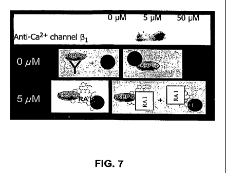
FIG 7 provides an immunoblot of the co-immunoprecipitate of the lysate of F11
cells exposed to various concentrations of the rapamycin analogue I(0 M, 5 M
or 50
M) precipitated using an anti-FKBP52 antibody. The immunoprecipitated
fractions
were immunoblotted with an anti-Ca2+ channel 0 1-subunit antibody. The lower
panels
13
CA 02676613 2009-07-27
WO 2008/094147 PCT/US2007/002656
provide diagrams summarizing the protein interactions. "RA I" represents
rapamycin
analogue I.
FIG 8 provides a bar graph depicting the effect of various concentrations of
rapamycin analogue I(50 M, 5 M, or 0 M) on neurite outgrowth of Fi l cells
using
neurofilament ELISA.
FIGS. 9A-9F depict the biological effect of Compounds 1 and 2 on calcium
currents_
FIG. 9A is a bar graph of the mean Ca2+ current density from whole-cell
recording in F-11 cells treated with 5 M of Compound 1, FK-506 or vehicle in
the bath
for 2 hrs. Recordings were performed from 7 cells in each condition.
FIG. 9B depicts representative Ca2+ currents with internally applied Compound
1
(10 M in pipette) at time 0 sec (bottom trace), 800 sec (middle trace) and in
the presence
of the L-type Caa+ channel blocker BAY-K 5552 (top trace) externally.
FIG. 9C depicts a graph of the time course of the experiment illustrated in
FIG.
9B. Whole cell, and subsequent diffusion of Compound 1 into the cell, begins
at time 0.
Once current stabilizes after 400 sec, 10 gM BayK-5552 is applied in the bath.
(n=3)
FIG. 9I? depicts similar conditions as in FIG. 9C, except that after 300 sec
100
nM wCTX MVIIA is applied via the bath. (n=2)
FIG. 9E depicts the Ca2+ current trace from hippocampal neuron immediately
upon break-in to whole-cell (control) and after 10 minutes of recording with
10 M
Compound 2 internally and wCTX GVIA externally.
FIG. 9F depicts the mean responses (+/- SEM) normalized to the initial current
from hippocampal neurons. Compound 2 (10 M) applied internally via the
recording
pipette, beginning at time 0, where indicated (= and =). External solution
contains 1 M
TTX + 100 nM=wCTX GVIA + 10 gM BAY-K 5552 (V, n=4) or 1 M TTX + 100 nM
wCTX GVIA (*, n=5). Control without compound (^) contained 100 nM wCTX GVIA
externally (n=3).
FIG 10Aprovides a graph demonstrating the effect of siRNA-driven reduction of
FKBP52 and CACNB 1 on neurite outgrowth.
14
CA 02676613 2009-07-27
WO 2008/094147 PCT/US2007/002656
FIG l OB provides a graph demonstrating the effect of siRNA-driven reduction
of
FKBP52 and CACNB 1 on neuronal survival.
FICz I OC shows Western blots confirming that siRNA treatment reduced lamin
A/C, CACNBI or FKBP52 protein expression in cortical neurons after 24 hours.
FIGS. 11A-11B provide the amino acid sequence and nucleotide sequence of
human Ca2+ channel )31 subunit isoform 1 (SEQ ID NOs: 1-2, respectively).
FIGS. 11C-11D provide the amino acid and nucleotide sequence of human CaZ+
channel,61 subunit isoform 2 (SEQ ID NOs: 3-4).
FIGS. 11E-11F provide the amino acid and nucleotide sequence of human Ca2+
channel,l31 subunit isoform 3 (SEQ ID NOs: 5-6).
FIGS. 11G-11H provide the amino acid and nucleotide sequence of a mouse (Mus
musculus) Ca2+ channel (.il subunit isoform A (SEQ ID NOs: 7-8).
FICxS 111-11J provide the amino acid sequence of a mouse (Mus musculus) Ca2+
channel 13, subunit isoform B (SEQ ID NOs: 9-10).
FIGS. 12A-12B provide the amin,o acid and nucleotide sequence of human
FKBP52 (SEQ ID NOs:11-12).
FIGS. 12C-12D provide the amino acid sequence of mouse (Mus musculus)
FKBP52 (SEQ ID NOs:13-14).
DETAILED DESCRIPTION
The present invention is based, at least in part, on the discovery that
immunophilin ligands, e.g., a rapamycin analogues modified at the mTOR binding
region, interact with, e.g., bind to, the immunophilin FKBP52 and/or the
voltage gated L-
type calcium channel (31 subunit. Inhibition of FKBP52 and/or CACNB 1 by these
compounds stimulates neurite outgrowth and/or neuronal survival. Thus,
interaction (and
complex formation) between these components is believed to inhibit the
activity of the 01
subunit and stimulate neurite outgrowth, implicating voltage gated L-type
calcium
channels in some of the neurotrophic and/or neuroprotective activities
exhibited by
immunophilin ligands, such as the rapamycin or meridamycin analogues described
herein.
CA 02676613 2009-07-27
WO 2008/094147 PCT/US2007/002656
Applicants have additionally sliown in the appended Examples that at least one
of
the immunophilin ligands disclosed herein (rapamycin analogue II) showed a
significant
increase in binding selectivity for FKBP52, relative to FKBP12 binding, of at
least 600
fold higher compared to rapamycin. Without being bound by theory, it is
believed that
inhibition of FKBP52 activity mediates neurite outgrowth, presumably by
activating
steroid, e_g., glucocorticoid receptors. Furthermore, treatment of cortical
neurons with
the immunophilin ligands disclosed herein caused an overall downregulation of
calcium
signaling pathways and partial inhibition of L-type calcium channels. A
significant effect
on neurite outgrowth of neuronal cells was also detected by selectively
reducing the
expression of the al subunit and FKBP52 in culture.
The data disclosed herein demonstrate that modification of rapamycin at the
mTOR binding region can provide significantly non-immunosuppressive compounds
with unusual selectivity for FKBP52 and potent neurotrophic activities. FKBP52
appears
to mediate immunophilin ligand-mediated neurite outgrowth, presumably by the
activation of steroid receptors (including glucocorticoid receptors), as
demonstrated by
neurite outgrowth observed in FKBP52 siRNA treated cortical neurons. Further,
the
ability of these rapamycin analogues to partially inhibit L-type Ca2+ channels
and reduce
transcription of_various Ca2} signaling proteins indicates that these
analogues can protect
neurons from Caa+ induced neuronal cell death, which is consistent with their
effect on
neuronal survival.
Calcium channels are present in various tissues, including neuronal and
cardiovascular tissues, and have important roles in a number of vital
processes in
animals, including neurotransmitter release, muscle contraction, pacemaker
activity, and
secretion of hormones and other substances. Entry of calcium into neuronal
cells through
voltage-gated calcium channels mediates a wide variety of cellular and
physiological
responses, including, but not limited to, modulating the activity of calcium-
dependent
enzymes such as protein kinase C and calmodulin-dependent protein kinase II;
controlling membrane potential and contributing to electrical properties such
as
excitability and repetitive firing patterns;. and increasing neurotransmitter
release. These
processes, are involved in human disorders, such as neurological and
cardiovascular
disorders. Therefore, methods of inhibiting the function of voltage-dependent
calcium
16
CA 02676613 2009-07-27
WO 2008/094147 PCT/US2007/002656
channels by forming immunophilin-calcium channel complexes are useful for
treating,
preventing and/or alleviating symptoms of calcium channel disorders, as
described in
more detail herein.
In order that the present invention may be more readily understood, certain
terms
are described in more detail herein and throughout the detailed description.
Calcium channels are membrane-spanning, multi-subunit proteins that allow
controlled entry of Ca2+ ions into cells from the extracellular fluid. The
most common
type of calcium channel is voltage dependent. "Excitable" cells in animals,
such as
neurons of the central nervous system (CNS), peripheral nerve cells, and
muscle cells
(including those of skeletal muscles, cardiac muscles, and venous and arterial
smooth
muscles) have voltage-dependent calcium channels. Voltage-gated calcium
channels
allow for influx of Caa+ ions into a cell, and typically require a
depolarization to a certain
level of the potential difference between the inside of the cell bearing the
channel and the
extracellular environment bathing the cell. Voltage-gated calcium channels
have been
classified by their electrophysiological and pharmacological properties into L-
, N-, P/Q-,
R- and T-types (reviewed in Catterall, 2000; Huguenard 1996; Dolphin, A.C.
(2003)
Pharmacological Reviews 55:607-627). The L-, N- and P/Q-type channels activate
at
positive potentials (high voltage-gated). T-type (or low voltage-gated)
channels describe
a broad class of molecules that transiently activate at negative potentials
and are highly
sensitive to changes in resting potential.
High voltage-gated calcium channels are composed of four distinct
polypeptides:
al, a25, P and y (reviewed by Stea et al., 1994; Catterall, 2000). The (3
subunit (also
referred to herein as "CACB 1") is a soluble intracellular protein encoded by
at least four
known separate genes, each of which is processed into multiple splice
variants. In
embodiments, the 0 subunit has one or.more of the following features: (i) an
amino acid
sequence of a naturally occurring mammalian (e.g., human or rodent) (31
subunit or a
fragment thereof, e.g., the amino acid sequence as shown in FIGS. 11A-11J (SEQ
ID
NOs:1-10) or a fragment thereof; (ii) an amino acid sequence substantially
homologous
to the amino acid sequence shown in FIGS. 11A-11J (SEQ ID NOs:1-10) or a
fragment
thereof; (iii) an amino acid sequence that is encoded by a naturally occurring
mammalian
(e.g., human or rodent) (31 subunit nucleotide sequence or a fragment thereof,
e.g., an
17
CA 02676613 2009-07-27
WO 2008/094147 PCT/US2007/002656
amino acid sequence encoded by the nucleotide sequence as shown in FIGS. 11A-
11J
(SEQ ID NOs:l-10) or a fragment thereof; (iv) an amino acid sequence encoded
by a
nucleotide sequence which is substantially homologous to the nucleotide
sequence shown
in FIGS. 11A 11J (SEQ ID NOs:1-10) or a fragment thereof; (v) an amino acid
sequence
encoded by a nucleotide sequence degenerate to a naturally occurring J31
subunit
nucleotide sequence or a fragment thereof, e.g., the nucleotide sequence shown
in FIGS.
11A-11J (SEQ ID NOs:1-10) or a fragment thereof; or (vi) a nucleotide sequence
that
hybridizes to one of the foregoing nucleotide sequences under stringent
conditions, e.g.,
highly stringent conditions. In some embodiments, the (3 subunit or functional
variant
(e.g., fragment) thereof exhibits one or more activities of the naturally-
occurring
sequence, including but not limited to, (i) forms a complex as described
herein; (ii)
interacts with, e.g., binds to, the a-subunit; (iii) facilitates the
localization or trafficking
of the voltage-gated calcium channel, e.g., the a, subunit, to the cellular
plasma
membrane; (iv) modulates gating of the channel (e.g., alters activation and
inactivation
kinetics, causes a leftward shift in the I-V curve and, at a single channel
level, induces an
increase in the channel opening probability); or (v) controls transcriptional
activity of one
or more of the genes described herein (e.g., calcium- influx channels, NMDA
receptors,
plasminogen activator (PLATJ), SHT3R channels).
In other embodiments, the (3 subunit has a sequence substantially identical to
that
disclosed in Powers et al. (1992) J. Biol. Chem. 267(32):22967-22972; Collin
et al.
(1993) Circ. Res. 72(6):1337-1344; Hogan, K. et al. (1999) Neurosei. Lett. 277
(2), 111-
114; Foell et al. (2004) Physiol. Genomics 17 (2), 183-200 (human (31 and (32
subunits);
Toba et al. (2005) Eur. J. Neurosci. 22 (1), 79-92 (murine beta 1 subunit
isoform);
Serikov et al. (2002) Biochem. Biophys. Res. Commun. 293 (5), 1405-1411;
Pragnell et
al. (1991) FEBSLett. 291 (2), 253-25&; Cahill et al. (2000) J. Neurosci. 20
(5), 1685-
1693 (2000) (bovine beta 1, 2 and 3 subunits); Rosenfeld et al. (1993) Ann.
Neurol. 33
(1), 113-120; Taviaux et al. (1997) Hum. Genet. 100 (2), 151-154 (human genes
for beta
2 and beta 4 subunits); Colecrafft et al. (2002) J. Physiol. (Lond.) 541 (Pt
2), 435-452
(human beta 2a, 2c, 2d and 2e subunits); Opatowsky et al. (2003) J. Biol.
Chem. 278
(52), 52323-52332 (rat beta 2 subunit); Yamada et al. (2001) J. Biol. Chem.
276 (50),
47163-47170 (2001) (rat beta 2 subunit); Strausberg et al. (2002) PNAS U.S.A.
99 (26),
18
CA 02676613 2009-07-27
WO 2008/094147 PCT/US2007/002656
16899-16903 (human beta 3 subunit, murine beta 4 subunit); Murakami et al.
(1996) Eur.
J. Biochem. 236 (1), 13 8-143 (1996) (murine calcium channel beta 3 subunit);
Yamada et
al. (1995) Genomics 27 (2), 312-319 (human calcium channel alpha 1 subunit
(CACNLIA2) and beta subunit (CACNLB3) genes); Chen et al. (2004) Nature 429
(6992), 675-680 (human beta 4 subunit); Helton et al. (2002) J. Neurosci. 22
(5), 1573-
1582 (2002) (beta 4 subunit); Badou et al. (2005) Science 307 (5706), 117-121
(2005)
(calcium channel beta4 subunit); the contents of all of which are hereby
incorporated by
reference. Other 0 subunit sequences are disclosed in Genbank Accession
Numbers: NP_
_ 666235, Q9Y698, Q02641, Q9MZL3 and P54288_2.
Immunophilins are soluble cytosolic proteins that form complexes with
immunophilin ligands, which in turn serve as ligands for other cellular
targets involved in
signal transduction. Classes of immunophilins include cyclophilins and FK506-
binding
proteins (e.g., FKBPs), such as FKBP-12 and FBBP-52. Cyclosporin A is a
macrolide
immunophilin ligand that binds to cyclophilins. Other macrolide immunophilin
ligands,
such as meridamycin, FK506, FK520, and rapasnycin, are understood to bind to
FKBPs.
Binding of FK506, FK520 and rapamycin to FKBP typically occurs through
structurally
similar segments of the polyketide molecules, referred to as "FKBP-binding
domain."
Gene sequences corresponding to more than two-dozen FKBPs have been found
in the human genome (Dornan et al., Curr. Top. Med. Chem. 3, 1392-1409
(2003)). They
are expressed 10-50 fold higher in central nervous system (CNS) and peripheral
nervous
system (PNS) tissue than in immune tissue (Lyons et al., .I. Neurosci. 15,
2985-2994
(1995)), and their expression is increased following the onset of neurological
disease
(Kihira et al., Neuropathology 22, 269-274 (2002)).. Interestingly, FKBP12,
FKBP12.6 and
FKBP52 were reported as channel-gating-FKBP proteins, modulating ryanodine
receptor
(RYR) (Huang et al., Proc. Natl. Acad. Sci. USA. 103, 3456-3461 (2006)),
inositol
1,4,5-trisphosphate receptor (IP3R) (Cameron et al., Proc. Natl. Acad. Sci. U
S A. 92,
1784-1788 (1995)) and transient receptor potential channels (TRPC) (Sinkins et
al., J.
Biol. Chem. 279, 34521-34529 (2004)). FKBP52 and FKBP51 associate with three
types
of steroid receptor complexes that mediate the down-stream responses to
estrogen,
androgen and glucocorticoid hormones (Steiner et al., Proc. Natl. Acad. Sci.
USA. 94,
2019-2024 (1997)). The nuclear FKBP25 regulates gene expression through
associating
19
CA 02676613 2009-07-27
WO 2008/094147 PCT/US2007/002656
with histone deacetylase, casein kinase II, nucleolin and transcription factor
YY1 (Yao
and Yang, Curr. Cancer Drug Targets .5, 595-610 (2005)). FKBP38 is
constitutively
inactive and located at the mitochondria and endoplasmic reticulum.
Interestingly, high
levels of Ca2+ and calmodulin (CaM) are required for FKBP38 to bind Bcl-2
(Edlich et
al., EMBO J. 24, 2688-2699 (2005)). Immunophilin ligands cause various down-
stream
biological activities by disruption of the natural FKBP-containing complexes
(Gold Drug
Metab. Rev. 31, 649-663 (1999); Edlich et al., J. Biol.Chem. 281, 14961-14970
(2006))
and by formation of novel complexes, such as FKBP12-FK506-calcineurin or
FKBP12-
rapamycin-mammalian target of rapamycin (mTOR) (Kissinger et al., Nature 378,
641-
644 (1995); Choi et al., Science 273, 239-42 (1996)). -
FKBP52 is a member of the FK506-binding class of immunophilins. Binding of
FK506 to the glucocoricoid receptor (GR)-associated FKBP52 caused increased
nuclear
translocation of GR in response to dexamethasone and potentiation of GR-
mediated gene
expression (Sanchez and Ning (1996) Methods: A Companion to Meth. Enzymol.
9:188-
200). Immunophilins such as FKBP52 and CyP40 and non- immunophilin proteins
such
as PP5, p60, and Mas70p, have one or more tetratricopeptide repeat (TPR)
domains
(Ratajczak et al. (1993) J. Biol. Chem. 268:13187-13192) that bind to the TPR-
binding
domain of hsp90. The number of TPR domains in a protein appears to correlate
with its
hsp90-binding affinity. Regions bordering the TPR domain also participate in
binding,
e.g., residues 232-271 of FKBP52 (Ratajczak and Carrello (1996) supra).
In some embodiments, the imrnunophilin has one or more of the following
features: (i) an amino acid sequence of a naturally occurring mammalian (e.g.,
human or
rodent) FKBP52 or a fragment thereof, e.g., the amino acid sequence as shown
in FIGS.
12A-12D (SEQ ID NOs: 11-14) or a fragment thereof; (ii) an amino acid sequence
substantially homologous to the amino acid sequence shown in FIGS. 12A-12D
(SEQ ID
NOs:11-14) or a fragment thereof; (iii) an amino acid sequence that is encoded
by a
naturally occurring mammalian (e.g., human or rodent) FKBP52 nucleotide
sequence or a
fragment thereof, e.g., an amino acid sequence that is encoded by the
nucleotide sequence
as shown in FIGS. 12A-12D (SEQ ID NOs:1 1-14) or a fragment thereof; (iv) an
amino
acid sequence encoded by a nucleotide sequence which is substantially
homologous to
the nucleotide sequence shown in FIGS. 12A-12D (SEQ ID NOs:l1-14) or a
fragment
CA 02676613 2009-07-27
WO 2008/094147 PCT/US2007/002656
thereof; (v) an amino acid sequence encoded by a nucleotide sequence
degenerate to a
naturally occurring FKBP52 nucleotide sequence or a fragment thereof, e.g.,
the
nucleotide sequence shown in FIGS. 12A-12D (SEQ ID NOs:11-14) or a fragment
thereof; or (vi) a nucleotide sequence that hybridizes to one of the foregoing
nucleotide
sequences under stringent conditions, e.g., highly stringent conditions. In
some
embodiments, the FKBP52 or functional variant (e.g., fragment) thereof
exhibits one or
more activities of the naturally-occurring sequence, including but not limited
to, forms a
complex as described herein; binds to FK506; increases nuclear translocation
of a
glucocorticoid receptor in response to dexamethasone; potentiates
glucocorticoid receptor
- mediated gene expression; and/or binds to a heat shock protein, e.g., hsp90.
Exemplary amino acid and nucleotide sequences for FKBP52 are disclosed in
Sanchez et al. (1990) Biochemistry 29 (21), 5145-5152; and Peattie et al.
(1992) Proc.
Natl. Acad. Sci. U.S.A. 89 (22), 10974-10978, the contents of both of which
are hereby
incorporated by reference.
In one embodiment, 0 subunit or immunophilin polypeptides of this invention
include, but are not limited to, fra.gments of native polypeptides from any
animal species
(including humans, rodents), and variants (e.g., functional variants) thereof
(human and
non-human) polypeptides and their fragments, provided that they have a
biological
activity in common with a respective native polypeptide. "Fragments" comprise,
in one
embodiment, regions within the sequence of a mature native polypeptide. Any
form of
the P subunit or inununophilin, e.g., FKBP52, of less than full length can be
used in the
methods and compositions of the preserit invention, provided that it is still
functional,
e.g., retains at least one activity of the naturally-occurring sequence (e.g.,
retains the
ability to form a complex as described herein). (3 subunits of less than full
length can be
produced by expressing a corresponding fragment of the polynucleotide encoding
the
full-length J3 subunit protein in a host cell. These corresponding
polynucleotide
fragments are also part of the present invention. Modified polynucleotides as
described
above may be made by standard molecular biology techniques, including
construction of
appropriate desired deletion mutants, site-directed mutagenesis methods or by
the
polymerase chain reaction using appropriate oligonucleotide primers.
21
CA 02676613 2009-07-27
WO 2008/094147 PCT/US2007/002656
A "variant" of a polypeptide, or fragment thereof, such as, for example, a
variant
of a(31 subunit or FKBP52 includes chimeric proteins, labeled proteins (e.g.,
radiolabeled
proteins), fusion proteins, mutant proteins, proteins having similar (e.g.,
substantially
similar) sequences (e.g., proteins having amino acid substitutions (e.g.,
conserved amino
acid substitutions), deletions, insertions), protein fragments, mimetics, so
long as the
variant has at least a portion of an amino'acid sequence of a native protein,
or at least a
portion of an amino acid sequence of substantial sequence identity to the
native protein.
A"functional variant" includes a variant that retains at least one function of
the native
protein, e.g., retains the ability to interact an immuno.philin ligand with
and/or form a
complex as described herein.
A "chimeric protein"or "fusion protein" is a fusion of a first amino acid
sequence
encoding a polypeptide with a second amino acid sequence, wherein the first
and second
amino acid sequences do not occur naturally as part of a single polypeptide
chain.
As used herein, the term "substantially similar" (or "substantially" or
"sufficiently" "homologous" or "identical") is used herein to refer to a first
amino acid or
nucleotide sequence that contains a sufficient number of identical or
equivalent (e.g.,
with a similar side chain, e.g., conserved amino acid substitutions) amino
acid residues or
nucleotides to a second amino acid or nucleotide sequence such that the first
and second
amino acid or nucleotide sequences have similar activities. Sequences similar
or
homologous (e.g., at least about 85% sequence identity) to the sequences
disclosed herein
are also part of this application. In some embodiments, the sequence identity
can be
about 90%, 91%, 92%, 93%, 94%, 95%, 96%, 97%, 98%, 99% or higher.
Alternatively,
substantial identity exists when the nucleic acid segments hybridizes under
selective
hybridization conditions (e.g., highly stringent hybridization conditions), to
the
complement of the strand. The nucleic acids may be present in whole cells, in
a cell
lysate, or in a partially purified or substantially pure form.
Calculations of "homology" or "sequence identity" between two sequences (the
terms are used interchangeably herein) are performed as follows. The sequences
are
aligned for optimal comparison purposes (e.g., gaps can be introduced in one
or both of a
first and a second amino acid or nucleic acid sequence for optimal alignment
and non-
homologous sequences can be disregarded for comparison purposes). Typically,
the
22
CA 02676613 2009-07-27
WO 2008/094147 PCT/US2007/002656
length of a reference sequence aligned for comparison purposes is at least
30%,
preferably at least 40%, more preferably at least 50%, even more preferably at
lo least
60%, and even more preferably at least 70%, 80%, 90%, 100% of the length of
the
reference sequence. The amino acid residues or nucleotides at corresponding
amino acid
positions or nucleotide positions are then compared. When a position in the
first
sequence is occupied by the same amino acid residue or nucleotide as the
corresponding
position in the second sequence, then the molecules are identical at that
position (as used
herein amino acid or nucleic acid "identity" is equivalent to amino acid or
nucleic acid
"homology"). The percent identity between the two sequences is a function of
the
number of identical positions shared by the sequences, taking into account the
number of
gaps, and the length of each gap, which need to be introduced for optimal
alignment of
the two sequences.
The comparison of sequences and determination of percent identity between two
sequences can be accomplished using a mathematical aigorithm. In one
embodiment, the
percent identity between two amino acid sequences is determined using the
Needleman
and Wunsch ((1970) J. Mol. Biol. 48:444-453) algorithm which has been
incorporated
into the commercially available GAP program in the GCG software package, using
either
a Blossum 62 matrix or a PAM250 matrix, and a gap weight of 16, 14, 12, 10, 8,
6, or 4
and a length weight of 1, 2, 3, 4, 5, or 6. In yet another embodiment, the
percent identity
between two nucleotide sequences is determined using the commercially
available GAP
program in the GCG software package, using a NWSgapdna.CMP matrix and a gap
weight of 40, 50, 60, 30 70, or 80 and a length weight of 1, 2, 3, 4, 5, or 6.
Parameters
typically used to determine percent homology are a Blossum 62 scoring matrix
with a gap
penalty of 12, a gap extend penalty of 4, and a frameshift gap penalty of 5.
The percent
identity between two amino acid or nucleotide sequences can also be determined
using
the s algorithm of E. Meyers and W. Miller ((1989) CABIOS 4:11-17) which has
been
incorporated into the ALIGN program (version 2.0), using a PAM120 weight
residue
table, a gap length penalty of 12 and a gap penalty of 4.
As used herein, the term "hybridizes under stringent conditions" describes
conditions for hybridization and washing. Stringent conditions are known to
those
skilled in the art and can be found in Current Protocols in Molecular Biology,
John
23
CA 02676613 2009-07-27
WO 2008/094147 PCT/US2007/002656
Wiley & Sons, N.Y. (1989), 6.3.1- 6.3.6. Aqueous and non-aqueous methods are
described in that reference and either can be used. An example of stringent
hybridization
conditions are hybridization in 6X sodium chloride/sodium citrate (SSC) at
about 45 C,
followed by one or more washes in 0.2X SSC, 0.1% SDS at 50 C. Another is
example of
stringent hybridization conditions are hybridization in 6X SSC at about 45 C,
followed
by one or more washes in 0.2X SSC, 0.1% SDS at 55 C. A further example of
stringent
hybridization conditions are hybridization in 6X SSC at about 45 C, followed
by one or
more washes in 0.2X SSC, 0.1% SDS at 60 C. Typically, stringent hybridization
conditions are hybridization in 6X SSC at about 45 C, followed by one or 20
more
washes in 0.2X SSC, 0.1% SDS at 65 C. More typically, the highly stringent
conditions
used are 0.5M sodium phosphate, 7% SDS at 65 C, followed by one or more washes
at 0.
2X SSC, 1% SDS at 65 C.
It is understood that the variants of the polypeptide disclosed herein may
have
additional conservative or non-essential amino acid substitutions, which do
not have a
substantial effect on antigen binding or other immunoglobulin functions. A
"conservative amino acid substitution" is one in which the amino acid residue
is replaced
with an amino acid residue having a similar side chain. Families of amino acid
residues
having similar side chains have been defined in the art. These families
include amino
acids with basic side chains (e.g., lysine, arginine, histidine), acidic side
chains (e.g.,
aspartic acid, glutamic acid), uncharged polar side chains (e.g., glycine,
asparagine,
glutamine, serine, threonine, tyrosine, cysteine), nonpolar side chains (e.g.,
alanine,
valine, leucine, isoleucine, praline, phenylalanine, methionine, tryptophan),
beta-
branched side chains (e.g., threonine, valine, isoleucine) and aromatic side
chains (e.g., s
tyrosine, phenylalanine, tryptophan, histidine).
A "non- essential" amino acid residue is a residue that can be altered from
the
wild-type sequence of a hybrid antibody, without abolishing or more
preferably, without
substantially altering a biological activity, whereas an "essential" amino
acid residue
results in such a change.
24
CA 02676613 2009-07-27
WO 2008/094147 PCT/US2007/002656
Immunophilin Ligands
Immunophilin ligands bind to iminunophilins to activate other cellular
targets,
primarily in the immune and nervous system. Several immunophilins are
immunosuppressive, e.g., cyclosporin A, FK506 and rapamycin, whereas other
less
immunosuppressive immunophilins show neurotrophic activities. For example,
meridamycin is substantially non-immimosuppressive and shows significant
neuroprotective activity in vitro (US 2005/0272133 by He, M. et al. published
on
December 8, 2005, and US 2005/0197356 by Graziani, E. et al. published on
September
8, 2005). Preferably, immunophilin ligands identified by, or used in, the
methods of the
invention are substantially non-immunosuppressive, but retain a desirable
activity, e.g., a
neurotrophic activity. Preferred immunophilin ligands increase the formation
of a
complex as described herein and/or reduce FKBP and/or calcium channel
activity.
In some embodiments, the immunophilin ligands are modified at the mTOR
binding domain. The mTOR binding domain of rapamycin is believed to localize
at the
macrocycle core at about positions 1-7 and 27-36 of FIG. 1A. For example, the
immunophilin ligands can have a heteroatom substituent at positions 1 and 4 of
the
rapamycin backbone (FIG. IA). In other embodiments, the rapamycin analogues
have a
cyclic structure at positions 1, 2, 3 and/or 4 (FIG. 1A). Such rapamycin
analogues are
disclosed in commonly assigned co-pending published application U.S.
2006/0135549
entitled "Rapamycin Analogues and the Uses Thereof in the Treatment of
Neurological,
Proliferative, and Inflammatory Disorders," published on June 22, 2006 from
U.S.S.N.
11/300,839, the entire content of which is hereby incorporated by reference.
In one embodiment, the rapamycin analogues have the formula I:
CA 02676613 2009-07-27
WO 2008/094147 PCT/US2007/002656
R4
R4.
R5
~ n..
O 0 OH
N
O O
HO R1s O Rs
O R7 Ra Ra
4
R1 R2
Rl and R2 in the above-noted formula are different, independent groups and are
selected from among OR3 and N(R3,)(R3>>) or RI and R2 are different, are
connected
through a single bond, and are selected from 0 and NR3. R3, R3=, and R3=, are
independently selected from among H, Cl to C6 alkyl, C, to C6 substituted
alkyl, C3 to C$
cycloalkyl, substituted C3 to C8 cycloalkyl, aryl, substituted aryl,
heteroaryl, and
substituted heteroaryl. R4 and R4, are (a) independently selected from among
H, OH,
O(Cl to C6 alkyl), O(substituted Cl to C6 alkyl), O(acyl), O(aryl),
O(substituted aryl), and
halogen; or (b) taken together to form a double bond to O. R5, R6, and R7 are
independently selected from among H, OH, and OCH3. R8 and R9 are connected
through
a (i) single bond and are CHZ or (ii) double bond and are CH. Rls is selected
from among
C=O, CHOH, and CH2 and n is 1 or 2; or pharmaceutically acceptable, salts,
prodrugs, or
metabolites thereof.
In further embodiments, R, and R2 are connected through a single bond and are
selected from 0 and NR3. In still a further embodiment, R, is 0 and R2 is NR3.
In one embodiment, R3, or R3 is an aryl or substituted aryl group, or a
substituted
benzene ring. In another embodiment, substituted benzene groups at R3. or R3-
include
rings of the following structure:
Rto R1:
R14 R13
R12
26
CA 02676613 2009-07-27
WO 2008/094147 PCT/US2007/002656
RIo, R>>, Ri a, R13, and RI 4 are independently selected from among H, Ct to
C6
alkyl, substituted C1 to C6 alkyl, aryl, substituted aryl, heteroaryl,
substituted heteroaryl,
halogen, acyl, OH, O(alkyl), O(substituted alkyl), O(aryl), O(substituted
aryl), O(acyl),
NH2, NH(alkyl), NH(substituted alkyl),'NH(aryl), NH(substituted aryl), and
NH(acyl).
In further embodiments, R3, R3, or Ry are phenyl optionally substituted by I
or 2
substituents selected from C, to C6 alkyl and halogen. In still further
embodiments, R3,
R3. or Ry. are phenyl optionally substituted with 1 or 2 methyl or chloro
substituents, e.g.
phenyi and 3-methyl, 4-chlorophenyl.
In one embodiment, R4 or R4, are OH or O(acyl), e.g., where the acyl is
-C(O)- optionally substituted alkyl, in particular where alkyl can be straight
or
branched and optionally substituted e.g. by heterocyclic such as aromatic
heterocyclic
such as pyridyl. An example is:
0
In other embodiments, rapamycin analogues of formula I include those where R5,
R6 and R7 are OCH3, those where the nitrogen containing ring at positions 17-
22 of the
rapamycin backbone is a piperidine ring, or where R15 is a carbonyl.
In one embodiment, the rapamycin analogues have the formula Ia:
OH
O''
O 0 OH
IV
O O `O O
HO O
O O Rs-Rs
R, R2
Ia
where Ri, R2, Rg, and R9 are defined as noted above.
In another embodiment, the rapamycin analogues have the following formula Ib:
27
CA 02676613 2009-07-27
WO 2008/094147 PCT/US2007/002656
OH
O'~
O O OH
N
O
HO O ~'O
O O'
O-N
Rm
Ib
In fonnula Ib, R is independently selected from among H, C, to C6 alkyl,
substituted Ci to C6 alkyl, aryl, substituted aryl, heteroaryl, substituted
heteroaryl,
halogen, acyl, OH; O(alkyl), O(substituted alkyl), O(aryl), O(substituted
aryl), O(acyl),
NH2, NH(alkyl), NH(substituted alkyl), NH(aryl), NH(substituted aryl), and
NH(acyl)
andrnislto5.
Specific raparnycin analogues are illustrated herein and include 9,27-
dihydroxy-3-
{2- [4-hydroxy-3-methoxycyclohexyl] -1-methylethyl } -10,21-dimethoxy-6,
8,12,14,2 0,26-
hexamethyl-37-phenyl-4,9,10,12,13,14,15,18,21,22,23,24,25,26,27,32,33,34,34a-
nonadecahydro-3H-23,27-epoxy-18,15-(epoxyimino)pyrido[2,1-
c][1,4]oxazacyclohentriacontine-1,5,11,28,29(6H,31H)-pentone; 9,27-dihydroxy-3-
{2-[4-
hydroxy-3-methoxycyclohexyl]-1-methylethyl}-10,21-dimethoxy-6,8,12,14,20,26-
hexamethyl-37-phenyl-
4,9,10,12,13,14,15,16,17,18,21,22,23,24,25,26,27,32,33,34,34a-
henicosahydro-3H-23,27-epoxy-18,15-(epoxyimino)pyrido[2,1-
c][ 1,4]oxazacyclohentriacontine-1,5,11,28,29(6H,31H)-pentone; 37-(4-chloro-3-
methylphenyl)-9,27-dihydroxy-3- {-2-[4-hydroxy-3-methoxycyclohexyl]-1-
methylethyl}-
.10,21-dimethoxy-6,8,12,14,20,26-hexamethyl-
4,9,10,12,13,14,15,18,21,22,23,24,25,26,27,32,33,34,34a-nonadecahydro-3H-23,27-
epoxy-18,15-(epoxyimino)pyrido[2,1-c] [ 1,4]oxazacyclohentriacontine-
1,5,11,28,29(6H,31H)-pentone; 37-(2,6-dichlorophenyl)-9,27-dihydroxy-3-{2-[4-
28
CA 02676613 2009-07-27
WO 2008/094147 PCT/US2007/002656
hydroxy-3-methoxycyclohexyl]-1-methylethyl} -10,21-dimethoxy-6,8,12,14,20,26-
hexamethyl-4,9,10,12,13,14,15,18,21,22,23,24,25,26,27,32,33,34,34a-
nonadecahydro-
3H-23,27-epoxy-18,15-(epoxyimino)pyrido[2,1-c] [ 1,4]oxazacyclohentriacontine-
1,5,11,28,29(6H,31H)-pentone; 9,27-dihydroxy-3-{-2-[4-hydroxy-3-
methoxycyclohexyl]-1-methylethyl }-10,21-dimethoxy-6,8,12,14,20,26-hexamethyl-
37-
phenyl-4,9,10,1.2,13,14,15,18,21,22,23,24,25,26,27,32,33,34,34a-nonadecahydro-
3H-
23,27-epoxy-18,15-(epoxyimino)pyrido[2,1-c] [ 1,4]oxazacyclohentriacontine-
1,5,11,28,29(6H,31H)-pentone ester with -2,2-dimethyl-3-(pyridin-2-yl)-
propionic acid;
37-(2,6-dichlorophenyl)-9,27-dihydroxy-3- {-2-[4-hydroxy-3-methoxycyclohexyl]-
1-
methylethyl } -10,21-dimethoxy-6,8,12,14,20,26-hexamethyl-
4,9,10,12,13,14,15,18,21,22,23,24,25,26,27,32,33,34,34a-nonadecahydro-3H-23,27-
epoxy-18,15-(epoxyimino)pyrido[2,1-c] [ 1,4]oxazacyclohentriacontine-
1,5,1 i,28,29(6H,31H)-pentone; or pharmaceutically acceptable, salts,
prodrugs, or
metabolites thereof. The invention is not limited to these illustrative
compounds.
In another embodiment, the specific compounds include the following:
Rapamycin I Rapamycin II
v,.. .....y
,. ...p.+ ...y
:r...~ ; ...
'M" XOA O I NO ~=r O I O O
oi N ~ M
N `O= O _6== õ~.O O `a=
a
N I V"' /-~ D
N O I ^""=\ .. O I XI r pi
' I~...
y
a ~ ..O-'~ O- Ia ral p~ -
p~
o-~
29
CA 02676613 2009-07-27
WO 2008/094147 PCT/US2007/002656
Rapamycin analogues I and II, referred to throughout the application, are
represented by the first and second chemical structures, respectively, shown
from the top
left.
Rapamycin analogues also include compounds where R, and R2 are connected
through a single bond; Rl is 0; R2 is NR3; R3 is phenyl; R4 is OH; R5-R7 are
OCH3; and
R8 and R9 are HC=CH; a compound where Rl is OR3; R2 is N(R3,)(R3 ); R3 is H;
R3, is
H; R3=. is phenyl; R4 is OH; R5-R7 are OCH3; and R8 and R9 are H2C-CH2; a
compound
where R, and R2 are connected through a single bond; Rl is 0; R2 is NR3; R3 is
phenyl;
R4 is OH; R5-R7 are OCH3; and R$ and R9 are H2C-CH2; a compound where Rt and
R2
are connected through a single bond; Rt is 0; R2 is NR3; R4 is OH; R5-R7 are
OCH3; R8
and R9 are HC=CH; and R3 is
cl
a compound where R, and R2 are connected through a single bond; Ri is 0; R2 is
NR3; R4 is OH; R5-R7 are OCH3; R8 and R9 are HC=CH; and R3 is
ci
a compound where Rl and R2 are connected through a single bond; Rl is 0; R2 is
NR3; R3 is phenyl; R5-R7 are OCH3; R8 and R9 are HC=CH; and R4 is
0
N
sv1 p
and a compound where R, and R2 are connected through a single bond; Rt is 0;
R2 is NR3; R4 is OH; RS-R7 are OCH3; R$ and R9 are H2C-CH2; and R3 is
cE.
CA 02676613 2009-07-27
WO 2008/094147 PCT/US2007/002656
The compounds can contain one or more asymmetric carbon atoms and some of
the compounds can contain one or more asymmetric (chiral) centers and can thus
give
rise to optical isomers and diastereomers. While shown without respect to
stereochemistry, when the compounds can contain one or more chiral centers,
preferably
at least one of the chiral centers is of S-stereochemistry. Thus, the compound
includes
such optical isomers and diastereomers; as well as the racemic and resolved,
enantiomerically pure stereoisomers; a's well as other mixtures of the R and S
stereoisomers, and pharmaceutically acceptable salts, hydrates, metabolites,
and prodrugs
thereof.
The term "alkyl" is used herein to refer to both straight- and branched-chain
saturated aliphatic hydrocarbon groups having 1 to 10 carbon atoms, and
desirably about
1 to 8 carbon atoms. The term "alkenyl" is used herein to refer to both
straight- and
branched-chain alkyl groups having one or more carbon-carbon double bonds and
containing about 2 to 10 carbon atoms. In one embodiment, the term alkenyl
refers to an
alkyl group having 1 or 2 carbon-carbon double bonds and having 2 to about 6
carbon
atoms. The term "alkynyl" group is used herein to refer to both straight- and
branched-
chain alkyl groups having one or morecarbon-carbon triple bond and having 2 to
8
carbon atoins. In another embodiment, the term alkynyl refers to an alkyl
group having 1
or 2 carbon-carbon triple bonds and having 2 to 6 carbon atoms.
The term "cycloalkyl" is used herein to refer to an alkyl group as previously
described that is cyclic in structure and has about 4 to 10 carbon atoms, or
about 5 to 8
carbon atoms.
The terms "substituted alkyl", "substituted alkenyl", and "substituted
alkynyl"
refer to alkyl, alkenyl, and al.kynyl groups, respectively, having one or more
substituents
including, without limitation, halogen, CN, OH, NOa, amino, aryl,
heterocyclic, alkoxy,
aryloxy, alkylcarbonyl, alkylcarboxy, and arylthio, which groups can be
optionally
substituted e.g. by 1 to 4 substituents including halogen, CN, OH; NO2, amino,
alkyl,
cycloalkyl, alkenyl, alkynyl, alkoxy, aryloxy, alkyloxy, alkylcarbonyl,
alkylcarboxy,
aminoalkyl, and arylthio. These substituents can be attached to any carbon of
an alkyl,
alkenyl, or alkynyl group provided that the attachment constitutes a stable
chemical
moiety.
31
CA 02676613 2009-07-27
WO 2008/094147 PCT/US2007/002656
The term "aryl" as used herein refers to an aromatic system, e.g., of 6-20
carbon
atoms, which can include a single ring or multiple aromatic rings fused or
linked together
(e.g. two or three) where at least one part of the fused or linked rings forms
the
conjugated aromatic system. The aryl groups can include, but are not limited
to, phenyl,
naphthyl, biphenyl, anthryl, tetrahydronaphthyl, phenanthryl, indene,
benzonaphthyl,
fluorenyl, and carbazolyl.
The term "substituted aryl" refers to an aryl group which is substituted with
one
or more substituents including halogen, CN, OH, NOZ, amino, alkyl, cycloalkyl,
alkenyl,
alkynyl, alkoxy, aryloxy, alkyloxy, alkylcarbonyl, alkylcarboxy, aminoalkyl,
and
arylthio, which groups can be optionally substituted. In one embodiment, a
substituted
aryl group is substituted with 1 to 4 substituents including halogen, CN, OH,
NO2, amino,
alkyl, cycloalkyl, alkenyl, alkynyl, alkoxy, aryloxy, alkyloxy, alkylcarbonyl,
alkylcarboxy, aminoalkyl, and arylthio.
The term "heterocyclic" as used herein refers to a stable 4- to 7-membered
monocyclic or multicyclic heterocyclic ring which is saturated, partially
unsaturated, or
wholly unsaturated, including aromatic such as pyridyl.. The heterocyclic ring
has
carbon atoms and one or more heteroatoms including nitrogen, oxygen, and
sulfur atoms.
In one embodiment, the heterocyclic ring has 1 to 4 heteroatoms in the
backbone of the
ring. When the heterocyclic ring contains nitrogen or sulfur atoms in the
backbone of the
ring, the nitrogen or sulfur atoms can be oxidized. The term "heterocyclic"
also refers to
multicyclic rings, e.g., of 9 to 20 ring members in which a heterocyclic ring
is fused to an
aryl ring. The heterocyclic ring can be attached to the aryl ring through a
heteroatom or
carbon atom, provided the resultant heterocyclic ring structure is chemically
stable. A
variety of heterocyclic groups are known in the art and include, without
limitation,
oxygen-containing rings, nitrogen-containing rings, sulfur-containing rings,
mixed
heteroatom-containing rings, fused heteroatom containing rings, and
combinations
thereof. Oxygen-containing rings include, but are not limited to, furyl,
tetrahydrofuranyl,
pyranyl, pyronyl, and dioxinyl rings. Nitrogen-containing rings include,
without
limitation, pyrrolyl, pyrazolyl, imidazolyl, triazolyl, pyridyl, piperidinyl,
2-
oxopiperidinyl, pyridazinyl, pyrimidinyl, pyrazinyl, piperazinyl, azepinyl,
triazinyl,
pyrrolidinyl, and azepinyl rings. Sulfur-containing rings include, without
limitation,
32
CA 02676613 2009-07-27
WO 2008/094147 PCT/US2007/002656
thienyl and dithiolyl rings. Mixed hetergatom containing rings include, but
are not
limited to, oxathiolyl, oxazolyl, thiazolyl, oxadiazolyl, oxatriazolyl,
dioxazolyl,
oxathiazolyl, oxathiolyl, oxazinyl, oxathiazinyl, morpholinyl,
thiamorpholinyl,
thiamorpholinyl sulfoxide, oxepinyl, thiepinyl, and diazepinyl rings. Fused
heteroatom-
containing rings include, but are not limited to, benzofuranyl, thionapthene,
indolyl,
benazazolyl, purindinyl, pyranopyrrolyl, isoindazolyl, indoxazinyl,
benzoxazolyl,
anthranilyl, benzopyranyl, quinolinyl, isoquinolinyl, benzodiazonyl,
naphthylridinyl,
benzothienyl, pyridopyridinyl, benzoxazinyl, xanthenyl, acridinyl, and purinyl
rings.
The term "substituted heterocyclic" as used herein refers to a heterocyclic
group
having one or more substituents including halogen, CN, OH, NO2, amino, alkyl,
cycloalkyl, alkenyl, alkynyl, alkoxy, aryloxy, alkyloxy, alkylcarbonyl,
alkylcarboxy,
aminoalkyl, and arylthio, which groups can be optionally substituted. In one
embodiment, a substituted heterocyclic group is substituted with 1 to 4
substituents.
The term "acyl" refers to a -C(O)- group, which is substituted at the carbon
atom.
The acyl group can be substituted or a terminal acyl group such as an HC(O)-
group. The
substituents can include any substituents noted above for alkyl groups, viz.
one or more
substituents including, without limitation, halogen, CN, OH, NO2, amino, aryl,
heterocyclic, alkoxy, aryloxy, alkylcarbonyl, alkylcarboxy, and arylthio,
which groups
can be optionally substituted. Examples include -C(O)-alkoxy (e.g. -OMe or -
OEt) or -
C(O)-alkyl where alkyl can be straight or branched and optionally substituted
e.g., by
heterocyclic (such as pyridyl). .
The term "alkoxy" as used hereiri refers to the O(alkyl) group, where the
point of
attachment is through the oxygen-atom and the alkyl group is optionally
substituted.
The term "aryloxy" as used herein refers to the O(aryl) group, where the point
of
attachment is through the oxygen-atom and the aryl group is optionally
substituted.
The term "alkyloxy" as used herein refers to the alkylOH group, where the
point
of attachment is through the alkyl group..
The term "arylthio" as used herein refers to the S(aryl) group, where the
point of
attachment is through the sulfur-atom and the aryl group can be optionally
substituted.
33
CA 02676613 2009-07-27
WO 2008/094147 PCT/US2007/002656
The term "alkylcarbonyl" as used herein refers to the C(O)(alkyl) group, where
the point of attachment is through the carbon-atom of the carbonyl moiety and
the alkyl
group is optionally substituted.
The term "alkylcarboxy" as used herein refers to the C(O)O(alkyl) group, where
the point of attachment is through the carbon-atom of the carboxy moiety and
the alkyl
group is optionally substituted.
The tenn "aminoalkyl" as used herein refers to both secondary and tertiary
amines
where the point of attachment is through the nitrogen-atom and the alkyl
groups are
optionally substituted. The alkyl groups can be the same or different.
The term "halogen" as used herein refers to Cl, Br, F, or I groups.
The rapamycin analogues can be prepared from a rapamycin starting material.
Preferably, the rapamycin starting material includes, without limitation,
rapamycin,
norrapamycin, deoxorapamycin, desmethylrapamycins, or desmethoxyrapamycin, or
pharmaceutically acceptable salts, prodrugs, or metabolites thereof. However,
one of
skill in the art would readily be able to select a suitable rapamycin starting
material that
can be utilized to prepare the novel rapamycin analogues of the present
invention.
The term "desmethylrapamycin" refers to the class of rapamycin compounds
which lack one or more methyl groups. Examples of desmethylrapamycins that can
be
used according to the present invention include 29-desmethylrapamycin (US
Patent No.
6,358,969), 7-0-desmethyl-rapamycin (US Patent No. 6,399,626), 17-
desmethylrapamycin (US Patent No. 6,670,168), and 32-0-desmethylrapamycin,
among
others.
The term "desmethoxyrapamycin" refers to the class of rapamycin compounds
which lack one or more methoxy groups and includes, without limitation, 32-
desmethoxyrapamycin.
The rapamycin analogues can be prepared by combining a rapamycin starting
material and a dienophile. The term "dienophile" refers to a molecule that
reacts with a
1,3-diene to give a [4+2] cycloaddition product. Preferably, the dienophile
utilized in the
present invention is an optionally substituted nitrosobenzene. A variety of
nitrosobenzenes can be utilized in the present invention and include
nitrosobenzene, 2,6-
dichloronitrosobenzene, and 1-chloro-2-rnethyl-4-nitrosobenzene, among others.
One of
34
CA 02676613 2009-07-27
WO 2008/094147 PCT/US2007/002656
skill in the art would readily be able to select the amount of nitrosobenzene
that would be
effective in preparing the rapamycin arialogues of the present invention.
Preferably, an
excess of the nitrosobenzene is utilized, and more preferably in a 5:1 ratio
of
nitrosobenzene to rapamycin starting material. However, even a 1:1, 2:1, or
3:1 ratio of
nitrosobenzene to rapamycin can be utilized as determined by one of skill in
the art.
The nitrosobenzene and rapamycin starting material is combined in a solvent.
The solvent preferably dissolves the nitrosobenzene and/or rapamycin on
contact, or
dissolves the nitrosobenzene and rapamycin as the reaction proceeds. Solvents
that can
be utilized in the present invention include, without limitation,
dimethylformamide,
dioxane such as p-dioxane, chloroform, alcohols such as methanol and ethanol,
ethyl
acetate, water, acetonitrile, tetrahydrofuran, dichloromethane, and toluene,
or
combinations thereof. However, one of skill in the art would readily be able
to select a
suitable solvent based upon the solubility of the rapamycin starting material
and
nitrosobenzene, as well as the reactivity..of the solvent with the same. The
amount of
solvent utilized depends upon the scale of the reaction and specifically the
amount of
rapamycin starting material and nitrosobenzene present in the reaction
mixture. One of
skill in the art would readily be able to determine the amount of solvent
required.
Typically, the solution containing the nitrosobenzene, rapamycin starting
material,
and solvent is maintained at elevated temperatures, and preferably a
temperature that
does not promote decomposition of the rapamycin and nitrosobenzene. In one
embodiment, the solution is maintained a temperature of about 30 to about 70
C, and
preferably about 50 C. The components are heated for a period of time
sufficient to
permit reaction between the rapamycin and nitrosobenzene. One of skill in the
art using
known techniques would readily be able to monitor the progress of the reaction
during
heating and thereby determine the amount of time required to perform the
reaction. In
one preferred embodiment, the rapamycin and nitrosobenzene are combined with p-
dioxane and maintained at a temperature of about 50 C.
Isolation and purification of the rapamycin analogue is well within one of
skill in
the art and include chromatography including, without limitation, and
recrystallization,
high performance liquid chromatography (HPLC) such as reverse phase HPLC, and
normal phase HPLC, and size-exclusion chromatography.
CA 02676613 2009-07-27
WO 2008/094147 PCT/US2007/002656
Once the rapamycin analogue is obtained, it can be reduced to form a more
saturated rapamycin analogue. One of skill in the art would readily be able to
select a
suitable reducing agent for use in the present invention. Preferably,
reduction of the
rapamycin analogue can be effected using a hydrogenation agent. One of skill
in the art
would readily be able to select a suitable hydrogenation agent for use in the
present
invention. Typically, transition metal catalysts or transition metals on a
support,
preferably a carbon support, among others, in the presence hydrogen gas, are
utilized to
carry out the reduction. In a preferred embodiment, the reduction is performed
using
palladium metal on carbon in the presence of hydrogen gas.
Reduction of the rapamycin analogue is typically carried out in a solvent. A
variety of solvents can be utilized in the reduction and include, without
limitation,
alcohols such as methanol. However, one of skill in the art would readily be
able to
select a suitable solvent for use in the present invention and depending on
the
hydrogenation catalyst and rapamycin analogue being reduced. The amount of
solvent
depends on the scale of the reaction, and specifically the amount of rapamycin
analogue
being reduced.
The amount of hydrogenation agent utilized in the present invention can
readily
be determined by one of skill in the art. However, one of skill in the art
would be able to
determine and adjust the amount of hydrogenation agent necessary to perform
the
reduction and to form the more saturated rapamycin analogues of the present
invention.
Further, a variety of apparatuses can be utilized to perfornz the
hydrogenation of the
present invention and include Parr apparatuses, among others. The selection of
the
particular apparatus for the hydrogenation is well within one of skill in the
art.
A preferred method of preparing the rapamycin analogues of the present
invention
is summarized in Scheme 1 below:
36
CA 02676613 2009-07-27
WO 2008/094147 PCT/US2007/002656
Scheme 1
R4 R~
~= R~.
Ry Rs
(" o o ~ QH dienophile ( N o o ~ oH
I I
~ o o solvent R~o o RB o
H R's o Re heat H
O R~ O R7
/ ~ / / -
R~ Rz
hydrogenation agent
solvent
R~
Rv
RS
~O O ~ OH
N
HO R'O O R O
O RT
/
R~-RZ
where Rl, R2, R4, R4,, R6, R7, R15, and n are defined above.
The rapamycin analogues can be utilized in the form of pharmaceutically
acceptable salts, prodrugs, or metabolites thereof derived from
pharmaceutically or
physiologically acceptable acids or bases. These salts include, but are not
limited to, the
following salts with mineral or inorganic acids such as hydrochloric acid,
sulfuric acid,
nitric acid, phosphoric acid and organic acids such as acetic acid, oxalic
acid, succinic
acid, and maleic acid. Other salts include salts with alkali metals or
alkaline earth metals,
such as sodium, potassium, calcium or magnesium in the form of esters,
carbamates and
other conventional "pro-drug" forms, which, when administered in such form,
convert to
the active moiety in vivo.
Additional synthetic routes and characterization of the rapamycin analogues
are
provided in Examples 1-3 of commonly assigned co-pending published application
US
2006/0135549, entitled "Rapamycin Analogues and the Uses Thereof in the
Treatment of
1 37
CA 02676613 2009-07-27
WO 2008/094147 PCT/US2007/002656
Neurological, Proliferative, and Inflammatory Disorders," published on June
22, 2006,
referenced heteinabove. ,
Other examples of rapamycin analogues that can be used in the methods of the
invention are disclosed in commonly owned published application U.S.
2006/0135550
entitled "Rapamycin Derivatives and the'Uses Thereof in the Treatment of
Neurological
Disorders," published on June 22, 2006, from U.S.S.N. 11/300,941, the entire
content of
which is hereby incorporated by reference.
In other embodiments, the inununophilin ligand is a meridamycin analogue.
Examples of meridamycin analogues that can be used in the methods of the
invention
include those disclosed in, e.g., U.S. 2005/0197379, U.S. 2005/0272133, U.S.
2005/0197356, WO 2005/084673, WO 2005/085257, as well as the following
commonly
owned provisional applications: U.S.S.N. 60/664,483 entitled "Meridamycin
Derivatives
and Uses Thereof," filed March 23, 2005 (publicly available through USPTO
PAIR; and
U.S.S.N. 60/779,940 entitled "Meridamycin Analogues for the Treatment of
Neurodegenerative Disorders," filed March 7, 2006. (The entire contents of all
of which
are hereby incorporated by reference.) Some of the neurotrophic effects of the
irnmunophilin ligands disclosed may be mediated by the formation of complexes
described herein. In one embodiment, the meridamycin analogue has the chemical
formula of compound I in U.S. 2005/0197379.
Several of the aforesaid rapamycin and meridamycin analogues have been
demonstrated to have potent neurotrophic (e.g., neuroprotective,
neuroregenerative and/or
stimulating neurite outgrowth) activities in cultured cortical, dopaminergic
and spinal
cord neurons.
Immunophilin Complexes
In one aspect, the invention relates to the discovery of , immunophilin
complexes.
In some embodiments, the complexes includes an immunophilin ligand (e.g., a
rapamycin
or a meridamycin analogue as described herein), an immunophilin (e.g., FKBP52)
or a
functional variant thereof, and a calcium channel subunit (e.g., a(31 subunit
of the voltage
gated L-type calcium channel) or a functional variant thereof.
38
CA 02676613 2009-07-27
WO 2008/094147 PCT/US2007/002656
As used herein, the terms "binding" and "complex formation" refer to a direct
or
indirect association between two or more molecules, e.g., polypeptides,
macrolides,
among others. Direct associations may include, for example, covalent,
electrostatic,
hydrophobic, ionic and/or hydrogen-bond interactions under physiological
conditions.
Indirect associations include, for example, two or more molecules that are
part of a
complex but do not have a direct interaction. In one embodiment, the
association
between the molecules is sufficient to maintain a stable complex under
physiological
conditions.
A complex of the invention may be obtained in isolated, recombinant, or
purified
form. The term "purified" or "isolated" as qualifiers of "protein" or
"complex" refers to a
preparation of a protein or proteins which are substantially free of other
proteins normally
associated with the protein (s) in a cell or cell lysate. For example, the
phrase
"substantially free" encompasses preparations comprising less than 40%, 30%,
20% (by
dry weight) contaminating protein, and typically comprises less than 5%
contaminating
protein. By "purified" or "isolated," it=is meant, when referring to component
protein
preparations used to generate a reconstituted protein mixture, that the
indicated molecule
is present in the substantial absence of other biological macromolecules, such
as other
proteins (particularly other proteins which may substantially mask, diminish,
confuse or
alter the characteristics of the component proteins either as purified
preparations or in
their function in the subject reconstituted mixture). The term "purified" or
"isolated' as
used herein preferably means at least 80% by dry weight, typically in the
range of 85 '0
by weight, more typically 95-99% or higher by weight, of biological
macromolecules of
the same type present (but water, buffers, and other small molecules,
especially
molecules having a molecular weight of less than 5000, can be present). In one
embodiment, the complex or protein is substantially free of purification
materials, e.g.,
matrices or other materials. In other embodiments, the complex or protein is
associated
with the purification materials.
The term "recombinant" "protein" or "complex" refers to a protein(s) that form
a
complex, which are produced by recombinant DNA techniques. Generally, the
DNA(s)
encoding the expressed protein(s) is inserted into a suitable expression
vector which is in
turn used to transform a host cell (also referred to herein as a "recombinant
cell") to
39
CA 02676613 2009-07-27
WO 2008/094147 PCT/US2007/002656
produce the heterologous protein. Moreover, the phrase "derived from," with
respect to a
recombinant gene encoding the recombinant protein is meant to include within
the
meaning of "recombinant protein" those proteins having an amino acid sequence
of a
native protein, or an amino acid sequence similar thereto which is generated
by mutations
including substitutions, insertions, and deletions of a naturally occurring
protein.
In an embodiment, the invention provides a complex prepared, for example, by
extraction from a cell, e.g., an immunophilin-treated cell, that comprises the
components
of the complex (e.g., a naturally occurring or a recombinant cell). Extraction
from a cell
may be accomplished by any of the methods known in the art. For example, a
complex
may be extracted from the cell by a series of traditional protein purification
steps, such as
centrifugation, gel filtration, ion exchange chromatography, affinity
chromatography
and/or affinity purification. It will generally be preferable to select
purification steps and
conditions that do not dissociate the complex. As described in the appended
Examples, a
lysis buffer (e.g., 6 ml; 50 mM Tris, pH'7.4, 250 mM NaC1, 5 mM EDTA, 50 mM
NaF, 1
mM Na3VO4, 1% Nonidet P40 (NP40), 0.1 fo mercaptoethan.ol and 2% protease
inhibitor
cocktails) can be used. For example, affinity matrices linking an immunophilin
ligand,
e.g., a rapamycin analog, to a resin can= be prepared as described by Fretz et
al. (1991) J.
Am. Chem. Soc. 113:1409). In one embodiment, affinity matrices can be prepared
by
using Affi-gellO resin through amino-phenyl-butyric acid (FIG. 1). Briefly,
the amino
group of amino-phenyl-butyric acid can be protected by treating with a
protecting group
such as diallyldicarbonate. The acid group of the resulting complex can be
activated with
PhOP(O)Cla DMF complex in CH2Cl2.. After the reaction is quenched, the ester
product
can be purified by, e.g., HPLC, and characterized by, e.g., MS and NMR. Afler
removing the allyloxycarbonyl group, the amino group of the product can be
linked to
Affigel-10 matrix. The resulting Affigel-immunophilin ligand affinity matrix
can be
washed and stored. After extraction, aliquots of cell lysated can be mixed
with affinity
beads, such as AffigellO-immunophilin ligand. Beads can be analyzed on, e.g.,
4-20%
SDS-PAGE gel. The protein bands can be digested and further analyzed by, e.g.,
FT-
ICR-MS analysis.
In other embodiments, the complex can be prepared by purifying recombinant
polypeptides expressed in cells, such as E. coli, and reconstituting the
complex in vitro.
CA 02676613 2009-07-27
WO 2008/094147 PCT/US2007/002656
In certain embodiments, one or more of the constituent polypeptides of a
complex is
expressed from*an endogenous gene of a cell. In certain embodiments, complexes
are
recombinant complexes wherein one or more of the constituent polypeptides are
expressed from a recombinant nucleic acid. In certain embodiments, the
invention also
includes labeled protein complexes, wherein at least one polypeptide of the
complex is
labeled. For example, the label is a detectable label can be chosen from,
e.g., one or
more of radioisotopes, fluorescent compounds, enzymes, and enzyme co-factors.
In
another embodiment, the label facilitates purification, isolation, or
detection of the
polypeptide. The label may be a polyhistidine, FLAQ Glu-Glu, glutathione S
transferase
(GST), thioredoxin, protein A, protein G, and an immunoglobulin heavy chain
constant
region. In one embodiment, the labeled protein is FKBP52. In another
embodiment, the
labeled protein is a calcium channel subunit. The labeled complex or a
component
thereof can be purified by an appropriate' aif'inity purification (e.g. as
described above, or
by contacting the complex with a nickel or copper resin in the case of a
hexahistidine tag,
contacting with a glutathione resin in the case of a GST tag).
In certain embodiment, a complex of the invention is in water-soluble form (a
"soluble complex"). For example, a soluble complex may include soluble
cytoplasmic
portions of an immunophilin and/or a calcium channel subunit. In other
embodiments,
the complex may be less soluble in water or in membrane-associated form. For
example,
a complex comprising a protein having a transmembrane domain will generally be
water
insoluble. Insoluble complexes may be prepared, for example, as lipid
micelles,
detergent micelles or mixed micelles comprising lipids, detergents and/or
other
components. Insoluble complexes may also be prepared as membrane fractions
from a
cell. A membrane fraction may be a crude membrane fraction, wherein the
membrane
portion is simply separated from the soluble portion of a cell by, for
example,
centrifugation or filtration. A membrane fraction may be further purified by,
for example,
affinity purification directed to an affinity tag present in one or more of
the proteins of a
complex. Where a complex is present in a lipid bilayer, the lipid bilayer may,
for
example, be a vesicle (optionally inverted, i.e., with the normaily
extracellular face facing
inwards towards the interior of the vesicle) or a planar bilayer.
Crystallized forms of the complex are also within the scope of the invention.
41
CA 02676613 2009-07-27
WO 2008/094147 PCT/US2007/002656
In one embodiment, the complex:is cross-linked. Crosslinked complexes can be
prepared using crosslinking reagents which are multifunctional or bifunctional
agents.
Such agents include the diamine group of compounds, such as, for example,
hexamethylenediamine, diaminooctane, ethylenediamine, 4-(4-N-
Maleimidophenyl)butyric acid hydrazide.HC1(MPBH), 4-(N-
Maleimidometliyl)cyclohexane-l-carboxy-hydrazide.HC1(M2 C2 H), and 3-(2-
Pyridyldithio)propionyl hydrazide (PDPH) and other amine alkenes. Examples of
such
crosslinking agents are glutaraldehyde, succinaldehyde, octanedialdehyde and
glyoxal.
Additional multifunctional crosslinking agents include halo-triazines, e.g.,
cyanuric
chloride; halo-pyrimidines, e.g., 2,4,6-trichloro/bromo-pyrimidine; anhydrides
or halides
of aliphatic or aromatic mono- or di-carboxylic acids, e.g., maleic anhydride,
(meth)acryloyl chloride, chioroacetyl chloride; N-methylol compounds, e.g., N-
methylol-
chioro acetamide; di-isocyanates or di-isothiocyanates, e.g., phenylene-1,4-di-
isocyanate
and aziridines. Other crosslinking agents include epoxides, such as, for
example, di-
epoxides, tri-epoxides and tetra-epoxides. For a representative listing of
other available
crosslinking reagents see, for example, the Pierce Catalog and Handbook,
Pierce
Chemical Company, Rockford, Ill. (1997) and also S. S. Wong, Chemistry of
Protein
Conjugation and Cross-Linking, CRC Press, Boca Raton, Fla. (1991).
Alternatively, reversible crosslinkers can be used. Examples of reversible
crosslinkers are described in T. W. Green, Protective Groups in Organic
Synthesis, John
Wiley & Sons (Eds.) (1981). Any variety of strategies used for reversible
protecting
groups can be incorporated into a crosslinker suitable for at least one
crosslinking in
producing carbohydrate crosslinked glycoprotein crystals capable of
feversible,
controlled solubilization. Various approaches are listed, in Waldmann's review
of this
subject, in Angewandte Chmie Intl. Ed. Engl., 35, p. 2056 (1996). Other types
of
reversible crosslinkers are disulfide bond-containing crosslinkers.
The invention further provides methods for modulating (e.g., increasing) the
formation and/or stability of a complex described herein. The method includes:
contacting an immunophilin, e.g., an FKBP52 (e.g., a human FKBP52) or a
functional
variant thereof; and a subunit of the voltage gated L-type calcium channel,
e.g., a(31
subunit (e.g., a human p 1 subunit), or a functional variant thereof, with an
immunophilin
, 42
CA 02676613 2009-07-27
WO 2008/094147 PCT/US2007/002656
ligand, e.g., a rapamycin or meridamycirj analogue as described herein, under
conditions
that allow the formation of the complex to occur. The contacting step can
occur in vitro,
e.g., in a cell lysate or in a reconstituted system. Alternatively, the method
can be
performed on cells (e.g., neuronal or cardiovascular cells) present in a
subject, e.g., a
human or an animal subject (e.g., an in vivo animal model).
The subject method can also be used on cells in culture. For example, cells
(e.g.,
purified or recombinant cells) can be cultured in vitro and the contacting
step can be
effected by adding the immunophilin ligand, e.g., the rapamycin or meridamycin
analogue, to the culture medium. Typically, the cell is a mammalian cell,
e.g., a human
cell. In some embodiments, the cell is a neuronal or a cardiovascular cell. In
some
embodiments, the cell is a recombinant cell, e.g., a host cell. Such methods
include (i)
introducing into the cell one or more polynucleotides encoding the
immunophilin and/or
the calcium channel subunit; (ii) contacting said cell with an immunophilin
ligand, e.g., a
rapamycin or meridamycin analog as described herein; (iii) thereby forming a
complex.
Host Cells
In another aspect, the invention features host cells comprising one or more
nucleic
acids encoding one or more of the polypeptide constituents of the complex
disclosed
herein. In one embodiment, the host cells contain a first nucleic acid that
includes a
nucleotide sequence encoding an immunophilin, e.g., an FKBP52 (e.g., a
mammalian
FKBP52 as described herein) or a functional variant thereof; and/or a second
nucleic acid
that includes a nucleotide sequence encoding a subunit of the voltage gated L-
type
calcium channel, e.g., a R 1 subunit (e.g., a mammalian P I subunit as
described herein), or
a functional variant thereof. In one embodiment, the first nucleic acid
comprises a
nucleotide sequence encoding the amino acid sequence shown as FIG 13A-13B (SEQ
ID
NOs:6-7), or a sequence substantially identical thereto. In other embodiments,
the
second nucleic acid comprises a nucleotide sequence encoding the amino acid
sequence
shown as FIG. 12A-12E (SEQ ID NO:1-5), or a sequence substantially identical
thereto.
"Host cells," "recombinant cells," and "recombinant host cells" are terms used
interchangeably herein. It is understood that such terms refer not only to the
particular
subject cell but to the progeny or potential progeny of such a cell. Because
certain
43
CA 02676613 2009-07-27
WO 2008/094147 PCT/US2007/002656
modifications may occur in succeeding generations due to either mutation or
environmental influences, such progeny may not, in fact, be identical to the
parent cell,
but are still included within the scope of the term as used herein.
The term "recombinant nucleic- acid" includes any nucleic acid that includes
at
least two sequences which are not present together in nature. A recombinant
nucleic acid
may be generated in vitro, for example by using the methods of molecular
biology, or in
vivo, for example by insertion of a nucleic acid at a novel chromosomal
location by
homologous or non-homologous recombination.
In some embodiments, host cells may be used, for example, for purifying,
making
or studying a protein or protein complex.. Optionally, host cells may be used,
for
example, for testing compounds in assay protocols such as those described
below.
In certain embodiments, recombinant expression of polypeptides of a complex of
the invention may be performed separately, and complexes formed therefrom. In
another
embodiment, recombinant expression of such polypeptides of a complex of the
invention
may be performed in the same cell, and complexes formed therefrom.
Suitable host cells for recombinant expression include bacteria such as E.
coli.,
Clostridium sp., Pseudomonas sp., yeast, plant cells, insect cells (such as)
and
mammalian cells such as fibroblasts, lymphocytes, U937 cells (or other
promonocytic
cell lines) and Chinese hamster ovary cells (CHO cells).
For the purpose of host cell expression, the recombinant nucleic acid may be
operably linked to one or more regulatory sequences in an expression
construct.
Regulatory nucleotide sequences will generally be appropriate for the host
cell used for
expression. Numerous types of appropriate expression vectors and suitable
regulatory
sequences are known in the art for a variety of host cells. Typically, said
one or more
regulatory nucleotide sequences may include, but are not limited to, promoter
sequences,
leader or signal sequences, ribosomal binding sites, transcriptional start and
termination
sequences, translational start and termination sequences, and enhancer or
activator
sequences. Constitutive or inducible promoters as known in the art are
contemplated by
the invention. The promoters may be either naturally occurring promoters, or
hybrid
promoters that combine elements of more than one promoter. An expression
construct
may be present in a cell on an episome, such as a plasmid, or the expression
construct
44
CA 02676613 2009-07-27
WO 2008/094147 PCT/US2007/002656
may be inserted in a chromosome. In a preferred embodiment, the expression
vector
contains a selectable marker gene to allow the selection of transformed host
cells.
Selectable marker genes are well known in the art and will vary with the host
cell used.
The expression vector may also include a fusion domain (typically provided by
the expression vector) so that the recombinant polypeptide of the invention is
expressed
as a fusion polypeptide with said fusioti domain. The main advantage of fusion
domains
are that they assist identification and/or purification of said fusion
polypeptide and also
enhance proteiri expression level and overall yield.
Antibodies
In yet another aspect, the invention features an antibody, or antigen-binding
fragment thereof that binds to the complexes disclosed herein. In certain
ernbodiinents,
the antibodies increase the formation and/or stability of a complex disclosed
herein. In
other embodiments, the antibodies, or antigen-binding fragments thereof,
decrease or
inhibit the formation and/or stability of a complex disclosed herein.
Exemplary antibody
molecules include full immunoglobulin molecules, or portions thereof that
contain, for
example, the antigen binding site (including those portions of inmmunoglobulin
molecules
known in the art as F(ab), F(ab), F(ab)2, humanized chimeric antibody, and
F(v)).
Polyclonal or monoclonal antibodies can`be produced by methods known in the
art.
(Kohler and Milstein (1975) Nature 256, 495-497; Campbell "Monoclonal Antibody
Technology, the Production and Characterization of Rodent and Human
Hybridomas" in
Burdon et al (eds.) (1985) "Laboratory Techniques in Biochemistry and
Molecular
Biology", Vol. 13, Elsevier Science Publishers, Amsterdam); Harlow and Lane
(eds)
(1988) In "Antibodies A Laboratory Manual", Cold Spring Harbor Press, Cold
Spring
Harbor, N.Y; the contents of all of which are hereby incorporated by
reference.
Purified complexes of the invention, or the polypeptide components thereof,
can
be used to immunize animals to obtain polyclonal and monoclonal antibodies
which
specifically react with the complex. Such antibodies may be obtained using the
entire
complex or full length polypeptide comppnents as an immunogen, or by using
fragments
thereof. Smaller fragments of the polypeptides may also be used to immunize
animals.
The peptide inununogens additionally may contain a cysteine residue at the
carboxyl
CA 02676613 2009-07-27
WO 2008/094147 PCT/US2007/002656
terminus and are conjugated to a hapteri such as keyhole limpet hemocyanin
(KLH).
Additional peptide immunogens may be generated by replacing tyrosine residues
with
sulfated tyrosine residues. Methods for synthesizing such peptides are known
in the art,
as described in, for example, Ausbel et al. (eds) (1987) In Current Protocols
In
Molecular Biology, John Wiley and Sons (New York, N.Y.).
Modified antibodies, or antigen-binding fragments thereof, can be generated by
techniques known in the art as disclosed in, e.g., Wood et al., International
Publication
WO 91/00906, Kucherlapati et al.; Intemational Publication WO 91/10741;
Lonberg et
al., International Publication WO 92/03918; Kay et al., International
Publication WO
92/03917; Lonberg et al. (1994) Nature 368:856-59; Green et al. (1994) Nat.
Genet.
7:13-21; Morrison et al. (1994) Proc. Natl. Acad. Sci. U.S.A. 81:6851-55;
Bruggeman et
al. (1993) Year Immunol. 7:33-40; Tuaillon et al. (1993) Proc. Natl. Acad.
Sci. U.S.A.
90:3720-24; Bruggeman et al. (1991) Eur. J. Immunol. 21:1323-1326; Larrick et
al.
(1991) Biotechniques 11:152-56 ; Robinson et al., Intemational Patent
Application
PCTIUS86/02269; Akira et al., European Patent Application 184,187; Taniguchi,
European Patent Application 171,496; Morrison et al., European Patent
Application
173,494; Neuberger et al. International Publication WO 86/01533; Cabilly et
al. U.S. Pat.
No. 4,816,567; Better et al. (1988) Science 240:1041-43; Liu et al. (1987)
Proc. Natl.
Acad. Sci. U.S.A. 84:3439-43; Liu et al. (1987) J. Immunol. 139:3521-26; Sun
et al.
(1987) Proc. Natl. Acad. Sci. U.S.A. 84:214-18; Nishimura et al. (1987) Canc.
Res.
47:999-1005; Wood et al. (1985) Nature=314:446-49; Shaw et al. (1988) J. Natl.
Cancer
Inst. 80:1553-59; Morrison (1985) Science 229:1202-07; Oi et al. (1986)
BioTechniques
4:214; and Queen et al. U.S. Pat. Nos. 5,585,089, 5,693,761 and 5,693,762, the
contents
of all of which are hereby incorporated by reference. Those methods include
isolating,
manipulating, and expressing the nucleic acid sequences that encode all or
part of
inununoglobulin Fv variable regions fi:om at least one of a heavy or light
chain. Sources
of such nucleic acids are known to those skilled in the art and, for example,
may be
obtained from a hybridoma producing an antibody agaitist a predetermined
target. The
recombinant DNA encoding the recombinant antibody, or fragment thereof, can
then be
cloned into an appropriate expression vector.
46
CA 02676613 2009-07-27
WO 2008/094147 PCT/US2007/002656
Assays for ldentifying Test Compounds that Modulate Formation of the Comnlex
In another aspect, the invention provides a method, or an assay, for
identifying a
test compound that modulates, e.g., inhibits or increases, the formation
and/or stability of
a complex that includes the test compound, an irnrnunophilin, and a calcium
channel
subunit. The rriethod, or the assay, includes: contacting a sample that
includes an
immunophilin or a functional variant thereof, and (3 subunit or a functional
variant thereof
with a test compound under conditions that allow the formation of the complex;
detecting
the presence of the complex in the sample contacted with the test compound
relative to a
reference sample (e.g., a control sample not exposed to the test agent, or a
control sample
exposed to rapamycin). A change (e.g.,-an increase or a decrease) in the level
of the
complex in the presence of the test compound, relative to the level of the
complex in the
reference sample, indicates that said test compound affects (e.g., increases
or decreases)
the formation and/or stability of said complex. Test compounds that increase
complex
formation by, e.g., about 1.5, 2, 5, 10 fold or higher, relative to a
reference sample are
preferred.
Test compounds can be obtained,=for example, from bacteria, actinomycetes
(e.g.,
S. hygroscopicus), yeast or other organisms (e.g., natural products), produced
chemically
(e. g., small molecules, including peptidomimetics), or produced
recombinantly. For
example, polyketides can be produced from naturally occurring or genetically
modified
Streptomyces species, as for example, described in U.S. 2005/0272133, U.S.
2005/0197379. Modified forms of the rapamycin and meridamycin analogues
disclosed
herein can be altematively by chemical synthesis.
The complex of the invention allows for the generation of new modified
macrolides, e.g., modified forms of the rapamycin and meridamycin analogues
disclosed
herein. The purified complex can be used for determination of a three-
dimensional
crystal structure, which can be used for ruodeling intermolecular
interactions. For
example, crystal structures of the complex can be determined and modifications
of the
structure can be generated by performing rational drug design using techniques
known in
the art. Numerous computer programs are available for rational drug design,
computer
modeling, model building as described in U.S. 2005/0288489A1, the contents of
which
are incorporated by reference herein.
47
CA 02676613 2009-07-27
WO 2008/094147 PCT/US2007/002656
A variety of assay formats will suffice and, in light of the present
disclosure, those
not expressly described herein will nevertheless be comprehended by one of
ordinary
skill in the art. Assay formats which approximate such conditions as formation
of protein
complexes, enzymatic activity, and may be generated in many different forms,
and
include assays based on cell-free systems, e.g., purified proteins or cell
lysates, as well as
cell-based assays which utilize intact cells. Simple binding assays can be
used to detect
compounds that inhibit or potentiate the interaction between components of the
complex,
or the binding of the complex to a substrate.
In certain embodiments, the present invention provides reconstituted protein
preparations including a polypeptide of the complex, and one or more
interacting
polypeptides of the complex. In one embodiments, all components or the complex
are
added simultaneously in a reaction mixture. In other embodiments, the reaction
mixture
is prepared by adding the components sequentially, e.g., forming a mixture of
the
immunophilin and the calcium channel, and adding the irnmunophilin ligand.
Alternatively, the immunophilin ligand can be added to the immunophilin or the
calcium
channel. Any order or combination of the components can be used. Assays of the
present invention include labeled in vitro protein-protein binding assays,
immunoassays
for protein binding, and the like. In one embodiment, the sample is a cell
lysate or a
reconstituted system. The reconstituted complex can comprise a reconstituted
mixture of
at least semi-purified proteins. By semi-purified, it is meant that the
proteins utilized in
the reconstituted mixture have been previously separated from other cellular
proteins.
For instance, in contrast to cell lysates, proteins involved in the complex
formation are
present in the mixture to at least 50% purity relative to all other proteins
in the mixture,
and more preferably are present at 90-95% purity. In certain embodiments, the
reconstituted protein mixture is derived by mixing highly purified proteins
such that the
reconstituted mixture substantially lacks other proteins (such as of cellular
origin) which
might interfere with or otherwise alter the ability to measure the complex
assembly
and/or disassembly. In certain embodiments, assaying in the presence and
absence of a
candidate compound, can be accomplished in any vessel suitable for containing
the
reactants. Examples include microtitre plates, test tubes, and micro-
centrifuge tubes.
48
CA 02676613 2009-07-27
WO 2008/094147 PCT/US2007/002656
In certain embodiments, drug screening assays can be generated which detect
test
compounds on the basis of their ability to interfere with assembly, stability,
or function of
a complex of the invention. Detection and quantification of the complex
provide a means
for determining the compound's efficacy at inhibiting (or potentiating)
interaction
between the components. The efficacy of the compound can be assessed by
generating
dose response curves from data obtained using various concentrations of the
test
compound. Moreover, a control assay can also be performed to provide a
baseline for
comparison. In the control assay, the formation of complexes is quantitated in
the
absence of the test compound.
In certain embodiments, association between any two polypeptides in a complex
or between the complex and a substrate polypeptide, may be detected by a
variety of
techniques, many of which are effectively described above. For instance,
modulation in
the formation of complexes can be quantitated using, for example, detectably
labeled
proteins (e.g., radiolabeled, fluorescently labeled, or enzymatically
labeled), by
immunoassay, or by chromatographic detection. Surface plasmon resonance
systems,
such as those available from Biacore International AB (Uppsala, Sweden), may
also be
used to detect protein-protein interaction.
In certain embodiments, one of the polypeptides of a complex can be
immobilized
to facilitate separation of the complex from uncomplexed forms of one of the
polypeptides, as well as to accommodate automation of the assay. Affinity
matrices or
beads are described herein that contain the immunophilin ligand (or other
components of
the complex) that permits other components of the complex to be bound to an
insoluble
matrix. Test compound are incubated under conditions conducive to complex
formation.
Following incubation, the beads are washed to remove any unbound interacting
protein,
and the matrix bead-bound radiolabel determined directly (e.g., beads placed
in
scintillant), or in the supematant after the complexes are dissociated, e.g.,
when
microtitre plate is used. Alternatively,'affter washing away unbound protein,
the
complexes can be dissociated from the matrix, separated by SDS-PAGE gel, and
the level
of interacting polypeptide found in the matrix-bound fraction quantitated from
the gel
using standard electrophoretic techniques.
49
CA 02676613 2009-07-27
WO 2008/094147 PCT/US2007/002656
Alternatively, the assays can be performed using cells in culture, e.g.,
purified
cultured or recombinant cells. For example, a two-hybrid assay (also referred
to as an
interaction trap assay) can be used for detecting the interaction of any two
polypeptides in
the complex, and for subsequently detecting test compounds which inhibit or
potentiate
binding of the proteins to one and other=(see also, U. S. Patent No. 5,283,
317;
W094/10300; Zervos et al. (1993) Cell 72: 223- 232; Madura et al. (1993) J.
Biol. Chem.
268: 12046-12054; Bartel et al. (1993) Biotechniques 14: 920-924; and Iwabuchi
et al.
(1993) Oncogene 8: 1693-1696), the contents of all of which are incorporated
by
reference.
In many. drug screening programs which test libraries of compounds and natural
extracts, high throughput assays are desirable in order to maximize the number
of
compounds surveyed in a given period of time. Assays of the present invention
which
are performed in cell-free systems, such as may be developed with purified or
semi-
purified proteins or with lysates, are often preferred as "primary" screens in
that they can
be generated to permit rapid development and relatively easy detection of an
alteration in
a molecular target which is mediated by a test compound. Moreover, the effects
of
cellular toxicity and/or bioavailability of the test compound can be generally
ignored in
the in vitro system, the assay instead being focused primarily on the effect
of the drug on
the molecular target as may be manifest in an alteration of binding affinity
with other
proteins or changes in enzymatic properties of the molecular target.
In certain embodiments, activities of a protein complex may include, without
limitation, a protein complex formation, which may be assessed by
immunoprecipitation
and analysis of co-immunoprecipitated proteins or affinity purification and
analysis of
co-purified proteins. Fluorescence Resonance Energy Transfer (FRET)-based
assays
may also be used to deternine complex formation. Fluorescent molecules having
the
proper emission and excitation spectra that are brought into close proximity
with one
another can exhibit FRET. The fluorescent molecules are chosen such that the
emission
spectrum of one of the molecules (the donor molecule) overlaps with the
excitation
spectrum of the other molecule (the acceptor molecule). The donor molecule is
excited
by light of appropriate intensity within the donor's excitation spectrum. The
donor then
emits the absorbed energy as fluorescerit light. The fluorescent energy it
produces is
CA 02676613 2009-07-27
WO 2008/094147 PCT/US2007/002656
quenched by the acceptor molecule. FRET can be manifested as a reduction in
the
intensity of the fluorescent signal from the donor, reduction in the lifetime
of its excited
state, and/or re-emission of fluorescent light at the longer wavelengths
(lower energies)
characteristic of the acceptor. When the fluorescent proteins physically
separate, FRET
effects are diminished or eliminated. FRET-based assays are described in U. S.
Patent
No. 5,981,200, the contents of which are incorporated by reference.
In general, where a screening assay is a binding assay (whether protein-
protein
binding, compound-protein binding, etc.), one or more of the molecules may be
joined to
a label, where the label can directly or indirectly provide a detectable
signal. Various
labels include radioisotopes, fluorescers, chemiluminescers, enzymes, specific
binding
molecules, particles, e.g., magnetic particles, and the like. Specific binding
molecules
include pairs, such as biotin and streptavidin, digoxin and antidigoxin etc.
For the
specific binding members, the complementary member would normally be labeled
with a
molecule that provides for detection, in accordance with known procedures.
A variety of other reagents may be included in the screening assay. These
include
reagents like salts, neutral proteins, e.g., albumin, detergents, etc that are
used to facilitate
optimal protein-protein binding and/or reduce nonspecific or background
interactions.
Reagents that improve the efficiency of the assay, such as protease
inhibitors, nuclease
inhibitors, anti-microbial compounds, etc. may be used. The mixture of
components are
added in any order that provides for the=requisite binding. Incubations are
performed at
any suitable temperature, typically between 4 and 40 C. Incubation periods are
selected
for optimum activity, but may also be optimized to facilitate rapid high-
throughput
screening.
In certain embodiments, the test compounds can be further assayed to identify
compounds that modulate calcium channel activity. For example, the effect of a
test
compound can be measured by testing calcium channel activity of a eukaryotic
cell
having a funetional calcium channel (e.g., a heterologous channel) when such
cell is
exposed to a solution containing the test compound and a calcium channel
selective ion,
and comparing the measured calcium channel activity to the calcium channel
activity of
the same cell or a substantially identical control cell in a solution not
containing the test
compound. The cell is maintained, in one embodiment, in a solution having a
51
CA 02676613 2009-07-27
WO 2008/094147 PCT/US2007/002656
concentration of calcium channel selective ions sufficient to provide an
inward current
when the channels open. Methods for practicing such assays are known to those
of skill
in the art. For example, for similar methods applied with Xenopus laevis
oocytes and
acetylcholine receptors, see, Mishina et al. (1985) Nature 313:364; Noda et
al. (1986)
Nature 322:826-828; Claudio et al. (1987) Science 238:1688-1694.
The assays are based on cells that express functional calcium channels and
measure functionally, such as electrophysiologically, the ability of a test
compound to
potentiate, antagonize or otherwise modulate the magnitude and duration of the
flow of
calcium channel selective ions, *such as Ca++ or Ba++, through the
heterologous functional
channel. The amount of current, which flows though the recombinant calcium
channels
of a cell may be determined, in one embodiment, directly, such as
electrophysiologically,
or, in another embodiment, by monitoring an independent reaction which occurs
intracellularly and which is directly influenced in a calcium (or other) ion
dependent
manner.
Any method for assessing the activity of a calcium channel may be used in
conjunction with the methods described herein. For example, in one embodiment
of the
method for testing a compound for its ability to modulate calcium channel
activity, the
amount of current is measured by its rriodulation of a reaction which is
sensitive to
calcium channel selective ions and uses a eukaryotic cell which expresses a
heterologous
calcium channel and also contains a transcriptional control element
operatively linked for
expression to a structural gene that encodes an indicator protein. The
transcriptional
control element used for transcription of the indicator gene is responsive in
the cell to a
calcium channel selective on, such as Caa+ and Ba . The details of such
transcriptional
based assays are described, for example, in PCT International Patent
Application No.
PCTlUS91/5625.
In other embodiments, electrophysiological methods for measuring calcium
channel activity, which are known to those of skill in the art and exemplified
herein may
be utilized for the indicated purposes. Any such methods may be used in order
to detect
the formation of functional calcium channels and to characterize the kinetics
and other
characteristics of the resulting currents. Pharmacological studies may be
combined with
52
CA 02676613 2009-07-27
WO 2008/094147 PCT/US2007/002656
the electrophysiological measurements, in other embodiments, in order to
further
characterize the calcium channels.
In general, activity of a given test compound in the nervous system can be
assayed by detecting the compound's ability to affect one of more of: promote
neurite
outgrowth, protect neurons from damage by chemical treatments, promote the
growth of
neurons or neuronal cells, recover lost or damaged motor, functional or
cognitive ability
associated with nervous tissue or organs of the nervous system, or regenerate
neurons.
For example, isolated neuronal cell cultures (e.g., dopaminergic, cortical,
DRG cell
cultures) can be isolated and cultured by methods known in the art (see e.g.,
Pong et al.
(1997) J. Neuroehem. 69:986-994; Porig et al. (2001) Exp Neurol. 171(l):84-
97).
Changes in neuronal activity, differentiation, survival can be detected and
quantified
using art recognized techniques as described in, e.g., US 2005/0197356
(describing
examples showing measuring changes in 3H-dopamine uptake and neurofilament
content
in cultured dopaminergic neurons and cortical neurons, respectively).
Alternatively,
neuronal activities can be characterized in cultured neural cell lines, e.g.,
neuroblastoma
cell lines, pheochromocytoma cells (PC12 cells), F11. Activities in vitro can
be useful in
identifying agents that can be used to treat andlor ameliorate a number of
human
neurodegenerative conditions, including but not limited to, Parkinson's
disease;
Alzheimer's disease; amyotrophic lateral sclerosis (ALS); traumatic injury;
spinal cord
injury; multiple sclerosis; diabetic neuropathy; neuropathy associated with
medical
treatments such as chemotherapy; ischemia or ischemia-induced injury; stroke,
among
others.
Methods for detecting neuronal activity include, for example, neuroprotective
assays where a compound is tested for its ability to protect against glutamate
neurotoxicity. Sensory neuronal cultures (DRG) can also be assayed for neurite
outgrowth, and assayed for neurotrophic activity. Cultured cells are treated
with an
immunophilin ligand and later assayed for the presence of new neurite fibers.
Immunohistochemistry can aid in the visualization and quantitation of neurites
as
compared to control.
A number of aniinal models and cell culture assays have been developed and can
be relied on for their clinical relevance to disease treatments, including the
human
53
CA 02676613 2009-07-27
WO 2008/094147 PCT/US2007/002656
diseases noted above. Each of the following references can be used as a source
for these
assays, and all of them are specifically incorporated herein by reference in
their entirety
for that purpose: Steiner, et al., Proc.lllatl. Acad. Sci. U.S.A. 94: 2019-
2024 (1997);
Hamilton, et al., Bioorgan. Med. Chem.Lett. 7:1785-1790 (1997); McMahon, et
al., Curr.
Opin. Neurobiol. 5:616-624 (1995); Gash, et al., Nature 380:252-255 (1996);
Gerlach, et
al., Eur. J. Pharmacol.-11Iol. Pharmacol: 208:273-286 (1991); Apfel, et al.,
Brain Res.
634:7-12 (1994); Wang, et al., J. Pharmacol. Exp. Therap. 282:1084-1093
(1997); Gold,
et al., Exp. Neurol. 147:269-278 (1997); Hoffer et al., J. Neural Transm.
[Suppi.] 49:1-10
(1997); and Lyons, et al., PNAS 91:3191-3195 (1994).
Therapeutic and Prophylactic Uses
In yet another aspect, the invention provides methods for modulating a
function
(e.g., calcium channel activity (e.g., voltage-gated calcium channel
activity), in a cell
(e.g., a mammalian cell) that expresses an immunophilin, e.g., an FKBP52 or a
functional
variant thereof and a subunit of the voltage gated L-type calcium channel,
e.g., a(31
subunit, or a functional variant thereof. In one embodiment, the calcium
channel or
FKBP52 activity or expression is inhibited. In those embodiments where calcium
channel activity is inhibited, neurite outgrowth and/or survival is preferably
stimulated.
Typically, the cell used in the methods of the invention is a mammalian cell,
e.g., a
human cell (e.g., a neuronal or a cardiovascular cell). In some embodiments,
the methods
include contacting the cell with an immunophilin ligand, e.g., a rapamycin or
a
meridamycin analogue as described herein, under conditions that allow the
formation of a
complex described herein to occur, thereby inhibiting the calcium channel
activity.
In related embodiments, the methods include contact the cell (e.g., a
dopaminergic, cholinergic, cortical, and spinal cord neuronal cell) with an
antagonist of a
calcium channel (3 subunit, e.g., a(31 subunit of the voltage gated L-type
calcium
channel. The antagonist can also be an inhibitor of activity and/or expression
of the
calcium channel (3 subunit. The term "antagonist" as used herein refers to an
agent which
reduces, inhibits or otherwise diminishes one or more biological activities of
a calcium
channel (3 subunit (e.g., (31 subunit). Antagonism does not necessarily
indicate a total
elimination of the calcium channel (3 subunit biological activity. In one
embodiment, the
54
CA 02676613 2009-07-27
WO 2008/094147 PCT/US2007/002656
antagonist is an immunophilin ligand, e.g., a rapamycin or meridamycin
analogue as
described herein. Typically, the immunophilin ligand is administered in an
amount
sufficient to form and/or stabilize a complex that includes the ligand, an
immunophilin or
a functional variant thereof, and a calcium channel subunit or a functional
variant thereof.
In other embodiment, the antagonist is an inhibitor of transcription of the
calcium channel
(3 subunit, e.g., a nucleic acid inhibitor (e.g., RNAi) as described in more
detail herein.
The methods of the invention can be performed in cells in cultured medium.
Alternatively, the method can be performed'on cells (e.g., neuronal or
cardiovascular
cells) present in a subject, e.g., as part of an in vivo (e.g., therapeutic or
prophylactic)
protocol, or in an animal subject (e.g., ari in vivo animal model).
Accordingly, methods of treating or preventing, in a subject, a disorder
associated
with calcium channel dysfunction, are encompassed by the present invention.
The
method includes administering to a subject an immunophilin ligand, e.g., a
rapamycin or
meridamycin analogue, in an amount sufficient to form and/or stabilize a
complex that
includes the ligand, an immunophilin or a functional variant thereof, and a
calcium
channel subunit or a functional variant thereof, thereby treating or
preventing the
disorder. The method can, optionally, include the step(s) of identifying
(e.g., evaluating,
diagnosing, screening, and/or selecting) a subject at risk of having, or
having, one or
more symptoms associated with a disorder involving calcium channel
dysfunction. The
subject can be a mammal, e.g., a human suffering from, e.g., a
neurodegenerative or a
cardiovascular disorder. For example, the subject is a human (e.g., a human
patient)
suffering from a disorder chosen from one or more of stroke, Parkinson's
disease,
migraine, cerebellar ataxia, angina, epilepsy, hypertension, ischemia, or
cardiac
arrhythmias.
As used herein, the term "subject" is intended to include human and non-human
animals. Preferred human animals include a human patient having a disorder
characterized by abnormal calcium channel activity. The term "non-human
animals"
includes vertebrates, e.g., rnammals and non-mammals, such as non-human
primates,
rodents, sheep, dog, cow, chickens, amphibians, reptiles, etc. The subject can
be, for
example, a mammal, e.g., a human suffering from, e.g., a neurodegenerative or
a
cardiovascular disorder.
CA 02676613 2009-07-27
WO 2008/094147 PCT/US2007/002656
The phrase "therapeutically effective amount" of an immunophilin ligand refers
to
an amount of an agent which is effective, upon single or multiple dose
administration to a
subject, e.g., a human patient, at treating-the subject. The term "treating"
or "treatment"
includes curing, reducing the severity of, ameliorating one or more symptoms
of a
disorder, or in prolonging the survival of the subject beyond that expected in
the absence
of such treatment. Similarly, the phrase "a prophylactically effective amount"
of an
immunophilin ligand refers to an amount of an agent which is effective, upon
single- or
multiple-dose administration to a subject; e.g., a human patient, in
preventing or delaying
the occurrence of the onset or recurrence of a disorder, e.g., a disorder as
described
herein.
The immunophilin ligand, e.g., the rapamycin analogue, can be administered
alone, or in combination with one or more agents, e.g., therapeutic agents.
The term "in
combination" in this context means that the agents are given substantially
contemporaneously, either simultaneously or sequentially. If given
sequentially, at the
onset of administration of the second compound, the first of the two compounds
is
preferably still detectable at effective concentrations at the site of
treatment. In one
embodiment, -the second agent is a caleium channel antagonist, e.g., an
antagonists of an
L-type calcium channel. Examples of antagonists of L-type calcium channels
include
dihydropyridines; phenylalkylamines (e.g., verapamil, gallpamil, and
thiapamil);
benzothiazepines; diphenylbutylpiperidine class of antischizophrenic
neuroleptic drugs
(e.g., pimozide, fluspiridine, penfluridol and clopimozide); as well as
nifedipine,
carbamazepine, diltiazem, nicardipine, nimodipine, and nitredipine.
Exemplary disorders associated with calcium channel dysfunction include
stroke;
Parkinson's disease; migraine (e.g., congenital migraine); cerebellar ataxia;
angina;
epilepsy; hypertension; ischemia (e.g., cardiac ischemia); cardiac
arrhythmias; stroke;
head trauma or spinal injury, or other injuries to the brain, peripheral
nervous, central
nervous, or neuromuscular system; chronic, neuropathic and acute pain; mood
disorders;
schizophrenia; depression; anxiety; psychoses; drug addiction; alcohol
dependence and
urinary incontinence.
Examples of other conditions associated with dysfunction of calcium (Ca2+) ion
channels, include, but not limited to, malignant hyperthermia, central core
disease,
56
CA 02676613 2009-07-27
WO 2008/094147 PCT/US2007/002656
cathecolaminergic polymorphic ventricular tachycardia, and arrhykhmogenic
right
ventricular dysplasia type 2 (ARVD-2). Examples of neurological disorders that
can be
treated using the methods of the invention include Alzheimer's disease;
Huntington's
disease; spinal cord injury; traumatic brain injury; Lewy body dementia;
Pick's disease;
Niewmann-Pick disease; amyloid angiopathy; cerebral amyloid angiopathy;
systemic
amyloidosis; hereditary cerebral hemorrhage with amyloidosis of the Dutch
type;
inclusion body myositis; mild cognitive impairment; Down's syndrome; and
neuromuscular disorders, including amyotrophic lateral sclerosis (ALS),
multiple
sclerosis, and muscular dystrophies including Duchenne dystrophy, Becker
muscular
dystrophy, Facioscapulohumeral (Landouzy-Dejerine) muscular dystrophy, and
limb-
girdle muscular dystrophy (LGNID). The immunophilin ligands are also useful as
neuroprotective and/or neuroregenerative agents, e.g., in restoring some
neurological
and/or neuromuscular or other function following onset of one of the above
conditions
and/or injury, stroke, or other trauma. =
Examples of additional cardiovascular disorders that can be treated include,
but
not limited to, congestive heart failure; arrhythmogenic syndromes, including
paroxysomal tachycardia, delayed after depolarizations, ventricular
tachycardia, sudden
tachycardia, exercise-induced arrhythmias, long QT syndromes, and
bidirectional
tachycardia; thromboembolic disorders, including arterial cardiovascular
thromboembolic
disorders, venous cardiovascular thromboembolic disorders, and thromboembolic
disorders in the.chambers of the heart; atherosclerosis; restenosis;
peripheral arterial
disease; coronary bypass grafting surgery; carotid artery disease; arteritis;
myocarditis;
cardiovascular inflammation; vascular inflammation; coronary heart disease
(CHD);
unstable angina (UA); unstable refractory angina; stable angina (SA); chronic
stable
angina; acute coronary syndrome (ACS); first or recurrent myocardial
infarction; acute
myocardial infaretion (AMI); myocardial infarction; non-Q wave myocardial
infarction;
non-STE myocardial infarction; coronary artery disease; ischemic heart
disease; ischemic
sudden death; transient ischemic attack; stroke; peripheral occlusive arterial
disease;
venous thrombosis; deep vein thrombosis; thrombophlebitis; arterial embolism;
coronary
arterial thrombosis; cerebral arterial thrombosis; cerebral embolism; kidney
embolism;
pulmonary embolism; thrombosis resulting from (a) prosthetic valves or other
implants,
57
CA 02676613 2009-07-27
WO 2008/094147 PCT/US2007/002656
(b) indwelling catheters, (c) stents, (d) cardiopulmonary bypass, (e)
hemodialysis, or (f)
other procedures in which blood is exposed to an artificial surface that
promotes
thrombosis; thrombosis resulting from atherosclerosis, surgery or surgical
complications,
prolonged immobilization, arterial fibrillation, congenital thrombophilia,
cancer, diabetes,
effects of medications or hormones, and complications of pregnancy; cardiac
arrhytrnias
including supraventricular arrhythmias, atrial arrhythmias, atrial flutter,
atrial fibrillation;
other diseases listed in Heart Disease: A Textbook of Cardiovascular Medicine,
2
Volume Set, 6th Edition, 2001, Eugene Braunwald, Douglas P. Zipes, Peter
Libby,
Douglas D. Zipes; and in the preparation of medicaments therefor.
In a further embodiment, the cardiovascular disease is chosen from one or more
of: atherosclerosis; coronary heart disease (CHD); restensosis; peripheral
arterial disease;
coronary bypass grafting surgery; carotid artery disease; arteritis;
myocarditis;
cardiovascular inflammation; vascular inflammation; unstable angina (UA);
unstable
refractory angina; stable angina (SA); chronic stable angina; acute coronary
syndrome
(ACS); myocardial infarction; or acute myocardial infaretion (AMI), including
first or
recurrent myocardial infarction, non-Q wave myocardial infarction, non-ST-
segment
elevation myocardial infarction and ST-segment elevation myocardial
infarction.
The amount or dosage requirements of the immunophilin ligands can vary
depending on the condition, severity of the symptoms presented and the
particular subject
being treated. One of skill in the art would readily be able to determine the
amount of the
immunophilin ligand required following the methods described herein.
Preferably, the
dosage of the immunophilin ligand is such-that it is sufficient to form and/or
stabilize a
complex that includes the ligand, an immunophilin or a functional variant
thereof, and a
calcium channel subunit or a functional variant thereof. In some embodiments,
the
dosage can be tested in vitro following the teachings of the invention. In one
embodiment, about 0.5 to 200 mg, about 0.5 to 100 mg, about 0.5 to about 75 mg
is
administered. In yet a further embodiment, about 1 to about 25 mg is
administered. In
another embodiment, about 0.5 to about 10 mg is administered, particularly
when used in
combination with another agent. In yet a further embodiment, about 2 to about
5 mg is
administered. In yet another embodiment, about 5 to about 15 mg is
administered.
58
CA 02676613 2009-07-27
WO 2008/094147 PCT/US2007/002656
Treatment can be initiated with dosages of the immunophilin ligand lower than
those required to produce a desired effect and generally less than the optimum
dose of the
ligand. Thereafter, the dosage can be increased until the optimum effect under
the
circumstances is reached. Precise dosages will be determined by the
administering
physician based on experience with the individual subject being treated. In
general, the
compositions are most desirably admirtistered at a concentration that will
generally afford
effective results without causing any harmful or deleterious side effects.
In certain embodiments, nucleic, acid antagonists are used to decrease
expression
of an endogenous gene encoding the calcium channel 0 subunit (e.g., the (3l
subunit). In
one embodiment, the nucleic acid antagonist is an siRNA that targets mRNA
encoding
the calcium channel (3 subunit. Other types of antagonistic nucleic acids can
also be
used, e.g., a dsRNA, a ribozyme, a triple-helix former, or an antisense
nucleic acid. In
some embodiments, nucleic acid antagonists can be directed to downstream
effector
targets of the calcium channel P subunit.
siRNAs are small double stranded RNAs (dsRNAs) that optionally include
overhangs. For example, the duplex region of an siRNA is about 18 to 25
nucleotides in
length, e.g., about 19, 20, 21, 22, 23, or 24 nucleotides in length.
Typically, the siRNA
sequences are exactly complementary to the target mRNA. dsRNAs and siRNAs in
particular can be used to silence gene expression in mammalian cells (e.g.,
human cells).
siRNAs also include short hairpin RNAs (shRNAs) with 29-base-pair stems and 2-
nucleotide 3' overhangs. See, e.g., Clemens et al. (2000) Proc. Natl. Acad.
Sci. USA
97:6499-6503; Billy et al. (2001) Proc. Natl. Scz. USA 98:14428-14433;
Elbashir et al.
(2001) Nature. 411:494-8; Yang et al. (2002) Proc. Natl. Acad. Sci. USA
99:9942-9947;
Siolas et al. (2005), Nat. Biotechnol. 23(2):227-3 1; 20040086884; U.S.
20030166282;
20030143204; 20040038278; and 20030224432.
Anti-sense agents can include, for example, from about 8 to about 80
nucleobases
(i.e. from about 8 to about 80 nucleotides), e.g., about 8 to about 50
nucleobases, or about
12 to about 30 nucleobases. Anti-sense compounds include ribozymes, external
guide
sequence (EGS) oligonucleotides (oligozymes), and other short catalytic RNAs
or
catalytic oligonucleotides which hybridize to the target nucleic acid and
modulate its
expression. Anti-sense compounds can include a stretch of at least eight
consecutive
59
CA 02676613 2009-07-27
WO 2008/094147 PCT/US2007/002656
nucleobases that are complementary to A sequence in the target gene. An
oligonucleotide
need not be 100% complementary to its target nucleic acid sequence to be
specifically
hybridizable. An oligonucleotide is specifically hybridizable when binding of
the
oligonucleotide to the target interferes with the normal function of the
target molecule to
cause a loss of utility, and there is a sufficient degree of complementarity
to avoid non-
specific binding of the oligonucleotide to non-target sequences under
conditions in which
specific binding is desired, i.e., under physiological conditions in the case
of in vivo
assays or therapeutic treatment or, in the case of in vitro assays, under
conditions in
which the assays are conducted.
Hybridization of antisense oligonucleotides with rnRNA (e.g., an mRNA encoding
the calcium channel (3 subunit) can interfere with one or more of the normal
functions of
mRNA. The functions of mRNA to be iriterfered with include all key functions
such as,
for example, translocation of the RNA to the site of protein translation,
translation of
protein from the RNA, splicing of the RNA to yield one or more mR.NA species,
and
catalytic activity which may be engaged in by the RNA. Binding of specific
protein(s) to
the RNA may also be interfered with by antisense oligonucleotide hybridization
to the
RNA.
Exemplary antisense compounds include DNA or RNA sequences that
specifically hybridize to the target nucleic acid, e.g., the mRNA encoding the
calcium
channel P subunit. The complementary region can extend for between about 8 to
about 80
nucleobases. The compounds can include one or more modified nucleobases.
Modified
nucleobases may include, e.g., 5-substituted pyrimidines such as 5-iodouracil,
5-
iodocytosine, and C5-propynyl pyrimidines such as C5-propynylcytosine and C5-
propynyluracil. Other suitable modified nucleobases include N4 --(Ct -C12)
alkylaminocytosines and N4,N4 --(Cl -Cta) dialkylaminocytosines. Modified
nucleobases
may also include 7-substituted-8-aza-7-deazapurines and 7-substituted-7-
deazapurines
such as, for example, 7-iodo-7-deazapurines, 7-cyano-7-deazapurines, 7-
aminocarbonyl-
7-deazapurines. Examples of these include 6-amino-7-iodo-7-deazapurines, 6-
amino-7-
cyano-7-deazapurines, 6-amino-7-aminocarbonyl-7-deazapurines, 2-amino-6-
hydroxy-7-
iodo-7-deazapurines, 2-amino-6-hydroxy-7-cyano-7-deazapurines, and 2-amino-6-
hydroxy-7-aminocarbonyl-7-deazapurines. Furthermore, N6 --(Cl -C]2)
CA 02676613 2009-07-27
WO 2008/094147 PCT/US2007/002656
alkylaminopurines and N6,N6 --(Cl -C12) dialkylarninopurines, including N6 -
methylaminoadenine and N6,N6 -dirnethylaininoadenine, are also suitable
modified
nucleobases. Similarly, other 6-substituted purines including, for example, 6-
thioguanine
may constitute appropriate modified niucleobases. Other suitable nucleobases
include 2-
thiouracil, 8-bromoadenine, 8-bromoguanine, 2-fluoroadenine, and 2-
fluoroguanine.
Derivatives of any of the aforementioned modified nucleobases are also
appropriate.
Substituents of any of the preceding compounds may include Cl -C30 alkyl, C2 -
C30
alkenyl, C2 -C30 alkynyl, aryl, aralkyl, heteroaryl, halo, amino, amido,
nitro, thio,
sulfonyl, carboxyl, alkoxy, alkylcarborlyl, alkoxycarbonyl, and the like.
Descriptions of other types of nucleic acid agents are also available. See,
e.g.,
U.S. Patent Nos. 4,987,071;. 5,116,742; and 5,093,246; Woolf et al. (1992)
Proc Natl
Acad Sci USA; Antisense RNA and DNA, D.A. Melton, Ed., Cold Spring Harbor
Laboratory, Cold Spring Harbor, N.Y. (1988); 89:7305-9; Haselhoff and Gerlach
(1988)
Nature 334:585-59; Helene, C. (1991) Anticancer Drug Des. 6:569-84; Helene
(1992)
Ann. N.Y. Acad. Sci. 660:27-36; and Maher (1992) Bioassays 14:807-15.
Pharmaceutical Compositions
In one aspect, the present invention includes methods of preparing a
pharmaceutical composition containing one or more immunophilin ligands. In
other
embodiments, pharmaceutical compositions containing the complexes described
herein
are disclosed. As used herein, compositions containing "an immunophilin
ligand" or "the
immunophilin ligand" are intended to encompass compositions containing one or
more
immunophilin ligands. The composition can be administered to a mammalian
subject by
several different routes and is desirably administered orally in solid or
liquid form_
Solid forms, including tablets, capsules, and caplets, containing the
immunophilin
ligand can be formed by blending the immunophilin ligand with one or more of
the
components described above. In one embodiment, the components of the
composition
are dry or wet blended. In another embodiment, the components are dry
granulated. In a
further embodiment, the components are suspended or dissolved in a liquid and
added to
a form suitable for administration to a mammalian subject.
61
CA 02676613 2009-07-27
WO 2008/094147 PCT/US2007/002656
Liquid forms containing the immunophilin ligand can be fortned by dissolving
or
suspending the immunophilin ligand iri a liquid suitable for administration to
a
mammalian subject.
The compositions described herein containing the immunophilin ligand can be
formulated in any form suitable for the desired route of delivery using a
pharmaceutically
effective amount of the immunophilin ligand. For example, the cornpositions of
the
invention can be delivered by a route such as oral, dermal, transdermal,
intrabronchial,
intranasal, intravenous, intramuscular, subcutaneous, parenteral,
intraperitoneal,
intranasal, vaginal, rectal, sublingual, intracranial, epidural,
intratracheal, or by sustained
release. Preferably, delivery is oral.
The oral dosage tablet composition of this invention can also be used to make
oral
dosage tablets containing derivatives of the immunophilin ligand, including,
but not
limited to, esters, carbarnates, sulfates, ethers, oximes, carbonates, and the
like which are
known to those of skill in the art.
A phannaceutically effective amount of the immunophilin ligand can vary
depending on the specific compound(s), mode of delivery, severity of the
condition being
treated, and any other active ingredients used in the composition. The dosing
regimen
can also be adjusted to provide the optimal therapeutic response. Several
divided doses
can be delivered daily, e.g., in divided doses 2 to 4 times a day, or a single
dose can be
delivered. The dose can however be proportionally reduced or increased as
indicated by
the exigencies of the therapeutic situation. In one embodiment, the delivery
is on a daily,
weekly, or monthly basis. In another embodiment, the delivery is on a daily
delivery.
However, dailydosages can be lowered or raised based on the periodic delivery.
The immunophilin ligands can be combined with one or more pharmaceutically
acceptable carriers or excipients including, without limitation, solid and
liquid carriers
which are compatible with the compositions of the present invention. Such
carriers
include adjuvants, syrups, elixirs, diluents, binders, lubricants,
surfactants, granulating
agents, disintegrating agents, emollients., metal chelators, pH adjustors,
surfactants,
fillers, disintegrants, and combinations thereof, among others. In one
embodiment, the
immunophilin ligand is combined with metal chelators, pH adjustors,
surfactants, fillers,
disintegrants, lubricants, and binders. Adjuvants can include, without
limitation,
62
CA 02676613 2009-07-27
WO 2008/094147 PCT/US2007/002656
flavoring agents, coloring agents, preservatives, and supplemental
antioxidants, which
can include vitamin E, ascorbic acid, butylated hydroxytoluene (BHT) and
butylated
hydroxyanisole (BHA).
Binders can include, without limitation, cellulose, methylcellulose,
hydroxymethylcellulose, carboxymethylcellulose calcium, carboxymethylcellulose
sodium, hydroxypropylcellulose, hydroxypropylmethylcellulose phthalate,
microcrystalline cellulose, noncrystalline cellulose, polypropylpyrrolidone,
polyvinylpyrrolidone (povidone, PVP), gelatin, gum arabic and acacia,
polyethylene
glycols, starch, sugars such as sucrose, kaolin, dextrose, and lactose,
cholesterol,
tragacanth, stearic acid, gelatin, casein, lecithin (phosphatides),
cetostearyl alcohol, cetyl
alcohol, cetyl esters wax, dextrates, dextrin, glyceryl monooleate, glyceryl
monostearate,
glyceryl palmitostearate, polyoxyethylene alkyl ethers, polyoxyethylene castor
oil
derivatives, polyoxyethylene stearates, polyvinyl alcohol, and gelatin, among
others. In
one embodiment, the binder is povidone, hydroxypropylmethylcellulose,
carboxymethylcellulose, or gelatin. In=another embodiment, the binder is
povidone.
Lubricants can include magnesium stearate, light anhydrous silicic acid, talc,
stearic acid, sodium lauryl sulfate, and sodium stearyl furamate, among
others. In one
embodiment, the lubricant is magnesium stearate, stearic acid, or sodium
stearyl
furamate. In another embodiment, the lubricant is magnesium stearate.
Granulating agents can include; without limitation, silicon dioxide,
microcrystalline cellulose, starch, calcium carbonate, pectin, crospovidone,
and
polyplasdone, among others.
Disintegrating agents or disintegrants can include croscarmellose sodium,
starch,
carboxymethylcellulose, substituted hydroxypropylcellulose, sodium
bicarbonate,
calcium phosphate, calcium citrate, sodium starch glycolate, pregelatinized
starch or
crospovidone, among others. In one embodiment, the disintegrant is
croscarmellose
sodium.
Emollients can include, without limitation, stearyl alcohol, mink oil, cetyl
alcohol,
oleyl alcohol, isopropyl laurate, polyethylene glycol, olive oil, petroleum
jelly, palmitic
acid, oleic acid, and myristyl myristate.
63
CA 02676613 2009-07-27
WO 2008/094147 PCT/US2007/002656
Surfactants can include polysorbates, sorbitan esters, poloxamer, or sodium
lauryl
sulfate. In one embodiment, the surfactant is sodium lauryl sulfate.
Metal chelators can include physiologically acceptable chelating agents
including
edetic acid, malic acid, or fumaric acid. In one embodiment, the metal
chelator is edetic
acid.
pH adjusters can also be utilized to adjust the pH of a solution containing
the
immunophilin ligand to about 4 to about 6. In one embodiment, the pH of a
solution
containing the immunophilin ligand is adjusted to a pH of about 4.6. pH
adjustors can
include physiologically acceptable agents including citric acid, ascorbic
acid, fumaric
acid, or malic acid, and salts thereof. In one embodiment, the pH adjuster is
citric acid.
Fillers that can be used according to the present invention include anhydrous
lactose, microcrystalline cellulose, mannitol, calcium phosphate,
pregelatinized starch, or
sucrose. In one embodiment, the filler.is anhydrous lactose. In another
embodiment, the
filler is microcrystalline cellulose.
In one embodiment, compositions containing the immunophilin ligand are
delivered orally by tablet, caplet or capsule, microcapsules, dispersible
powder, granule,
suspension, syrup, elixir, and aerosol. Desirably, when compositions
containing the
immunophilin ligand are delivered orally, delivery is by tablets and hard- or
liquid-filled
capsules. In another embodiment, the compositions containing the immunophilin
ligand
can be delivered intravenously, intramuscularly, subcutaneously, parenterally
and
intraperitoneally in the form of sterile inj.ectable solutions, suspensions,
dispersions, and
powders which are fluid to the extent that easy syringe ability exits. Such
injectable
compositions are sterile and stable under conditions of manufacture and
storage, and free
of the contaminating action of microorganisms such as bacteria and fungi. In a
further
embodiment, compositions containing:the immunophilin ligand can be delivered
rectally
in the form of a conventional suppository. In another embodiment, compositions
containing the immunophilin ligand can be delivered vaginally in the form of a
conventional suppository, cream, gel, ring, or coated intrauterine device
(IUD).
In another embodiment, compo'sitions containing the inimunophilin ligand can
be
delivered via coating or impregnating of a supporting structure, i.e., a
framework capable
of containing of supporting pharmaceutically acceptable carrier or excipient
containing a
64
CA 02676613 2009-07-27
WO 2008/094147 PCT/US2007/002656
compound of the invention, e.g., vascular stents or shunts, coronary stents,
peripheral
stents, catheters, arterio-venous grafts, by-pass grafts, and drug delivery
balloons for use
in the vasculature. In one embodiment, coatings suitable for use include, but
are not
limited to, polymeric coatings composed,of any polymeric material in which the
compound of the invention is substantially soluble. Supporting structures and
coating or
impregnating methods, e.g., those described in United States Patent No.
6,890,546, are
known to those of skill in the art and are not a limitation of the present
invention.
In yet another embodiment, compositions containing the immunophilin ligand can
be delivered intranasally or intrabronchially in the form of an aerosol.
Solutions or suspensions of these active compounds as a free base or
pharmacologically acceptable salt are prepared in water suitably mixed with a
surfactant
such as hydroxypropylcellulose. Dispersions are also prepared in glycerol,
liquid,
polyethylene glycols and mixtures thereof in oils. Under ordinary conditions
of storage
and use, these preparations contain a preservative to prevent the growth of
microorganisms.
The pharmaceutical forms suitable for injectable use include sterile aqueous
solutions or dispersions and sterile powders for the extemporaneous
preparation of sterile
injectable solutions or dispersions. In all cases, the form is sterile and
fluid to the extent
that easy syringe ability exits. It is stable under conditions of manufacture
and storage
and is preserved against the contaminatirig action of microorganisms such as
bacterial
and fungi. The carrier is a solvent or dispersion medium containing, for
example, water,
ethanol (e.g., glycerol, propylene glycol and liquid polyethylene glycol),
suitable
mixtures thereof, and vegetable oil.
The present invention also provides kits or packages containing the
immunophilin
ligands. Kits of the present invention can include the ligand and a carrier
suitable for
administration to a mammalian subject as discussed above. The kits can also
contain the
reagents required to prepare the immunophilin ligands. Also within the scope
of the
invention are kits comprising the complexes, components thereof, and/or
reagents and
instructions for use.
The following examples are provided to illustrate the invention and do not
limit
the scope thereof. One skilled in the art will appreciate that although
specific reagents
CA 02676613 2009-07-27
WO 2008/094147 PCT/US2007/002656
and conditions are outlined in the following examples, modifications can be
made which
are meant to be=encompassed by the spirit and scope of the invention.
Example 1. Synthesis of Rapamycin Analogues I and II
The complexes of FK506 and rapamycin with their respective protein targets
result in immunosuppressive activity that may be undesirable in the context of
a therapy
for chronic neurodegeneration (Lam et al, J. Biol. Chem. 270, 26511-22
(1995)).
Therefore, to develop non-immunosuppressive immunophilin ligands, rapamycin
analogues I and II were prepared from rapamycin via a[4+2] cycloaddition
reaction with
nitrosobenezene at the C1,C3 diene in order to disrupt the interaction with
mTOR while
leaving the FKBP binding portion intact (Fig. 1A) as described in more detail
below.
Synthesis ofRapamycin Analogue I
Chemicals were purchased from Sigma-Aldrich (St. Louis, MO).
Rapamycin (0.3 g, 0.328 mmol) was dissolved in 5 mL toluene with gentle
heating. To this solution was added, dropwise, a solution of nitrosobenzene
(0.1 g, 3 eq)
in 5 mL toluene. The reaction mixture was stirred at 70 C for 16 hours, and
then the
products were chromatographed via reversed-phase high performance liquid
chromatography (HPLC) (column: 250 x 20 mm YMC ODS-A with 50 x 20 guard,
mobile phase: 80 to 85 % methanol:water in 40 minutes, flow = 20 mL/min) to
yield
0.139 g of the product (42% yield, tR = 12.1 min, analytical HPLC conditions:
column =
YMC ODS-A S-3 120 A, mobile phase/gradient: 95% water (+ 0.025% formic
acid)/acetonitrile (+ 0.025% formic acid) to 5% water in 6 minutes, hold at 5%
for 9
minutes, flow = 0.30 mL/min). 1H-NMR (500 MHz, CD3CN): 57.29 (m, 2H, H57),
7.02
(m, 2H, H56), 6.90 (m, 1H, H58), 6.25 (m, 1H, H2), 5.65 (m, 1H, H3), 5.25 (m,
1H,
H29), 5.16 (m, 1H, H5), 5.12 (m, 1H, H25), 5.07 (m, 1H, H4), 4.37 (m, 1H,
H22), 4.09
(m, 1H, H31), 3.95 (m, 1H, H32), 3.74 (m, 1H, H9), 3.69 (m, 1H, HI), 3.59 (m,
1H, 31-
OH), 3.44 (m, 1H, H28), 3.44 (m, 1H, H28), 3.33 (s, 3H, Me54), 3.29 (m, 3H,
Me53),
3.27 (m, IH, H42), 3.07 (m, 1H, H34), 3.06 (s, 3H, Me52), 2.94 (m, 1H, H18),
2.86 (m,
1H, H41), 2.84 (m, 1H, H26), 2.64 (m, 1H, H26'), 2.14 (m, 1H, H21), 2.09 (m,
1H, H12),
2.05 (m, 1H, H40), 2.01 (m, 1H, H36),.2.00 (m, 1H, H35), 1.87 (m, 1H, H37),
1.85 (m,
66
CA 02676613 2009-07-27
WO 2008/094147 PCT/US2007/002656
1H, H43), 1.81 (m, 1H, H21'), 1.78 (s, 3H, Me48), 1.74 (m, 1H, H19), 1.74 (m,
1H,
H20), 1.69 (m, 1H, H8), 1.64 (m, 111, H44), 1.63 (m, 1H, H8'), 1.60 (m, 1H,
H11), 1.55
(m, IH, H44'), 1.51 (s, 3H, Me45), 1.43 (m, 2H, H10), 1.42 (m, 1H, H19'), 1.39
(m, 1H,
H20'), 1.37 (m, 1H, H39), 1.27 (m, 1H, H38), 1.12 (d, 3H, Me50), 1.06 (d, 3H,
Me47),
1.04 (m, 1H, H38'), 1.03 (d, 3H, Me49), 0.89 (d, 3H, Me51), 0.83 (d, 3H,
Me46), 0.63
(m, 1H, H40'); '13C-NMR (125 MHz, CD3CN): 6215.4 (s, C33), 209.5 (s, C27),
198.5 (s,
C15), 170.6 (s, C23), 166.4 (s, C16), 149.2 (s, C55), 139.9 (s, C6), 138.7 (s,
C30), 130.0
(d, C57), 128.0 (d, C3), 127.9 (d, C29), 127.2 (d, C5), 127.0 (d, C2), 121.5
(d, C58),
116.1 (d, C56), 99.5 (s, C13), 87.3 (d, C32), 85.2 (d, C41), 84.8 (d, C7),
78.2 (d, C31),
77.0 (d, C25), 74.5 (d, C42), 68.4 (d, C4), 68.3 (d, C9), 60.3 (d, Cl), 58.6
(q, C53), 57.4
(d, C22), 56.9 (q, C54), 56.1 (q, C52), 46.9 (d, C28), 42.8 (d, C34), 41.7 (t,
C26), 39.5 (t,
C18), 39.5 (t, C8), 38.6 (t, C35), 38.5 (t, C38), 37.5 (d, C36), 35.6 (d,
C12), 35.3 (t, C40),
33.9 (d, C37), 33.8 (d, C39), 32.9 (t, C43), 32.2 (t, C10), 32.2 (t, C44),
28.2 (t, C21), 27.7
(t, C11), 25.1 (t, C19), 21.6 (t, C20), 18.5 (q, C50), 18.0 (q, C49), 16.7 (q,
C51), 16.3 (q,
C46), 16.0 (q, C47), 12.4 (q, C48), 10.8 (q, C45); FT-ICRMS (mlz): [M+H]} calc
for
C57H$SN2014, 1021.59954; found, 1021.59780.
Synthesis of Rapamycin Analogue II
Rapamycin analogue I(0.29 g, 0.284 mmol) was dissolved in 7 mL methanol in
an 18 mm test-tube, and a spatula tip of Pd/C catalyst (Aldrich) was added.
The mixture
was hydrogenated on a Parr apparatus for 15 minutes at 2.0 atmosphere H2. The
products
were chromatographed via reversed-phase HPLC (column: 250 x 20 mm YMC ODS-A
with 50 x 20 guard, mobile phase: 80 % methanol:water for 15 minutes, then to
85% in 5
minutes, then held at 85% for 20 minutes, flow = 20 mL/min) to yield 0.089 g
of the
product (31 /a yield, tR = 12.6 min, analytical HPLC conditions: column = YMC
ODS-A
S-3 120 A, mobile phase/gradient: 95% water (+ 0.025% formic acid)
/acetonitrile (+
0.025% formic acid) to 5% water in 6 minutes, hold at 5% for 9 minutes, flow =
0.30
mL/min). 'H-NMR (500 MHz, CD3CN):. 57.25 (m, 2H, H57), 6.91 (m, 2H, H56), 6.79
(m, IH, H58), 5.44 (m, 1H, H29), 5.35 (m, IH, H5), 5.24 (m, 1H, H25), 5.11 (m,
IH,
H22), 4.50 (m, 1H, H4), 4.42 (m, IH, 13-OH), 4.00 (m, 1H, H31), 3.80 (m, 1H,
H9), 3.77
(m, 1H, H32), 3.67 (m, 1H, H7), 3.57 (m, 1H, 31-OH), 3.43 (m, 1H, H28), 3.35
(m, 1H,
H18), 3.35 (s, 3H, Me54), 3.34 (m, 1H, H1), 3.32 (m, 1H, H18'), 3.32 (s, 3H,
Me53),
67
CA 02676613 2009-07-27
WO 2008/094147 PCT/US2007/002656
3.27 (m, 1H, H42), 3.16 (m, 1H, H34), 3.08 (s, 3H, Me52), 3.00 (m, 111, 42-
OH), 2.87
(m, 1H, H41), 2.79 (m, 1H, H26), 2.71.(m, 1H, H26'), 2.29 (m, IH, H21), 2.18
(m, 1H,
H36), 2.10 (m, 1H, H40), 1.95 (m, 1H, H35), 1.95 (m, IH, H37), 1.86 (m, 1H,
H43), 1.85
(m, IH, H2), 1.85 (m, 1H, H3), 1.82 (m, 1H, H12), 1.79 (m, 1H, H2'), 1.77 (m,
IH,
H20), 1.71 (m, 1H, H8), 1.69 (m, IH, H19), 1.68 (m, 1H, H21'), 1.66 (s, 3H,
Me48), 1.64
(m, 1H, H44), 1.63 (m, 1H, 118'), 1.61 (m, 1H, H10), 1.60 (m, 2H, H11), 1.50
(m, 1H,
Me45), 1.46 (m, 1H, H3'), 1.43 (m, 1H, 1119'), 1.39 (m, 1H, H20), 1.39 (m, 1H,
H39),
1.35 (m, 1H, H10'), 1.29 (m, IH, H38), 1.26 (m, 1H, H43'), 1.13 (d, 3H, Me47),
1.12 (m,
1H, H38'), 1.07 (d, 3H, Me49), 1.03 (rh, 1H, H35'), 1.03 (d, 3H, Me46), 1.00
(m, 1H,
H44'), 0.97 (d, 3H, Me50), 0.91 (d, 3H, Me51), 0.66 (m, 1H, H40');13C-NMR (125
MHz, CD3CN): 5216.1 (s, C33), 210.3 (s, C27), 198.3 (s, C15), 170.3 (s, C23),
168.3 (s,
C16), 149.9 (s, C55), 139.9 (s, C30), 139.4 (s, C6), 130.2 (d, C57), 129.4 (d,
C5), 128.1
(d, C29), 119.7 (d, C58), 114.2 (d, C56), 98.4 (s, C13), 88.5 (d, C32), 85.4
(d, C41), 85.0
(d, C7), 77.7 (d, C31), 76.3 (d, C25), 74.8 (d, C42), 72.3 (d, C4), 68.5 (d,
C9), 60.0 (d,
C1), 59.2 (q, C53), 57.1 (q, C54), 56.0 (q, C52), 52.0 (d, C22), 46.5 (d,
C28), 45.1 (t,
C18), 42.7 (d, C34), 42.1 (t, C26), 40.8 (t, C35), 39.1 (t, C38), 38.3 (t,
C8), 35.7 (t, C40),
35.0 (d, C12), 34.3 (d, C37), 34.1 (d, C39), 33.1 (t, C43), 32.5 (t, C44),
32.1 (t, C10),
32.0 (d, C36), 29.1 (t, C11), 28.0 (t, C21), 26.8 (t, C3), 25.9 (t, C19), 21.7
(t, C20), 20.6
(t, C2), 19.0 (q, C49), 17.5 (q, C47), 17.4 (q, C50), 16.8 (q, C46), 16.4 (q,
C51), 13.1 (q,
C48), 10.4 (q, C45); FT-ICRMS (m/z): [M+H]+ calc for C57H$7N2014, 1023.61519;
found, 1023.61722.
Biological Activities ofRapamycin Analogues I and II
Methods
Neurite outgrowth measurements
Cortical neurons were fixed using 2% paraformaldehyde for 5 min followed by
4% paraformaldehyde for 5 min. Cells were incubated in blocking solution (0.2%
Triton-
X+ 1.5 `o normal goat serum in PBS) followed by primary (anti-neuronal class
III (3-
tubulin (TUJI) (Covance Innovative Antibodies, Berkeley, CA) and secondary
antibody
(Alexa Fluor 488 goat anti-mouse) (Molecular Probes, Carlsbad, CA). Each step
was
performed at room temperature for 1 hr. 'Total neurite outgrowth for each
condition was
68
CA 02676613 2009-07-27
WO 2008/094147 PCT/US2007/002656
analyzed using the Neuronal Profiling Bioapplication on an ArrayScan HCS
Reader
(Cellomics, Pittsburgh, PA).
Neuronal survival assay (neurofilament ELISA)
Cultures were fixed for 30 min with 4 % paraformaldehyde at 37 C. Nonspecific
binding was blocked by incubating with PBS containing 0.3 % Triton X-100 and 5
%
fetal bovine serum (FBS) for 45 min. Cultures were then incubated overnight at
4 C
,
with an anti-neurofilament (200kD) monoclonal antibody (1:1000, clone RT-97,
Chemicon, Temecula, CA). After washing, a peroxidase-conjugated secondary
antibody
(1:1000, Vector Labs, Burlingame, CA) was applied for 2 h. After three washes,
the
peroxidase substrate K-BlueMax (Neogen, Lexington, KY; Young et al., 1999) was
added to the cultures and incubated for 10 min on an orbital shaker. The
peroxidase
substrate is highly soluble in the K-B1ueMax solution. Optical density is then
readily
measured using a Molecular Devices Spectramax Plus colorimetric plate reader
at 650
nm.
Immunosuppression assay
Human CD4+ T cells were purified by negative selection from peripheral blood
lymphocytes using RosetteSep as per manufacture's instructions (StemCell
Technologies,
Inc. Vancouver, British Columbia). Tosyl-activated magnetic microspheres
(Dynal, Great
Neck, NY) were coated with anti-CD3 Ab (1 g/107 microspheres), and anti-
CD28Ab
(0.5 g/107 microspheres) as described in Blair et al. J. Immunol., 160:12,
1998. Murine
IgG was used to saturate the binding capacity of the microspheres (total
protein = 5
g/107 microspheres). Protein-coated microspheres were added to purified CD4+ T
cells
(2 x10⁶ cells/mL, ratio 1 bead: 1 cell) and activated for 72 hours in RPMI,
10% fetal calf
serum, 2 mM glutamine media. Cells were harvested, washed, and cultured
ovemight in
fresh media and re-stimulated with IL-2 as described in Bennett et al., J.
Immunol.
170:711, 2003. Briefly, overnight rested cells were recounted, plated (105
cells/well) in
flat-bottomed 96 well microtiter plates and stimulated with 1 ng/mL human IL-2
(R&D
Systems, Minneapolis, MN) in the presence of increasing concentrations of
compound.
69
CA 02676613 2009-07-27
WO 2008/094147 PCT/US2007/002656
Seventy-two hours after culture re-stimulation, plates were pulsed with 1
Ci/we11
tritiated thymidine and incubated for a 6-16 hour period.
Results
As described above, rapamycin analogs I and II were prepared from rapamycin
via a [4+2] cycloaddition reaction with nitrosobenezene at the C1,C3 diene in
order to
disrupt the interaction with mTOR while, leaving the FKBP binding portion of
the
compound intact. (Fig. 1A). Compound II showed no detectable inhibition of IL-
2
stimulated CD4+ T-cell proliferation up to 1 M, in contrast to rapamycin
(IC50 = 0.005
M). Moreover, Compound I was found to promote neuronal survival, as measured
by
neurofilament ELISA, in cultured rat cortical neurons (Fig. 1B), and to
promote neurite
outgrowth in both cortical neurons (Fig. 1C) and F-11 cells (Fig. 1D).
Importantly, 10
and 30 mg/kg of Compound 2 significantly reduced infarct volume by 24% and
23%,
respectively, in a transient mid-cerebral artery occlusion model for ischemic
stroke (see
Example 9 of US 06/0135549). Given the therapeutic potential of these
compounds, the
cellular target(s) of these compounds were identified to evaluate their roles
in promoting
neuronal survival and neurite outgrowth.
Example 2. Chemical Synthesis and Preparation of Afrinity Matrix
To identify the target proteins, affinity matrices containing rapamycin
analogue I,
rapamycin analogue II and the meridamycin analogue were prepared by linking
the
compound to Affi-Gel 10 resin through amino-phenyl-butyric acid (Fig. 2)
according to
the methods published by Fretz et al. supra. Briefly, the amino group of amino-
phenyl-
butyric acid (1200 mg) was protected with an allyloxycarbonyl group by
treating with
diallyldicarbonate (1200 M) in dioxane: water (3:1; 50 ml) for 3 h at room
temperature.
The acid group of the resulting 4-(para-N-Alloc-aminophenyl) butanylester (80
mg) was
activated by PhOP(O)C12-DMF complex in CH2Cl2 (1 ml) at 4 C, and reacted with
the
42-hydroxyl group of the rapamycin analogue I(80 mg) in the presence of
pyridine (90
ZCM) at room temperature for 30 min. The reaction was quenched with methanol
and the
ester product was purified by HPLC with a purity of 99 "o and characterized by
MS and
NMR. After removing the allyloxycarbonyl group of the ester product (40 mg) by
CA 02676613 2009-07-27
WO 2008/094147 PCT/US2007/002656
treatment with Pd(PPh3)4 (2 mg) and diinedone (7 mg) in THF (1.2 ml), the
amino group
of the product was linked to Affigel-10 matrix (4 ml) in the presence of 2%
pyridine in
THF. The resulting Affi-Gel-rapamycin analogue I affinity matrix was washed
with
ethanol, water and ethanolamine-50 mM Hepes pH 8.0 buffer and stored in 40%
ethanol.
Similar approaches were used to prepare affinity matrix containing rapamycin
analogue II, a meridamycin analogue disclosed as compound I in U.S.
2005/0197379,
FK506 and rapamycin.
Example 3. Affinity Precipitation of.Target Proteins
The matrices prepared in Exarnple 2 were used to precipitate target proteins
from
the lysates of F-11 (a hybrid of rat dorsal root ganglia neurons (DRG) and
mouse
neuroblastoma) cells (Platika, D. et al. (1985) Proc. Natl. Acad. Sci USA
82:3499-3503).
Experiment A.
F11 cells were grown in culture medium, DMEM supplemented with 10% FBS
and 1% pen/Strep, in 75 cm2 vented flasks in 37 C incubator with 5% CO2. Cells
were
harvested at 80% confluence and washed with PBS buffer. Lysis buffer (6 ml; 50
mM
Tris, pH 7.4, 250 mM NaCI, 5 mM EDTA, 50 mM NaF, 1 mM Na3VO4, 1 fo Nonidet
P40 (NP40), 0.1% mercaptoethanol and 2% protease inhibitor cocktails) was
added to 109
cells. Cells were broken by forcing them through a 26-gauge needle, and S-100
supematant was collected after 15 min centrifugation at 4 C. Aliquots (2 ml)
were
mixed with affinity beads (100-150 l; such as Affigell 0, AffigellO-FK506 and
AffigellO-rapamycin analogue I) at 4 C:overnight. After washes with lysis
buffer (2 ml)
and then PBS (2 ml), the beads were analyzed on 4-20% SDS-PAGE gel. Figure 2
shows
the following lanes: lysate of F 11 cells, blank (proteins bind to Affgel-10
beads), FK506
(proteins bind to Affigel-l0-FK506 beads), rapamycin analogue II (proteins
bind to
Affigel-l0-rapamycin analogue I beads), marker (protein standards). The
protein bands
(Fig. 3) were cut out and digested with trypsin (0.3 gg) in digestion buffer
(30 1; 0.2%
NH4HCO3) at 30 C overnight. The resulting peptides were purified on Cl 8-
resin and
submitted for FT-ICR-MS analysis. The FT-ICR-MS data was manually edited and
used
to search protein databases. The results are shown in Fig. 4 and have the
following
scores. FK506-binding protein (FKBP.52) (P30416, score: 94, expect: 9.6e-05);
MS Data
71
CA 02676613 2009-07-27
WO 2008/094147 PCT/US2007/002656
of the 59 kDaband: 2753.35; 1710.94; 2215.13; 2363.15; 1298.71; 1215.59;
1000.51;
1000.46; 1790.93; 1381.70; 2746.36; 1.316.71; and 1171.60. Voltage dependent L-
type
calcium channel i61 subunit (Q8R3Z5-03-00-00, score: 133, expect: le-09); MS
Data of
the 52 kDa band: 651.38; 663.39; 779.54; 853.55; 1014.50; 1347.75; 1217.78;
1346.67;
1297.75; 1231.77; 877.52; 853.47; 919.48; 1014.56; 1041.63; 1217.74; 1231.74;
1296.84; 869.58 (major).
Thirteen fragments of the -60 kDa band matched the partial sequence of the
FKBP52 protein with a p-value of 9.6e-5, and 19 fragments of the -50 kDa band
matched
the partial sequence of the,61 subunit of, the voltage gated L-type calcium
channel
(CACB1). Other minor components were skeleton proteins (actin and myosin).
Therefore, immunophilin FKBP52 and CACB1 were identified as binding candidates
for
rapamycin analogue I.
Experiment B.
In another experiment, F 11 cells were grown in culture medium, DMEM
suppleinented with 10% FBS and 1% Pen/Strep, in 75 cm2 vented flasks in a 37 C
incubator with 5% CO2. Cells were harvested at 80% confluence and washed with
PBS
buffer. To 3 x 10⁸ cells, lysis buffer (2 ml; 50 mM Tris, pH 7.4, 250 mM NaCI,
5 mM
EDTA, 50 mM NaF, 1 mM Na3VO4, 1% Nonidet P40, 0.1% mercaptoethanol and 2%
protease inhibitor cocktails) was added, and its S-100 supernatant was
collected after 15
min centrifugation at 4 C. Aliquots (2 ml) were incubated with affinity beads
(100-150
l) at 4 C. After wash with lysis buffer (2 ml) and then PBS -buffer (2 ml),
beads were
analyzed by SDS-PAGE. The protein bands were cut and digested with trypsin
(0.3 g)
in digestion buffer (30 l; 0.2% NH4HCO3) at 30 C. The resulting peptides (2
l) were
loaded into a nanoelectrospray tip of FT ICR-MS and mixed with 1% forrn.ic
acid in
methanol (2 l). A high voltage about -800 V was applied between the
nanoelectrospray
tip and the glass capillary. The resulting mass spectra data were externally
calibrated
using HP tuning mix, and used for Mascot search in NCBI protein databases.
Reasonable
protein candidates were selected based`on confident scores (p value). For
Western
analysis, the precipitated proteins were separated on by SDS-PAGE, transferred
to PVDF
membranes by electroblotting (IOOV, 1 hr), immunoblotted with the anti- CACNBI
or
anti-FKBP4 antibody, and visualized by 3,3',5,5'-tetramethylbenzidine (TMB)
staining.
72
CA 02676613 2009-07-27
WO 2008/094147 PCT/US2007/002656
Results
As shown in Fig. 5A, three strong bands (220 kDa, 60 kDa and 50 kDa) and two
very weak bands (25 kDa and 12 kDa) were found in both rapamycin analogue I
and II
pull-down fractions. FT-ICR-MS spectra of each band were used for Mascot
search in
the NCBI database (see Table 1 below). FKBP52 (Gold, B.G. Drug Metab. Rev. 31,
649-
663 (1999)) and the,(31 subunit (CACNB 1) of the voltage gated L-type calcium
channel
(VGCC) (Opatowsky, Y. et al. Neuron 42, 387-399 (2004). Structural analysis of
the
voltage-dependent calcium channel beta subunit functional core and its complex
with the
alpha 1 interaction domain were identified as major targets of rapamycin
analogue I and
II, and their presence was confirmed by Western analysis (Fig. 5B). FKBP25 and
FKBP12 were identified in the weak bands, whereas myosin and actin were found
in all
fractions, indicating non-specific binding to the resin.
Table 1. FT-ICR-MS analysis of proteins that bind to rapamycin analogues I and
II
MS Data Identified protein
2753.35, 1710.94, 2215.13, 2363.15, 1298.71, 1215.59, 1000.51, Major band, 59
kDa
1000.46, 1790.93, 1381.70, 2746.36, 1316.71, 1171.60 FKBP52, P= 1e-09
651.38, 663.39, 779.54, 853.55, 1014.50, =1347.75, 869.58 (major), Major band,
52 kDa
877.52, 853.47, 919.48, 1014.56, 1041.63, 1217.74, 1231.74, CACNB1, P= Ie-09
1296.84, 1346.67
616.32, 701.42, 802.42, 888.15, 908.97, 980.48, 1010.56, 1132.55, Minor band
(<5%), 25 kDa,
1405.67, 1424.71, 1611.90, 1764.81, 2328.12, 2366.15, 2342.22, FKBP25, P= 3.8e-
12
2365.15, 2442.22, 2458.17, 2493.20, 2525.20, 3101.49, 3277.66,
3235.66, 3275.65,
739.47, 881.58, 1190.68, 1314.66, 1011.69, 1533.69, 903.6 Minor band (<5%), 12
kDa,
FKBP12, P = 0.03
Example 4. Characterization of the Precipitated Targets by Western and Kinetic
Analysis
Methods
Cloning and expressing recombinant genes and binding assays.
Using Gateway cloning methods developed by Invitrogen (Carlsbad, CA),
cacnbl/CACNBI, cacnb4/CACNB4, fkbp3/FKBP25, fkbp4/FKBP52, ppid, ppif and
fkbp81FKBP38 genes were cloned into the pDEST17 (N-His6 tag) vector. The His6-
73
CA 02676613 2009-07-27
WO 2008/094147 PCT/US2007/002656
CACNBI:TGG548TAA were generated from pDEST17-CACNB1 using QuikChange
site-directed mutagenesis kit (Stratagene, LaJolla, CA). The His6 tagged
protein was
purified on a Ni-NTA column (Qiagen, Valencia, CA). Proteins showed above 95%
purity by SDS-PAGE analysis, and were used fresh. FKBP38 was tested in the
presence
of 2 mM Ca2+ and 5 M CaM (Edlich, F. et al.. J. Biol.Chem. 281, 14961-14970
(2006).
The binding to rapamycin analogue II was measured by SDS-PAGE based on the
amount
of proteins retained on rapamycin analogue II matrix in comparison with blank
Affi-Gel
beads. The binding of rapamycin analogue I was measured by quantifying the14C
radioactivity coeluted with the protein through TopTip P-4 column, after
reacting each
purified protein (10 gM) with [14C]-rapamycin analogue I(10 M, 241Ci/mol) at
37 C_
The protein fluorescent quenching induced by rapamycin analogue I was measured
by
titrating His6-CACNB1:TGG548TAA protein (0-8 M) with rapamycin analogue I(1
M).
Kinetic Analysis
Binding of immunophilin ligands to His6-tagged FKBP 12 and FKBP52 proteins
was measured by quantitation of 3H FK506 retained on Ni-chelated FLASH plate
in 0.1
ml reaction mixtures containing 50 mM Hepes, pH 7.4, 0.1% Tween-20, (0-10 M)
immunophilin ligands, 3 nM [3H]-FK506 (87 Ci/mmol), and (5 nM) enzyme.
Reactions
were carried out in triplicate at 25 C for 30 min. Kd were calculated using
methods
described by Carreras (Anal. Biochem. 298, 57-61 (2001)).
The following materials used in the examples described herein were obtained
from the following commercially available sources: Antibodies were from Abcam
(Cambridge, MA). Media, human ORF clones (cacnbl, cacnb4, fkbp3, fkkbp4,
fkbp8,
ppiF, and ppiD), plasmids (pDEST 17), and SUPERSCRIPT System were from
Invitrogen (Carlsbad, CA). Protein purification kits were from Pieres
(Rockford, IL) or
Qiagen (Valencia, CA). TOPTip P-4 column was from Glygen (Columbia, MD). Ni-
chelated Flash plates and [3H]-FK506 were from PerkinElmer Life Science
(Boston,
MA). PCR reagents and Affi-Gel 10 were from BioRad (Hercules, CA). Rat Genome
230 2.0 GENECHI.P is from AFFYly1ETRIX (Santa Clara, CA). FT-ICR-MS
analysis was carried out on a Bruker (Billerica, MA) APEXII FT-ICR mass
spectrometer
74
CA 02676613 2009-07-27
WO 2008/094147 PCT/US2007/002656
equipped with an actively shielded 9.4 Tesla superconducting magnet (Magnex
Scientific
Ltd., UK), and an external Bruker APOLLO ESI source.
Results _
Table 2 shows that both FK506 and rapamycin bind to FKBP12 and FKBP52
with comparable affinities (Kd(FKBP12)/Kd(FKBP52) = 0.46 and 0.23
respectively). In
contrast, Compound 2 showed a marked preference of binding to FKBP52 relative
to
FKBP12 (Kd(FKBP12)/Kaa(FKBP52) = 229). This is unexpected because Compound 2
has the same pipecolate moiety for FKBP binding as rapamycin, and the site of
modification is distant. X-ray structures lhave shown that the isomerase
domains of
FKBP52 and FKBP12 are very similar (Wu et al., Proc Natl Acad Sci US A. 101,
8348-
53 (2004), and sequence alignment of their active site residues showed only
one amino
acid difference (His87 in FKBP12 versus Ser118 in FKBP52) (Dornan et al.,
Curr. Top.
Med. Chem. 3, 1392-1409 (2003)). Rapamycin and its analogs are known to exist
as a set
of major and minor solution conformers, due to rotation about the amide bond
(Kessler et
al., Helv. Chim. Acta 76, 117-130 (1993)). The additional moiety NO-phenyl
moiety
affects the overall global population of macrolactone conformers, which in
turn affects
immunophilin selectivity. This observation appears consistent with the
dramatic
differences in binding affinities for Compound 2 towards different yet
homologous
immunophilins appears consistent with the dramatic differences in binding
affinity
reported for FKBP25 between rapamycin and FK506 (Galat et al., Biochemistry
31,
2427-2434 (1992)). This shows non-scaffold modifications to rapamycin that
enhance
binding to specific FKBPs.
To further validate the specificity of the compounds for immunophilins and the
related cyclophilins, the binding of Compound 1 and Compound 2 to purified
recombinant FKBP25, FKBP38, cyclophilin F (PPID), cyclophilin D (PPIF) was
measured. These targets were chosen, in light of the in vivo activity of
Compound 2 (vide
infra), because of their reported importance in stroke models (Edlich et al.,
J. Biol. Chem.
281, 14961-14970 (2006); Baines et al., Nature 434, 658-662 (2005); Edlich et
al.,
EMBO J. 24, 2688-2699 (2005)). The binding results of [14C]-1 to the various
putative
targets are shown in Figure 5C. At a 10 M concentration, [14C]-1 binds to
FKBPs well,
CA 02676613 2009-07-27
WO 2008/094147 PCT/US2007/002656
PPID weakly, and PPIF and FKBP38/Ca2+/CaM negligibly. Compound 2 also binds to
FKBP52, FKBP25 and FKBP12 with a similar selectivity profile (Fig. 5D, Table
2).
Table 2. Binding of immunophilin ligands to FKBP12 and FKBP52
Compounds FKBP 12 (Kd, nM) FKBP 52 (Kd, nM)
FK506 0.33 f 0.03 (Lit. 0.4 0.72 f 0.07
Rapamycin analogue II 110 t 11 0.48 :E 0.04
Rapamycin analogue I 4.7 f 0.4 0.55 0.05
Rapamycin 0.33 f 0.03 1.4 - 0.1
GPI-1046 >110 - >12
The other major binding protein identified in the affinity purification and
confirmed by Western analysis (Fig. 6A), CACNB 1, is one of the 6 subunits
associated
with the L-type Ca2+ channels in primary neurons. To further validate this
specific
subunit as a binding partner for Compounds 1 and 2, binding to the fl4 subunit
(CACNB4) of the VGCC and C-terminal truncated CACNBI was determined (Fig. 6B).
Recombinant His6-CACNB1:TGG548TAA protein was prepared by removing 51 C-
terminal residues from CACNB 1. Binding to full length CACNB4 was also tested
because of its sequence homology to CACNBI (Opatowsky, Y. et al. Neuron 42,
387-
399 (2004)). At a 10 M concentration; [14C]-Compound 1 binds to the mutant
His6-
CACNBI:TGG548TAA weakly and CACNB4 negligibly. To further confirm the binding
of Compound 1 to the His6-CACNB 1:TGG548TAA mutant, we measured protein
fluorescent quenching induced by Compound 1. Fig. 6C shows a linear dose
response
curve, indicating binding of Compound 1 to CACNB 1. Compound 2 also binds to
CACNBI with a similar selectivity profile (Fig. 6D)
The existence of the drug targets or binding candidates for rapamycin analogue
I
(immunophilin FKBP52 and CACB1) was also confirmed by Western blotting using
the
corresponding antibodies. The proteins on the affinity beads were separated by
4-20%
SDS-PAGE gel, and transferred to PVDF membrane atlOO V for lh. The membranes
were blotted with blocking solution, primary antibody (anti-FKBP52 or anti-
Ca2+
channel-(31 subunit antibodies; 1:200 dilution), and secondary antibody
(peroxidase
conjugated anti-rabbit IgG antibody; 1:1000 dilution). The existence of the
target
proteins was visualized after TMB staining, as shown in Fig. 8.
76
CA 02676613 2009-07-27
WO 2008/094147 PCT/US2007/002656
Western analysis demonstrated the binding of both proteins to rapamycin
analogue I, but not to the blank beads. FXBP52 was detected in the fractions
of
rapamycin analogue I beads and FK506 beads, but not in the blank beads. The
voltage
dependent L-type calcium channel (fl1 $ubunit was only detected in the
fraction of the
rapamycin analogue I beads. This indicates that rapamycin analogue I
specifically bound
to FKBP52 and. the 161 subunit of the voltage gated L-type calcium channel.
Example 5. Formation of a Novel Complex, FKBP52-Rapamycin Analogue 1-Ca2+
Channel (31 Subunit
Co-immunoprecipitation was used to investigate the complex formation among
FKBP52, rapamycin analogue I and the voltage gated calcium channel,61 subunit.
Briefly, aliquots (1.8 ml) of F11 cell lysate were mixed with 0, 5, and 50 M
rapamycin
analogue I, respectively, at 4 C for 5 h. Anti-FKBP52 antibody was added at
1:200
dilution to each aliquot and incubated at 4 C for 5 h. Protein A beads (50-100
JtM) were
then added to precipitate the anti-FKBP52-antibody-associated complex. The
proteins
immunoprecipitated on the beads were washed with PBS buffer, separated on 4-
20%
SDS-PAGE gel, transferred to PVDF, and immunoblotted with anti- Ca2} channel
(31
subunit antibody (1:500 dilution) to detect the 01 subunit.
Results
The results are shown in Fig. 7, The Ca2+ chan.nel (31 subunit did not
precipitate
with FKBP52 in the absence of rapamycin analogue I, indicating that the Caz+
channel Q1
subunit does not associate with FKBP52. In the presence of rapamycin analogue
I(5
M), a large amount of Caa{ channel (31 subunit was co-immunoprecipitated with
FKBP52, indicating a complex formation. However, an excess amount of rapamycin
analogue I(50 M) reduced the amount of precipitated fll subunit, indicating
lower
amount of complex formation, which may be caused by saturation of the compound
binding sites on both FKBP52 and,61 subunit in a limited amount of lysate.
77
CA 02676613 2009-07-27
WO 2008/094147 PCT/US2007/002656
Example 6. Complex Formation Correlates with Neurite Outgrowth
Neurofilament ELISA was used to measure the neurite outgrowth of F11 cells
grown in the absence or presence of rapamycin analogue I. Briefly, F 11 cells
were grown
in DMEM supplemented with 10% FBS, 1% pen/Strep, and rapamycin analogue 1(0,
5,
or 50 M) for 96 hrs. Cells were fixed with 4% paraformaldehyde for 30 min at
37 C.
Nonspecific binding was blocked by incubating with PBS containing 0.3 % Triton
X-100
and 5 % fetal bovine serum (FBS) for 45 min. Cultures were then incubated
overnight at
4 C with an anti-neurofilament (200kD) monoclonal antibody (1:1000). After
washing, a
peroxidase-conjugated anti-mouse secondary antibody (1:1000) was applied for 2
h.
After three washes, the peroxidase substrate K-BlueMax was added to the
cultures and
incubated for 10 rnin. Optical density was determined at 650 nm.
Results
The results are shown in Fig. 8. The cells treated with 5 M rapamycin
analogue
I showed 4-5 fold higher neurofilament content than those treated with 50 M
rapamycin
analogue I or no compound control, indicating strong neurite outgrowth at 5 M
rapamycin analogue I. This directly correlated with the complex formation in
the
presence of the identical concentration of rapamycin analogue I.
Example 7. Evaluation of the Electrophysiological Properties of the Calcium
Channel in F-11 Cells, Following 77reatment with Rapamycin Analogues
Methods. Whole-cell patch clamp recordings
The whole-cell configuration of the patch-clamp technique was used to record
calcium currents from the cells at room temperature using an EPC-9 amplifier
(HEKA,
Instrutech Corp.) with the acquisition and analysis program Pulse-PulseFit
from HEKA.
(Lambrecht, Germany). Electrodes were fabricated using a P-87 puller (Sutter
Instrument). Electrodes had a resistance of 2-5 MQ when filled with recording
solution
(140 mM CsCI, 10 mM EGTA, 10 mM HEPES, 5 mM MgC12, 2 M ATP, 1 mM cAMP,
pH 7.2). The standard bath recording solution is Ca2+ and Mga+ free HBSS (pH
7.4)
containing 10 mM HEPES, 10mM dextrose, and 4mM BaC12. Currents were filtered
at 3
78
CA 02676613 2009-07-27
WO 2008/094147 PCT/US2007/002656
kHz, and the inward CaZ+ currents were recorded from cells held at -90 mV with
10 mV
depolarizing steps from -80 mV to 60 mV for 50 ms.
Results
CACNBI, is one of the P subunits associating with the L-type Ca2+ channels in
primary neuron (Pichler et al., J. Biol. Chem. 272, 13877-13882 (1997)). The
O1b, 163
and 134 subunits are known to enhance L-type Ca2+ channel current, whereas the
/32
subunit plays a negative role (Opatowsky et al, Neuron 42, 387-399 (2004);
Schjott et al.,
J. Biol. Chem. 278, 33936-33942 (2003)). If binding of our rapalogs to (31b
subunit
inhibits the function of this subunit, the L-type Ca2+ current is expected to
be reduced.
Therefore, the electrophysiological properties of the Caa+ channel in F-11
cells following
treatment with rapamycin analogue 1 were measured.
Whole-cell Ca2+ currents recorded in F-11 cells was not affected by bath
application of Compound 1 for short time periods (10 min application; FK506
inhibited
the C2+ current within this time period), so cells were exposed to 5 M 1 for
2 hrs and
then compared to vehicle treated controls. This treatment paradigm strongly
reduced the
CaZ+ currents detected in the cells, reducing the current density from 6.5+/-
0.5 pA/pF to
3.2+/-0.3 pA/pF, a 49% decrease (Fig.=9A). FK506 also was found to produce a
similar
effect on the Ca2+ currents (current was reduced to 2.9+/-0.1 pA/pF, a 55%
decrease), as
has been described for calcineurin dependent action on Ca2+ currents
((Yasutsune, et al.
British Journal of Pharmacology 126(3), 717-729 (1999); Fauconnier, J., et al.
Am J
Physiol Heart Circ Physiol. 288, H778-H786 (2005)). This, combined with the
large size
of Compound 1, required that for subsequent experiments, the compound be added
into
the cell directly by way of the recording patch pipette.
Internal application of Compound 1 via diffusion into the cell beginning when
the
whole-cell configuration was achieved (time 0) produced an inhibition of Ca2+
current
immediately, reaching a steady state level of current block within several
minutes.
Interestingly, the compound's effect in F-11 was quite variable, but as a
hybrid of DRG
and neuroblastoma cells, the expression profiles of N- and L- type Ca2+
channels are
known to differ among individual F-i l cells (Boland, L M. et al. Journal
ofPhysiology
420, 223-245 (1990)). Some cells (Fig. 9C) contained predominantly the L-type
CaZ+
79
CA 02676613 2009-07-27
WO 2008/094147 PCT/US2007/002656
channel as determined by inhibition with BAY-K5552 (L-type blocker), while
others (Fig.
9D) contained mainly the N-type channpl that was inhibited by wCTX MVIIA (N-
type
blocker). Figure 9C shows that treatment with Compound 1 reduces the Ca2+
current, in
cells responding to BAY-K5552, while Figure 9D illustrates how cells not
responding to
Compound I contained Ca2+ current sensitive to eo CTX MVIIA. In the former
case,
internal application of Compound 1 (10 jiM) reduced the Caz+ current by an
average of
46+/-1.8% within 10 min. (Fig. 9C). No significant current reduction was found
in cells
responding significantly to wCTX MVIIA (Fig. 9D). Further validation of
rapalog effects
on Ca2+ currents was performed on cultured rat hippocampal neurons. When N-
type Ca2+
channels were blocked and Compound 2 (10 gM) was added to the internal pipette
solution, the current was slowly inhibited by 74.5+/-8.8 after 10 min (Fig.
9E,F). This
effect was due at least partly to an inhibition of L-type channels, as block
of both N- and
L-type Ca2+ channels reduced the inhibition to only 21.3+/-4.4% of the
remaining current
in the cells (Fig. 9F).
Example 8. Transcriptional Profiling Following Treatment with Rapamycin
Analogues
Methods
Transcriptional profiling
Cortical neuron cultures were prepared from E16 rat embryos. After plating for
24 hrs, cultures were treated with 10 gM immunophilin ligands and the
corresponding
vehicle. After treatment for 4 hrs, 12 hrs, 24 hrs and 48 hrs, cells were
lysed. Total RNA
from each sample was extracted with the RNEASYC@ Mini Kit (QIAGEN ). Double
stranded cDNA was synthesized from 2 g of each RNA sample using the
SUPERSCRIPT System (INVITROGEN ), purified, transcribed in vitro to prepare
biotinylated cRNA using T7 RNA polymerase in the presence of biotin labeled
UTP and
CTP. The fragmented cRNAs were hybridized to a Rat Genome 230 2.0 GENECHIP
(AFFYMETRIX@, Santa Clara, CA) as=recommended by the manufacturer. Hybridized
arrays were stained according to manufacture protocols on a Fluidics Station
450 and
subsequently scanned on an AFFYMETRIXG scanner 3000. The raw data was
generated
CA 02676613 2009-07-27
WO 2008/094147 PCT/US2007/002656
using AFFYMETRIX MAS 5.0 Software. Transcriptional profiling data were
analyzed
in Ingenuity.
Results
To further analyze downstreani consequences of rapamycin analogue binding,
transcriptional profiling data of rat cortical neuron cultures treated with 10
M of
rapamycin analogue I or II were obtained.
Transcriptional profiling revealed overall down-regulation of Ca2+ signaling
pathways after rapamycin analogue I or II treatment (see Table 3A). Rapamycin
analogue I caused down-regulation of inajor plasma membrane Ca2+ influx
channels,
such as VGCC, transient receptor potential channels, N-methyl D-aspartate
subtype of
glutamate receptors (NMDA), and SHT3R channels. Among these channels, Ca2"
influx
through the NMDA channel is a major event leading to apoptosis (Ghosh et al.,
Science
268, 239-247 (1995)). Plasminogen activator (PLAU), known to cleave the NMDA
peptide and activate C2+ influx (Traynelis et al., Nat. Med. 7, 17-18 (2001)),
was
significantly down regulated (-40 fold by rapamycin analogue I, -10 fold by
rapamycin
analogue II); this is likely to reduce the Ca2+ influx through NMDA channeis.
Also,
down regulation of IP3 receptor might reduce Ca2+ release from internal
storage, and
down regulation of calmodulin and calinodulin kinases (e.g. PNCK, -20 fold)
would
reduce the cytosolic Caa+ signaling. The observed attenuation of Ca2+ influx
and Caa+
signaling pathways may be critical for the treatment of stroke and traumatic
brain injury,
because Caa+ overload of neurons is generally considered the critical event
triggering the
Ca2+ dependent processes that eventually lead to neuronal death (Ghosh et al.,
Science
268, 239-247 (1995)). In addition, lowering cellular Caa+ levels may suppress
apoptosis by
FKBP38/Ca2+/CaM activation of Bc12 (Edlich et al., J. Biol.Chem. 281, 14961-
14970
(2006)), or PPID associated mitochondrial permeability transition pore (Baines
et al.,
Nature 434, 658-662 (2005)).
Significant upregulation of cholesterol biosynthesis genes (e.g. LSS, +13
fold)
was observed, indicating activation of steroid receptors (Wang et al., J.
Lipid Res. 47,
778-786 (2006)) (see Table 3B). Because activation of steroid receptors by
FK506,
steroid hormones or geldanamycin has been reported to stimulate neurite
outgrowth, it is
81
CA 02676613 2009-07-27
WO 2008/094147 PCT/US2007/002656
possible that binding of rapamycin analogue I and II to FKBP52 activates
steroid
receptors and promotes neurite outgrowth.
Table 3A: Calcium signaling pathway genes
Gene Gene Name fold p-value 2 fold p-value I location family
Symbol change 2 vs DMSO change 1 vs DMSO
vs DMSO vs DMSO
ACTAI actin, alpha 1, -1.6 <0.001 -1.53 <0.001 Cytoplasm other
skeletal muscle
ACTA2 actin, alpha 2, 1.37 0.089 1.41 <0.001 Cytoplasm other
smooth muscle,
aorta
AKAPS -- -1.5 0.17 -2.56 0.003 Plasma other
Membrane
ASPH aspartate beta- -3.27 <0.001 -3.19 <0.001 Cytoplasm enzyme
h dro lase
ATP2A2 ATPase, Ca++ 2 <0.001 = 1.85 <0.001 Cytoplasm transporter
transporting,
cardiac muscle,
slow twitch 2
ATP2B1 ATPase, Ca++ 1.81 0.003 1.53 0.016 Plasma transporter
transporting, Membrane
plasma
membrane 1
ATP2B3 ATPase, Ca++ -1.45 0.019 -1.54 0.009 Plasma transporter
transporting, Membrane
plasma
membrane 3
ATP2CI ATPase, Ca++ -1.32 0.002 -1.38 <0.001 Cytoplasm transporter
transporting,
type 2C;
member 1
CABIN1 calcineurin 1.34 0.01 1.33 0.011 Nucleus other
binding protein 1
CACNAI calcium channel, -1.45 0.001 -1.77 <0.001 Plasma ion channel
B voltage- Membrane
dependent, L
type, alpha 1 B
subunit
CACNAI calcium channel, 1.82 0.001 . 1.48 0.001 Plasma ion channel
C voltage- Membrane
dependent, L
type, alpha 1C
subunit
CACNAI calcium channel, -1.28 0.21 -1.72 0.017 Plasma ion channel
D voltage- Membrane
dependent, L
type, alpha 1 D
subunit
CACNA2 calcium channel, 5.73 0.009 , 3.47 0.03 Plasma ion channel
Dl voltage- Membrane
dependent,
alpha 2/delta
subunit 1
82
CA 02676613 2009-07-27
WO 2008/094147 PCT/US2007/002656
Gene Gene Name fold p-value 2' fold p-value 1 location family
Symbol change 2 vs DMSO change I vs DMSO
vsDMSO vs DMSO
CACNBI calcium channel, -1.14 0.051 -1.32 0.026 Plasma ion channel
voltage- Membrane
dependent, beta
1 subunit
CACNG2 calcium channel, -2 <0.001 -1.8 <0.001 Plasma ion channel
voltage- Membrane
dependent, gamma subunit
2
CACNG3 calcium channel, -2.46 0.001 -3.73 <0.001 Plasma ion channel
voltage- Membrane
dependent,
gamma subunit
3
CALM1 calmodulin 1 -1.63 <0.001 -1.74 <0.001 Plasma other
(phosphorylase Membrane
kinase, delta)
CALM2 calmodulin 2 -1.33 0.007 -1.22 0.028 Plasma other
(phosphorylase Membrane
kinase, delta)
CALM3 calmodulin 3 -1.52 0.005 -1.34 0.01 Plasma other
(phosphorylase Membrane
kinase, delta)
CALR calreticulin 1.19 0.016 . 1.18 0.023 Nucleus transcription
regulator
CAMKI calcium/calmodu -1.43 0.006 -1.6 0.001 Cytoplasm kinase
iin-dependent
protein kinase I
CAMK4 calcium/calmodu -1.33 0.46 -1.42 0.049 Nucleus kinase
lin-dependent
rotein kinase IV
CAMK1G calcium/calmodu -2.26 <0.001 -2.47 <0.001 Plasma kinase
lin-dependent Membrane
protein kinase
IG
CAMK2A calcium/calmodu -4.13 0.06 -2.03 0.004 Cytoplasm kinase
lin-dependent
protein kinase
(CaM kinase) II
alpha
CAMK2B calcium/calmodu -1.88 <0.001 -1.82 <0.001 Cytoplasm kinase
lin-dependent
protein kinase
(CaM kinase) II
taeta
CAMK2D calcium/calmodu -1.48 0.002 -1.45 0.004 Cytoplasm kinase
lin-dependent
protein kinase
(CaM kinase) II
delta
CHP calcium binding 1.54 0.001 1.65 0.001 Cytoplasm transporter
rotein P22
CREBBP CREB binding 5.84 0.004 4.14 0.009 Nucleus transcription
protein regulator
(Rubinstein-
Ta bi s ndrome
83
CA 02676613 2009-07-27
WO 2008/094147 PCT/US2007/002656
Gene Gene Name fold p-value 2 fold p-value I location family
Symbol change 2 vs DMSO change 1 vs DMSO
vs DMSO - vs DMSO
DSCR1 Down syndrome -1.82 <0.001 -1.93 <0.001 Nucleus transcription
critical region regulator
ene 1
DSCR1 L Down syndrome -4.73 <0.001 --4.44 <0.001 Unknown other
1 critical region
ene 1-like 1
GRIA1 glutamate -2.16 0.001 -2.78 0.008 Plasma ion channel
receptor, Membrane
ionotropic,
AMPA 1
GRIA2 glutamate 2.71 0.002 2.74 <0.001 Plasma ion channel
receptor, Membrane
ionotropic,
AMPA 2=
GRIN1 glutamate -2.8 <0.001 -2.41 <0.001 Plasma ion channel
receptor, Membrane
ionotropic, N-
methyl D-
as artate 1
GRIN2B glutamate -1.57 0.052 -1.94 0.008 Plasma ion channel
receptor, Membrane
ionotropic, N-
methyl D-
as artate 2B
GRIN3A glutamate -2.94 0.063 -1.84 0.013 Plasma ion channel
receptor, Membrane
ionotropic, N-
methyl-D-
as artate 3A
GRINA glutamate -1.2 0.009 -1.21 0.007 Unknown Ion channel
receptor,
ionotropic, N-
methyl D-
asparate-
associated
protein 1
(glutamate
binding)
HDAC5 histone 1.8 0.014 1.78 0.016 Nucleus transcription
deacetylase 5 re ulator
HDAC6 histone -1.13 0.015 -1.18 0.005 Nucleus transcription
deacetylase 6 re ulator
HDAC7A histone 1.27 0.007 1.24 0.011 Nucleus transcription
deace lase 7A regulator
HTR3A 5- -1.64 0.005 -1.57 0.005 Plasma ion channel
hydroxytryptami Membrane
ne (serotonin)
rece tor 3A
ITPR3 inositol 1,4,5- -1.93 0.002 -1.85 0.019 Cytoplasm ion channel
triphosphate
receptor, type 3
MAPK1 mitogen- 1.38 0.009 1.29 0.029 Cytoplasm kinase
activated protein
kinase I
MAPK3 mitogen= -1.19 0.046 -1.31 0.01 Cytoplasm kinase
activated protein
kinase 3
84
CA 02676613 2009-07-27
WO 2008/094147 PCT/US2007/002656
Gene Gene Name fold p-value 2 fold p-value I location family
Symbol , change 2 vs DMSO change I vs DMSO
vs DMSO vs DMSO
MYH1 myosin, heavy -2.17 0.008 =2.18 0.006 Cytoplasm enzyme
polypeptide 1,
skeletal muscle,
adult
MYH6 myosin, heavy -5.93 0.017 -7.18 0.02 Cytoplasm other
polypeptide 6,
cardiac muscle,
alpha
(cardiomyopathy
, h ertro hic 1
MYH7 myosin, heavy -5.88 <0.001 -6.11 <0.001 Cytoplasm other
polypeptide 7,
cardiac muscle,
beta
MYL6B myosin, light -1.68 <0.001 -1.69 0.001 Cytoplasm other
polypeptide 6B, -
alkali, smooth
muscle and non-
muscie
PPP3CB protein -1.27 <0.001 -1.3 <0.001 Unknown phosphatase
phosphatase 3
(formerly 213),
catalytic subunit,
beta isoform
(calcineurin A
beta
PPP3CC protein -1.27 0.007 -1.18 0.037 Unknown phosphatase
phosphatase 3
(formerly 213),
catalytic subunit,
gamma isoform
(calcineurin A
amma
PPP3R1 protein -1.44 0.039 -1.49 0.031 Cytoplasm phosphatase
phosphatase 3
(formerly 2B),
regulatory
subunit B,
19kDa, alpha
isoform
(calcineurin B.
itype
PRKAG1 protein kinase, -1.24 0.005 -1.21 0.012 Unknown kinase
AMP-activated,
gamma 1 non-
catal ic subunit
PRKARI protein kinase, -1.1 0.001 1.08 0.015 Cytoplasm kinase
A cAMP-
dependent,
regulatory, type
I, alpha (tissue
specific
extinguisher 1)
PRKAR1 protein kinase, -3.18 <0.001 -3.36 <0.001 Cytoplasm kinase
B cAMP-
dependent,
regulatory, type
I, beta
CA 02676613 2009-07-27
WO 2008/094147 PCT/US2007/002656
Gene Gene Name fold p-value 2 fold p-value I location family
Symbol change 2 vs DMSO change I vs DMSO
vs DMSO vsDMSO
PRKAR2 protein kinase, -1.23 0.26 -1.56 0.047 Cytoplasm kinase
A cAMP-
dependent,
regulatory, type
II alpha
PRKAR2 protein kinase, -1.5 <0.001 ~-1.56 <0.001 Cytoplasm kinase
B cAMP-
dependent,
regulatory, type
II, beta
RAP1B RAP1B, -1.33 0.002 -1.29 <0.001 Cytoplasm enzyme
member of RAS
onc ene famil
TPM1 tropomyosin 1 -2.83 0.002 -2.55 <0.001 Cytoplasm other
a1 ha
TPM3 tropomybsin 3 -1.99 0.001 -2.04 <0.001 Cytoplasm other
TRPC1 transient -1.08 0.33 -1.21 0.033 Plasma ion channel
receptor Membrane
potential cation
channel,
subfamily C,
member 1 =
TRPC3 transient -1.95 0.003 -2.4 0.015 Plasma ion channel
receptor Membrane
potential cation
channel;
subfamily C,
member 3
TRPC4 transient -5.42 0.023 -4.56 0.005 Plasma ion channel
receptor Membrane
potential cation
channel,
subfamily C,
member 4
TRPV6 transient 1.73 <0.001 1.65 <0.001 Plasma ion channel
receptor- Membrane
potential cation
channel,
subfamily V,
member 6
86
CA 02676613 2009-07-27
WO 2008/094147 PCT/US2007/002656
Table 3B: Sterol biosynthesis pathway'genes
Gene Gene Name fold p-value 2 fold change p-value 1 location family
Symbol change vs DMSO. I vs DMSO vs DMSO
2 vs
DMSO
CYP27B1 cytochrome -1.38 0.008 -1.27 0.055 Cytoplasm enzyme
P450, family 27,
subfamily B,
ol e tide 1
DHCR7 7- 1.73 0.001 1.84 <0.001 Cytoplasm enzyme
dehydrocholest
erol reductase
EBP emopamil 1.32 0.034 1.45 0.012 Cytoplasm enzyme
binding protein
(sterol
isomerase)
FDFT1 farnesyl- 1.89 <0.001 1.94 <0.001 Cytoplasm enzyme
diphosphate
farnesyltransfer
ase 1
FDPS famesyl 2.07 <0.001 2.34 <0.001 Cytoplasm enzyme
diphosphate
synthase
(famesyl
pyrophosphate
synthetase,
dimethylallyltran
stransferase,
geranyltranstra
nsferase)
HMGCR 3-hydroxy-3- 2-26 <0.001 2.24 <0.001 Cytoplasm enzyme
methylglutaryl-
Coenzyme A
reductase
IDI1 isopentenyl- 2.85 <0.001 - 3.24 <0.001 Cytoplasm enzyme
diphosphate
delta isomerase
1
LSS lanosterol 13.19 0.009 13.3 0.009 Cytoplasm enzyme
synthase (2,3-
o)ddosqualene-
lanosterol
c clase
MVD mevalonate 1.81 0.005 = 1.94 0.004 Cytoplasm enzyme
(diphospho)
decarbo lase
MVK mevalonate 1.6 0.001 1.6 <0.001 Cytoplasm kinase
kinase
(mevalonic
aciduria
NQO1 NAD(P)H 1.64 0.001 1.8 <0.001 Cytoplasm enzyme
dehydrogenase,
quinone 1
PMVK phosphomevalo 1.25 0.024 1.33 0.009 Cytoplasm kinase
nate kinase
87
CA 02676613 2009-07-27
WO 2008/094147 PCT/US2007/002656
Gene Gene Name fold p-value 2 fold change p-value 1 location family
Symbol change vs DMSO = I vs DMSO vs DMSO
2 vs
DMSO
SC5DL sterol-C5- 1.94 <0.001 2.01 0.001 Cytoplasm enzyme
desaturase
(ERG3 delta-5-
desaturase
homolog,
fun al -like
SQLE squalene 1.9 <0.001 1.92 <0.001 Cytoplasm enzyme
e oxidase
Example 10. Reduction of #1 subunit of Voltage-Gated L-Type Calcium Channel
Stimulates Neurite Outgrowth
RNAi technology was used to reduce the transcription levels of the CACB1 (Ca2+
channel (31 subunit) and the FKBP4 (FKBP52) genes, and the biological effect
was
examined by growth phenotype.
Methods. '
Neuronal cultures
Briefly, cortical neuron cultures were prepared from embryonic day 15 (E15)
rat
embryos (Sprague-Dawley, Charles River Laboratories, Wihnington, MA). The
embryos
were collected, their brains were removed, and the cortices were dissected out
in ice-cold
phosphate-buffered saline (PBS) without Ca2+ and Mg2 Dissected pieces of
cortical
tissue were pooled together and transferred to an enzymatic dissociation media
containing 20 IU/ml papain in Earle's balanced salt solution (Worthington
Biochemical,
Freehold, NJ) and incubated for 30 min at 37 C. After enzymatic dissociation,
the
papain solution was aspirated and the tissue mechanically triturated with a
fire-polished
Pasteur pipette in complete media [Neurobasal Medium with B-27 supplement
(Gibco,
Grand Island, NY), 100 IU/ml penicillin, 100 g/mi streptomycin, 3.3 g/ml
aphidicolin,
0.5 mM glutamate] containing 2,000 IU/ml DNase and 10-mg/ml ovomucoid protease
inhibitor.
88
CA 02676613 2009-07-27
WO 2008/094147 PCT/US2007/002656
Transient transfection of siR1VA into primary cortical neurons
For each condition, 5 x 10⁵ cortical neurons were transfected with 200 ng of
siGLO Lamin A1C siRNA (Dharmacon RNA Technologies, Boulder, CO), L-type
calcium channel j31 subunit siRNA (GGAGAAGUACAAUAAUGACTT (SEQ ID
NO:15) (sense) and GUCAUUAUUGUACUUCUCCTT (SEQ ID NO:16) (antisense)) or
FKBP4 siRNA (CCUAGCUAUGCUUUUGGCATT (SEQ ID NO:17) (sense) AND *
UGCCAAAGCAUAGCUAGGTT (SEQ ID NO:18)(antisense) (Ambion, Inc., Austin,
TX) using program DC- 104 on the 96-well shuttle (amaxa biosystems,
Gaithersburg,
MD). 25 l from each transfection reaction were added to a poly-D-lysine-
coated 96 well
((4 wells per experiment). Transfected cortical neurons were maintained in
culture for 24
h.
Western blotting
Cortical, neurons treated with scrambled siRNA, lamin A/C, CACNB 1, or
FKBP52 siRNA were lysed in RIPA buffer containing protease inhibitor cocktail
and
phosphatase inhibitors and protein concentrations were measured using a
Bradford assay
(Bio-Rad Laboratories, Hercules, CA). 2 g of protein per condition were
loaded into
each well and separated via SDS-PAGE. Proteins were transferred onto
nitrocellulose
and incubated with an antibody against lamin A/C (Upstate), CACNBI (abcam,
Cambridge, MA), or FK.BP52 (Santa Cruz Biotechnology, Inc.) and actin (Sigma)
as a
loading control. Bands were developed and quantified using an Odyssey Infrared
Imaging System and Odyssey software (Li-Cor Biosciences, Lincoln, NE). Protein
expression knock down was calculated as the ratio to actin as a percentage of
scrambled
siRNA expression.
Results
To further demonstrate that inhibition of both FKBP52 and CACNB1 by
rapamycin analogue I or II contributes to the neurite outgrowth and neuronal
survival, we
transfected rat cortical neurons with siRNA against lamin A/C (to serve as a
control),
FKBP52, CACNBI, or FKBP52 + CACNB1 and measured total neurite outgrowth after
24 h. Total neurite outgrowth compared to control was essentially unchanged in
89
CA 02676613 2009-07-27
WO 2008/094147 PCT/US2007/002656
CACNB 1 siRNA-treated neurons, but significantly increased in FKBP52 siRNA-
(125
12% of control) and FKBP52 + CACNB 1 siRNA- treated (126 14% of control)
neurons
(Fig. l0A), indicating inhibition of FKBP52 stimulates neurite outgrowth. In
parallel, we
assessed the effects of siRNA on neuronal survival by an ELISA assay to
quantify
neurofilament expression. Percent neuronal survival compared to control was
decreased
in CACNB 1 siRNA-treated cells (80 3% of control) and mildly increased in
FKBP52
siRNA- (112 2% of control) and significantly in FKBP52 + CACNBI siRNA-
treated
(152 2% of control) cells (Fig. 10B), indicating that reducing both FKBP52
and
CACNB 1 promotes neuronal survival. Western blots were performed to verify
that
siRNA treatment reduced lamin A/C, CACNBI or FKBP52 protein expression in
cortical
neurons after 24 h. A representative blot is shown in Fig 10C.Lamin A/C
expression was
reduced by 79.21 13.68 %, CACNB 1 expression was reduced by 70.79 20.79 %
and
FKBP52 expression was reduced by 86.83 7.03 %(n=3).
These experiments demonstrate that rapamycin analogue I forms a novel complex
with FKBP52 and the voltage gated L-type calcium channel (31 subunit. The
complex
formation inhibited the activity of the (31 subunit, and stimulated neurite
outgrowth.
They also demonstrate that two substantially non-immunosuppressive
immunophilin
ligands, rapamycin analogues I and II, prepared by modification of rapamycin
at the
mTOR binding region (Abraham et al., Annu. Rev. Immunot. 14, 483-510 (1996)),
demonstrated potent neurite outgrowth activity. Affinity purification revealed
that both
bound to the immunophilin FKBP52 and the )31-subunit of L-type voltage
dependent Caz+
channels (CACNBI). R.a.pamycin analogue II showed 687-fold higher binding
selectivity
for FKBP52 versus FKBP12 than that of rapamycin. Furthermore, rat cortical
neurons
treated with the cornpounds demonstrated an overall down regulation of CaZ+
signaling
pathways, and partial inhibition of L-type Caz} channel was observed in
treated F-11
cells. Genetic reduction of FKBP52 and/or CACNB 1 in rat cortical neurons
promoted
neurite,outgrowth and neuronal survival. Without being bound to theory,
Applicants
believe that immunophilin ligands can potentially protect neurons from Ca2+
induced cell
death by modulating CaZ+ signaling, and promote neurite outgrowth by
activation of
steroid receptors via FKBP52 binding. This novel mechanism of neuroprotective
action
provides valuable insights for the treatment of many diseases.
CA 02676613 2009-07-27
WO 2008/094147 PCT/US2007/002656
The contents of all references, peinding patent applications and published
patents,
cited throughout this application are hereby expressly incorporated by
reference.
Equivalents
Those skilled in the art will recognize, or be able to ascertain using no more
than
routine experimentation, many equivalents of the specific embodiments of the
invention
described herein. Such equivalents are intended to be encompassed by the
following
claims.
91