Note: Descriptions are shown in the official language in which they were submitted.
CA 02676742 2009-08-26
TISSUE FUSION JAW ANGLE IMPROVEMENT
BACKGROUND
Background
The present disclosure relates to electrosurgical forceps for assuring
uniform sealing of tissue when performing electrosurgical procedures. More
particularly, the present disclosure relates to open, laparoscopic, or
endoscopic
bipolar forceps that improve the uniformity of current distribution through
tissue and
create a seal having a substantially uniform tissue thickness, by improving
parallelism of the electrode faces of the bipolar forceps.
Technical Field
Forceps utilize mechanical action to constrict, grasp, dissect and/or
clamp tissue. Electrosurgical forceps utilize both mechanical clamping action
and
electrical energy to effect hemostasis by heating the tissue and blood
vessels. By
controlling the intensity, frequency and duration of the electrosurgical
energy applied
through jaw members to the tissue, the surgeon can coagulate, cauterize and/or
seal
tissue.
In order to effect a proper seal with larger vessels or thick tissue, two
predominant mechanical parameters must be accurately controlled-- the pressure
applied to the tissue and the gap distance between the electrodes. As can be
appreciated, both of these parameters are affected by thickness of vessels or
tissue.
More particularly, accurate application of pressure is important for several
reasons:
to oppose the walls of the vessels; to reduce the tissue impedance to a low
enough
value that allows enough electrosurgical energy through the tissue; to
overcome the
forces of expansion during tissue heating; and to contribute to the end tissue
thickness which is an indication of a good seal. It has been determined that a
fused
vessel wall is optimum between 0.001 and 0.006 inches. Below this range, the
seal
1
CA 02676742 2009-08-26
may shred or tear and above this range the lumens may not be properly or
effectively sealed.
With respect to smaller vessels, the pressure applied to the tissue
tends to become less relevant whereas the gap distance between the
electrically
conductive tissue sealing surfaces becomes more significant for effective
sealing. In
other words, the chances of two electrically conductive sealing surfaces
touching
during activation increases as the vessels become smaller.
Electrosurgical methods may be able to seal larger vessels using an
appropriate electrosurgical power curve, coupled with an instrument capable of
applying a large closure force to the vessel walls. It is thought that the
process of
coagulating small vessels is fundamentally different than electrosurgical
tissue
vessel sealing. For the purposes herein "coagulation" is defined as a process
of
desiccating tissue wherein the tissue cells are ruptured and dried and vessel
sealing
is defined as the process of liquefying the coliagen in the tissue so that it
reforms into
a fused mass. Thus, coagulation of small vessels is sufficient to permanently
close
them. Larger vessels need to be sealed to assure permanent closure.
Numerous bipolar electrosurgical forceps have been proposed in the
past for various surgical procedures. However, some of these designs may not
provide uniformly reproducible pressure to the blood vessel and may result in
an
ineffective or non-uniform seal. Complicating matters further is the fact that
a non-
uniform pressure applied to a blood vessel creates varying tissue thickness
along the
length of the forceps. The result is varying pressure being applied, varying
tissue
thickness, and varying amount of electrosurgical energy passing through the
tissue.
All of these conditions reduce the effectiveness of the seal
2
CA 02676742 2009-08-26
SUMMARY
A bipolar forceps for sealing tissue includes an end effector assembly
having opposing first and second jaw members each having a proximal end and a
distal end. The jaw members are moveable relative to one another from a first
spaced apart position to a second position wherein the jaw members cooperate
to
grasp tissue.
Each of the jaw members includes an electrode having an electrically
conductive tissue sealing surface. An electrical energy source may be
connected to
the tissue sealing surfaces so that the sealing surfaces can conduct energy to
tissue.
The tissue sealing surfaces may include at least one electrically non-
conductive
insulating member disposed thereon to prevent shorting between the sealing
surfaces. The insulating member may also be an insulating ridge disposed along
a
length of the tissue sealing surface.
In one embodiment, one or both electrodes may be hingedly connected
to a respective jaw member at distal ends thereof to promote parallel closure
of the
respective electrically conductive tissue sealing surfaces against tissue
disposed
between the jaw members. The electrodes may be hingedly connected to the jaw
members at a distal end of the electrode.
The electrodes are hingedly connected to their respective jaw member
by a resilient member. In embodiments, the resilient member is a piece of
spring
metal.
In embodiments, a recess is defined in at least one of the jaws.
3
CA 02676742 2009-08-26
BRIEF DESCRIPTION OF THE DRAWINGS
Various embodiments of the present disclosure are described herein
with reference to the drawings wherein:
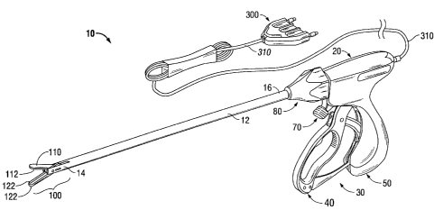
Fig. 1 is a perspective view of an electrosurgical forceps in accordance
with an embodiment of the present disclosure;
Fig. 2A is a side view of a pair jaw members including individually
pivoting electrodes pivotally connected thereto in a first spaced apart
position in
accordance with the present disclosure;
Fig. 2B is a side view of the jaw members in a second grasping tissue
position in accordance with the present disclosure;
Fig. 2C is a side view of the jaw members including an insulating
member disposed on each tissue sealing surface of each electrode, the jaw
members being disposed in the first position in accordance with another
embodiment
of the present disclosure;
Fig. 2D is a side view of the jaw members of Fig. 2C in the second
position in accordance with the present disclosure;
Fig. 3A is a side view of the jaw members including a wedge shaped
electrode disposed at a distal end of each jaw member in accordance with
another
embodiment of the present disclosure;
Fig. 3B is a side view of the jaw members of Fig 3A shown in the
second grasping position;
Fig. 3C is a side view of the jaw members including an insulating
member disposed on each tissue sealing surface of each electrode, the jaw
members being disposed in the first position in accordance with another
embodiment
the present disclosure;
Fig. 3D is a side view of the jaw members of Fig. 3C in the second
position in accordance with the present disclosure;
4
CA 02676742 2009-08-26
Fig. 4A is a side view of jaw members having opposing electrodes
thereof pivotally connected at the distal end and connected by a spring at the
proximal end, in accordance with the present disclosure;
Fig. 4B is a side view of the jaw members of Fig. 4A in the second
grasping position in accordance with the present disclosure;
Fig. 4C is a side view of the jaw members including an insulating
member disposed on each tissue sealing surface of each electrode, in the first
position in accordance with another embodiment of the present disclosure;
Fig. 4D is a side view of the jaw members of Fig. 4C in the second
position in accordance with the present disclosure;
Fig. 5A is a side view of a pair of jaw members connected by a
trapezoidal pivot mechanism including electrodes disposed at a distal end
thereof
and shown in an open, spaced apart position;
Fig. 5B is a side view of the jaw members of Fig. 5A having an
insulating member disposed on each of the tissue sealing surfaces of the
electrodes;
Fig. 5C is a side view of the jaw members of Fig. 5A shown in the
second grasping position;
Fig. 5D is a side view of the jaw members of Fig. 5B shown in the
second position;
Fig. 6A is a side view of jaw members having opposing electrodes
hingedly connected at the distal ends thereof in accordance with the present
disclosure;
Fig. 6B is a side view of the jaw member of Fig. 6A shown in the
second position grasping thick tissue;
Fig. 6C is a side view of the jaw member of Fig. 6A shown in the
second position grasping thin tissue;
Fig. 7A is a side view of jaw members having a recess disposed
therein and opposing electrodes hingedly connected at distal ends thereof in
accordance with the present disclosure;
CA 02676742 2009-08-26
Fig. 7B is a side view of the jaw members of Fig. 7A shown in the
second position grasping thick tissue; and
Fig. 7C is a side view of the jaw members of Fig. 7A shown in the
second position grasping thin tissue.
DETAILED DESCRIPTION
Various embodiments of the present disclosure are described
hereinbelow with reference to the accompanying drawings. Well-known functions
or
constructions are not described in detail to avoid obscuring the present
disclosure in
unnecessary detail. Those skilled in the art will understand that the present
disclosure
may be adapted for use with a laparoscopic instrument, an endoscopic
instrument, or
an open instrument; however, different electrical and mechanical connections
and
considerations may apply to each particular type of instrument. The novel
aspects with
respect to vessel and tissue sealing are generally consistent with respect to
the open,
laparoscopic, and endoscopic designs. In the drawings and in the description
that
follows, the term "proximal", as is traditional, will refer to the end of the
forceps that is
closer to the user, while the term "distal" will refer to the end of the
forceps that is
further from the user.
Referring now to Fig. 1, a bipolar electrosurgical forceps according to an
embodiment of the present disclosure is shown including electrosurgical
forceps 10
configured to support end effector assembly 100. Forceps 10 typically includes
various
conventional features (e.g., a housing 20, a handle assembly 30, a rotating
assembly
80, a trigger assembly 70, etc.) that enable forceps 10 and end effector
assembly 100
to mutually cooperate to grasp, seal and, if warranted, divide tissue. Forceps
10
6
CA 02676742 2009-08-26
generally includes housing 20 and handle assembly 30 that includes moveable
handle
40 and handle 50 which is integral with housing 20. Handle 40 is moveable
relative to
handle 50 to actuate end effector assembly 100 to grasp and treat tissue.
Forceps 10
also includes shaft 12 that has distal end 14 that mechanically engages end
effector
assembly 100 and proximal end 16 that mechanically engages housing 20
proximate
rotating assembly 80 disposed at the distal end of housing 20. Rotating
assembly 80 is
mechanically associated with shaft 12. Movement of rotating assembly 80
imparts
similar rotational movements to shaft 64 which, in turn, rotates end effector
assembly
100.
As explained in more detail below, with respect to Figs. 2A-2D, end
effector assembly 100 includes jaw members 110 and 120 having proximal ends
111a,
121 a and distal ends 111 b, 121 b. Jaw members 110 and 120 are moveable from
a first
position wherein jaw members 110 and 120 are spaced relative to one another,
to a
second position wherein jaw members 110 and 120 are closed and cooperate to
grasp
tissue therebetween. Each jaw member 110, 120 includes respective electrodes
112
and 122 having an electrically conductive tissue sealing surface, 114 and 124,
respectively, disposed on an inner-facing surface thereof. Electrically
conductive tissue
sealing surfaces 114 and 124 cooperate to seal tissue held therebetween upon
the
application of electrosurgical energy.
Referring now to Figs. 2A-2D, end effector assembly 100 includes jaw
members 110 and 120 connected at their respective proximal ends, 111a and
121a, via
a suitable pivot mechanism 130. Jaw members 110 and 120 are rotatable about
pivot
pin 132 to effect grasping and sealing of tissue 600 (see Fig. 2B). Jaw
members 110
and 120 include similar component features that cooperate to permit facile
rotation
about pivot pin 132. Other systems and methods for closing the jaws are
possible and
7
CA 02676742 2009-08-26
are within the purview of those skilled in the art. The jaw configuration may
also be
bilateral or unilateral.
Electrodes 112 and 122 are pivotally connected to the corresponding jaw
members 110 and 120 via respective pivot mechanisms 142 and 162. As mentioned
above, each electrode 112 and 122 has an electrically conductive tissue
sealing
surface 114, 124, respectively disposed thereon that are positioned to
generally
oppose one another, for grasping tissue therebetween.
As shown in Fig. 2B, as jaw members 110 and 120 are moved about
pivot mechanism 130 relative to one another to grasp tissue 600, electrodes
112 and
122 tilt about respective pivots 142 and 162 such that electrically conductive
tissue
sealing surfaces 114 and 124 mutually cooperate in a substantially parallel
manner to
engage tissue. By assuring that the sealing surfaces 114 and 124 grasp tissue
in a
substantially parallel manner, the tissue thickness between electrodes 112 and
122
remains substantially uniform along the length of the sealing surfaces 114 and
124.
This allows the surgeon to selectively apply a uniform closure pressure and a
uniform
amount of electrosurgical energy to tissue 600 between electrodes 112 and 122.
As shown in Figs. 2C-2D, a pair of non-conductive insulating members
190 are disposed on electrically conductive tissue sealing surfaces 114 and/or
124 to
prevent unintended shorting between the two electrically conductive tissue
sealing
surfaces 114 and 124. Insulating members 190 may also be used to maintain an
effective gap distance between sealing surfaces 114 and 124 to promote tissue
sealing, e.g. about 0.001 inches to about 0.006 inches. Insulating member 190
may
also be configured as an insulating ridge disposed along a length of
electrically
conductive tissue sealing surface 114 or 124.
8
CA 02676742 2009-08-26
Referring now to Figs. 3A-3D, in another embodiment, end effector
assembly 200 includes jaw members 210 and 220 that are connected at their
respective proximal ends, 211a and 221a, by a suitable pivot mechanism 230 and
rotatable about pivot pin 232. The electrodes 212 and 222 are configured to be
wedge-shaped, such that the thickness of electrodes 212 and 222 increases
distally
along a length thereof. Any suitable angle may be incorporated into the
electrode to
form the wedge-shape.
As shown in Fig 3B, the wedge-shaped configuration of the electrodes
212 and 222 promotes parallel closure of respective electrically conductive
tissue
sealing surfaces 214 and 224 against tissue 600 disposed between jaw members
210 and 220. As the jaw members 210 and 220 move from the first position, as
shown
in Figs. 3A and 3C, to the second position, as shown in Figs. 3B and 3D,
tissue 600 is
squeezed toward the distal ends 211b and 221b of jaw members 210 and 220,
respectively. At the same time, the wedged-shaped electrodes 212 and 222
squeeze
tissue 600 toward the proximal ends 211 a and 221 a of jaw members 210 and
220, until
tissue sealing surfaces 214 and 224 become parallel. Substantially parallel
tissue
sealing surfaces 214 and 224, as shown in Figs. 3B and 3D, ensure that tissue
thickness between electrodes 212 and 222 remains substantially uniform along a
length of sealing surfaces 214 and 224. This enables a surgeon to apply
accurate
closure pressure and a proper amount of electrosurgical energy in a uniform
fashion to
seal tissue 600.
Figs. 3C-3D show a pair of non-conductive insulating members 290 are
disposed on the electrically conductive tissue sealing surfaces 214 and/or 224
to
prevent unintended shorting between the two tissue sealing surfaces 214 and
224.
Insulating members 290 may also be used to maintain an effective gap distance
9
CA 02676742 2009-08-26
between sealing surfaces 214 and 224 to promote tissue sealing, e.g., about
0.001
inches to about 0.006 inches. Insulating members 290 may also be configured as
insulating ridges disposed along a length of electrically conductive tissue
sealing
surface 214 and 224.
Referring now to Fig. 4A-4D, in another embodiment, end effector
assembly 600 includes jaw members 410 and 420 pivotally connected to one
another at proximal ends 411a and 421a via a suitable pivot mechanism 430
including pivot pin 432. A recess 415 and 425 (see Fig. 4D) may be defined
within
each jaw member 410 and 420, respectively. Electrodes 412 and 422 are disposed
within each respective recess 415 and 425 and are pivotally connected to
respective
jaw members 410 and 420 at the distal ends 413b and 423b thereof.
Alternatively,
electrodes 412 and 422 may be connected to an inner facing surface of jaw
members 410 and 420, respectively, similar to that shown in Figs. 2A-2D. Each
respective electrode 412 and 422 is also connected at the proximal end 413a
and
423a thereof to jaw members 412 and 422, respectively, via resilient members
472
and 492, such that resilient members 472 and 492 bias each electrode 412 and
422
against tissue 600 disposed between jaw members 410 and 420. Resilient members
472 and 492 may be any compressible and/or flexible segment as is within the
purview of those skilled in the art. In embodiments, resilient members 472 and
492
are springs. As shown in Figs. 4B and 4D, as jaw members 410 and 420 are
rotated
about pivot pin 432 to the second position in order to grasp tissue 600
therebetween,
electrodes 412 and 422 tilt about pivots 442 and 462 against springs 472 and
492 to
compress tissue in a more parallel manner. As mentioned above in regards to
previous embodiments, closing the electrodes and engaging tissue in a
substantially
parallel manner ensures that the tissue thickness between electrodes 412 and
422
CA 02676742 2009-08-26
remains substantially uniform along a length of sealing surfaces 414 and 424,
thus
allowing the surgeon to apply a uniform closure pressure and a uniform amount
of
electrosurgical energy to tissue 600 between electrodes 412 and 422.
Figs. 4C and 4D show a pair of opposing insulating members 490
disposed on electrically conductive sealing surfaces 414 and 424 configured as
insulating ridges disposed along a length of electrically conductive tissue
sealing
surface 414 and 424, as described above in relation to previous embodiments.
Insulating members 490 prevent unintended shorting between the two tissue
sealing
surfaces 414 and 424. Insulating members 490 may also maintain an effective
gap
distance between sealing surfaces 414 and 424 to promote tissue sealing, e.g.,
about 0.001 inches to about 0.006 inches.
In yet another embodiment, as shown in Figs. 5A-5D, jaw members
510 and 520 of end effector assembly 500 include electrodes 512 and 522,
respectively, disposed on opposing surfaces thereon. Electrodes 512 and 522
include electrically conductive sealing surfaces 514 and 524, respectively. A
trapezoidal pivot mechanism 580 operably connects jaw members 510 and 520 to
one another via pivot connections 582. Pivot connections 584 connect an
actuator
rod 586 to trapezoidal pivot mechanism 580. When closure of jaw members 510
and
520 is required, e.g., by squeezing handle assembly 40, in order to grasp
tissue
therebetween, actuator rod 586 is advanced distally such that trapezoidal
pivot
mechanism 580 promotes a more parallel closure of jaw members 510 and 520, as
shown in Figs. 5C-5D. This results in parallel closure of tissue sealing
surfaces 514
and 524, which ensures that tissue thickness between electrodes 512 and 522
remains
substantially uniform along a length of sealing surfaces 514 and 524. The
surgeon can
11
CA 02676742 2009-08-26
selectively apply a uniform closure pressure and a uniform amount of
electrosurgical
energy to tissue 600 between electrodes 512 and 522.
As shown in Figs. 5B and 5D, non-conductive insulating members 590
may also be disposed on electrically conductive tissue sealing surfaces 514
and 524 to
prevent unintended shorting between the two electrically conductive tissue
sealing
surfaces 514 and 526. Insulating members 590 may also maintain an effective
gap
distance between sealing surfaces 514 and 524 to promote tissue sealing, e.g.,
about 0.001 inches to about 0.006 inches.
Referring now to Figs. 6A-6C, end effector assembly 601 includes jaw
members 610 and 620 pivotally connected to one another at proximal ends 611a
and
621a via a suitable pivot mechanism 630 including pivot pin 632. Electrodes
612
and 622 are hingedly connected to respective jaw members 610 and 620 at the
distal ends 613b and 623b thereof via resilient members 672 and 692 such that
resilient members 672 and 692 bias each electrode 612 and 622 against tissue
600
disposed between jaw members 610 and 620. Resilient members 672 and 692 may
be substantially straight or shaped pieces of spring metal or other stiff, yet
bendable
segments as is within the purview of those skilled in the art to provide a
balanced
force on tissue. As shown in Figs. 6B and 6C, as jaw members 610 and 620 are
rotated about pivot pin 632 to the second position in order to grasp tissue
600
therebetween, resilient members 672 and 692 bend back with some force such
that
electrodes 612 and 622 tilt to compress tissue in a more parallel manner. As
mentioned above with regard to previous embodiments, closing the electrodes
and
engaging tissue in a substantially parallel manner ensures that the tissue
thickness
between electrodes 612 and 622 remains substantially uniform along a length of
sealing surfaces 614 and 624, thus allowing the surgeon to apply a uniform
closure
12
CA 02676742 2009-08-26
pressure and a uniform amount of electrosurgical energy to tissue 600 between
electrodes 612 and 622.
Figs. 6A-6C also show optional pairs of opposing insulating members
690 disposed on electrically conductive sealing surfaces 614 and 624
configured as
insulating ridges disposed along a length of electrically conductive tissue
sealing
surface 614 and 624, as described above in relation to previous embodiments.
Insulating members 690 prevent unintended shorting between the two tissue
sealing
surfaces 614 and 624. Insulating members 690 may also maintain an effective
gap
distance between sealing surfaces 614 and 624 to promote tissue sealing, e.g.,
about 0.001 inches to about 0.006 inches.
Referring now to Fig. 7A-7C, in another embodiment, end effector
assembly 700 includes jaw members 710 and 720 pivotally connected to one
another at proximal ends 711a and 721a via a suitable pivot mechanism 730
including pivot pin 732. A recess 715 and 725 is defined within each jaw
member
710 and 720, respectively. Electrodes 712 and 722 are disposed proximal to
each
respective recess 715 and 725 and are hingedly connected to respective jaw
members 710 and 720 at the distal ends 713b and 723b thereof via resilient
members 772 and 792 such that resilient members 772 and 792 bias each
electrode
712 and 722 against tissue 600 disposed between jaw members 710 and 720.
Resilient members 772 and 792 may be substantially straight or shaped pieces
of
spring metal or other stiff, yet bendable segments as is within the purview of
those
skilled in the art to provide a balanced force on tissue held between jaw
members
710 and 720. As shown in Figs. 7B and 7C, as jaw members 710 and 720 are
rotated about pivot pin 732 to the second position in order to grasp tissue
600
therebetween, electrodes 712 and 722 tilt against resilient members 772 and
792 to
13
CA 02676742 2009-08-26
compress tissue in a more parallel manner. As mentioned above in regards to
previous embodiments, closing the electrodes and engaging tissue in a
substantially
parallel manner ensures that the tissue thickness between electrodes 712 and
722
remains substantially uniform along a length of sealing surfaces 714 and 724,
thus
allowing the surgeon to apply a uniform closure pressure and a uniform amount
of
electrosurgical energy to tissue 600 between electrodes 712 and 722.
Figs. 7A-7C also show an optional pair of opposing insulating members
790 disposed on electrically conductive sealing surfaces 714 and 724
configured as
insulating ridges disposed along a length of electrically conductive tissue
sealing
surface 714 and 724, as described above in relation to previous embodiments.
Insulating members 790 prevent unintended shorting between the two tissue
sealing
surfaces 714 and 724. Insulating members 790 may also maintain an effective
gap
distance between sealing surfaces 614 and 624 to promote tissue sealing, e.g.,
about 0.001 inches to about 0.006 inches.
While several embodiments of the disclosure have been shown in the
drawings and/or discussed herein, it is not intended that the disclosure be
limited
thereto, as it is intended that the disclosure be as broad in scope as the art
will allow
and that the specification be read likewise. Therefore, the above description
should
not be construed as limiting, but merely as exemplifications of particular
embodiments. Those skilled in the art will envision other modifications within
the
scope and spirit of the claims appended hereto.
14