Note: Descriptions are shown in the official language in which they were submitted.
CA 02677383 2009-08-05
WO 2008/100816 PCT/US2008/053398
ANTIBODY DISPLAY
1. BACKGROUND OF THE INVENTION
[0001] Recombinant antibodies have become increasingly prevalent therapeutics
over
the past decade; currently they represent over 30% of biopharmaceuticals in
clinical trials.
As such, the rapid generation, characterization and optimization of
recombinant antibodies is
critically important for the biopharmaceutical industry. An early solution for
the problem
was provided by the use of phage display libraries of simplified antibody
fragments. The
possibility to generate large libraries and the ease with which a library is
screened made
antibody fragment phage display technology a powerful tool for the development
of new
therapeutics against various human diseases.
[0002] Phage display technologies, however, have a disadvantage in that they
rely on
the screening of antibody fragments as opposed to full length antibodies.
Given that most
therapeutic applications call for the use of divalent IgG antibodies, an
isolated antibody
fragment with the desired binding properties is usually converted into a full
length antibody.
The conversion process is not only labor intensive, but may also result in the
loss of antigen
binding specificity.
[0003] Additional display technologies have been developed to address these
problems. Recently, the a type agglutinin mediated display of antibody
fragments (scFv and
Fab) on the yeast cell wall has been emerging as an effective alternative to
the phage display
technology.
[0004] There is still a need, however, in the art for the development of new
technologies that allow the rapid screening of large libraries of full length
antibodies.
2. SUMMARY OF THE INVENTION
[0005] The present invention relates to an antibody or a fragment thereof that
may be
displayed on the extracellular surface of the plasma membrane of a cell (e.g.,
yeast cell),
referred to herein as an "antibody of the invention" and like terms. In
certain embodiments,
an antibody of the invention comprises a heavy chain or a fragment thereof and
optionally a
light chain or a fragment thereof, wherein either the heavy chain or light
chain further
comprises an amino acid sequence that targets the antibody or a fragment
thereof to the
extracellular surface of the plasma membrane. In one embodiment, an antibody
of the
invention comprises a full length heavy chain having an amino acid sequence
that targets the
1
CA 02677383 2009-08-05
WO 2008/100816 PCT/US2008/053398
antibody to the extracellular surface of the plasma membrane, wherein said
amino acid
sequence is fused to the C terminus of said heavy chain, and wherein said
antibody may
further comprise a full length light chain. In still another embodiment, an
antibody of the
invention comprises a portion of a heavy chain having an amino acid sequence
that targets
the antibody to the extracellular surface of the plasma membrane, wherein said
amino acid
sequence is fused to the C terminus of said heavy chain portion, and wherein
said antibody
may further comprise a light chain or a fragment thereof. In a specific
embodiment, said
amino acid sequence that targets an antibody of the invention to the
extracellular surface of
the plasma membrane is a transmembrane domain. In another embodiment, said
amino acid
sequence that targets an antibody of the invention to the extracellular
surface of the plasma
membrane is a GPI anchor domain.
[0006] The present invention further relates to vectors comprising
polynucleotides
encoding an antibody or a fragment thereof that may be displayed on the
extracellular surface
of the plasma membrane of a cell (e.g., yeast cell), referred to herein as a
"vector of the
invention". In one embodiment, a vector of the invention is operable in a host
cell to direct
the expression and the display of an antibody or a fragment thereof on the
extracellular
surface of the plasma membrane. In a specific embodiment, a vector of the
invention is a set
of two vectors wherein a first vector comprises a polynucleotide encoding a
heavy chain of
an antibody or a fragment thereof and a second vector comprises a
polynucleotide encoding a
light chain of an antibody or a fragment thereof, wherein said antibody or a
fragment thereof
may be displayed on the extracellular surface of the plasma membrane.
[0007] The present invention also provides host cells comprising an antibody
or a
fragment thereof that may be displayed on the extracellular surface of the
plasma membrane,
referred to herein as a "host cell of the invention". In one embodiment, a
host cell of the
invention is a eukaryotic cell selected from the Ascomycota phylum. In a
specific
embodiment, a host cell of the invention is Saccharomyces cerevisiae,
Hansenula
polymorpha, Kluyveromyces lactis, Pichia pastoris, Schizosaccharomyces pombe,
or
Yarrowia lipolytica. In one embodiment, a host cell of the invention comprises
a genetic
mutation wherein said genetic mutation renders the cell wall permeable to an
antibody
binding agent (e.g. antigens, Fc receptors, antibodies).
[0008] The present invention also relates to libraries comprising
polynucleotides
encoding a heterogeneous population of antibodies or a fragment thereof that
may be
displayed on the extracellular surface of the plasma membrane of a cell (e.g.,
yeast cell),
referred to herein as a "polynucleotide library of the invention". In one
embodiment, a
2
CA 02677383 2009-08-05
WO 2008/100816 PCT/US2008/053398
polynucleotide library of the invention may comprise polynucleotides encoding
antibodies or
a fragment thereof comprising a heterogeneous population of heavy chain
variable regions.
In another embodiment, a polynucleotide library of the invention may comprise
polynucleotides encoding antibodies or a fragment thereof comprising a
heterogeneous
population of light chain variable regions. In a further embodiment, a
polynucleotide library
of the invention may comprise polynucleotides encoding antibodies or a
fragment thereof
comprising a heterogeneous population of Fc regions, including variant Fc
regions. In one
embodiment, a population of host cells comprises a polynucleotide library of
the invention.
[0009] The present invention also relates to libraries comprising a
heterogeneous
population of antibodies or a fragment thereof that may be displayed on the
extracellular
surface of the plasma membrane of a cell (e.g., yeast cell), referred to
herein as an "antibody
library of the invention". In one embodiment, an antibody library of the
invention may
comprise a heterogeneous population of heavy chain variable regions. In
another
embodiment, an antibody library of the invention may comprise a heterogeneous
population
of light chain variable regions. In a further embodiment, an antibody library
of the invention
may comprise a heterogeneous population of Fc regions, including variant Fc
regions. In
one embodiment, a population of host cells comprises an antibody library of
the invention.
[0010] The invention also provides methods of screening a polynucleotide
library of
the invention or an antibody library of the invention. In one embodiment, a
method of
screening a library allows the identification of an antibody or a fragment
thereof that binds a
specific antigen. In one embodiment, a method of screening a library allows
the
identification of an antibody or a fragment thereof having an altered binding
to a specific
antigen. In one embodiment, a method of screening a library allows the
identification of an
antibody or fragment having an altered binding to effector molecules (e.g.,
FcyRs and/or
Clq).
[0011] The invention also provides methods of expressing a polynucleotide
library of
the invention in a host cell. In one embodiment, a polynucleotide library of
the invention
may encode a heterogeneous population of antibodies or a fragment thereof that
may be
displayed on the extracellular surface of the plasma membrane of a cell (e.g.,
yeast cell).
3. BRIEF DESCRIPTION OF THE FIGURES
[0012] For the purpose of illustrating representative embodiments of the
invention,
drawings are provided herein.
3
CA 02677383 2009-08-05
WO 2008/100816 PCT/US2008/053398
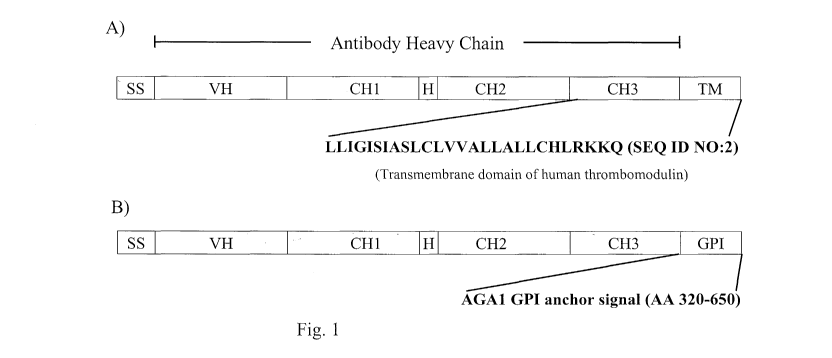
[0013] Figure 1. Schematic representation of heavy chain fusion polypeptides
of an
antibody of the invention. Panel A) depicts a heavy chain targeted for display
on the
extracellular surface of the yeast plasma membrane. It comprises a signal
sequence (SS), a
heavy chain variable region (VH), a heavy chain constant region 1(CHl), a
hinge region (H),
a heavy chain constant region 2 (CH2), a heavy chain constant region 3 (CH3),
and a
transmembrane domain (TM). The amino acid sequence of the human thrombomodulin
transmembrane domain is shown as a non limiting example (SEQ ID NO:2). Panel
B)
depicts a heavy chain targeted for display on the yeast cell wall. It
comprises a signal
sequence (SS), a heavy chain variable region (VH), a heavy chain constant
region 1(CHl) , a
hinge region (H), a heavy chain constant region 2 (CH2), a heavy chain
constant region 3
(CH3), and a GPI anchor domain (GPI). The GPI anchor domain comprising
residues 320-
650 of the Agal protein is given as a non limiting example.
[0014] Figure 2. (A) Schematic representation of a yeast vector that may be
used to
control the expression of the heavy chain of an antibody or a fragment thereof
that may be
displayed on the surface of a yeast cell. (B) Schematic representation of a
yeast vector that
may be used to control the expression of the light chain of an antibody or a
fragment thereof
that may be displayed on the surface of a yeast cell.
[0015] Figure 3. Fluorescence intensity profile of immunostained spheroplasts
expressing the 10C121ight chain and a cell surface displayed heavy chain
fusion polypeptide.
"Agalp GPI", "hThrm TM", "Axl2p TM", and "Swplp TM" denotes the data obtained
with
spheroplasts expressing the 10C12 heavy chain fused with the Agalp GPI signal,
the human
thrombomodulin transmembrane region, the Axl2p transmembrane region, and the
Swplp
transmembrane region, respectively. Spheroplasts not expressing an antibody
serve as
negative control ("Uninduced"). Samples were stained with FITC conjugated anti-
human
IgG(H+L) antibody. "hThrm TM", "Axl2p TM", and "Swplp TM" samples show
significant staining; the staining profile of "Agalp GPI" resembles that of
the negative
control sample.
[0016] Figure 4. Mean fluorescence intensity of immunostained spheroplasts.
Spheroplasts expressing the lOCl2light chain and a lOCl2 heavy chain fused to
either
human thrombomodulin transmembrane domain (10C12-hThrMTM, light gray bars) or
Agal
GPI signal (10C12-AgaGPI, dark grey bars) were immunostained with FITC
conjugated anti-
human IgG(H+L) antibody. Spheroplasts were generated by lyticase treatment at
the
indicated pH in the presence or absence of 0.5 M sorbitol. Control samples of
immunostained intact yeast cells ("no lyticase") were also analyzed. The
hThrmTM fused
4
CA 02677383 2009-08-05
WO 2008/100816 PCT/US2008/053398
10C 12 antibody is positively stained on spheroplasts, but not intact yeast
cells. Lyticase
treatment, on the other hand, significantly reduces the signal obtained with
the AgaGPI fused
antibody.
[0017] Figure 5. Fluorescence intensity profile of immunostained intact yeast
cells
expressing the 10C12 light chain and the 10C12 heavy chain fused to the Agal
GPI signal
(lOCl2-AgaGPI); and immunostained spheroplasts expressing the lOCl2 light
chain and the
10C12 heavy chain fused to the human thrombomodulin transmembrane domain
(10C12-
hThrMTM). Samples were stained with biotinylated EphA4-Fc, a ligand for 10C12,
and PE
conjugated streptavidin. Spheroplasts without lOCl2 expression were included
as a negative
control ("uninduced"). Positive staining is detected for both the GPI anchored
and ThrmTM
anchored 10C12 antibody.
[0018] Figure 6. Fluorescence intensity profile of immunostained spheroplasts
expressing the 3F2 anti-EphA2 Fab fused to the human thrombomodulin
transmembrane
domain (hThrmTM). hThrmTM is fused to either the heavy chain (3F2Fab-
hThrmTM(HC))
or light chain (3F2Fab-hThrmTM(LC)) of the Fab. Spheroplasts without Fab
expression
were included as a negative control ("uninduced"). Spheroplasts were stained
with (A)
biotinylated EphA2-Fc/ PE conjugated streptavidin or (B) FITC conjugated anti-
IgG(H+L).
The heavy chain and light chain anchored Fabs are stained equally well with
FITC
conjugated anti-IgG(H+L); they also bind the biotinylated EphA2-Fc ligand.
[0019] Figure 7. Fluorescence intensity profile of immunostained spheroplasts
expressing the 3F2 scFv comprising a FLAG tag and a human thrombomodulin
transmembrane domain fused to its N and C terminus, respectively. Spheroplasts
were
stained with a FITC conjugated anti-FLAG antibody; spheroplasts without scFv
expression
were used as negative control ("uninduced"). 3F2 scFv is readily detected on
the cell surface.
[0020] Figure 8. Fluorescence intensity profile of immunostained intact yeast
cells.
MAT alpha, mnn9, ura3, leu2, his4 yeast cells expressing the 3F2 anti-EphA2
scFv-Fc fused
to the human thrombomodulin transmembrane domain (3F2scFv-Fc-hThrmTM) or the
3F2
heavy chain fused to the human thrombomodulin transmembrane domain (3F2HC-
hThrmTM) were stained with FITC conjugated anti-human Fc antibody. Cells
without an
expression construct were used as negative controls. Cells expressing 3F2scFv-
Fc-
hThrmTM, but not 3F2HC-hThrmTM show positive staining.
[0021] Figure 9. Fluorescence intensity profile of immunostained intact yeast
cells.
MAT alpha, mnn9, ura3, leu2, his4 yeast cells expressing the 3F2 anti-EphA2
scFv-Fc fused
to the human thrombomodulin transmembrane domain (3F2scFv-Fc-hThrmTM) or the
3F2
5
CA 02677383 2009-08-05
WO 2008/100816 PCT/US2008/053398
heavy chain fused to the human thrombomodulin transmembrane domain (3F2HC-
hThrmTM) were stained with biotinylated EphA2/ PE conjugated streptavidin.
Cells without
an expression construct were used as negative controls. Cells expressing
3F2scFv-Fc-
hThrmTM, but not 3F2HC-hThrmTM show positive staining.
[0022] Figure 10. Schematic representation of the 2 -scFv-TM vector.
[0023] Figure 11. Experimental flow chart of a single round of antibody
selection.
[0024] Figure 12. Efficiency of antibody gene amplification from plasmid DNA
purified from yeast spheroplast. PCR product amplified form DNA purified from
1000,
2000, 3000, 4000, 5000, 6000, and 10000 cells were run in lanes 1 to 7,
respectively, on an
agarose gel. Lane 8 shows the PCR product of direct amplification from 2000
cells. All PCR
reactions yielded a DNA fragment of the same size. The staining intensity of
the various
PCR products was comparable.
[0025] Figure 13. Fluorescent intensity profile of various artificial
libraries of
antibody displaying spheroplasts during the first round of selection.
[0026] Figure 14. Fluorescent intensity profile of various artificial
libraries of
antibody displaying spheroplasts during the second round of selection.
[0027] Figure 15. Schematic representation of the pPICZ+scFv-FC vector.
[0028] Figure 16. Fluorescent intensity profile of EphA4-Fc-biotin/
streptavidin-PE
stained P. pastoris expressing a GPI anchored l OC 12 anti-EphA4 scFv, scFv-Fc
or full IgG
on the cell surface. The mean fluorescence intensity of the antibody
expressing yeast cells is
separated from the parental negative control yeast cells by at least 21ogs.
[0029] Figure 17. Zymolase digestion dependence of antigen binding by membrane
anchored 10C12 anti-EphA4 His/FLAG-tagged-scFv (1OC12ScFvHF-TM) or scFv-Fc
(l OC12ScFvFc-TM) molecules expressed in P. pastoris. Spheroplasts prepared by
0 min, 2
min, 5 min, 10 min, 20 min or 30 min zymolase digestion (2.5 units zymolase
per 108 cells)
were stained with EphA4-Fc-biotin/ streptavidin-PE. Fluorescent intensity
profile of the
stained spheroplasts is shown. Similarly stained parental P. pastoris
spheroplasts were
included as negative control. The fluorescent intensity profile of TM anchored
scFv and
scFv-Fc expressing cells is almost identical at all time points tested. The
separation between
the mean fluorescent intensity (MFI) of antibody expressing and control cells
reaches a 21og
maximum after 2 minutes of zymolase digestion.
[0030] Figure 18. P. pastoris expressed TM anchored 10C12 anti-EphA4 scFv or
scFv-Fc molecules are accessible to antigen binding in absence of zymolase
treatment. Cells
6
CA 02677383 2009-08-05
WO 2008/100816 PCT/US2008/053398
were washed with PBS or 50 mM DTT/ 1 M sorbitol, incubated with 10 g/ml, 1
g/ml or
0.1 g/ml EphA4-Fc-biotin, and stained with streptavidin-PE. Similarly treated
parental P.
pastoris cells were included as negative control. The fluorescent intensity
profile of PBS
washed TM anchored scFv and scFv-Fc expressing cells are identical at each
concentration of
EphA4-Fc-biotin tested; the separation between the MFI of antibody expressing
cells and
parental cells is not dependent on antigen concentration within the range
tested. The
fluorescent intensity profile of DTT/sorbitol washed TM anchored scFv-Fc
expressing cells is
higher than that of the scFv expressing cells at all EphA4-Fc-biotin
concentrations tested; the
separation between the MFI of antibody expressing cells and parental cells is
not dependent
on antigen concentration within the range tested..
[0031] Figure 19. Episomal expression of P. pastoris surface displayed
antibodies.
Fluorescent intensity profile of 10 ug/ml EphA4-Fc-biotin/ streptavidin-PE
stained cells.
Surface displayed antibodies were expressed from an episomal vector. Similarly
treated
parental P. pastoris cells were included as negative control.
4. DEFINITIONS
[0032] As used herein, the term "polynucleotide encoding an antibody or a
fragment
thereof' encompasses a composition of polynucleotides comprising one or more
polynucleotide chains encoding the individual polypeptide chains of said
antibody or a
fragment thereof.
[0033] As used herein, "vector" refers to any element capable of serving as a
vehicle
of genetic transfer, gene expression, or replication or integration of a
foreign polynucleotide
in a host cell. A vector can be an artificial chromosome or plasmid, and can
be integrated
into the host cell genome or exist as an independent genetic element (e.g.,
episome, plasmid).
A vector can exist as a single polynucleotide or as two or more separate
polynucleotides. A
"vector of the invention" is capable, in an appropriate host, of directing
expression of at least
one chain of an antibody or fragment thereof that may be displayed on the
plasma membrane
of the host cell. Vectors according to the present invention can be single
copy vectors or
multicopy vectors (indicating the number of copies of the vector typically
maintained in the
host cell). Vectors of the present invention include yeast expression vectors,
2 vectors and
centromere vectors. A "shuttle vector" (or bi-functional vector) is known in
the art as any
vector that can replicate in more than one species of organism. For example, a
shuttle vector
that can replicate in both Escherichia coli (E. coli) and Saccharomyces
cerevisiae (S.
7
CA 02677383 2009-08-05
WO 2008/100816 PCT/US2008/053398
cerevisiae) can be constructed by linking sequences from an E. coli plasmid
with sequences
from the yeast 2 plasmid.
[0034] The term "library" refers to a mixture of heterogeneous polypeptides or
polynucleotides. A library is composed of members that have similar
polypeptide or
polynucleotide sequences. Where the library is a polynucleotide library, it
encodes a
heterogeneous population of polypeptides (e.g., a heterogeneous population of
antibody
polypeptides). Sequence differences between library members are responsible
for the
diversity present in the library. The library can take the form of a simple
mixture of
polypeptides or polynucleotides, or can be in the form of organisms or cells,
for example
yeast cells and the like, that are transformed with a library of
polynucleotides.
Advantageously, polynucleotides are incorporated into expression vectors, in
order to allow
expression of the polypeptides encoded by the polynucleotides. In one
embodiment, a library
can take the form of a population of host cells, each cell containing one or
more copies of an
expression vector containing a one or more polynucleotide encoding an antibody
or a
fragment thereof that can be expressed to produce its corresponding antibody
or a fragment
thereof . Thus, the population of host cells has the potential to encode a
heterogeneous
population of antibodies or a fragment thereof.
[0035] As used herein, the terms "antibody" and "antibodies" refer to
monoclonal
antibodies, multispecific antibodies, human antibodies, humanized antibodies,
camelised
antibodies, chimeric antibodies, single-chain Fvs (scFv), disulfide-linked Fvs
(sdFv), Fab
fragments, F (ab') fragments, and anti-idiotypic (anti-Id) antibodies
(including, e.g., anti-Id
antibodies to antibodies of the invention), and epitope-binding fragments of
any of the above.
In particular, antibodies include immunoglobulin molecules and immunologically
active
fragments of immunoglobulin molecules, i.e., molecules that contain an antigen
binding site,
these fragments may or may not be fused to another immunoglobulin domain
including but
not limited to, an Fc region or a fragment thereof. The term "antibody" and
"antibodies" also
encompass any polypeptide comprising an immunoglobulin molecule or an
immunologically
active fragment thereof (i.e., molecules that contain an antigen binding site)
fused to any
polypeptide fusion partner. As used herein, the terms "antibody" and
"antibodies" also
include the Fc variants, full-length antibodies and Fc variant-fusions
comprising Fc regions,
or a fragment thereof. Fc variant- fusions include but are not limited to,
scFv-Fc fusions,
variable region (e.g., VL and VH) -Fc fusions, scFv-scFv-Fc fusions.
Antibodies can be of
8
CA 02677383 2009-08-05
WO 2008/100816 PCT/US2008/053398
any type (e.g., IgG, IgE, IgM, IgD, IgA and IgY), class (e.g., IgGl, IgG2,
IgG3, IgG4, IgAl
and IgA2) or subclass.
[0036] The complementarity determining regions (CDRs) residue numbers referred
to
herein are those of Kabat et al. (1991, NIH Publication 91-3242, National
Technical
Information Service, Springfield, VA). Specifically, residues 24-34 (CDRl), 50-
56 (CDR2)
and 89-97 (CDR3) in the light chain variable domain and 31-35 (CDRl), 50-65
(CDR2) and
95-102 (CDR3) in the heavy chain variable domain. Note that CDRs vary
considerably from
antibody to antibody (and by definition will not exhibit homology with the
Kabat consensus
sequences). Maximal alignment of framework residues frequently requires the
insertion of
"spacer" residues in the numbering system, to be used for the Fv region. It
will be
understood that the CDRs referred to herein are those of Kabat et al. supra.
In addition, the
identity of certain individual residues at any given Kabat site number may
vary from antibody
chain to antibody chain due to interspecies or allelic divergence.
[0037] As used herein "Fc region" includes the polypeptides comprising the
constant
region of an antibody excluding the first constant region immunoglobulin
domain. Thus Fc
refers to the last two constant region immunoglobulin domains of IgA, IgD, and
IgG, and the
last three constant region immunoglobulin domains of IgE and IgM, and the
flexible hinge N-
terminal to these domains. For IgA and IgM Fc may include the J chain. For
IgG, Fc
comprises immunoglobulin domains Cgamma2 and Cgamma3 (Cy2 and Cy3) and the
hinge
between Cgammal (Cyl) and Cgamma2 (Cy2). Although the boundaries of the Fc
region
may vary, the human IgG heavy chain Fc region is usually defined to comprise
residues C226
or P230 to its carboxyl-terminus, wherein the numbering is according to the EU
index as in
Kabat et al. (1991, NIH Publication 91-3242, National Technical Information
Service,
Springfield, VA). The "EU index as set forth in Kabat" refers to the residue
numbering of
the human IgGl EU antibody as described in Kabat et al. supra. Fc may refer to
this region
in isolation, or this region in the context of an antibody, antibody fragment,
or Fc fusion
protein. An Fc variant protein may be an antibody, Fc fusion, or any protein
or protein
domain that comprises an Fc region. Also encompassed are proteins comprising
variant Fc
regions, which are non-naturally occurring variants of an Fc region. The amino
acid
sequence of a non-naturally occurring Fc region (also referred to herein as a
"variant Fc
region") comprises a substitution, insertion and/or deletion of at least one
amino acid residue
compared to the wild type amino acid sequence. Any new amino acid residue
appearing in
the sequence of a variant Fc region as a result of an insertion or
substitution may be referred
to as a non-naturally occurring amino acid residue. Note: Polymorphisms have
been
9
CA 02677383 2009-08-05
WO 2008/100816 PCT/US2008/053398
observed at a number of Fc positions, including but not limited to Kabat 270,
272, 312, 315,
356, and 358, and thus slight differences between the presented sequence and
sequences in
the art may exist.
[0038] As used herein, the term "transmembrane domain" refers to the domain of
a
peptide, polypeptide or protein that is capable of spanning the plasma
membrane of a cell.
These domains can be used to anchor an antibody on the plasma membrane.
[0039] A "chimeric antibody" is a molecule in which different portions of the
antibody are derived from different immunoglobulin molecules such as
antibodies having a
variable region derived from a non-human antibody and a human immunoglobulin
constant
region. Methods for producing chimeric antibodies are known in the art. See
e.g., Morrison,
1985, Science 229:1202; Oi et al., 1986, BioTechniques 4:214; Gillies et al.,
1989, J.
Immunol. Methods 125:191-202; and U.S. Patent Nos. 5,807,715, 4,816,567, and
4,816,397,
CDR-grafting (EP 239,400; PCT Publication No. WO 91/09967; and U.S. Patent
Nos.
5,225,539, 5,530,101, and 5,585,089), veneering or resurfacing (EP 592,106; EP
519,596;
Padlan, 1991, Molecular Immunology 28(4/5): 489-498; Studnicka et al., 1994,
Protein
Engineering 7:805; and Roguska et al., 1994, PNAS 91:969), and chain shuffling
(U.S. Patent
No. 5,565,332).
[0040] A "humanized antibody" is an antibody or its variant or a fragment
thereof
which is capable of binding to a predetermined antigen and which comprises a
framework
region having substantially the amino acid sequence of a human immunoglobulin
and a CDR
having substantially the amino acid sequence of a non-human immunoglobulin. A
humanized antibody comprises substantially all of at least one, and typically
two, variable
domains (Fab, Fab', F(ab')2, Fabc, Fv) in which all or substantially all of
the CDR regions
correspond to those of a non-human immunoglobulin (i.e., donor antibody) and
all or
substantially all of the framework regions are those of a human immunoglobulin
consensus
sequence. In one embodiment, a humanized antibody also comprises at least a
portion of an
immunoglobulin constant region (Fc), typically that of a human immunoglobulin.
Ordinarily,
the antibody will contain both the light chain as well as at least the
variable domain of a
heavy chain. The antibody also may include the CHl, hinge, CH2, CH3, and CH4
regions of
the heavy chain. The humanized antibody can be selected from any class of
immunoglobulins, including, but not limited to, IgM, IgG, IgD, IgA and IgE,
and any isotype,
including, but not limited to, IgGl, IgG2, IgG3 and 1gG4. In another
embodiment, the
constant domain is a complement fixing constant domain where it is desired
that the
humanized antibody exhibit cytotoxic activity, and the class is typically
IgGl. Where such
CA 02677383 2009-08-05
WO 2008/100816 PCT/US2008/053398
cytotoxic activity is not desirable, the constant domain may be of the IgG 2
class. The
humanized antibody may comprise sequences from more than one class or isotype,
and
selecting particular constant domains to optimize desired effector functions
is within the
ordinary skill in the art. The framework and CDR regions of a humanized
antibody need not
correspond precisely to the parental sequences, e.g., the donor CDR or the
consensus
framework may be mutagenized by substitution, insertion or deletion of at
least one residue
so that the CDR or framework residue at that site does not correspond to
either the consensus
or the import antibody. Such mutations, however, will not be extensive. In one
embodiment,
at least 75%, at least 90%, and or at least 95% of the humanized antibody
residues will
correspond to those of the parental framework region (FR) and CDR sequences.
Humanized
antibody can be produced using variety of techniques known in the art,
including but not
limited to, CDR-grafting (European Patent No. EP 239,400; PCT Publication No.
WO
91/09967; and U.S. Patent Nos. 5,225,539, 5,530,101, and 5,585,089), veneering
or
resurfacing (European Patent Nos. EP 592,106 and EP 519,596; Padlan, 1991,
Molecular
Immunology 28(4/5): 489-498; Studnicka et al., 1994, Protein Engineering 7(6):
805-814;
and Roguska et al., 1994, PNAS 91:969-973), chain shuffling (U.S. Patent No.
5,565,332),
and techniques disclosed in, e.g., U.S. Pat. No. 6,407,213, U.S. Pat. No.
5,766,886, PCT
Patent Publication WO 93/17105, Tan et al., 2002, J. Immunol. 169:1119-25,
Caldas et al.,
2000, Protein Eng. 13: 353-60, Morea et al., 2000, Methods 20: 267-79, Baca et
al., 1997, J.
Biol. Chem. 272: 10678-84, Roguska et al., 1996, Protein Eng. 9: 895-904,
Couto et al.,
1995, Cancer Res. 55 (23 Supp): 5973s - 5977s, Couto et al., 1995, Cancer Res.
55: 1717-22,
Sandhu JS, 1994, Gene 150: 409-10, and Pedersen et al., 1994, J. Mol. Biol.
235: 959-73).
Often, framework residues in the framework regions will be substituted with
the
corresponding residue from the CDR donor antibody to alter and/or improve
antigen binding.
These framework substitutions are identified by methods well known in the art,
e.g., by
modeling of the interactions of the CDR and framework residues to identify
framework
residues important for antigen binding and sequence comparison to identify
unusual
framework residues at particular positions. (See, e.g., Queen et al., U.S.
Patent No.
5,585,089; and Riechmann et al., 1988, Nature 332:323).
[0041] The term "ADCC" (antibody-dependent cell-mediated cytotoxicity) refers
to a
cell-mediated reaction in which nonspecific cytotoxic cells that express Fc
receptors (FcR)
(e.g. natural killer (NK) cells, neutrophils, and macrophages) recognize bound
antibody on a
target cell and subsequently cause lysis of the target cell. The primary cells
for mediating
ADCC, NK cells, express FcyRIII only, whereas monocytes express FcyRI, FcyRII
and
11
CA 02677383 2009-08-05
WO 2008/100816 PCT/US2008/053398
FcyRIII. To assess ADCC activity of a molecule of interest, an in vitro ADCC
assay (e.g.
such as that described in U.S. Pat. No. 5,500,362 and U.S. Pat. No. 5,821,337)
may be
performed. Useful effector cells for such assays include peripheral blood
mononuclear cells
(PBMC) and natural killer (NK) cells.
[0042] "Complement dependent cytotoxicity" and "CDC" refer to the lysing of a
target cell in the presence of complement. The complement activation pathway
is initiated by
the binding of the first component of the complement system (C l q) to a
molecule, an
antibody for example, complexed with a cognate antigen. To assess complement
activation, a
CDC assay, e.g. as described in Gazzano-Santoro et al., 1996, J. Immunol.
Methods, 202:163,
may be performed.
5. DETAILED DESCRIPTION
[0043] The present invention relates to an antibody or a fragment thereof that
may be
displayed on the extracellular surface of the plasma membrane of a cell (e.g.,
yeast cell),
referred to herein as an "antibody of the invention" and like terms.
[0044] In one embodiment, an antibody of the invention is a murine antibody, a
primate antibody, a chimeric antibody, a primatized antibody, a humanized
antibody or a
human antibody. In one embodiment, an antibody of the invention is a human
antibody.
[0045] In one embodiment, an antibody of the invention is of an immunoglobulin
type selected from the group consisting of IgA, IgE, IgM, IgD, IgY and IgG.
[0046] In one embodiment, an antibody of the invention comprises a heavy chain
or a
fragment thereof having an amino acid sequence that targets the antibody to
the extracellular
surface of the plasma membrane of a cell (e.g., yeast cell). In one
embodiment, an antibody
of the invention comprises a heavy chain or a fragment thereof having an amino
acid
sequence that targets the antibody to the extracellular surface of the plasma
membrane,
wherein said amino acid sequence is fused to the C terminal end of said heavy
chain or a
fragment thereof.
[0047] In another embodiment, an antibody of the invention comprises a light
chain
or a fragment thereof having an amino acid sequence that targets the antibody
to the
extracellular surface of the plasma membrane of a cell (e.g., yeast cell). In
a specific
embodiment, an antibody of the invention comprises a light chain or a fragment
thereof
having an amino acid sequence that targets the antibody to the extracellular
surface of the
plasma membrane, wherein said amino acid sequence is fused to the C terminal
end of said
light chain or a fragment thereof.
12
CA 02677383 2009-08-05
WO 2008/100816 PCT/US2008/053398
[0048] In yet another embodiment, an antibody of the invention comprises a
single
chain antigen binding domain, including but not limited to an scFv, having an
amino acid
sequence that targets the single chain antibody to the extracellular surface
of the plasma
membrane of a cell (e.g., yeast cell). In certain embodiments, the amino acid
sequence that
targets the single chain antibody to the extracellular surface of the plasma
membrane of a cell
is a transmembrane domain or a GPI anchor domain (Figure 1). In certain
embodiments, the
single chain antibody is fused to an Fc region and said membrane targeting
domain is fused to
the C-terminus of the single chain antibody Fc fusion protein.
[0049] In one embodiment, said amino acid sequence that targets the antibody
to the
extracellular surface of the plasma membrane is a transmembrane domain (Figure
1). In
another embodiment, said amino acid sequence that targets the antibody to the
extracellular
surface of the plasma membrane is a GPI anchor domain.
[0050] In one embodiment, an antibody of the invention comprises a heavy chain
or a
fragment thereof having a transmembrane domain that targets the antibody to
the
extracellular surface of the plasma membrane of a cell (e.g., yeast cell). In
a specific
embodiment, an antibody of the invention comprises a heavy chain or a fragment
thereof
having a transmembrane domain that targets the antibody to the extracellular
surface of the
plasma membrane, wherein said transmembrane domain is fused to the C terminal
end of said
heavy chain or a fragment thereof.
[0051] In one embodiment, a cell surface displayed antibody or a fragment
thereof of
the current invention comprises the transmembrane domain of thrombomodulin
having an
amino acid sequence of SEQ ID NO:2 or a functional fragment thereof. In
another
embodiment, a cell surface displayed antibody or a fragment thereof of the
current invention
comprises a transmembrane domain that is at least 70%, or at least 80%, or at
least 90%, or at
least 95%, or at least 97%, or at least 99% identical to SEQ ID NO:2.
[0052] In one embodiment, a cell surface displayed antibody or a fragment
thereof of
the current invention comprises the transmembrane domain of Axl2p having an
amino acid
sequence of SEQ ID NO:4 or a functional fragment thereof. In another
embodiment, a cell
surface displayed antibody or a fragment thereof of the current invention
comprises a
transmembrane domain that is at least 70%, or at least 80%, or at least 90%,
or at least 95%,
or at least 97%, or at least 99% identical to SEQ ID NO:4.
[0053] In one embodiment, a cell surface displayed antibody or a fragment
thereof of
the current invention comprises the transmembrane domain of Swplp having an
amino acid
sequence of SEQ ID NO:6 or a functional fragment thereof. In another
embodiment, a cell
13
CA 02677383 2009-08-05
WO 2008/100816 PCT/US2008/053398
surface displayed antibody or a fragment thereof of the current invention
comprises a
transmembrane domain that is at least 70%, or at least 80%, or at least 90%,
or at least 95%,
or at least 97%, or at least 99% identical to SEQ ID NO:6.
[0054] In another embodiment, an antibody of the invention comprises a light
chain
or a fragment thereof having a transmembrane domain that targets the antibody
to the
extracellular surface of the plasma membrane of a cell (e.g., yeast cell). In
a specific
embodiment, an antibody of the invention comprises a light chain or a fragment
thereof
having a transmembrane domain that targets the antibody to the extracellular
surface of the
plasma membrane, wherein said transmembrane domain is fused to the C terminal
end of said
light chain or a fragment thereof.
[0055] In one embodiment, an antibody of the invention comprises a full length
heavy
chain having an amino acid sequence that targets the antibody to the
extracellular surface of
the plasma membrane of a cell (e.g., yeast cell), wherein said amino acid
sequence is fused to
the C terminus of said heavy chain; and may further comprise a full length
light chain or a
fragment thereof. In still another embodiment, an antibody of the invention
comprises a
portion of a heavy chain having an amino acid sequence that targets the
antibody to the
extracellular surface of the plasma membrane, wherein said amino acid sequence
is fused to
the C terminus of said heavy chain portion; and may further comprise a light
chain or a
fragment thereof. In yet another embodiment, an antibody of the invention
comprises a
single chain antigen binding domain, including but not limited to an scFv,
having an amino
acid sequence that targets the antibody to the extracellular surface of the
plasma membrane of
a cell (e.g., yeast cell), wherein said amino acid sequence is fused to either
the N-terminus or
C-terminus of the single chain antibody. In certain embodiments, the single
chain antibody is
fused to an Fc region and said amino acid sequence is fused to the either the
N-terminus or C-
terminus of the single chain antibody Fc fusion protein.
[0056] In one embodiment, an antibody of the invention comprises a full length
heavy
chain having a transmembrane domain that targets the antibody to the
extracellular surface of
the plasma membrane of a cell (e.g., yeast cell), wherein said transmembrane
domain is fused
to the C terminus of said heavy chain; and may further comprise a full length
light chain or a
fragment thereof. In still another embodiment, an antibody of the invention
comprises a
portion of a heavy chain having a transmembrane domain that targets the
antibody to the
extracellular surface of the plasma membrane, wherein said transmembrane
domain is fused
to the C terminus of said heavy chain portion; and may further comprise a
light chain or a
fragment thereof. In yet another embodiment, an antibody of the invention
comprises a
14
CA 02677383 2009-08-05
WO 2008/100816 PCT/US2008/053398
single chain antigen binding domain, including but not limited to an scFv,
having a
transmembrane domain that targets the single chain antibody to the
extracellular surface of
the plasma membrane of a cell (e.g., yeast cell), wherein said transmembrane
domain is fused
to either the N-terminus or C-terminus of the single chain antibody. In
certain embodiments,
the single chain antibody is fused to an Fc region and said transmembrane
domain is fused to
the either the N-terminus or C-terminus of the single chain antibody Fc fusion
protein.
[0057] The current invention also relates to polynucleotides encoding an
antibody or
a fragment thereof that may be displayed on the extracellular surface of the
plasma membrane
of a cell (e.g., yeast cell), referred to herein as a "polynucleotide of the
invention". In one
embodiment, a polynucleotide of the invention comprises two operatively linked
coding
regions, wherein a first coding region encodes an antibody polypeptide or a
fragment thereof
and a second coding region encodes a transmembrane domain. In another
embodiment, a
polynucleotide of the invention comprises two operatively linked coding
regions, wherein
said coding regions are immediately juxtaposed. In a further embodiment, a
polynucleotide
of the invention comprises two operatively linked coding regions, wherein said
coding
regions are separated by at least one codon.
[0058] The present invention further relates to vectors comprising a
polynucleotide
encoding an antibody or a fragment thereof that may be displayed on the
extracellular surface
of the plasma membrane of a cell (e.g., yeast cell), referred to herein as a
"vector of the
invention". In one embodiment, a vector of the invention is a shuttle vector.
In a particular
embodiment, a vector of the invention is a shuttle vector that is capable of
replication in an E.
coli host cell, as well as in a eukaryotic host cell capable of expressing an
antibody of the
invention. In a further embodiment, a vector of the invention is a shuttle
vector that is
capable of replication in an E. coli host cell and in a yeast host cell. In a
specific
embodiment, a vector of the invention is a shuttle vector that is capable of
replication in an E.
coli host cell and in a S. cerevisiae host cell.
[0059] In one embodiment, a vector of the invention is an episomal vector. In
another
embodiment, a vector of the invention is a integrating vector. In one
embodiment, a vector of
the invention is a single copy vector. In another embodiment, a vector of the
invention is a
low copy number vector. In a further embodiment, a vector of the invention is
a high copy
number vector. In a specific embodiment, a vector of the invention is an
autonomously
replicating low copy-number yeast vector.
[0060] In one embodiment, a vector of the invention is operable in a host cell
to direct
the expression and display of an antibody or a fragment thereof on the
extracellular surface of
CA 02677383 2009-08-05
WO 2008/100816 PCT/US2008/053398
the plasma membrane of a cell (e.g., yeast cell). In one embodiment, a vector
of the
invention may comprise a promoter or a transcription terminator operatively
linked to a
polynucleotide encoding a plasma membrane displayed antibody or a fragment
thereof. In
another embodiment, a vector of the invention may comprise a first and second
polynucleotide wherein said first polynucleotide encoding a signal sequence
peptide is
operatively linked to a second polynucleotide encoding an antibody or a
fragment thereof that
may be displayed on the plasma membrane.
[0061] In one embodiment, a vector of the invention is a single vector. In
another
embodiment, a vector of the invention is a set of vectors. In a further
embodiment, a vector
of the invention is a set of two vectors wherein a first vector comprises a
polynucleotide
encoding a heavy chain of an antibody or a fragment thereof and a second
vector comprises a
polynucleotide encoding a light chain of an antibody or a fragment thereof,
wherein said
antibody or a fragment thereof may be displayed on the extracellular surface
of the plasma
membrane.
[0062] The present invention also relates to host cells comprising an antibody
or a
fragment thereof displayed on the extracellular surface of the plasma
membrane, referred to
herein as a "host cell of the invention". In one embodiment, a host cell of
the invention is a
eukaryotic cell selected from the Ascomycota phylum. In another embodiment, a
host cell of
the invention is a cell belonging to the Saccharomyces, Pichia, Hansenula,
Schizosaccharomyces, Kluyveromyces, Yarrowia, or Candida genera. In a specific
embodiment, a host cell of the invention is Saccharomyces cerevisiae,
Hansenula
polymorpha, Kluyveromyces lactis, Pichia pastoris, Schizosaccharomyces pombe,
or
Yarrowia lipolytica. In a further embodiment, a host cell of the invention is
S. cerevisiae. In
another embodiment, a host cell of the invention is Pichia pastoris.
[0063] The present invention also relates to libraries comprising a
heterogeneous
population of antibodies or a fragment thereof that may be displayed on the
extracellular
surface of the plasma membrane of a cell (e.g., yeast cell), referred to
herein as an "antibody
library of the invention". In one embodiment, an antibody library of the
invention may
comprise a heterogeneous population of heavy chain variable regions. In
another
embodiment, an antibody library of the invention may comprise a heterogeneous
population
of light chain variable regions. In yet another embodiment, an antibody
library of the
invention may comprise a heterogeneous population of single chain antigen
binding domains,
including but not limited to scFv domains. In certain embodiments, the library
may comprise
a heterogeneous population of single chain antibodies each fused to an Fc
region. In a further
16
CA 02677383 2009-08-05
WO 2008/100816 PCT/US2008/053398
embodiment, an antibody library of the invention may comprise a heterogeneous
population
of Fc regions, including variant Fc regions.
[0064] Persons skilled in the art will appreciate that an antibody or fragment
thereof
may comprise a heavy chain variable region, a light chain variable region and
an Fc region,
any one of which, or any combination of which, may constitute a heterogeneous
population
within an antibody library of the invention.
[0065] In one embodiment, an antibody library of the invention is a library of
full
length antibodies, wherein said antibody library of the invention comprises a
heterogeneous
population of heavy chain variable regions and/or a heterogeneous population
of light chain
variable regions. In another embodiment, an antibody library of the invention
is a library of
antibody fragments, wherein said antibody library of the invention comprises a
heterogeneous
population of heavy chain variable regions and/or a heterogeneous population
of light chain
variable regions. In yet another embodiment, an antibody library of the
invention comprises
a heterogeneous population of single chain antigen binding domains, including
but not
limited to scFv domains. In certain embodiments, the library comprises a
heterogeneous
population of single chain antibodies each fused to an Fc region. In a further
embodiment, an
antibody library of the invention is a library of full length antibodies or Fc
fusion proteins,
wherein said antibody library of the invention comprises a heterogeneous
population of Fc
regions, including variant Fc regions. In another embodiment, an antibody
library of the
invention is a library of antibody fragments, wherein said antibody library of
the invention
comprises a heterogeneous population of Fc regions, including variant Fc
regions.
[0066] The present invention also relates to libraries comprising
polynucleotides
encoding a heterogeneous population of antibodies or a fragment thereof that
may be
displayed on the extracellular surface of the plasma membrane of a cell (e.g.,
yeast cell),
referred to herein as a"polynucleotide library of the invention". In one
embodiment, a
polynucleotide library of the invention may comprise polynucleotides encoding
antibodies or
a fragment thereof comprising a heterogeneous population of heavy chain
variable regions.
In another embodiment, a polynucleotide library of the invention may comprise
polynucleotides encoding antibodies or a fragment thereof comprising a
heterogeneous
population of light chain variable regions. In yet another embodiment, a
polynucleotide
library of the invention may comprise polynucleotides encoding a heterogenous
population of
single chain antigen binding domains, including but not limited to scFv
domains. In certain
embodiments, the library comprises polynucleotides encoding a heterogeneous
population of
single chain antibodies each fused to an Fc region. In a further embodiment, a
polynucleotide
17
CA 02677383 2009-08-05
WO 2008/100816 PCT/US2008/053398
library of the invention may comprise polynucleotides encoding antibodies or a
fragment
thereof comprising a heterogeneous population of Fc regions, including variant
Fc regions.
[0067] Persons skilled in the art will appreciate that a polynucleotide
encoding an
antibody or fragment thereof may encode a heavy chain variable region, a light
chain variable
region and an Fc region, any one of which, or any combination of which, may
constitute a
heterogeneous population within a polynucleotide library of the invention.
[0068] In one embodiment, a polynucleotide library of the invention is a
library of
polynucleotides each encoding a full length antibody, wherein said
polynucleotide library of
the invention comprises polynucleotides encoding a heterogeneous population of
heavy chain
variable regions and/or a heterogeneous population of light chain variable
regions. In another
embodiment, a polynucleotide library of the invention is a library of
polynucleotides each
encoding an antibody fragment, wherein said polynucleotide library of the
invention
comprises polynucleotides encoding a heterogeneous population of heavy chain
variable
regions and/or a heterogeneous population of light chain variable regions. In
still another
embodiment, a polynucleotide library of the invention is a library of
polynucleotides
encoding single chain antigen binding domains, including but not limited to
scFv domains. In
certain embodiments, a polynucleotide library of the invention is a library of
polynucleotides
encoding single chain antibodies each fused to an Fc region. In a further
embodiment, a
polynucleotide library of the invention is a library of polynucleotides each
encoding a full
length antibody, wherein said polynucleotide library of the invention
comprises
polynucleotides encoding a heterogeneous population of Fc regions, including
variant Fc
regions. In another embodiment, a polynucleotide library of the invention is a
library
polynucleotides each encoding an antibody fragment, wherein said
polynucleotide library of
the invention comprises polynucleotides encoding a heterogeneous population of
Fc regions,
including variant Fc regions. In yet another embodiment, a polynucleotide
library of the
invention is a library polynucleotides encoding single antibody domains
wherein said
polynucleotide library of the invention comprises polynucleotides encoding a
heterogeneous
population of Fc regions, including variant Fc regions.
[0069] In one embodiment, a vector of the invention comprises a member of a
polynucleotide library of the invention. Accordingly, the present invention
also provides a
population of vectors each comprising a member of a polynucleotide library of
the invention.
[0070] In one embodiment, a population of host cells of the invention comprise
a
polynucleotide library of the invention. A population of host cells comprising
the library is
also referred to herein as a "host cell library of the invention." It will be
understood by one
18
CA 02677383 2009-08-05
WO 2008/100816 PCT/US2008/053398
of skill in the art that each member of the population of host cells (i.e.,
each host cell)
generally comprises a limited number of members of a polynucleotide library of
the
invention. In one embodiment, each host cell comprises between one and six
members of a
polynucleotide library of the invention. Accordingly, it is contemplated that
each host cell
will express no more than one, or no more than two, or no more than three
antibodies of the
invention on the extracellular surface of the plasma membrane.
[0071] In one embodiment, a population of host cells comprise a library of the
invention, wherein said host cells are selected from the group consisting of:
Saccharomyces
cerevisiae, Hansenula polymorpha, Kluyveromyces lactis, Pichia pastoris,
Schizosaccharomyces pombe, or Yarrowia lipolytica. In a specific embodiment, a
population
of host cells comprise a library of the invention, wherein said host cell is
Pichia pastoris. In a
another embodiment, a population of host cells comprise a library of the
invention, wherein
said host cell is Saccharomyces cerevisiae.
[0072] The invention also provides methods of screening a library of the
invention.
The invention also provides methods of screening that facilitate the
identification of an
antibody or a fragment thereof with desired characterisitcs. In one
embodiment, a method of
screening a library allows the identification of an antibody or a fragment
thereof that binds a
specific antigen. In one embodiment, a method of screening a library allows
the
identification of an antibody or a fragment thereof having an altered binding
for a specific
antigen. In one embodiment, a method of screening a library allows the
identification of an
antibody or fragment having an altered binding for effector molecules (e.g.,
FcyRs and/or
Clq).
[0073] The present invention provides a method for selecting host cells
comprising an
antibody or a fragment thereof with desirable binding characteristics wherein
said method
comprises: a) introduction into host cells a library of polynucleotides
encoding an antibody or
a fragment thereof that may be displayed on the extracellular surface of the
plasma
membrane; b) culturing host cells comprising the library to allow expression
and display on
the extracellular surface of the plasma membrane each antibody or a fragment
thereof; c)
contacting the host cells with an antibody binding reagent (e.g., antigen,
FcyRs, Clq); and d)
isolating the host cells comprising a plasma membrane displayed antibody or a
fragment
thereof that binds to the antibody binding reagent. The present invention
further provides
methods for 1) recovering nucleic acids from the isolated host cells; 2)
amplifying nucleic
acids encoding at least one antibody variable region from the nucleic acids;
3) inserting the
19
CA 02677383 2009-08-05
WO 2008/100816 PCT/US2008/053398
amplified nucleic acids into a second vector, wherein said second vector, with
the inserted
nucleic acids, encodes a secreted soluble antibody and 4) transforming a host
cell with said
second vector.
[0074] The present invention also provides methods for screening antibodies
based on
antibody dependent cell-mediated cytotoxicity (ADCC) effect. In one
embodiment, a library
of the invention comprises antibody Fc variants.
5.1 Antibodies
[0075] Essentially all types of antibodies may be utilized in accordance with
the
invention. These include, but are not limited to, synthetic antibodies,
monoclonal antibodies,
recombinantly produced antibodies, intrabodies, multispecific antibodies,
diabodies,
bispecific antibodies, human antibodies, humanized antibodies, chimeric
antibodies, synthetic
antibodies, single-chain Fvs (scFv), Fab fragments, F(ab') fragments,
disulfide-linked Fvs
(sdFv), and anti-idiotypic (anti-Id) antibodies, epitope-binding fragments of
any of the above
and Fc-fusion of any of the above. Antibodies used in the methods of the
present invention
include immunoglobulin molecules and immunologically active portions of
immunoglobulin
molecules. The immunoglobulin molecules of the invention can be of any type
(e.g., IgG,
IgE, IgM, IgD, IgA and IgY), class (e.g., IgGl, IgG2, IgG3, IgG4, IgAl and
IgA2) or
subclass of immunoglobulin molecule.
[0076] Antibodies or antibody fragments may be from any animal origin
including
birds and mammals (e.g., human, murine, donkey, sheep, rabbit, goat, guinea
pig, camel,
horse, or chicken). In one embodiment, the antibodies are human or humanized
monoclonal
antibodies. As used herein, "human" antibodies include antibodies having the
amino acid
sequence of a human immunoglobulin and include antibodies isolated from human
immunoglobulin libraries or from mice that express antibodies from human
genes.
Antibodies or antibody fragments used in accordance with the present invention
may be
monospecific, bispecific, trispecific or of greater multispecificity.
Multispecific antibodies
may specifically bind to different epitopes of desired target molecule or may
specifically bind
to both the target molecule as well as a heterologous epitope, such as a
heterologous
polypeptide or solid support material. See, e.g., PCT Publication Nos. WO
93/17715, WO
92/08802, WO 91/00360, and WO 92/05793; Tutt, et al., 1991, J. Immunol. 147:60-
69; U.S.
Patent Nos. 4,474,893, 4,714,681, 4,925,648, 5,573,920, and 5,601,819; and
Kostelny et al.,
1992, J. Immunol. 148:1547-1553. The present invention may also be practiced
with single
domain antibodies, including camelized single domain antibodies (see e.g.,
Muyldermans et
CA 02677383 2009-08-05
WO 2008/100816 PCT/US2008/053398
al., 2001, Trends Biochem. Sci. 26:230; Nuttall et al., 2000, Cur. Pharm.
Biotech. 1:253;
Reichmann and Muyldermans, 1999, J. Immunol. Meth. 231:25; PCT Publication
Nos. WO
94/04678 and WO 94/25591; U.S. Patent No. 6,005,079).
[0077] Embodiments of the invention include antibodies that bind to any
target. Any
antibody known in the art may be engineered to be displayed on the
extracellular surface of
the plasma membrane. Accordingly, any antibody may be used to generate a
library of
variants with altered binding characteristics. The variation between members
of a library
may be restricted to the heavy chain variable region, the light chain variable
region, the Fc
region, or any combination thereof.
[0078] Antibodies may be from any species, be chimeric antibodies or humanized
antibodies. In one embodiment, the antibodies are human antibodies. In one
embodiment,
the antibodies are humanized antibodies.
[0079] It is also contemplated that a library of plasma membrane displayed
variants
may be generated from an antibody already described in the art including but
not limited to
anti-fluorescein monoclonal antibody, 4-4-20 (Kranz et al., 1982 J. Biol.
Chem. 257(12):
6987-6995), a humanized anti-TAG72 antibody (CC49) (Sha et al., 1994 Cancer
Biother.
9(4): 341-9), an antibody that specifically bind an Eph Receptor including,
but not limited to
those disclosed in PCT Publication Nos. WO 04/014292, WO 03/094859 and U.S.
Patent
Application Serial No. 10/863,729, antibodies that specifically bind Integrin
av03 including,
but not limited to, LM609 (Scripps), the murine monoclonal LM609 (PCT
Publication WO
89/015155 and U.S. Patent No. 5,753,230); the humanized monoclonal antibody
MEDI-522
(a.k.a. VITAXIN , Medlmmune, Inc., Gaithersburg, MD; Wu et al., 1998, PNAS USA
95(11): 6037-6042; PCT Publications WO 90/33919 and WO 00/78815), an antibody
against
interferon alpha as disclosed in WO/2005/05059106, an antibody against the
interferon
receptor 1 as disclosed in WO/2006/059106, ErbituxTM (also known as IMC-C225)
(ImClone
Systems Inc.), a chimerized monoclonal antibody against EGFR; HERCEPTIN
(Trastuzumab) (Genentech, CA) which is a humanized anti-HER2 monoclonal
antibody for
the treatment of patients with metastatic breast cancer; REOPRO (abciximab)
(Centocor)
which is an anti-glycoprotein IIb/IIIa receptor on the platelets for the
prevention of clot
formation; ZENAPAX (daclizumab) (Roche Pharmaceuticals, Switzerland) which is
an
immunosuppressive, humanized anti-CD25 monoclonal antibody for the prevention
of acute
renal allograft rejection. Other examples are a humanized anti-CD 18 F(ab')z
(Genentech);
CDP860 which is a humanized anti-CD 18 F(ab')z (Celltech, UK); PR0542 which is
an anti-
HIV gp120 antibody fused with CD4 (Progenics/Genzyme Transgenics); C14 which
is an
21
CA 02677383 2009-08-05
WO 2008/100816 PCT/US2008/053398
anti-CD14 antibody (ICOS Pharm); a humanized anti-VEGF IgGl antibody
(Genentech);
OVAREXTM which is a murine anti-CA 125 antibody (Altarex); PANOREXTM which is
a
murine anti-17-IA cell surface antigen IgG2a antibody (Glaxo
Wellcome/Centocor); IMC-
C225 which is a chimeric anti-EGFR IgG antibody (ImClone System); VITAXINTM
which is
a humanized anti-aV(33 integrin antibody (Applied Molecular
Evolution/MedImmune);
Campath 1H/LDP-03 which is a humanized anti CD52 IgGl antibody (Leukosite);
Smart
M195 which is a humanized anti-CD33 IgG antibody (Protein Design Lab/Kanebo);
RITUXANTM which is a chimeric anti-CD20 IgGl antibody (IDEC Pharm/Genentech,
Roche/Zettyaku); LYMPHOCIDETM which is a humanized anti-CD22 IgG antibody
(Immunomedics); Smart ID 10 which is a humanized anti-HLA antibody (Protein
Design
Lab); ONCOLYMTM (Lym-1) is a radiolabelled murine anti-HLA DR antibody
(Techniclone); anti-CDl la is a humanized IgGl antibody (Genetech/Xoma); ICM3
is a
humanized anti-ICAM3 antibody (ICOS Pharm); IDEC-114 is a primatized anti-CD80
antibody (IDEC Pharm/Mitsubishi); ZEVALINTM is a radiolabelled murine anti-
CD20
antibody (IDEC/Schering AG); IDEC-131 is a humanized anti-CD40L antibody
(IDEC/Eisai); IDEC- 151 is a primatized anti-CD4 antibody (IDEC); IDEC- 152 is
a
primatized anti-CD23 antibody (IDEC/Seikagaku); SMART anti-CD3 is a humanized
anti-
CD3 IgG (Protein Design Lab); 5G1.1 is a humanized anti-complement factor
5(C5)
antibody (Alexion Pharm); IDEC-151 is a primatized anti-CD4 IgGl antibody
(IDEC
Pharm/SmithKline Beecham); MDX-CD4 is a human anti-CD4 IgG antibody
(Medarex/Eisai/Genmab); CDP571 is a humanized anti-TNF-a IgG4 antibody
(Celltech);
LDP-02 is a humanized anti-a407 antibody (LeukoSite/Genentech); OrthoClone
OKT4A is a
humanized anti-CD4 IgG antibody (Ortho Biotech); ANTOVATM is a humanized anti-
CD40L
IgG antibody (Biogen); ANTEGRENTM is a humanized anti-VLA-4 IgG antibody
(Elan);
MDX-33 is a human anti-CD64 (FcyR) antibody (Medarex/Centeon); rhuMab-E25 is a
humanized anti-IgE IgGl antibody (Genentech/Norvartis/Tanox Biosystems); IDEC-
152 is a
primatized anti-CD23 antibody (IDEC Pharm); ABX-CBL is a murine anti CD-147
IgM
antibody (Abgenix); BTI-322 is a rat anti-CD2 IgG antibody (Medimmune/Bio
Transplant);
Orthoclone/OKT3 is a murine anti-CD3 IgG2a antibody (ortho Biotech);
SIMULECTTM is a
chimeric anti-CD25 IgGl antibody (Novartis Pharm); LDP-01 is a humanized anti-
(3z-integrin
IgG antibody (LeukoSite); Anti-LFA-1 is a murine anti CD 18 F(ab')2 (Pasteur-
Merieux/Immunotech); CAT-152 is a human anti-TGF-(32 antibody (Cambridge Ab
Tech);
and Corsevin M is a chimeric anti-Factor VII antibody (Centocor).
22
CA 02677383 2009-08-05
WO 2008/100816 PCT/US2008/053398
[0080] Additional antibodies which may be utilized in accordance with the
present
invention may specifically bind a cancer or tumor antigen for example,
including, but not
limited to, KS 1/4 pan-carcinoma antigen (Perez and Walker, 1990, J. Immunol.
142: 3662-
3667; Bumal, 1988, Hybridoma 7(4): 407-415), ovarian carcinoma antigen (CA125)
(Yu et
al., 1991, Cancer Res. 51(2): 468-475), prostatic acid phosphate (Tailor et
al., 1990, Nucl.
Acids Res. 18(16): 4928), prostate specific antigen (Henttu and Vihko, 1989,
Biochem.
Biophys. Res. Comm. 160(2): 903-910; Israeli et al., 1993, Cancer Res. 53: 227-
230),
melanoma-associated antigen p97 (Estin et al., 1989, J. Natl. Cancer Instit.
81(6): 445-446),
melanoma antigen gp75 (Vijayasardahl et al., 1990, J. Exp. Med. 171(4): 1375-
1380), high
molecular weight melanoma antigen (HMW-MAA) (Natali et al., 1987, Cancer 59:
55-63;
Mittelman et al., 1990, J. Clin. Invest. 86: 2136-2144), prostate specific
membrane antigen,
carcinoembryonic antigen (CEA) (Foon et al., 1994, Proc. Am. Soc. Clin. Oncol.
13: 294),
polymorphic epithelial mucin antigen, human milk fat globule antigen,
colorectal tumor-
associated antigens such as: CEA, TAG-72 (Yokata et al., 1992, Cancer Res. 52:
3402-3408),
C017-1A (Ragnhammar et al., 1993, Int. J. Cancer 53: 751-758); GICA 19-9
(Herlyn et al.,
1982, J. Clin. Immunol. 2: 135), CTA-1 and LEA, Burkitt's lymphoma antigen-
38.13, CD19
(Ghetie et al., 1994, Blood 83: 1329-1336), human B-lymphoma antigen-CD20
(Reff et al.,
1994, Blood 83:435-445), CD33 (Sgouros et al., 1993, J. Nucl. Med. 34:422-
430), melanoma
specific antigens such as ganglioside GD2 (Saleh et al., 1993, J. Immunol.,
151, 3390-3398),
ganglioside GD3 (Shitara et al., 1993, Cancer Immunol. Immunother. 36:373-
380),
ganglioside GM2 (Livingston et al., 1994, J. Clin. Oncol. 12: 1036-1044),
ganglioside GM3
(Hoon et al., 1993, Cancer Res. 53: 5244-5250), tumor-specific transplantation
type of cell-
surface antigen (TSTA) such as virally-induced tumor antigens including T-
antigen DNA
tumor viruses and Envelope antigens of RNA tumor viruses, oncofetal antigen-
alpha-
fetoprotein such as CEA of colon, bladder tumor oncofetal antigen (Hellstrom
et al., 1985,
Cancer. Res. 45:2210-2188), differentiation antigen such as human lung
carcinoma antigen
L6, L20 (Hellstrom et al., 1986, Cancer Res. 46: 3917-3923), antigens of
fibrosarcoma,
human leukemia T cell antigen-Gp37 (Bhattacharya-Chatterjee et al., 1988, J.
ofImmun.
141:1398-1403), neoglycoprotein, sphingolipids, breast cancer antigen such as
EGFR
(Epidermal growth factor receptor), HER2 antigen (pl85xER), polymorphic
epithelial mucin
(PEM) (Hilkens et al., 1992, Trends in Bio. Chem. Sci. 17:359), malignant
human
lymphocyte antigen-APO-1 (Bernhard et al., 1989, Science 245: 301-304),
differentiation
antigen (Feizi, 1985, Nature 314: 53-57) such as I antigen found in fetal
erythrocytes,
primary endoderm I antigen found in adult erythrocytes, preimplantation
embryos, I(Ma)
23
CA 02677383 2009-08-05
WO 2008/100816 PCT/US2008/053398
found in gastric adenocarcinomas, M18, M39 found in breast epithelium, SSEA-1
found in
myeloid cells, VEP8, VEP9, Myl, VIM-D5, D156-22 found in colorectal cancer,
TRA-1-85
(blood group H), C14 found in colonic adenocarcinoma, F3 found in lung
adenocarcinoma,
AH6 found in gastric cancer, Y hapten, Ley found in embryonal carcinoma cells,
TL5 (blood
group A), EGF receptor found in A431 cells, Ei series (blood group B) found in
pancreatic
cancer, FC 10.2 found in embryonal carcinoma cells, gastric adenocarcinoma
antigen, CO-514
(blood group Lea) found in Adenocarcinoma, NS-10 found in adenocarcinomas, CO-
43
(blood group Leb), G49 found in EGF receptor of A431 cells, MH2 (blood group
ALeb/Ley)
found in colonic adenocarcinoma, 19.9 found in colon cancer, gastric cancer
mucins, T5A7
found in myeloid cells, R24 found in melanoma, 4.2, GD3, D 1. 1, OFA- 1, GMZ,
OFA-2, GD2,
and Ml :22:25:8 found in embryonal carcinoma cells, and SSEA-3 and SSEA-4
found in 4 to
8-cell stage embryos. In one embodiment, the antigen is a T cell receptor
derived peptide
from a Cutaneous Tcell Lymphoma (see, Edelson, 1998, The Cancer Journal 4:62).
5.2 Transmembrane domains
[0081] The present invention relates to an antibody or a fragment thereof that
may be
displayed on the extracellular surface of the yeast plasma membrane, referred
to herein as an
"antibody of the invention" and like terms. In certain embodiments, an
antibody of the
invention may comprise a heavy chain or a fragment thereof and a light chain
or a fragment
thereof, wherein either the heavy chain or light chain comprises a
transmembrane domain that
targets the antibody or a fragment thereof to the extracellular surface of the
yeast plasma
membrane. The transmembrane domain may be derived from natural sources or may
be of
synthetic origin. Synthetic transmembrane domains are described in U.S. Patent
No.
7,052,906 to Lawson, et al.
[0082] Transmembrane regions of proteins are highly hydrophobic or lipophilic
domains that are the proper size to span the lipid bilayer of the cellular
membrane, thereby
anchoring the protein in the cell membrane. They will typically, but not
always, comprise
15-30 amino acids. See Chou et al. (1999 Biotechnology and Bioengineering
65(2):160-
169), which describes using a number of transmembrane domains derived from
different
source proteins to display a wide array of proteins on the cell surface. One
skilled in the art
can adapt the method performed in Chou et al., 1999 to optimize or screen
different
transmembrane domains for use in the present invention.
[0083] A given transmembrane protein may contain a single or multiple
transmembrane domains. For example, receptor tyrosine kinases, certain
cytokine receptors,
24
CA 02677383 2009-08-05
WO 2008/100816 PCT/US2008/053398
receptor guanylyl cyclases and receptor serine/threonine protein kinases
contain a single
transmembrane domain. Other proteins, for example membrane channel components
and
adenylyl cyclases, contain numerous transmembrane domains as do a group of
cell surface
receptors classified as "seven transmembrane domain" proteins, based on the
shared property
of having seven membrane spanning regions. Examples of receptor proteins with
a
transmembrane domain include, but are not limited to, insulin receptor,
insulin-like growth
factor receptor, human growth hormone receptor, glucose transporters,
transferrin receptor,
epidermal growth factor receptor, low density lipoprotein receptor, epidermal
growth factor
receptor, leptin receptor, and interleukin receptors (e.g. IL-1 receptor, IL-2
receptor).
[0084] Without wishing to be bound by any theory, the majority of secreted and
membrane-bound proteins in eukaryotes are translocated across the endoplasmic
reticulum
membrane concurrently with their translation (Wicker and Lodish, Science
230:400 (1985);
Verner and Schatz, Science 241:1307 (1988); Hartmann et al., Proc. Nat'l Acad.
Sci. USA
86:5786 (1989); Matlack et al., Ce1192:381 (1998)). In the first step of this
co-translational
translocation process, an N-terminal hydrophobic segment of the nascent
polypeptide, the
"signal sequence," forms a complex with a signal recognition particle (SRP),
that is
subsequently anchored to the endoplasmic reticulum membrane via interaction
between the
SRP and the membrane bound SRP receptor. As a result of these interactions,
the signal
sequence traverses the endoplasmic reticulum membrane and the rest of the
nascent
polypeptide chain begins to pass through the translocation apparatus into the
endoplasmic
reticulum lumen. At the end of the transfer process, the signal peptide is
cleaved off of the
translocated polypeptide chain by a dedicated protease leading to the
disassembly of the SRP
receptor/ SRP/ signal peptide complex.
[0085] Single pass transmembrane proteins may be inserted into the plasma
membrane via one of the following two basic mechanisms. In case of type I
transmembrane
domain proteins, a signal sequence located at the N terminus of the nascent
polypeptide
initiates translocation across the membrane as described above. Translocation
stops once the
transmembrane domain is inserted into the membrane. The mature membrane
anchored
protein is released from the translocation machinery by cleaving off the
signal sequence
peptide. The insertion process of a type I transmembrane domain protein always
leads to an
orientation where the C terminus of the mature protein is located in the
cytosol.
Consequently, the N terminus of a plasma membrane bound type I transmembrane
domain is
located in the extracellular space. In case of type II and III transmembrane
proteins,
translocation is initiated by an internal "signal anchor domain" that serves
both as a
CA 02677383 2009-08-05
WO 2008/100816 PCT/US2008/053398
transmembrane domain and an internal signal sequence element. The "signal
anchor
domain" is recognized by the SRP and is brought to the endoplasmic reticulum
membrane via
SRP-SRP receptor interaction. The "signal anchor domain" inserts itself into
the membrane
to initiate protein translocation and remains membrane bound at the end of the
translocation
process. The signal anchor domain is not cleaved; it is part of the mature
membrane bound
protein. The orientation of the initial interaction between the "signal anchor
domain" and the
protein translocation machinery determines the final orientation of the mature
transmembrane
protein. Type II and III transmembrane domain proteins have their N and C
terminus,
respectively, in the cytosol. Consequently, plasma membrane bound type II and
III
transmembrane domains have their C and N terminus, respectively, in the
extracellular space.
[0086] In one embodiment, the transmembrane domain is from a type I membrane
protein. In another embodiment, the transmembrane domain is a type II or type
III signal
anchor domain.
[0087] Described herein are examples of transmembrane domains, but the
transmembrane domain of the fusion proteins of the invention can be any amino
acid
sequence that will span the plasma membrane and can anchor other domains to
the
membrane. Characteristics of transmembrane domains include generally
consecutive
hydrophobic amino acids that may be followed by charged amino acids.
Therefore, upon
analysis of the amino acid sequence of a particular protein, the localization
and number of
transmembrane domains within the protein may be predicted by those skilled in
art. A
transmembrane domain may comprise hydrophobic regions or amphipathic regions.
Hydrophobic regions contain hydrophobic amino acids, which include, but are
not limited to,
phenylalanine, methionine, isoleucine, leucine, valine, cysteine, tryptophan,
alanine,
threonine, glycine and serine and include hydrophobic alpha-helices.
[0088] Amphipathic regions may have both hydrophobic and hydrophilic amino
acids
and moieties and include amphipathic alpha-helices. Hydrophilic amino acids
include, but
are not limited to, arginine, aspartate, lysine, glutamate, asparagine,
glutamine, histidine,
tyrosine and proline. Transmembrane domains that form stable alpha helices
have been
previously described in the art.
[0089] Essentially any transmembrane domain is compatible with the present
invention. Transmembrane domains include, but are not limited to, those from:
a member of
the tumor necrosis factor receptor superfamily, CD30, platelet derived growth
factor receptor
(PDGFR, e.g. amino acids 514-562 of human PDGFR; Chestnut et al. 1996
Jlmmunological
Methods 193:17-27; also see Gronwald et al. 1988 PNAS 85:3435); nerve growth
factor
26
CA 02677383 2009-08-05
WO 2008/100816 PCT/US2008/053398
receptor, Murine B7-1 (Freeman et al. 1991 JExp Med 174:625-631),
asialoglycoprotein
receptor Hl subunit (ASGPR; Speiss et al. 1985 JBiol Chem 260:1979-1982),
CD27, CD40,
CD120a, CD120b, CD80 (Freeman et al. 1989 Jlmmunol 143:2714-22) lymphotoxin
beta
receptor, galactosyltransferase (E.G. GenBank accession number AF155582),
sialyly
transferase (E. G. GenBank accession number NM-003032), aspartyl transferase
1(Aspl;
e.g. GenBank accession number AF200342), aspartyl transferase 2 (Asp2; e.g.
GenBank
accession number NM-012104), syntaxin 6 (e.g. GenBank accession number NM-
005819), ubiquitin, dopamine receptor, insulin B chain, acetylglucosaminyl
transferase (e.g.
GenBank accession number NM-002406), APP (e.g. GenBank accession number
A33292),
a G-protein coupled receptor, thrombomodulin (Suzuki et al. 1987 EMBO J 6,
1891) and
TRAIL receptor. In one embodiment, the transmembrane domain is from a human
protein.
For the purposes of the present invention all or part of a transmembrane
domain from a
protein may be utilized. In specific embodiments, the transmembrane domain is
residues
454-477 of the Asp2, residues 598-661 of APP (e.g., of APP 695), residues 4-27
of
galactosyltransferase, residues 470-492 of Aspl, residues 10-33 of
sialyltransferase, residues
7-29 of acetylglucosaminyl transferase or residues 261-298 of syntaxin 6.
Examples of
transmembrane domains are also described in Patent Publications WO 03/104415
and
US20040126859. In one embodiment, the transmembrane domain is derived from a
human
protein.
[0090] In one embodiment, a cell surface displayed antibody or a fragment
thereof of
the current invention comprises the transmembrane domain of thrombomodulin
having an
amino acid sequence of SEQ ID NO:2 or a functional fragment thereof. In
another
embodiment, a cell surface displayed antibody or a fragment thereof of the
current invention
comprises a transmembrane domain that is at least 70%, or at least 80%, or at
least 90%, or at
least 95%, or at least 97%, or at least 99% identical to SEQ ID NO:2.
[0091] In one embodiment, a cell surface displayed antibody or a fragment
thereof of
the current invention comprises the transmembrane domain of Axl2p having an
amino acid
sequence of SEQ ID NO:4 or a functional fragment thereof. In another
embodiment, a cell
surface displayed antibody or a fragment thereof of the current invention
comprises a
transmembrane domain that is at least 70%, or at least 80%, or at least 90%,
or at least 95%,
or at least 97%, or at least 99% identical to SEQ ID NO:4.
[0092] In one embodiment, a cell surface displayed antibody or a fragment
thereof of
the current invention comprises the transmembrane domain of Swplp having an
amino acid
sequence of SEQ ID NO:6 or a functional fragment thereof. In another
embodiment, a cell
27
CA 02677383 2009-08-05
WO 2008/100816 PCT/US2008/053398
surface displayed antibody or a fragment thereof of the current invention
comprises a
transmembrane domain that is at least 70%, or at least 80%, or at least 90%,
or at least 95%,
or at least 97%, or at least 99% identical to SEQ ID NO:6.
5.3 GPI anchors
[0093] A wide range of cell-surface proteins, including enzymes, coat
proteins,
surface antigens, and adhesion molecules, are attached to plasma membranes via
GPI anchors
(Burikofer et al. 2002 FASEB J 15:545). GPI is a post-translationally added
lipid anchor;
therefore, unlike conventional polypeptide anchors which have different
transmembrane
domains and connect to specific cytoplasmic extensions, GPI anchors use a
common lipid
structure to attach to the membrane, which is irrespective of the proteins
linked with it
(Englund et al., Annul Rev. Biochem. 62:121 (1993)). GPI anchor domains have
been
identified for many proteins (for example, see Cares et al., Science 243:1196
(1989)). The
GPI anchor signals have been successfully engineered onto the C- terminus of
other proteins,
and these GPI anchored proteins are coated on the cell surface and are
functional. (Anderson
et al., P.N.A.S. 93:5894 (1996); Brunschwig et al., J. Immunother. 22:390
(1999)). GPI
anchors are proposed to function in protein targeting, transmembrane
signaling, and in the
uptake of small molecules (endocytosis). GPI anchors of plasma membrane
proteins are
present in eukaryotes from protozoa and fungi to vertebrates. For examples of
GPI anchor
domains, which may be utilized in the present invention, see Doering, T. L. et
al. (1990) J.
Biol. Chem. 265:61 1-614; McConville, M. J. et al. (1993) Biochem. J. 294:305-
324; and
PCT Publication WO 03/017944).
[0094] In Saccharomyces cerevisiae, GPI-associated proteins have several
common
structural characteristics: a signal sequence for secretion in the N terminus
and a GPI signal
for attachment to GPI in the C terminus (see, Hamada et al., JBacteriol.,
181(13):3886-3889
(1999)). The GPI signal itself has three domains: the region containing the
GPI attachment
site (the co site) plus the first and second amino acids downstream of the co
site, a spacer of 5
to 10 amino acids, and a hydrophobic stretch of 10 to 15 amino acids. A
protein containing
the GPI signal is cleaved at the co site, and the resulting carboxy terminus
of the protein is
covalently bound to a GPI moiety. This reaction occurs in the endoplasmic
reticulum. Being
associated with membranes by means of the GPI moiety, GPI-attached proteins
are then
transported to the cell surface and remain on the plasma membrane as GPI-
anchored proteins;
however, some of them are further processed. They are incorporated into the
cell wall by
detaching themselves from the GPI moiety and then by linking themselves to 0-
1,6-glucan of
28
CA 02677383 2009-08-05
WO 2008/100816 PCT/US2008/053398
the cell wall. These different processes suggest that each GPI-attached
protein has a signal
for selecting either to be incorporated into the cell wall or to remain on the
plasma
membrane. Two kinds of amino acid sequences in the region upstream of the co
site (the co-
minus region) have been proposed as being responsible for the selection:
dibasic residues for
remaining on the plasma membrane and specific amino acid residues at sites 4
or 5 and 2
amino acids upstream of the co site (co-4/5 and co-2 sites, respectively) for
incorporation into
the cell wall.
[0095] Essentially any GPI anchor domain is compatible with the present
invention.
Non-limiting examples of S. cerevisiae GPI anchors useful for the present
invention are listed
in Table l.
Table 1. GPI anchor domains
ORF/Gene GPI anchor sequence SEQ ID
NO:
YILOl1W/YIBl EKSTNSSSSATSKNAGAAMDMGFFSAG 7
VGAAIAGAAAMLL
YOL030W/GAS5 SLLKSAASATSSSQSSSKSKGAAGIIEIPLI 8
FRALAELYNLVL
YDR055W SSGASSSSSKSSKGNAAIMAPIGQTTPLV 9
GLLTAIIMSIM
YBR078w/ECM33 AQANVSASASSSSSSSKKSKGAAPELVP 10
ATSFMGVVAAVGVALL
YNL190W GPGEKARKNNAAPGPSNFNSIKLFGVTA 11
GSAAVAGALLLL
YDR144C/YAP2 SSTGMLSPTSSSSTRKENGGHNLNPPFFA 12
RFITAIFHHI
YIR039C/YAP6 SSFSSSGGSSESTTKKQNAGYKYRSSFSF 13
SLLSFISYFLL
YLR194C YKSTVNGKVASVMSNSTNGATAGTHIA 14
YGAGAFAVGALLL
YLR120C/YAP3 SGNLTTSTASATSTSSKRNVGDHIVPSLP 15
LTLISLLFAFI
YDR522C/SPS2 GKNGAKSQGSSKKMENSAPKNIFIDAFK 16
MSVYAVFTVLFSIIF
YMR215W/GAS3 TGSSSASSSSKSKGVGNIVNVSFSQSGYL 17
ALFAGLISALL
YMR008C/PLBI ASGSSTHKKNAGNALVNYSNLNTNTFIG 18
VLSVISAVFGLI
YOL 132W/GAS4 EDADEDKDDLKRKHRNSASISGPLLPLG 19
LCLLFFTFSLFF
29
CA 02677383 2009-08-05
WO 2008/100816 PCT/US2008/053398
5.4 Polynucleotides
[0096] The current invention also relates to polynucleotides encoding an
antibody or
a fragment thereof that may be displayed on the extracellular surface of the
plasma
membrane, referred to herein as a "polynucleotides of the invention". In one
embodiment, a
polynucleotide of the invention comprises two operatively linked coding
regions, wherein the
first coding region encodes an antibody polypeptide (e.g., a heavy chain or a
fragment
thereof) and the second coding region encodes an amino acid sequence that
targets the
antibody for display on the extracellular surface of the plasma membrane
(e.g.,
transmembrane domain). In one embodiment, a polynucleotide of the invention
comprises
two operatively linked coding regions wherein the first and second coding
regions are linked
in-frame so as one polypeptide is formed during translation. In a specific
embodiment, the
first polynucleotide encodes an immunoglobulin heavy chain or a fragment
thereof and the
second polynucleotide encodes the transmembrane domain of human thrombomodulin
(SEQ
ID NO: 1)
[0097] In one embodiment, a polynucleotide of the invention comprises two
operatively linked coding regions, wherein said coding regions are immediately
juxtaposed.
In a further embodiment, a polynucleotide of the invention comprises two
operatively linked
coding regions wherein said coding regions are separated by at least one
codon. In one
embodiment, said at least one codon separating said first end second coding
regions encodes
a linker or spacer sequence separating the antibody polypeptide (e.g., heavy
chain) and the
plasma membrane targeting polypeptide (e.g., transmembrane domain).
5.5 Vectors
[0098] The present invention further relates to vectors comprising
polynucleotides
encoding an antibody or a fragment thereof that may be displayed on the
extracellular surface
of the plasma membrane, referred to herein as a "vector of the invention". In
certain
embodiments, a vector of the invention is operable in a host cell to direct
the expression and
display of an antibody or a fragment thereof on the extracellular surface of
the plasma
membrane. In one embodiment, a vector of the invention is operable in a yeast
cell to direct
the expression and plasma membrane display of an antibody or a fragment
thereof.
[0099] A wide range of vectors are known in the art and available commercially
which meet various requirements for recombinant gene expression in yeast. Most
yeast
vectors are shuttle vectors, which contain sequences permitting them to be
selected and
propagated in E. coli, thus allowing for convenient amplification and
subsequent alteration in
CA 02677383 2009-08-05
WO 2008/100816 PCT/US2008/053398
vitro. The most common yeast shuttle vectors originated from pBR322. They
contain an
origin of replication promoting high copy-number maintenance in E. coli (e.g.,
ColEl origin
of replication), and a selectable antibiotic marker (e.g., the (3-lactamase
gene, tetracycline
resistance gene conferring resistance to, respectively, ampicillin and
tetracycline). Specific
yeast shuttle vectors include, but are not limited to those described in U.S.
Patent No.'s
5,866,404 and 6,897,353. Additional yeast vectors useful for practicing the
inventions
described herein, fore example, but not limited to, the expression of a cell
surface displayed
antibody in a Pichic pastoris host cell, are described in U.S. Patent Nos.
5,707,828,
6,730,499, U.S. Patent Publication No. 2006/0270041, and PCT Publication Nos.
05/040395,
04/04165 and 02/31178.
[0100] All yeast vectors contain marker genes that allow selection of
transformants
containing the desired plasmid. Examples of the most commonly used yeast
marker genes
include, but are not limited to, URA3, HIS3, LEU2, TRPI and LYS2, which
complement
specific auxotrophic mutations in the host cell, such as ura3-52, his3-01,
leu2-01, trpl-01
and lys2-201. The URA3 and LYS2 yeast marker genes have an additional
advantage because
they allow the use of both positive and negative selection schemes. Selectable
marker genes
conferring dominant drug resistant phenotype to the yeast host cell, such as
the hph and nat
genes conferring resistance to hygromycin B and nourseothricin, respectively
(see, Sato et al.,
Yeast 22:583-591 (2005)) may also be used.
[0101] Most currently used yeast shuttle vectors fall into one of the
following three
broad categories: (i) integrative vectors, (ii) autonomously replicating high
copy-number
vectors, or (iii) autonomously replicating low copy-number vectors.
Integrative vectors do
not replicate autonomously, but integrate into the genome at low frequencies
by homologous
recombination. The site of integration can be targeted by cutting the yeast
segment in an
integrative with a restriction endonuclease and transforming the yeast strain
with the
linearized plasmid. Integrative vectors typically integrate as a single copy.
However
multiple integration do occur at low frequencies, a property that can be used
to construct
stable strains overexpressing specific genes. Strains transformed with
integrative vectors are
extremely stable, even in the absence of selective pressure. Examples for
integrative vectors
include, but are not limited to pRS304 (ATCC), pRS305 (ATCC) and pRS306 (ATCC)
(see,
Sikorski and Hieter, Genetics, 122(1):19-27, (1989)).
[0102] Autonomously replicating high copy-number vectors are episomal vectors
based
on the 2 m yeast plasmid. Their replication is governed by the 2 m
replication origin (ori)
31
CA 02677383 2009-08-05
WO 2008/100816 PCT/US2008/053398
resulting in high copy-number and high frequency of transformation. The
vectors contain
either a full copy of the 2 m plasmid, or, as with most of these kinds of
vectors, a region
which encompasses the ori and the REP3 gene. The REP3 gene is required in cis
to the ori
for mediating the action of the trans-acting REP] and REP2 genes which encode
products
that promote partitioning of the plasmid between cells at division. Therefore,
2 m plasmids
containing the region encompassing only ori and REP3 must be propagated in
cir+ hosts
containing the native 2 m plasmid. Examples for 2 m vectors include, but are
not limited
to, pYES2/GS (Invitrogen), pRegal (Invitrogen), pBridge (Clontech).
[0103] Autonomously replicating low copy-number vectors contain a yeast
centromere
sequence (CEN), and an autonomously replicating sequence (ARS). These vectors
are
typically present at 1 to 3 copies per cell, and are lost in approximately 10-
2 cells per
generation without selective pressure. The ARS sequences are believed to
correspond to the
natural replication origins of yeast chromosomes. The CEN function is
dependent on three
conserved domains, designated I, II, and III; all three of these elements are
required for
mitotic stabilization of the vectors. The stability and low copy-number of
autonomously
replicating low copy-number vectors make them a good choice for library
construction.
ARS], which is in close proximity to TRPI, is one of the most commonly used
ARS elements.
CEN3, CEN4 and CENll are examples of commonly used centromeres. Examples for
autonomously replicating low copy-number vectors include, but are not limited
to pYD 1
(Invitrogen), pRS314 (ATCC), pRS315 (ATCC) and pRS316 (ATCC) (see, Sikorski
and
Hieter, Genetics, 122(1):19-27, (1989)).
[0104] To achieve effective cellular expression of a plasma membrane displayed
antibody, the polynucleotides encoding each antibody polypeptide are
operatively linked to a
transcriptional promoter to regulate expression of the polypeptide chains. The
effective
promoter must be functional in the eukaryotic host cell. The promoter can be a
constitutive
promoter or an inducible promoter. In order to achieve balanced expression and
to ensure
simultaneous induction of expression, a vector construct that utilizes the
same promoter for
each chain is preferred. Promoters useful in the present invention and
functional linkages
thereto are numerous and well known in the art, and the present invention is
not limited by
the use thereof. Promoters specifically contemplated include those useful in
yeast expression
vectors, such as the AOXl promoter, galactose inducible promoters (pGALl,
pGALl-10,
pGal4, and pGal l0), phosphoglycerate kinase promoter (pPGK), cytochrome c
promoter
(pCYCl), and alcohol dehydrogenase I promoter (pADHl).
32
CA 02677383 2009-08-05
WO 2008/100816 PCT/US2008/053398
[0105] In one embodiment, a vector of the invention comprises the pGALl
galactose
inducible promoter.
[0106] Polynucleotides encoding a plasma membrane displayed antibody
polypeptide
are operatively linked to a transcription terminator sequence to facilitate
proper mRNA
processing. In eukaryotes, transcription termination by RNA polymerase II is
linked to the
process of cleavage and polyadenylation of the precursor RNA. In yeast cells,
transcription
termination occurs within 100 nucleotides downstream of polyA sites,
consistent with the
short intergenic regions found in this organism. Yeast regulatory elements
required for
precursor RNA cleavage and polyA formation consist of three components: (1)
the efficiency
element that enhances the efficiency of a downstream positioning element, (2)
the positioning
element that positions the polyA site, and (3) the actual polyA site. Some of
the sequence
elements regulating 3' RNA processing are also directly involved in
transcription
termination. Transcription termination elements useful in the present
invention and
functional linkages thereto are numerous and well known in the art, and the
present invention
is not limited by the use thereof. Examples of transcription termination
elements include, but
are not limited to, the 3' flank sequences of several yeast genes, such as
CYCI, ADHI, AR04,
TRPI, ACT] AND YPTI.
[0107] In one embodiment, a vector of the invention comprises a transcription
terminator element derived from the CYC 1 genomic locus (SEQ ID NO:20). In
another
embodiment, a cell surface displayed antibody or a fragment thereof of the
current invention
comprises a transcription terminator element that is at least 70%, or at least
80%, or at least
90%, or at least 95%, or at least 97%, or at least 99% identical to SEQ ID
NO:20.
[0108] In one embodiment, a vector of the invention comprises a transcription
terminator element derived from the alpha factor locus (SEQ ID NO:21:). In
another
embodiment, a cell surface displayed antibody or a fragment thereof of the
current invention
comprises a transcription terminator element that is at least 70%, or at least
80%, or at least
90%, or at least 95%, or at least 97%, or at least 99% identical to SEQ ID
NO:21.
[0109] To facilitate cellular processing and transport to the surface of an
antibody of
the invention, a polynucleotide encoding said antibody may be operatively
linked to a
polynucleotide element encoding a signal sequence peptide. In certain
embodiments, an
antibody of the invention may comprise a heavy chain or a fragment thereof and
a light chain
or a fragment thereof, wherein the nascent form of heavy chain and/or light
chain comprises a
signal sequence that targets the antibody or a fragment thereof for membrane
insertion.
33
CA 02677383 2009-08-05
WO 2008/100816 PCT/US2008/053398
[0110] Signal sequences are small N terminal peptide elements that target a
nascent
polypeptide for transport into or across a cellular membrane. Within the
eukaryotic proteome
there are a number of different signal sequences that can target a protein for
transport across a
membrane into a specific subcellular compartment (e.g., mitochondria).
Specifically
contemplated are signal sequences that target a nascent polypeptide to the
endoplasmic
reticulum (ER) for secretion across or insertion into the plasma membrane.
Signal sequences
for the endoplasmic reticulum (ER) are approximately 15-30 amino acids long
peptide
elements located at the N terminus of the nascent polypeptide chain. They
usually comprise a
block of 5-10 hydrophobic amino acids. The secondary structure and general
biochemical
characteristics of a signal sequence appear to be more important for its
function than the
primary sequence itself. Signal sequences for the ER from different proteins
of a eukaryotic
genus frequently function interchangeably.
[0111] In one embodiment, individual subunits of an antibody of the invention
are
expressed in a eukaryotic host and transported to the endoplasmic reticulum
(ER) for
assembly and transport to the plasma membrane for extracellular display. In
one
embodiment, the nascent form of each individual polypeptide subunit of an
antibody of the
current invention comprises an ER specific signal sequence at its N terminus.
The signal
sequence can be the same or different for the nascent form of each subunit of
an antibody of
the invention. The signal sequence can be native to the host or heterologous,
as long as it is
operable to effect extracellular transport or membrane insertion of the
polypeptide to which it
is fused. Numerous signal sequences operable in the present invention are
known to persons
skilled in the art (e.g., Aga2p signal sequence, Mfal prepro-peptide, Mfal pre-
peptide, acid
phosphatase Pho5, Invertase SUC2 signal sequence). The signal sequences may be
derived
from native secretory proteins of the host cell, for example the eukaryotic
signal sequences of
a-mating factor of yeast, a-agglutinin of yeast, invertase of Saccharomyces,
inulinase of
Kluyveromyces, and the signal peptide of the Aga2p subunit of yeast a-
agglutinin. In one
embodiment, the signal sequence at the amino terminus of the nascent
polypeptide is cleaved
during post-translational processing of the protein. In one embodiment, a
native signal
sequence is retained.
[0112] In one embodiment, a cell surface displayed antibody or a fragment
thereof of
the current invention comprises in its nascent form the signal sequence of
Aga2p having an
amino acid sequence of SEQ ID NO:23 or a functional fragment thereof. In
another
embodiment, a cell surface displayed antibody or a fragment thereof of the
current invention
34
CA 02677383 2009-08-05
WO 2008/100816 PCT/US2008/053398
comprises in its nascent form a signal sequence that is at least 70%, or at
least 80%, or at
least 90%, or at least 95%, or at least 97%, or at least 99% identical to SEQ
ID NO:23.
[0113] In one embodiment, a cell surface displayed antibody or a fragment
thereof of
the current invention comprises in its nascent form the signal sequence of the
a-mating factor
having an amino acid sequence of SEQ ID NO:25 or a functional fragment
thereof. In
another embodiment, a cell surface displayed antibody or a fragment thereof of
the current
invention comprises in its nascent form a signal sequence that is at least
70%, or at least 80%,
or at least 90%, or at least 95%, or at least 97%, or at least 99% identical
to SEQ ID NO:25.
[0114] In one embodiment, a vector of the invention is an expression vector
operable in
a host cell to direct the expression of both an antibody heavy chain or a
fragment thereof and
an antibody light chain or a fragment thereof. In one embodiment, an antibody
expression
vector may contain two separate expression cassettes: one for the heavy chain
and one for the
light chain. The two cassettes may comprise the same or different
transcriptional regulatory
elements (e.g., promoter and transcription terminator). When different
transcriptional
regulatory elements are used, care should be taken that the expression levels
of heavy and
light chain polypeptides are balanced. In another embodiment, polynucleotides
encoding the
heavy and light chain polypeptides are expressed from a single promoter and
transcribed into
a single RNA molecule. A number of different molecular techniques known in the
art may
be used to translate such a polycistronic RNA message into separate heavy and
light chain
polypeptides. Useful techniques include, but are not limited to, the use of an
IRES element,
trans-splicing ribozyme, and inteins. IRES (internal ribosomal entry site) is
a sequence
element derived from the 5' untranslated regions of certain viral and cellular
genes that allows
for cap-independent translation of protein synthesis. The use of an IRES
elements operative
in a wide variety of eukaryotic cells including , but not limited to yeast has
been described in
U.S. Patent No.'s 6,376,745, and 6,933,378. Trans-splicing ribozymes are RNA-
based
catalysts capable of splicing RNA sequences from one transcript specifically
into a separate
target transcript. In doing so, a chimeric mRNA can be produced that is
functional as mRNA
or encodes a protein to be expressed in the target cells (see, e.g., Ayre et
al., Nucleic Acids
Research, Vol. 30(24):e141 (2002); U.S. Patent Publication 2006/0246422A1).
Inteins are
internal portions of protein sequences that are post-translationally excised
while the flanking
regions are spliced together, making an additional protein product. Modified
inteins had been
described that promote the excision of an intein from a polyprotein but
prevent the ligation
reactions normally associated with protein splicing (see, U.S. Patent No.
7,026,526).
CA 02677383 2009-08-05
WO 2008/100816 PCT/US2008/053398
Additional methods are known in the art, for example the use of a host cell
protease cleavable
peptide linker to release heavy and light chain from a single nascent
polypeptide chain (see,
e.g., U.S. Patent Publication No. 20060252096A1).
[0115] In one embodiment, a vector of the invention is a set of two vectors
wherein a
first vector comprises a polynucleotide encoding a heavy chain of an antibody
or a fragment
thereof and a second vector comprises a polynucleotide encoding a light chain
of an antibody
or a fragment thereof, wherein said antibody or a fragment thereof a first and
second vector
comprises a polynucleotide encoding a first and second, respectively,
polypeptide chain of an
antibody or a fragment thereof that may be displayed on the extracellular
surface of the
plasma membrane. To achieve effective cellular expression of an antibody, the
polynucleotides encoding each of the antibody chains are, linked to a
transcriptional promoter
to regulate expression of the polypeptide chains. The effective promoter must
be functional
in the host cell. The promoter can be a constitutive promoter or an inducible
promoter. In
order to achieve balanced expression and to ensure simultaneous induction of
expression, a
vector set that utilizes the same promoter for each chain may be used. A
number of
promoters useful in the present invention are known in the art. Promoters
contemplated
include those useful in yeast expression vectors, such as galactose inducible
promoters
(pGAL 1, pGALl-10, pGal4, and pGal 10), phosphoglycerate kinase promoter
(pPGK),
cytochrome c promoter (pCYCl), and alcohol dehydrogenase I promoter (pADHl).
In one
embodiment, a vector of the invention is a set of a first vector and a second
vector wherein
said first and second vectors comprise the galactose inducible promoter pGALl
operatively
linked to a polynucleotide of the invention.
[0116] In one embodiment, to achieve optimal cellular expression of an
antibody,
polynucleotides encoding each of the antibody chains are operatively linked to
a transcription
terminator element. In one embodiment, a vector of the invention is a set of a
first vector and
second vector wherein said first vector comprises a transcription terminator
element derived
from the CYCl genomic locus (SEQ ID NO:20), and said second vector comprises a
transcription terminator element derived from the alpha mating factor locus
(SEQ ID NO:21).
In another embodiment, a vector of the invention is a set of a first vector
and second vector
wherein said first vector comprises a transcription terminator element that is
at least 70%, or
at least 80%, or at least 90%, or at least 95%, or at least 97%, or at least
99% identical to
SEQ ID NO:20, and said second vector comprises a transcription terminator
element that is at
least 70%, or at least 80%, or at least 90%, or at least 95%, or at least 97%,
or at least 99%
identical to SEQ ID NO:21.
36
CA 02677383 2009-08-05
WO 2008/100816 PCT/US2008/053398
[0117] It is contemplated that each of the polynucleotides encoding an
antibody chain
is also linked to a signal sequence. An effective signal sequence must be
functional in the
host cell. Polynucleotides encoding the antibody chains are typically directly
linked, in frame
(either immediately adjacent to the polynucleotide or optionally linked via a
linker or spacer
sequence), to a polynucleotide encoding a signal sequence, thus generating a
polypeptide
chain-signal sequence peptide fusion protein. In certain embodiments, each
chain of a multi-
chain polypeptide is fused to a separate signal sequence peptide. The
polynucleotide
encoding a signal sequence peptide can be the same or different for each chain
of the multi-
chain polypeptide. The signal sequence can be native to the host or
heterologous, as long as
it is operable to effect extracellular transport of the polypeptide to which
it is fused. In one
embodiment, a vector of the invention is a set of a first and second vector
wherein said first
vector comprises a polynucleotide encoding the signal sequence of Aga2p having
an amino
acid sequence of SEQ ID NO:23 or a functional fragment thereof and said second
vector
comprises a polynucleotide encoding the signal sequence of the a-mating factor
having an
amino acid sequence of SEQ ID NO:25 or a functional fragment thereof. In
another
embodiment, a vector of the invention is a set of a first vector and second
vector wherein said
first vector comprises a polynucleotide encoding a signal sequence that is at
least 70%, or at
least 80%, or at least 90%, or at least 95%, or at least 97%, or at least 99%
identical to SEQ
ID NO:23, and said second vector comprises a polynucleotide encoding a signal
sequence
that is at least 70%, or at least 80%, or at least 90%, or at least 95%, or at
least 97%, or at
least 99% identical to SEQ ID NO:25.
[0118] An antibody of the invention may be displayed on the extracellular
surface of
the plasma membrane. Display on the plasma membrane is achieved by fusing at
least one
polypeptide chain of an antibody of the invention with a transmembrane domain.
More than
one polypeptide chain of an antibody of the invention may be fused with a
transmembrane
domain, but only one chain need be fused to achieve plasma membrane display.
Polynucleotides encoding an antibody chains are typically directly linked, in
frame (either
immediately adjacent to the polynucleotide or optionally linked via a linker
or spacer
sequence), to a polynucleotide encoding a transmembrane domain, thus
generating an
antibody chain-transmembrane domain fusion protein. In one embodiment, a
vector of the
invention is a set of two vectors, wherein at least one vector comprises a
polynucleotide
encoding a transmembrane domain operatively linked to a polynucleotide
encoding a
polypeptide chain of an antibody of the invention (e.g., a heavy chain or
fragment thereof).
37
CA 02677383 2009-08-05
WO 2008/100816 PCT/US2008/053398
In one embodiment, a vector of the invention is a set of two vectors, wherein
at least one
vector comprises a polynucleotide encoding the transmembrane domain of
thrombomodulin
having an amino acid sequence of SEQ ID NO:2 operatively linked to a
polynucleotide
encoding a polypeptide chain of an antibody of the invention. In one
embodiment, a vector
of the invention is a set of two vectors, wherein at least one vector
comprises a
polynucleotide encoding the transmembrane domain of Axl2p having an amino acid
sequence
of SEQ ID NO:4 operatively linked to a polynucleotide encoding a polypeptide
chain of an
antibody of the invention. In one embodiment, a vector of the invention is a
set of two
vectors, wherein at least one vector comprises a polynucleotide encoding the
transmembrane
domain of Swplp having an amino acid sequence of SEQ ID NO:6 operatively
linked to a
polynucleotide encoding a polypeptide chain of an antibody of the invention
(e.g., a heavy
chain or a fragment thereof).
[0119] Given proper selection of expression vector components and compatible
host
cells, the chains of the multi-chain polypeptide (e.g., antibody or fragment
thereof) will be
displayed on the plasma membrane of a eukaryotic host cell. Persons skilled in
the art will
appreciate that this can be achieved using any of a number of variable
expression vector
constructs, and that the present invention is not limited thereby. The display
vector itself can
be constructed or modified from any of a number of genetic vectors and genetic
control
elements known in the art and commercially available (e.g., from Invitrogen,
Clontech,
Stratagene, ATCC). Essentially, a vector of the present invention encompasses
any vector
capable of expressing an antibody or a fragment thereof having a transmembrane
domain for
effective display on the extracellular surface of the plasma membrane of a
eukaryotic host
cell transformed with said vector.
[0120] The vector pair depicted in Figure 2 A and B is a non-limiting example
of a
vector of the invention.
5.6 Libraries
[0121] The present invention also provides libraries comprising antibodies or
a
fragment thereof that may be displayed on the extracellular surface of the
plasma membrane,
referred to herein as an "antibody library of the invention". In one
embodiment, an antibody
library of the invention may comprise a heterogeneous population of heavy
chain variable
regions. In another embodiment, an antibody library of the invention may
comprise a
heterogeneous population of light chain variable regions. In yet another
embodiment, an
antibody library of the invention may comprise a heterogeneous population of
single chain
38
CA 02677383 2009-08-05
WO 2008/100816 PCT/US2008/053398
antigen binding domains, including but not limited to scFv domains. In certain
embodiments,
the library may comprise a heterogeneous population of single chain antibodies
each fused to
an Fc region. In a further embodiment, an antibody library of the invention
may comprise a
heterogeneous population of Fc regions, including variant Fc regions.
[0122] Persons skilled in the art will appreciate that a heterogeneous
population of
sequences may exist within the context of an antibody or a fragment thereof
within the
library. For example, a heterogeneous population of heavy chain variable
regions may exist
in the context of a library of full length antibodies.
[0123] In one embodiment, an antibody library of the invention is a library of
full
length antibodies, wherein said antibody library of the invention comprises a
heterogeneous
population of heavy chain variable regions and/or a heterogeneous population
of light chain
variable regions. In another embodiment, an antibody library of the invention
is a library of
antibody fragments, wherein said antibody library of the invention comprises a
heterogeneous
population of heavy chain variable regions and/or a heterogeneous population
of light chain
variable regions. In yet another embodiment, an antibody library of the
invention comprises
a heterogeneous population of single chain antigen binding domains, including
but not
limited to scFv domains. In certain embodiments, the library comprises a
heterogeneous
population of single chain antibodies each fused to an Fc region. In a further
embodiment, an
antibody library of the invention is a library of full length antibodies, or
Fc fusion proteins
wherein said antibody library of the invention comprises a heterogeneous
population of Fc
regions, including variant Fc regions. In another embodiment, an antibody
library of the
invention is a library of antibody fragments, wherein said antibody library of
the invention
comprises a heterogeneous population of Fc regions, including variant Fc
regions.
[0124] An antibody library of the invention may comprise a plurality of
essentially any
type of antibodies. These include, but are not limited to, synthetic
antibodies, monoclonal
antibodies, recombinantly produced antibodies, intrabodies, multispecific
antibodies,
diabodies, bispecific antibodies, human antibodies, humanized antibodies,
chimeric
antibodies, synthetic antibodies, single-chain Fvs (scFv), Fab fragments,
F(ab') fragments,
disulfide-linked Fvs (sdFv), and anti-idiotypic (anti-Id) antibodies, epitope-
binding fragments
of any of the above and Fc fusions of any of the above. Antibodies may be
monospecific,
bispecific, trispecific or of greater multispecificity. Multispecific
antibodies may specifically
bind to different epitopes of desired target molecule or may specifically bind
to both the
target molecule as well as a heterologous epitope, such as a heterologous
polypeptide or solid
support material. A library of the invention may comprise a plurality of
immunoglobulin
39
CA 02677383 2009-08-05
WO 2008/100816 PCT/US2008/053398
molecules and an immunologically active portion of immunoglobulin molecules.
The
immunoglobulin molecules can be of any type (e.g., IgG, IgE, IgM, IgD, IgA and
IgY), class
(e.g., IgGl, IgG2, IgG3, IgG4, IgAl and IgA2) or subclass of immunoglobulin
molecule.
[0125] The present invention also relates to libraries comprising
polynucleotides
encoding a heterogeneous population of antibodies or a fragment thereof that
may be
displayed on the extracellular surface of the plasma membrane, referred to
herein as a
"polynucleotide library of the invention". Any polynucleotide library encoding
an antibody
library described herein is a polynucleotide library of the invention.
[0126] In one embodiment, a polynucleotide library of the invention may
comprise
polynucleotides encoding antibodies or a fragment thereof comprising a
heterogeneous
population of heavy chain variable regions. In another embodiment, a
polynucleotide library
of the invention may comprise polynucleotides encoding antibodies or a
fragment thereof
comprising a heterogeneous population of light chain variable regions. In a
further
embodiment, a polynucleotide library of the invention may comprise
polynucleotides
encoding antibodies or a fragment thereof comprising a heterogeneous
population of Fc
regions, including variant Fc regions.
[0127] Persons skilled in the art will appreciate that a polynucleotide
encoding an
antibody or fragment thereof may comprise a polynucleotide encoding a heavy
chain variable
region, a polynucleotide encoding a light chain variable region and a
polynucleotide encoding
an Fc region, any one of which, or any combination of which, may constitute a
heterogeneous
population within a polynucleotide library of the invention.
[0128] In one embodiment, a polynucleotide library of the invention is a
library of
polynucleotides each encoding a full length antibody, wherein said
polynucleotide library of
the invention comprises polynucleotides encoding a heterogeneous population of
heavy chain
variable regions and/or a heterogeneous population of light chain variable
regions. In another
embodiment, a polynucleotide library of the invention is a library of
polynucleotides each
encoding an antibody fragment, wherein said polynucleotide library of the
invention
comprises polynucleotides encoding a heterogeneous population of heavy chain
variable
regions and/or a heterogeneous population of light chain variable regions. In
still another
embodiment, a polynucleotide library of the invention is a library of
polynucleotides
encoding single chain antigen binding domains, including but not limited to
scFv domains. In
certain embodiments, a polynucleotide library of the invention is a library of
polynucleotides
encoding single chain antibodies each fused to an Fc region. In a further
embodiment, a
polynucleotide library of the invention is a library of polynucleotides each
encoding a full
CA 02677383 2009-08-05
WO 2008/100816 PCT/US2008/053398
length antibody, wherein said polynucleotide library of the invention
comprises
polynucleotides encoding a heterogeneous population of Fc regions, including
variant Fc
regions. In another embodiment, a polynucleotide library of the invention is a
library
polynucleotides each encoding an antibody fragment, wherein said
polynucleotide library of
the invention comprises polynucleotides encoding a heterogeneous population of
Fc regions,
including variant Fc regions.
[0129] A polynucleotide library of the present invention can be constructed by
any
number of methods know to those skilled in the art. Briefly, a library of
polynucleotides
comprising a diverse repertoire of nucleic acid sequences encoding an antibody
or a fragment
thereof is isolated. For example, the repertoire of nucleic acid sequences
encoding an
antibody heavy chain can be isolated from, for example, an antibody cDNA
library, a library
of cDNA molecules generated from nucleic acids (e.g., poly A+ RNA) isolated
from any
tissue or cell population expressing antibodies. The repertoire of coding
sequences may then
be amplified, for example by PCR, and cloned into a vector using standard
methods known
in the art (see, e.g., U.S. Patent Publication No's 2005/00048617A1,
2005/00042664A1; Wu,
H., Methods Mol Biol., 207:197-212 (2003); Wu, H. & Ann, L.L., Methods Mol
Biol.,
207:213-33 (2003)). Libraries of antibody coding sequences are also
commercially available.
In one embodiment, the library is constructed using coding regions from human
antibodies.
In some embodiments, the library expresses at least 2, 10, 100, 103, 104, 105,
106, 5x10⁶, 107
,
5x10⁷> 10g> 5x10g> 109> 5x10⁹> 1010, 5x10i0> 1011> 5xl0ii or 1012 different
antibodies.
[0130] Once a library of polynucleotides encoding an antibody or a fragment
thereof is
obtained, it may be cloned into a vector of the invention. Essentially any
methods known for
cloning nucleic acids into a vector can be utilized. These methods include,
but are not limited
to, restriction enzyme digestion and ligation, SOEing PCR or recombination
(see, e.g.,
Horton, et al., 1989, Gene, 77, 61-68; Wu, H., Methods Mol Biol., 207:197-212
(2003); Wu,
H. & Ann, L.L., Methods Mol Biol., 207:213-33 (2003)).
[0131] A library of the invention can be derived from any source. It can be a
large
library isolated from a vertebrate (e.g., human, mouse, rat, donkey, goat,
cat, dog, chicken,
camel) and essentially represent a complete repertoire of antibodies or a
fragment thereof
from a single specimen or a population of the source organism. In one
embodiment, a library
is representative of the entire repertoire of a single human or a human
population. In one
embodiment, a library is representative of the entire repertoire of a mouse or
a group of mice.
A library may be isolated from a immunologically naive individual. A library
may also be
isolated from a vertebrate that has been previously immunized with the antigen
of interest.
41
CA 02677383 2009-08-05
WO 2008/100816 PCT/US2008/053398
The library therefore may be enriched for antibodies that bind the antigen of
interest. In
another embodiment, a phage display antibody library is screened against the
antigen of
interest. The antibody library for the present invention is then created from
those phage that
express an antibody that binds the antigen of interest. Again, the library is
enriched for
antibodies that bind the antigen of interest. In still another embodiment, an
antibody library
for the present invention is generated from a library of humanized antibody
fragments.
Humanized antibody fragments may be generated by any method known to one of
skill in the
art including, but not limited to, framework shuffling (e.g., PCT Publication
WO 05/042743)
and low homology humanization (e.g., PCT Publication WO 05/035575). In another
embodiment, the library is a mutant CDR library derived from an antibody that
binds the
antigen of interest. A mutant CDR library is a library coding for antibodies
that are CDR
mutations of a parent antibody's CDR sequences. Mutant CDR include, but are
not limited
to, libraries created by mutating CDR amino acids that are determined to be
contact residues
by crystallographic studies (e.g., Dall'Acqua et al. 1996 Biochemistry 35:9667-
76); libraries
created by retaining one native CDR (e.g. the one believed to have the highest
binding
efficiency) and combining with a library of CDRs in place of the other 5 CDRs
(e.g., Rader et
al., 1998, PNAS 95:8910-15); a library created by "CDR walking" (e.g., Yang et
al., 1995, J
Mol Biol 254:392-403; and a library created by a method of separately mutating
each CDR
of a parental antibody (e.g., Wu et al., 1998, PNAS 95:6037-42). Therefore,
any library of
antibodies may be used in accordance with the present invention to express the
library on the
plasma membrane. In one embodiment, the library is an affinity maturation
library derived
from a parental antibody.
[0132] In another embodiment, a phage display antibody library is screened
against the
antigen of interest. The antibody library for the present invention is then
created from those
phage that express an antibody that binds the antigen of interest. In one
embodiment, both
the heavy chain and light chain variable regions from each selected phage are
cloned into a
vector of the invention, wherein each vector encodes a heavy and light chain.
Therefore, the
same heavy and light chain combinations are maintained. In another embodiment,
the heavy
chain and light chain variable regions are isolated and combined in a random
matter.
Therefore, theoretically the library comprises every combination of each pre-
selected heavy
chain variable sequence with each pre-selected light chain sequence. For
example, if the
initial phage screen resulted in 1,000 unique phage and antibody sequences, a
library
comprised of every combination of each pre-selected heavy chain variable
sequence with
each pre-selected light chain sequence would now account for 106 possible
unique antibody
42
CA 02677383 2009-08-05
WO 2008/100816 PCT/US2008/053398
sequences that would be cloned into a viral vector of the invention. This
method creates an
even more diverse repertoire than the initial phage library. Additionally,
while the phage
display method is limited to certain antibody fragments, the method described
herein allows a
screen of whole antibodies. A population of antibody fragments selected in a
first phage
display screen and converted into full length antibodies may therefore be
subjected to a
second round of screening. Examples of phage display methods that can be used
to make the
antibody libraries of the present invention include those disclosed in
Brinkman et al., 1995, J.
Immunol. Methods 182:41-50; Ames et al., 1995, J. Immunol. Methods 184:177-
186;
Kettleborough et al., 1994, Eur. J. Immunol. 24:952-958; Persic et al., 1997,
Gene 187:9-18;
Burton et al., 1994, Advances in Immunology 57:191-280; PCT Publication Nos.
WO
90/02809, WO 91/10737, WO 92/01047, WO 92/18619, WO 93/11236, WO 95/15982, WO
95/20401, and W097/13844; and U.S. Patent Nos. 5,698,426, 5,223,409,
5,403,484,
5,580,717, 5,427,908, 5,750,753, 5,821,047, 5,571,698, 5,427,908, 5,516,637,
5,780,225,
5,658,727, 5,733,743 and 5,969,108.
[0133] For further details and methods for cloning antibody libraries see
e.g., PCT
Patent Publication WO 2005/063817; WO 95/15393; U.S. Patent Publication No's
2005/00048617A1, 2005/00042664A1; Wu, H., Methods Mol Biol., 207:197-212
(2003);
Wu, H. & Ann, L.L., Methods Mol Biol., 207:213-33 (2003); Higuchi et al. 1997
J
Immunological Methods 202:193-204.
[0134] A antibody library of the invention may comprise Fc variant antibodies.
In one
embodiment, a library of Fc variants comprises antibodies having the same
variable regions
or Fab regions but different Fc regions. In a specific embodiment, a library
of Fc variants
may contain variants of the hinge domain, CH3 domain, CH2 domain or any
combination
thereof.
[0135] Methods for constructing Fc variants and Fc variant antibody libraries
are know
in the art. For examples, see Patent Publication Nos. WO 05/0037000; WO
06/023420; and
WO 06/023403.
[0136] A polynucleotide library of the invention comprises polynucleotides
encoding
an antibody or a fragment thereof that may be displayed on the extracellular
surface of the
plasma membrane. A polynucleotide library of the invention may comprise
polynucleotides
encoding a plurality of heavy chains or a fragment thereof, it may further
comprise
polynucleotides encoding a plurality of light chains or a fragment thereof,
and it may further
comprise polynucleotides encoding a plurality of variant Fc regions or a
fragment thereof. It
will be apparent to one skilled in the art that a polynucleotide encoding an
antibody or a
43
CA 02677383 2009-08-05
WO 2008/100816 PCT/US2008/053398
fragment thereof may comprise sequence elements encoding a heavy chain
variable region, a
light chain variable region and a Fc region. Any one of these sequence
elements, or any
combination of these sequence elements may encode a heterogeneous population
of antibody
polypeptides within the context of a polynucleotide library of the invention.
[0137] A polynucleotide library of the invention may be cloned into a vector
to
generate a vector library of the invention. The mode of introducing the
polynucleotides into a
vector includes any of the applicable methods of recombinant DNA technology
known in the
art. A vector may comprise a single vector or a set of two vectors. In one
embodiment, a
vector library of the invention is generated by introducing a polynucleotide
library of the
invention into a vector; wherein said vector is a single vector; and wherein
said single vector
comprises polynucleotides encoding a heavy chain or a fragment thereof and a
light chain or
a fragment thereof. In another embodiment, a vector library of the invention
is generated by
introducing a polynucleotide library of the invention into a vector; wherein
said vector is a set
of a first vector and a second vector; and wherein said first vector comprises
polynucleotides
encoding a heavy chain or a fragment thereof, and said second vector comprises
polynucleotides encoding a light chain or a fragment thereof.
5.7 Host cells
[0138] The present invention also relates to host cells comprising an antibody
or a
fragment thereof that may be displayed on the extracellular surface of the
plasma membrane,
referred to herein as a "host cell of the invention" and like terms. A host
cell of the invention
is any eukaryotic cell capable of expressing an antibody or a fragment thereof
that may be
displayed on the extracellular surface of the host cell plasma membrane. A
host cell of the
invention can be any eukaryotic cell, of any genotype, differentiated or
undifferentiated,
unicellular or multi-cellular, depending on the practitioner's particular
interest and
requirements. In one embodiment, the host cell is an undifferentiated,
unicellular, haploid or
diploid cellular organism. In another embodiment, a host cell of the invention
is a Fungus.
In a further embodiment, a host cell of the invention is part of the phylum
Ascomycota. In
one embodiment, a host cell of the invention is of the genera Neurospora,
Saccharomyces,
Pichia, Hansenula, Schizosaccharomyces, Kluyveromyces, Yarrowia, Debaryomyces,
and
Candida. In another embodiment, a host cell of the invention is selected from
the group
consisting of: Saccharomyces cerevisiae, Hansenula polymorpha, Kluyveromyces
lactis,
Pichia pastoris, Schizosaccharomyces pombe and Yarrowia lipolytica. In a
specific
44
CA 02677383 2009-08-05
WO 2008/100816 PCT/US2008/053398
embodiment, a host cell of the invention is Saccharomyces cerevisiae. In a
further
embodiment, a host cell of the invention is Pichia pastoris.
[0139] The basic life cycle of eukaryotic cells involves an alternation
between diploid
and haploid states. For many fungi, the haploid stage of the life cycle
predominates.
Importantly, the natural recombination and re-mixing of genetic material that
results from the
cellular fusion of separate haploid cells to produce a diploid cell is a
powerful process that
can be utilized in biological research. In a particular embodiment, a host
cell of the invention
is a cell suitable for cell fusion. For example, yeast cells of opposite
mating type can be
"mated" to produce fused diploid cells. In addition, yeast protoplasts or
spheroplasts suitable
for cell fusion are also suitable eukaryotic host cells for the purposes of
the invention.
[0140] A host cell comprising an antibody of the invention may be generated by
transforming a host cell with a polynucleotide encoding an antibody or a
fragment thereof
that may be displayed on the extracellular surface of the plasma membrane. The
polynucleotide encoding an antibody or a fragment thereof can be introduced
into the host
cell via one or more vectors. The mode of introducing the vector(s) into the
host cell includes
any of the methods for introducing genetic material into a cell known in the
art. Examples
for such methods include, but are not limited to, electroporation,
microinjection, viral
transfer, ballistic insertion, and the like.
[0141] In one embodiment, a host cell of the invention comprises an antibody
or
fragment thereof that may be displayed on the extracellular surface of the
plasma membrane.
In one embodiment, a host cell of the invention comprises a vector of the
invention wherein
said vector is a single vector comprising a polynucleotide sequence encoding a
heavy chain
of an antibody or a fragment thereof and a light chain of an antibody or a
fragment thereof;
wherein said antibody or a fragment thereof may be displayed on the
extracellular surface of
the plasma membrane. In another embodiment, a host cell of the invention
comprises a
vector of the invention wherein said vector is a vector set of a first vector
and a second
vector. In one embodiment, a host cell of the invention comprises a set of two
vectors;
wherein a first vector comprises a polynucleotide encoding a heavy chain of an
antibody or a
fragment thereof, and a second vector comprises a polynucleotide encoding a
light chain of
an antibody or a fragment thereof; wherein said antibody or a fragment thereof
may be
displayed on the extracellular surface of the plasma membrane.
[0142] In one embodiment, a host cell of the invention is a diploid cell
comprising a
vector of the invention, wherein said vector is a single vector comprising a
polynucleotide
sequence encoding a heavy chain of an antibody or a fragment thereof and a
light chain of an
CA 02677383 2009-08-05
WO 2008/100816 PCT/US2008/053398
antibody or a fragment thereof; wherein said antibody or a fragment thereof
may be displayed
on the extracellular surface of the plasma membrane.
[0143] In one embodiment, a host cell of the invention is a diploid cell. In
one
embodiment, a host cell of the invention is a diploid cell comprising a set of
two vectors;
wherein a first vector comprises a polynucleotide encoding a heavy chain of an
antibody or a
fragment thereof, and a second vector comprises a polynucleotide encoding a
light chain of
an antibody or a fragment thereof; wherein said antibody or a fragment thereof
may be
displayed on the extracellular surface of the plasma membrane.
[0144] In one embodiment, a host cell of the invention is a diploid cell that
was
generated by cell fusion. In one embodiment, a diploid host cell of the
invention is generated
by fusing a first haploid host cell and a second haploid host cell, wherein
said first and second
haploid host cells are of opposite mating types; and wherein said first
haploid host cell
comprises a polynucleotides encoding a heavy chain or a fragment thereof of an
antibody of
the invention; and wherein said second haploid host cell comprises a
polynucleotide encoding
a light chain or a fragment thereof of an antibody of the invention. For
further details and
methods see U.S. Patent Publication Nos. 2003/0186374, 2006/0270041, and PCT
Publication No. 05/040395.
[0145] A polynucleotide library of the invention may be introduced into a
population of
host cells to generate a host cell library of the invention. Advantageously, a
polynucleotide
library of the invention may be introduced into a host cell population by
transforming said
host cell population with a vector library of the invention. The mode of
introducing a vector
library of the invention into a population of host cells includes any of the
methods for
introducing genetic material into a cell known in the art. Examples for such
methods include,
but are not limited to electroporation, microinjection, viral transfer,
ballistic insertion, and the
like (e.g., Gietz, D. et al., Nucleic Acids Res., 20:1425 (1992)). In a
specific embodiment, a
polynucleotide library of the invention may be introduced into a population of
host cells by
utilizing the host cell's gap repair mechanism (see, e.g., Swers JS et al.
Biochem Biophys Res
Commun. 350(3):508-13 (2006)).
[0146] In one embodiment, a vector library of the invention is generated by
introducing
a polynucleotide library of the invention into a vector; wherein said vector
is a single vector;
and wherein said single vector comprises polynucleotides encoding a heavy
chain or a
fragment thereof and a light chain or a fragment thereof. Said vector library
of the invention
may be introduced into a host cell population via any of the methods for
introducing genetic
material into a cell known in the art. Examples for such methods include, but
are not limited
46
CA 02677383 2009-08-05
WO 2008/100816 PCT/US2008/053398
to electroporation, microinjection, viral transfer, ballistic insertion, and
the like (e.g., Gietz,
D. et al., Nucleic Acids Res., 20:1425 (1992)). In a specific embodiment, a
polynucleotide
library of the invention may be introduced into a population of host cells by
utilizing the host
cell's gap repair mechanism (see, e.g., Swers JS et al. Biochem Biophys Res
Commun.
350(3):508-13 (2006)).
[0147] In one embodiment, a vector library of the invention is generated by
introducing
a polynucleotide library of the invention into a vector; wherein said vector
is a set of a first
vector and a second vector; and wherein said first vector comprises
polynucleotides encoding
a heterogeneous population of heavy chains or a fragment thereof, and said
second vector
comprises polynucleotides encoding a heterogeneous population of light chains
or a fragment
thereof. Said vector library of the invention may be introduced into a host
cell population via
fusion of first haploid host cell population and a second haploid host cell
population, wherein
said first and second haploid host cell populations are of opposite mating
types; and wherein
said first haploid host cell population comprises polynucleotides encoding a
heterogeneous
population of heavy chains or a fragment thereof; and wherein said second
haploid host cell
population comprises polynucleotides encoding a heterogeneous population of
light chains or
a fragment thereof. The natural recombination and re-mixing of genetic
material that results
from the cellular fusion of separate haploid cells to produce a diploid cell
is a powerful
process that can be utilized in biological research. For example, a library of
full length
antibodies with increased diversity may be generated by recombining via cell
fusion a heavy
chain only library of lower diversity with a light chain only library of lower
diversity. For
further details and methods see U.S. Patent Publication No. 2003/0186374,
2006/0270041,
and PCT Publication No. 05/040395.
5.8 Methods of screening
[0148] The invention also provides methods of screening a library comprising
antibodies or a fragment thereof that may be displayed on the extracellular
surface of the
plasma membrane, hereinafter referred to as a "screening method of the
invention". In one
embodiment, a screening method of the invention allows the identification of
an antibody or a
fragment thereof that binds a specific antigen. In one embodiment, a screening
method of the
invention allows the identification of an antibody or a fragment thereof
having an altered
binding for a specific antigen. In one embodiment, a screening method of the
invention
allows the identification of an antibody or fragment having an altered binding
for effector
molecules (e.g., FcyRs and/or Clq).
47
CA 02677383 2009-08-05
WO 2008/100816 PCT/US2008/053398
[0149] The present invention provides a method for selecting host cells
comprising an
antibody or a fragment thereof with desirable binding characteristics wherein
said method
comprises: a) introduction into host cells of a library of polynucleotides
encoding an antibody
or a fragment thereof that may be displayed on the extracellular surface of
the plasma
membrane of a cell (e.g., yeast cell); b) culturing said host cells comprising
the library (also
referred to herein as a "host cell library of the invention") to allow
expression and display on
the extracellular surface of the plasma membrane each antibody or a fragment
thereof; c)
contacting said host cells with an antibody binding reagent (also referred to
herein as an
"antibody ligand" or simply as a "ligand"); and d) isolating the host cells
comprising a
plasma membrane displayed antibody or a fragment thereof that binds to the
antibody binding
reagent. Antibody binding agents include, but are not limited to antigens,
FcyRs and Clq.
Desirable binding characteristics include, but are not limited to, binding to
a specific antigen,
increased binding to a specific antigen, reduced binding to a specific
antigen. Desirable
binding characteristics further include, but are not limited to, binding to an
effector molecule
(e.g., Clq, FcyRI, FcyRII, FcyRIIIA), increased binding to an effector
molecule, and reduced
binding to an effector molecule. The present invention further provides
methods for 1)
recovering nucleic acids from the isolated host cells; 2) amplifying nucleic
acids encoding at
least one antibody variable region from the nucleic acids; 3) inserting the
amplified nucleic
acids into a second vector, wherein said second vector, with the inserted
nucleic acids,
encodes a secreted soluble antibody and 4) transforming a host cell with said
second vector.
[0150] It will be understood by one of skill in the art that each host cell of
the host cell
library generally comprises a limited number of polynucleotides encoding an
antibody or a
fragment thereof that may be displayed on the extracellular surface of the
plasma membrane
of a cell. In one embodiment, each host cell comprises between one and six
polynucleotides
encoding an antibody or a fragment thereof that may be displayed on the
extracellular surface
of the plasma membrane of a cell. Accordingly, it is contemplated that each
host cell will
express no more than one, or no more than two, or no more than three
antibodies of the
invention on the extracellular surface of the plasma membrane.
[0151] In one embodiment, a host cell of the invention is a yeast cell. Yeast
cells are
protected by a cell envelope consisting of three major constituents (inside
out): the plasma
membrane, the periplasmic space, and the cell wall. The cell envelope plays a
major role in
controlling the osmotic and permeability properties of the cell. The cell
wall, comprising 15-
25% of the dry cell mass, acts as the primary gatekeeper by preventing large
molecular
48
CA 02677383 2009-08-05
WO 2008/100816 PCT/US2008/053398
weight compounds (e.g., antibodies) from entering the periplasmic space. Main
structural
components of the cell wall are (3-glucans, chitin and mannose containing
glycoproteins
(mannoproteins). The porosity of the yeast cell wall is mainly controlled by
the
mannoproteins (see, Zlotnik et al., J. Bacteriology, 159:1018-1026 (1984); De
Nobel et al., J.
Gen. Microbiol. 135:2077-2084 (1989)). An antibody of the invention displayed
on the
extracellular surface of the plasma membrane, and thus located in the
periplasmic space, is
shielded from the outside environment by the cell wall and may be prevented
from interacting
with some antibody binding reagents such as a protein antigen. It will be
apparent to one of
skill in the art that a screening method of the invention exploiting
differential interaction
between an antibody and an interacting ligand (e.g., antigen, FcyRs, C l q)
may not be
feasible in the presence of an intact cell wall. To facilitate the screening
procedure, methods
are provided herein to increase the porosity of the cell wall allowing access
to the antibody
displayed in the periplasmic space.
[0152] The porosity of the yeast cell wall may be increased by a number of
different
methods (e.g., the use of mutant host cells, chemical inhibitors of cell wall
synthesis, cell wall
degrading enzyme treatment of host cells) and the present invention is not
limited by the use
thereof. In one embodiment, a host cell of the invention is a yeast cell
comprising a mutation
that leads to the increased porosity of the cell wall. In a specific
embodiment, a host cell of
the invention comprises a mutation that reduces or eliminates the function of
a gene selected
from the group consisting of: mnnl, mnn2, mnn9 and orthologues of mnnl, mnn2,
mnn9 ;
wherein said mutation leads to the increased porosity of the cell wall.
[0153] In one embodiment, a method of the invention comprises the step of
culturing a
yeast host cell in the presence of a chemical inhibitor, wherein the activity
of the chemical
inhibitor leads to the formation of a cell wall with increased porosity. In a
specific
embodiment, said chemical inhibitor is tunicamycin. In one embodiment, a
method of the
invention comprises the step of treating a host cell of the invention with an
enzyme, wherein
said enzyme treatment increases the porosity of the cell wall. In a specific
embodiment, said
enzyme is selected from the group consisting of: lyticase and zymolase. In one
embodiment,
a method for selecting yeast host cells having an antibody or a fragment
thereof with
desirable binding characteristics comprises: a) introduction into yeast host
cells a library of
polynucleotides encoding an antibody or a fragment thereof that may be
displayed on the
extracellular surface of the plasma membrane of a cell (e.g., yeast cell); b)
culturing yeast
host cells comprising the library to allow expression and display on the
extracellular surface
49
CA 02677383 2009-08-05
WO 2008/100816 PCT/US2008/053398
of the plasma membrane each antibody or a fragment thereof; c) contacting said
yeast host
cells with an enzyme that renders the cell wall sufficiently porous to make it
permeable for an
antibody ligand; d) contacting said yeast host cells with an antibody binding
reagent (e.g.,
antigen; FcyRs, C l q); and e) isolating the host cells comprising a plasma
membrane
displayed antibody or a fragment thereof that binds to the antibody binding
reagent. The
method of the invention may further incorporate the additional steps of 1)
recovering nucleic
acids from the isolated yeast host cells; 2) amplifying nucleic acids encoding
at least one
antibody variable region from the nucleic acids; 3) inserting the amplified
nucleic acids into a
second vector, wherein said second vector, with the inserted nucleic acids,
encodes a secreted
soluble antibody and 4) transforming a yeast host cell with said second
vector.
[0154] Once a host cell library of the invention is generated, it may be
screened against
at least one antigen of interest. A host cell library of the invention may
also be screened to
identify an Fc variant with desirable characteristics. It will be appreciated
by those of skill in
the art that numerous variations for screening may be made without departing
from the
invention as described herein.
[0155] A host cell library of the invention is cultured to allow expression of
the
antibody library of the invention, which may be displayed on the cell surface.
The cells are
then screened to identify and select the ones expressing an antibody with the
desired ligand
(e.g., antigen, FcyRs, Clq) binding properties or other desired
characteristics. The cells can
be screened and selected by methods described herein and those known to one
skilled in the
art. Cells expressing antibodies with the desired properties are then selected
by separation
from the other cells.
[0156] The screening and selection step can be accomplished using any of a
variety of
techniques known in the art including those described herein. Most frequently
used
techniques comprise the steps of: a) incubation of a host cell library of the
invention with the
ligand of interest (e.g., antigen, FcyRs, Clq) under conditions that allow the
specific binding
of the ligand of interest to an antibody but prevent nonspecific association
of the ligand of
interest with an antibody or the host cell; b) detection of host cell bound
ligand of interest; c)
separation of host cells with bound ligand of interest from host cells without
bound ligand of
interest. Useful binding conditions include, but are not limited to the use of
PBS buffer (pH
7.6) with 0.1 % Triton-X 100. In certain embodiments, the antibody binding
reagent of
interest is tagged to facilitate detection and/or separation as described
infra.
CA 02677383 2009-08-05
WO 2008/100816 PCT/US2008/053398
[0157] The ligand of interest may be detected using any one of a large number
of
methods know to one skilled in the art and the current invention is not
limited by the use
thereof (e.g., US Patent No. 5,994,519, 6,180,336, 6,489,123). The ligand of
interest may be
detected with a specific binding reagent (e.g. a ligand specific antibody or a
fragment thereof,
a ligand specific aptamer) that is itself labeled to aid detection (e.g.,
fluorescently labeled
reagent, magnetic bead conjugated reagent, Sepharose bead conjugated reagent).
The ligand
may be modified to aid direct detection (e.g. fluorescently labeled ligand,
GFP protein fusion
ligand). The ligand may be tagged or modified to aid indirect detection (e.g.,
biotinylated
ligand, HA affinity tag fusion ligand, FLAG tag fusion ligand, MBP tag fusion
ligand,
streptavidin fusion ligand); and the ligand is detected indirectly via the use
of a labeled (e.g.,
fluorescently labeled, magnetic bead conjugated, Sepharose bead conjugated)
secondary
reagent that specifically interacts with the tag or modification of the ligand
(e.g., streptavidin
for biotinylated ligands, anti-biotin antibody or a fragment thereof for
biotinylated ligands,
anti-HA antibody for HA tagged ligands, anti-FLAG antibody for FLAG tagged
ligands).
[0158] In one embodiment, the ligand of interest (e.g., antigen, FcyRs, Clq)
may be
tagged (e.g. fluorescent marker) and used to bind to antibodies on the host
cell surface; thus
labeling the host cells expressing antibodies that bind to the ligand. In one
embodiment the
fluorescent label is selected from the group consisting of Aqua, Texas-Red,
FITC, rhodamine,
rhodamine derivatives, fluorescein, fluorescein derivatives, cascade blue,
Cy5,
phycoertythrin, GFP or a GFP derivative e.g., EGFP. Numerous fluorescent
labels are known
in the art and commercially available (see, e.g., Molecular Probes: Handbook
of Fluorescent
Probes and Research Chemicals, R. P. Haugland, 9th ed., Molecular Probes, (OR,
2004)). In
one embodiment, the ligand of interest is biotin-labeled. The cells bind the
ligand of interest
and a label conjugated to streptavidin is used to label ligand bound cells. In
one embodiment,
PE-conjugated streptavidin is used. In one embodiment, the ligand of interest
comprises
streptavidin and labeled biotin is used as the detection reagent. In one
embodiment, the
ligand of interest is recombinantly produced and incorporates a peptide tag
(e.g., FLAG, HIS
tag). Antibodies to the peptide tag can be used to detect and sort/select for
or exclude cells
that bind the particular antigen.
[0159] The method for separation of host cells with specifically bound ligand
of
interest from host cells without bound ligand of interest may be achieved by
using any one of
a large number of methods know to one skilled in the art and the current
invention is not
limited by the use thereof. For example, in the case of a fluorescently tagged
ligand, the host
51
CA 02677383 2009-08-05
WO 2008/100816 PCT/US2008/053398
cells can be separated/sorted, for example, by a flow cytometer and sorted
based on
fluorescence. For examples, see PCT Publication Nos. WO 04/014292, WO
03/094859, WO
04/069264, WO 04/028551, WO 03/004057, WO 03/040304, WO 00/78815, WO 02/070007
and WO 03/075957, U.S. Patent Nos. 5,795,734, 6,248,326 and 6,472,403, Pecheur
et al.,
2002, FASEB J. 16: 1266-1268; Almed et al., 2002, J.Histochemistry &
Cytochemistry
50:1371-1379. In another embodiment, fluorescently labeled host cells are
observed using a
fluorescent microscope and may be isolated directly using standard
micromanipulation
techniques such as a fine glass pipette, micropipettor or a micromanipulator.
In another
embodiment, the cells can be sorted/separated using beads (e.g., US Patent
No.'s. 6,342,588,
5,665,582, and 4,219,411; Chestnut et al., 1996, Jlmmunological Methods 193:17-
27). For
example, an antigen can be biotinylated, and cells expressing antibodies that
bind to the
antigen can be isolated using streptavidin-coated magnetic beads.
[0160] More than one antigen may be utilized in the selection step, for
example, if
screening for antibodies that bind a first antigen but not a second antigen.
In this case, a
negative selection step could be carried out by sorting for cells expressing
antibodies that do
not bind the second antigen, followed by a positive selection step that sorts
for antibodies that
bind the first antigen. In one embodiment, the positive selection step is
carried out prior to
the negative selection step. In one embodiment, the positive and negative
selection step is
carried out essentially simultaneously. For example, first and second antigen
is labeled with
different fluorescent molecules. Both antigens are incubated together with the
cells
displaying the antibodies. The concentration of the antibodies may be
optimized for this
embodiment. The cells are then simultaneously sorted for those that bind the
first, but not the
second antigen (e.g., two-color FACS analysis). One skilled in the art, based
on the teachings
herein, can negatively and/or positively screen for binding to a multitude of
antigens by
employing consecutive screening/selection steps and by multi-color FACS
analysis.
[0161] In some embodiments antibodies that bind to a particular cell type
(target cell)
can be selected. Such selections in relation to phage-displayed antibodies are
described in
e.g. Huts et al., 2001, Cancer Immunol. Immunother. 50:163-171. The target
cells can be
fixed or unfixed, which may for example, offer an opportunity to select
antibodies that bind
to cell surface antigens that are altered by fixation. A particular cell type
can be selected with
reference to the biological function to be screened/selected for in the
process. An appropriate
cell type would be one that antibodies with the desired biological function
would be expected
to bind. For example, if it is desired to isolate antibodies capable of
inhibiting the
proliferation of cancer cells, it would be expected that such antibodies could
bind to cancer
52
CA 02677383 2009-08-05
WO 2008/100816 PCT/US2008/053398
cells. Thus, it would be appropriate to initially select for antibodies that
can bind to cancer
cells. To select for antibodies that bind to cells, the cells displaying
antibodies can be
screened for binding to the target cell using conditions conducive to binding.
For example, a
biotin-conjugated antibody that binds to the target cells, but not to the
cells expressing the
antibodies, can be bound to streptavidin-coated magnetic beads. These beads
are then used to
immobilize the target cells. The antibody-expressing cells can be combined
with the
immobilized cells, and those that bind to the magnetic beads can be isolated.
Selection for
antibodies that bind to cells, rather than specific, known antigens, has the
advantage that there
is a possibility of selecting for antibodies that bind to previously unknown
antigens displayed
on a cell surface. Such an antigen need not be a protein and may comprise more
than one cell
surface molecule. A selection step for binding to a chosen kind of cells or a
particular
molecule can be repeated once or multiple times, for example, at least about
2, 3, 4, 5, 6, 7, or
more times. If desired, two or more different pre-selection steps can be
performed either
simultaneously or in succession. For example, antibodies that bind to two
different kinds of
cancer cells can be selected.
[0162] Optionally, further refinement can be achieved by one or more negative
selection steps, which can be performed either before or after the positive
selection step. For
example, if selecting for antibodies that bind to cancer cells, the cells
displaying antibodies
can be allowed to bind non-cancerous cells (e.g. as described above), and
antibodies that do
not bind to these cells can be retained for further testing. Such a negative
selection can
eliminate at least some of the antibodies that bind nonspecifically to non-
target cells.
Alternatively, the non-target protein(s) (e.g. unrelated or similar antigen as
compared to the
target antigen) is affixed to a solid support and utilized in a negative
selection step to
eliminate antibody expressing cells that bind to the non-target protein(s). In
another example,
it may be desired to isolate antibodies to a certain receptor, wherein this
receptor is part of a
family of receptors that have closely related structures. To increase the
probability of
isolating an antibody specific to this particular receptor, a negative
selection step may be
performed using one, some or all of the other receptors from the family. In a
negative
selection step, cells expressing antibodies that do not bind the non-target
antigen can be
retained for further testing. This selection can eliminate at least some of
the antibodies that
bind nonspecifically to the solid support or to a non-target protein(s).
Similarly, if selecting
for cells displaying antibodies that bind to a particular protein, the cells
can be mixed with an
unrelated or similar protein to compete for binding with the target protein.
53
CA 02677383 2009-08-05
WO 2008/100816 PCT/US2008/053398
[0163] Following a first round of selection, polynucleotides encoding the
selected
antibodies may be isolated and used to express the corresponding antibodies
for further
characterization. The positively isolated cells expressing the desired
antibodies may also be
expanded and subjected to another round of screening. Screening methods of the
present
invention may employ 1, 2, 3, 4, 5, 6, 7, 8 or more selection steps.
[0164] In one embodiment, a host cell library of the invention is a library of
diploid
host cells comprising a first and second vector having a first and second
polynucleotide
encoding a first and second chain of an antibody of the invention. In one
embodiment a host
cell library of the invention is a diploid host cell library that was
generated via fusion of first
haploid host cell population and a second haploid host cell population;
wherein said first and
second haploid host cell populations are of opposite mating types; and wherein
said first
haploid host cell population comprises polynucleotides encoding a
heterogeneous population
of heavy chains or a fragment thereof; and wherein said second haploid host
cell population
comprises polynucleotides encoding a heterogeneous population of light chains
or a fragment
thereof. A first round of screening is performed to isolate diploid host cells
displaying an
antibody on the surface of the plasma membrane with desired characteristics.
DNA is
isolated from the isolated host cells to recover the vectors encoding the
heavy and light
chains of the antibody which was displayed on the surface of the plasma
membrane. The
isolated DNA may optionally be amplified in E. Coli. The isolated vector DNA
is used to
establish a secondary host cell library comprising diploid host cells
expressing effectively all
possible combinations of the heavy and light chains isolated from the primary
screen. The
secondary library may be screened for antibodies with further improved
characteristics.
[0165] Once cells comprising antibodies with the desired properties are
identified in
the preceding selection steps, polynucleotides encoding said antibodies can be
isolated and
retested to ensure that they encode antibodies with the desired biological
properties. If
individual transformants or pools of transformants are isolated, recombinant
nucleic acids can
be obtained from these for retesting. For example, if individual transformants
have been
isolated, nucleic acids encoding the antibodies can be purified and used to
transfect
mammalian cells, which can then be characterized with regards to their binding
properties for
the antigen. If pools of transformants have been isolated, nucleic acids
encoding the
antibodies from pools testing positive can be used to transform cells to
generate individual
transformants expressing one antibody.
[0166] Nucleic acids encoding the antibodies from these individual
transformants can
be used to transfect cells and antibodies can be expressed, isolated and
tested for function,
54
CA 02677383 2009-08-05
WO 2008/100816 PCT/US2008/053398
thereby identifying proteins or antibodies having the desired function. If
individual
transformants or pools of transformants have not been isolated, nucleic acids
encoding the
protein or at least the antibody variable regions can be obtained from the
transfectants or
pools of transfectants that have tested positive, for example, by amplifying
the expressed
antibody variable region-encoding sequences by PCR. These sequences, which may
be
amplified by PCR, can also then be re-inserted into a suitable vector and used
to generate
individual transformants. Recombinant DNA from these transformants can be used
to
transfect mammalian cells in order express the antibodies and to retest for
function.
5.9 Specific Antigens and Fusion Partners of the Invention
[0167] As described above, the methods of the present invention may be applied
to any
antibody. For example an Fc variant library may be generated from, or a
variant Fc region of
the invention may be introduced into any antibody. Furthermore, a variant Fc
region may be
utilized to generate an Fc fusion protein. Accordingly, virtually any molecule
may be
targeted by and/or incorporated into an antibody and/or Fc fusion protein
which may be
utilized in accordance with the present invention including, but not limited
to, the following
list of proteins, as well as subunits, domains, motifs and epitopes belonging
to the following
list of proteins: renin; a growth hormone, including human growth hormone and
bovine
growth hormone; growth hormone releasing factor; parathyroid hormone; thyroid
stimulating
hormone; lipoproteins; alpha- 1- antitrypsin; insulin A-chain; insulin B-
chain; proinsulin;
follicle stimulating hormone; calcitonin; luteinizing hormone; glucagon;
clotting factors such
as factor VII, factor VIIIC, factor IX, tissue factor (TF), and von
Willebrands factor; anti-
clotting factors such as Protein C; atrial natriuretic factor; lung
surfactant; a plasminogen
activator, such as urokinase or human urine or tissue-type plasminogen
activator (t-PA);
bombesin; thrombin; hemopoietic growth factor; tumor necrosis factor (TNF)
proteins such
as TNF-alpha, TNF-beta, TNFbeta2, TNFc, TNFalphabeta, 4-1BBL as well as
members of
the TNF superfamily members such as, TNF-like weak inducer of apoptosis
(TWEAK), and
LIGHT, B lymphocyte stimulator (B1yS); members of the TNF receptor superfamily
including TNF-RI, TNF-RII, TRAIL receptor-l, CD137, Transmembrane activator
and
CAML interactor (TACI) and OX40L; Fas ligand (FasL); enkephalinase; RANTES
(regulated on activation normally T-cell expressed and secreted); human
macrophage
inflammatory protein (MIP-1-alpha); a serum albumin such as human serum
albumin;
Muellerian-inhibiting substance; relaxin A-chain; relaxin B-chain; prorelaxin;
mouse
gonadotropin-associated peptide; a microbial protein, such as beta-lactamase;
DNase; IgE; a
CA 02677383 2009-08-05
WO 2008/100816 PCT/US2008/053398
cytotoxic T-lymphocyte associated antigen (CTLA), such as CTLA-4; inhibin;
activin;
vascular endothelial growth factor (VEGF); receptors for hormones or growth
factors such as,
for example, EGFR (ErbB-l), VGFR, CTGF (connective tissue growth factor);
interferons
such as alpha interferon (a-IFN), beta interferon (0-IFN) and gamma interferon
(y-IFN);
interferon alpha receptor (IFNAR) subunits 1 and/or 2 and other receptors such
as, Al,
Adenosine Receptor, Lymphotoxin Beta Receptor, BAFF-R, endothelin receptor;
protein A
or D; rheumatoid factors; a neurotrophic factor such as bone-derived
neurotrophic factor
(BDNF), neurotrophin-3,-4,-5, or -6 (NT-3, NT-4, NT-5, or NT-6), or a nerve
growth factor;
platelet-derived growth factor (PDGF); fibroblast growth factor such as aFGF
and (3FGF;
epidermal growth factor (EGF); transforming growth factor (TGF) such as TGF-
alpha and
TGF-beta, including TGF-l, TGF-2, TGF-3, TGF-4, or TGF-5; insulin-like growth
factor-I
and-II (IGF-I and IGF-II); des (l-3)-IGF-I (brain IGF-I), insulin-like growth
factor binding
proteins, keratinocyte growth factor; growth factor receptors such as, FGFR-3,
IGFR,
PDGFRa; CD proteins such as CD2, CD3, CD3E, CD4, CD 8, CDl l, CD11a, CD14,
CD16,
CD18, CD19, CD20, CD22, CD23, CD25, CD27, CD27L, CD28, CD29, CD30, CD30L,
CD32, CD33 (p67 protein), CD34, CD38, CD40, CD40L, CD44, CD45, CD52, CD54,
CD55, CD56, CD63, CD64, CD80; CD137 and CD147; IL-2R/IL-15R Beta Subunit
(CD 122); erythropoietin; osteoinductive factors; immunotoxins; a bone
morphogenetic
protein (BMP); an interferon such as interferon-alpha,-beta, and-gamma; colony
stimulating
factors (CSFs), such as M-CSF, GM-CSF, and G-CSF; interleukins (ILs), e.g., IL-
l to IL-13
and IL-15, IL-18, IL-23; EPO; superoxide dismutase; T-cell receptors
alpha/beta; surface
membrane proteins; decay accelerating factor; viral antigen such as, for
example, a portion of
the AIDS envelope, e.g.,gp120; transport proteins; homing receptors;
addressins; regulatory
proteins; chemokine family members such as the eotaxins, the MIPs, MCP-l,
RANTES; cell
adhesion molecules such as selectins (L-selectin, P-selectin, E-selectin) LFA-
l, LFA-3, Mac
1, p150.95, VLA- 1, VLA-4, ICAM- 1, ICAM-3, EpCAM and VCAM, a4/p7 integrin,
and
Xv/p3 integrin, integrin alpha subunits such as CD49a, CD49b, CD49c, CD49d,
CD49e,
CD49f, alpha7, alpha8, alpha9, alphaD, CDlla, CDllb, CD51, CDllc, CD41,
alphaIIb,
alphaIELb; integrin beta subunits such as, CD29, CD 18, CD61, CD104, beta5,
beta6, beta7
and beta8; Integrin subunit combinations including but not limited to, aV03,
aV05 and a407;
cellular ligands such as, TNF-related apoptosis-inducing ligand (TRAIL), A
proliferation-
inducing ligand (APRIL), B Cell Activating Factor (BAFF), a member of an
apoptosis
pathway; IgE; blood group antigens; flk2/flt3 receptor; obesity (OB) receptor;
mpl receptor;
CTLA-4; protein C; an Eph receptor such as EphA2, EphA4, EphB2, etc.; immune
system
56
CA 02677383 2009-08-05
WO 2008/100816 PCT/US2008/053398
markers, receptors and ligands such as CTLA-4, T cell receptor, B7-1, B7-2,
IgE, Human
Leukocyte Antigen (HLA) such as HLA-DR, CBL; complement proteins such as
complement
receptor CRl, CIRq and other complement factors such as C3, and C5; blood
factors
including tissue factor, factor VII; a glycoprotein receptor such as Gplba,
GPIIb/IIIa and
CD200; and fragments of any of the above-listed polypeptides.
[0168] Also contemplated are cancer related proteins including, but not
limited to,
ALK receptor (pleiotrophin receptor), pleiotrophin; KS 1/4 pan-carcinoma
antigen; ovarian
carcinoma antigen (CA125); prostatic acid phosphate; prostate specific antigen
(PSA);
prostate specific membrane antigen (PSMA); melanoma-associated antigen p97;
melanoma
antigen gp75; high molecular weight melanoma antigen (HMW-MAA); prostate
specific
membrane antigen; carcinoembryonic antigen (CEA); carcinoembryonic antigen-
related cell
adhesion molecule (CEACAMl); cytokeratin tumor-associated antigen; human milk
fat
globule (HMFG) antigen; CanAg antigen; tumor-associated antigen expressing
Lewis Y
related carbohydrate; colorectal tumor-associated antigens such as: CEA, tumor-
associated
glycoprotein-72 (TAG-72), C017-1A, GICA 19-9, CTA-1 and LEA; Burkitt's
lymphoma
antigen-38.13; CD19; human B-lymphoma antigen-CD20; CD22; CD33; melanoma
specific
antigens such as ganglioside GD2, ganglioside GD3, ganglioside GM2 and
ganglioside GM3;
tumor-specific transplantation type cell-surface antigen (TSTA); virally-
induced tumor
antigens including T-antigen, DNA tumor viruses and Envelope antigens of RNA
tumor
viruses; oncofetal antigen-alpha-fetoprotein such as CEA of colon, 5T4
oncofetal trophoblast
glycoprotein and bladder tumor oncofetal antigen; differentiation antigen such
as human lung
carcinoma antigens L6 and L20; antigens of fibrosarcoma; human leukemia T cell
antigen-
Gp37; neoglycoprotein; sphingolipids; breast cancer antigens such as EGFR
(Epidermal
growth factor receptor); NY-BR-16; NY-BR-16 and HER2 antigen (p 185xER2);
Her2/neu
(ErbB-2), Her3 (ErbB-3), Her4 (ErbB-4), polymorphic epithelial mucin (PEM)
antigen;
epithelial membrane antigen (EMA); Melanoma-associated antigen MUC18; MUC 1;
malignant human lymphocyte antigen-APO-l; differentiation antigen such as I
antigen found
in fetal erythrocytes; primary endoderm I antigen found in adult erythrocytes;
preimplantation embryos; I(Ma) found in gastric adenocarcinomas; M18, M3 9
found in
breast epithelium; SSEA-1 found in myeloid cells; VEP8; VEP9; Myl; VIM-D5;
D156-22
found in colorectal cancer; TRA-1-85 (blood group H); SCP-1 found in testis
and ovarian
cancer; C14 found in colonic adenocarcinoma; F3 found in lung adenocarcinoma;
AH6 found
in gastric cancer; Y hapten; Ley found in embryonal carcinoma cells;
Colonocyte
differentiation antigen found in colorectal tumors, Carbonic anhydrase IX
found in renal cell
57
CA 02677383 2009-08-05
WO 2008/100816 PCT/US2008/053398
carcinoma, FAPa in the stroma around numerous tumor types, Folate binding
protein found
in ovarian tumors, PD 1; death receptor proteins, DR5; TL5 (blood group A);
EGF receptor
found in A431 cells; Ei series (blood group B) found in pancreatic cancer;
FC10.2 found in
embryonal carcinoma cells; gastric adenocarcinoma antigen; CO-514 (blood group
Lea)
found in Adenocarcinoma; NS- 10 found in adenocarcinomas; CO-43 (blood group
Leb); G49
found in EGF receptor of A431 cells; MH2 (blood group ALeb/Ley) found in
colonic
adenocarcinoma; 19.9 found in colon cancer; gastric cancer mucins; T5A7 found
in myeloid
cells; R24 found in melanoma; 4.2, GD3, D 1.1, OFA-1, GM2, OFA-2, GD2, and M
1:22:25 : 8
found in embryonal carcinoma cells and SSEA-3 and SSEA-4 found in 4 to 8-cell
stage
embryos; Cutaneous T cell Lymphoma antigen; MART-1 antigen; Sialy Tn (STn)
antigen;
Anaplastic lymphoma kinase (ALK) found in large cell lymphoma; Colon cancer
antigen
NY-CO-45; Lung cancer antigen NY-LU-12 variant A; Adenocarcinoma antigen ARTl;
Paraneoplastic associated brain-testis-cancer antigen (onconeuronal antigen
MA2;
paraneoplastic neuronal antigen); Neuro-oncological ventral antigen 2 (NOVA2);
Hepatocellular carcinoma antigen gene 520; TUMOR-ASSOCIATED ANTIGEN CO-029;
Tumor-associated antigens MAGE-C1 (cancer/testis antigen CT7), MAGE-B1 (MAGE-
XP
antigen), MAGE-B2 (DAM6), MAGE-2, MAGE-4a, MAGE-4b and MAGE-X2; Cancer-
Testis Antigen (NY-EOS-1); placental alkaline phosphatase (PLAP) and
testicular PLAP-like
alkaline phosphatase, transferrin receptor; Heparanase I; EphA2 associated
with numerous
cancers; DNA/histone Hl complexes that are found in the necrotic cores of many
tumor
types; amino phospholipids such as phosphatidylserine; Placental Alkaline
Phosphatase
(PALP); cell surface glycoproteins such as CSl, gp-3, gp4 and gp9 that are
associated with
numerous tumor types and fragments of any of the above-listed polypeptides.
[0169] Other exemplary proteins which may be targeted by and/or incorporated
into Fc
variant proteins include but not limited to the following list of proteins, as
well as subunits,
domains, motifs, and epitopes belonging to the following list of microbial
proteins: B.
anthracis proteins or toxins; human cytomegalovirus (HCMV) proteins such as,
envelope
glycoprotein, gB, internal matrix proteins of the virus, pp65 and pp150,
immediate early (IE)
proteins; human immunodeficiency virus (HIV) proteins such as, Gag, Pol, Vif
and Nef
(Vogt et al., 1995, Vaccine 13: 202-208); HIV antigens gp120 and gp160 (Achour
et al.,
1995, Cell. Mol. Biol. 41: 395-400; Hone et al., 1994, Dev. Biol. Stand. 82:
159-162); gp4l
epitope of human immunodeficiency virus (Eckhart et al., 1996, J. Gen. Virol.
77: 2001-
2008); hepatitis C virus (HCV) proteins such as, nucleocapsid protein in a
secreted or a
nonsecreted form, core protein (pC); El (pEl), E2 (pE2) (Saito et al., 1997,
Gastroenterology
58
CA 02677383 2009-08-05
WO 2008/100816 PCT/US2008/053398
112: 1321-1330), NS3, NS4a, NS4b and NS5 (Chen et al., 1992, Virology 188:102-
113);
severe acute respiratory syndrome (SARS) corona virus proteins include but are
not limited
to, the S (spike) glycoprotein, small envelope protein E (the E protein), the
membrane
glycoprotein M (the M protein), the hemagglutinin esterase protein (the HE
protein), and the
nucleocapsid protein (the N-protein) See, e.g., Marra et al., "The Genome
Sequence of the
SARS-Associated Coronavirus," Science Express, May 2003); Mycobacterium
tuberculosis
proteins such as the 30-35 kDa (a.k.a. antigen 85, alpha-antigen) that is
normally a
lipoglycoprotein on the cell surface, a 65-kDa heat shock protein, and a 36-
kDa proline-rich
antigen (Tascon et al. (1996) Nat. Med. 2: 888-92), Ag85A, Ag85b (Huygen et
al., 1996, Nat.
Med. 2: 893-898), 65-kDa heat shock protein, hsp65 (Tascon et al., 1996, Nat.
Med. 2: 888-
892), MPB/MPT51 (Miki et al., 2004, Infect. Immun. 72:2014-21), MTSPl l,
MTSP17 (Lim
et al., 2004, FEMSMicrobiol. Lett. 232:51-9 and supra); Herpes simplex virus
(HSV)
proteins such as gD glycoprotein, gB glycoprotein; proteins from intracellular
parasites such
as Leishmania include LPG, gp63 (Xu and Liew, 1994, Vaccine 12: 1534- 1536; Xu
and
Liew, 1995, Immunology 84: 173-176), P-2 (Nylen et al., 2004, Scand. J.
Immunol. 59:294-
304), P-4 (Kar et al. 2000, JBiol. Chem. 275:37789-97), LACK (Kelly et al.,
2003, JExp.
Med. 198:1689-98); microbial toxin proteins such as Clostridium perfringens
toxin; C.
difficile toxin A and B; in addition, exemplary antigen peptides of human
respiratory
syncytial virus (hRSV), human metapneumovirus (HMPV) and Parainfluenza virus
(PIV) are
detailed in: Young et al., in Patent publication W004010935A2.
[0170] One skilled in the art will appreciate that the aforementioned lists of
proteins
refers not only to specific proteins and biomolecules, but the biochemical
pathway or
pathways that comprise them. For example, reference to CTLA-4 as a target
antigen and/or
fusion partner implies that the ligands and receptors that make up the T cell
co-stimulatory
pathway, including CTLA-4, B7-1, B7-2, CD28, and any other undiscovered
ligands or
receptors that bind these proteins, are also useful as target antigens and/or
fusion partners.
Thus, the present invention encompasses not only a specific biomolecule, but
the set of
proteins that interact with said biomolecule and the members of the
biochemical pathway to
which said biomolecule belongs. One skilled in the art will also appreciate
that antibodies
and/or antigen binding fragments thereof, which bind to a protein, the ligands
or receptors
that bind them, or other members of their corresponding biochemical pathway,
may be
derived by methods will known in the art, such as those described below, and
that such
antibodies and/or antigen binding fragments may be engineered to comprise a
variant Fc
region or fragment thereof including, but not limited to, those described
herein. One skilled
59
CA 02677383 2009-08-05
WO 2008/100816 PCT/US2008/053398
in the art will further appreciate that any of the aforementioned proteins,
the ligands or
receptors that bind them, or other members of their corresponding biochemical
pathway, may
be operably linked to a variant Fc region or fragment thereof including, but
not limited to,
those described herein in order to generate an Fc fusion. Thus for example, an
Fc fusion that
targets EGFR could be constructed by operably linking a variant Fc region to
EGF, TGFa, or
any other ligand, discovered or undiscovered, that binds EGFR. Accordingly, a
variant Fc
region could be operably linked to EGFR in order to generate an Fc fusion that
binds EGF,
TGFa, or any other ligand, discovered or undiscovered, that binds EGFR. Thus
virtually any
polypeptide, whether a ligand, receptor, or some other protein or protein
domain, including
but not limited to the aforementioned targets and the proteins that compose
their
corresponding biochemical pathways, may be utilized as a fusion partner to
generate an Fc
variant protein. It is contemplated that the resulting Fc variant proteins
(e.g., antibodies, Fc
fusions) targeting and/or incorporating one or more of the molecules listed
supra are
formulated in accordance with the present invention.
5.10 Downstream engineering
[0171] It is contemplated that one or more of the polypeptides isolated using
the
screening methods of the present invention may be further modified. For
example, an
antibody isolated in accordance with the present invention may be modified
(i.e., by the
covalent attachment of any type of molecule to the antibody such that covalent
attachment).
For example, but not by way of limitation, the antibody derivatives include
antibodies that
have been modified, e.g., by glycosylation, acetylation, pegylation,
phosphorylation,
amidation, derivatization by known protecting/blocking groups, proteolytic
cleavage, linkage
to a cellular ligand or other protein, etc. Any of numerous chemical
modifications may be
carried out by known techniques, including, but not limited to, specific
chemical cleavage,
acetylation, formylation, etc. In certain embodiments antibodies, or a
fragment thereof,
isolated in accordance with the present invention are fused to a bioactive
molecule including,
but not limited to, peptides, polypeptides, proteins, small molecules, mimetic
agents,
synthetic drugs, inorganic molecules, and organic molecules. In other
embodiments
antibodies, or a fragment thereof, isolated in accordance with the present
invention are
conjugated to a diagnostic, detectable or therapeutic agent. Such agents and
method for
conjugation are well known to one of skill in the art and are disclosed in
numerous sources
(see, e.g., Amon et al., "Monoclonal Antibodies For Immunotargeting Of Drugs
In Cancer
Therapy", in Monoclonal Antibodies And Cancer Therapy, Reisfeld et al. (eds.),
pp. 243-56
CA 02677383 2009-08-05
WO 2008/100816 PCT/US2008/053398
(Alan R. Liss, Inc. 1985); Hellstrom et al., "Antibodies For Drug Delivery",
in Controlled
Drug Delivery (2nd Ed.), Robinson et al. (eds.), pp. 623-53 (Marcel Dekker,
Inc. 1987);
Thorpe, "Antibody Carriers Of Cytotoxic Agents In Cancer Therapy: A Review",
in
Monoclonal Antibodies `84: Biological And Clinical Applications, Pinchera et
al. (eds.), pp.
475-506 (1985); "Analysis, Results, And Future Prospective Of The Therapeutic
Use Of
Radiolabeled Antibody In Cancer Therapy", in Monoclonal Antibodies For Cancer
Detection
And Therapy, Baldwin et al. (eds.), pp. 303-16 (Academic Press 1985), and
Thorpe et al.,
1982, Immunol. Rev. 62:119; International Publication Nos. WO 93/15199; WO
93/15200;
WO 97/33899; WO 97/34911; WO 01/77137; WO 03/075957; U.S. Patent Publications
2006/0040325).
[0172] Alternatively or optionally, the antibody, or a fragment thereof,
isolated in
accordance with the present invention may be fused to a polypeptide moiety.
Methods for
fusing or conjugating antibodies to polypeptide moieties are known in the art.
See, e.g., U.S.
5,336,603; 5,622,929; 5,359,046; 5,349,053; 5,447,851, and 5,112,946; EP
307,434; EP
367,166; PCT Publications WO 96/04388 and WO 91/06570; Ashkenazi et al., 1991,
PNAS
USA 88:10535; Zheng et al., 1995, J lmmunol 154:5590; and Vil et al., 1992,
PNAS USA
89:11337; each incorporated by reference in their entireties. The fusion of an
antibody, or a
fragment thereof, to a moiety does not necessarily need to be direct, but may
occur through
linker sequences. Such linker molecules are commonly known in the art and
described in
Denardo et al., 1998, Clin Cancer Res 4:2483; Peterson et al., 1999, Bioconjug
Chem 10:553;
Zimmerman et al., 1999, Nucl Med Bio126:943; Garnett, 2002, Adv Drug Deliv Rev
53:171.
[0173] In one embodiment, antibodies, or a fragment thereof, isolated in
accordance
with the present invention are recombinantly fused or chemically conjugated
(including both
covalent and non-covalent conjugations) to a heterologous protein or
polypeptide (or a
fragment thereof, preferably to a polypeptide of at least 10, at least 20, at
least 30, at least 40,
at least 50, at least 60, at least 70, at least 80, at least 90 or at least
100 amino acids) to
generate fusion proteins. Alternatively, or optionally, antibodies, or a
fragment thereof, may
be used to target heterologous polypeptides to particular cell types, either
in vitro or in vivo,
by fusing or conjugating the antibodies to antibodies specific for particular
cell surface
receptors. Alternatively, an antibody can be conjugated to a second antibody
to form an
antibody heteroconjugate as described by Segal in U.S. Patent No. 4,676,980.
61
CA 02677383 2009-08-05
WO 2008/100816 PCT/US2008/053398
5.11 Specific embodiments
[0174] 1. An antibody that may be displayed on the extracellular surface of
the
plasma membrane of a yeast cell.
[0175] 2. The antibody of embodiment 1, comprising an amino acid sequence
that targets the antibody to the extracellular surface of the plasma
membrane, wherein said amino acid sequence is fused to the heavy
chain or the light chain of the antibody.
[0176] 3. The antibody of embodiment 2, wherein said amino acid sequence is
fused to the C-terminal end of the heavy chain or the light chain of the
antibody.
[0177] 4. The antibody of embodiment 2, wherein said amino acid sequence
comprises a transmembrane domain.
[0178] 5. The antibody of embodiment 2, wherein said amino acid sequence
comprises a GPI anchor domain.
[0179] 6. The antibody of embodiment 4, wherein said transmembrane domain is
derived from thrombomodulin, Axl2p, or Swplp.
[0180] 7. The antibody of embodiment 4, wherein said transmembrane domain
comprises a polypeptide having an amino acid sequence set forth as
SEQ ID NO:2, 4, or 6.
[0181] 8. The antibody of embodiment 1, wherein said antibody is from an
immunoglobulin type selected from the group consisting of IgA, IgE,
IgM, IgD, IgY and IgG.
[0182] 9. The antibody of embodiment 1, wherein said antibody is a murine
antibody, a chimeric antibody, a humanized antibody or human
antibody.
[0183] 10. The antibody of embodimentl, wherein said antibody is a human
antibody.
[0184] 11. The antibody of embodimentl, wherein said antibody is an antigen
binding fragment.
[0185] 12. The antibody of embodimentl, wherein the antigen binding fragment
is
selected from the group consisting of a single-chain Fv (scFv); an Fab
fragment; an F(ab') fragment; and an Fd fragment.
62
CA 02677383 2009-08-05
WO 2008/100816 PCT/US2008/053398
[0186] 13. The antibody of embodiment 11 or 12, wherein said antigen binding
fragment is fused to an Fc region.
[0187] 14. The antibody of embodiment 13, wherein said antibody comprises an
amino acid sequence that targets said antibodies to the cell surface,
wherein said amino acid sequence is fused to the C-terminal end of the
Fc region.
[0188] 15. The antibody of embodiment 1 or 13, wherein said antibody comprises
a variant Fc region.
[0189] 16. The antibody of embodiment 1, wherein said antibody comprises a
heavy chain variable region, a light chain variable region or both a
heavy chain and a light chain variable region.
[0190] 17. The antibody of embodiment 1, wherein said antibody comprises a
heavy chain, a light chain or both a heavy chain and a light chain.
[0191] 18. A polynucleotide encoding the antibody of any one of embodiments 1-
17.
[0192] 19. A vector comprising the polynucleotide of embodiment 18, wherein
said vector is operable in a yeast host cell to direct the expression and
the display on the extracellular surface of the plasma membrane of an
antibody.
[0193] 20. The vector of embodiment 19, wherein said vector is a set of two
vectors, wherein a first vector comprises a polynucleotide encoding a
heavy chain of an antibody and a second vector comprises a
polynucleotide encoding a light chain of an antibody, wherein said
antibody may be displayed on the extracellular surface of the plasma
membrane.
[0194] 21. The vector of embodiment 20, wherein said first vector further
comprises (a) an inducible promoter, (b) a signal sequence, (c) a poly
A signal and (d) a transcription termination element operatively linked
to said polynucleotide encoding a heavy chain of an antibody; and
wherein said second vector further comprises (a) an inducible
promoter, (b) a signal sequence, (c) a poly A signal and (d) a
transcription termination element operatively linked to said
polynucleotide encoding a light chain of an antibody.
63
CA 02677383 2009-08-05
WO 2008/100816 PCT/US2008/053398
[0195] 22. The vector of embodiment 19, wherein said vector further comprises
(a) an inducible promoter, (b) a signal sequence, and (c) a poly A
signal and (d) a transcription termination element operatively linked to
said polynucleotide encoding a yeast expressed recombinant antibody
that may be displayed on the extracellular surface of the plasma
membrane.
[0196] 23. The vector of embodiment 19, wherein said vector further comprises
a
selectable marker.
[0197] 24. The vector of embodiment 20, wherein said first and/or said second
vector further comprises a selectable marker.
[0198] 25. The vector of embodiment 19, wherein said vector is a shuttle
vector.
[0199] 26. The vector of embodiment 19, wherein said vector is an autonomously
replicating low copy vector, an autonomously replicating high copy
vector or an integrating vector.
[0200] 27. A yeast cell comprising the vector of embodiment 19.
[0201] 28. The yeast cell of embodiment 27, wherein said yeast cell comprises
a
genetic mutation rendering the cell wall sufficiently porous to make it
permeable for a protein antigen or an antibody.
[0202] 29. The yeast cell of embodiment 28, wherein said yeast cell comprises
a
genetic mutation in mnn9 or an orthologue of mnn9.
[0203] 30. The yeast cell of embodiment 27, wherein said yeast cell is of a
genus
selected from the group consisting of: Saccharomyces, Pichia,
Hansenula, Schizosaccharomyces, Kluyveromyces, Yarrowia,
Debaryomyces and Candida.
[0204] 31. The yeast cell of embodiment 30,wherein said yeast cell is selected
from the group consisting of: Saccharomyces cerevisiae, Hansenula
polymorpha, Kluyveromyces lactis, Pichia pastoris,
Schizosaccharomyces pombe and Yarrowia lipolytica.
[0205] 32. The yeast cell of embodiment 31, wherein said yeast cell is
Saccharomyces cerevisiae.
[0206] 33. The yeast cell of embodiment 31, wherein said yeast cell is Pichia
pastoris.
64
CA 02677383 2009-08-05
WO 2008/100816 PCT/US2008/053398
[0207] 34. A library comprising polynucleotides encoding a heterogeneous
population of antibodies that may be displayed on the extracellular
surface of the plasma membrane of a yeast cell.
[0208] 35. The library of embodiment 34, wherein said heterogeneous population
of antibodies comprise a library of heavy chain variable region
sequences.
[0209] 36. The library of embodiment 34, wherein said heterogeneous population
of antibodies comprise a library of light chain variable region
sequences.
[0210] 37. The library of embodiment 34, wherein said heterogeneous population
of antibodies comprise a library of single chain antibody sequences.
[0211] 38. The library of embodiment 37, wherein said single chain antibody
sequences further comprise an Fc region.
[0212] 39. The library of embodiment 34, wherein said heterogeneous population
of antibodies comprise a library of variant Fc regions.
[0213] 40. The library of any one of embodiments 34-39, wherein said
antibodies
comprise an amino acid sequence that targets said antibodies to the cell
surface, wherein said amino acid sequence is fused to the heavy chain
or the light chain or the Fc region of said antibodies.
[0214] 41. The library of embodiment 40, wherein said amino acid sequence is
fused to the C-terminal end of the heavy chain or the light chain or the
Fc region of the antibodies.
[0215] 42. The library of embodiment 41, wherein said amino acid sequence
comprises a transmembrane domain.
[0216] 43. The library of embodiment 41, wherein said amino acid sequence
comprises a GPI anchor domain.
[0217] 44. The library of embodiment 42, wherein said transmembrane domain is
derived from thrombomodulin, Axl2p, or Swplp.
[0218] 45. The library of embodiment 44, wherein said transmembrane domain
comprises a polypeptide having an amino acid sequence corresponding
to SEQ ID NO:2, 4, or 6.
[0219] 46. A population of yeast cells comprising the library of any one of
embodiments 34-45.
CA 02677383 2009-08-05
WO 2008/100816 PCT/US2008/053398
[0220] 47. The population of yeast cells of embodiment 46, wherein said
population of yeast cells comprise a genetic mutation rendering the cell
wall sufficiently porous to make it permeable for a protein antigen or
an antibody.
[0221] 48. The population of yeast cells of embodiment 47, wherein said
population of yeast cells comprise a genetic mutation in mnn9 or an
orthologue of mnn9.
[0222] 49. The population of yeast cells of embodiment 46, wherein said
population of yeast cells are of a genus selected from the group
consisting of: Saccharomyces, Pichia, Hansenula,
Schizosaccharomyces, Kluyveromyces, Yarrowia, Debaryomyces and
Candida.
[0223] 50. The population of yeast cells of embodiment 49,wherein said
population of yeast cells are selected from the group consisting of:
Saccharomyces cerevisiae, Hansenula polymorpha, Kluyveromyces
lactis, Pichia pastoris, Schizosaccharomyces pombe and Yarrowia
lipolytica.
[0224] 51. The population of yeast cells of embodiment 50, wherein said
population of yeast cells are Saccharomyces cerevisiae.
[0225] 52. The population of yeast cells of embodiment 50, wherein said
population of yeast cells are Pichia pastoris.
[0226] 53. A method of isolating an antibody having a desirable binding
characteristic comprising:
a) culturing a population of yeast cells comprising a library under
conditions that allow display of an antibody on the extracellular
surface of the plasma membrane;
b) contacting said yeast cells with an enzyme that renders the cell
wall sufficiently porous to make it permeable for an antibody ligand;
c) contacting said yeast cells with an antibody ligand; and
d) sorting said yeast cells based on the binding of said antibody
ligand thereby isolating at least one cell expressing an antibody having
the desired binding characteristic;
66
CA 02677383 2009-08-05
WO 2008/100816 PCT/US2008/053398
wherein said library comprises polynucleotides encoding a
heterogeneous population of antibodies that may be displayed on the
extracellular surface of the plasma membrane of a yeast cell.
[0227] 54. A method of isolating an antibody having a desirable binding
characteristic comprising:
a) culturing a population of yeast cells comprising a library under
conditions that allow display of an antibody on the extracellular
surface of the plasma membrane;
b) contacting said yeast cells with an antibody ligand; and
c) sorting said yeast cells based on the binding of said antibody
ligand thereby isolating at least one cell expressing an antibody having
the desired binding characteristic;
wherein said library comprises polynucleotides encoding a
heterogeneous population of antibodies that may be displayed on the
extracellular surface of the plasma membrane of a yeast cell.
[0228] 55. The method of any one of embodiments 53-54, wherein said ligand is
labeled with a detectable agent.
[0229] 56. The method of embodiment 55, wherein said detectable agent is
selected from the group consisting of a fluorescent marker, biotin,
streptavidin, and a peptide tag.
[0230] 57. The method of any one of embodiments 53-56, further comprising the
step of isolating a polynucleotide from the isolated yeast cell, wherein
said polynucleotide encodes an antibody having a desirable binding
characteristic.
[0231] 58. The method of any one of embodiments 53-56, wherein said desirable
binding characteristic is binding to a specific antigen.
[0232] 59. The method of any one of embodiments 53-56, wherein said desirable
binding characteristic is increased binding to a specific antigen.
[0233] 60. The method of any one of embodiments 53-56, wherein said desirable
binding characteristic is decreased binding to a specific antigen.
[0234] 61. The method of any one of embodiments 53-56, wherein said desirable
binding characteristic is binding to an effector molecule.
[0235] 62. The method of any one of embodiments 53-56, wherein said desirable
binding characteristic is reduced binding to an effector molecule.
67
CA 02677383 2009-08-05
WO 2008/100816 PCT/US2008/053398
[0236] 63. The method of any one of embodiments 53-56, wherein said desirable
binding characteristic is increased binding to an effector molecule.
[0237] 64. The method of any one of embodiments 61 to 63, wherein said
effector
molecule is selected from the group consisting of Clq, FcyRI, FcyRII
and FcyRIIIA.
[0238] 65. The method of any one of embodiments 53-56, wherein said
heterogeneous population of antibodies comprise a library of heavy
chain variable region sequences.
[0239] 66. The method of any one of embodiments 53-56, wherein said
heterogeneous population of antibodies comprise a library of light
chain variable region sequences.
[0240] 67. The method of any one of embodiments 53-56, wherein said
heterogeneous population of antibodies comprise a library of single
chain antibody sequences.
[0241] 68. The method of any one of embodiment 67, wherein said single chain
antibody sequences further comprise an Fc region.
[0242] 69. The method of any one of embodiments 53-56, wherein said
heterogeneous population of antibodies comprise a library of variant
Fc regions.
[0243] 70. The method of any one of embodiments 53-69, wherein said antibodies
comprise an amino acid sequence that targets said antibodies to the cell
surface, wherein said amino acid sequence is fused to the heavy chain
or the light chain or the Fc region of said antibodies.
[0244] 71. The method of embodiment 70, wherein said amino acid sequence is
fused to the C-terminal end of the heavy chain or the light chain or the
Fc region of the antibodies.
[0245] 72. The method of embodiment 71, wherein said amino acid sequence
comprises a transmembrane domain.
[0246] 73. The method of embodiment 71, wherein said amino acid sequence
comprises a GPI anchor domain.
[0247] 74. The method of embodiment 72, wherein said transmembrane domain is
derived from thrombomodulin, Axl2p, or Swplp.
68
CA 02677383 2009-08-05
WO 2008/100816 PCT/US2008/053398
[0248] 75. The method of embodiment 74, wherein said transmembrane domain
comprises a polypeptide having an amino acid sequence corresponding
to SEQ ID NO:2, 4, or 6.
[0249] 76. The method of embodiment 53, further comprising the steps of:
i) cloning a polynucleotide encoding said antibody having the desired
binding characteristic from the at least one cell isolated in step (d)
and subcloning said polynucleotide into a vector adapted for
expression in a eukaryotic cell; and
ii) expressing said antibody in said eukaryotic cell, wherein said
desired binding characteristic is confirmed by determining the
properties of said antibody.
[0250] 77. The method of embodiment 53, further comprising the steps of:
i) cloning a polynucleotide encoding said antibody having the desired
binding characteristic into a vector adapted for expression in a
prokaryote; and
ii) expressing said antibody in said prokaryote, wherein said desired
binding characteristic is confirmed by determining the properties of
said antibody.
[0251] 78. The method of embodiment 76, wherein said eukaryotic cell is
selected
from the group consisting of mammalian, insect and yeast cells.
[0252] 79. The method of embodiment 54, further comprising the steps of
i) cloning a polynucleotide encoding said antibody having the desired
binding characteristic into a vector adapted for expression in a
eukaryotic cell; and
ii) expressing said antibody in said eukaryotic cell, wherein said
desired binding characteristic is confirmed by determining the
properties of said antibody.
[0253] 80. The method of embodiment 54, further comprising the steps of:
i) cloning a polynucleotide encoding said antibody having the desired
binding characteristic into a vector adapted for expression in a
prokaryote; and
ii) expressing said antibody in said prokaryote, wherein said desired
binding characteristic is confirmed by determining the properties of
said antibody.
69
CA 02677383 2009-08-05
WO 2008/100816 PCT/US2008/053398
[0254] 81. The method of embodiment 79, wherein said eukaryotic cell is
selected
from the group consisting of mammalian, insect and yeast cells.
[0255] 82. A method of expressing a library in a population of yeast cells,
comprising:
a) transforming a population of yeast cells with said library; and
b) incubating said population of yeast cells under conditions
sufficient for the production of antibodies that may be displayed on the
extracellular surface of the plasma membrane of said population of
yeast cells;
wherein said library comprises polynucleotides encoding a
heterogeneous population of antibodies that may be displayed on the
extracellular surface of the plasma membrane of a yeast cell.
[0256] 83. The method of embodiment 82, wherein said population of yeast cells
comprise a genetic mutation rendering the cell wall sufficiently porous
to make it permeable for a protein antigen or an antibody.
[0257] 84. The method of embodiment 83, wherein said population of yeast cells
comprise a genetic mutation in mnn9 or an orthologue of mnn9.
[0258] 85. The method of any one of embodiments 82-84, wherein said
population of yeast cells are of a genus selected from the group
consisting of: Saccharomyces, Pichia, Hansenula,
Schizosaccharomyces, Kluyveromyces, Yarrowia, Debaryomyces and
Candida.
[0259] 86. The method of embodiment 85,wherein said population of yeast cells
are selected from the group consisting of: Saccharomyces cerevisiae,
Hansenula polymorpha, Kluyveromyces lactis, Pichia pastoris,
Schizosaccharomyces pombe and Yarrowia lipolytica.
[0260] 87. The method of embodiment 86, wherein said population of yeast cells
are Saccharomyces cerevisiae.
[0261] 88. The method of embodiment 81, wherein said population of yeast cells
are Pichia pastoris.
[0262] 89. The method of embodiment 82, wherein said heterogeneous population
of antibodies comprise a library of heavy chain variable region
sequences.
CA 02677383 2009-08-05
WO 2008/100816 PCT/US2008/053398
[0263] 90. The method of embodiment 82, wherein said heterogeneous population
of antibodies comprise a library of light chain variable region
sequences.
[0264] 91. The method of embodiment 82, wherein said heterogeneous population
of antibodies comprise a library of single chain antibody sequences.
[0265] 92. The method of embodiment 86, wherein said single chain antibody
sequences further comprise an Fc region.
[0266] 93. The method of embodiment 82, wherein said heterogeneous population
of antibodies comprise a library of variant Fc regions.
[0267] 94. The method of any one of embodiments 82 or 89-93, wherein said
antibodies comprise an amino acid sequence that targets said
antibodies to the cell surface, wherein said amino acid sequence is
fused to the heavy chain or the light chain or the Fc region of said
antibodies.
[0268] 95. The method of embodiment 94, wherein said amino acid sequence is
fused to the C-terminal end of the heavy chain or the light chain or the
Fc region of the antibodies.
[0269] 96. The method of embodiment 95, wherein said amino acid sequence
comprises a transmembrane domain.
[0270] 97. The method of embodiment 95, wherein said amino acid sequence
comprises a GPI anchor domain.
[0271] 98. The method of embodiment 96, wherein said transmembrane domain is
derived from thrombomodulin, Axl2p, or Swplp.
[0272] 99. The method of embodiment 96, wherein said transmembrane domain
comprises a polypeptide having an amino acid sequence corresponding
to SEQ ID NO:2, 4, or 6.
[0273] 100. A kit comprising: a) the library of any one of embodiments 34-45.
[0274] 101. The kit of embodiment 100, further comprising a yeast cell.
[0275] 102. The kit of embodiment 101, wherein said yeast cell is of a genus
selected from the group consisting of: Saccharomyces, Pichia,
Hansenula, Schizosaccharomyces, Kluyveromyces, Yarrowia,
Debaryomyces and Candida.
[0276] 103. The kit of embodiment 102,wherein said yeast cell is selected from
the
group consisting of: Saccharomyces cerevisiae, Hansenula
71
CA 02677383 2009-08-05
WO 2008/100816 PCT/US2008/053398
polymorpha, Kluyveromyces lactis, Pichia pastoris,
Schizosaccharomyces pombe and Yarrowia lipolytica.
[0277] 104. The kit of embodiment 103, wherein said yeast cell is
Saccharomyces
cerevisiae.
[0278] 105. The kit of embodiment 103, wherein said yeast cell is Pichia
pastoris.
6. EXAMPLES
[0279] The invention is now described with reference to the following
examples.
These examples are provided for the purpose of illustration only and the
invention should in
no way be construed as being limited to these examples but rather should be
construed to
encompass any and all variations which become evident as a result of the
teachings provided
herein.
6.1 Example 1. Antibody display on yeast plasma membrane
[0280] The following sections describe the generation and characterization of
antibody
fusion polypeptides that are efficiently displayed on the extracellular
surface of the yeast
plasma membrane.
6.1.1 Construction of light chain display vector
[0281] A novel Nhel restriction site was introduced into pYD 1(Invitrogen) at
the 3'
end of the nucleotide sequences encoding the Aga2 signal peptide using the
QuickChange kit
(Stratagene) and the YDl (SEQ ID NO:26) and YD2 (SEQ ID NO:27) primers. A
polynucleotide encoding the full length light chain of the 12G3H11 anti-EphA2
antibody
(SEQ ID NO:60) was PCR amplified using the YD3 (SEQ ID NO:28) and YD4 (SEQ ID
NO:29) primers, digested with Nhe I restriction endonuclease, and ligated into
the Nhel Pmel
digested modified pYD 1 vector described above. The resulting light chain
display vector is
designated as pYD-LC; a schematic representation of pYD-LC is depicted in
Figure 2B.
Primers used herein were designed to ensure that the polynucleotides encoding
the Aga2
signal peptide and the light chain were operatively linked in the light chain
display vector.
Cloning procedures were performed following standard protocols. Identity of
pYD-LC was
confirmed using restriction digestion and dideoxynucleotide sequencing.
6.1.2 Construction of heavy chain display vectors
[0282] Schematic representation of the pYC2a-HC heavy chain display vector is
shown
in Figure 2A. pYC2a-HC was built using the pYC2-E vector (Invitrogen) as a
scaffold.
First, a polynucleotide encoding the alpha factor signal peptide (SEQ ID
NO:24) was
amplified from total RNA isolated from the BJ5457 (ATCC) yeast strain using
primers YD5
72
CA 02677383 2009-08-05
WO 2008/100816 PCT/US2008/053398
(SEQ ID NO:30) and YD6 (SEQ ID NO:31) following a standard RT-PCR protocol.
The
amplified polynucleotide was digested with HindIII restriction endonuclease
and ligated into
a PvuII HindIII cleaved pYC2-E vector to generate pYCa-E. Second, a
polynucleotide
encoding a fusion polypeptide consisting of a full length heavy chain of the
12G3H11 anti-
EphA2 antibody (SEQ ID NO:59) and the transmembrane domain of human
thrombomodulin
(SEQ ID NO:l) was generated via overlap PCR. The template for the reaction is
an
expression construct comprising the heavy chain gene of the 12G3H11 anti-EphA2
antibody
with an engineered Afl II restriction site at its 3' end. The reaction mix
comprised a single
forward primer, YD7 (SEQ ID NO:32), and a set of two reverse primers, YD8 (SEQ
ID
NO:33) and YD9 (SEQ ID NO:34). The overlap PCR reaction was performed
following a
standard protocol. The amplified polynucleotide is digested with Xhol
restriction
endonuclease and ligated into an Xhol Pmel cleaved pYCa-E vector to generate
pYCa-HC-
ThrmTM. The pYCa-HC-ThrmTM vector may be used to target a heavy chain for
display on
the surface of the plasma membrane. Third, a polynucleotide encoding the Agal
gene GPI
anchor domain was amplified from total RNA isolated from the BJ5457 yeast
strain using
primers YD10 (SEQ ID NO:35) and YDl1 (SEQ ID NO:36) following a standard RT-
PCR
protocol. The PCR product was digested with AflII and HindIII restriction
endonucleases
and cloned into a similarly digested pYCa-HC-ThrmTM vector to generate pYC2a-
HC-
AgaGPI. The pYC2a-HC-AgaGPI vector may be used to target a heavy chain for
display on
the cell wall. Fourth, a polynucleotide encoding the Axl2p transmembrane
domain was
amplified from total RNA isolated from the BJ5457 yeast strain using primers
YD14 (SEQ
ID NO:39) and YD15 (SEQ ID NO:40) following a standard RT-PCR protocol. The
PCR
product was digested with AflII and HindIII restriction endonucleases and
cloned into a
similarly digested pYCa-HC-ThrmTM vector to generate pYC2a-HC-Ax12TM. The
pYC2a-
HC-Ax12TM vector may be used to target a heavy chain for display on the
surface of the
plasma membrane. Fifth, a polynucleotide encoding the Swplp transmembrane
domain was
amplified from total RNA isolated from the BJ5457 yeast strain using primers
YD12 (SEQ
ID NO:37) and YD13 (SEQ ID NO:38) following a standard RT-PCR protocol. The
PCR
product was digested with AflII and HindIII restriction endonucleases and
cloned into a
similarly digested pYCa-HC-ThrmTM vector to generate pYC2a-HC-SwplTM. The
pYC2a-
HC-SwplTM vector may be used to target a heavy chain for display on the
surface of the
plasma membrane. Cloning procedures were performed following standard
protocols.
Identity of the various vectors was confirmed using restriction digestion and
dideoxynucleotide sequencing.
73
CA 02677383 2009-08-05
WO 2008/100816 PCT/US2008/053398
Table 2 List of primers used to generate the antibody display vectors.
YD1 CAATATTTTCTGTTATTGCTAGCGTTTTAGCACAGGAACTGAC SEQ ID NO:26
YD2 GTCAGTTCCTGTGCTAAAACGCTAGCAATAACAGAAAATATTG SEQ ID NO:27
YD3 GTTATTGCTAGCGTTTTAGCAGACATCCAGATGACCCAGTCT SEQ ID NO:28
YD4 TTGCGGCCGCTATACTAGTGACATCGATTCACTAACACTCT SEQ ID NO:29
YD5 AACTAGTAAAAGAATGAGATTTCCTTCAATTTTTACTGC SEQ ID NO: 30
YD6 GGTAATAAGCTTGAATTCAGCTTCAGCCTCTCTTTTCTCGAGAGA SEQ ID NO: 31
YD7 GTATCTCTCGAGAAAAGACAAATGCAGCTGGTGCAGTCT SEQ ID NO: 32
YD8 AGCGCCACCACCAGGCACAGGCTCGCGATGGAGATGCCTATG SEQ ID NO: 33
AGCAAGCCTCCTTTACCCGGAGACAGGCTTAAG
YD9 TAGCGGCCGCTCACTGCTTCTTGCGCAGGTGGCAGAGGAGCGC SEQ ID NO: 34
CAAAAGCGCCACCACCAGGCACAGGCT
YD10 AAGAGCTTAAGCCTGTCTCCGGGTAAAGGCGGTGGCGGAAGC SEQ ID NO: 35
GCCAAAAGCTCTTTTATC
YD11 AGCGGGTTTGCGGCCGCTCATTAGAATAGCAGGTACGACAAAAG SEQ ID NO: 36
YD12 GAAGAGCTTAAGCCTGTCTCCGGGTAAACAACTCAACCTGAACTT SEQ ID NO: 37
CGATGTAG
YD13 CGGGTTTGCGGCCGCTCATTAAGTGACACCGGTCGGGATGTTATTG SEQ ID NO: 38
YD14 GAAGAGCTTAAGCCTGTCTCCGGGTAAAACAAGTTCTTACACATCT SEQ ID NO: 39
TCTAC
YD15 CGGGTTTGCGGCCGCTCATTAGGAAGCATCATCATCAAAGGGGTT SEQ ID NO: 40
G
YD16 GGTATCTCTCGAGAAAAGAGAGGTGCAGCTGGTGGAGTCTGGGGG SEQ ID NO:41
YD17 AGACAGGCTTAAGCAAGATTTGGGCTCAACTCTCTTGTCCACCTT SEQ ID NO:42
YD18 GTTATTGCTAGCGTTTTAGCACAGTCTGTGCTGACGCAGCCGCC SEQ ID NO:43
YD19 TCCTTTACCCGGAGACAGGCTTAAGGAACATTCTGTAGGGGCCACT SEQ ID NO:44
GT
YD20 TTAAGCCTGTCTCCGGGTAAAGGA SEQ ID NO:45
YD21 GTCACGCTTACATTCACGC SEQ ID NO:46
YD22 GGGTTTGCGGCCGCTCATTAACAAGATTTGGGCTCAACTCTCTT SEQ ID NO: 47
YD23 TCTCTCGAGAAAAGAGACTACAAAGATGACGATGACAAAGAGGTG SEQ ID NO: 48
CAGCTGGTGGAGTCT
YD24 GGAGCCGCCGCCGCCAGAACCACCACCACCTGAGGAGACGGTGAC SEQ ID NO: 49
CATGGT
YD25 TCTGGCGGCGGCGGCTCCGGTGGTGGTGGTTCTGCCATCCAGTTGA SEQ ID NO: 50
CTCAGTCT
YD26 GACAGGCTTAAGCCTTTGATCTCCAGCTTGGTCCCT SEQ ID NO: 51
TD27 TCTCTCGAGAAAAGAGACTACAAAGATGACGATGACAAACAGGTG SEQ ID NO: 52
CAGCTGCAGGAGTC
YD28 GGAGCCGCCGCCGCCAGAACCACCACCACCTGAGGAGACGGTGAC SEQ ID NO: 53
CAGGGT
YD29 TCTGGCGGCGGCGGCTCCGGTGGTGGTGGTTCTCAGTCTGTGTTGA SEQ ID NO: 54
CGCAGCCG
YD30 GACAGGCTTAAGCCTAGGACGGTCAGCTTGGTCCCT SEQ ID NO: 55
74
CA 02677383 2009-08-05
WO 2008/100816 PCT/US2008/053398
YD31 TTTGGGGTCGACTTTGATCTCCAGCTTGGTCCCT SEQ ID NO: 56
YD32 GTCGACCCCAAATCTAGTGACAAAACTCAC SEQ ID NO: 57
YD33 GGCCCTTGGTCGACGCTGAGGAGACGGTGACCATGGTGCCC SEQ ID NO; 58
6.1.3 Antibody display on the yeast plasma membrane
[0283] The strain of S. cerevisiae cells used in the experiments described
herein is
BJ5457 (ATCC) with a genotype of MATalpha ura3-52 trpl lys2-801 leu2-deltal
his3-
delta200 pep4::HIS3 prbl-deltal.6R canl GAL unless otherwise stated.
[0284] The pYD-LC-10C12 vector comprising a polynucleotide encoding the light
chain of the 10C12 anti-EphA4 antibody was generated by: 1) PCR amplifying a
polynucleotide encoding the light chain variable region of 10C12; 2) cloning
the Nhel/ BsiWI
digested isolated PCR fragment into a similarly cut pYD-LC vector. The pYCa-HC-
AgaGPI-
1 OC12, pYCa-HC-ThrmTM-lOCl2, pYCa-HC-Ax12TM-lOC12, and pYCa-HC-Swp1TM-
10C12 vectors, each of which comprises a polynucleotide encoding the heavy
chain of the
10C12 anti-EphA4 antibody, were generated by: 1) PCR amplifying a
polynucleotide
encoding the heavy chain variable region of 10C12; 2) cloning the Xhol/ Sall
digested
isolated PCR fragment into a similarly cut pYCa-HC-AgaGPI, pYCa-HC-ThrmTM,
pYCa-
HC-Ax12TM, and pYCa-HC-Swp1TM, vector, respectively.
[0285] S. cerevisiae cells are co-transformed with a light chain and heavy
chain
expression vector comprising the 10C12 anti-EphA4 antibody using a S.c.
EasyCompTM
Transformation Kit (Invitrogen). The following vector pairs were tested: pYD-
LC-lOC12
withpYCa-HC-AgaGPI-lOCl2, pYD-LC-lOC12 withpYCa-HC-ThrmTM-10C12, pYD-LC-
10C12 withpYCa-HC-Ax12TM-10C12, andpYD-LC-10C12 withpYCa-HC-Swp1TM-
10C12. Transformed cells were plated onto Trp- Ura- SDCAA plates and incubated
at 30 C
for 36 hours. 4 ml SD-CAA 2% dextrose containing yeast minimal growth medium
is
inoculated with a single colony and grown overnight on an orbital shaker (30
C, 300rpm).
Cells are harvested by centrifugation, resuspended in 20 ml of SG-CAA
galactose containing
yeast minimal induction medium and incubated at 20 C overnight on an orbital
shaker (300
rpm) to allow for antibody expression. Induced cells are harvested and
digested with lyticase
(Sigma, 100U/ ml stock, final concentration is lU/ 106 cells) in the presence
of 1 M sorbitol,
0.1% beta-mercaptoethanol, and 0.1mM EDTA at 30 C for 30 minutes to generate
spheroplasts. Spheroplasts are subsequently incubated with FITC conjugated
anti-human
IgG(H+L) antibody (PIERCE) in the presence of 1 M sorbitol for 30 minutes
followed by
CA 02677383 2009-08-05
WO 2008/100816 PCT/US2008/053398
analysis on a flow cytometer. Spheroplasts prepared from uninduced control
yeast cells are
used as negative control.
[0286] Spheroplasts prepared from galactose induced yeast cells comprising the
pYCa-
HC-ThrmTM-lOCl2, pYCa-HC-Ax12TM-lOC12, orpYCa-HC-Swp1TM-lOCl2 heavy
chain display vectors show significant positive staining compared to the
negative control
samples. Spheroplasts prepared from galactose induced yeast cells comprising
the pYCa-
HC-AgaGPI-10C12 heavy chain display vector are stained at a level comparable
to that of the
negative control sample, presumably because the lyticase digestion removed the
cell wall
displayed antibody along with the cell wall itself. The observed results
indicate that a yeast
spheroplast plasma membrane displayed antibody is accessible to large
molecular weight
reagents. A representative example of the flow cytometry results is shown in
Figure 3.
6.1.4 Optimization of spheroplast formation
[0287] S. cerevisiae cells comprising either the pYD-LC-10C12/ pYCa-HC-AgaGPI-
10C12 or the pYD-LC-10C12/ pYCa-HC-ThrmTM-10C12 antibody display vector pairs
are
subjected to galactose induction as described in 6.1.3. Cells are harvested
and subjected to
lyticase treatment using 100 U/ml lyticase in the presence or absence of 0.5 M
sorbitol.
Lyticase digestion is performed at 30 C for 30 minutes. The effect of pH
changes on
spheroplast generation is assayed by performing lyticase treatment in buffers
with different
pH levels (e.g., pH 7.4, pH 7.6, pH 7.9, pH 9.0, pH9.7). Spheroplasts are
incubated with
FITC conjugated anti-human IgG(H+L) antibody in a PBS buffer (pH 7.2)
containing 0.5%
BSA and 1mM EDTA at room temperature for 30 minutes. Stained samples are
analyzed on
a flow cytometer. "No lyticase" control samples are prepared by incubating the
galactose
induced yeast cells in a 0.5 M sorbitol containing lyticase treatment buffer
(pH 9.6) with no
lyticase enzyme for 30 minutes at 30 C. Staining and analysis of control
samples is done
using the same condition as used for the experimental samples.
[0288] As expected, the staining intensity of spheroplasts comprising the pYD-
LC-
10C12/ pYCa-HC-ThrmTM-10C12 antibody display vector pair is higher than that
of the
intact cells comprising the same vector pair; indicating that enzymatic
removal of the cell
wall renders the plasma surface displayed antibody accessible to the FITC
labeled binding
reagent. On the other hand, spheroplasts comprising the pYD-LC-10C 12/ pYCa-HC-
AgaGPI-lOCl2 antibody display vector pair display significantly lower staining
intensity
than that of the intact cells comprising the same vector pair; indicating that
the enzymatic
removal of the cell wall also removes the cell wall displayed antibody.
Staining intensity of
spheroplasts comprising the pYD-LC-lOCl2/ pYCa-HC-ThrmTM-lOCl2 antibody
display
76
CA 02677383 2009-08-05
WO 2008/100816 PCT/US2008/053398
vector pair varies with the pH of the lyticase treatment buffer. Highest
staining intensity is
seen with samples that were digested in a buffer with a pH of or above 7.9.
The presence or
absence of 0.5 M sorbitol in the lyticase digestion buffer does not seem to
significantly affect
the final staining intensity of spheroplasts under the conditions used. A
chart summarizing
the results of a representative experiment is shown in Figure 4.
6.2 Example 2. Antigen binding properties of yeast plasma membrane displayed
antibodies
[0289] The following sections describe experiments demonstrating that
antibodies
displayed on the extracellular surface of the yeast plasma membrane can
efficiently bind their
target antigen
6.2.1 A yeast plasma membrane displayed 10C12 antibody efficiently binds its
target
antigen
[0290] S. cerevisiae cells comprising the pYD-LC-10C12/ pYCa-HC-ThrmTM-10C12
antibody display vector pair are subjected to galactose induction as described
in 5.1.3.
Following induction, cells are harvested and treated with 100 U/ml lyticase in
a 0.5 M
sorbitol containing buffer pH 9.6 for 30 minutes at 30 C. Spheroplasts are
incubated with
biotinylated EphA4-Fc fusion protein (50 g/ml) in FACS buffer (PBS (pH 7.2),
0.5% BSA,
1mM EDTA) for 30 minutes at room temperature. Spheroplasts are subsequently
washed
with FACS buffer, followed by incubation with 1:200 fold diluted PE conjugated
streptavidin
(Pierce) on ice for 30 minutes. Stained spheroplast are analyzed using a flow
cytometer.
Two control samples are prepared and analyzed in parallel with the
experimental sample.
The first control sample is spheroplasts prepared from uninduced yeast cells
comprising the
pYD-LC-lOC12/ pYCa-HC-ThrmTM-1OC12 antibody display vector pair; spheroplast
preparation and staining is identical to experimental sample. The second
control sample is
prepared from galactose induced yeast cells comprising the pYD-LC-10C12/ pYCa-
HC-
AgaGPI-lOCl2 antibody display vector pair; the second control sample comprises
intact
yeast cells immunostained following the same protocol as the one used for the
experimental
sample.
[0291] Spheroplasts prepared from galactose induced yeast cells comprising the
pYD-
LC-10C12/ pYCa-HC-ThrmTM-10C12 antibody display vector pair show a
significantly
higher staining intensity than that of the spheroplasts prepared from
uninduced yeast cells;
indicating that enzymatic removal of the cell wall renders the plasma surface
displayed
antibody accessible to the biotinylated antigen and PE conjugated
streptavidin. As expected,
77
CA 02677383 2009-08-05
WO 2008/100816 PCT/US2008/053398
intact galactose induced yeast cells comprising the pYD-LC-10C12/ pYCa-HC-
AgaGPI-
l OCl2 antibody display vector pair also displayed high staining intensity;
indicating that the
cell wall displayed 10C 12 antibody is capable of antigen binding. The
observed high staining
intensity of galactose induced spheroplast comprising the pYD-LC-10C12/ pYCa-
HC-
ThrmTM-10C 12 antibody display vector pair indicates that the yeast plasma
membrane
displayed 10C12 anti-EphA4 antibody is capable of antigen binding. A
representative
example of the flow cytometry results is shown in Figure 5.
6.2.2 A yeast plasma membrane displayed 3F2 Fab efficiently binds its target
antigen
[0292] The p3F2Fd-ThrmTM antibody display vector having a polynucleotide
encoding a fusion polypeptide comprising (i) the Fd heavy chain fragment of
the 3F2 anti-
EphA2 antibody and (ii) the transmembrane domain of human thrombomodulin is
constructed as follows. The Fd region of 3F2, a humanized anti-EphA2 antibody,
is PCR
amplified using primers YD 16 and YD 17, digested by Xho I and Afl II
restriction
endonucleases, and ligated into a similarly digested pYCa-HC-ThrmTM vector to
generate
p3F2Fd-ThrmTM.
[0293] The p3F2LC-ThrmTM antibody display vector having a polynucleotide
encoding a fusion polypeptide comprising the light chain of the 3F2 anti-EphA2
antibody and
the transmembrane domain of human thrombomodulin (3F2LC-ThrmTM) is constructed
as
follows. A first polynucleotide encoding the 3F21ight chain polypeptide is
generated by
PCR amplification using primers YD18 and YD19. A second polypeptide encoding
the
transmembrane domain of human thrombomodulin is generated by PCR amplification
from
the pYCa-HC-ThrmTM template using primers YD20 and 21. A third polynucleotide
encoding the 3F2LC-ThrmTM fusion polypeptide is generated by fusing the first
and second
polynucleotide via overlap-PCR using primers the YD 18 and YD21 primers. The
third
polynucleotide is digested with Nhel and Notl, and ligated into a similarly
digested pYD-LC
vector to generate p3F2LC-ThrmTM.
[0294] The p3F2Fd antibody display vector having a polynucleotide encoding the
Fd
heavy chain fragment of the 3F2 anti-EphA2 antibody is constructed as follows.
The Fd
region of the 3F2 heavy chain is PCR amplified using primers YD16 and YD22,
digested by
Xho I and Not I, and cloned into similarly digested pYCa-HC-ThrmTM to generate
vector
p3F2Fd.
[0295] The pYD-LC-3F2 vector comprising a polynucleotide encoding the light
chain
of the 3F2 anti-EphA2 antibody was generated by: 1) PCR amplifying a
polynucleotide
78
CA 02677383 2009-08-05
WO 2008/100816 PCT/US2008/053398
encoding the light chain variable region of 3F2; 2) cloning the Nhel/ BsiWI
digested isolated
PCR fragment into a similarly cut pYD-LC vector.
[0296] S. cerevisiae cells comprising the pYD-LC-3F2/ p3F2Fd-ThrmTM or p3F2LC-
ThrmTM/ p3F2Fd antibody display vector pair are subjected to galactose
induction as
described in 5.1.3. Following induction, cells are harvested and treated with
100 U/ml
lyticase in a 0.5 M sorbitol containing buffer (pH 9.4) for 30 minutes at 30
C. Spheroplasts
are divided into two batches. The first batch is incubated with 18
microgram/ml biotinylated
EphA2-Fc fusion protein (R&D Systems) in FACS buffer (PBS (pH 7.2), 0.5% BSA,
1mM EDTA) for 30 minutes at room temperature, then washed with FACS buffer,
and
subsequently stained with 1:2000 fold diluted PE conjugated streptavidin
(Pierce) on ice for
30 minutes. The second batch of spheroplasts is stained with FITC conjugated
anti-human
IgG(H+L) antibody (PIERCE) in a PBS buffer (pH 7.2) containing 0.5% BSA and
1mM
EDTA at room temperature for 30 minutes. Stained spheroplast are analyzed
using a flow
cytometer. A control sample of spheroplasts prepared from uninduced yeast
cells comprising
the pYD-LC-3F2/ p3F2Fd-ThrmTM antibody display vector pair is included in the
experiment.
[0297] Spheroplasts prepared from galactose induced yeast cells comprising the
pYD-
LC-3F2/ p3F2Fd-ThrmTM or p3F2LC-ThrmTM/ p3F2Fd antibody display vector pair
show
identical high staining intensity with FITC conjugated anti-human IgG(H+L)
antibody
(PIERCE); indicating that an antibody fragment is efficiently displayed on the
surface of the
plasma membrane when either the heavy chain fragment or the light chain
comprises a
transmembrane domain. The staining intensity of galactose induced spheroplasts
is
significantly higher than that of the control uninduced spheroplasts;
indicating that galactose
induced expression of the plasma membrane displayed antibody fragment is a
necessary
condition for getting a positive staining signal. Spheroplasts prepared from
galactose induced
yeast cells comprising the pYD-LC-3F2/ p3F2Fd-ThrmTM or p3F2LC-ThrmTM/ p3F2Fd
antibody display vector pair show identical high staining intensity with
biotinylated EphA2-
Fc/ PE conjugated streptavidin; indicating that a plasma membrane displayed
antibody
fragment efficiently binds its cognate antigen regardless of whether the heavy
chain
fragment or the light chain comprises the transmembrane domain. The staining
intensity of
galactose induced spheroplasts is significantly higher than that of the
control uninduced
spheroplasts; indicating that enzymatic removal of the cell wall renders the
plasma surface
displayed antibody fragment accessible to the FITC labeled binding reagent.
The observed
results indicate that a yeast plasma membrane displayed Fab fragment anchored
by either the
79
CA 02677383 2009-08-05
WO 2008/100816 PCT/US2008/053398
heavy chain Fd fragment or the light chain can efficiently bind its target
antigen. A
representative example of the flow cytometry results is shown in Figure 6.
6.2.3 A yeast plasma membrane displayed 3F2 scFv efficiently binds its target
antigen
[0298] The pFLAG3F2scFv-ThrmTM antibody display vector having a polynucleotide
encoding a fusion polypeptide comprising (i) the FLAG tag, (ii) the 3F2 anti-
EphA2 scFv,
and (iii) the transmembrane domain of human thrombomodulin is constructed as
follows.
Polynucleotides encoding the FLAG tagged VH and the untagged VL regions of the
3F2 anti-
EphA2 antibody are PCR amplified using primer pairs YD23/YD24 and YD25/YD26,
respectively. These two polynucleotides are fused into a single polynucleotide
encoding the
FLAG tagged 3F2 scFv by over-lapping PCR using primers YD23 and YD26. The
assembled PCR product is digested by Xho I/Afl II and cloned into a similarly
digested
pYCa-HC-ThrmTM vector to generate pFLAG3F2scFv-ThrmTM.
[0299] The pFLAGlOC12scFv-ThrmTM antibody display vector having a
polynucleotide encoding a fusion polypeptide comprising (i) the FLAG tag, (ii)
the l OC 12
anti-EphA4 scFv, and (iii) the transmembrane domain of human thrombomodulin is
constructed as follows. Polynucleotides encoding the FLAG tagged VH and
untagged VL
regions of the 12C 12 anti-EphA4 antibody are PCR amplified using primer pairs
YD27/YD28 and YD29/YD30, respectively. These two polynucleotides are fused
into a
single polynucleotide encoding the FLAG tagged 10C12 scFv by over-lapping PCR
using
primers YD27 and YD30. The assembled PCR product is digested by Xho I/Afl II
and
cloned into a similarly digested pYCa-HC-ThrmTM vector to generate pFLAGl
OC12scFv-
ThrmTM.
[0300] S. cerevisiae cells comprising the pFLAG3F2scFv-ThrmTM antibody display
vector are subjected to galactose induction as described in 5.1.3. Following
induction, cells
are harvested and treated with 100 U/ml lyticase in a 0.5 M sorbitol
containing buffer (pH
9.4) for 30 minutes at 30 C. Spheroplasts are stained with 10 microgram/ml
FITC
conjugated anti-FLAG antibody in a PBS buffer (pH 7.2) containing 0.5% BSA and
1mM
EDTA at room temperature for 30 minutes. Stained spheroplast are analyzed
using a flow
cytometer. A negative control sample of spheroplasts prepared from uninduced
yeast cells is
also analyzed.
[0301] Spheroplasts prepared from galactose induced yeast cells comprising the
pFLAG3F2scFv-ThrmTM antibody display vector show significantly higher staining
than
that of the spheroplasts prepared from uninduced yeast cells. The observed
results indicate
that a yeast spheroplast plasma membrane displayed scFv is accessible for
interaction with
CA 02677383 2009-08-05
WO 2008/100816 PCT/US2008/053398
large molecular weight reagents (e.g., antibodies). A representative example
of the flow
cytometry results is shown in Figure 7.
6.2.4 A yeast plasma membrane displayed 3F2 scFv-Fc antibody efficiently binds
its
antigen
[0302] The p3F2scFv-Fc-ThrmTM antibody display vector having a polynucleotide
encoding a fusion polypeptide comprising (i) the 3F2 anti-EphA2 scFv, (ii) a
heavy chain Fc
region, and (iii) the transmembrane domain of human thrombomodulin is
constructed as
follows. A polynucleotide encoding the 3F2scFv fragment is PCR amplified from
the
pFLAG3F2scFv-ThrmTM template using the YD16 and YD31 primers. A polynucleotide
encoding the IgGl Fc region fused to the transmembrane domain of human
thrombomodulin
is PCR amplified from the pYCa-HC-ThrmTM template using the YD32 and YD21
primers.
These two PCR products are assembled into a single polynucleotide encoding the
3F2scFv-
Fc-TM fusion polypeptide by overlap-PCR using primer pair YD16/YD21. The final
PCR
product is digested by Xho I/Not I and ligated into a similarly digested pYCa-
HC-ThrmTM
vector to generate p3F2scFv-Fc-ThrmTM.
[0303] The pYCa-HC-ThrmTM-3F2 antibody display vector having a polynucleotide
encoding a fusion polypeptide comprising (i) the heavy chain of the 3F2 anti-
EphA2
antibody, and (ii) the transmembrane domain of human thrombomodulin is
constructed as
follows. A polynucleotide encoding the VH domain of 3F2 is PCR amplified using
primers
YD16 and YD33, digested by Xho I/Sal I and cloned into a similarly digested
pYCa-HC-
ThrmTM vector to generate pYCa-HC-ThrmTM-3F2.
[0304] S. cerevisiae strain LB 3003-4B (MATalpha mnn9 ur3 leu2 his4) cells
(ATCC),
hereinafter denoted as mnn9 cells, comprising either the p3F2scFv-Fc-TM or
pYCa-HC-
ThrmTM-3F2 antibody display vector are grown in 3 ml SD-CAA (-Ura, -Trp, -Ade)
medium overnight at 30 C on an orbital shaker (300 rpm). Cells are
transferred into 30 ml
galactose containing SG-CAA (-Ura, -Trp, -Ade) medium and incubated at 20 C
on an
orbital shaker (300 rpm) for 20 hrs to induce the expression of the plasma
membrane
displayed 3F2scFv-Fc-TM antibody. Following induction, the cell density is
determined by
measuring the absorbance of the culture at 600 nm wavelength. 0.1 absorbance
unit,
corresponding to approximately 1x10⁶ cells, of the culture is harvested by 2
minutes
centrifugation at 14,000 rpm and washed with PBS/ 1% BSA buffer. A control
sample of
mnn9 cells having no antibody display vector is also subjected to the above
described
galactose induction. Induced cells are stained either with FITC conjugated
anti-human Fc
antibody or biotinylated EphA2/ PE conjugated streptavidin as follows.
81
CA 02677383 2009-08-05
WO 2008/100816 PCT/US2008/053398
[0305] FITC conjugated anti-human Fc antibody staining: Cells are resuspended
in
50 1 of 1:150 fold diluted FITC conjugated anti-human Fc antibody (PIERCE) in
PBS/
1% BSA and incubated on ice for 30-60 minutes. Cells are washed with PBS/ 1%
BSA,
resuspended in 350 l of PBS/ 1% BSA and analyzed on a flow cytometer.
Positive staining
signal indicates that the scFv-Fc antibody is displayed on the surface of the
plasma
membrane, and is accessible to staining.
[0306] Biotinylated EphA2/ PE conjugated streptavidin staining: Cells are
resuspended in 50 l of 18 mg/ml biotinylated EphA2 (R&D Systems)/ PBS/ 1% BSA
and
incubated on ice for 60 minutes. Cells are washed with PBS/ 1% BSA,
resuspended in 50 l
of 1:200 fold diluted PE conjugated streptavidin (PIERCE)/ PBS/ 1% BSA and
incubated on
ice for 30 minutes. Cells are washed with PBS/ 1% BSA, resuspended in 350 l
of PBS/
1% BSA and analyzed on a flow cytometer. Positive staining signal indicates
that the plasma
membrane displayed scFv-Fc antibody is accessible to the antigen, and is
capable of antigen
binding.
[0307] The staining intensity patterns observed with FITC conjugated anti-
human Fc
antibody and biotinylated EphA2/ PE conjugated streptavidin are identical. In
both cases the
staining intensity of galactose induced mnn9 cells comprising the p3F2scFv-Fc-
ThrmTM
antibody display vector is significantly higher than that of the control mnn9
cells comprising
no antibody display vector. The staining intensity of mnn9 cells comprising
the pYCa-HC-
ThrmTM-3F2 antibody display vector is the same as that of the control mnn9
cells
comprising no antibody display vector; indicating that the 3F2 heavy chain
fusion
polypeptide comprising a transmembrane domain is not efficiently displayed on
the surface
of the plasma membrane in the absence of the 3F21ight chain. The observed data
demonstrates that a) 3F2scFv-Fc-ThrmTM antibody is efficiently displayed on
the yeast
plasma membrane, b) in mnn9 cells a plasma membrane displayed antibody is
accessible for
interaction high molecular weight reagents (e.g., anti-human Fc antibody,
biotinylated EphA2
protein), c) a yeast plasma membrane displayed 3F2scFv-Fc-ThrmTM antibody is
capable to
efficiently bind its cognate antigen. A representative example of the flow
cytometry results is
shown in Figure 8 and 9.
6.3 Example 3. Method for screening an artificial yeast plasma membrane
displayed
full length antibody library
[0308] The following section describes a non-limiting example of method for
screening and isolating cells displaying a full antibody on their plasma
membrane from a
82
CA 02677383 2009-08-05
WO 2008/100816 PCT/US2008/053398
population of yeast cells comprising an artificial library. The population of
yeast cells
comprising an artificial library is generated by mixing yeast cells comprising
either the pYD-
LC-10C12/ pYCa-HC-ThrmTM-10C12 (full length antibody) or the pYD-LC-3F2/
p3F2Fd-
ThrmTM (antibody fragment) antibody display vector pair at a ratio of 1 to
104.
[0309] First round of selection: Yeast cells comprising the pYD-LC-l OC12/
pYCa-
HC-ThrmTM-l OC12 or the pYD-LC-3F2/ p3F2Fd-ThrmTM antibody display vector pair
are
grown in CAA/ 2% glucose medium overnight at about 30 C on an orbital shaker
(about
300 rpm). Cells are harvested, resuspended in CAA-RGD (2% raffinose, 2%
galactose,
0.1 % glucose) medium and incubated overnight at about 20 C on an orbital
shaker (about
300 rpm). Cell density is determined by measuring the absorbance of the
cultures at 600 nm
wavelength. OD 1 corresponds approximately to a cell density of lx10'
cells/ml. 104 cells
comprising the pYD-LC-l OC 12/ pYCa-HC-ThrmTM-l OC 12 antibody display vector
pair are
mixed with 10g cells comprising the pYD-LC-3F2/ p3F2Fd-TM antibody display
vector pair
to create an artificial library. The cells are harvested by 2 minutes
centrifugation at about
14,000 rpm, resuspended in of sodium carbonate buffer (pH 9.6), digested with
100 U/ml
lyticase for 30 minutes at 30 C, and are twice washed with PBS (pH 9.6).
Spheroplasts are
incubated in of 1:150 fold diluted immunoPure FITC conjugated rabbit anti-
human IgG(Fc)
antibody (e.g., PIERCE) in PBS (pH 7.4)/ 1% BSA for about 30 minutes at room
temperature. Stained spheroplasts are twice washed with PBS/ 1% BSA and sorted
on a
FACS machine. Positively stained cells are grown in CAA/ 2% glucose medium
with
Carbenicillin (50 g/ml)/ Tetracycline (5 g/ml) (e.g., Sigma) for 2 days at
30 C. A small
amount (-l l) of the liquid culture is plated onto a 100 mm CAA-glucose plate
and
incubated at 30 C for 3 days; the remainder of the liquid culture is subjected
to a second
round of selection.
[0310] Second round of selection: Cells expanded from the first round of
selection are
harvested and resuspended in CAA-RGD (2% raffinose, 2% galactose, 0.1 %
glucose)
medium and incubated overnight at 20 C on an orbital shaker (300 rpm). Cell
density is
determined by measuring the absorbance of the cultures at 600 nm wavelength.
OD 1
corresponds approximately to a cell density of lx10' cells/ml. 5x10' cells are
harvested by 2
minutes centrifugation at 14,000 rpm, resuspended in of sodium carbonate (pH
9.6) buffer,
digested with 100 U/ml lyticase for 30 minutes at 30 C, and are twice washed
with PBS
(pH 9.6). Spheroplasts are incubated in 1:150 fold diluted immunoPure FITC
conjugated
rabbit anti-human IgG(Fc) antibody (e.g., PIERCE) in PBS (pH 7.4)/ 1% BSA for
30 minutes
83
CA 02677383 2009-08-05
WO 2008/100816 PCT/US2008/053398
a t room temperature. Stained spheroplasts are twice washed with PBS/ 1% BSA
and sorted
on a FACS machine. Positively stained cells are grown in CAA/ 2% glucose
medium with
Carbenicillin (50 g/ml)/ Tetracycline (5 g/ml) (e.g., Sigma) for 2 days at
37 C. A small
amount (-l l) of the liquid culture is plated onto a 100 mm CAA-glucose plate
and
incubated at 37 C for 3 days.
[0311] Well isolated clones are picked from the plates with the positive cells
recovered
from the first and second round of selection. Individual CAA-RGD cultures are
inoculated
with the colonies and grown overnight at 30 C. Yeast cells from the overnight
cultures are
pelleted and used as templates in a Fc region specific PCR reaction (primers
YD21 and
YD32).
[0312] Positive clones recovered from the first round of selection will give a
positive
PCR signal for the presence of the Fc region. The validity of any PCR positive
clones from
the screen maybe further confirmed by immunostaining of spheroplasts with FITC
conjugated anti-human Ig(Fc) antibody.
[0313] The method of screening described above may also be used, with small
modifications, to screen a host cell library of the invention with the goal of
isolating a cell
displaying an antibody that is capable of binding an antigen (e.g., protein
antigen). Briefly, a
host cell library is incubated as described to allow for the cell surface
display of antibodies or
fragment thereof. Following incubation, the host cells are processed to
generate spheroplasts.
Spheroplasts are incubated with a labeled form of the antigen of interest
(e.g., biotinylated
antigen, FLAG-tagged antigen) to allow for specific binding of antigen to
antibodies or a
fragment thereof displayed on the plasma membrane of the spheroplasts.
Subsequently,
antigen bound spheroplasts are incubated with a fluorescently labeled
secondary reagent (e.g.,
PE-streptavidin for biotinylated antigens, FITC conjugated anti-FLAG antibody
for FLAG
tagged antigens) that is capable of specific binding to the antigen.
Spheroplasts with surface
bound antigen/ secondary reagent complexes are detected and isolated via
fluorescently
activated cell sorting. Vector DNA is recovered from the isolated cells. A
second round of
selection may be performed as described above. Isolated cells displaying an
antibody with
the desired antigen binding characteristics are processed as described above.
6.4 Example 4. Yeast plasma membrane displayed naive human antibody library
[0314] The following sections describe a non-limiting example of a protocol
that may
be used for the generation of a yeast plasma membrane displayed naive human
antibody
library.
84
CA 02677383 2009-08-05
WO 2008/100816 PCT/US2008/053398
6.4.1 cDNA Library Synthesis
[0315] First, total RNA is isolated from the peripheral blood mononuclear
cells
(PBMC) of several healthy donors e.g., by using QlAgen RNeasy kit. In
addition, a pool of
mRNA is obtained by combining material from several sources (e.g., Bioscience,
Cat#
636170, BD Bioscience Cat. 6594-1, Origene technologies and Biochain
Institute, Inc.
Cat#M1234246). A human cDNA library is synthesized by using Superscript III RT
kit (e.g.,
Invitrogen) following the manufacturer's instructions.
6.4.2 pYC2a-Heavy Chain Library Construction
[0316] Rearranged VH segments are PCR amplified from a human cDNA library (see
5.4.1). Primers which may be used are listed in Table 3. The heavy chain
variable regions
may be amplified from the cDNA library using Taq DNA polymerase (Invitrogen,
cat.
18038-018), 50 pmol of yMedieu-VHl-15 and 50 pmol of the pooled reverse-Medieu-
JHl,
JH2 and JH3 primers as follows. After 5 minutes of denaturing, the template is
amplified for
8 cycles at 95 C for 30 sec, 52 C for 60sec and 72 C for 60 sec; the template
is further
amplified for 32 cycles at 95 C for 30 sec, 62 C for 30 sec, 72 C for 60 sec
and held at 72 C
for 7 minutes. The amplified VH fragments are agarose gel purified,
resuspended in 20 1 TE
pH 8.0 buffer and the concentration of each fragment determined. A mixture
containing an
equal amount of each VH fragment is digested with Xhol and SalI restriction
endonucleases
(New England Biolabs) and cloned into the similarly digested pYC2a-HC vector
to create the
VH library. The ligation product is phenol-chloroform extracted, precipitated
and
transformed into DHlOB electrocompetent cells. Transformed cells are plated
onto LB plates
with 100 mg/ml Ampicillin and grown overnight at 37 C. 96 well isolated
clones are
sequenced to determine the library's degree of diversity. The library
repertoire is recovered
by flooding the plates with liquid medium and harvesting the cells. Library is
frozen in small
aliquots in 15% glycerol. 100 ml liquid culture medium with 100 g/ml
Ampicillin is
inoculated with a small aliquot of the frozen library and incubated at 37 C
overnight. pYD-
LC plasmid DNA comprising the naive human heavy chain repertoire is recovered
from the
cells using a commercially available DNA purification kit (e.g. Qiagen DNA
purification kit).
Table 3. Primers used for human naive antibody library generation.
Human V heavy specific forward primers
yMedieu-VH1 GTATCTCTCGAGAAAAGACAGGTKCAGCTGGTGCAGTCTGG (SEQ ID NO:68)
yMedieu-VH2 GTATCTCTCGAGAAAAGACAGGTCCAGCTTGTGCAGTCTGG (SEQ ID NO:69)
yMedieu-VH3 GTATCTCTCGAGAAAAGASAGGTCCAGCTGGTACAGTCTGG (SEQ ID NO:70)
CA 02677383 2009-08-05
WO 2008/100816 PCT/US2008/053398
yMedieu-VH4 GTATCTCTCGAGAAAAGACARATGCAGCTGGTGCAGTCTGG (SEQ ID NO:71)
yMedieu-VH5 GTATCTCTCGAGAAAAGACAGATCACCTTGAAGGAGTCTGG (SEQ ID NO:72)
yMedieu-VH6 GTATCTCTCGAGAAAAGACAGGTCACCTTGAAGGAGTCTGG (SEQ ID NO:73)
yMedieu-VH7 GTATCTCTCGAGAAAAGAGARGTGCAGCTGGTGGAGTCT (SEQ ID NO:74)
yMedieu-VH8 GTATCTCTCGAGAAAAGACAGGTGCAGCTGGTGGAGTCTGG (SEQ ID N750:)
yMedieu-VH9 GTATCTCTCGAGAAAAGAGAGGTGCAGCTGTTGGAGTCTGG (SEQ ID N076:)
yMedieu-VH10 GTATCTCTCGAGAAAAGAGAGGTGCAGCTGGTGCAGWCYGG (SEQ ID NO:77)
yMedieu-VH11 GTATCTCTCGAGAAAAGACAGSTGCAGCTGCAGGAGTCSGG (SEQ ID NO:78)
yMedieu-VH12 GTATCTCTCGAGAAAAGACAGGTGCAGCTACAGCAGTGGGG (SEQ ID NO:79)
yMedieu-VH13 GTATCTCTCGAGAAAAGAGARGTGCAGCTGGTGCAGTCTGG (SEQ ID NO:80)
yMedieu-VH14 GTATCTCTCGAGAAAAGACAGGTACAGCTGCAGCAGTCAGG (SEQ ID NO:81)
yMedieu-VH15 GTATCTCTCGAGAAAAGACAGGTGCAGCTGGTGCAATCTGG (SEQ ID NO:82)
Human V heavy specific reverse primers
Medieu-JH1 GAAGACGGATGGGCCCTTGGTCGACGCTGAGGAGACRGTGACCAGGGT (SEQ ID
NO:83)
Medieu-JH2 GAAGACGGATGGGCCCTTGGTCGACGCTGAAGAGACGGTGACCATTGT (SEQ ID
NO:84)
Medieu-JH3 GAAGACGGATGGGCCCTTGGTCGACGCTGAGGAGACGGTGACCGTGGT (SEQ ID
NO:85)
Human V kappa specific forward primers
yMedieu-Vx1 GTTATTGCTAGCGTTTTAGCARACATCCAGATGACCCAGTCTCC (SEQ ID NO:86)
yMedieu-Vx2 GTTATTGCTAGCGTTTTAGCAGMCATCCRGWTGACCCAGTCTCC (SEQ ID NO:87)
yMedieu-Vx3 GTTATTGCTAGCGTTTTAGCAGTCATCTGGATGACCCAGTCTCC (SEQ ID NO:88)
yMedieu-Vx4 GTTATTGCTAGCGTTTTAGCAGATATTGTGATGACCCAGACTCC (SEQ ID NO:89)
yMedieu-Vx5 GTTATTGCTAGCGTTTTAGCAGATRTTGTGATGACWCAGTCTCC (SEQ ID NO:90)
yMedieu-Vx6 GTTATTGCTAGCGTTTTAGCAGAAATTGTGTTGACRCAGTCTCC (SEQ ID NO:91)
yMedieu-Vx7 GTTATTGCTAGCGTTTTAGCAGAAATAGTGATGACGCAGTCTCC (SEQ ID NO:92)
yMedieu-Vx8 GTTATTGCTAGCGTTTTAGCAGAAATTGTAATGACACAGTCTCC (SEQ ID NO:93)
yMedieu-Vx9 GTTATTGCTAGCGTTTTAGCAGACATCGTGATGACCCAGTCTCC (SEQ ID N0:94)
yMedieu-Vx10 GTTATTGCTAGCGTTTTAGCAGAAACGACACTCACGCAGTCTCC (SEQ ID NO:95)
yMedieu-Vx11 GTTATTGCTAGCGTTTTAGCAGAAATTGTGCTGACTCAGTCTCC (SEQ ID NO:96)
Human V kappa specific reverse primers
Ckappa GCATGCTCGACATCGATTCACTAACACTCTCCCCTGTTGAAGCTC (SEQ ID NO:97)
Human V lambda specific forward primers
yMedieu-Vk1 GTTATTGCTAGCGTTTTAGCACAGTCTGTGCTGACTCAGCCACC (SEQ ID NO:98)
yMedieu-Vk2 GTTATTGCTAGCGTTTTAGCACAGTCTGTGYTGACGCAGCCGCC (SEQ ID NO:99)
yMedieu-Vk3 GTTATTGCTAGCGTTTTAGCACAGTCTGCCCTGACTCAGCCT (SEQ ID NO: 100)
yMedieu-Vk4 GTTATTGCTAGCGTTTTAGCATCCTATGWGCTGACWCAGCCA (SEQ ID NO:101)
yMedieu-Vk5 GTTATTGCTAGCGTTTTAGCATCCTATGAGCTGACACAGCTACC (SEQ ID NO:102)
yMedieu-Vk6 GTTATTGCTAGCGTTTTAGCATCTTCTGAGCTGACTCAGGACC (SEQ ID NO:103)
yMedieu-Vk7 GTTATTGCTAGCGTTTTAGCATCCTATGAGCTGATGCAGCCAC (SEQ ID NO:104)
yMedieu-Vk8 GTTATTGCTAGCGTTTTAGCACAGCYTGTGCTGACTCAATC (SEQ ID NO:105)
86
CA 02677383 2009-08-05
WO 2008/100816 PCT/US2008/053398
yMedieu-Vk9 GTTATTGCTAGCGTTTTAGCACWGSCTGTGCTGACTCAGCC (SEQ ID NO:106)
yMedieu-Vk10 GTTATTGCTAGCGTTTTAGCAAATTTTATGCTGACTCAGCCCCA (SEQ ID NO:107)
yMedieu-Vk11 GTTATTGCTAGCGTTTTAGCACAGRCTGTGGTGACYCAGGAGCC (SEQ ID NO:108)
yMedieu-Vk12 GTTATTGCTAGCGTTTTAGCACAGGCAGGGCTGACTCAGCCACC (SEQ ID NO:109)
Human Vlambda specific reverse primers
GCATGCTCGACATCGATTCACTATGAACATTCTGTAGGGGCCACTG (SEQ ID
Clambdal NO:110)
GCATGCTCGACATCGATTCACTAAGAGCATTCTGCAGGGGCCACTG (SEQ ID
Clambda2 NO:111)
6.4.3 pYD-LC Library Construction
[0317] Rearranged VL segments are PCR amplified from a human cDNA library (see
5.4.1). Primers which may be used are listed in Table 3. Twelve yMedieu-Vk
forward
primers are paired with two k reverse primers to amplify the antibody k light
chain variable
and constant regions. Similarly, eleven yMedieu-yVx forward primers are paired
with the K
reverse primer to amplify the antibody K light chain variable and constant
regions. Using Pfu
Ultra (Stratagene), each reaction is done separately using 10 pmol of each
primer as follows:
after the initial 3 minutes denaturation, the PCR reaction is amplified for 30
cycles at 95 C
for 30 sec, 52 C for 30 sec, 68 C for 90 sec and held at 68 C for 10 minutes.
Equal amounts
of the PCR products are pooled, agarose gel purified and digested with Nhel
and Clal
restriction endonucleases. Using similarly digested pYD-LC vector, the
products are T4
DNA ligated, phenol-chloroform extracted, precipitated and transformed into
DHlOB
electrocompetent cells. Transformed cells are plated onto LB plates with 100
g/ml
Ampicillin and grown overnight at 37 C. 96 well isolated clones are sequenced
to determine
the library`s degree of diversity. The library repertoire is recovered by
flooding the plates
with liquid medium and harvesting the cells. Library is frozen in small
aliquots in
15% glycerol. A small aliquot of frozen cells is used to inoculate 100 ml
liquid culture with
100 g/ml Ampicillin and incubated at 37 C overnight. pYD-LC plasmid DNA
comprising
the naive human light chain repertoire is recovered from the cells using a
commercially
available DNA purification kit (e.g. Qiagen DNA purification kit).
6.4.4 Generation of a yeast cell population comprising the plasma membrane
displayed naive human antibody library
[0318] A yeast cell population comprising a library of plasma membrane
displayed
naive human antibodies may be generated by transforming a suitable host cell
(e.g., LB 3003-
87
CA 02677383 2009-08-05
WO 2008/100816 PCT/US2008/053398
4B (MATalpha mnn9 ur3 leu2 his4) cells (ATCC)) with both the naive human light
and
heavy chains. Transformation may be performed using a protocol developed for
library scale
transformation (e.g., DSY kit from Dualsystems Biotech). The yeast cell
population
comprising a library of plasma membrane displayed naive human antibodies may
then be
used in conjunction with a screening method described herein.
[0319] Alternatively, a yeast cell population comprising a library of plasma
membrane
displayed naive human heavy and light chains may also be generated using the
methods
described in US Patent publication 2003/0186374 to Hufton. According to this
method, a
haploid yeast cell population comprising an expression library of antibody
heavy chains is
mated with a haploid yeast population of the opposite mating type comprising
an expression
library of antibody light chains to generate a population of diploid yeast
cells comprising an
expression library of full length antibodies. An outline of the protocol is
provided below.
[0320] The pYD-HC plasmid DNA preparation comprising the naive human heavy
chain repertoire is used to transform a haploid yeast strain (e.g., EGY48
(MATa, ura3, his3,
trpl, LexAop(x6)-LEU2) from Clontech). Transformation is performed using a
protocol
developed for library scale transformation (e.g., DSY kit from Dualsystems
Biotech).
Transformed cells are elected on -Ura plates. The pYD-LC plasmid DNA
preparation
comprising the naive human light chain repertoire is used to transform a
haploid yeast strain
of the opposite mating type (e.g., YM4271 (MATa, ura3, his3-200, lys2-801,
ade2-101, ade5,
trpl-901, leu2-3,112, tyr1-501,ga14A, gal80A, ade5::hisG) from Clontech).
Transformation
is performed using a protocol developed for library scale transformation
(e.g., DSY kit from
Dualsystems Biotech). Transformed cells are selected on -Trp plates.
Transformation is
performed using a protocol developed for library scale transformation (e.g.,
DSY kit from
Dualsystems Biotech). pYD-HC and pYD-LC comprising haploid cells are mixed and
plated
onto -Trp, -Ura plates to select for diploid cells comprising both a heavy and
light chain
display vector. The diploid yeast cell population comprising a library of
plasma membrane
displayed naive human antibodies may be used in conjunction with a screening
method
described herein (see Examples 1 and 2).
6.5 Example 5. Methods of screening a 2 -scFv-TM plasmid based S. cerevisiae
library.
[0321] 2 -scFv-TM plasmid construction: A polynucleotide fragment encoding the
antibody fragment fused to the transmembrane anchor domain was PCR amplified
from the
pYC-scFv-TM vector using the HinAlphaF (SEQ ID NO:112) and CycR (SEQ ID
NO:113)
88
CA 02677383 2009-08-05
WO 2008/100816 PCT/US2008/053398
primers. The amplified polynucleotide fragment was digested with the HindIII
and Notl
restriction endonucleases and ligated into a HindIIl/ Notl digested pYes2_CT
(Invitrogen)
vector. The resulting 2 -scFv-TM vector (Figure 10) was recovered using
standard
laboratory techniques.
[0322] Fluorescent labeling of spheroplasts: A single yeast colony was
inoculated
into SDCAA growth medium and grown at 30 C overnight to reach OD600nm of 1-3.
The
yeast cells were pelleted and resuspended in induction medium SGCAA and grown
overnight
at 20 C to induce antibody expression. Spheroplasts were generated by washing
approximately 108 yeast cells twice with lmL SE buffer (1M sorbitol, 100 mM
EDTA in
H20), resuspending them in lmL lyticase digestion buffer containing 100
Units/ml lyticase
(SigmaAldrich), and incubating the resuspended yeast cells at 30 C for 30
minutes.
Spheroplasts were collected by centrifugation at 500g for 4 minutes and washed
twice with
0.5 ml PBSA (1% BSA in PBS) to remove any lyticase. For dual detection by flow
cytometry, spheroplasts were resuspended in 0.5mL PBSA containing 10 g/ml
anti-FLAG
M2 antibody and 25 g/ml biotinylated antigen (e.g., EphA4-Fc-biotin or EphA2-
Fc-Biotin).
Spheroplasts were stained at 4 C for 1 hour and washed twice with 0.5m1 PBSA.
Secondary
staining was performed by incubating the cells with goat anti mouse IgG-FITC
conjugate
(Molecular Probes) and streptavidin- allophycocyanin (Caltag) at 4 C for
30minutes.
Spheroplasts were washed twice with 0.5m1 PBSA and resuspend in 0.5m1 PBSA for
flow
cytometry. The anti-FLAG / anti IgG-FITC FITC antibody staining visualizes the
presence
of a cell surface displayed FLAG-tagged antibody. Positive staining for
biotinylated antigen/
strepavidin-allophycocyanin on the other hand demonstrates that the cell
surfaced displayed
antibody is capable of specifically bind its target antigen.
[0323] FACS sorting and antibody gene amplification: A single drop of
propidium
iodide (PI) solution was added to the cell suspension prior to FACS. Gates
were set to only
analyze the PI negative spheroplasts. Sorting gates were set to only include
FITC and
allophycocyanin double positive cells. The double positives accounted for
approximately
0.1% of the PI negative spheroplasts (see, for example, Figure 13).The double
positive cells
were collected into Chargeswitch EasyPlexTM gDNA PCR tubes (Invitrogen)
containing 100
llysis/binding buffer. Tubes were incubated at room temperature for 30
minutes. Reaction
tubes were washed twice with 120 1 wash buffer. DNA was recovered following
the
manufacturer's protocol. Polynucleotide fragments encoding the antibody and
the
transmembrane domain were amplified using the AlphaFC (SEQ ID NO:l 14)/His6R
(SEQ
89
CA 02677383 2009-08-05
WO 2008/100816 PCT/US2008/053398
ID NO:115) and His6F (SEQ ID NO:116)/HcRev (SEQ ID NO:117) primer pairs,
respectively. Data presented in Figure 12 demonstrates the robustness of the
PCR reactions
used. The two fragments were gel purified and assembled in a single PCR
reaction using the
AlphaFD (SEQ ID NO:118) and HTMRD (SEQ ID NO:119) primers. The single PCR
product was purified and combined with AflII/Xhol digested 2 -scFv-TM vector
at a 10 to 1
mass ratio. The PCR fragment/ vector mix was precipitated with ethanol and
resuspended in
1 of H20 for gap repair/ yeast transformation.
[0324] Yeast transformation by gap repair: The yeast transformation/ gap
repair
was performed substantially as described by Swers JS et al. Biochem Biophys
Res Commun.
10 350(3):508-13 (2006). Briefly, a single colony of the BJ5457 yeast strain
was grown in YPD
medium overnight at 30 C. The overnight cell culture was diluted into 50 ml of
fresh YPD to
reach a final OD600nm of 0.1 and grown for about 5 hours to log phase (OD6ooõm
=1.2-1.5).
Cells were harvested by centrifugation, resuspended in 50 ml of freshly
prepared 10 mM Tris
pH 8.0, 25 mM dithiothreitol (DTT) in YPD, and shaken for 20 min at 30 C.
Cells were
washed once with 15 ml of buffer E (10 mM Tris pH 7.5, 270 mM sucrose, 1 mM
MgC1z)
and resuspended in about 150 l buffer E at a density of 2x 10⁸ cells per 50
l aliquot of
electrocompetent cells. A aliquot of the gap repair DNA mix comprising 1 g of
Afl II/Xho I
digested 2 -scFv-TM vector and 10 g of PCR product was added to 50 l of
electrocompetent cells and incubated on ice for 5 minutes. The yeast cells
were
electroporated using a 0.2 cm cuvette at 0.54kV/ 25 F, transfered into lml of
YPD, and
incubated for 1 hour at 30 C. The electroporated cells were collected by
centrifugation,
plated onto SDCAA plates (-URA, +TRP) and incubated at 30 C overnight.
6.5.1 Proof-of-principle screen of a 2 -scFv-TM plasmid based artificial S.
cerevisiae
library.
[0325] The following section describes a non-limiting example of a method for
screening and isolating a desired full antibody from a 2 -scFv-TM plasmid
based artificial S.
cerevisiae plasma membrane displayed antibody library. The screen utilizes
successive
rounds comprising the steps of (1) selection of spheroplast s displaying on
their surface an
antibody with desired characteristics (e.g., binding to a specific antigen);
(2) amplification of
antibody encoding polynucleotides from the selected cells; (3) generation of a
secondary
library of cells expressing the antibodies selected in step 1(Figure 11).
[0326] The screen was performed on several artificial libraries generated by
mixing
yeast cells expressing the 10C12 anti-EphA4 and 3F2 anti-EphA2 TM anchored
antibody
CA 02677383 2009-08-05
WO 2008/100816 PCT/US2008/053398
fragment at ratios of 1:100, 1:330, and 1:1000. Spheroplasts were stained with
anti-FLAG /
anti IgG-FITC antibodies and biotinylated EphA2/ streptavidin-allophycocyanin.
Fluorescent
intensity profile of the various cell populations in the first round of
selection are shown in
Figure 13. The experiment included pure populations of 10C 12 and 3F2 antibody
displaying
cells. Double positive cells were selected in the first round and used to
prepare a secondary
library according to the methods described above. Fluorescent intensity
profile of the cell
population representing the various secondary libraries is shown in Figure 14.
The frequency
of double positive cells is significantly increased in each of the secondary
libraries over the
frequency seen in the corresponding primary libraries.
[0327] The efficiency of the selection process was evaluated by analyzing -50
randomly selected clones comprising antibody genes amplified from the double
positive cell
population of the first and second round of selection. The results are
presented in Figure 14.
4/63 (6%) and 38/52 (73%) of the random clones representing the double
positive cells
isolated from the 1:10001ibrary in the first and second, respectively, round
of selection
contained the 10C 12 antibody gene. This represents a 60-fold and 730-fold
total enrichment
after the first and second, respectively, round of selection. 11/59 (19%) and
29/52 (56%) of
the random clones representing the double positive cells isolated from the
1:3301ibrary in the
first and second, respectively, round of selection contained the 10C 12
antibody gene. This
represents a 63-fold and 185-fold total enrichment after the first and second,
respectively,
round of selection. 20/44 (45%) of the random clones representing the double
positive cells
isolated from the 1:1001ibrary in the first round of selection contained the l
OC 12 antibody
gene. This represents a 45-fold enrichment. The double positive cells isolated
in the first
round form the 1:1001ibrary were not processed in the second selection round.
6.6 Methods related to cell surface display of antibodies in P. pastoris.
[0328] Vectors and Strains: Pichia pastoris host strain X-33 and plasmid
pPICZa
were obtained from Invitrogen. GPI-signal and transmembrane domain anchored
scFv and
scFv-Fc antibody fragments were excised from S. cerevisiae plasmid constructs
described
herein and cloned into Xho I/Not I digested vector pPICa. Cloning procedures
were
performed according to standard protocols. A schematic representation of the
pPICZ+scFv-
Fc vector is shown in Figure 15 as a representative example of the vectors
generated.
[0329] For full length antibody (IgG) display, polynucleotide fragments
encoding the
antibody heavy and light chains were separately cloned into pPICZa vector to
generate the
pPICZa-Hc and pPICZa-Lc constructs, respectively. A recognition site for the
Pme I
91
CA 02677383 2009-08-05
WO 2008/100816 PCT/US2008/053398
restriction endonuclease was removed from the AOXl promoter region of the
pPICZa-Hc
plasmid using the QuickChange Kit (Stratagene) and the PmeIF (SEQ ID NO: 120)
/PmeIR
(SEQ ID NO:121) primers. The light chain expression cassette of the pPICZa-Lc
vector was
isolated as a Bg1II/BamHI fragment and cloned into a Bg1II cleaved pPICZa-Hc
vector to
generate the 10C 12 P. pastoris full length antibody display vector.
[0330] An episomal antibody expression vector was generated by inserting the
PARSl
autonomous replication sequence into the PICZa10C12ScFvFc-GPI and
PICZalOC12ScFvHF-GPI vectors. A PARSl comprising polynucleotide sequence was
amplified in a single reaction using the PARS I F l(SEQ ID NO: 122), PARS I F2
(SEQ ID
NO:123), PARSIRl (SEQ ID NO:124), PARSIR2 (SEQ ID NO:125) primers. The Pcil
restriction endonuclease digested PCR product was cloned into the Pcil
digested vectors.
Yeast transformation were performed according to standard protocols.
[0331] Pichia Transformation: Antibody display vectors were linearized by Pme
I
digestion and electroporated into X-33 Pichia pastoris electrocompetent cells.
Transformed
Pichia pastoris cells comprising an integrated antibody gene were selected for
growth on
YPD (1 % yeast extract, 2% tryptone, 2% dextrose) plates supplemented with 100
ug/ml of
Zeocin. Episomal vector transformation was performed according to the same
protocol with
the exception that the episomal vector was not linearized prior to
transformation.
[0332] Antibody expression: Single colonies of transformed X-33 cells were
grown
overnight at 30 C in 5 ml of BMGY ( 1% yeast extract, 2% peptone, 100 mM
potassium
phosphate, pH6.0, 1.34% YNB, 4 x l0-s% biotin, 1% glycerol) in the presence of
Zeocin (100
ug/ml). Cells were pelleted and resuspended in methanol containing BMYY (1 %
yeast
extract, 2% peptone, 100 mM potassium phosphate, pH6.0, 1.34% YNB, 4 x l0-s%
biotin,
0.5% methanol) medium at OD600 of 1.0 to induce antibody expression for 1 or 2
days.
Induction was performed according to the manufacturer's protocol (Invitrogen).
[0333] Spheroplasts Preparation: Following induction, -10g yeast cells were
washed
twice with sterile water, once with 1 ml of fresh SED (50 mM DTT in 1M
sorbitol), and once
with 1M sorbitol. Cells were resuspended in 100 ul of SEC buffer (1M sorbitol,
1 mM
EDTA and 10 mM sodium citrate buffer, pH 5.8) containing 2.5 units of
Zymolyase and
incubated for 2 min at room temperature to generate spheroplast. The
spheroplasts were
washed once with 1M sorbitol and once with 1% BSA in PBS.
[0334] Flow cytometry of P. pastoris spheroplasts: The effect of extended
zymolase
digestion on antigen binding by trans-membrane domain anchored 10C12 anti-
EphA4
92
CA 02677383 2009-08-05
WO 2008/100816 PCT/US2008/053398
His/FLAG-tagged-scFv (1OC12ScFvHF-TM) or scFv-Fc (1OC12ScFvFc-TM) molecules
expressed in P. pastoris was examined. P. pastoris cells comprising various
plasma
membrane displayed antibody expression vectors were induced following standard
protocols.
Spheroplasts were prepared as described above. Zymolase digestion was allowed
to proceed
for 0 min, 2 min, 5 min, 10 min, 20 min or 30 min. Spheroplasts were stained
with EphA4-
Fc-biotin/ streptavidin-PE and analysed on a flow cytometer. Fluorescent
intensity profile of
the stained spheroplasts is shown in Figure 17. Similarly stained parental P.
pastoris
spheroplasts were included as negative control. The fluorescent intensity
profile of TM
anchored scFv and scFv-Fc expressing cells is almost identical at all zymolase
digestion time
points tested. The separation between the mean fluorescent intensity (MFI) of
antibody
expressing and control cells reaches a 21og maximum after 2 minutes of
zymolase digestion.
Further incubation with zymolase does not appear to increase staining
intensity of the
spheroplasts.
[0335] Flow cytometry of P. pastoris cells: Approximately 5 million cells
expressing
trans-membrane anchored 10C12 anti-EphA4 scFv or scFv-Fc were washed with
buffer (1%
BSA/PBS) twice and resuspended in 100 ul buffer. Antigen binding of cell
surface displayed
antibodies were detected by EphA4-Fc-biotin (1 ug/ml)/ streptavidin-PE
staining. Anti-
hIgGFc-FITC was used to detect human Fc on cell surface displayed human Fc
domains.
Staining and flow cytometry was performed as described above.
[0336] Accessibility of the P. pastoris expressed TM anchored 10C 12 anti-
EphA4 scFv
or scFv-Fc molecules to antigen in intact cells was assessed. P. pastoris
cells comprising an
integrated plasma membrane displayed antibody expression vector were induced
following
standard protocols. Approximately 5 million cells were washed either with PBS
or 50 mM
DTT/ 1 M sorbitol . Following the wash, cells were incubated with 10 g/ml, 1
g/ml or 0.1
g/ml EphA4-Fc-biotin, stained with streptavidin-PE and analyzed on a flow
cytometer.
Similarly stained parental P. pastoris cells were included as negative
control. Results are
shown in Figure 18. The fluorescent intensity profile of PBS washed TM
anchored scFv and
scFv-Fc expressing cells are identical at each concentration of EphA4-Fc-
biotin tested; the
separation between the MFI of antibody expressing cells and parental cells is
not dependent
on antigen concentration within the range tested. The fluorescent intensity
profile of
DTT/sorbitol washed TM anchored scFv-Fc expressing cells is higher than that
of the scFv
expressing cells at all EphAn-Fc-biotin concentrations tested; the separation
between the MFI
93
CA 02677383 2009-08-05
WO 2008/100816 PCT/US2008/053398
of antibody expressing cells and parental cells is not dependent on antigen
concentration
within the range tested.
[0337] MFI of P. pastoris cells comprising GPI anchored antibodies is shown in
Figure
16. P. pastoris cells comprising various GPI anchored plasma membrane
displayed antibody
expression vectors were induced following standard protocols. Induced cells
were stained
with EphA4-Fc-biotin/ streptavidin-PE and analysed on a flow cytometer as
described above.
[0338] Episomal expression of cell surface displayed antibodies in P.
pastoris. P.
pastoris cells comprising an episomal surface displayed antibody expression
vector were
induced following standard protocols. Approximately 5 million cells were
washed with PBS
or 50 mM DTT/ 1 M sorbitol. Following the wash, cells were incubated with 10
g/ml
EphA4-Fc-biotin, stained with streptavidin-PE and analyzed on a flow
cytometer. Similarly
stained parental P. pastoris cells were included as negative control. Results
are shown in
Figure 19. The MFI of lOCl2ScFvFc-GPI and 1OC12ScFvHF-GPI expressing cells
were
1.5-21ogs higher than that of the parental cells.
[0339] Whereas, particular embodiments of the invention have been described
above
for purposes of description, it will be appreciated by those skilled in the
art that numerous
variations of the details may be made without departing from the invention as
described in
the appended claims.
[0340] All publications, patents and patent applications mentioned in this
specification
are herein incorporated by reference into the specification to the same extent
as if each
individual publication, patent or patent application was specifically and
individually indicated
to be incorporated herein by reference. In addition, U.S. Provisional
Application No
60/889,019, filed February 9, 2007, is incorporated by reference in its
entirety for all
purposes.
94