Note: Descriptions are shown in the official language in which they were submitted.
CA 02677811 2009-08-10
WO 2008/101027 PCT/US2008/053876
REPLACEABLE AND/OR EASILY REMOVABLE NEEDLE SYSTEMS
FOR DERMAL AND TRANSDERMAL CRYOGENIC REMODELING
CROSS-REFERENCES TO RELATED APPLICATIONS
[0001] NOT APPLICABLE
STATEMENT AS TO RIGHTS TO INVENTIONS MADE UNDER
FEDERALLY SPONSORED RESEARCH AND DEVELOPMENT
[0002] NOT APPLICABLE
REFERENCE TO A "SEQUENCE LISTING," A TABLE, OR A COMPUTER
PROGRAM LISTING APPENDIX SUBMITTED ON A COMPACT DISK.
[0003] NOT APPLICABLE
BACKGROUND OF THE INVENTION
[0004] The present invention is generally directed to medical devices,
systems, and methods,
particularly for cooling-induced remodeling of tissues. Embodiments of the
invention include
devices, systems, and methods for applying cryogenic cooling to dermatological
tissues so as to
selectively remodel one or more target tissues along and/or below an exposed
surface of the skin.
Embodiments may be employed for a variety of cosmetic conditions, optionally
by inhibiting
undesirable and/or unsightly effects on the skin (such as lines, wrinkles, or
cellulite dimples) or
on other surrounding tissue. Other embodiments may find use for a wide range
of medical
indications. The remodeling of the target tissue may achieve a desired change
in its behavior or
composition.
[0005] The desire to reshape various features of the human body to either
correct a deformity
or merely to enhance one's appearance is common. This is evidenced by the
growing volume of
cosmetic surgery procedures that are performed annually.
[0006] Many procedures are intended to change the surface appearance of the
skin by reducing
lines and wrinkles. Some of these procedures involve injecting fillers or
stimulating collagen _
production. More recently, pharmacologically based therapies for wrinkle
alleviation and other
cosmetic applications have gained in popularity.
[0007] Botulinum toxin type A(BOTOX ) is an example of a pharmacologically
based
therapy used for cosmetic applications. It is typically injected into the
facial muscles to block
CA 02677811 2009-08-10
WO 2008/101027 PCT/US2008/053876
muscle contraction, resulting in temporary enervation or paralysis of the
muscle. Once the
muscle is disabled, the movement contributing to the formation of the
undesirable wrinkle is
temporarily eliminated. Another example of pharmaceutical cosmetic treatment
is mesotherapy,
where a cocktail of homeopathic medication, vitamins, and/or drugs approved
for other
indications is injected into the skin to deliver healing or corrective
treatment to a specific area of
the body. Various cocktails are intended to effect body sculpting and
cellulite reduction by
dissolving adipose tissue, or skin resurfacing via collagen enhancement.
Development of non-
pharmacologically based cosmetic treatments also continues. For example,
endermology is a
mechanical based therapy that utilizes vacuum suction to stretch or loosen
fibrous connective
tissues which are implicated in the dimpled appearance of cellulite.
100081 While BOTOX and/or mesotherapies may temporarily reduce lines and
wrinkles,
reduce fat, or provide other cosmetic benefits they are not without their
drawbacks, particularly
the dangers associated with injection of a known toxic substance into a
patient, the potential
dangers of injecting unknown and/or untested cocktails, and the like.
Additionally, while the
effects of endermology are not known to be potentially dangerous, they are
brief and only mildly
effective.
[0009] In light of the above, it would be desirable to provide improved
medical devices,
systems, and methods, particularly for treatment of wrinkles, fat, cellulite,
and other cosmetic
defects. It would be particularly desirable if these new techniques provided
an alternative visual
appearance improvement mechanism which could replace and/or compliment known
bioactive
and other cosmetic therapies, ideally allowing patients to decrease or
eliminate the injection of
toxins and harmful cocktails while providing similar or improved cosmetic
results. It would
also be desirable if such techniques were performed percutaneously using only
local or no
anesthetic with minimal or no cutting of the skin, no need for suturing or
other closure methods,
no extensive bandaging, and limited or no bruising or other factors
contributing to extended
recovery or patient "down time". It would further be desirable to provide new
devices, systems,
and methods for treatment of other cosmetic and/or dermatological conditions
(and potentially
other target tissues), particularly where the treatments may be provided with
greater accuracy
and control, less collateral tissue injury and/or pain, and greater ease of
use.
BRIEF SUMMARY OF THE INVENTION
[0010] The present invention generally provides improved medical devices,
systems, and
methods. Embodiments may be particularly well suited for the treatment of
dermatological
2
CA 02677811 2009-08-10
WO 2008/101027 PCT/US2008/053876
and/or cosmetic defects, and alternative embodiments may be configured for
treatment of a wide
range of target tissues. Some embodiments of the present invention apply
cooling with at least
one small, tissue-penetrating probe, the probe often comprising a needle
having a size suitable
for inserting through an exposed surface of the skin of a patient without
leaving a visible scar.
The cooling may remodel one or more target tissue so as to effect a desired
change in a
composition of the target tissue and/or a change in its behavior. Unlike the
large format
cryogenic cooling systems of the past, small cryogenic cooling needle probes
may dull or be
damaged by insertion. Exemplary embodiments make use of replaceable needle
probes
supported by a probe body handle, with small needle probes often being
replaced during
treatment of a single patient. Careful control over the cryogenic cooling
fluid introduced into a
needle probe can allow the length of the active cooling to be controlled
through depletion of
evaporating cryogenic cooling liquid. Hence, even needles having similar
external structures
may provide differing lengths of effective remodeling along the needle axis.
Surprisingly, small
cryogenic cooling needles and/or other cryogenic cooling probes having a
lubricious coating will
allow safe removal of the probe from the treatment region while at a least a
portion of the tissue
remains frozen, significantly decreasing the overall time for a procedure
involving many
insertion/freeze/removal cycles.
[0011] In a first aspect, the invention provides a method for treating tissue
of a patient. The
method comprises inserting a first needle through a first insertion point and
into a first target
region of the tissue by manipulating handle. The handle supports the first
needle via a needle
interface. The first target region is cooled with the first needle and the
first needle is removed
from the patient. The first needle is replaced in the needle interface with a
second needle. The
second needle is inserted through the second insertion point and into a second
target region of the
tissue by manipulating the handle. The second target region is cooled with the
second needle.
[00121 The second needle may optionally have size and/or cooling
characteristics which are
similar to those of the first needle. Such needle replacement may be
particularly useful when
using small needles that can become dull after a limited number of insertions
into the patient. In
other embodiments, the second needle may have size and/or cooling
characteristics that differ
from those of the first needle, such as having a different length, needle
gauge size or diameter,
active cooling length, or the like. In some embodiments, the first needle may
be included in a
first needle assembly that has only a single needle, while the second needle
is included in a
needle assembly having a plurality of needles. The needles of the second
needle assembly may
be simultaneously inserted into the target tissue, with the needles often
being substantially
3
CA 02677811 2009-08-10
WO 2008/101027 PCT/US2008/053876
parallel. A cooling fluid supply tube (and its associated lumen) may extend
from a common
cooling fluid supply of the needle interface, and cooling fluid vaporization
lumens of each needle
may flow to a common pressure-regulated exhaust path, also often via the
needle interface. In
many embodiments, cooling with the plurality of needles of the second needle
assembly may be
performed so that the cooled tissues are remodeled throughout a contiguous
treatment zone. In
other embodiments, the needle spacing and the like may result in a plurality
of discrete
remodeled zones.
[0013] Typically, the first and second needles will each have a sharpened
distal tip and a 20-
gauge needle size or less. The needles may be disposed of after use to avoid
inserting a dull
needle into the patient, with the needles optionally being inserted a single
time, or alternatively
being inserted a plurality of times (often less than ten times, and in many
cases, less than five
times) through the patient's skin. The handle of the system may be included in
a probe body, and
a fluid supply cartridge and battery may be supported and/or housed by the
probe body. The
probe body may be disposed of so that one or all of these components are used
to treat only a
single patient. Such a structure also helps avoid any requirement for a
tether, power port,
flexible supply line, or the like, which might otherwise inhibit manipulation
and use of the hand-
held probe. Cooling will often be terminated by closing a cooling fluid
shutoff valve disposed
along a cooling fluid supply path between a cooling fluid source and the
lumen. As cooling may
be performed by evaporating liquid cooling fluid within a lumen of the needle,
a volume of the
supply path between the valve and the lumen will preferably be quite low
(typically being less
than .05 cubic inches, optionally being less than .005 cubic inches) so as to
allow more accurate
control of the treatment time. The supply path between the valve and the
needle lumen is
preferably vented when the valve is closed so as to avoid continuing cooling
by any residual
cryogenic liquid within that volume.
[0014] In another aspect, the invention provides a method for treating a
target tissue of a
patient. The method comprises inserting a cooling probe distally through a
collateral tissue and
into the target tissue. The cooling probe has a lumen with a distal portion
adjacent the target
tissue and a proximal portion adjacent the collateral tissue. Cooling fluid is
introduced into the
distal portion of the lumen, and evaporation of liquid from the cooling fluid
into gas occurs as
the cooling fluid flows proximally within the distal portion of the lumen.
This evaporation
occurs so that the evaporation cools the target tissue sufficiently for the
desired remodeling
treatment. Additionally, the evaporation occurs so that the liquid is depleted
from the cooling
4
CA 02677811 2009-08-10
WO 2008/101027 PCT/US2008/053876
fluid sufficiently when the gas passes through the proximal portion of the
lumen to inhibit
cooling of the collateral tissue.
[0015] The target tissue along the distal portion of the lumen can be cooled
to a treatment
temperature which is in a first temperature range. The collateral tissue along
the proximal
portion of the lumen will typically be cooled to a collateral tissue
temperature in a second
temperature range that is warmer than the first temperature range. Note that
the differential in
cooling effects between the distal and proximal lumen portions may occur
despite the structure
of the needle having a substantially uniform and/or consistent cross-section
along the proximal
and distal portions. Advantageously, a length of the distal, tissue remodeling
portion may be
selected from among a plurality of alternative lengths by selecting the probe
for mounting to a
probe body. Alternative probes may include differing cooling fluid supply
paths so as to
introduce differing cooling fluid supply flows with corresponding differing
liquid depletion
characteristics. More specifically, using otherwise similar probe structures
having differing
cooling fluid supply tubes with differing inner diameters and/or differing
lengths may effectively
vary the axial length of tissue that is remodeled, particularly where a
significant portion of the
metering of the cooling fluid flow is effected by the flow resistance of the
cooling fluid supply
lumen. Advantageously, the treatment temperatures along the distal portion may
remain
substantially uniform so long as there continues to be a sufficient mixture of
cooling liquid and
evaporated gas in the cooling fluid flow. As the cooling fluid liquid is
depleted from that flow,
temperatures of the flow may increase and/or the heat transfer from the
surrounding probe
structure (and tissue) may significantly decrease, with the change in cooling
during a relatively
short and predictable axial length of the probe.
[0016] In another aspect, the invention provides a method for remodeling a
target tissue of a
patient. The method comprises inserting a cooling probe distally into the
target tissue. The
target tissue is cooled sufficiently to freeze a region of the target tissue.
The cooling probe is
removed from the target tissue while the region remains frozen.
[0017] In many embodiments, the cooling probe may be removed less than 15
seconds after
the termination of cooling, with the probe typically being removed less than
10 seconds after the
cooling (or even less than 5 seconds after the cooling). Such counterintuitive
removal of a
cryogenic cooling probe from a still-frozen treatment region may be safely
performed, for
example, where the cooling is effected using a cooling probe having a cross-
sectional size of a
20-gauge needle or less, the needle often being 25 gauge or less, and ideally
being 30 gauge. A
melted zone may be relatively quickly formed between such a probe and the
surrounding frozen
5
CA 02677811 2009-08-10
WO 2008/101027 PCT/US2008/053876
tissue to facilitate safe removal of the probe, despite the region remaining
frozen. Hence, not all
of the initially-frozen tissue may remain frozen during removal, although the
majority of the
tissue that has been frozen may remain frozen in many embodiments.
[0018] Many embodiments of the present invention may facilitate removal of a
cryogenic
treatment probe from a still-frozen tissue region by cooling the target tissue
through a lubricious
coating of the probe. Although the lubricious coating will often have a
thermal conductivity
which is significantly lower than that of the underlying probe material (the
probe material
typically comprising stainless steel hypotube or the like for small needle
probes), the total
thermal transfer from the target tissue can be facilitated by using a
lubricious coating having a
thickness which is significantly less than that of the probe material.
Additionally, the internal
temperature of a cryogenic fluid vaporization chamber or lumen may be selected
to generate the
desired cooling characteristics despite the thermal insulation of the
lubricious coating.
Nonetheless, overall treatment times will be significantly shorter,
particularly where a large
number of insertion/cooling/removal cycles are employed, and/or where the
total cooling time is
relatively short compared to the time for a total thaw of the frozen tissue.
[0019] In another aspect, the invention provides a system for treating tissue
of a patient. The
system comprises a first needle having a proximal end, a distal tissue-
penetrating end, a lumen
therebetween, and a cooling fluid supply lumen extending distally to a port
within the needle
lumen. The needle has a size of a 20-gauge needle or less. A second needle has
a proximal end,
a distal tissue-penetrating end and a lumen therebetween. A cooling fluid
supply lumen extends
distally to a port within the lumen of the second needle, the needle also
having a size of a 20-
gauge needle or less. A probe body has a handle supporting a cooling fluid
source and a needle
interface for sequentially receiving the first and second needles.
Vaporization within the lumen
of the received needle cools the tissue when the needle is inserted therein
and cooling fluid is
introduced from the cooling fluid supply through the port.
[0020] In another aspect, the invention provides a system for treatment of the
target tissue of a
patient. The patient has a collateral tissue adjacent the target tissue, and
the system comprises a
probe having a proximal end and a distal end. The distal end is insertable
through the collateral
tissue and into the target tissue. The inserted probe has a lumen with a
proximal portion adjacent
the target tissue and a distal portion adjacent the collateral tissue when the
distal end is inserted.
A cooling fluid source is in fluid communication with the distal portion of
the lumen. The
source is configured so that, when cooling fluid flows from the source into
(and proximally
along) the lumen of the inserted probe, liquid of the cooling fluid evaporates
into gas within the
6
CA 02677811 2009-08-10
WO 2008/101027 PCT/US2008/053876
distal portion of the lumen such that the evaporation cools the target tissue
sufficiently for the
treatment. Additionally, the liquid is depleted sufficiently when the cooling
fluid passes through
the proximal portion of the lumen to inhibit cooling of the collateral tissue.
[0021] In yet another aspect, the invention provides a system for remodeling a
target tissue of
the patient. The system comprises a cooling probe insertable distally into the
target tissue. The
cooling probe has a cooling surface for cooling the target tissue sufficiently
to freeze a region of
the target tissue. A lubricious coating is disposed over the cooling surface
of the probe to
facilitate removing the cooling probe from the target tissue while the region
remains frozen.
[0022] Exemplary lubricious and/or hydrophobic coatings include polymers, such
as a PTFE
TeflonTM polymers, a silicone, or the like. Typical thicknesses of the coating
may be from about
0.00005 inches to about 0.001 inches, with an exemplary PTFE polymer coating
having a
thickness of about 0.0005 inches and exemplary silicone coatings being
thinner. In some
embodiments, a portion of the probe (such as a distal end or small region near
the distal end)
may be free of the coating so as to allow use of the coating-free region as an
electrode or the like.
BRIEF DESCRIPTION OF THE DRAWINGS
[0023] Fig. 1 A is a perspective view of a self-contained subdermal cryogenic
remodeling
probe and system, according to an embodiment of the invention.
100241 Fig. 1 B is a partially transparent perspective view of the self-
contained probe of Fig.
1A, showing internal components of the cryogenic remodeling system and
schematically
illustrating replacement treatment needles for use with the disposable probe.
[0025] Fig. 2 schematically illustrates components that may be included in the
treatment
system.
[0026] Fig. 3 is a schematic cross-sectional view of an embodiment of a distal
portion of the
probe and system of Fig. 1B, showing a replaceable needle and an pressure
relief valve with a
limited exhaust volume.
[0027] Fig. 3A illustrates an exemplary fused silica cooling fluid supply tube
for use in the
replaceable needle of Fig. 3.
[0028] Fig. 4 is a more detailed view of a replaceable needle assembly for use
in the system of
Figs 1A and 1B.
7
CA 02677811 2009-08-10
WO 2008/101027 PCT/US2008/053876
100291 Figs. 5A-5C illustrate an exemplary supply valve for use in the probe
and system of
Figs. 1 A and 1 B.
[0030] Figs. 6-8 illustrate skin-engaging surfaces that selectably limit an
effective insertable
length of the needle, that apply pain-dulling pressure, and that apply
inflammation-inhibiting
cooling to the skin before and/or during treatment of the target tissue,
respectively.
[0031] Figs. 9, 9A, and 9B schematically illustrate a needle having an
elongate cross-section to
enhance the volume of treated tissue.
[0032] Fig. 10 schematically illustrates a thermal model of a cryogenic
microprobe needle.
[0033] Figs. l0A-lOC graphically illustrate aspects of cryogenic cooling using
nitrous oxide in
the microprobe needles described herein.
[0034] Figs. 11 A and 11 B schematically illustrate cross-sectional views
cooling with a one
needle system and a multiple needle system.
[0035] Fig. 12 graphically illustrates non-uniform cooling that can result
from inadequate
evaporation space within a small cryogenic needle probe.
[0036] Figs. 13A-13D graphically illustrate effects of changes in exhaust
volume on the
cooling response by a small cryogenic needle probe.
[0037] Fig. 14 schematically illustrates a cryogenic microprobe needle system
being used for a
dermatological treatment.
[0038] Fig. 15 is a flow chart schematically illustrating a method for
treatment using the
disposable cryogenic probe and system of Fig. 1 B.
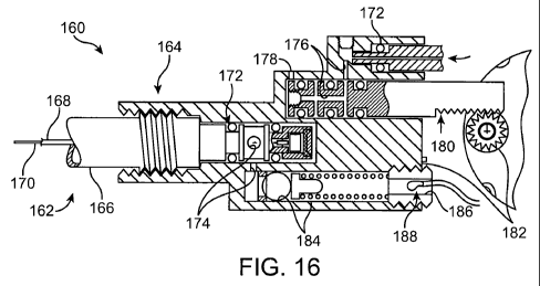
[0039] Fig. 16 is a schematic cross-sectional view showing an alternative
exemplary needle
interface, along with the adjacent structures of the needle assembly and probe
body.
[0040] Figs. 17A and 17B are partial cross-sectional views schematically
illustrating removal
of a cryogenic cooling probe needle while at least a portion of the tissue
remains frozen.
[0041] Figs. 18A and 18B are partial cross-sectional views schematically
illustrating how a
depletion of liquid from a vaporizing cryogenic cooling fluid can be used to
limit an effective
treatment length on a portion of a cryogenic probe.
8
CA 02677811 2009-08-10
WO 2008/101027 PCT/US2008/053876
DETAILED DESCRIPTION OF THE INVENTION
[0042] The present invention provides improved medical devices, system, and
methods.
Embodiments of the invention will facilitate remodeling of tissues disposed at
and below the
skin, optionally to treat a cosmetic defect, a lesion, a disease state, and/or
so as to alter a shape of
the overlying skin surface.
[0043] Among the most immediate applications of the present invention may be
the
amelioration of lines and wrinkles, particularly by inhibiting muscular
contractions which are
associated with these cosmetic defects so as so improve an appearance of the
patient. Rather
than relying entirely on a phanmacological toxin or the like to disable
muscles so as to induce
temporary paralysis, many embodiments of the invention will at least in part
employ cold to
immobilize muscles. Advantageously, nerves, muscles, and associated tissues
may be
temporarily immobilized using moderately cold temperatures of 10 C to -5 C
without
permanently disabling the tissue structures. Using an approach similar to that
employed for
identifying structures associated with atrial fibrillation, a needle probe or
other treatment device
can be used to identify a target tissue structure in a diagnostic mode with
these moderate
temperatures, and the same probe (or a different probe) can also be used to
provide a longer term
or permanent treatment, optionally by ablating the target tissue zone and/or
inducing apoptosis at
temperatures from about -5 C to about -50 C. In some embodiments, apoptosis
may be induced
using treatment temperatures from about -1 C to about -15 C, or from about -1
C to about -
19 C, optionally so as to provide a permanent treatment that limits or avoids
inflammation and
mobilization of skeletal muscle satellite repair cells. Hence, the duration of
the treatment
efficacy of such subdermal cryogenic treatments may be selected and
controlled, with colder
temperatures, longer treatment times, and/or larger volumes or selected
patterns of target tissue
determining the longevity of the treatment. Additional description of
cryogenic cooling for
treatment of cosmetic and other defects may be found in co-pending US Patent
Application
No. 11/295,204, filed on December 5, 2005 and entitled "Subdermal Cryogenic
Remodeling of
Muscle, Nerves, Connective Tissue, and/or Adipose Tissue (Fat)," the full
disclosure of which is
incorporated herein by reference.
[0044] In addition to cosmetic treatments of lines, wrinkles, and the like,
embodiments of the
invention may also find applications for treatments of subdermal adipose
tissues, benign, pre-
malignant lesions, malignant lesions, acne and a wide range of other
dermatological conditions
(including dermatological conditions for which cryogenic treatments have been
proposed and
additional dermatological conditions), and the like. Embodiments of the
invention may also find
9
CA 02677811 2009-08-10
WO 2008/101027 PCT/US2008/053876
applications for alleviation of pain, including those associated with muscle
spasms. Hence, a
variety of embodiments may be provided.
[0045] Referring now to Figs. lA and 1B, a system for cryogenic remodeling
here comprises a
self-contained probe handpiece generally having a proximal end 12 and a distal
end 14. A
handpiece body or housing 16 has a size and shape suitable for supporting in a
hand of a surgeon
or other system operator. As can be seen most clearly in Fig. 1B, a cryogenic
cooling fluid
supply 18 and electrical power source 20 are found within housing 16, along
with a circuit 22
having a processor for controlling cooling applied by self-contained system 10
in response to
actuation of an input 24. Some embodiments may, at least in part, be manually
activated, such as
through the use of a manual supply valve and/or the like, so that processors,
electrical power
supplies, and the like may be absent.
[0046] Extending distally from distal end 14 of housing 16 is a tissue-
penetrating cryogenic
cooling probe 26. Probe 26 is thermally coupled to a cooling fluid path
extending from cooling
fluid source 18, with the exemplary probe comprising a tubular body receiving
at least a portion
of the cooling fluid from the cooling fluid source therein. The exemplary
probe 26 comprises a
30 g needle having a sharpened distal end that is axially sealed. Probe 26 may
have an axial
length between distal end 14 of housing 16 and the distal end of the needle of
between about 1/2
mm and 5 cm, preferably having a length from about 1 cm to about 3 cm. Such
needles may
comprise a stainless steel tube with an inner diameter of about .006 inches
and an outer diameter
of about .012 inches, while alternative probes may comprise structures having
outer diameters
(or other lateral cross-sectional dimensions) from about .006 inches to about
.100 inches.
Generally, needle probe 26 will comprise a 16 g or smaller size needle, often
comprising a 20 g
needle or smaller, typically comprising a 25 g or smaller needle.
[0047] Addressing some of the components within housing 16, the exemplary
cooling fluid
supply 18 comprises a cartridge containing a liquid under pressure, with the
liquid preferably
having a boiling temperature of the less than 37 C. When the fluid is
thermally coupled to the
tissue-penetrating probe 26, and the probe is positioned within the patient so
that an outer surface
of the probe is adjacent to a target tissue, the heat from the target tissue
evaporates at least a
portion of the liquid and the enthalpy of vaporization cools the target
tissue. A valve (not
shown) may be disposed along the cooling fluid flow path between cartridge 18
and probe 26, or
along the cooling fluid path after the probe so as to limit the temperature,
time, rate of
temperature change, or other cooling characteristics. The valve will often be
powered
electrically via power source 20, per the direction of processor 22, but may
at least in part be
CA 02677811 2009-08-10
WO 2008/101027 PCT/US2008/053876
manually powered. The exemplary power source 20 comprises a rechargeable or
single-use
battery.
[0048] The exemplary cooling fluid supply 18 comprises a single-use cartridge.
Advantageously, the cartridge and cooling fluid therein may be stored and/or
used at (or even
above) room temperature. The cartridges may have a frangible seal or may be
refillable, with the
exemplary cartridge containing liquid N20. A variety of alternative cooling
fluids might also be
used, with exemplary cooling fluids including fluorocarbon refrigerants and/or
carbon dioxide.
The quantity of cooling fluid contained by cartridge 18 will typically be
sufficient to treat at least
a significant region of a patient, but will often be less than sufficient to
treat two or more
patients. An exemplary liquid N20 cartridge might contain, for example, a
quantity in a range
from about 7 g to about 30 g of liquid.
[0049] Processor 22 will typically comprise a programmable electronic
microprocessor
embodying machine readable computer code or programming instructions for
implementing one
or more of the treatment methods described herein. The microprocessor will
typically include or
be coupled to a memory (such as a non-volatile memory, a flash memory, a read-
only memory
("ROM"), a random access memory ("RAM"), or the like) storing the computer
code and data to
be used thereby, and/or a recording media (including a magnetic recording
media such as a hard
disk, a floppy disk, or the like; or an optical recording media such as a CD
or DVD) may be
provided. Suitable interface devices (such as digital-to-analog or analog-to-
digital converters, or
the like) and input/output devices (such as USB or serial I/O ports, wireless
communication
cards, graphical display cards, and the like) may also be provided. A wide
variety of
commercially available or specialized processor structures may be used in
different
embodiments, and suitable processors may make use of a wide variety of
combinations of
hardware and/or hardware/software combinations. For example, processor 22 may
be integrated
on a single processor board and may run a single program or may make use of a
plurality of
boards running a number of different program modules in a wide variety of
alternative
distributed data processing or code architectures.
[0050] Referring now to Fig. 2, the flow of cryogenic cooling fluid from fluid
supply 18 is
controlled by a supply valve 32. Supply valve may comprise an electrically
actuated solenoid
valve or the like operating in response to control signals from controller 22,
and/or may comprise
a manual valve. Exemplary supply valves may comprise structures suitable for
on/off valve
operation, and may provide venting of the cooling fluid path downstream of the
valve when
11
CA 02677811 2009-08-10
WO 2008/101027 PCT/US2008/053876
cooling flow is halted so as to limit residual cryogenic fluid vaporization
and cooling. More
complex flow modulating valve structures might also be used in other
embodiments.
[0051] The cooling fluid from valve 32 flows through a lumen 34of a cooling
fluid supply tube
36. Supply tube 36 is, at least in part, disposed within a lumen 38 of needle
26, with the supply
tube extending distally from a proximal end 40 of the needle toward a distal
end 42. The
exemplary supply tube 36comprises a fused silica tubular structure 36a having
a polymer coating
36b (see Fig. 3A) and extends in cantilever into the needle lumen 38. Supply
tube 36 may have
an inner lumen with an effective inner diameter 36c of less than about 200 m,
the inner
diameter often being less than about 100 m, and typically being less than
about 40 m.
Exemplary embodiments of supply tube 36 have inner lumens of between about 15
and 50 m,
such as about 30 m. An outer diameter or size 36d of supply tube 36 will
typically be less than
about 1000 m, often being less than about 800 m, with exemplary embodiments
being
between about 60 and 150 m, such as about 90 m or 105 m. The tolerance of
the inner
lumen diameter of supply tubing 36 will preferably be relatively tight,
typically being about +/-
10 m or tighter, often being +/- 5 m or tighter, and ideally being +/- 3 m
or tighter, as the
small diameter supply tube may provide the majority of (or even substantially
all of)the metering
of the cooling fluid flow into needle 26.
[0052] Though supply tubes 36 having outer jackets of polyimide (or other
suitable polymer
materials) may bend within the surrounding needle lumen 38, the supply tube
should have
sufficient strength to avoid collapsing or excessive blow back during
injection of cooling fluid
into the needle. Polyimide coatings may also provide durability during
assembly and use, and
the fused silica/polymer structures can handle pressures of up to 100 kpsi.
The relatively thin
tubing wall and small outer size of the preferred supply tubes allows adequate
space for
vaporization of the nitrous oxide or other cooling fluid within the annular
space between the
supply tube 36 and surrounding needle lumen 38. Inadequate space for
vaporization might
otherwise cause a buildup of liquid in that annular space and inconsistent
temperatures, as
illustrated in Fig. 12. Exemplary structures for use as supply tube 36 may
include the flexible
fused silica capillary tubing sold commercially by Polymicro Technologies, LLC
of Phoenix,
Arizona under model names TSP, TSG, and TSU, optionally including model
numbers
TSP 020090, TSP040105, and/or others.
[0053] Referring now to Figs. 2 and 3, the cooling fluid injected into lumen
38 of needle 26
will typically comprises liquid, though some gas may also be injected. At
least some of the
liquid vaporizes within needle 26, and the enthalpy of vaporization cools the
tissue engaged by
12
CA 02677811 2009-08-10
WO 2008/101027 PCT/US2008/053876
the needle. Controlling a pressure of the gas/liquid mixture within needle 26
substantially
controls the temperature within lumen 38, and hence the treatment temperature
range of the
tissue. A relatively simple mechanical pressure relief valve 46 may be used to
control the
pressure within the lumen of the needle, with the exemplary valve comprising a
valve body 48
(here in the form of a ball bearing) urged against a valve seat 50 by a
biasing spring 52.
[0054] During initiation of a cooling cycle, a large volume along the cooling
fluid pathway
between the exit from the supply tube and exit from the pressure relief valve
46 may cause
excessive transients. In particular, a large volume in this area may result in
initial temperatures
that are significantly colder than a target and/or steady state temperature,
as can be seen in
Fig. 13D. This can be problematic, particularly when (for example) the target
temperature is
only slightly warmer than an undesirable effect inducing temperature, such as
when remodeling
through apoptosis or the like while seeking to inhibit necrosis. To limit such
transients, the
pressure relief valve 46 may be integrated into a housing 54 supporting needle
26, with the valve
spring 52 being located outside the valve seat (and hence the pressure-control
exit from pressure
relief valve 46). Additionally, where needle 26 is included in a replaceable
needle assembly
26A, pressure relief valve 46 is also located adjacent the interface between
the needle assembly
and probe handpiece housing 54. A detent 56 may be engaged by a spring
supported catch to
hold the needle assembly releasably in position, and the components of the
needle assembly 26A
(such as a brass or other metallic housing, a polyimide tubing 58, needle 26,
and the like) may be
affixed together using adhesive. Alternatively, as illustrated in Figs. 1 B
and 4, the needle
assembly and handpiece housing may have corresponding threads for mounting and
replacement
of the needle assembly. 0-rings 60 can seal the cooling fluid pathway.
[0055] Figs. 13A-13C present additional details on the effects of exhaust
volume on cooling
transients. In each case, a graph of temperature over time is shown for the
outside temperature
of an in vivo 30g cooling needle with a target temperature of about -12 C. The
devices were
constructed with different exhaust volumes, with the volume being greater than
about 0.009 in3
in the embodiment of Fig. 13A. The embodiment of Figs. 13B and 13C had exhaust
volumes of
about 0.009 in3 and about.0025 in3, respectively. The data collection rate was
about 0.7 sec for
the embodiment of Fig. 13A, while the embodiments of Figs. 13B and 13C both
had data
collection rates of about 0.1 sec, so that the actual nadir for the embodiment
of Fig. 13A may
have actually been significantly lower than that shown. Regardless, the
exhaust volume is
preferably less than about 0.05 in3., typically being less than 0.01 in3
and/or 0.009 in3, and
ideally being less than 0.005 in3.
13
CA 02677811 2009-08-10
WO 2008/101027 PCT/US2008/053876
[0056] Alternative methods to inhibit excessively low transient temperatures
at the beginning
of a refrigeration cycle might be employed instead of or together with the
limiting of the exhaust
volume. For example, the supply valve might be cycled on and off, typically by
controller 22,
with a timing sequence that would limit the cooling fluid flowing so that only
vaporized gas
reached the needle lumen (or a sufficiently limited amount of liquid to avoid
excessive dropping
of the needle lumen temperature). This cycling might be ended once the exhaust
volume
pressure was sufficient so that the refrigeration temperature would be within
desired limits
during steady state flow.
[0057] Additional aspects of the exemplary supply valves 32 can be understood
with reference
to Figs. 2, 3, and 5A-5C. In Fig. 3, the valve is shown in the "on"
configuration, with 0-rings 60
sealing either side of the cooling fluid flow path and the cooling fluid
flowing around the
movable valve member. In Figs. 5A-5C, the cooling fluid flows through a
passage 64 that
extends axially along an alternative valve body of valve body 32' when the
valve is in the on
configuration (seen in Fig. 5B), with the 0-rings being disposed between
recesses in the movable
valve body so as to allow the valve to operate when the valve body is in any
rotational
orientation about its axis. In both embodiments, the cooling fluid flow path
downstream of the
valve is vented when the valve is in the "off' configuration (in the
embodiment of Fig. 3, by
channe166, and in the embodiment of Figs. 5A-5C by the vaporizing cooling
fluid flowing
through the annular space between the valve body and the adjacent housing 54
so as to preserve
the cooling fluid within the movable valve body).
[0058] Venting of the cooling fluid from the cooling fluid supply tube 36 when
the cooling
fluid flow is halted by supply valve 32, 32' is advantageous to provide a
rapid halt to the cooling
of needle 26. For example, a 2.5 cm long 30 g needle cooled to an outside
temperature of -15 C
might use only about 0.003 g/sec of nitrous oxide after the system approaches
or reaches steady
state (for example, 10 seconds after initiation of cooling). If the total
volume along the cooling
fluid path from supply valve to the distal end or release port of supply tube
36 is about 0.1 cc, the
minimum time to flow all the vaporizing liquid through the supply tube might
be calculated as
follows:
0.1 cc *(0.7 g/cc) = 0.07g of liquid nitrous oxide,
0.07 g/(.003 g/sec) = 23 sec.
These calculation assume a fused silica supply tube sized to allow the minimum
flow of nitrous
oxide when fluid supply has a pressure of about 900 psi. When the supply valve
is shut off, the
14
CA 02677811 2009-08-10
WO 2008/101027 PCT/US2008/053876
pressure on the needle side of the supply valve would decay, causing the
actual residual run time
to be longer, with only a partial cooling near the distal tip of needle 16.
Regardless, it is
desirable to limit the flow of cooling fluid into the needle to or near that
which will vaporize in
the needle so as to facilitate use of a simple disposable cooling fluid supply
cartridge 18.
Analytical models that may be used to derive these cooling flows include that
illustrated in
Fig. 10, which may be combined with the properties of the cooling fluid (such
as the
pressure/enthalpy diagram of nitrous oxide seen in Fig. 10A) and the thermal
properties of tissue
shown in Table I to determine theoretical minimum cooling fluid flow rates
(see Fig. 10B),
theoretical minimum cooling fluid quantities (see Fig. 10C), and the like.
Table I
Property Units Value
Upper temperature bond of freezing (T2) C -1
Peak of phase transition temperature (T3) C -3
Lower Temperature bond of freezing (TI) C -8
Thermal conductivity in unfrozen region (ku) W/(mm - C) 0.00063
Thermal conductivity in frozen region (kf) W/(mm - C) 0.00151
Volumetric specific heat in unfrozen region ({ptct)f) J/(mm3 - C 0.00316
Volumetric specific heat infrozen region J/mm3 - C 0.00193
Latent heat of solidification (HF) J/ mm 0.300
[0059] Referring now to Figs. 3 and 4, a wide variety of alternative
embodiments and
refinements may be provided. Fluid supply 18 may be initially opened for use
by penetrating a
frangible seal of the cartridge with a pierce point 70 (such as by tightening
a threaded cartridge
support coupled to housing 54), with the nitrous being filtered by a filter 72
before being
transmitted further along the cooling fluid path. Suitable filters may have
pore sizes of from
about 6 to about 25 m, and may be available commercially from Porex of
Georgia (or a variety
of alternative suppliers), or may comprise a fine stainless steel screen (such
as those having a
mesh size of 635 with 0.0009" wire and spacing between the wire edges of
approximately
0.0006"), or the like. A wide variety of epoxy or other adhesives 74 may be
used, and the
replaceable needle housing 24A and other structural components may comprise a
wide variety of
metals or polymers, including brass or the like. Fins 76 may be included to
help vaporize excess
cooling liquid traveling proximally of the insertable length of needle 26.
[0060] Very fine needles will typically be used to deliver to cooling at
and/or below the
surface of the skin. These needles can be damaged relatively easily if they
strike a bone, or may
otherwise be damaged or deformed before or during use. Fine needles well help
inhibit damage
CA 02677811 2009-08-10
WO 2008/101027 PCT/US2008/053876
to the skin during insertion, but may not be suitable for repeated insertion
for treatment of
numerous treatment sites or lesions of a particular patient, or for sequential
treatment of a large
area of the patient. Hence, the structures shown in Figs. 1 B, 3, and 4 allow
the use probe bodies
16, 54 with a plurality of sequentially replaceable needles. 0-rings 60 help
to isolate the cooling
fluid supply flow (which may be at pressures of up to about 900 psi) from the
exhaust gas (which
may be at a controlled pressure in a range between about 50 and 400 psi,
depending on the
desired temperature). Exemplary 0-rings may comprise hydrogenated Buna-N 0-
rings, or the
like.
[00611 It may be advantageous to increase the volume of tissue treated by a
single treatment
cycle. As it is often desirable to avoid increasing the needle size
excessively, along with
selecting needles of different lengths, needle assemblies having differing
numbers of needles in a
needle array may also be selected and mounted to the probe body. Other
embodiments may
employ a single needle array fixedly mounted to the probe body, or a plurality
of replaceable
needle assemblies which all include the same number of needles. Regardless,
cooling fluid flow
to a plurality of needles may be provided, for example, by inserting and
bonding a plurality of
fused silica supply tubes into a 0.010 polyimide tubing 58 or header within
the needle assembly,
and by advancing the distal end of each supply tube into a lumen of an
associated needle 26. The
needles might vent into a common exhaust space coaxially around polyimide
tubing 58 in a
manner similar to the single needle design shown. This can increase the
quantity of tissue
treated adjacent and/or between needles, as can be seen by comparing the
theoretical 15 second
exposures to one and two needles having a-15 C probe surface, as shown in
Figs. 11A and 11B,
respectively.
[0062] Referring now to Fig. 6, it may be desirable to allow a system user to
select a treatment
depth, and/or to treat the skin surface to a temperature similar to that of
the underlying target
tissue along needle 26. A distally oriented surface 82 supported by probe body
54 adjacent
and/or around the proximal end of the needles may be configured to limit heat
transfer to or from
the skin when the needle 26 is inserted so that surface 82 engages the skin
and cooling fluid
flows into the needle. Exemplary heat transfer limiting surfaces may be
formed, for example,:
from a small rigid foam pad or body 84. Closed cell polyethylene foam or
StyrofoamTM foam
bodies may be used. As seen in Fig. 6, an alternatively selectable set of
bodies may also have
differing thicknesses between the skin engaging-surface 82 and a surface 86
that engages the
distal portion of the probe body. A user can then select an insertable length
of the needle by
selecting an appropriate probe body 84, 84a, 84b and mounting the selected
probe body onto the
16
CA 02677811 2009-08-10
WO 2008/101027 PCT/US2008/053876
needles. Skin engaging surface 82 of bodies 84, 84a, and 84b (or some other
skin engaging
surface adjacent the distal end of the needle) may be used to apply pressure
to the skin, lesion,
and/or target tissue during treatment. Alternative insertable length varying
arrangements may
also be provided, including those having threaded or other articulatable
structures supporting the
skin engaging surface 82 relative to the adjacent probe body 54 and the like.
[0063] Referring now to Fig. 7, the application of pressure before, during,
and/or after cooling
may help dull or otherwise inhibit sharp pain. Such pain may otherwise result
from the skin
penetration, cooling, or thawing of the target and/or collateral tissues. It
may also be beneficial
to obscure the patient's view of the cooling needles, and/or to cover the
needles when not in use
so as to inhibit needle-stick injuries and potential disease transmission.
Toward that end, skin-
engaging surface 82 may be supported by an articulatable support structure
having a first
configuration (shown in solid in Fig. 7) and a second configuration (shown
dashed in Fig. 7). A
simple spring mechanism may be used to apply a desired contact force between
the skin-
engaging surface 82 and the patient before insertion and during cooling. More
sophisticated
arrangements can also be employed in which the needle is driven distally and
then proximally
relative to the skin engaging surface appropriate times after sufficient
pressure is applied to the
patient, and the like.
[0064] Referring now to Fig. 8, still further alternative embodiments may be
provided, in this
case to apply different cooling temperatures to the patient, and/or to apply
cooling to the skin
surface and to a target tissue adjacent needle 26. For example, in the case of
acne it may be
desirable to have two different cooling target temperatures, with cooling on
the skin surface to
inhibit inflammation (such as to about -10 C), and (see Fig. 14) cooling of a
target tissue
TT cylinder around needle 26 sufficient to kill bacteria in the sebaceous
gland and enlarged
follicle opening (such as to about -20 C). This dual temperature treatment may
be particularly
beneficial for severe forms of acne involving cysts or nodules. To provide
cooling of tissue
engaging surface 82, that surface may be thermally coupled to a chamber 88.
Cooling fluid may
be transmitted into chamber 88 by a port of a cooling fluid supply tube 36,
and the pressure of
chamber 88 (and hence the temperature within the chamber) can optionally be
controlled by a
dedicated additional pressure relief valve 46a. As the pressure within chamber
88 may differ
from that within the needle, different treatment temperatures may be provided.
The structures
described herein can also be combined, for example, with the dual skin
surface/needle
temperature treatment structure of Fig. 8 being compatible with the
replaceable needle systems
of Figs. 1 B, 3, and/or 4. The dual skin surface/needle treatment systems and
methods may also
17
CA 02677811 2009-08-10
WO 2008/101027 PCT/US2008/053876
be compatible, for example, with the articulatable skin surface supports of
Fig. 7 so as to apply
cooled pressure to the skin prior to and/or during needle insertion using a
flexible fluid supply
tube or the like.
[0065] Still further alternatives may also be provided, including systems that
generate a high rate
of cooling to promote necrosis of malignant lesions or the like. High cooling
rates limit osmotic
effects in the target tissue. Slow cooling may tend to promote ice formation
between cells rather
than within cells due to the osmotic effect. While such slow cooling can be
provided where
necrosis is not desired (such as through the use of a proportion supply valve
to modulate flow, a
processor generated on/off cycle during initial cooling, or the like), the
needle probes described
herein will often be well suited to induce rapid cooling rates of the target
tissue by vaporizing the
cooling fluid in close thermal and spatial proximity to that target tissue.
Hence, where necrosis
of cells by intracellular ice formation is desired, cooling rates of about 25
C/sec or more, or even
about 50 C/sec or more can be provided.
[0066] Referring now to Figs 9, 9A, and 9B, needles having circular cross-
sectional shapes can
often be used, but may not always provide the desired surface area for the
cross-sectional area. of
the needle. Increased surface area may decrease the amount of time the needle
is inserted to cool
a volume of tissue to a temperature in a target range. Hence, a needle with an
elongate outer
cross-section such as elliptical needle 90 may be desirable. A distal cutting
edge 92 at the distal
tip may facilitate insertion and a circular cross-section 94 near the proximal
end may limit
cooling adjacent the skin, while cooling of the target tissue therebetween is
enhanced by
elliptical cross-section 96.
[0067] Referring now to Fig. 15, a method 100 facilitates treating a patient
using a cryogenic
cooling system having a self-contained disposable handpiece and replaceable
needles such as
those of Fig. 1 B. Method 100 generally begins with a determination I 10 of
the desired tissue
remodeling and results, such as the alleviation of specific cosmetic wrinkles
of the face, the
inhibition of pain from a particular site, the alleviation of unsightly skin
lesions or cosmetic
defects from a region of the face, or the like. Appropriate target tissues for
treatment are
identified 112 (such as the subdermal muscles that induce the wrinkles, a
tissue that transmits the
pain signal, or the lesion-inducing infected tissues), allowing a target
treatment depth, target
treatment temperature profile, or the like to be determined 114. An
appropriate needle assembly
can then be mounted 116 to the handpiece, with the needle assembly optionally
having a needle
length, skin surface cooling chamber, needle array, and/or other components
suitable for
18
CA 02677811 2009-08-10
WO 2008/101027 PCT/US2008/053876
treatment of the target tissues. Simpler systems may include only a single
needle type, and/or a
first needle assembly mounted to the handpiece.
[0068] As described above, pressure, cooling, or both may be applied 118 to
the skin surface
adjacent the needle insertion site before, during, and/or after insertion 120
and cryogenic cooling 5 122 of the needle and associated target tissue. The
needle can then be retracted 124 from the
target tissue. If the treatment is not complete 126 and the needle is not yet
dull 128, pressure
and/or cooling can be applied to the next needle insertion location site 118,
and the additional
target tissue treated. However, as small gauge needles may dull after being
inserted only a few
times into the skin, any needles that are dulled (or otherwise determined to
be sufficiently used to
warrant replacement, regardless of whether it is after a single insertion, 5
insertions, or the like)
during the treatment may be replaced with a new needle 116 before the next
application of
pressure/cooling 118, needle insertion 120, and/or the like. Once the target
tissues have been
completely treated, or once the cooling supply cartridge included in the self-
contained handpiece
is depleted, the used handpiece and needles can be disposed of 130.
[0069] A variety of target treatment temperatures, times, and cycles may be
applied to differing
target tissues to as to achieve the desired remodeling. For example, (as more
fully described in
patent application 11/295204, previously incorporated herein by reference)
desired temperature
ranges to temporarily and/or permanently disable muscle, as well as protect
the skin and
surrounding tissues, may be indicated by Table II as follows:
Table II
Temperature Skin Muscle/Fat
37 C baseline baseline
C cold sensation
18 C reflex vasodilation of deep
blood vessels
15 C cold pain sensation
12 C reduction of spasticity
10 C very cold sensation
reduction of chronic
oedema
Hunting response
5 C pain sensation
0 C freezing point
-1 C Phase transition begins
-2 C minimal apoptosis
-3 C Peakphase transition
-5 C tissue damage moderate apoptosis
-8 C Completion ofphase transition
19
CA 02677811 2009-08-10
WO 2008/101027 PCT/US2008/053876
Temperature Skin Muscle/Fat
----------------
-10 C considerable apoptosis
-15 C extensive apoptosis
mild-moderate necrosis
-19 C adoptosis in some skeletal
muscle tissues
-40 C extensive necrosis
[0070] To provide tissue remodeling with a desired or selected efficacy
duration, tissue
treatment temperatures may be employed per Table III as follows:
Table III
Cooled Time Effectiveness Purpose
Tem erature Ran e
> 0 C Treatment lasts only while the Can be used to identify target
needle is inserted into the tissues.
target tissue.
From 0 C to -5 C Often lasts days or weeks, and Temporary treatment. Can be
target tissue can repair itself used to evaluate effectiveness
Embodiments may last hours of remodeling treatment on
or days. skin surface shape or the like.
From -5 C to -15 C Often lasts months to years; Long term, potentially
and may be permanent. permanent cosmetic benefits.
Limited muscle repair. Can be deployed in limited
Embodiments may last weeks doses over to time to achieve
to months. staged impact, controlling
outcome and avoiding negative
outcome. May be employed as
the standard treatment.
From -15 C to -25 C Often lasts weeks or months. May result in Mid-term
Muscle may repair itself via cosmetic benefits, and can be
satellite cell mobilization. used where permanent effects
Embodiments may last years. are not desired or to evaluate
outcomes of potentially
permanent dosing.
Embodiments may provide
permanent treatment.
[0071] There is a window of temperatures where apoptosis can be induced. An
apoptotic
effect may be temporary, long-term (lasting at least weeks, months, or years)
or even permanent.
While necrotic effects may be long term or even permanent, apoptosis may
actually provide
more long-lasting cosmetic benefits than necrosis. Apoptosis may exhibit a non-
inflammatory
cell death. Without inflammation, normal muscular healing processes may be
inhibited.
Following many muscular injuries (including many injuries involving necrosis),
skeletal muscle
CA 02677811 2009-08-10
WO 2008/101027 PCT/US2008/053876
satellite cells may be mobilized by inflammation. Without inflammation, such
mobilization may
be limited or avoided. Apoptotic cell death may reduce muscle mass and/or may
interrupt the
collagen and elastin connective chain. Temperature ranges that generate a
mixture of these
apoptosis and necrosis may also provide long-lasting or permanent benefits.
For the reduction of
adipose tissue, a permanent effect may be advantageous. Surprisingly, both
apoptosis and
necrosis may produce long-term or even permanent results in adipose tissues,
since fat cells
regenerate differently than muscle cells.
[0072] Referring now to Fig. 16, an exemplary interface 160 between a
cryogenic cooling
needle probe 162 and the associated probe body structure 164 are illustrated,
along with adjacent
portions of the needle, valve, probe body, and the like. Needle probe 162 is
included in a needle
assembly having a needle hub 166 with a lumen containing a polyimide tube 168
around a fused
silica cooling fluid supply tube with its polyimide jacket 170. 0-rings 172
seal in exhaust gas
path 174 and inlet cooling fluid path 176, with the inlet path having a vent
178 to minimize run-
on cooling when the cooling fluid supply is shut off by a valve 180, as
generally described
above. The valve is here actuated by a motor 182, while the exhaust gas
pressure is controlled
using a biasing spring and ball valve 184 as described above. A hollow set
screw 186 can be
used to assemble and/or adjust the pressure relief valve, and a thermistor 188
can be used to
sense cooling gas flow.
[0073] Referring now to Figs. 17A and 17B, cryogenic cooling probes 196, 198
are inserted
into a target tissue TT and a flow of cryogenic cooling fluid is injected into
the needle as
generally described above. A region 200 of target tissue TT is cooled
sufficiently to freeze and
effect the desired remodeling of at least a portion of the target tissue.
Rather than waiting for the
frozen target tissue to thaw, in the embodiment of Fig. 17A a lubricious
coating 202 facilitates
removal of the needle while at least a portion of the frozen target tissue
remains frozen. The
lubricious coating 202 may comprise a material having a thermal conductivity
which is
significantly less than that of the underlying probe structure 204. Coating
202 may have a
thickness which is significantly less than that of the underlying probe
structure 204, limiting the
total insulation effect of the coating, and/or an interior temperature of
probe 196 may be reduced
so as to provide the desired overall cooling treatment. While it may be
counterintuitive to cool
the target tissue through a thermally insulating lubricious coating, the
ability to more rapidly
remove probe 196 from the patient can significantly increase the speed with
which procedures
may be performed, particularly when a large number of
insertion/cooling/removal cycles are
21
CA 02677811 2009-08-10
WO 2008/101027 PCT/US2008/053876
involved, and/or when the thaw time is at least half as long as (often being
as long as or longer
than) the active cooling time.
[0074] Note that a small surface 206 of probe 196 may be free of lubricious
coating 202.
Where the underlying probe structure 204 comprises an electrical conductor
such as stainless
steel or some alternative metal, the uncovered surface portion 206 may be used
as an electrode
for neurostimulation during positioning of probe 196 or the like.
[0075] In the embodiment of Fig. 17B, the use of cryosurgical probes of small
diameter may
facilitate removal of the probe without having to wait for a complete thaw of
region 200. In this
embodiment, microneedle probe 198 has a cross-sectional size of a 20-gauge
needle or less,
preferably comprising a 25-gauge needle or smaller, and ideally comprising a
30-gauge needle.
These small diameter microneedle probes have little thermal mass and can be
warmed relatively
quickly by conduction from adjacent tissues and/or by any warm fluids flowing
therein. As a
result, while a major portion 208 of the target tissue remains frozen a layer
210 disposed between
the still-frozen region and probe 198 may facilitate safe removal of the probe
from the patient.
Thawed layer 210 may comprise thawed target tissue, thawed extracellular
fluids, or the like.
Small needles also have small probe/tissue interface surface areas which may
limit the total
stiction between the probe and frozen tissue. Regardless of any particular
mechanism of action,
the use of small diameter cryogenic microneedles may allow safe removal of the
probe from a
treated tissue in a time which is significantly less than that associated with
complete thaw of the
iceball that has been formed. Exemplary embodiments using a lubricious coating
and/or small
diameter probe may allow the probe to be removed within about 10 seconds of
the cooling,
optionally allowing safe removal within about 5 seconds of cooling or even
within about 3
seconds of cooling.
[0076] Referring now to Figs. 18A and 18B, appropriate metering of the cooling
fluid into a
cryogenic cooling probe 220, 222, can be used to control the length of the
probe that applies a
therapeutic cooling. Probes 220, 222 are replaceably supported by a probe body
224 via a needle
receptacle or interface, as generally described above. Each probe includes a
lumen 226 with a
cooling fluid supply tube 228 extending to a distal port 230. Through proper
selection of the
length of the cooling fluid supply tube 228 and/or an inner diameter of the
lumen within the
supply tube, the supply tube can be used to meter cooling fluid. More
specifically, as noted
above, cooling of the target tissue TT along a distal portion 232 of probe 228
is cooled by
evaporation of the liquid included in the cryogenic cooling fluid. As shown in
Fig. 18A, cooling
of a collateral tissue CT proximal of the target tissue TT may be limited by
controlling the
22
CA 02677811 2009-08-10
WO 2008/101027 PCT/US2008/053876
amount of cooling fluid flow so that the vaporizing liquid is depleted by the
time the flow
reaches a proximal portion 234 of the probe. In the embodiment of Fig. 18B, a
greater length of
probe 222 is cooled by providing a relatively larger quantity of cooling fluid
(and liquid) flowing
from the supply tube 238 into lumen 226 via port 230, so that liquid remains
present for
vaporization throughout a longer distal portion 232 of the probe. Note that
the difference in
lengths of the cooled portion 232 may be provided despite making use of an
outer probe structure
that is similar in cross section and/or overall length.
[0077] While the proximal portion 234 of probes 220, 222 may be cooled
somewhat (via
conduction from the distal portion 232 of the probe, from the passage of gas
vaporized from the
gas of the cooling fluid, or the like), a temperature of collateral tissue CT
may remain above the
remodeling treatment temperature of a treatment zone 238 within the target
tissue. Hence, the
collateral tissue may avoid injury despite the absence of any additional
insulation on the
proximal portion of the probe. This also facilitates the use of differing
treatment zones 238 at
different locations for a particular patient through the selection of needle
assemblies having
appropriate cooling fluid supply paths with the desired differing cooling
fluid flow
characteristics.
[0078] While the exemplary embodiments have been described in some detail for
clarity of
understanding and by way of example, a number of modifications, changes, and
adaptations may
be implemented and/or will be obvious to those as skilled in the art. For
example, one or more
temperature feedback loops may be used to control the treatments, with the
tissue temperature
optionally being taken using a temperature sensing needle having a temperature
sensor disposed
adjacent an outer cooled skin engaging surface of the needle. Hence, the scope
of the present
invention is limited solely by the independent claims.
23