Note: Descriptions are shown in the official language in which they were submitted.
CA 02678304 2009-08-10
WO 2008/102359 PCT/IL2008/000230
ANTIBODIES AND PHARMACEUTICAL COMPOSITIONS CONTAINING
SAME USEFUL FOR INHIBITING ACTIVITY OF METALLOPROTEINS
FIELD AND BACKGROUND OF THE INVENTION
The present invention relates to hapten molecules and antibodies directed
thereagainst, which can be used to inhibit' activity of metalloproteins, such
as
metalloproteases, and to methods which utilize the antibodies for treating
diseases
such as metastatic cancer which are associated with abnormal activity of a
metalloprotein.
The matrix metalloproteins (MMPs) are key enzymes participating in
remodeling of the extracellular matrix (ECM). These enzymes are capable of
destroying a variety of connective tissue components of articular cartilage or
basement
membranes.
The human MMP gene family consists of at least 28 structurally related
proteins (see Figure 1), which share a similar overall spherical topology
(Figure 2 and
Borkakoti, 1998). Each MMP is secreted as an inactive, latent pro-enzyme. The
catalytic zinc domain is composed of about 180 amino acids wherein the highly
conserved sequence HE-GH-LGL-H provides the three histidine (i.e., H) residues
which bind to the metal Zn(2+) ion. The forth-binding site of the catalytic
zinc ion in
the pro-enzyme is bound to a cystein residue (Morgunova et al., 1999), which
upon
enzyme activation dissociates from the active site (Van Wart and Birkedal-
Hansen,
1990). As a result, the forth-binding site in the activated MMPs is taken up
by a water
molecule, which is also hydrogen-bonded to a conserved glutamic residue. This
process facilitates the hydrolysis of a peptide bond of the target substrate
with the
activated water molecule.
The uncontrolled breakdown of connective tissue by metalloproteases is a
feature of many pathological conditions, probably resulting from an excess of
MMP
activity or from an imbalanced ratio between the natural MMP tissue inhibitors
(TIMPs) and MMPs. TIMPs inhibit MMPs by forming stoichiometric complexes with
the active zinc binding site of MMPs (Gomez et al., 1997; Henriet at al.,
1999; Bode et
al., 1999; Will et al., 1996). When TIMPs levels are insufficient, a
progressive slow
degradation of the ECM may lead to loss of cartilage matrix in rheumatoid
arthritis
(Walakovits et al., Arthritis Rheum, 35:35-42, 1992) and osteoarthritis (Dean
et al., J.
Clin. Invest. 84:678-685, 1989) or bone matrix degradation in osteoporosis
(Hill et al.,
CA 02678304 2009-08-10
WO 2008/102359 PCT/IL2008/000230
2
Biochem. J. 308: 167-175, 1995). In other situations, such as congestive heart
failure,
rapid degradation of the heart's ECM may occur (Armstrong et al., Canadian J.
Cardiol. 10: 214-220, 1994).
Additionally, MMPs are known to play a role in the maturation of cytokines
and chemokines such as galectin-3 (Ochieng J. , Biochemistry, 1994
33(47):14109-14)
plasminogen (Patterson, BC., JBC, 1997 272(46):28823-5, interleukin-8,
connective
tissue activating peptide III, platelet factor-4 (Van den Steen, 2000 Blood.
2000 Oct
15;96(8):2673-81.), pro-interleukin-1(3 (Schonbeck, 1998), interleukin-2
receptor a
chain [Sheu, B. C, Hsu, S. M., Ho, H., Lien, H. C., Huang, S. C., Lin, R. H. A
novel
role of metalloproteinase in cancer-mediated immunosuppression Cancer Research
(2001) 61, 237-242], and pro-transforming growth factor-(3 [TGF-(3, Yu, Q.
Stamenkovic, I. Cell surface-localized matrix metalloproteinase-9
proteolytically
activates TGF-beta and promotes tumor invasion and angiogenesis Genes Dev
(2000)
14, 163-176].
Other pathological conditions, which are also related to unregulated activity
of
MMPs, include the rapid remodeling of the ECM by metastatic tumor cells. In
such
conditions the activated MMPs are either expressed by the cancer cells or by
the
surrounding tissues. There is considerable evidence that MMPs are involved in
the
growth and spread of tumors (e.g., see Davidson et al., Chemistry & Industry,
258-
261, 1997, and references therein). In the process of tumor metastasis, MMPs
are tised
to break down the ECM, allowing primary tumor cancer cells to invade
neighboring
blood vessels where they are transported to different organs and establish
secondary
tumors. The invasive growth at these secondary sites is mediated by MMPs,
which
break down the tissue. In addition, MMP activity contributes to the invasive
in-growth
of new blood vessels, also termed angiogenesis, which is required for tumors
to grow
above a certain size. Among the members of MMP family, the secreted human MMP-
9, also known as gelatinase B, has been shown to have key roles not only in
extracellular matrix (ECM) catabolism but. also in the processing of protein
substrates
that are relevant in neurological diseases such as multiple sclerosis (MS)
(Opdenakker,
2003). Recent studies showed that MMP-9 has a critical role in promoting
autoimmune diseases by cleaving pre-processed type II collagen (Van den Steen,
2004). The products are collagen type II fragments that are remnant epitopes
thought
to generate autoimmune diseases.
CA 02678304 2009-08-10
WO 2008/102359 PCT/IL2008/000230
3
Given the broad role of MMPs in human physiology and pathology, it is not
surprising that numerous efforts have been affected to design drugs, which
inhibit
MMP excessive activity.
Drug discovery efforts have focused on inhibitor classes that contain a
functional group which coordinates the zinc ion to thereby inactivate the
target MMP.
One such inhibitor class is the hydroxamate inhibitors, small peptide analogs
of
fibrillar collagens, which specifically interact in a bidentate manner via the
hydroxyl
and carbonyl oxygens of the hydroxamic group with the zinc ion in the
catalytic site
[Grams et al., (1995), Biochem. 34: 14012-14020; Bode et al., (1994), EMBO J.,
13:
1263-1269].
Hydroxamate-based MMP inhibitors are usually composed of either a carbon
back-bone (WO 95/29892, WO 97/24117, WO 97/49679 and EP 0780386), a peptidyl
back-bone (WO 90/05719, WO 93/20047, WO 95/09841 and WO 96/06074) or a
peptidomimetic back-bone [Schwartz et al., Progr. Med. Chem., 29: 271-
334(1992);
Rasmussen et al., Pharmacol. Ther., 75: 69-75 (1997); Denis et al., Invest.
New Drugs,
15: 175-185 (1997)]. Alternatively, they contain a sulfonamido sulfonyl group
which
is bonded on one side to a phenyl ring and a sulfonamido nitrogen which is
bonded to
an hydroxamate group via a chain of one to four carbon atoms (EP 0757984 Al).
Other peptide-based MMP inhibitors are thiol amides which exhibit
collagenase inhibition activity (U.S. Pat. No. 4,595,700), N-carboxyalkyl
derivatives
containing a biphenylethylglycine which inhibit MMP-3, MMP-2 and collagenase
(Durette, et al., WO-9529689), lactam derivatives which inhibit MMPs, TNF-
alpha
and aggrecanase (see US 6,495,699) and Tricyclic sulfonamide compounds (see US
6,492,422).
Although peptide-based MMP inhibitors have a clear therapeutic potential their
use in clinical therapy is limited. Peptide-based hydroxamate are costly to
produce
and have low metabolic stability and oral bioavailability [e.g., batimastat
(BB-94)].
These compounds are rapidly glucuronidated, oxidized to carboxylic acid and
excreted
in the bile [Singh et al., Bioorg. Med. Chem. Lett. 5: 337-342, 1995; Hodgson,
"Remodelling MMPIs", Biotechnology 13: 554-557, 1995)]. In addition, peptide-
based MMP inhibitors often exhibit the same or similar inhibitory effects
against each
of the MMP enzymes. For example, batimastat is reported to exhibit IC50 values
of
about 1 to about 20 nM against each of MMP-1, MMP-2, MMP-3, MMP-7, and
MMP-9 [Rasmussen et al., Pharmacol. Ther., 75(l): 69-75 (1997)]. Furthermore,
the
CA 02678304 2009-08-10
WO 2008/102359 PCT/IL2008/000230
4
use of several hydroxamate inhibitors was associated with severe side effects
such as
muscoloskeletal problems with marimastat (BB-2516), widespread maculopapular
rash with CGS27023A (Novartis) [Levitt et al., 2001, Clin. Cancer Res. 7: 1912-
1922]
and liver abnormalities, anemia, shoulder and back pain, thrombocytopenia,
nausea,
fatigue, diarrhea and deep vein thrombosis with BAY12-9566 (Bayer) [Heath et
al.,
2001, Cancer Chemother. Pharmacol. 48: 269-274]. Moreover, phase III clinical
trials
on advanced cancer patients with marimastat, prinomastat (AG 3340, Agouron)
and
Bay 12-9566 demonstrated no clinical efficacy in inhibiting metastasis (Zucker
et al.,
2000, Oncogene 19: 6642-50).
Other MMP inhibitors are the chemically modified nonmicrobial tetracyclines
(CMTs) that were shown to block expression of several MMPs in vitro. However,
in
vivo efficacy of these compounds was found to be limited, e.g., the CMT
inhibitor,
doxycycline, reduced tissue levels of MMP-1 but not MMP-2, 3, or -9 in
atherosclerotic carotid plaques in human patients (Axisa et al., 2002, Stroke
33: 2858-
2864).
Recently, a mechanism-based MMP inhibitor, SB-3CT, was designed
according to the X-ray crystallographic information of the MMP active site
(Brown et
al., 2000). X-ray absorption studies revealed that binding of this molecule to
the
catalytic zinc reconstructs the conformational environment around the active
site metal
ion back to that of the pro-enzyme [Kleifeld et al., 2001, J Biol. Chem. 276:
17125-
31 ]. However, the therapeutic efficacy obtained with this agent is yet to be
determined.
Another class of natural inhibitors is monoclonal antibodies. Several
antibodies have been raised against specific peptide sequences within the
catalytic
domain MMP-1 (Galvez et al., 2001, J. Biol. Chem., 276: 37491-37500). However,
although these antibodies could inhibit the in-vitro activity of MMP, results
demonstrating the in-vivo effectiveness of such antibodies have not been
demonstrated.
As described hereinabove, the catalytic site of MMPs includes a coordinated
metal ion which becomes available for substrate binding following enzyme
activation
(see Figures 2a-c). It is thus conceivable that conventional antibodies
directed at the
primary amino acid sequence of the enzyme would not distinguish the active
form
from the inactive form of the enzyme and hence would not serve as potent
inhibitors
of such enzymes.
CA 02678304 2009-08-10
WO 2008/102359 PCT/IL2008/000230
The present inventors have previously shown that antibodies which recognize
both electronic and structural determinants of the catalytic site of MMPs are
potent
inhibitors thereof and as such can be used to treat diseases associated with
imbalanced
MMP activity (see PCT Publication WO 2004/087042).
5 There is thus, a widely recognized need for and it would be highly desirable
to
have specific hapten compounds which mimic the electronic and structural
determinants of the catalytic site of metalloproteins as well as specific
antibodies
which are directed thereagainst.
SUMMARY OF THE INVENTION
According to one aspect of the present invention there is provided a compound
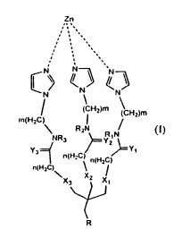
having the general Formula (I):
Zn
,. .
,. .
,
CN N
N
N N
' ((CHZ)m \ Hz)m
/
m(H2C) \ I
Y3 R2N RIN NR3 Y2
Y,
n(H2C) n(HZC)
n(H2C) ~ J
x3 xZ x/
1
T
R
wherein:
m and n are each independently an integer from I to 6;
Xi-X3 and Yi-Y3 are each independently 0 or S;
Ri-R3 are each indepdnently selected from the group consisting of hydrogen,
alkyl, and cycloalkyl; and
R is (CH2)x-C(=O)NR'-(CH2)y-NR'R"
whereas:
x and y are each independently an integer from I to 6; and
CA 02678304 2009-08-10
WO 2008/102359 PCT/IL2008/000230
6
R' and R" are each independently selected from the group consisting of
hydrogen, alkyl, and cycloalkyl.
According to further features in preferred embodiments of the invention
described below, the compound has the Formula (II):
Zn
,, .
., ,
0, `
(N N N/
HN
NH OHN
o p
O w
R .
wherein R = -CH2-C(=0)NH-CH2-CH2-NH2
According to another aspect of the present invention there is provided a
compound having the Formula (II):
Zn
\~ -\
iS N \N
HN
NH OHN
o p
A O w
R wherein R = -CH2-C(=0)NH-CH2-CHZ-NH2
According to yet another aspect of the present invention there is provided an
antibody comprising an antigen recognition region capable of specifically
binding the
above compound.
CA 02678304 2009-08-10
WO 2008/102359 PCT/IL2008/000230
7
According to still further features in the described preferred embodiments the
antigen recognition region comprises a CDR amino acid sequence selected from
the
group consisting of SEQ ID NO: 7, 8, 9, 10, 11 and 12.
According to still further features in the described preferred embodiments the
CDR amino acid sequence is encoded by a nucleic acid sequence selected from
the
group consisting of SEQ ID NO: 13, 14, 15, 16, 17 and 18.
According to still further features in the described preferred embodiments the
antibody is capable of inhibiting an activity of a metalloproein.
According to still further features in the described preferred embodiments the
metalloprotein is a matrix metalloprotease.
According to still further features in the described preferred embodiments the
matrix metalloprotease is a gelatinase.
According to still further features in the described preferred embodiments the
gelatinase is selected from the group of MMP-2 and MMP-9.
According to still another aspect of the present invention there is provided a
method of producing a metalloprotein inhibitor, the method comprising
generating
antibodies directed at the above compound, thereby producing the
metalloprotein
inhibitor.
According to still further features in the described preferred embodiments the
antibodies are polyclonal antibodies.
According to still further features in the described preferred embodiments the
antibodies are monoclonal antibodies.
According to an additional aspect of the present invention there is provided a
pharmaceutical composition comprising the antibody and a pharmaceutically
acceptable carrier.
According to an additional aspect of the present invention there is provided a
method of treating a disease associated with imbalanced or abnormal activity
of
metalloproteins in a subject in need thereof, the method comprising
administering to
the subject a therapeutically effective amount of any one of the antibodies of
claim 4-
10, thereby treating a disease associate with imbalanced or abnormal activity
of
metalloproteins in the subject.
According to still further features in the described preferred embodiments the
disease is an inflammatory bowel disease.
CA 02678304 2009-08-10
WO 2008/102359 PCT/IL2008/000230
8
According to an additional aspect of the present invention there is provided a
method of inhibiting matrix metalloprotease activity in a cell, the method
comprising
contacting the cell with any one of the antibodies of claim 4-10, thereby
inhibiting the
matrix metalloprotease activity in the cell.
The present invention successfully addresses the shortcomings of the presently
known configurations by providing a novel hapten composition which can be used
to
generate antibodies which recognize both electronic and structural
determinants of the
catalytic site of metalloproteins.
Unless otherwise defined, all technical and scientific terms used herein have
the same meaning as commonly understood by one of ordinary skill in the art to
which
this invention belongs. Although methods and materials similar or equivalent
to those
described herein can be used in the practice or testing of the present
invention, suitable
methods and materials are described below. In case of conflict, the patent
specification, including definitions, will control. In addition, the
materials, methods,
and exaxnples are illustrative only and not intended to be limiting.
BRIEF DESCRIPTION OF THE DRAWINGS
The invention is herein described, by way of example only, with reference to
the accompanying drawings. With specific reference now to the drawings in
detail, it
is stressed that the particulars shown are by way of example and for purposes
of
illustrative discussion of the preferred embodiments of the present invention
only, and
are presented in the cause of providing what is believed to be the most useful
and
readily understood description of the principles and conceptual aspects of the
invention. In this regard, no attempt is made to show structural details of
the invention
in more detail than is necessary for a fundamental understanding of the
invention, the
description taken with the drawings making apparent to those skilled in the
art how the
several forms of the invention may be embodied in practice.
In the drawings:
FIGs. lA-D are schematic representations of the molecular structure of
Co/ZnTCPP - [meso-Tetrakis (4- carboxyphenyl)-porphyrinato] cobalt/zinc (II)
(Figures IA-B, Imisdp - [2-(2-minoethylcarbomoyl)-ethoxymethyl] -tris-[2-(N-(3-
imidazol-1-yl-propyl)) -ethoxymethyl] methane, and the conserved zinc-protein
ligation at the catalytic zinc site in MMPs.
CA 02678304 2009-08-10
WO 2008/102359 PCT/IL2008/000230
9
FIGs. 1 E-H are three dimensional schemes of the structures displayed in
Figures 1 A-d. Note, the ZnTCPP retains planar conformation while the CoTCPP
exhibit a distorted microcycle conformation. Remarkably, the misdp structure
is
highly analogous to the nearest environment of the catalytic zinc ion in MMP-9
as
demonstrated in Figure 1 G.
FIG. 2A is a structural overlay between the three dimensional calculated
structures of Imisdp (green carbon atoms) and the three conserved histidines
at the
active site of MMP-9 (PDB code I GKC, grey carbon atoms). The catalytic zinc
ion is
depicted as an orange ball, water molecule is depicted as a blue ball,
nitrogens are
colored blue, oxygens red.
FIG. 2B is a structural overlay between ZnTCPP porphyrinic ring (CSD code
AKICOM) (green carbon atoms) and the three conserved histidines at the active
site of
MMP-9 (grey carbon atoms PDB code IGKC), the catalytic zinc ion is depicted as
an
orange ball, nitrogens are colored blue
FIGs. 3A-C are western blot images showing the ability of mouse IgG -
Agarose immobilized mAbs to pull down recombinant MMP-2 catalytic domain
(MMP-2cat) or Pro-MMP-2 and Pro-MMP-9 from solution. Antibodies used for each
experiment are 6C6, 13E11, and 13E15. Figure 3A - MMP-2cat (2 g) was incubated
with anti-mouse IgG - Agarose (cntl, lanel) or anti CoTCPP, ZnTCPP and Imisdp
mAb (10 g) -anti-mouse IgG - Agarose for 2 hr at 20 C, immunoprecipitates
(lane
2,3,5) were centrifuged and washed three times, separated on SDS/PAGE gel and
visualized by Coomassie-staining. Figure 3B - Pro-MMP-2, Pro-MMP-9 were
incubated with mAbs-anti-mouse IgG - Agarose in the same manner as in A.
Immunoprecipitates (lane 2,4,6 left and 1,3,5 right) and unbound fraction
(lane 1,3,5
left and 2,4,6 right) were separated on SDS/PAGE gel and visualized by
Coomassie-
staining. Figure 3C - conditioned medium of HT1080 cells that either underwent
activation with APMA (left) or did not (right), was immunoprecipitated with
anti
CoTCPP mAb and analyzed by western blot with specific antibodies against MMP-
2.
FIGs. 4A-B are Lineweaver-Burk plots of anti CoTCPP mAb inhibition of
MMP-2 (A) and MMP-9 (B). Velocity units are in mol/sec 1, and substrate units
are
in M-1. Figure 4A - MAb concentrations were 6 (closed triangles), 18 (closed
squares), 24 (open circles), and 0 M (open squares). MMP-2cat concentration
was
200 nM. Figure 4B - Inhibition of full length APMA activated MMP-9, mAb
CA 02678304 2009-08-10
WO 2008/102359 PCT/IL2008/000230
concentrations were 6 (open squares), 12 (closed triangles), 24 (open
squares), and 0
gM (closed squares). MMP-9 concentration was 20nM. The inhibition pattern
indicates that anti CoTCPP mAb behaves as a competitive inhibitor of MMP-2 and
MMP-9.
5 FIG. 5 is a plot showing MMP-2 and MMP-9 inhibition by anti Imisdp mAb.
MMP-9 catalytic domain (20 nM) (closed circles) or full length APMA activated
MMP-2 (closed triangles, 5 nM) was added to mixtures of the fluorogenic
substrate
OCAcPLGLA2pr(Dnp)-AR-NH2 (10 M) in buffer R containing increasing
concentrations of mAb.
10 The lines represent nonlinear least-squares fits to the Equation:
vilvo=(Km+[S])l(Km(1+[I]lKi)+[S]), using the program Origin.
FIG. 6A shows zinc k-edge spectra of active and anti CoTCPP mAb inhibited
forms of MMP-2cat. Normalized raw XAS data of zinc K-edge region of active
(dotted) and MMP-2cat -mAb (solid) complex are shown.
FIG. 6B shows the edge position the MMP-2cat -mAb complex (solid) shifts to
a higher energy relative to active MMP-2cat (dotted).
FIG. 6C shows EXAFS results for active (black) and inhibited (green) forms of
MMP-2cat are shown. The results are presented in R-space and back-transformed
to
the k-space.
FIGs. 7A-B are photographs showing the ability of anti CoTCPP mAb to
inhibit cell surface gelatinase activity. Representative fluorescent
micrographs of
HT1080 cells plated on cover slips coated with DQ-gelatin in the presence or
absence
of 1 uM of 13E I 1 mAb. Cell surface gelatinolytic activity was assayed as a
measure of
fluorescence emitted by degraded gelatin. Untreated cells exhibited
significant cell
surface gelatinase activity, which was significantly inhibited in the presence
of 1 uM of
anti CoTCPP mAb. 4'-6-Diamidino-2-phenylindole (DAPI) staining, in blue,
indicates
the location of the nuclei of the cells.
FIG. 8 is a scheme showing the configuration of the various MMP active sites
(S 1 pocket).
FIG. 9 is the Imisdp synthesis scheme.
FIG. 10 shows the amino acid sequences of the antibodies of the present
invention with CDR regions highlighted.
FIGs. I lA-D are photographs and models illustrating that 6C6 binds only the
active conformation of MMP9 and MMP2. Figure 11 A: Detection of active MMP9
CA 02678304 2009-08-10
WO 2008/102359 PCT/IL2008/000230
11
that co-purified with 6C6 from mice ascites fluid. MAb (10 g) purified from
mice
ascites fluid containing MMP9, was subjected to western blot (WB) analysis
using
commercial anti MMP9 antibody. Non related IgG mAb that has been purified in
the
same manner, served as negative control (MAb Control). Human ProMMP9 purified
from Hilla transfected cells served as molecular weight marker to discern the
active
species. Purification was done by affinity chromatography using protein G
beads
which bind mAb via its constant domain, leaving the antigen binding site free
to
interact with the antigen. Figures 11 B,C: 6C6 mAb immobilized to protein A
beads
was analyzed for its ability to pull down ProMMP2, ProMMP9, or MMP2 catalytic
fragment (lacking the hemopexin and pro domains) from solution. MAbs 6C6 (10
g)
immobilized to protein A Sepharose beads was incubated with MMP2 catalytic
fragment (1 g) - Figure 11 B, ProMMP9 - Figure 11 C top, or ProMMP2 (2 g)
(Figure 11C bottom, for 2 hours at 20 C. Bead-bound mAb complex was separated
by centrifugation and washed three times, separated on SDS/PAGE gel and
visualized
by Coomassie-staining. Immunoprecipitates (6C6) and unbound fractions were
separated on SDS/PAGE gel and visualized by Coomassie-staining. As negative
control for non specific adsorption enzyme alone was incubated with protein A
Sepharose beads. Figure 11 D: The three-dimensional structure of MMP2 lacking
the
hemopexin domain with (bottom) and without (top) the pro-domain is shown in
surface representation (PDB ID: 1 CK7). The catalytic and the fibronectin
domains are
shown in cyan and pro-peptide in red. The catalytic zinc ion is depicted as an
orange
sphere and bound to three conserved histidines shown as yellow sticks. As
shown the
pro-peptide domain sterically blocks the active site.
FIGs. 12A-B are graphs and data relating to the inhibition mechanism of
MMP-9 by 6C6 mAb. Figure 12A: MMP-9 recombinant catalytic fragment (without
the hemopexin and pro domain) was preincubated with varying amounts of mAb.
The
residual enzymatic activity was measured after addition of fluorogenic peptide
substrate (10 M). Ki was evaluated by fitting to equation of competitive
inhibition
(vi/vo=Km+[S]/(Km(1+I/Ki)+[S]) Km=9.14f0.8) (Inset) Active MMP-9 (at a fixed
concentration of 2 nM) was preincubated for 60 minutes at 37 C in the absence
(0) or
presence of 0.7 (m) or 2 M (0) mAb, in 100 mM NaC1,10 mM CaCl2, 100 mM Tris
pH 7.5. Fluorogenic peptide substrate (Mca-Pro-Leu-Gly-Leu-Dpa-Ala-Arg-NH2)
was
then added to achieve the final concentrations indicated (S) in the range of 0-
30 M,
and the initial velocity of substrate hydrolysis was determined by measurement
of
CA 02678304 2009-08-10
WO 2008/102359 PCT/IL2008/000230
12
increased fluorescence. The values of apparent Km and Vmax were derived by
fitting
the experimental data to Mxchaelis-Menten equation. The derived values were
used to
reconstruct double reciprocal Linweaver-Butk plot the intersection points
indicate
competitive inhibitxon of MMP-9 by 6C6. Figure 12I3: The differerit MMPs were
preincubated with varying amounts of mAb. The residual enzymatic activity was
measured after addition of fluorogenic peptide substrate (10 ltM). K'z was
evaluated by
fitting to equation of competitive inhibition (vi/vo=Krn+[S]/(Kin(1+I/Ki)+[S])
Kaxi=2.46:L0.34 for full lerigtb. MMP2 purified from Hila cells, K,m=16 1 for
catalytic
domain of .MT1-MIvMP). Effective inhibition of 6C6 was also detected using
full length
MMP-2 and MMP-9 (data are not shown).
FIG. 13 is a structural overlay of different MMPs showing the conserved
overall topology of the active site with variations mostly within the
peripheral loops.
MMP9 (PDB IGKC) -cyan, MMP2 (PDB 1QIS)-magenta, MTI-MMP (PDB 1BUV)-
orange, MMP7 (PDB 1MMQ)- red, TACE (PDB 2147)- yellow. Conserved histidines
are shown as sticks, catalytic zinc ion is depicted as orange ball.
Remarkably, the
overall topography of the peripheral loops of MMP-2 and MMP-9 is similar. This
may
explain the selectivity of 6C6 to MMP-2 and MMP-9 in the tested group of
enzymes.
k'TGs. 14A-C are fluorescent znicrogxaphs illustrating that 6C6 inhibits cell
surface gelatinase activity. Representative fluorescent mxcrographs (generated
by in
situ zymography assay) of FIT1080 cells plated on cover-slips coated with DQ-
gelatin
in the absence (Figure 14A) or presence (Figure 14B) of 5 M mAb or 15 P.M SB-
3CT
mechanism based nanomolar inhibitor of gelatinases (Figure 14C), Cell surface
gelatinolytic activity was assayed as a measure of fluorescezice emitted by
degrading
gelatin. Untreated cells exhibited significant cell surface gelatinase
activity (green),
whxch was signifiGatxtly inhibited in the presence ofm,A,b.
FIGs. 15A-C are graphs illustrating the effect of 6C6 treatment on the various
manifestations of acute DSS colitis in C57I3L/6 mice. Disease was iiYduced by
2 %
DSS for 5 days. 6C6 treattnent, 5 or 1.5 mg/kg mouse, was administered by
daily i.p.
injection starting from day 0. Figure 15A: Clinical score was evaluated by
daily
monitoring of DAI (which is the combined score of body weight, rectal bleeding
and
stool consistency, on a scale of 0-4). Data are expressed as the dot
distribution of a
mean for each animal of days 6 to 10. Figure 15B: Colon length. Figure 15C:
Ivloxtality. The data presented are the combined results of two experiments,
with a total
of 15 mice per group.*, significant effect over coliti~-untreated mice
(p<0.05).
RECTIFIED SHEET(RULE 91)
ISA/EP
CA 02678304 2009-08-10
WO 2008/102359 PCT/IL2008/000230
13
FIG. 16 is a graph of results from X-ray absorption spectroscopy at the zinc K
edge of active MMP9 (black) and inhibited MMP9-6C6 complex (red). The results
are presented in the form of radial distribution from the zinc ion. The edge
position the
MMP-9 catalytic domain -mAb complex (red) shifts to a higher energy relative
to
active MMP-9 (inset) indicating binding to the catalytic zinc ion. Structural
analysis of
the X-ray spectroscopy data indicates that 6C6 directly binds the zinc ion and
forms
pentacoordinate zinc-protein complex. Remarkably, this mode of binding is
analogous
to the binding of TIMPs at the active site of MMPs.
DESCRIPTION OF THE PREFERRED EMBODIMENTS
The present invention is of antibodies and fragments thereof, which can be
used to inhibit metalloprotein activity. Specifically, the antibodies of the
present
invention can be used to treat diseases associated with imbalanced matrix
metalloprotease activity such as multiple sclerosis, autoimmune diseases and
metastatic cancers.
The principles and operation of the present invention may be better understood
with reference to the drawings and accompanying descriptions.
Before explaining at least one embodiment of the invention in detail, it is to
be
understood that the invention is not limited in its application to the details
set forth in
the following description or exemplified by the Examples. The invention is
capable of
other embodiments or of being practiced or carried out in various ways. Also,
it is to
be understood that the phraseology and terminology employed herein is for the
purpose of description and should not be regarded as limiting.
Matrix metalloproteases participate in many biological processes, ranging from
cell proliferation, differentiation and remodeling of the extracellular matrix
(ECM) to
vascularization and cell migration. These processes require a delicate balance
between
the functions of the matrix metalloproteases (MMPs) and natural tissue
inhibitors
thereof (TIMPs). The loss of this balance is the hallmark of numerous
pathological
conditions including metastatic tumors, neurodegenerative diseases and
osteoarthritis.
Numerous MMP inhibitors are known in the art including small peptide
inhibitors such as hydroxomate, non-microbial tetracyclins and monoclonal
antibodies.
While the former are limited by the high cost of production, high
degradability, low
oral bioavailability and lack of specificity, none of the latter have
demonstrated in-
vivo therapeutic efficacy.
CA 02678304 2009-08-10
WO 2008/102359 PCT/IL2008/000230
14
The present inventors have previously uncovered that antibodies which
recognize both electronic and structural determinants of the catalytic site of
metalloenzymes can be used as potent inhibitors thereof. Using haptens mimic
the
metal-bound catalytic site of metalloenzymes as immunogens enabled the
generation of
highly efficient therapeutic antibodies which can be used to treat clinical
conditions
characterized by elevated metalloprotein activity (see W02004/087042 to the
present
inventors).
While reducing the present invention to practice, the present inventors
designed
a novel hapten compound which closely mimic the local structure and
conformation of
the reactive zinc site inMMPs. The compound [2-(2-minoethylcarbomoyl)-
ethoxymethyl] -
tris-[2-(N-(3-imidazol-1-yl-propyl)) -ethoxymethyl]methane, termed, Imisdp
(see Figure 1),
can mimic a 4-coordination geometry and similar force field induced by the
zinc ion on
coordinated three histidine array and water. A nearly tetrahedral conformation
is
formed by three imidazole bases and water molecule as the fourth ligand.
Figure 2A
shows an overlay of the constructed 3D model of the Imisdp compound with the
catalytic site of MMP-9 (PDB IGKC) that has been modified to represent the
tetrahedral geometry of the zinc ligands. The modifications include replacing
the ligand
present in the X-ray structure (an hydroxamate inhibitor) with a water
molecule and
optimization of the full enzyme to a local minimum by a multilayer QM/MM
approach
(see materials and methods). High similarity exists between the calculated
histidine zinc
motif in MMP-9 and Imisdp in terms of distances of the Histidines' s-nitrogen
from the
zinc ion (2.04 0.06 and 2.02 respectively) and the relative orientation of the
three
histidines toward the metal.
As is illustrated hereinbelow and in the Examples section which follows, the
present inventors have immunized mice with Imisdp and screened for an MMP
antibody cross-reactive with MMP-2 amd MMP-9. That antibody was termed 6C6
(See
Figure 10 and Examples 1-2 of the Examples section which follows). 6C6 was
found
to bind MMP-2/9 and competitively inhibit the activity of MMP-9, MMP-2 (Ki
range 1
pM-5 pM) and MTI-MMP (Ki of 15 pM, see Table 4 below).The binding and
inhibition of MMP-9 and MMP-2 was demonstrated in-vitro and in-situ by variety
of
biochemical and biophysical tools (see Examples 4-7 and 9). Importantly, 6C6
binds
only the activated form of MMP-9 and MMP-2 (see Example 3 and Example 8). This
enzyme form is lacking the pro-domain which shields the catalytic zinc complex
CA 02678304 2009-08-10
WO 2008/102359 PCT/IL2008/000230
residing within the enzyme moiety. The present inventors showed that
antibodies
generated according to the present method are capable of binding in vivo to
MMP-9
(Figure 11A). Furthermore, the present inventors showed that the antibodies of
the
present invention comprised therapeutic potential for the treatment of
inflammatory
5 bowel disease (Example 10).
Altogether, the present findings support the use of Imisdp as an important
reagent (platform) for the production of metalloprotein inhibitors, and 6C6
and derived
peptides and peptidomimetics as a valuable therapeutic tool.
These results demonstrates the potential in using these antibodies as a
platform
10 for the design of selective peptide inhibitors for individual MMPs by means
of phage
desplay and point mutations of the mAbs or their fragments.
Thus, according to one aspect of the present invention there is provided a
compound having the general Formula (1):
Zn
,, .
. ,
`/~~
CN~ N \N
(CHZ)m
(CHZ)m
m(H2C) I
R2N
NR3 ~ RjN
~ Z
Y n(HZC) n(HZC) t
3 Y
n(H2C)
Xg X2 X1
1
\
T
15 R
wherein:
m and n are each independently an integer from 1 to 6;
Xi-X3 and YI-Y3 are each independently 0 or S;
Rl-R3 are each indepdnently selected from the group consisting of hydrogen,
alkyl, and
cycloalkyl; and
R is (CH2)x-C(=O)NR'-(CH2)y-NR'R"
whereas:
CA 02678304 2009-08-10
WO 2008/102359 PCT/IL2008/000230
16
x and y are each independently an integer from I to 6; and
R' and R" are each independently selected from the group consisting of
hydrogen,
alkyl, and cycloalkyl.
According to a preferred embodiment of this aspect of the present invention
the
compound is [2-(2-minoethylcarbomoyl)-ethoxymethyl] -tris-[2-(N-(3-imidazol-1-
yl-propyl))
-ethoxymethyl]methane, termed, Imisdp, having the general Formula (II):
Zn
CI
\N" N N
HN
NH OHN
O O
A O O
R wherein R = -CH2-C(=0)NH-CH2-CH2-NH2
Synthesis of Imisdp is described in Example 7 of the Examples section which
follows.
Since Imisdp mimics the local structure and transient conformation of the
reactive zinc site in MMP-9 and MMP-2 it can be used for the production of
metalloprotein inhibitors.
Thus, according to one aspect of the present invention, there is provided a
method of producing a metalloprotein inhibitor.
The method is effected by generating antibodies or antibody fragments directed
at the above-described compound (i.e., Imisdp). See Examples 1-2 as well as
the
"Materials and Methods" section of the Examples section which follows.
The "metalloprotein" of the present invention refers to a metal-bound protein,
in which the metal binding site forms a part of an ezyme's catalytic domain,
which
both electronically and structurally resembles that of Imisdp.
CA 02678304 2009-08-10
WO 2008/102359 PCT/IL2008/000230
17
The rrietalloprotein of this aspect of the present invention is preferably a
metalloprotease - MMP (e.g., gelatinase such as MMP-2 and MMP-9).
It will be appreciated that all members of the MMP family are translated as
latent enzymes, which upon activation are converted into active enzymes in
which the
metal ion in the active site is accessible for substrate binding. For example,
the
"cysteine switch model" has been previously suggested to explain MMP in vitro
activation. The cysteine switch model suggests that upon activation, the
latent zinc-
binding site is converted to a catalytic zinc-binding site by dissociation of
the thiol
(Cys)-bearing propeptide from the zinc atom. Cleavage of the propeptide
results in a
breakdown of the pro-domain structure of the enzyme, and the shielding of the
catalytic zinc ion is withdrawn. Consequently, the metal ion and the active
site pocket
are accessible for substrate binding and hydrolysis [Van Wart and Birkedal-
Hansen
(1990) Proc. Natl. Acad. Sci. USA 87, 5578-5582].
Antibodies and antibody fragments generated according to the teachings of the
present invention serve as potent inhibitors of MMPs, due to their ability to
bind both
the metal ion and the coordinating amino acids within the catalytic zinc site,
thereby
specifically inhibiting the active conformation of these enzymes which are
directly
involved in pathological processes as described above.
As used herein the term "antibody", refers to an intact antibody molecule and
the phrase "antibody fragment" refers to a functional fragment thereof, such
as Fab,
F(ab')2, and Fv that are capable of binding to macrophages. These functional
antibody fragments are defined as follows: (i) Fab, the fragment which
contains a
monovalent antigen-binding fragment of an antibody molecule, can be produced
by
digestion of whole antibody with the enzyme papain to yield an intact light
chain and
a portion of one heavy chain; (ii) Fab', the fragment of an antibody molecule
that can
be obtained by treating whole antibody with pepsin, followed by reduction, to
yield
an intact light chain and a portion of the heavy chain; two Fab' fragments are
obtained
per antibody molecule; (iii) (Fab')2, the fragment of the antibody that can be
obtained
by treating whole antibody with the enzyme pepsin without subsequent
reduction;
F(ab')2 is a dimer of two Fab' fragments held together by two disulfide bonds;
(iv) Fv,
defined as a genetically engineered fragment containing the variable region of
the
light chain and the variable region of the heavy chain expressed as two
chains; (v)
CA 02678304 2009-08-10
WO 2008/102359 PCT/IL2008/000230
18
Single chain antibody ("SCA"), a genetically engineered molecule containing
the
variable region of the light chain and the variable region of the heavy chain,
linked by
a suitable polypeptide linker as a genetically fused single chain molecule;
and (vi)
Peptides coding for a single complementarity-determining region (CDR)..
Methods of generating antibodies (i.e., monoclonal and polyclonal) are well
known in the art. Antibodies may be generated via any one of several methods
known in the art, which methods can employ induction of in vivo production of
antibody molecules, screening immunoglobulin libraries or panels of highly
specific
binding reagents as disclosed [Orlandi D.R. et al. (1989) Proc. Natl. Acad.
Sci.
86:3833-3837, Winter G. et al. (1991) Nature 349:293-299] or generation of
monoclonal antibody molecules by continuous cell lines in culture. These
include but
are not limited to, the hybridoma technique, the human B-cell hybridoma
technique,
and the Epstein-Bar-Virus (EBV)-hybridoma technique [Kohler G., et al. (1975)
Nature 256:495-497, Kozbor D., et al. (1985) J. Immunol. Methods 81:31-42,
Cote
R.J. et al. (1983) Proc. Natl. Acad. Sci. 80:2026-2030, Cole S.P. et al.
(1984) Mol.
Cell. Biol. 62:109-120].
In cases where the invention compounds are too small to elicit a strong
immunogenic response, such antigens (haptens) can be coupled to antigenically
neutral
carriers such as keyhole limpet hemocyanin (KLH) or serum albumin [e.g.,
bovine
serum albumine (BSA)] carriers (see U.S Pat. Nos. 5,189,178 and 5,239,078 and
Examples 2 of the Examples section). Coupling to carrier can be effected using
methods well known in the art; For example, direct coupling to amino groups
can be
effected and optionally followed by reduction of imino linkage formed.
Alternatively,
the carrier can be coupled using condensing agents such as dicyclohexyl
carbodiimide
or other carbodiimide dehydrating agents. Linker compounds can also be used to
effect
the coupling; both homobifunctional and heterobifunctional linkers are
available from
Pierce Chemical Company, Rockford, 111. The resulting immunogenic complex can
then be injected into suitable mammalian subjects such as mice, rabbits, and
the like.
Suitable protocols involve repeated injection of the immunogen in the presence
of
adjuvants according to a schedule which boosts production of antibodies in the
serum.
The titers of the immune serum can readily be measured using immunoassay
procedures which are well known in the art.
The antisera obtained can be used directly or monoclonal antibodies may be
obtained as described hereinabove.
CA 02678304 2009-08-10
WO 2008/102359 PCT/IL2008/000230
19
Antibody fragments can be obtained using methods well known in the art. (See
for example, Harlow and Lane, Antibodies: A Laboratory Manual, Cold Spring
Harbor
Laboratory, New York, 1988, incorporated herein by reference). For example,
antibody fragments according to the present invention can be prepared by
proteolytic
hydrolysis of the antibody or by expression in E. coli or mammalian cells
(e.g. Chinese
hamster ovary cell culture or other protein expression systems) of DNA
encoding the
fragment.
Alternatively, antibody fragments can be obtained by pepsin or papain
digestion of whole antibodies by conventional methods. For example, antibody
fragments can be produced by enzymatic cleavage of antibodies with pepsin to
provide
a 5S fragment denoted F(ab')2. This fragment can be further cleaved using a
thiol
reducing agent, and optionally a blocking group for the sulfhydryl groups
resulting
from cleavage of disulfide linkages, to produce 3.5S Fab' monovalent
fragments.
Alternatively, an enzymatic cleavage using pepsin produces two monovalent Fab'
fragments and an Fc fragment directly. These methods are described, for
example, by
Goldenberg, U.S. Pat. Nos. 4,036,945 and 4,331,647, and references contained
therein,
which patents are hereby incorporated by reference in their entirety. See also
Porter, R.
R., Biochem. J., 73: 119-126, 1959. Other methods of cleaving antibodies, such
as
separation of heavy chains to form monovalent light-heavy chain fragments,
further
cleavage of fragments, or other enzymatic, chemical, or genetic techniques may
also
be used, so long as the fragments bind to the antigen that is recognized by
the intact
antibody.
Fv fragments comprise an association of VH and VL chains. This association
may be noncovalent, as described in Inbar et al., Proc. Nat'l Acad. Sci. USA
69:2659-
62, 1972. Alternatively, the variable chains can be linked by an
intermolecular
disulfide bond or cross-linked by chemicals such as glutaraldehyde.
Preferably, the Fv
fragments comprise VH and VL chains connected by a peptide linker. These
single-
chain antigen binding proteins (sFv) are prepared by constructing a structural
gene
CA 02678304 2009-08-10
WO 2008/102359 PCT/IL2008/000230
comprising DNA sequences encoding the VH and VL domains connected by an
oligonucleotide. The structural gene is inserted into an expression vector,
which is
subsequently introduced into a host cell such as E. coli. The recombinant host
cells
synthesize a single polypeptide chain with a linker peptide bridging the two V
5 domains. Methods for producing sFvs are described, for example, by Whitlow
and
Filpula, Methods, 2: 97-105, 1991; Bird et al., Science 242:423-426, 1988;
Pack et al.,
Bio/Technology 11:1271-77, 1993; and Ladner et al., U.S. Pat. No. 4,946,778.
CDR peptides ("minimal recognition units") can be obtained by constructing
genes encoding the CDR of an antibody of interest. Such genes are prepared,
for
10 example, by using the polymerase chain reaction to synthesize the variable
region
from RNA of antibody-producing cells. See, for example, Larrick and Fry,
Methods,
2: 106-10, 1991.
It will be appreciated that for human therapy or diagnostics, humanized
antibodies are preferably used. Humanized forms of non-human (e.g., murine)
15 antibodies are chimeric molecules of immunoglobulins, immunoglobulin
_chains or
fragments thereof (such as Fv, Fab, Fab', F(ab')2 or other antigen-binding
subsequences of antibodies) which contain minimal sequence derived from non-
human
immunoglobulin. Humanized antibodies include human immunoglobulins (recipient
antibody) in which residues form a complementary determining region (CDR) of
the
20 recipient are replaced by residues from a CDR of a non-human species (donor
antibody) such as mouse, rat or rabbit having the desired specificity,
affinity and
capacity. In some instances, Fv framework residues of the human immunoglobulin
are
replaced by corresponding non-human residues. Humanized antibodies may also
comprise residues which are found neither in the recipient antibody nor in the
imported CDR or framework sequences. In general, the humanized antibody will
comprise substantially all of at least one, and typically two, variable
domains, in which
all or substantially all of the CDR regions correspond to those of a non-human
immunoglobulin and all or substantially all of the FR regions are those of a
human
immunoglobulin consensus sequence. The humanized antibody optimally also will
include at least a portion of an immunoglobulin constant region (Fc),
typically that of a
human immunoglobulin [Jones et al., Nature, 321:522-525 (1986); Riechmann et
al.,
Nature, 332:323-329 (1988); and Presta, Curr. Op. Struct. Biol., 2:593-596
(1992)].
CA 02678304 2009-08-10
WO 2008/102359 PCT/IL2008/000230
21
Methods for humanizing non-human antibodies are well known in the art.
Generally, a humanized antibody has one or more amino acid residues introduced
into
it from a source which is non-human. These non-human amino acid residues are
often
referred to as import residues, which are typically taken from an import
variable
domain. Humanization can be essentially performed following the method of
Winter
and co-workers [Jones et al., Nature, 321:522-525 (1986); Riechmann et al.,
Nature
332:323-327 (1988); Verhoeyen et al., Science, 239:1534-1536 (1988)], by
substituting rodent CDRs or CDR sequences for the corresponding sequences of a
human antibody. Accordingly, such humanized antibodies are chimeric antibodies
(U.S. Pat. No. 4,816,567), wherein substantially less than an intact human
variable
domain has been substituted by the corresponding sequence from a non-human
species. In practice, humanized antibodies are typically human antibodies in
which
some CDR residues and possibly some FR residues are substituted by residues
from
analogous sites in rodent antibodies.
Human antibodies can also be produced using various techniques known in the
art, including phage display libraries [Hoogenboom and Winter, J. Mol. Biol.,
227:381
(1991); Marks et al., J. Mol. Biol., 222:581 (1991)]. The techniques of Cole
et al. and
Boemer et al. are also available for the preparation of human monoclonal
antibodies
(Cole et al., Monoclonal Antibodies and Cancer Therapy, Alan R. Liss, p. 77
(1985)
and Boemer et al., J. Immunol., 147(1):86-95 (1991)]. Similarly, human can be
made
by introducing of human immunoglobulin loci into transgenic animals, e.g.,
mice in
which the endogenous immunoglobulin genes have been partially or completely
inactivated. Upon challenge, human antibody production is observed, which
closely
resembles that seen in humans in all respects, including gene rearrangement,
assembly,
and.antibody repertoire. This approach is described, for example, in U.S. Pat.
Nos.
5,545,807; 5,545,806; 5,569,825; 5,625,126; 5,633,425; 5,661,016, and in the
following scientific publications: Marks et al., Bio/Technology 10, 779-783
(1992);
Lonberg et al., Nature 368 856-859 (1994); Morrison, Nature 368 812-13 (1994);
Fishwild et al., Nature Biotechnology 14, 845-51 (1996); Neuberger, Nature
Biotechnology 14, 826 (1996); Lonberg and Huszar, Intern. Rev. Immunol. 13 65-
93
(1995).
Once antibodies are obtained, they may be tested for metalloprotein inhibitory
activity. Appropriate assay conditions for metalloprotein inhibition activity
are
described in Knight et al., FEBS Letters 296(3):263-266(1992), Cawston et al.,
Anal.
CA 02678304 2009-08-10
WO 2008/102359 PCT/IL2008/000230
22
Biochem, 99:340-345 (1979), Cawston et at., Methods in Enzymology 80:771 et
seq.
(1981); Cawston et at., Biochem. J., 195:159-165 (1981), Weingarten et al.,
Biochem. Biophys. Res. Comm., 139:1184-1187 (1984) and U.S. Pat. Nos.
4,743,587 and 5,240,958.
As mentioned, using the above-methodology, the present inventors were able
to produce a matrix metalloprotease (MMP) inhibitory antibody for MMP-2 and
MMP-9, termed 6C6, a sequence of which is provided in SEQ ID NO: 1. CDR
sequences are provided in SEQ ID NOs.7, 8, 9, 10, 11 and 12.
Thus, the present invention provides for any (poly)peptide sequence which
comprises at least one of the above-mentioned CDR sequences as well as
homologs
and fragments thereof as long as its metalloprotein inhibitory activity is
retained
(specific inhibition of the catalytic activity of the metalloprotein). An
example of
such a polypeptide is an antibody (see above).
The term "polypeptide" as used herein encompasses native peptides (either
15- degradation products, synthetically synthesized peptides or recombinant
peptides) and
peptidomimetics (typically, synthetically synthesized peptides), as well as
peptoids and
semipeptoids which are peptide analogs, which may have, for example,
modifications
rendering the peptides more stable while in a body or more capable of
penetrating into
cells.. Such modifications include, but are not limited to N terminus
modification, C
terminus modification, peptide bond modification, including, but not limited
to, CH2-
NH, CH2-S, CH2-S=O, O=C-NH, CH2-O, CH2-CH2, S=C-NH, CH=CH or CF=CH,
backbone modifications, and residue modification. Methods for preparing
peptidomimetic compounds are well known in the art and are specified, for
example, in
Quantitative Drug Design, C.A. Ramsden Gd., Chapter 17.2, F. Choplin Pergamon
Press (1992), which is incorporated by reference as if fully set forth herein.
Further
details in this respect are provided hereinunder.
Peptide bonds (-CO-NH-) within the peptide may be substituted, for example,
by N-methylated bonds (-N(CH3)-CO-), ester bonds (-C(R)H-C-O-O-C(R)-N-),
ketomethylen bonds (-CO-CH2-), a-aza bonds (-NH-N(R)-CO-), wherein R is any
alkyl, e.g., methyl, carba bonds (-CH2-NH-), hydroxyethylene bonds (-CH(OH)-
CH2-
), thioamide bonds (-CS-NH-), olefinic double bonds (-CH=CH-), retro amide
bonds (-
NH-CO-), peptide derivatives (-N(R)-CH2-CO-), wherein R is the "normaP" side
chain,
naturally presented on the carbon atom.
CA 02678304 2009-08-10
WO 2008/102359 PCT/IL2008/000230
23
These modifications can occur at any of the bonds along the peptide chain and
even at several (2-3) at the same time.
Natural aromatic amino acids, Trp, Tyr and Phe, may be substituted for
synthetic non-natural acid such as Phenylglycine, Tic, naphtylalanine (Nal),
phenylisoserine, threoninol, ring-methylated derivatives of Phe, halogenated
derivatives of Phe or o-methyl-Tyr.
In addition to the above, the peptides of the present invention may also
include
one or more modified amino acids or one or more non-amino acid monomers (e.g.
fatty
acids, complex carbohydrates etc).
As used herein in the specification and in the claims section below the term
"amino acid" or "amino acids" is understood to include the 20 naturally
occurring
amino acids; those amino acids often modified post-translationally in vivo,
including,
for example, hydroxyproline, phosphoserine and phosphothreonine; and other
unusual
amino acids including, but not limited to, 2-aminoadipic acid, hydroxylysine,
isodesmosine, nor-valine, nor-leucine and omithine. Furthermore, the term
"amino
acid" includes both D- and L-amino acids.
Tables 1 and 2 below list naturally occurring amino acids (Table 1) and non-
conventional or modified amino acids (e.g., synthetic, Table 2) which can be
used
with the present invention.
CA 02678304 2009-08-10
WO 2008/102359 PCT/IL2008/000230
24
Table 1
Amino Acid Tkree-Letter Abbreviation One-letter Symbol
alanine Ala A
Arginine Arg R
Asparagine Asn N
Aspartic acid Asp D
Cysteine Cys C
Glutamine Gln Q
Glutamic Acid Glu E
glycine Gly G
Histidine His H
isoleucine lie I
leucine Leu L
Lysine Lys K
Methionine Met M
phenylalanine Phe F
Proline Pro P
Serine Ser S
Threonine Thr T
tryptophan Trp W
tyrosine Tyr Y
Valine Val V
Any amino acid as above Xaa X
Table 2
on-conventional amino acid ode on-conventional amino acid ode
-aminobutyric acid bu -N-methylalanine mala
-amino-oc-methylbutyrate gabu -N-methylarginine marg
minocyclopropane- pro -N-methylasparagine masn
arboxylate -N-methylaspartic acid masp
minoisobutyric acid ib -N-methylcysteine mcys
minonorbornyl orb -N-methylglutamine 4mgin
arboxylate -N-methylglutamic acid 4mglu
yclohexylalanine hexa -N-methylhistidine mhis
yclopentylalanine pen -N-methylisolleucine 4mile
-alanine al -N-methylleucine mleu
arginine arg -N-methyllysine mlys
CA 02678304 2009-08-10
WO 2008/102359 PCT/IL2008/000230
-aspartic acid asp -N-methylmethionine 4mmet
-cysteine cys -N-methylnorleucine 4mnle
glutarnine gln -N-methylnorvaline 4mnva
glutamic acid glu -N-methylornithine morn
-histidine his -N-methylphenylalanine 1mphe
-isoleucine ile -N-methylproline 4mpro
-leucine leu -N-methylserine 4mser
-tysine lys -N-methylthreonine 4mthr
-methionine met -N-methyltryptophan mtrp
-ornithine orn -N-methyltyrosine mtyr
-phenylalanine phe -N-methylvaline 4mval
-proline pro -N-methylethylglycine metg
-serine ser -N-methyl-t-butylglycine mtbug
-threonine thr -norleucine 4le
tryptophan trp -norvaline 4va
-tyrosine tyr -methyl-aminoisobutyrate aib
-valine val -methyl-'y-aminobutyrate gabu
a,-methylalanine mala -methylcyclohexylalanine chexa
-a-methylarginine marg -methylcyclopentylalanine cpen
-a-methylasparagine masn -methyl-a-napthylalanine anap
-a-methylaspartate masp - methylpenicillamine pen
-oC-methy(cysteine mcys -(4-aminobutyl)glycine glu
-oc,-methylglutamine mgln -(2-aminoethyl)glycine aeg
a-methylhistidine rnh-s 4-(3-aminopropyl)glycine 4orn
-ot-methylisoleucine mile 14- amino-a-methylbutyrate maabu
-a-methylleucine mleu L-napthylalanine nap
-oc,-methyllysine mlys -benzylglycine phe
a-methylmethionine mmet 4-(2-carbamylethyl)glycine gln
-oC-methylornithine morn -(carbamylmethyl)glycine asn
a-methylphenylalanine mphe 4-(2-carboxyethyl)glycine 1glu
-a-methylproline mpro -(carboxymethyl)glycine asp
-at-methylserine mser -cyclobutylglycine cbut
a-methylthreonine mthr 4-cycloheptylglycine 1chep
-a-methyltryptophan mtrp -cyclohexylglycine chex
CA 02678304 2009-08-10
WO 2008/102359 PCT/IL2008/000230
26
-a-methyltyrosine mty -cyclodecylglycine cdec
-a-methylvaline mval -cyclododeclglycine cdod
-a-methylalnine nmala -cyclooctylglycine coct
-a-methylarginine nmarg -cyclopropylglycine cpro
-a-methylasparagine nmasn -cycloundecylglycine cund
-a-methylasparatate nmasp -(2,2-diphenylethyl)glycine bhm
a-methylcysteine nmcys -(3,3-diphenylpropyl)glycine bhe
-N-methylleucine nmleu -(3-indolylyethyl) glycine htrp
N-methyllysine nmlys -methyl-'y-aminobutyrate mgabu
methylcyclohexylalanine 4mchexa -N-methylmethionine nmmet
-N-methylornithine nmorn -methylcyclopentylalanine 4mcpen
4-methylglycine 4ala -N-methylphenylalanine nmphe
methylaminoisobutyrate maib -N-methylproline nmpro
(1-methylpropyl)glycine ile -N-methylserine nmser
14-(2-methylpropyl)glycine ile -N-methylserine nmser
(2-methylpropyl)glycine leu -N-methylthreonine nmthr
-N-methyltryptophan nmtrp 4-(1-methylethyl)glycine 4va
-N-methyltyrosine nmtyr 4-methyla-napthylalanine 4manap
-N-methylvaline nmval 4-methylpenicillamine 1mpen
-aminobutyric acid abu -(p-hydroxyphenyl)glycine htyr
-t-butylglycine bug 4-(thiomethyl)glycine cys
-ethylglycine tg enicillamine en
-homophenylalanine phe -a-methylalanine ala
-a-methylarginine arg -a-methylasparagine asn
-a-methylaspartate asp -a-methyl-t-butylglycine tbug
-a-methylcysteine cys -methylethylglycine etg
-a-methylglutamine gln -a-methylglutamate glu
-oC-methylhistidine is -a-methylhomo phenylalanine hphe
-a-methylisoleucine ile -(2-methylthioethyl)glycine met
-N-methylglutamine nmgln 4-(3-guanidinopropyl)glycine 4arg
-N-methylglutamate nmglu 4-(1-hydroxyethyl)glycine 4thr
-N-methylhistidine nmhis 4-(hydroxyethyl)glycine ser
-N-methylisoleucine nmile 4-(imidazolylethyl)glycine his
N-methylleucine nmleu 4-(3-indolylyethyl)glycine 4htrp
CA 02678304 2009-08-10
WO 2008/102359 PCT/IL2008/000230
27
-N-methyllysine nmlys -methyl-Y-aminobutyrate mgabu
-methylcyclohexylalanine mchexa -N-methylmethionine nmmet
-N-methylornithine nmorn -methylcyclopentylalanine 1mcpen
methylglycine a1a -N-methylphenylalanine nmphe
4-methylaminoisobutyrate maib -N-methylproline nmpro
1-(1-methylpropyl)glycine i1e -N-methylserine nmser
4-(2-methylpropyl)glycine 1eu -N-methylthreonine nmthr
N-methyltryptophan nmtrp 4-(l-methylethyl)glycine 4val
N-methyltyrosine nmtyr 1-methyla-napthylalanine 4manap
-N-methylvaline nmval 4-methylpenicillamine 4mpen
v-aminobutyric acid abu -(p-hydroxyphenyl)glycine htyr
-t-butylglycine bug 4-(thiomethyl)glycine 4cys
-ethylglycine tg enicillamine en
-homophenylalanine phe -a-methylalanine ala
-oc-methylarginine arg -a-methylasparagine asn
-a-methylaspartate asp -a-methyl-t-butylglycine tbug
-a-methylcysteine cys -methylethylglycine etg
-a-methylglutamine gln -cc,-methylglutamate glu
-a-methylhistidine is -a-methylhomophenylalanine hphe
-oc-methylisoleucine ile -(2-methylthioethyl)glycine met
-a-methylleucine leu -a-methyllysine lys
-oc,-methylmethionine met -a,-methy1norleucine nle
-oc,-methylnorvaline nva -(x-methylornithine orn
-oc-methylphenylalanine phe -oc,-methylproline pro
-a-methylserine ser -a-methylthreonine thr
-a-methylvaline trp -oc-methyltyrosine tyr
-a-methylleucine val Nnbhm -N-methylhomophenylalanine mhphe
4-(N-(2,2-diphenylethyl) 4-(N-(3,3-diphenylpropyl)
arbamylmethyl-glycine 1nbhm arbamylmethyl(1)glycine 4nbhe
1-carboxy-1-(2,2-diphenyl 4mbc
thylam ino)cyc lopropane
CA 02678304 2009-08-10
WO 2008/102359 PCT/IL2008/000230
28
Peptides with improved affinity to a metalloprotease of interest or enhanced
biological activity may be generated by methods well known in the art
including phage
display and computational biology.
The peptides of the present invention may be synthesized by any techniques
that are known to those skilled in the art of peptide synthesis. For solid
phase peptide
synthesis, a summary of the many techniques may be found in: Stewart, J. M.
and
Young, J. D. (1963), "Solid Phase Peptide Synthesis," W. H. Freeman Co. (San
Francisco); and Meienhofer, J (1973). "Hormonal Proteins and Peptides," vol.
2, p. 46,
Academic Press (New York). For a review of classical solution synthesis, see
Schroder, G. and Lupke, K. (1965). The Peptides, vol. 1, Academic Press (New
York).
For recombinant techniques see references further below.
Also contemplates are nucleic acid sequences which encode the above-
described polypeptide sequences (see SEQ ID NOs. 13, 14, 15, 16; 17 and 18).
As is mentioned hereinabove, one specific use for the antibodies of the
present
invention is prevention or treatment of diseases associated with imbalanced or
abnormal activity of metalloproteins such as metalloproteases.
Examples of such disease include, but are not limited to, arthritic diseases,
such
as osteoarthritis (OA), rheumatoid arthritis (RA), septic arthritis, soft
tissue
rheumatism, polychondritis and tendonitis; metastatic tumors, periodontal
diseases;
comeal ulceration, such as induced by alkali or other burns, by radiation, by
vitamin E
or retinoid deficiency; glomerular diseases, such as proteinuria, dytrophobic
epidermolysis bullosa; bone resorption diseases, such as osteoporosis, Paget's
disease,
hyperparathyroidism and cholesteatoma; birth control through preventing
ovulation or
implantation; angiogenesis relating to tumor growth or to the
neovascularization
associated with diabetic retinopathy and macular degeneration; coronary
thrombosis
associated with atherosclerotic plaque rupture; pulmonary emphysema, wound
healing
and HIV infection.
As illustrated in Example 10, the present inventors have shown that the
antibodies of the present invention may be used to treat an irritable bowel
disease.
Inflammatory bowel diseases (IBD) are severe gastrointestinal disorders
characterized by intestinal inflammation and tissue remodeling, that increase
in
frequency and may prove disabling for patients. The major forms of IBD,
ulcerative
colitis (UC) and Crohn's disease are chronic, relapsing conditions that are
clinically
characterized by abdominal pain, diarrhea, rectal bleeding, and fever.
CA 02678304 2009-08-10
WO 2008/102359 PCT/IL2008/000230
29
Thus, according to another aspect of the present invention there is provided a
method of inhibiting matrix metalloprotease activity in a subject in need
thereof.
Preferred individual subjects according to the present invention are animals
such
as mammals (e.g., canines, felines, ovines, porcines, equines, bovines,
primates)
preferably, humans.
The method comprises providing to the subject a therapeutically effective
amount of the MMP inhibitor of the present invention (i.e., the antibody or
antibody
fragments, described hereinabove).
As is further detailed hereinbelow, the MMP inhibitor can be provided via
direct administration (e.g., oral administration or injection) or it can be
expressed from
a polynucleotide construct administered to target cells of the individual.
The MMP inhibitors of the present invention can be provided to an individual
per se, or as part of a pharmaceutical composition where it is mixed with a
pharmaceutically acceptable carrier.
As used herein a "pharmaceutical composition" refers to a preparation of one
or more of the active ingredients described herein with other chemical
components
such as physiologically suitable carriers and excipients. The purpose of a
pharmaceutical composition is to facilitate administration of a compound to an
organism.
Herein the term "active ingredient" refers to the antibody preparation, which
is
accountable for the biological effect.
Hereinafter, the phrases "physiologically acceptable carrier" and
"pharmaceutically acceptable carrier" which may be interchangeably used refer
to a
carrier or a diluent that does not cause significant irritation to an organism
and does
not abrogate the biological activity and properties of the administered
compound. An
adjuvant is included under these phrases. One of the ingredients included in
the
pharmaceutically acceptable carrier can be for example polyethylene glycol
(PEG), a
biocompatible polymer with a wide range of solubility in both organic and
aqueous
media (Mutter et al. (1979).
Herein the term "excipient" refers to an inert substance added to a
pharmaceutical composition to further facilitate administration of an active
ingredient.
Examples, without limitation, of excipients include calcium carbonate, calcium
phosphate, various sugars and types of starch, cellulose derivatives, gelatin,
vegetable
oils and polyethylene glycols.
CA 02678304 2009-08-10
WO 2008/102359 PCT/IL2008/000230
Techniques for formulation and administration of drugs may be found in
"Remington's Pharmaceutical Sciences," Mack Publishing Co., Easton, PA, latest
edition, which is incorporated herein by reference.
Suitable routes of administration may, for example, include oral, rectal,
5 transmucosal, especially transnasal, intestinal or parenteral delivery,
including
intramuscular, subcutaneous and intramedullary injections as well as
intrathecal, direct
intraventricular, intravenous, inrtaperitoneal, intranasal, or intraocular
injections.
Alternately, one may administer a preparation in a local rather than systemic
manner, for example, via injection of the preparation directly into a specific
region of
10 a patient's body.
Pharmaceutical compositions of the present invention may be manufactured by
processes well known in the art, e.g., by means of conventional mixing,
dissolving,
granulating, dragee-making, levigating, emulsifying, encapsulating, entrapping
or
lyophilizing processes.
15 Pharmaceutical compositions for use in accordance with the present
invention
may be formulated in conventional manner using one or more physiologically
acceptable carriers comprising excipients and auxiliaries, which facilitate
processing
of the active ingredients into preparations which, can be used
pharmaceutically.
Proper formulation is dependent upon the route of administration chosen.
20 For injection, the active ingredients of the invention may be formulated in
aqueous solutions, preferably in physiologically compatible buffers such as
Hank's
solution, Ringer's solution, or physiological salt buffer. For transmucosal
administration, penetrants appropriate to the barrier to be permeated are used
in the
formulation. Such penetrants are generally known in the art.
25 For oral administration, the compounds can be formulated readily by
combining the active compounds with pharmaceutically acceptable carriers well
known in the art. Such carriers enable the compounds of the invention to be
formulated as tablets, pills, dragees, capsules, liquids, gels, syrups,
slurries,
suspensions, and the like, for oral ingestion by a patient. Pharmacological
30 preparations for oral use can be made using a solid excipient, optionally
grinding the
resulting mixture, and processing the mixture of granules, after adding
suitable
auxiliaries if desired, to obtain tablets or dragee cores. Suitable excipients
are, in
particular, fillers such as sugars, including lactose, sucrose, mannitol, or
sorbitol;
cellulose preparations such as, for example, maize starch, wheat starch, rice
starch,
CA 02678304 2009-08-10
WO 2008/102359 PCT/IL2008/000230
31
potato starch, gelatin, gum tragacanth, methyl cellulose, hydroxypropylmethyl-
cellulose, sodium carbomethylcellulose; and/or physiologically acceptable
polymers
such as polyvinylpyrrolidone (PVP). If desired, disintegrating agents may be
added,
such as cross-linked polyvinyl pyrrolidone, agar, or alginic acid or a salt
thereof such
as sodium alginate.
Dragee cores are provided with suitable coatings. For this purpose,
concentrated sugar solutions may be used which may optionally contain gum
arabic,
talc, polyvinyl pyrrolidone, carbopol gel, polyethylene glycol, titanium
dioxide,
lacquer solutions and suitable organic solvents or solvent mixtures. Dyestuffs
or
pigments may be added to the tablets or dragee coatings for identification or
to
characterize different combinations of active compound doses.
Pharmaceutical compositions, which can be used orally, include push-fit
capsules made of gelatin as well as soft, sealed capsules made of gelatin and
a
plasticizer, such as glycerol or sorbitol. The push-fit capsules may contain
the active
ingredients in admixture with filler such as lactose, binders such as
starches, lubricants
such as talc or magnesium stearate and, optionally, stabilizers. In soft
capsules, the
active ingredients may be dissolved or suspended in suitable liquids, such as
fatty oils,
liquid paraffin, or liquid polyethylene glycols. In addition, stabilizers may
be added.
All formulations for oral administration should be in dosages suitable for the
chosen
route of administration.
For buccal administration, the compositions may take the form of tablets or
lozenges formulated in conventional manner.
For administration by nasal inhalation, the active ingredients for use
according
to the present invention are conveniently delivered in the form of an aerosol
spray
presentation from a pressurized pack or a nebulizer with the use of a suitable
propellant, e.g., dichlorodifluoromethane, trichlorofluoromethane, dichloro-
tetrafluoroethane or carbon dioxide. In the case of a pressurized aerosol, the
dosage
unit may be determined by providing a valve to deliver a metered amount.
Capsules
and cartridges of, e.g., gelatin for use in a dispenser may be formulated
containing a
powder mix of the compound and a suitable powder base such as lactose or
starch.
The preparations described herein may be formulated for parenteral
administration, e.g., by bolus injection or continuous infusion. Formulations
for
injection may be presented in unit dosage form, e.g., in ampoules or in
multidose
containers with optionally, an added preservative. The compositions may be
CA 02678304 2009-08-10
WO 2008/102359 PCT/IL2008/000230
32
suspensions, solutions or emulsions in oily or aqueous vehicles, and may
contain
formulatory agents such as suspending, stabilizing and/or dispersing agents.
Pharmaceutical compositions for parenteral administration include aqueous
solutions of the active preparation in water-soluble form. Additionally,
suspensions of
the active ingredients may be prepared as appropriate oily or water based
injection
suspensions. Suitable lipophilic solvents or vehicles include fatty oils such
as sesame
oil, or synthetic fatty acids esters such as ethyl oleate, triglycerides or
liposomes.
Aqueous injection suspensions may contain substances, which increase the
viscosity of
the suspension, such as sodium carboxymethyl cellulose, sorbitol or dextran.
Optionally, the suspension may also contain suitable stabilizers or agents
which
increase the solubility of the active ingredients to allow for the preparation
of highly
concentrated solutions.
Alternatively, the active ingredient may be in powder form for constitution
with a suitable vehicle, e.g., sterile, pyrogen-free water based solution,
before use.
The preparation of the present invention may also be formulated in rectal
compositions such as suppositories or retention enemas, using, e.g.,
conventional
suppository bases such as cocoa butter or other glycerides.
Pharmaceutical compositions suitable for use in context of the present
invention include compositions wherein the active ingredients are contained in
an
amount effective to achieve the intended purpose. More specifically, a
therapeutically
effective amount means an amount of active ingredients effective to prevent,
alleviate
or ameliorate symptoms of disease or prolong the survival of the subject being
treated.
Determination of a therapeutically effective amount is well within the
capability of those skilled in the art.
For any preparation used in the methods of the invention, the therapeutically
effective amount or dose can be estimated initially from in vitro assays. For
example,
a dose can be formulated in animal models and such information can be used to
more
accurately determine useful doses in humans.
Toxicity and therapeutic efficacy of the active ingredients described herein
can
be determined by standard pharmaceutical procedures in vitro, in cell cultures
or
experimental animals. The data obtained from these in vitro and cell culture
assays
and animal studies can be used in formulating a range of dosage for use in
human.
The dosage may vary depending upon the dosage form employed and the route of
administration utilized. The exact formulation, route of administration and
dosage can
CA 02678304 2009-08-10
WO 2008/102359 PCT/IL2008/000230
33
be chosen by the individual physician in view of the patient's condition. [See
e.g.,
Fingl, et al., (1975) "The Pharmacological Basis of Therapeutics", Ch. 1 p.1].
Depending on the severity and responsiveness of the condition to be treated,
dosing can be of a single or a plurality of administrations, with course of
treatment
lasting from several days to several weeks or until cure is effected or
diminution of the
disease state is achieved.
The amount of a composition to be administered will, of course, be dependent
on the subject being treated, the severity of the affliction, the manner of
administration, the judgment of the prescribing physician, etc.
Compositions including the preparation of the present invention formulated in
a compatible pharmaceutical carrier may also be prepared, placed in an
appropriate
container, and labeled for treatment of an indicated condition.
Compositions of the present invention may, if desired, be presented in a pack
or dispenser device, such as an FDA approved kit, which may contain one or
more unit
dosage forms containing the active ingredient. The pack may, for example,
comprise
metal or plastic foil, such as a blister pack. The pack or dispenser device
may be
accompanied by instructions for administration. The pack or dispenser may also
be
accommodated by a notice associated with the container in a form prescribed by
a
governmental agency regulating the manufacture, use or sale of
pharmaceuticals,
which notice is reflective of approval by the agency of the form of the
compositions or
human or veterinary administration. Such notice, for example, may be of
labeling
approved by the U.S. Food and Drug Administration for prescription drugs or of
an
approved product insert.
As described hereinabove, the antibody inhibitors of the present invention can
be expressed from a nucleic acid construct.
It will be appreciated that polynucleotides encoding the antibodies of the
present invention preferably further encode a signal peptide which allows
secretion or
trafficking of the antibodies into a subcellular or extracellular localization
of interest.
For example, when the target metalloprotein is an MMP, a secretory signal
peptide is
preferably conjugated inframe to the polynucleotide encoding antibody segment.
It will be further appreciated that recombinant single-chain Fv (ScFv)
fragments may be preferably expressed because of their considerably less
complicated
structure as compared to whole antibody molecules. As described hereinabove
ScFvs
are proteins consisting of the VL and VH antibody polypeptide chains
synthesized as a
CA 02678304 2009-08-10
WO 2008/102359 PCT/IL2008/000230
34
single chain with the carboxyl terminus of VL linked by a peptide bridge to
the amino
terminus of VH Methods for recombinantly producing these peptides are well
known in
the art [see Bird et al., Science 242:423-426 (1988); Huston et al., Proc.
Nat'1 Acad.
Sci. USA 85:5879-5883 (1988); and de Kruif et al., J. Mol. Biol. 248:97-105
(1995)].
According to embodiments of this aspect of the present invention, following
immunization with the compounds of the present invention, splenic, mRNA is
harvested from the immunized animal and used to produce a cDNA library in a
bacteriophage which displays the ScFv fragments. Phage particles are then
screened
to determine those that interact specifically and preferably with the
activated form of
the metallop[rotein of interest. ScFv segments are recovered from these phage
particles, and cloned into an expression construct (see U.S. Pat. No.
5,800,814).
The nucleic acid constructs of this aspect of the present invention can be
administered to target cells of the individual subject (i.e., in-vivo gene
therapy).
Alternatively, the nucleic acid construct is introduced into a suitable cell
via an
appropriate gene .delivery vehicle/method (transfection, transduction,
homologous
recombination, etc.) and an expression system as needed and then the modified
cells
are expanded in culture and returned to the individual (i.e., ex-vivo gene
therapy).
To enable cellular expression of the antibodies or antibody fragments of the
present invention, the nucleic acid construct of the present invention further
includes
at least one cis acting regulatory element. As used herein, the phrase "cis
acting
regulatory element" refers to a polynucleotide sequence, preferably a
promoter, which
binds a trans acting regulator and regulates the transcription of a coding
sequence
located downstream thereto.
Any available promoter can be used by the present methodology. In a
preferred embodiment of the present invention, the promoter utilized by the
nucleic
acid construct of the present invention is active in the specific cell
population
transformed. Examples of cell type-specific and/or tissue-specific promoters
include
promoters such as albumin that is liver specific [Pinkert et al., (1987) Genes
Dev.
1:268-277], lymphoid specific promoters [Calame et al., (1988) Adv. Immunol.
43:235-275]; in particular promoters of T-cell receptors [Winoto et al.,
(1989) EMBO
J. 8:729-733] and immunoglobulins; [Banerji et al. (1983) Cell 33729-740],
neuron-
specific promoters such as the neurofilament promoter [Byrne et al. (1989)
Proc.
Natl. Acad. Sci. USA 86:5473-5477], pancreas-specific promoters [Edlunch et
al.
(1985) Science 230:912-916] or mammary gland-specific promoters such as the
milk
CA 02678304 2009-08-10
WO 2008/102359 PCT/IL2008/000230
whey promoter (U.S. Pat. No. 4,873,316 and European Application Publication
No.
264,166). The nucleic acid construct of the present invention can further
include an
enhancer, which can be adjacent or distant to the promoter sequence and can
function
in up regulating the transcription therefrom.
5 The constructs of the present methodology preferably further include an
appropriate selectable marker and/or an origin of replication. Preferably, the
construct utilized is a shuttle vector, which can propagate both in E. coli
(wherein the
construct comprises an appropriate selectable marker and origin of
replication) and be
compatible for propagation in cells, or integration in a gene and a tissue of
choice.
10 The construct according to the present invention can be, for example, a
plasmid, a
bacmid, a phagemid, a cosmid, a phage, a virus or an artificial chromosome.
Currently preferred in vivo , nucleic acid transfer techniques include
transfection with viral or non-viral constructs, such as adenovirus,
lentivirus, Herpes
simplex I virus, or adeno-associated virus (AAV) and lipid-based systems.
Useful
15 lipids for lipid-mediated transfer of the gene are, for example, DOTMA,
DOPE, and
DC-Chol [Tonkinson et al., Cancer Investigation, 14(1): 54-65 (1996)]. The
most
preferred constructs for use in gene therapy are viruses, most preferably
adenoviruses,
AAV, lentiviruses, or retroviruses. A viral construct such as a retroviral
construct
includes at least one transcriptional promoter/enhancer or locus-defining
element(s),
20 or other elements that control gene expression by other means such as
alternate
splicing, nuclear RNA export, or post-translational modification of messenger.
Such
vector constructs also include a packaging signal, long terminal repeats
(LTRs) or
portions thereof, and positive and negative strand primer binding sites
appropriate to
the virus used, unless it is already present in the viral construct. In
addition, such a
25 construct typically includes a signal sequence for secretion of the peptide
or antibody
from a host cell in which it is placed. Preferably the signal sequence for
this purpose
is a mammalian signal sequence. Optionally, the construct may also include a
signal
that directs polyadenylation, as well as one or more restriction sites and a
translation
termination sequence. By way of example, such constructs will typically
include a 5'
30 LTR, a tRNA binding site, a packaging signal, an origin of second-strand
DNA
synthesis, and a 3' LTR or a portion thereof. Other vectors can be used that
are non-
viral, such as cationic lipids, polylysine, and dendrimers.
Preferred modes for executing gene therapy protocols are provided in Somia
and Verma [(2000) Nature Reviews 1:91-99], Isner (2002) Myocardial gene
therapy.
CA 02678304 2009-08-10
WO 2008/102359 PCT/IL2008/000230
36
Nature 415:234-239; High (2001) Gene therapy: a 2001 perspective. Haemophilia
7:23-27; and Hammond and McKirnan (2001) Angiogenic gene therapy for heart
disease: a review of animal studies and clinical trials. 49:561-567.
Because of the ability of the antibodies of the present invention to
differentially recognize the activated form of metalloprotein (see Example 3
of the
Examples section), they can be used as potent diagnostic and prognostic tools,
such as
by monitoring MMP activity in a biological sample [i.e., any body sample such
as
blood (serum or plasma), sputum, ascites fluids, pleural effusions, urine,
biopsy
specimens, isolated cells and/or cell membrane preparation]. This is of
special
significance when evaluating the metastatic features of cancer cells, wherein
imbalanced activation of MMPs facilitate tumor invasion. Likewise, the
antibodies of
the present invention can be used in monitoring therapeutic dosage of MMP
inhibitors. For such applications the antibodies of the present invention are
preferably labeled with each of any radioactive, fluorescent, biological or
enzymatic
tags or labels of standard use in the art. U.S. Patents concerning the use of
such
labels include U.S. Pat. No. 3,817,837; 3,850,752; 3,939,350; 3,996,345;
4,277,437;
4,275,149 and 4,366,241.
It will be appreciated that such detection methods can also be used for high
throughput screening of novel MMPs. Briefly, multiple biological samples can
be
contacted with the antibodies of the present invention, where activated MMPs
can bind
thereto. Measures are taken to use biological samples, which include activated
MMPs
such as those derived from tumor cell-lines. Typically, a radioactive label is
used to
reduce the assay volume.
Alternatively, the antibodies of the present invention can be used to purify
active metalloenzymes from biological samples.
Numerous protein purification methods are known in the art. For example,
the antibodies or antibody fragments of the present invention can be used in
affinity
chromatography for isolating the metalloenzymes. Columns can be prepared where
the antibodies are linked to a solid substrate, e.g., particles, such as
agarose,
Sephadex, and the like, and the biological sample, such as a cell lysate may
be passed
through the column, the column washed, followed by increasing concentrations
of a
mild denaturant, whereby the purified metalloenzyme will be released.
The antibodies or fragments thereof generated according to the teachings of
the present invention can be included in a diagnostic or therapeutic kit.
Antibodies or
CA 02678304 2009-08-10
WO 2008/102359 PCT/IL2008/000230
37
antibody fragments can be packaged in a one or more containers with
appropriate
buffers and preservatives and used for diagnosis or for directing therapeutic
treatment.
Thus, the antibodies or fragments thereof can be each mixed in a single
container or placed in individual containers. Preferably, the containers
include a
label. Suitable containers include, for example, bottles, vials, syringes, and
test tubes.
The containers may be formed from a variety of materials such as glass or
plastic.
In addition, other additives such as stabilizers, buffers, blockers and the
like
may also be added. The antibodies of such kits can also be attached to a solid
support, such as beads, array substrate (e.g., chips) and the like and used
for
diagnostic purposes. The kit can also include instructions for determining if
the
tested subject is suffering from, or is at risk of developing, a condition,
disorder, or
disease associated with expression of an MMP of interest.
Additional objects, advantages, and novel features of the present invention
will
become apparent to one ordinarily skilled in the art upon examination of the
following
examples, which are not intended to be limiting. Additionally, each of the
various
embodiments and aspects of the present invention as delineated hereinabove and
as
claimed in the claims section below finds experimental support in the
following
examples.
EXAMPLES
Reference is now made to the following examples, which together with the
above descriptions, illustrate the invention in a non limiting fashion.
Generally, the nomenclature used herein and the laboratory procedures utilized
in the present invention include molecular, biochemical, microbiological and
recombinant DNA techniques. Such techniques are thoroughly explained in the
literature. See, for example, "Molecular Cloning: A laboratory Manual"
Sambrook et
al., (1989); "Current Protocols in Molecular Biology" Volumes 1-111 Ausubel,
R. M.,
ed. (1994); Ausubel et al., "Current Protocols in Molecular Biology", John
Wiley and
Sons, Baltimore, Maryland (1989); Perbal, "A Practical Guide to Molecular
Cloning",
John Wiley & Sons, New York (1988); Watson et al., "Recombinant DNA",
Scientific
American Books, New York; Birren et al. (eds) "Genome Analysis: A Laboratory
Manual Series", Vols. 1-4, Cold Spring Harbor Laboratory Press, New York
(1998);
CA 02678304 2009-08-10
WO 2008/102359 PCT/IL2008/000230
38
methodologies as set forth in U.S. Pat. Nos. 4,666,828; 4,683,202; 4,801,531;
5,192,659 and 5,272,057; "Cell Biology: A Laboratory Handbook", Volumes I-III
Cellis, J. E., ed. (1994); "Current Protocols in Immunology" Volumes I-III
Coligan J.
E., ed. (1994); Stites et al. (eds), "Basic and Clinical Immunology" (8th
Edition),
Appleton & Lange, Norwalk, CT (1994); Mishell and Shiigi (eds), "Selected
Methods
in Cellular Immunology", W. H. Freeman and Co., New York (1980); available
immunoassays are extensively described in the patent and scientific
literature, see, for
example, U.S. Pat. Nos. 3,791,932; 3,839,153; 3,850,752; 3,850,578; 3,853,987;
3,867,517; 3,879,262; 3,901,654; 3,935,074; 3,984,533; 3,996,345; 4,034,074;
4,098,876; 4,879,219; 5,011,771 and 5,281,521; "Oligonucleotide Synthesis"
Gait, M.
J., ed. (1984); "Nucleic Acid E-Iybridization" Hames, B. D., and Higgins S.
J., eds.
(1985); "Transcription and Translation" Hames, B. D., and Higgins S. J., Eds.
(1984);
"Animal Cell Culture" Freshney, R. I., ed. (1986); "Immobilized Cells and
Enzymes"
IRL Press, (1986); "A Practical Guide to Molecular Cloning" Perbal, B., (1984)
and
"Methods in Enzymology" Vol. 1-317, Academic Press; "PCR Protocols: A Guide To
Methods And Applications", Academic Press, San Diego, CA (1990); Marshak et
al.,
"Strategies for Protein Purification and Characterization - A Laboratory
Course
Manual" CSHL Press (1996); all of which are incorporated by reference as if
fully set
forth herein. Other general references are provided throughout this document.
The
procedures therein are believed to be well known in the art and are provided
for the
convenience of the reader. All the information contained therein is
incorporated
herein by reference.
Materials and Methods
Recombinant enzymes - The catalytic domain of MMP-2 (amino acids 110-
467 of GenBank Accession NO. NP 032636.1) was expressed under the T7 promoter
in BL-21 cells. The cells were induced with 1 mM isopropyl-(3-D-
thiogalactopyranoside for 5 h. The cell pellet was resuspended in 50 mM Tris,
pH 8.0,
0.5 mM EDTA, 50 mM NaCI, 5 % glycerol and I % Triton X-100 at a 1:25 ratio of
the buffer to the original culture volume. The suspension was centrifuged for
10 min
at 15,000 rpm, and the pellet was dissolved in 50 mM Tris, pH 8.0, 0.5 mM
EDTA, 50
mM NaCl, 5 % glycerol, and 0.2 % Sarkosyl followed by a 30-min incubation on
ice.
The supematant fraction was loaded onto a 5-ml gelatin-Sepharose column
(prepacked, Amersham Biosciences), preequilibrated, and washed with dialysis
buffer
CA 02678304 2009-08-10
WO 2008/102359 PCT/IL2008/000230
39
(50 mM Tris, pH 8.0, 50 mM NaCI, 5 mM CaC12, 10 M ZnC1Z, 0.02 % Brij). The
protein was eluted with 50 mM Tris, pH 8.0, 1 M NaCl, 5 mM CaC12, 10 M ZnC12,
0.02 % Brij, and 15 % Me2SO [ Rosen, 0., Inhibition of MMPs by Monoclonal
Antibodies. 2001] and assayed using SDS-PAGE, and its catalytic activity was
measured by fluorogenic peptide degradation [ Knight, C.G., F. Willenbrock,
and G.
Murphy, A novel coumarin-labelled peptide for sensitive continuous assays of
the
matrix metalloproteinases. FEBS Lett, 1992. 296(3): p. 263-6].
Pro-MMP-9 [lacking the hinge region and the hemopexin domain, Alal-
G1y424 IP14780IMMP9_HUMAN Matrix metalloproteinase-9 precursor (MMP-9) (EC
3.4.24.35)] was expressed in Escherichia coli ER2566 in a pTWIN expression
vector
and was purified to homogeneity from inclusion bodies as described earlier [
Bjorklund, M., P. Heikkila, and E. Koivunen, Peptide inhibition of catalytic
and
noncatalytic activities of matrix metalloproteinase-9 blocks tumor cell
migration and
invasion. J Biol Chem, 2004. 279(28): p. 29589-97]. Pro-MMP-9 was activated
with 1
mM p-aminophenylmercuric acetate (APMA, ICN Biomedicals Inc., Ohio, USA),
dissolved in 200 mM Tris, for 30 min at 37 C.
Human recombinant pro-MMP-2 and pro-MMP-9, were expressed in HeLa S3
cells infected with the corresponding recombinant vaccinia viruses and
purified to
homogeneity as previously described [Olson, M. W., Gervasi, D. C., Mobashery,
S.,
and Fridman, R. (1997) J. Biol. Chem. 272, 29975-29983; Fridman, R., Fuerst,
T. R.,
Bird, R. E., Hoyhtya, M., Oelkuct, T. M., Kraus, S., Komarek, D., Liotta, L.
A.,
Berman, M. L.; and Stetler-Stevenson, W. G. (1992) J. Biol. Chem. 267, 15398-
15405].
Tetra-carboxy phenyl porphyrin Co(II)/Zn(II) (CoTCPP/ZnTCPP) - The ZnTCPP
was synthesized by the reaction of ZnCIZ, and TCPP in N,N-dimethylformamide
(DMF) as described [ Harada, A., et al., Control of photoinduced electron
transfer
from zinc-porphyrin to methyl viologen by supramolecular formation between
monoclonal antibody and zinc-porphyrin. Photochem Photobiol, 1999. 70(3): p.
298-
302]. CoTCPP was synthesized by the reaction of Co(OAc)2=4H20 and TCPP in DMF
as described [Harada, A., et al., Control of photoinduced electron transfer
from zinc-
porphyrin to methyl viologen by supramolecular formation between monoclonal
antibody and zinc-porphyrin. Photochem Photobiol, 1999. 70(3): p. 298-302] and
purified.
Synthesis of Imisdp - Described in Example 7 hereinbelow.
CA 02678304 2009-08-10
WO 2008/102359 PCT/IL2008/000230
Hapten conjugation to protein - The haptens (4 mg) were activated for
conjugation by adding 1,1'-Carbonyldiimidazole in DMF (at a molar ratio of
1:1) and
incubating for I h. One to 50 pmoles of activated hapten were added to 20mg/mL
BSA or keyhole limpet hemocyanin (KLH) in 0.1 M carbonate buffer pH 8. The
5 solution was stirred at room temperature for 3 h and then extensively
dialyzed against
PBS.
Immunization and Fusion - Each of adjuvant (KLH) conjugated CoTCPP,
ZnTCPP or Imisdp were used to immunize BALB/c mice. Immunization and
subsequent fusion to the NSO myeloma cell line were performed according to
standard
10 procedures [ Harlow, E., and Lane, D., Using Antibodies: A Laboratory
Manual
Portable Protocol No. L 1998].
Antibody screening
ELISA - Supernatants of the growing hybridomas were screened for antibodies
reactive with ZnTCPP, CoTCPP or Imisdp using direct ELISA in which respective
15 hapten -BSA (3 g/ml in PBS) was coated to Nunc maxisorp plates. The
coating was
performed at 4 C overnight and incubation with antibodies at 20 C for 1 h.
HRP-
conjugated anti-mouse mAb (Sigma) was used as the secondary antibody and 2,2'-
azino-bis (3-ethylbenzthiazoline-6-sulphonic acid, ABTS, Sigma) was used as
substrate. PBS containing 0.005 % v/v Tween 20 (PBST) was used as washing
20 reagent. The dilution buffer was PBS. A602 was recorded by microplate
reader in
SPECTRAFIuor Plus spectrometer (Tecan, Austria). As control supernatants were
incubated with BSA coated plates in the same manner. Absorbance values above
0.5
millioptical density were considered as positive.
Competitive ELISA - Different dilutions of hybridoma supernatant were
25 incubated with hapten-BSA coated plates following the steps described
earlier.
Titration curve was plotted and the titer dilution was determined at 50 % of
binding.
Supernatants diluted to the titer concentration were preincubated with soluble
ZnTCPP,
CoTCPP or Imisdp compounds for 30 minutes and then transferred to hapten-
coated
microtiter plates following the steps described previously. The estimated
dissociation
30 constant was the concentration of soluble hapten required to attain 50 %
binding.
Production and purification - Selected hybridomas were subcloned twice by
limited dilutions, followed by large-scale production by ascites tumors primed
with
pristine (2,6,10,14 tetrametylpentadecane) injected BALB/c mice. MAb were
purified
by the affinity chromatography on protein G Sepharose 4 Fast Flow (Amersham
CA 02678304 2009-08-10
WO 2008/102359 PCT/IL2008/000230
41
Biosciences). Ascites was centrifuged at 12,000 g for 15 min to remove the
insoluble
particles and lipid. The 1-mL ascites was diluted into 5 times volume with PBS
then
loaded to the 5-mL column volume protein G Sepharose. The elution peak was
analyzed by SDS-PAGE.
Isotype determination - Culture supematants obtained from cloned hybridomas
grown in culture flasks were used as a source of mAb. Each antibody was
isotyped by
a Mouse Monoclonal Antibody Isotyping Kit (HyCult biotechnology b.v., The
Netherlands).
Immunoblot analysis of purified antibodies - Purified antibodies were
separated in 8 % SDS-polyacrylamide gel, transferred to NC membranes (Bio-
Rad),
and subsequently subjected to immunoblot analysis using anti MMP-9 antibody
(Sigma). The goat anti-mouse IgG conjugated with horseradish peroxidase
(Sigma)
was used as the secondary antibody. Signals were detected using ECL (Pierce).
Binding assay using purified proteins - MAbs (10 g) were incubated with anti
mouse IgG Agarose beads (Sigma) overnight at 4 C in PBS. After washing unbound
antibody, purified Pro-MMP-2, Pro-MMP-9 MMP-2 catalytic domain, MTI catalytic
domain or TACE (2 g), were added following 2 h incubation at RT. The beads
were
collected by centrifugation and washed three times with PBS. The proteins that
remained bound to the beads were eluted with SDS sample buffer, fractionated
by SDS-
PAGE, and detected by staining with Coomassie blue.
Immunoprecipitation and Western Blot - HT 1080 cells were seeded in petri
dishes. After reaching 80 % confluence, the medium (DMEM supplemented with 10
% FCS, nonessential amino acids, penicillin, streptomycin, sodium pyruvate,
and L-
glutamine) was changed to serum free medium (without FCS). Following another
24 h
of incubation, conditioned medium (CM) was harvested from the adherent cells
and
concentrated using Millipore Centricon- l 0(Bedford, MA). Concentrated
supematants
were used for immunoprecipitations. CM was incubated with anti-I (CoTCPP) mAb
(15 g/ml) overnight at 4 C. Protein A Sepharose (CL-4B Amersham Biosciences)
was added to the samples and mixed for 2h at RT. Beads were washed 3 times
with
PBS, suspended in SDS sample buffer, and heated to 95 C for 3 min.
Immunoprecipitates were recovered by centrifugation and subjected to SDS/PAGE.
After separation, proteins were transferred to nitrocellulose (NC) membranes
and
probed with anti-MMP-2 antibody.
CA 02678304 2009-08-10
WO 2008/102359 PCT/IL2008/000230
42
To activate ProMMP-2 produced by HT1080 cells, 1 mM of 4-aminophenyl
mercuric acetate (APMA) was added to the concentrated CM followed by 6 h
incubation in 37 C. After activation, the CM was dialyzed (x3) against PBS at
4 C, to
remove APMA. Immunoprecipitation with the activated medium was performed as
described above.
Binding to active MMP-9 using direct ELISA - MMP-9 Catalytic domain (2
g/ml) was immobilized in microtiter wells. mAbs (lmg/ml) were added to the
wells
following the same procedure as described for ELISA screen. anti MMP-9
antibody
(Sigma) served as positive control and unrelated mouse IgG affinity purified
from
ascites served as negative control.
Kinetic assay - The enzymatic activity of MMPs was measured as described
previously [ Solomon, A., et al., Pronounced diversity in electronic and
chemical
properties between the catalytic zinc sites of tumor necrosis factor-alpha-
converting
enzyme and matrix metalloproteinases despite their high structural similarity.
J Biol
Chem, 2004. 279(30): p. 31646-54]. The activity of MMP-9, MMP-2 and MT1-MMP
was measured by monitoring the degradation of the fluorogenic peptide Mca-Pro-
Leu-
Gly-Leu-Dpa-Ala-Arg-NHZ at ~,X 340 nm and ),,m 390 nm as described by Knight
et al. [FEBS Lett, 1992. 296(3): p. 263-6] purchased from Calbiochem-
Novabiochem
AG. The standard assay mixture contained 50 mM Tris buffer, pH 7.5, 200 mM
NaCI,
5 mM CaC1Z, 20 M ZnClZ and 0.05 % Brij. The enzymatic activity of TACE was
measured by monitoring the degradation of fluorogenic peptide QF-45 (Mca-Ser-
Pro-
Leu-Ala-Gln-Ala-Val-Arg-Ser-Ser-Ser-Arg-Lys(dinitrophenyl)-NH2) purchased from
Calbiochem-Novabiochem AG.
In situ zyn:ography - To localize net gelatinolytic activity of MMPs by in
situ
zymography, fluorescein isothiocyanate-labeled DQ gelatin that is
intramolecularly
quenched (Molecular Probes) was used as a substrate for degradation by
gelatinases.
Proteolysis by gelatinases yields cleaved fluorescein isothiocyanate-gelatin
peptides
and the localization of this fluorescence indicates the sites of net
gelatinolytic activity.
Briefly, Human fibrosarcoma HT 1080 cells (which produce MMP-2,MMP-9 and MT 1-
MMP) were plated on 12-mm coverslips. After 24 h incubation, cells were
treated with
luM of 13E11 mAb, for 30 min at 37 C. Untreated cells served as negative
control for
this experiment. Cells were washed with PBS and then incubated with zymography
reaction buffer (0.05 M Tris-HCI, 0.15 M NaCl, 5 mM CaCl2, and 0.2 mM NaN3, pH
7.6, the high concentration of azide prevented the gelatin from being
phagocytosed and
CA 02678304 2009-08-10
WO 2008/102359 PCT/IL2008/000230
43
thus allowing cell surface gelatinolytic activity to occur) containing 60
ug/ml DQ
gelatin at 37 C overnight. The zymography buffer contained luM of the CoTCPP
mAb for the treated cells. At the end of the incubation period, without
fixation or
further washes, gelatinolytic activity of the MMPs was localized and
photographed by
fluorescence microscopy and images were acquired by Spot digital camera.
EXAMPLE 1
Conformational mimcry of the zinc active site by small organometalic compounds
The zinc ion in the active site of MMPs is uniformly coordinated by three
conserved histidine residues. During zymogen activation and substrate
proteolysis zinc
coordination varies from 4-coordination, tetrahedral geometry, in the non
catalytic
stages to 5-coordination, trigonal bipyramidal [ Auld, D.S., Zinc coordination
sphere in
biochemical zinc sites. Biometals, 2001. 14(3-4): p. 271-313] in the catalytic
stages.
The conserved histidines can therefore assume different geometries with
respect to the
zinc ion. To sample these conformations, two compounds were selected as models
for
zinc environment mimicry Imisdp and Co/ZnTCPP (Figure 1). Imisdp (synthesis is
provided in Example 7 below) compound can mimic the 4-coordination geometry.
In
this case, a nearly tetrahedral conformation is formed by three imidazole
bases and
water molecule as the fourth ligand.
Figure 2A shows an overlay of the constructed 3D model of the Imisdp
molecule with the catalytic site of MMP-9 (PDB 1 GKC) [ Rowsell, S., et al.,
Crystal
structure of human MMP9 in complex with a reverse hydroxamate inhibitor. J Mol
Biol, 2002. 319(1): p. 173-81] that has been modified to represent the
tetrahedral
geometry of the zinc ligands. The modifications include replacing the ligand
present
in the X-ray structure (an hydroxamate inhibitor) with a water molecule and
optimization of the full enzyme to a local minimum by a multilayer QM/MM
approach (see materials and methods). High similarity exists between the
calculated
histidine zinc motif in MMP-9 and Imisdp in terms of distances of the
Histidines' s-
nitrogen from the zinc ion (2.04 0.06 and 2.02 respectively) and the relative
orientation of the three histidines toward the metal. The second molecule -
Zn/CoTCPP, has four imidazole bases in coordination with zinc, or its
analogous
metal cobalt, in a co-planar conformation with respect to the metal ion [
Stevens,
E.D., Electronic Structure of Metalloporphyrins. 1. Experimental Electron
Density
Distribution of (meso-Tetraphenylporphinato)cobalt( II). J. Am. Chem. SOC,
1981.
CA 02678304 2009-08-10
WO 2008/102359 PCT/IL2008/000230
44
103(17): p. 5087-5095]. This configuration imitates the conformation of two of
the
three histidines in 5-coordination trigonal bipyramidal geometry where metal
is
almost coplanar with the two histidines which form the base of the pyramid.
Figure
2B shows the crystal structure of MMP-9 (PDB 1 GKC) where the zinc is
coordinated
by 5 ligands (two additional ligands are contributed by the hydroxamate
inhibitor) the
orientation of the two histidines at the base of the pyramid and their
distances from
the zinc ion (2.2 0.02, 2.03 0.04 and 1.95 for MMP-9, ZnTCPP and CoTCPP
respectively) are comparable to Co/ZnTCPP molecule.
EXAMPLE 2
Monoclonal antibodies generation and selection
Monoclonal antibodies against CoTCPP, ZnTCPP and Imisdp (Figure 1) were
produced by immunization of mice and selection of specific antibodies by an
ELISA
screen with the respective compound as the coated antigen. Three antibodies
were
selected for extensive study. Notably, these clones were chosen because they
displayed the best affinity toward their immunizing hapten respectively, based
on
competitive ELISA screen. Their binding constants, ranging from 0.01-0.09 M,
(Table 3, below), are characteristic of high affinity mAbs. MAbs were
propagated as
ascites in mice and purified with protein G beads.
Table 3- summary of isotype and ELISA competition analysis of anti- CoTCPP,
Zn TCPP and Imisdp monoclonal antibodies.
Immunizing Hapten Antibody Isotype Kd [pM] * Name
CoTCPP IgG2b 0.09 13E11
ZnTCPP IgG2a 0.01 15E12
Imisdp IgG2a 0.09 6C6
* -Binding affinities (Kd) of the antibodies toward their immunizing hapten
were determined by
competitive ELISA (for details, see Materials and Methods).
EXAMPLE 3
Monoclonal antibodies cross react with MMP-2 and MMP-9
To determine whether mAbs raised against synthetic compounds that mimic
the zinc histidine conformation in the catalytic site of MMPs, cross react
with the
CA 02678304 2009-08-10
WO 2008/102359 PCT/IL2008/000230
exposed zinc histidine motif within the active sites of MMP-2 and MMP-9,
monoclonal antibodies were first screened for binding MMP-9 using direct
ELISA.
The three mAbs bound MMP-9 catalytic domain directly adsorbed to
microtiter plate wells (commercial anti MMP-9 antibody served as positive
control
5 and unrelated IgG served as negative control). Interestingly, mAbs that have
been
propogated as ascites in mice co purified with active MMP-9 present in mice
ascites
fluid. Western blot analysis, of the purified antibodies alone, with anti MMP-
9
antibody as the primary antibody showed a clear band corresponding to the
expected
molecular mass of about 82 KDa for active MMP-9. Thus, mAbs formed a complex
in
10 vivo with the native enzyme.
Monoclonal antibodies were next screened for binding MMP-2 using Immuno
Affinity based assay. Antibodies were incubated with MMP-2 catalytic domain
(MMP-2cat) in vitro, followed by pull down with anti mouse IgG Agarose beads.
As
Figure 3A shows, all mAbs bound MMP-2cat.
15 To establish that binding occurs through direct interaction with the active
site,
mAbs were analyzed for their ability to bind Pro-MMP-2 and Pro-MMP-9. In the
latent enzymes the pro domain structure shields the catalytic cleft. Hence,
blocking of
the active site by the pro-domain structure should prevent mAbs binding,
providing it
recognizes the histidine zinc motif within the active site. Under the same
conditions,
20 no binding to the pro enzymes was detected (Figure 3B). This mode of
binding to
active MMP-2 but not to Pro-MMP-2 was further challenged in an in vivo like
environment with full length native MMP-2 secreted by human fibrosarcoma
(HT1080) cell cultures. Immunoprecipitation of HT1080 conditioned medium with
anti-CoTCPP antibody followed by western blot analysis showed binding to
active
25 but not Pro-MMP-2 (Figure 3C). These results demonstrate that all three
antibodies
cross react with MMP-2 and MMP-9. Exposure of the active site cleft is
essential for
antibody binding, confirming that mAbs interact directly with the active sites
of
MMP-2 and 9.
30 EXAMPLE 4
Anti CoTCPP and anti Imisdp mAbs inhibit MMP-2 and MMP-9 in-vitro
Anti Imisdp and anti CoTCPP mabs inhibited the proteolytic activity of MMP-
2 and MMP-9 in the micromolar range (Figure 5). Kinetic analysis of the
inhibition of
MMPs by mAbs was performed in a continuous fluorometric assay with a quenched
CA 02678304 2009-08-10
WO 2008/102359 PCT/IL2008/000230
46
fluorescent peptide substrate. Surprisingly anti ZnTCPP mAb did not show
inhibitory
effect.
To determine the nature of the inhibition by anti CoTCPP mAb, experiments
were carried out using enzymes in the presence or absence of mAb with
different
concentrations of fluorescent peptide substrate. The data presented in a
Lineweaver-
Burk plot shown in Figures 4A-B is characteristic of competitive inhibition
profile
with Ki values of l3 M and 24 M for MMP-9 and MMP-2 respectively. The
competitive inhibition profile indicated that the mAb bound to the same site
as the
peptide substrate. This mode of inhibition is a further verification of the
direct
interaction with the active site. Remarkably, Anti Imisdp mAb showed
concentration-
dependent inhibitory effect toward MMP-2 and MMP-9, assuming competitive
inhibition, calculated Ki's are 5.8 M and 3 M toward MMP-9 and MMP-2
respectively (Figure 5). Since mAbs recognize the binding site of MMP-2 and
MMP-
9, further optimization of the interface complementarities between the mAbs
and
MMPs, both structurally and electrostatically is achievable by affinity
maturation
methods (Paul J. Carter Nature Reviews Immun. Vol. 6 2006 343-357). Using this
approach may lead to highly specific inhibitors, that will take advantage of
specificity
features that are either inside or outside the active site.
CA 02678304 2009-08-10
WO 2008/102359 PCT/IL2008/000230
47
EXAMPLE 5
In Situ zymography
To confirm the inhibitory activity of anti CoTCPP mAb at cellular level, the
effect of the antibody was examined on gelatinolytic activity of human
fibrosarcoma
HT1080 cells that constitutively secrete MMP-2 and 9 by in situ zymography. To
localize gelatinolytic activity of MMPs by in situ zymography, fluorescein
isothiocyanate-labeled gelatin that is intramolecularly quenched (DQ-gelatin)
was
used as substrate. Proteolysis by gelatinases yields cleaved fluorescein
isothiocyanate-gelatin peptides and the localization of this fluorescence
indicates the
sites of net gelatinolytic activity.
Untreated human fibrosarcoma HT1080 cells (Figure 7A) exhibited significant
cell surface gelatinolytic activity. In the presence of 1 M mAb (Figure 7B),
gelatinase activity was reduced as compared to that observed in control cells.
These
results demonstrate that anti CoTCPP mAb inhibited MMP-2 and MMP-9 at the
cellular level.
EXAMPLE 6
Selectivity of mAbs of the present invention
The antibody selectivity was tested by examining the binding and inhibitory
effect of anti CoTCPP and anti Imisdp mAbs toward MMP-14 (MTl-MMP) and
TNF-a-converting enzyme (TACE) a zinc-dependent metalloproteinase belonging to
the related ADAM (a disintegrin and metalloproteinase) family (ADAM-17).
Inhibitory effect toward MT1-MMP and TACE was tested by in-vitro fluorescence
enzymatic activity assay with the appropriate peptide substrates. Anti CoTCPP
mAb
showed no inhibitory effect toward MT 1-MMP or TACE. To determine whether it
binds TACE and MTI-MMP without consequential inhibition, immuno affinity based
experiments were performed with the purified enzymes, yet no binding was
detected.
In contrast to anti CoTCPP mAb, anti Imisdp mAb inhibited MTI-MMP, with Ki
value of 10 M but did not display inhibitory effect toward TACE. Results are
listed
in Table 4 below.
CA 02678304 2009-08-10
WO 2008/102359 PCT/IL2008/000230
48
Table 4
MMP 6C6 (ic50 M> 13E11 (Ic50 pM) 15E12 (iC50 ftM)
MMP-2 3 f 0.2 24 I NI
MMP-9 4.5 0.2 15t0.8 NI
MTI-MMP 14.4 0.7 NI NI
TACE NI NI NI
NI, "not inhibiting" at concentrations up to
High structural similarity at the active site exists among MMP family members
and TACE specifically the three-dimensional structural elements surrounding
the zinc-
binding site are almost identical, due to the need to accommodate the
substrates'
peptide backbone and the presence of conserved zinc-binding motif EXXHXXGXXH
[Solomon, A., et al., Pronounced diversity in electronic and chemical
properties
between the catalytic zinc sites of tumor necrosis factor-alpha-converting
enzyme and
matrix metalloproteinases despite their high structural similarity. J Biol
Chem, 2004.
279(30): p. 31646-54; Lukacova, V., et al., A comparison of the binding sites
of
matrix metalloproteinases and tumor necrosis factor-alpha converting enzyme:
implications for selectivity. J Med Chem, 2005. 48(7): p. 2361-70]. Therefore
mAb
selectivity among MMPs is not expected based solely on recognition of the
conserved
histidine zinc motif. However, unlike small molecular weight synthetic
inhibitors, an
antibody being a large protein molecule must have limited accessibility
towards an
active site cleft that is buried within the framework of the protein.
Particularly since
mAbs were shown to specifically interact with the catalytic zinc ion, the
degree of
exposure of the zinc ion to solution must be critical for antibody binding. MT
1-MMP
and to a larger extent TACE are distinguished by a deep S 1 pocket correlated
with
relatively buried catalytic zinc ion as exhibited by their crystal structures.
This
difference in the depth of the active site may account for antibodies' lack of
inhibitory
effect toward TACE. These results suggest that selectivity may be achieved
based on
the degree of exposure of the catalytic zinc ion. Another important factor
that should
be considered when comparing MMPs and TACE, are differences in the active site
pocket in terms of chemistry such as hydrophobicity and polarity (see Figure
8). The
active site of TACE for example, is significantly more polar than the active
sites of
most MMPs. Solomon et al demonstrated that such variation in the polarity of
the
CA 02678304 2009-08-10
WO 2008/102359 PCT/IL2008/000230
49
active site directly influence the orientation of the active site histidine
imidazole rings
toward the catalytic zinc ion [ Solomon, A., et al., Pronounced diversity in
electronic
and chemical properties between the catalytic zinc sites of tumor necrosis
factor-
alpha-converting enzyme and matrix metalloproteinases despite their high
structural
similarity. J Biol Chem, 2004. 279(30): p. 31646-54].
The selectivity of anti CoTCPP and anti Imisdp was further challenged, by
testing their cross reactivity with non related zinc dependent enzymes -
Carbonic
Anhydrase (CA) and brockii alcohol dehydrogenase (TbADH). Similar to active
MMPs CA contains a zinc ion that is. tetrahedraly coordinated to three
histidine
residues and a water molecule, TbADH contains a catalytic zinc ion that is
tetraheadrally coordinated to four different amino acid residues, histidine,
cysteine,
aspartate and glutamate. Appropriate in vitro functional inhibition
experiments, as
well as similar immuno affinity based experiments were performed to examine
cross
reactivity with these enzymes, however no binding or inhibition was observed.
Anti
CoTCPP mAb was also tested for its cross reactivity with related physiological
porphyrins such as the Heme group within Myoglobin and Hemoglobin and vitamin.
No cross was detected in competitive ELISA as well as immuno affinity assay.
Carbonic anhydrase, and alcohol dehydrogenase all have rather buried active
sites, similarly, the porphyrin moiety in Myoglobin and Hemoglobin is not
exposed.
Vitamin B 12 contains metal at the center of planer imidazol structure yet the
axial
ligands may interfere with the binding of the mAb. Altogether these results
substantiate that anti CoTCPP mAb recognizes relatively exposed metal-
imidazole
configuration with no interference of axial metal-coordinating residues.
EXAMPLE 7
Synthesis of [2-(2-minoethylcarbomoyl)-ethoxymethyl]-tris-[2-(N-(3-imidazol-l-
yl-propyl))-ethoxymethyl]methane Zinc(II) (3), Figure 9
(i) Synthesis of Tetra(2-pentachloro-phenoxycarbonyl-ethoxymethyl)methane
Synthesis of pentachlorophenol-substitution tetra-active ester was carried out
as in the
procedure of Haim Weizmann et al., JACS 1996, 118, 12368-12375.
(a) Prepration of mono-substituted tri active ester : Tetra active ester (1)
(1 g, 0.69
mmol) and BocNHCH2CH2NH2 (100 mg, 0.62 mmol) were dissolved in 20 ml of dry
dichloromethane,. The solution was stirred overnight while maintain pH-8 with
triethyl amine. The solution was concentrated and purified by flash
chromatography
CA 02678304 2009-08-10
WO 2008/102359 PCT/IL2008/000230
with CHC13 :ethylacetate (90:10) to give (152 mg, 15% yield). IH NMR 250MHz (
CDC13 ) S: 1.4(s, 9H, Boc); 2.4 ( t, 2H, J=6 Hz, -CH2-CH2-CONH); 2.9 (t, 6H,
J=6
Hz, -CH2-CH2-COOPCP); 3.2 (q, 2H, J= 6 Hz, -CONH-CH2-CH2-NHBoc); 3.31(t,
2H, J=6 Hz, -CONH-CH2-CH2-NHBoc ); 3.38(s, 2H, -C-CH2-O-CHZ-CHZ-CONH-);
5 3.42(s, 6H, -C-CH2-O-CH2-CH2-COOPCP); 3.61(t, 2H, J=6 Hz, -C-CH2-O-CH2-
CH2-CONH-); 3.78(t, 6H, J=6 Hz, -C-CH2-O-CH2-CH2-CONH-); 5.03(t, 1H, NH);
6.7(t, 1 H, NH).
(b) Preparation of tris (imidazole) : The mono-substituted triactive ester (
150 mg,
10 0.11 mmol) and 1-(3-aminopropyl)-imidazole (33 lt, 0.39 mmol) were
dissolved in (
20 ml) dry THF and stirred overnight at room temparture. The white color
solution
was concentrated and purified by column chromatography using silica of ( 0.063-
0.200 mm) with CHC13:methanol(50-90%) to give ( 45 mg 44% yield). 'H NMR
250MHz (CDC13/MeOD) S: 1.45(s, 9H, Boc); 2.0(m, 6H, J= 6 Hz, -CONH-CH2-
15 CH2-CH2-imi); 2.4(t, 6H, J=6 Hz, -O-CH2-CHZ-CONH-); 2.5 (t, 2H, J= 6 Hz, -
CH2-
CH2-CONH-CH2-CH2-NHBoc) 3.0 ( m, 8H, J= 6 Hz, -CONH-CH2-CH2-CH2-imi, -
CH2-CH2-CONH-CH2-CH2-NHBoc); 3.1(t, 2H, J= 6 Hz, -CONH-CH2-CH2-
NHBoc); 3.4 (b, 8H, -C-CH2-O-CHZ-CH2-CONH- CH2-CH2-NHBoc, -C-CHZ-O-
CHZ-CHZ-CONH-); 3.6 (m, 8H, J=6 Hz, -C-CHz-O-CHZ-CHZ-CONH-, -C-CH2-O-
20 CH2-CH2-CONH- CH2-CH2-NHBoc,); 4.0 (t, 6H, J= 6 Hz, -CONH-CH2-CH2-CH2-
imi); 5.5(t, IH, NH); 6.98(s, 3H, Imi); 7.06(s, 3H, Imi) 7.32(t, 3H, NH);
7.57(s,
3H, Imi). ESI-MS : 910.87[M+Na]+,925.98 [M+K]+.
(c) Preparation of tris(imidazole) with free amine (2) : Tris(imidazole) (40
mg,
0.045 mmol) was dissolved in 6 ml of dichloromethane and trifluroacitic acid
(2:1)
25 mixture and stirred for an hour. The reaction mixture was concentrated and
evaporated several times with carbon tetrachloride and dried under high vacuum
to
remove TFA from the mixture to obtain ( 30 mg, 85% yield, b).~H NMR 250MHz (
CDC13/MeOD) fi: .1.9 (m, 6H, J=6 Hz, -CONH-CH2-CH2-CH2-imi); 2.3 (m, 8H, J=6
Hz, -O-CH2-CHZ-CONH-, -CH2-CHZ-CONH-CH2-CH2-NH2); 2.9 (t, 2H, J= 6 Hz, -
30 CONH-CHZ-CHz-CHZ-imi); 3.0 (t, 2H, J=14Hz, -CONH-CH2-CH2-NH2); 3.31(t,
2H, J=6 Hz, -CH2-CH2-CONH-CH2-CH2-NH2); 3.4 (b, 8H, -C-CH2-O-CH2-CH2-
CONH- CH2-CH2-NH2, -C-CH2-O-CH2-CH2-CONH-); 3.6 (m, 8H, J=6 Hz, -C-CH2-
O-CHZ-CH2-CONH-, -C-CH2-O-CHZ-CHZ-CONH- CH2-CH2-NH2); 4.0 (t, 6H, J= 6
CA 02678304 2009-08-10
WO 2008/102359 PCT/IL2008/000230
51
Hz, -CONH-CH2-CH2-CH2-imi); 7.26(s, 3H, Imi); 7.32(s, 3H, Imi); 8.82(s, 3H,
Imi).
3. Preparation of tris(imidazole)-Zn(II) complex (3) : Compound 2( 30 mg,
0.038
mmol) was dissolved in lml of methanol. To this 2-3 drops of IN NaOH solution
and
ZnC12 ( 5 mg, 0.04 mmol) was added and stirred for half an hour. The white
color
precipitate was filtered to obtain ( 12 mg, '37% yield). 'H NMR 250MHz
(MeOD/D20) 8: 1.8 (m, 6H, J=6 Hz, -CONH-CH2-CH2-CH2-imi); 2.4 (m, 8H, J=6
Hz,_ -O-CH2-CH2-CONH-, -CH2-CH2-CONH-CH2-CH2-NH2); 3.0 (t, 2H, J= 6 Hz, -
CONH-CH2-CH2-CH2-imi); 3.0 (t, 2H, J=6 Hz,. -CONH-CH2-CH2-NH2); 3.31(b,
2H, -CH2-CH2-CONH-CH2-CH2-NH2); 3.4 (b, 8H, -C-CH2-O-CH2-CH2-CONH-
CH2-CHZ-NH2, -C-CH2-O-CH2-CH2-CONH-); 3.6 (m, 8H, -C-CH2-O-CHZ-CHZ-
CONH-, -C-CH2-O-CH2-CH2-CONH- CH2-CH2-NH2); 4.2 (b, 6H, -CONH-CH2-
CH2-CH2-imi); 7.19(s, 3H, Imi); 7.28(s, 3H, Imi); 8.55(s, 3H, Imi). ESI-MS
:852.09[M+1 ]+.
Zn
N'
C-N ~~
~ N
N N~
HN
NH -diN
O_ io
O
O
R
R = O-CH2-CH2-CONH-CH2-CH2-NH2
[2-(2-minoethylcarbomoyl)-ethoxymethyl]-tris-[2-(N-(3-imidazol-1-yl-propyl))-
etho xymethyl ] methane.
CA 02678304 2009-08-10
WO 2008/102359 PCT/IL2008/000230
52
EXAMPLE 8
6C6 cross reacted with the catalytic sites of gelatinases
It was discovered that some amount of 6C6 had co-purified with active MMP9
from ascitic fluid. The presence of detectable amounts of MMP9 in ascetic
tumor,
induced in mice to propagate mAbs was revealed by Western blot and gelatin
zymography (data not shown). MMP9-antibody complex was purified from mouse
ascites fluid using Protein G affinity chromatography (Protein G binds to the
antibody's
constant domain, leaving the variable domain free to interact with the
antigen). As
shown in Figure 1IA, co-purified MMP9 was detected by western blotting
purified
6C6-MMP9 complex using comercially available anti-MMP9 antibody. A band with
molecular weight of -82 kDa, corresponding to active MMP9 lacking the pro-
domain
was identified. This band was not detected in irrelevant mouse mAb control
that was
purified and analyzed in the same manner. These results showed that 6C6 formed
a
specific in vivo complex with endogenous, active, mouse MMP9.
To further check for binding to the active form of highly homologous MMP2
enzyme, analogous immunoprecipitation experiments were performed in vitro. 6C6
was incubated with purified MMP2 catalytic fragment in 3:1 molar ratio. SDS-
PAGE
analysis of protein A sepharose immunoprecipitates, revealed formation of a
specific
complex of 6C6 with active MMP2 catalytic fragment (Figure 11 B). Protein A
beads
alone did not immunoprecipitate MMP2. Next, binding to the inactive zymogenic
(latent) forms of MMP2 and 9 was tested. As all MMPs are produced as inactive
zymogens, they have N-terminal propeptides of approximately 80-90 amino acids
that
block the active sites [Bode, W. and K. Maskos, Biol Chem, 2003. 384(6): p.
863-72]
(Figure 11D). Immunoprecipitation experiments with pro-MMP2 and 9 were
performed in a similar manner. Importantly, the antibody did not bind to the
latent
enzyme (Figure 11C). Significantly, 6C6 bound only to the active enzyme
conformation in which the active site zinc protein complex is exposed to
solution.
These results confirmed that 6C6 antibody, raised and screened against active-
site-mimic bioinorganic hapten cross reacted with the protein active sites of
MMP2 and
30. 9. Apparently, the zinc-tripod hapten was able to mimic the three-
dimensional structure
of the respective zinc-histidine epitope in the native protein. Remarkably,
recognition
of this minimal metal- protein structural epitope was enough to elicit cross
reactivity
with the native enzyme. Binding only to the activated enzymes and not their
latent
form in which the pro domain blocked access to the catalytic zinc protein
epitope
CA 02678304 2009-08-10
WO 2008/102359 PCT/IL2008/000230
53
(Figure 11 D), indicated direct interaction of 6C6 with the zinc catalytic
site. Notably,
6C6 bound native MMP9 in vivo demonstrating that the antibody can form a
specific
complex with the enzyme in a complex protein environment.
Discerning the activated enzyme species from the latent form is unique and
valuable functional property of 6C6. This activity is unique to 6C6, as
opposed to
other antibodies raised against MMP9. This is because immunization with
proteins
typically yields epitopes directed toward surface loops, while the catalytic
amino acids
are mostly buried inside a cleft on the enzyme's surface. This part of the
molecule is
regarded to be of low immunogenicity. Hence, neutralizing monoclonal
antibodies
raised by conventional methods (against native proteins or protein fragments)
generally
interact with regions contiguous or adjacent to the active site rather then
the active site
catalytic residues and inhibit via steric hindrance mechanism. Such antibodies
typically
bind to the inactive precursor as well as the active form. The present unique
active-site-
mimic hapten immunization approach may have enabled the production of
antibodies
that recognize the catalytic metal protein residues in MMPs, which is not
attainable by
a conventional protein immunization approach.
EXAMPLE 9
6C6 selectively inhibit gelatinases in vitro and in situ
To determine the enzyme-inhibiting capacity of 6C6 towards MMP9 and
MMP2, inhibition assays were performed using small fluorogenic peptide
substrates (7
amino acids) that spans the active site cleft of gelatinases. The initial
reaction
velocities were measured for several concentrations of the mAb. 6C6 inhibited
the
catalytic activity of both enzymes (Figures 12A-B). Competitive mechanism of
inhibition was determined by analyzing MMP9 activity in the presence of
various
concentrations of inhibitory antibody, as a function of substrate
concentration. The data
shown in Figure 12A in the form of double reciprocal Linweaver-Burk plot,
demonstrates competitive inhibition profile. Fitting the inhibition data to
equation of
competitive inhibition systems, Ki of 1 0.1 M and 1.4 0.16 M for MMP9 and 2
respectively was obtained. It was also determined that 6C6 was not cleaved by
MMP-9
after overnight incubations with high concentrations (30 M) of MMP9,
demonstrating
that the observed inhibition of MMP9 by 6C6 was not due to cleavage of
competitor
substrate. The kinetic analysis of MMP9 was taken as representative of
inhibition
mechanism by 6C6 as it was designed to recognize the same epitope in the
different
CA 02678304 2009-08-10
WO 2008/102359 PCT/IL2008/000230
54
MMPs. Inhibitory effect was consistent for catalytic fragment species of MMP2
and 9
as well as full length enzyme forms of gelatinases. Specifically, recombinant
MMP9
and MMP2 catalytic fragments, containing the catalytic domain as well as
fibronectin
domain but not the hemopexin domain; as well as MMP9 recombinant minimal
catalytic unit containing only the catalytic domain and lacking both
fibronectin and
hemopexin domain were all inhibited similarly to full length (p-
aminophenylmercuric
acetate (APMA) activated) gelatinases purified from the media of HeLa S3 cells
infected with a recombinant vaccinia virus encoding the full-length cDNA of
human
pro-MMP2 and 9 as described previously [Olson, M.W., et al., J Biol Chem,
2000.
275(4): p. 2661-8]. These results confirmed that inhibition is mediated by
direct
interaction with the catalytic domain and is not dependent on interaction with
either the
hemopexin or the fibronectin domains. The competitive inhibition profile
provided a
further indication of direct interaction with the catalytic zinc site. A non
relevant mAb
prepared in a similar manner did not interfere with the enzyme's photolytic
activity.
Thus, the observed inhibition was not due to trace amounts of co-purified
contaminants. Antibodies for these experiments were purified from tissue
culture and
did not contain detectable amounts of active MMP9 in the purified antibody
fraction.
To explore the selectivity of 6C6, its reactivity was tested toward different
matrix metalloproteinase subgroups including matrilysin (MMP7), membrane type
MMP (MTI-MMP) and related disintegrin (ADAMs) tumor necrosis factor-a-
converting enzyme (TACE). The core structures of these enzymes, are highly
similar,
varying mostly within the peripheral loops. Specifically the zinc -histidine
scaffold is
well conserved, showing a consensus helix followed by a loop that serves as a
scaffold
for the three histidine residues that coordinate the catalytic zinc ion
(Figure 13).
Similar inhibition assays were performed with appropriate flourogenic peptide
substrates. Interestingly, neither MMP-7 nor TACE were inhibited to any
measurable
extent upon incubation with 6C6 at concentrations of up to 30 M, indicating a
substantial level of selectivity toward gelatinases. MT 1-MMP was inhibited by
6C6,
less potently, with Ki of 14.4f0.75 M. Interestingly, the origin of this
selectivity can
not be elucidated based exclusively on the antibody's design to recognize the
conserved
zinc-histidine scaffold since the core structures particularly in the active
site are highly
similar. Sequence variations, mostly within the peripheral loops dictate
differences in
the extent of exposure of the zinc-histidine motif, in the shape of the active
site and its
surface electrostatic may account for this selective inhibitory pattern.
CA 02678304 2009-08-10
WO 2008/102359 PCT/IL2008/000230
6C6 was also tested for cross reactivity with different zinc dependent metallo-
proteases, Carbonic Anhydrase and Alcohol Dehydrogenase. Analogous to MMPs,
Carbonic Anhydrase (CA) has a catalytic zinc ion tetrahedraly coordinated to
three
histidine ligands and a water molecule. Consequently, several potent small
molecule
5 MMP inhibitors (of the sulfonylated amino acid hydroxamate type) also act as
efficient
CA inhibitors and vice versa. Some N-hydroxysulfonamides investigated
previously as
CA inhibitors also show inhibitory properties against MMPs [Scozzafava, A. and
C.T.
Supuran, J Med Chem, 2000. 43(20): p. 3677-87]. The active site of Alcohol
Dehydrogenase from thermophilic bacterium (TbADH) includes a different zinc-
10 protein moiety in which zinc is bound to histidine, cysteine, aspartate and
glutamate
located inside a crevice. Appropriate functional inhibition experiments in the
presence
of mAb concentrations up to 30 M, displayed no inhibitory effect toward both
enzymes. Notably, the active site of CA, located in the central region of a 10-
stranded,
twisted R-sheet, is comprised of cone-shaped cleft, 15A deep, with the
tetrahedral Znz+
15 ion at the bottom of the cleft. Unlike small molecule inhibitors, the zinc
ion must be
too deeply buried for interaction with an antibody. Importantly these
experiments
further demonstrate selective inhibitory profile of 6C6.
At the cellular environment, the inhibitory effect of 6C6 toward gelatinases
was further tested using gelatinases' natural substrate - gelatin, by in situ
zymography.
20 Human fibrosarcoma, HT1080 cells, grown in culture expressing membrane
bound
MTl-MMP and secreting MMP-2 and 9 [Giambernardi, T.A., et al., Matrix Biol,
1998.
16(8): p. 483-96] were overlaid with fluorescein-conjugated gelatin (DQ
gelatin). As
shown in Figures 14A-C, untreated HT1080 cells exhibited -significant cell
surface
gelatinolytic activity. Treatment with 5 M mAb significantly decreased
surface
25 gelatinolytic activity, analogous to inhibition observed with mechanism
based
gelatinase inhibitor, SB-3CT. SB-3CT has analogous inhibitory profile, as it
inhibits
both gelatinases and MT1-MMP (Ki values are 28, 400, and 110nM for MMP2, MMP9
and MT 1-MMP respectively).
In summary 6C6 inhibited both synthetic peptide cleavage in vitro and natural
30 macromolecular substrate in situ. 6C6 displayed competitive mode of
inhibition
toward MMP9, analogous to TIMP's mechanism of inhibition. Competitive
inhibitory
profile is a further indication of direct interaction with the catalytic zinc
moiety.
Importantly, 6C6 showed selective inhibitory profile toward gelatinases. The
origin of
this selectivity cannot be explained by the antibody targeting of the
conserved zinc-
CA 02678304 2009-08-10
WO 2008/102359 PCT/IL2008/000230
56
histidine motif. These results suggest that the antibody interacts with
additional
determinants on the enzyme's surface that account for the observed
specificity.
EX4,MP.LE .l U
Effect of 6C'6 MA6 Treatment on DSS-induced Colitis in Mice
Thexe is growing evidence that MMPs are ir.nplicated in tissue remodeling and
destruction associated with several inflammatory conditions, including
infiainmatory
bowel disease (IBD) [Baugh, M.D., et al., Gastroenterology, 1999. 117(4): p.
814-22;
kieuschkel, R.B., et al., Gut, 2004. 47(1): p. 57-62; von Lampe, B., et al.,
Gut, 2000.
47(1): p. 63-73; kirlcegaa.rd, T., et al., Gut, 2004. 53(5): p. 701-9].
Thexefore, the present inventors exarnined the anti gelatinase irihxbitory
effect
of 6C6 in vivo in mice experimental model of inflammatory bowel disease.
To explore - the inhibitory activity of 6C6, the ability of mAb treatment to
ameliorate DSS induced aaute colitis was examined. Specifica.lly, 2 'o DSS was
provided to the highly susceptible mouse strain. C57aL/6 for five days. 6C6
treatment
was given daily by intraperitoneal injections of 1.5 or 5 mg/kg mouse,
starting at the
day of induction. Mice exposed to 2 % DSS developed symptoms of acute colitis,
with
diarrhea, rectal bleeding and severe weight loss.
The effect of mAb treatment on the daily monitored disease activity index
(DAI), (combined score of body weight, bleeding and stool consistency) is
shown in
Figure 15A. 141A.b tzeated mice had decreased disease activity compared to
control
(significant from day 6). An additional macroscopic manifestation of DSS-
induced
colitis is the reduction in colon length (Figure 15B). Thus 30 % decrease in
colonic
length was found in untreated mice in comparison with naKve mice, 11 days
after DSS
induction. In contrast only an average of 22 % or 16 % reduction was obtained
in 6C6-
treated mice dosed with 1.5 and 5 mg/kg mouse respeGtively. The protective
effects of
6C6 were also confirmed by the mortality rate from the disease. Mortality rate
of 60 %
was found in the untreated mice, 11 days after inductiorA, whereas only a 33 %
mortality rate was qbserved in the 6C6 treated mice (Figure 15C). Thus,
tt'eatmexxt of
C57BL/6 mice with 6C6 resulted in improved survival rate, in addition to the
reduced
manifestations of I)SS-intduced colitis.
Overall, these results deznonstrated the tllerapeutac potential of 6C6 as
gelatinase
inhibitor.
RECTIFIED SHEET(RULE 91)
ISA/EP
CA 02678304 2009-08-10
WO 2008/102359 PCT/IL2008/000230
57
EXAMPLE 11
Characterization of the MMP9-6C6 mAb complex By X-ray Absorption Spectroscopy
In order to further study the differences between active MMP9 and an inhibited
MMP9-6C6 complex, X ray absorption spectroscopy was performed. Figure 16 shows
the fluorescence XAS data collected. The data is presented in the form of
Fourier
transform (FT) spectra to provide the radial distribution of the various atoms
within the
first and second coordination shells of the catalytic zinc ion in MMP9.
Apparent
change in the radial distribution spectra of the free and inhibited enzyme can
be
observed above the noise level. These spectral changes indicate that the local
environment of the catalytic zinc ion undergoes structural changes upon
binding to
6C6. The observed deviation in both spatial distribution and peak intensities
of the FT
spectral features between the active and the inhibited enzyme indicate
unequivocally
that the local structure of the catalytic zinc changes upon mAb complex
formation.
-
It is appreciated that certain features of the invention, which are, for
clarity, described
in the context of separate embodiments, may also be provided in combination in
a
single embodiment. Conversely, various features of the invention, which are,
for
brevity, described in the context of a single embodiment, may also be provided
separately or in any suitable subcombination.
Although the invention has been described in conjunction with specific
embodiments thereof, it is evident that many alternatives, modifications and
variations
will be apparent to those skilled in the art. Accordingly, it is intended to
embrace all
such alternatives, modifications and variations that fall within the spirit
and broad
scope of the appended claims. All publications, patents and patent
applications
mentioned in this specification are herein incorporated in their entirety by
reference
into the specification, to the same extent as if each individual publication,
patent or
patent application was specifically and individually indicated to be
incorporated herein
by reference. In addition, citation or identification of any reference in this
application
shall not be construed as an admission that such reference is available as
prior art to
the present invention.
CA 02678304 2009-08-10
WO 2008/102359 PCT/IL2008/000230
58
REFERENCE LIST
(Additional references are cited in the text)
1. Nagase, H. and Woessner, J.F.Jr. (1999). Matrix
metalloproteiases.Minireview. J. Biol. Chem. 274: 21491-21494.
2. Bode, W., Femandez-Catalan, C., Nagase, H., and Maskos, K. (1999).
Endoproteinase- protein inhibitor interactions. APMIS 107, 3-10.
3. Bode, W., Fernandez-Catalan, C., Tschesche, H., Grams, F., Nagase,
H., and Maskos, K. (1999). Structural properties of matrix metalloproteinases.
Cell. Mol. Life. Sci. 55, 639-652.
4. Borkakoti, N. (1998). Matrix metalloproteases: variations on a theme.
Prog. Biophys. Mol. Biol. 70, 73-94.
5. Brown, S., Bernardo, M.M., Li, Z.H., Kotra, L.P., Tanaka, Y.,
Fridman, R., and Mobashery, S. (2000). Potent and Selective Mechanism-
Based Inhibition of Gelatinases. J. Am. Chem. Soc., 122, 6799 -6800.
6. Fridman, R., Fuerst, T.R., Bird, R.E., Hoyhtya, M., Oelkuct, M.,
Kraus, S., Komarek, D., Liotta, L.A., Berman, M.L., and Stetler-Stevenson,
W.G. (1992). Domain structure of human 72-kDa gelatinase/type IV
collagenase. Characterization of proteolytic activity and identification of
the
tissue inhibitor of metalloproteinase-2 (TIMP-2) binding regions. J Biol.
Chem. 267, 15398-405.
7. Gogly, B., Groult, N., Hornebeck, W., Godeau, G., and Pellat, B.
(1998). Collagen Zymography as a Sensitive and Specific Technique for the
Determination of Subpicogram Levels of Interstitial Collagenase. Anal.
Biochem. 255, 211-216.
8. Gomez, D.E., Alonso, D.F., Yoshiji, H., and Thorgeirsson, U.P.
(1997). Tissue inhibitors of metalloproteinases: structure, regulation and
biological functions. Eur. J. Cell. Biol. 74, 111-122.
CA 02678304 2009-08-10
WO 2008/102359 PCT/IL2008/000230
59
9. Henriet, P., Blavier,. L., and Declerck, Y.A. (1999). Tissue inhibitors of
metalloproteinases (TIMP) in invasion and proliferation. APMIS 107, 111-
119.
10. Kleifeld, 0., Kotra, L.P., Gervasi, D.C., Brown, S., Bemardo, M.M.,
Fridman, R., Mobashery, S., and Sagi, I. (2001). X-ray Absorption Studies of
Human Matrix Metalloproteinase-2 (MMP-2) Bound to a Highly Selective
Mechanism-based Inhibitor. Comparison with the latent and active forms of
the enzyme. J. Biol. Chem. 276, 17125-17131.
11. Korkhin, Y., Kalb(Gilboa), A.J., Peretz, M., Bogin, 0., Burstein, Y.,
and Frolow, F. (1998). NADP-dependent Bacterial Alcohol Dehydrogenases:
Crystal Structure, Cofactor-binding and Cofactor Specificity of the ADHs of
Clostridium beijerinckii and Thermoanaerobacter brockii. J. Mol. Biol. 278,
967-981.
12. Morgunova, E., Tuuttila, A., Bergmann, U., Isupov, M., Lindqvist, Y.,
Schneider, G., and Tryggvason, K. (1999). Structure of Human Pro-Matrix
Metalloproteinase-2: Activation Mechanism Revealed. Science 284, 1667-
1670.
13. Bode, W., Fernandez-Catalan, C., Tschesche, H., Grams, F., Nagase,
H., and Maskos, K. (1999). Structural properties of matrix metalloproteinases.
Cell. Mol. Life. Sci. 55, 639-652.
14. Netzel-Arnett, S., Mallya, S.K., Nagase, H., Birkedal-Hansen, H., and
Van Wart, H.E. (1991). Continuously recording fluorescent assays optimized
for five human matrix metalloproteinases. Anal. Biochem. 195, 86-92.
15. Rehr, J.J., Mustre de leon, J., Zabinsky, S.I., and Albers, R.C. (1991).
J. Am. Chem. Soc. USA 113, 5135-5138.
CA 02678304 2009-08-10
WO 2008/102359 PCT/IL2008/000230
16. Reponen, P., Sahlberg, C., Huhtala, P., Hurskainen, T., Thesleff, I., and
Tryggvason, K. (1992). Molecular cloning of murine 72-kDa type IV
collagenase and its expression during mouse development. J. Biol. Chem. 267,
7856-7862.
17. Stem, E.A., Newville, M., Ravel, B., Yacoby, Y., and Haskel, D.
(1995). The UWXAFS analysis package: philosophy and details. Physica B
208/209, 117-122.
18. Van Wart, H., and Birkedal-Hansen, H. (1990). The Cysteine Switch:
A Principle of Regulation of Metalloproteinase Activity with Potential
Applicability to the Entire Matrix Metalloproteinase Gene Family. Proc. Natl.
Acad. Sci. USA 87, 5578-5582.
19. Will, H., Atkinson, SJ., Butler, G.S., Smith, B., and Murphy G.
(1996). The soluble catalytic domain of membrane type 1 matrix
metalloproteinase cleaves the propeptide of progelatinase A and initiates
autoproteolytic activation. J. Biol. Chem. 271, 17119-17123.
20. Zabinsky, S.I., Rehr, J.J., Ankudinov, A., Albers, R.C., and Eller, M. J.
(1995). Multiple-scattering calculations of x-ray-absorption spectra. Phys.
Rev. B 52, 2995-3009.