Note: Descriptions are shown in the official language in which they were submitted.
CA 02678487 2009-08-17
DESCRIPTION
FLEXIBLE ENDOSCOPE SUITABLE FOR MRI
TECHNICAL FIELD
[0001]
This invention relates to a flexible endoscope suitable for use with a
MRI (Magnetic Resonance Imaging) apparatus, and particularly relates to a
flexible tube in an insertion portion of the flexible endoscope.
BACKGROUND ART
[0002]
As is well known, a MRI apparatus is used to obtain tomographic
images of living bodies such as human bodies by magnetic nuclear resonance.
An endoscope is inserted in a living body to observe inside the living body.
An operation or a treatment can be performed through the endoscope. Many
of metallic parts of an endoscope are generally made of stainless steel. This
makes the endoscope not suitable for use with MRI because stainless steel
affects magnetic fields.
[0003]
One example of a possible solution is disclosed in Patent Document 1,
in which low magnetic susceptiblility metals such as brass are used for
metallic parts of an endoscope in order to make the endoscope suitable for use
in the vicinity of a MRI apparatus.
Patent Document 1: Japanese Published Patent Application No.H10-305014
DISCLOSURE OF THE INVENTION
PROBLEM TO BE SOLVED BY THE INVENTION
[0004]
-1-
CA 02678487 2009-08-17
In recent years, there is a demand in the medical field for an endoscope
that allows an operation or treatment to be performed endoscopically while an
observation is being made with MRI. One way to meet this demand may be
to make parts of an endoscope out of materials that affect magnetic field of a
MRI apparatus as little as possible, as in the endoscope disclosed in Patent
Document 1 mentioned above. However, it may reduce strengths of the
endoscope, such as tensile strength, compressive strength and torsional
strength, making the endoscope unsuitable for operation inside a living body,
thus rendering the endoscope useless.
An alternative may be a rigid endoscope for which flexibility is not
required. It may be relatively easy to make a rigid endoscope out of
materials that satisfies strength requirements mentioned above and yet does
not affect magnetic fields. However, a flexible endoscope needs to be freely
bendable and at the same time to possess strength to maintain cross-sectional
shape (cross-section is not deformed) even when curved to the maximum
degree in addition to the strengths mentioned above. Moreover, to reduce
burden on patient, the endoscope should be as small as possible in diameter.
It is an objective of the present invention to provide a flexible
endoscope that meets basic requirements as mentioned above and, at the same
time, is suitable for use with a MRI apparatus.
MEANS FOR SOLVING THE PROBLEM
[0005]
In order to achieve the objective mentioned above, the present
invention provides a flexible endoscope having a flexible insertion portion to
be inserted in an observation zone of a MRI apparatus,
characterized in that the insertion portion includes an inner tube made
of resin, triple helical tubes covering the inner tube and an outer tube made
of
resin and covering the triple helical tubes; and that the triple helical tubes
-2-
CA 02678487 2009-08-17
include a first helical band made of a low magnetic susceptibility material
and
helically wound around an outer periphery of the inner tube, a second helical
band made of a low magnetic susceptibility material and helically wound
around an outer periphery of the first helical band in a direction opposite to
the winding direction of the first helical band, and a third helical band made
of a low magnetic susceptibility material and helically wound around an outer
periphery of the second helical band in a direction opposite to the winding
direction of the second helical band.
[0006]
Preferably, the low magnetic susceptibility materials for making the
first to third helical bands have elasticity that allow the helical bands to
expand and contract and to bend when being helically wound.
The low magnetic susceptibility materials (low magnetic permeability
materials) mean materials whose magnetic susceptibility (magnetic
permeability) are so low that they can hardly affect magnetic fields of MRI
apparatus or materials whose magnetic susceptibility is substantially OH/m.
Specifically, magnetic susceptibility of substantially OH/m means magnetic
susceptibility (H/m) in the order of 1 x 10⁻⁶ or less. The helical bands may
be made of diamagnetic materials or paramagnetic materials.
Preferably, the low magnetic susceptibility material is phosphor
bronze or copper-silver alloy. Phosphor bronze and cupper-silver alloy
affect the magnetic fields of the MRI apparatus substantially less than
nonmagnetic stainless steel, etc. Phosphor bronze and cupper-silver alloy
are less expensive than titanium and have sufficient elasticity and tensile
strength.
[0007]
Examples of resins suitable for forming the inner tube include
preferably polyolefin resins such as polypropylene and polyethylene.
Polyamide resins such as nylon are also acceptable.
-3-
CA 02678487 2009-08-17
Examples of resins suitable for forming the outer tube include
polyolefin resins such as polypropylene and polyethylene and polyamide
resins such as nylon.
[0008]
Preferably, a wire for bending a bendable portion in a distal end
portion of the insertion portion is received in the inner tube; and the wire
includes a string and adhesive, the string made by braiding a plurality of
resin
fibers, the adhesive impregnated and hardened in the string. Impregnation
and hardening of the adhesive provides the string with adequate hardness and
tension. This prevents the string from being elongated deformed even when
tensile force is applied to the string during the bending operation.
Various known means of braiding, weaving and twisting, etc. may be
adopted for the braiding.
[0009]
Preferably, the adhesive is impregnated and hardened with a
predetermined tensile force applied to the string. In this way, the wire can
be homogenized in thickness. The tensile force is preferably slightly greater
than a load applied to the wire during the bending operation of the bendable
portion.
The string preferably includes a main portion received in the flexible
insertion portion and a distal end portion passed through joint rings of the
bendable portion, which are arranged in a line. Preferably, the adhesive is
not impregnated in the distal end portion or is impregnated in the distal end
portion to a less degree than in the main portion.
This prevents the string from being excessively hard in the bendable
portion, thus facilitating smooth bending of the bendable portion.
Here, the degree of impregnation means the amount of adhesive
impregnated in a unit length of the string.
-4-
CA 02678487 2009-08-17
EFFECT OF THE INVENTION
[0010]
The present invention provides a flexible endoscope that has a
required strength and flexibility and a small enough diameter and that can be
used with a MRI apparatus.
BRIEF DESCRIPTION OF THE DRAWINGS
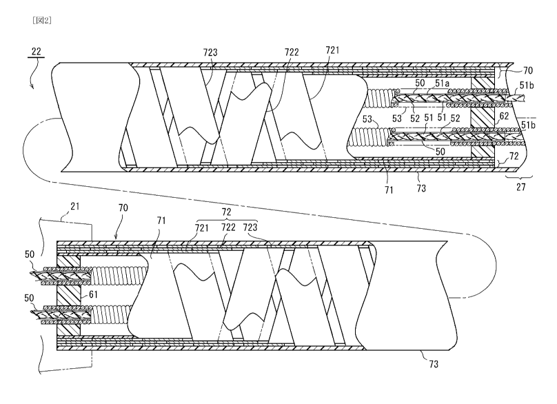
[0011]
FIG. 1 is a side view of a flexible endoscope suitable for use with a
MRI apparatus, showing an entire structure of the flexible endoscope
according to one embodiment of the present invention.
FIG. 2 is a side view showing an inner structure of a flexible portion of
an insertion portion of the flexible endoscope.
FIG. 3 is a side view showing an inner structure of a distal end portion
(a bendable portion and a distal piece) of the insertion portion.
DESCRIPTION OF THE REFERENCE NUMERALS
[0012]
MRI (Magnetic Resonance Imaging) apparatus
11 observation zone
endoscope
21 endoscope body
22 insertion portion
23 operation knob
24 ocular lens
forceps introduction portion
26 light guide
27 bendable portion
28 distal piece
-5-
CA 02678487 2009-08-17
29 joint ring
30 image guide
31 objective lens
40 working channel tube
41 mesh tube
50 bending operation wire (operation wire)
51 string
52 adhesive
53 bending operation wire guide (wire guide)
61, 62 securing piece
70 flexible tube (insertion portion body)
71 inner tube
72 triple helical tubes
721 first helical band
722 second helical band
723 third helical band
73 outer tube
BEST MODE FOR CARRYING OUT THE INVENTION
[0013]
A preferred embodiment of the present invention will now be described.
In FIG. 1, reference numeral 10 refers to a MRI (Magnetic Resonance
Imaging) apparatus and reference numeral 20 refers to an endoscope to be
inserted in an observation zone 11 of the MRI apparatus 10 when to be used.
The endoscope 20 includes an endoscope body 21 and an insertion portion 22.
The endoscope body 21 has an operation knob 23 disposed in a side portion
thereof, an ocular portion 24 disposed in a basal end portion thereof and a
forceps introduction portion 25 disposed in an upper side portion thereof. A
light guide 26 is drawn from a lower side portion of the endoscope body 21
-6-
CA 02678487 2009-08-17
and connected to a light source which is not shown. The insertion portion 22
extends from a distal end portion of the endoscope body 21. The insertion
portion 22 is to be inserted in the observation zone 11.
[0014]
As shown in FIGS. 1 and 3, the insertion portion 22 is flexible and
tubular. The insertion portion 22 has a bendable portion 27 in a distal end
portion thereof and a distal piece 28 in a distal end side of the bendable
portion 27.
[0015]
As shown in FIG. 3, a plurality of joint rings 29 are disposed in the
bendable portion 27. The joint rings 29 are made of low magnetic
susceptibility materials (nonmagnetic materials, low magnetic permeability
materials) such as brass. The joint rings 29 are arranged in a line, through
which a distal end portion 5lb of a bending operation wire (operation wire) 50
is passed. The bendable portion 27 as a whole can be bent by operating the
operation knob 23 to operate the bending operation wire 50 (see the chain
double-dashed line in FIG. 1).
Although the bendable portion 27 shown in the drawings can be bent in
two directions, the bendable portion 27 may be bendable in one direction only
or may be bendable in four directions.
[0016]
The distal piece 28 is made of low magnetic susceptibility materials
(nonmagnetic materials, low magnetic permeability materials) such as brass.
Surfaces of the distal piece 28 are plated with gold, which is a low magnetic
susceptibility material (nonmagnetic material, low magnetic permeability
material).
[0017]
The light guide 26, an image guide 30, a working channel tube 40 and
the bending operation wire 50 are received inside the insertion portion 22
-7-
CA 02678487 2009-08-17
(including the bendable portion 27 and the distal piece 28).
[0018]
The light guide 26 and the image guide 30 are composed of bundles of
optical fibers, which are nonmagnetic materials. Although not shown in the
drawings, a distal end of the light guide 26 reaches a distal end surface of
the
distal piece 28. Illumination light from the light source is transmitted along
the light guide 26 and emitted therefrom to illuminate objects of observation.
[0019]
An objective lens 31 is disposed at a distal end of the image guide 30.
The objective lens 31 is made of optical glass or plastic, which are
nonmagnetic materials. The objective lens 31 faces the distal end surface of
the distal piece 28. Images of the objects of observation are transmitted
through the objective lens 31 along the image guide 30 and can be observed
through the ocular lens 24.
[0020]
The working channel tube 40 is made of resin such as TeflonTM, which
is a nonmagnetic material, and is pleated like an accordion to provide
flexibility. A distal end portion of the working channel tube 40 reaches the
distal end surface of the distal piece 28. A Surgical instrument such as
forceps is to be inserted through the forceps introduction portion 25 into the
working channel tube 40. The surgical instrument is protruded from the
distal end surface of the distal piece 28 so that it can be used for surgery.
An
outer peripheral surface of the working channel tube 40 is covered with a
mesh tube 41 for restraining elongation. The mesh tube 41 is made of resin
such as NyronTM, which is a nonmagnetic material.
[0021]
As shown in FIGS. 2 and 3, a bending operation wire guide 53 (wire
guide) is received in a flexible portion of the insertion portion 22 other
than
the bendable portion 27 and the distal piece 28. The bending operation wire
-8-
CA 02678487 2009-08-17
guide 53 receives a main portion 51a of the bending operation wire 50 therein.
The bending operation wire guide 53 is made of a material that has a required
elasticity and low magnetic susceptibility (nonmagnetic materials, low
magnetic permeability materials) into a coiled configuration. In this
embodiment, copper-silver alloy is used as an example material for the
bending operation wire guide 53. Phosphor bronze may be used instead of
the copper-silver alloy. A distal end portion and a basal end portion of the
bending operation wire guide 53 are secured to a peripheral wall of the
insertion portion 22 via securing pieces 61, 62. The securing pieces 61, 62
are made of resin which is a nonmagnetic material.
[0022]
As shown in FIG. 2, a flexible tube 70 constituting the peripheral wall
(body) of the insertion portion 22 includes an inner tube 71, triple helical
tubes 72 and an outer tube 73. Parts including the image guide 30, the light
guide 26, the working channel tube 40, the bending operation wire guide 53
and the bending operation wire 50 are received inside the inner tube 71.
[0023]
The inner tube 71 is made of resin which is a nonmagnetic material.
Preferably, the resin forming the inner tube 71 has flexibility and also has
sufficient tensile strength and compressive strength. Examples of the
preferable resin include olefin resins such as polyethylene and polypropylene.
In this embodiment, polyethylene (IrraxTM by Sumitomo Electric Industries),
as an example, is used to form the inner tube 71.
[0024]
Outer side of the inner tube 71 is covered with the triple helical tubes
72. The triple helical tubes 72 are composed of a first helical band 721, a
second helical band 722 and a third helical band 723. Each of the first to
third helical bands 721, 722, 723 is composed of a helical band. The first
helical band 721 is closely wound around an outer peripheral surface of the
-9-
CA 02678487 2009-08-17
inner tube 71. The second helical band 722 is closely wound around an
outer peripheral surface of the first helical band 721. A direction of winding
of the second helical band 722 is opposite to that of the first helical band
721.
The third helical band 723 is closely wound around an outer peripheral surface
of the second helical band 722. A direction of winding of the third helical
band 723 is opposite to that of the second helical band 722 and same as that
of
the first helical band 721. Opposite end portions of the triple helical tubes
72 are respectively fixed between the securing pieces 61, 62 and the outer
tube 73.
[0025]
The first to third helical bands 721, 722, 723 are made of a material
that has a required elasticity, tensile strength and low magnetic
susceptibility
(nonmagnetic material, low magnetic permeability material). In this
embodiment, phosphor bronze is used to make the first to third helical bands
721, 722, 723. Copper-silver alloy may be used instead of the phosphor
bronze. The phosphor bronze and the copper-silver alloy sufficiently meet
the requirements mentioned above.
[0026]
The outer tube 73 is made of resin such as polypropylene and
polyethylene, which are nonmagnetic materials. An external diameter of the
outer tube 73 is 10 mm or smaller, and may be approximately 7 mm, for
example. The outer tube 73 covers not only the flexible portion of the
insertion portion 22 but also the bendable portion 27 and reaches the distal
piece 28.
[0027]
Detailed description of the bending operation wire 50 is provided
below.
The bending operation wire 50 includes a string 51 made by braiding
resin fibers which are nonmagnetic materials. The bending operation wire
-10-
CA 02678487 2009-08-17
50 is made by impregnating and hardening adhesive 52 which is a
nonmagnetic material in the string 51. Olefin resin, such as polypropylene
and polyethylene for example, is used as a material for the resin fibers to be
made into the string 5 1 in this embodiment. Various methods of braiding can
be employed, including various methods of knitting, weaving and twisting.
[0028]
Various adhesive 52 can be employed for impregnating the string 51.
Examples of suitable adhesive include commercially available products such
as CyanobondTM
The string 51 is pulled while being impregnated. Preferably, the
string is pulled with a tensile force slightly greater than a load applied to
the
bending operation wire 50 when the bendable portion 27 is bent
(approximately 2 kgf).
Example procedures for the impregnation include: suspending a weight
having a weight corresponding to the load mentioned above by tying the
weight at one end portion of the string 51; and dropping the adhesive 52 from
an upper end portion of the string 51. The adhesive 52 is absorbed in mesh
openings of the string 51, and impregnated in the string 51. Preferably, the
tensile force is continuously applied to the string 51 until the adhesive 52
is
hardened.
The impregnation and the hardening of the adhesive 52 provide the
string 51 with appropriate hardness and tension.
[0029]
The adhesive 52 is impregnated in the string 51, other than the distal
end portion 51b thereof received in the bendable portion 27. In other words,
the adhesive is impregnated in a portion of the string 51 received in the
endoscope body 21 and the main portion 51a of the string 51 received in the
inner tube 71 (flexible portion of the insertion portion 22).
The adhesive 52 is not impregnated in the distal end portion 51b of the
-11-
CA 02678487 2009-08-17
string received in the bendable portion 27.
[0030]
In the flexible endoscope 20, since the parts of the insertion portion 22
are made of nonmagnetic materials, placement of the insertion portion 22 in
the vicinity of observation zone 11 of the MRI apparatus does not
substantially affect the magnetic field of the MRI apparatus. Thus
interference on observation with the MRI can be limited or eliminated. This
enables an operator to perform a surgery, etc. using the flexible endoscope 20
while observing the site with MRI.
[0031]
Tensile strength, compressive strength, torsional strength and other
kinds of strength required for the insertion portion 22 can be provided by the
inner tube 71 and the triple helical tubes 72. Particularly, the inner tube 71
can provide the tensile strength and the compressive strength and the triple
helical tubes 72 can provide the torsional strength. Moreover, when the
flexible insertion portion 22 is bent at a certain point, a cross-sectional
shape
of the inner tube 71 at the certain point can be maintained by the triple
helical
tubes 72. Thus the inner tube 71 can be prevented from having the
cross-section thereof deformed even when it is bent. Moreover, curvature of
the insertion portion 22 can be limited by the triple helical tubes 72 to
prevent
the insertion portion 22 from being bent to an excessive degree.
Since the inner tube 71 can be reinforced by the triple helical tubes 72
as mentioned above, the outer diameter of the inner tube 71 can be reduced as
much as possible and a thickness of a tube wall of the inner tube 71 can be
reduced as much as possible. This serves to reduce the diameter of the
insertion portion 22 as much as possible, thus reducing burden on patients.
[0032]
Since the bending operation wire 50 includes the string 51 made of
resin fibers, the bending operation wire 50 can be less expensive than those
-12-
CA 02678487 2009-08-17
made of a metal wire, such as a stainless steel wire. The impregnation and
hardening of the adhesive 52 in the string 51 can serve to prevent the
bendable
wire 50 made of resin from being elongated even when tensile force is applied
thereto during the bending operation of the bendable portion 27.
Pulling the string 51 during the impregnation and hardening of the
adhesive 52 can serve to homogenize the thickness of the bending operation
wire 50.
Since the adhesive 52 is not impregnated in the distal end portion 51b
of the string in the bendable portion 27, the bendable portion 27 can be bent
easily.
[0033]
The present invention is not limited to the embodiment described
above, and various modifications can be made without departing from the
spirit of the present invention.
For example, the parts mentioned above can be made of any other
materials than those mentioned in the above embodiment as long as they have
the low magnetic susceptibility (low magnetic permeability) and other
required properties.
Other polyolefin resins such as polypropylene, polyamide resins such
as nylon or any other resins can be used instead of polyethylene as a resin
material for the inner tube 71.
Resins such as polypropylene or polyethylene may be used instead of
the non-magnetic metals (alloys) such as phosphor bronze or copper-silver
alloy to form the first to third helical bands 721, 722, 723.
Resins such as polypropylene or polyethylene may be used instead of
the non-magnetic metals (alloys) such as copper-silver alloy or phosphor
bronze to form the bending operation wire guide 53.
Polyamide resins such as nylon may be used instead of the polyolefin
resins such as polypropylene or polyethylene as the resin fibers to form the
-13-
CA 02678487 2009-08-17
string 51 of the bending operation wire 50.
The adhesive 52 may be impregnated in the string 51 by other methods
than those disclosed in the above embodiment.
The tensile force applied to the string 51 during the impregnation and
hardening of the adhesive 52 does not have to be slightly greater than the
load
applied to the bending operation wire 50 during the bending operation of the
bendable portion 27. The tensile force may be generally equal to or
considerably greater than the load mentioned above. Alternatively, the
tensile force may be smaller than the load mentioned above, and the string 51
does not have to be pulled during the impregnation of the adhesive 52.
The adhesive 52 may also be impregnated in the distal end portion
51b of the string in the bendable portion 27. The degree of impregnation of
the adhesive 52 may be smaller in the distal end portion 51b of the string
than
in the main portion 51 a.
INDUSTRIAL APPLICABILITY
[0034]
The present invention may be applied to an endoscope for use with a
MRI apparatus.
-14-