Note: Descriptions are shown in the official language in which they were submitted.
CA 02678747 2009-08-19
WO 2008/106531
PCT/US2008/055160
- I -
METHOD AND APPARATUS FOR REPLACING A PROSTHETIC
VALVE
FIELD
[001] The present invention relates to embodiments of a method and apparatus
for replacing a previously implanted prosthetic valve, such as a surgically
implanted prosthetic heart valve, without removing the previously implanted
valve from the body.
BACKGROUND
[002] Prosthetic valves, such as prosthetic heart valves, are implanted in the
body to replace a failing or diseased natural valve. Should the prosthetic
valve
begin to fail, it also may need to be replaced with another prosthetic valve.
Surgically implanted, prosthetic heart valves, such as a prosthetic aortic
valve,
typically are replaced about every 15 years. The current method for replacing
a
surgically implanted, prosthetic heart valve involves open heart surgery
wherein
the patient's chest is opened and the existing prosthetic valve is removed and
replaced with a new prosthetic valve. As can be appreciated, this is a
traumatic
and high risk procedure accompanied by substantial morbidity and mortality,
and in some cases, cannot even be attempted due to the advanced age and/or
medical condition of the patient.
[003] Therefore, it would be preferable to replace a prosthetic heart valve
with
a percutaneously implanted valve that is delivered to the implantation site
via
the patient's vasculature and deployed within the previously implanted valve.
However, because existing prosthetic heart valves can vary widely in size and
shape, there are substantial difficulties associated with the development and
validation of a percutaneously delivered replacement valve that is compatible
with different types of existing prosthetic heart valves. More particularly,
difficulties arise because a replacement valve that does not conform to the
geometry of the previously implanted valve may not be able to adequately
CA 02678747 2009-08-19
WO 2008/106531
PCT/US2008/055160
- 2 -
anchor to the previously implanted valve and/or form an effective seal with
the
previously implanted valve.
SUMMARY
[004] In one aspect, the present disclosure concerns a percutaneously
delivered
adapter stent that is deployed within a previously implanted prosthetic valve
and
serves as an anchor or platform for implanting a percutaneously delivered
replacement valve within the previously implanted valve. The replacement
valve can be any known percutaneous valve. The adapter stent can be adapted
to provide a suitable mounting platform for implanting a percutaneous
replacement valve in a wide range of existing surgical valves, which typically
vary widely in size and shape from patient to patient. In one advantageous
feature, the adapter stent increases the frictional forces between the
percutaneous replacement valve and the failing surgical valve, thereby
providing a more predictable orientation and securement of the percutaneous
replacement valve. Hence, this technique is particularly suited for replacing
a
surgically implanted prosthetic heart valve, but also could be used for
replacing
a percutaneously implanted prosthetic valve.
[005] The adapter stent can be delivered to the implantation site via the
patient's vasculature and positioned within the previously implanted valve.
The
stent can then be deployed to cause the stent to expand and become anchored to
the inner surface of the previously implanted valve. Subsequently, the
replacement valve can be positioned within the adapter stent and deployed to
cause the replacement valve to expand and become anchored to the adapter
stent.
[006] In particular embodiments, the adapter stent and the replacement valve
can be mounted on the same delivery catheter for delivery to the implantation
site. In one implementation, for example, the adapter stent and the
replacement
valve can be crimped around respective first and second balloons of a double-
balloon catheter. In this approach, the adapter stent is positioned in the
previously implanted valve and expanded into contact with the previously
CA 02678747 2009-08-19
WO 2008/106531
PCT/US2008/055160
-3 -
implanted valve by inflating the first balloon. The catheter is then re-
positioned
to place the replacement valve in the deployed adapter stent, after which the
valve is expanded into contact with the adapter stent by inflating the second
balloon. In another implementation, the adapter stent and the replacement
valve
are self-expandable. The self-expandable adapter stent and valve can be
mounted on a common delivery catheter adapted to retain the stent and the
valve in compressed positions while they are advanced through the patient's
vasculature. Using the catheter, the adapter stent and the valve can be
successively positioned and deployed within the previously implanted valve.
[007] The adapter stent in exemplary embodiments can comprise an
expandable frame that mounts a flexible annular sealing member. The sealing
member provides a seal between the previously implanted valve and the
replacement valve to prevent or at least minimize blood flow between the
original and replacement valves.
[008] The adapter stent may be configured to have a length that is greater
than
the length of the previously implanted valve that needs to be replaced. This
allows the stent to extend over the entire inner surface of the previously
implanted valve to provide sufficient surface area for anchoring the
replacement
valve and to ensure that the previously implanted valve does not interfere
with
the positioning and deployment of the replacement valve. In certain
embodiments, the adapter stent, when expanded, has enlarged end portions that
flare or extend radially outwardly past the adjacent ends of the previously
implanted valve to assist in securing the adapter stent in place.
[009] In one representative embodiment, a method is provided for
percutaneously implanting a replacement prosthetic valve at a site occupied by
a
previously implanted prosthetic valve. The method includes positioning an
adapter stent within the previously implanted valve, deploying the adapter
stent
to cause the adapter stent to become anchored to the previously implanted
valve, positioning the replacement valve within the deployed adapter stent,
and
deploying the replacement valve to cause the replacement valve to become
anchored to the adapter stent.
CA 02678747 2009-08-19
WO 2008/106531
PCT/US2008/055160
- 4 -
[010] In another representative embodiment, a method of percutaneously
implanting a replacement prosthetic valve in a patient at a site occupied by a
previously implanted prosthetic valve includes advancing a catheter carrying
an
adapter stent through the patient's vasculature to position the adapter stent
within the previously implanted valve. The catheter also carries the
replacement valve. The method further includes deploying the adapter stent to
cause the adapter stent to become anchored to the previously implanted valve,
re-positioning the catheter to position the replacement valve within the
deployed
adapter stent, and deploying the replacement valve to cause the replacement
valve to become anchored to the adapter stent.
[011] In another representative embodiment, an assembly is provided for
percutaneous replacement of a previously implanted prosthetic valve without
removal of the previously implanted valve. The assembly comprises an adapter
stent comprising a frame and an annular sealing member. The adapter stent is
adapted to be deployed within the previously implanted valve. The assembly
also includes a percutaneous, replacement prosthetic valve comprising a frame
and a flexible valve member. The valve is adapted to be deployed within the
deployed adapter stent such that the sealing member provides a seal between
the
previously implanted valve and the replacement valve.
[012] In yet another representative embodiment, an assembly for percutaneous
replacement of a previously implanted prosthetic valve comprises a
percutaneous, replacement prosthetic valve comprising a frame and a flexible
valve member. The assembly also includes means for anchoring and sealing the
replacement valve to the previously implanted valve, said means being
separately deployable within the previously implanted valve prior to deploying
the replacement valve within said means.
[013] The foregoing and other features and advantages of the invention will
become more apparent from the following detailed description, which proceeds
with reference to the accompanying figures.
CA 02678747 2009-08-19
WO 2008/106531
PCT/US2008/055160
- 5 -
BRIEF DESCRIPTION OF THE DRAWINGS
[014] FIG. 1 is a side elevation view of one embodiment of an assembly
comprising a percutaneous prosthetic valve and an adapter stent for anchoring
the prosthetic valve within a previously implanted prosthetic valve.
[015] FIG. 2 is a perspective view of the prosthetic valve shown in FIG. I.
[016] FIG. 3 is a schematic side view of an embodiment of a double-balloon
catheter showing the prosthetic valve and the adapter stent of FIG. 1 crimped
around respective balloons on the catheter for percutaneous delivery to an
implantation site.
[017] FIGS. 4A-4G illustrate the successive steps of one specific embodiment
of an implantation procedure employing the double-balloon catheter shown in
FIG. 2 for implanting the adapter stent and the prosthetic valve inside a
failing
surgically implanted, prosthetic valve previously implanted in the aortic
orifice
of a patient.
[018] FIG. 5 is a schematic side view of one embodiment of delivery catheter
that can be used to implant a self-expanding adapter stent and replacement
valve
inside a previously implanted valve.
[019] FIG. 6 is a side elevation view of another embodiment of an adapter
stent that can be used to anchor a replacement valve within a previously
implanted prosthetic valve.
[020] FIG. 7 illustrates another embodiment of an implantable assembly for
replacing a previously implanted prosthetic valve.
[021] FIG. 8 illustrates the assembly of FIG. 7 deployed within a previously
implanted surgical valve.
DETAILED DESCRIPTION
[022] As used herein, the singular forms "a," "an," and "the" refer to one or
more than one, unless the context clearly dictates otherwise.
[023] As used herein, the term "includes" means "comprises." For example, a
device that includes or comprises A and B contains A and B but may optionally
CA 02678747 2009-08-19
WO 2008/106531
PCT/US2008/055160
- 6 -
contain C or other components other than A and B. A device that includes or
comprises A or B may contain A or B or A and B, and optionally one or more
other components such as C.
[024] In one aspect, the present disclosure concerns a percutaneously
delivered
adapter stent that is deployed within a previously implanted prosthetic valve
and
serves as an anchor or platform for implanting a percutaneously delivered
replacement valve within the previously implanted valve. As used herein, the
term "stent" refers generally to any lumina! structure. The replacement valve
can be any known percutaneous valve. The adapter stent can be advanced
through the patient's vasculature and positioned within the previously
implanted
valve. The adapter stent can then be deployed to cause the adapter stent to
expand and become anchored to the inner surface of the previously implanted
valve. The replacement valve can then be positioned within the adapter stent
and deployed to cause the replacement valve to expand and become anchored to
the adapter stent. In one respect, the adapter stent is configured to increase
the
frictional forces between the replacement valve and the failing previously
implanted valve, thereby providing a more predictable orientation and
securement of the replacement valve. In the following description, the adapter
stent and the replacement valve are shown and described in connection with
replacing a previously implanted aortic valve. However, the embodiments
described herein can also be used to replace prosthetic valves implanted at
other
locations in the heart or in other body channels having native valves, such as
veins or other organs.
[025] FIG. 1 shows an assembly 10 comprising a percutaneous prosthetic heart
valve 12 and an adapter stent 30, according to one embodiment. The adapter
stent 30 can be deployed within a failing, previously implanted valve, such as
the prosthetic aortic valve 60 shown in FIG. 4A. Once the adapter stent 30 is
deployed within the previously implanted valve, the new valve 12 can be
deployed within the adapter stent 30 to replace the previously implanted valve
60. The previously implanted valve 60 shown in the figures is a surgical valve
(i.e., a valve implanted via open heart surgery), although the adapter stent
30
CA 02678747 2009-08-19
WO 2008/106531
PCT/US2008/055160
- 7 -
and the replacement valve 12 can also be deployed within a previously
implanted percutaneous valve.
[026] The valve 12 and the adapter stent 30 are each crimpable or
compressible to a reduced diameter for percutaneous delivery to the
implantation site, such as using a delivery catheter. When expanded to their
functional size (FIG. 1), the outer diameter of the valve 12 desirably is
approximately equal to the inner diameter of the adapter stent and the outer
surface of the valve 12 generally conforms to an inner surface portion of the
adapter stent 30 to promote attachment of the valve 12 to the adapter stent
30.
Methods for implanting the adapter stent 30 and the valve 12 are described in
greater detail below.
[027] As shown in FIGS. 1 and 2, the valve 12 in the illustrated embodiment
includes an annular frame 14 that mounts a flexible valve member 16. The
frame 14 in the illustrated embodiment comprises a plurality of angularly-
spaced axial struts, or support members, 18 that extend axially
(longitudinally)
along the frame and a plurality of support posts, or beams, 20 (one of which
is
shown in FIGS. 1 and 2) spaced in the illustrated example at 120-degree
intervals from each other around the frame 14. The support posts 20 can be
formed with apertures 22 to facilitate attachment of the valve member 16 to
the
posts 20, such as, for example, by suturing the valve member 16 to the posts.
The frame 14 can also include a plurality of axially-spaced, circumferential
bands, or struts, 24 attached to the axial struts 18 and the support posts 20.
The
struts 24 are formed with multiple bends that allow the frame 14 to be crimped
to a smaller diameter for delivery to an implantation site and expanded to its
functional size for anchoring the valve assembly to the adapter stent 30 at
the
implantation site. For example, each of the struts 24 in the illustrated
configuration includes a plurality of linear strut members 26a, 26b arranged
in a
zig-zag or saw-tooth configuration defining bends between adjacent strut
members.
[028] In alternative embodiments, the frame can have other configurations.
For example, one or more of the circumferential bands 24 can have a curved or
CA 02678747 2014-08-12
- 8 -
serpentine shape rather than a zig-zag shape. Further, the frame 14 can
include
various attachment elements (not shown), such as barbs, staples, flanges, and
the like for enhancing the ability of the frame to anchor to the adapter stent
30.
[029] The frame 14 can be made from any of various suitable ductile and/or
elastic materials and is typically made of a metal, such as stainless steel,
titanium, or other biocompatible metals. The frame 14 or components thereof
can also he made from a shape memory alloy such as nickel titanium (NiTi)
shape memory alloys, as marketed, for example, under the trade name Nitinol.
The shape-memory components allow the valve 12 to be self-expandable; that
is, the valve 12, when restrained in a radially compressed state by an outer
restraint (e.g., a sheath covering the valve), automatically expands to its
functional size when the outer restraint is removed.
[030] The valve member 16 can have a leafed-valve configuration, such as the
tricuspid valve configuration shown in the illustrated embodiment. 'Be valve
member 16 can be formed from three pieces of pliant material connected to
each other at seams aligned with posts 20 to form collapsible leaflets 28
(FIG.
2). The valve member 16 can be made from biological matter, such as natural
tissue, pericardial tissue (such as bovine, porcine or equine pericardium), a
harvested natural valve or other biological tissue. Alternatively, the valve
member 16 can be made from biocompatible polymers or similar materials.
[031] Various other prosthetic valve configurations also can be used.
Examples of other valves that can be utilized are disclosed in U.S. Patent No.
6,730, 118, U.S. Patent No. 6,767,362, and U.S. Patent No. 6,908,481.
[032] The adapter stent 30 in exemplary embodiments includes an expandable
frame 32 that mounts a flexible annular sealing member 34. The frame 32 is
shown in HG. 1 in its expanded, functional size, and is configured to be
crimpable to a reduced diameter for percutaneous delivery, such as on a
delivery catheter. The frame 32 can be made from any of various suitable
ductile and/or elastic materials and is typically made of a metal, such as
stainless steel, titanium, or other biocompatible metals. The frame 14 or
CA 02678747 2009-08-19
WO 2008/106531
PCT/US2008/055160
- 9 -
components thereof can also be made from a shape memory material, which
allows the stent 30 to be self-expandable.
[033] The frame 32 is the illustrated embodiment comprises a plurality of
longitudinally extending, zig-zag struts 36 joined to each other at junctures
38.
The frame 32 has a length L measured between the opposite ends thereof that
desirably is greater than the length of the previously implanted valve that
needs
to be replaced. In this manner, the frame 32, when deployed within the
previously implanted valve, can extend over the entire inner surface area of
the
previously implanted valve to provide sufficient surface area for anchoring
the
replacement valve 12 and to ensure that the previously implanted valve does
not
interfere with the positioning and deployment of the replacement valve 12. In
particular embodiments, for example, the length L of the frame is about 10 mm
to about 40 mm, with about 30 mm being a specific example.
[034] As shown, the frame 32 in exemplary embodiments has a generally
cylindrical intermediate portion 44 extending between the opposite end
portions
40, 42, which are enlarged or flared relative to the intermediate portion 44
when
the frame is expanded. Each end portion 40, 42 desirably expands to a diameter
that is greater than the diameter of the previously implanted valve. Hence,
when the adapter stent 30 is deployed within the previously implanted valve,
the
end portions 40, 42 can extend radially outwardly past the adjacent ends of
the
previously implanted valve to assist in securing the adapter stent in place.
[035] In alternative embodiments, the frame 32 can have various other shapes
or configurations. For example, the frame 32 can be generally cylindrical or
tubular along its entire length without enlarged end portions. The frame 32
optionally can be provided with various attachment elements (not shown), such
as barbs, staples, flanges, and the like for enhancing the ability of the
frame to
anchor to the previously implanted valve 60 (FIG. 4A). If desired, the frame
32
may be provided with attachment elements along the inner surface for
enhancing the ability of the frame 32 to securely engage the frame 14 of the
percutaneously delivered replacement valve 12.
CA 02678747 2009-08-19
WO 2008/106531
PCT/US2008/055160
- 10 -
[036] The sealing member 34 provides a seal between the previously
implanted valve 60 and the replacement valve 12 to prevent or at least
minimize
blood flow between the valves. As shown in FIG. 1, the sealing member 34
desirably extends nearly the entire length of the frame 32 to maximize the
surface area that can contact the previously implanted valve 60 and the
replacement valve 12. In other embodiments, however, the sealing member can
extend along only a portion of the frame 32, such as the intermediate portion
44.
With reference to the embodiment shown in FIG. 1, the sealing member 34 is
secured to the inner surface of the frame 32. Alternatively, the sealing
member
can be secured to the outer surface of the frame 32 as shown in FIG. 6 to
prevent the leakage of blood. In another implementation, a sealing member 34
can be secured to both the inner and outer surfaces of the frame 32.
[037] In particular embodiments, the sealing member 34 is made of a natural
or synthetic biocompatible elastomeric material, such as foam rubber,
thermoplastic elastomers (e.g., polyurethanes) or other polymeric elastomers,
such as a polymeric sponge. The sealing member 34 can be secured to or
formed on the frame using any suitable techniques or mechanisms, such as by
suturing the sealing member to the frame or co-molding the sealing member to
the frame. The sealing member 34 also can be formed on the frame using
conventional coating techniques, such as spray coating, dip coating, or roll
coating.
[038] The valve 12 and the adapter stent 30 can be implanted using a double-
balloon catheter. FIG. 3, for example, shows the distal end portion of an
exemplary embodiment of a double-balloon catheter, indicated at 70. The
catheter 70 includes a shaft 72, on which there are mounted first and second,
spaced-apart balloons 74, 76, respectively, between a respective pair of rings
80, 82. The adapter stent 30 and the replacement valve 12 are crimped around
the first balloon 74 and the second balloon 76, respectively. The shaft 72
contains two lumens (not shown), each of which is fluidly connected to a
respective balloon 74, 76 for successive and separate inflation of each
balloon.
The shaft 72 also contains another lumen to accept a guide wire 78 so that the
CA 02678747 2009-08-19
WO 2008/106531
PCT/US2008/055160
- 1 I -
catheter can be advanced over the guide wire 78 for guiding the catheter
through the patient's vasculature.
[039] The catheter 70 can be introduced percutaneously into the patient's
vasculature (e.g., into a peripheral artery such as the femoral artery) and
advanced to the implantation site. For example, for replacing a prosthetic
aortic
valve, the catheter in certain embodiments has a length of at least about 80
cm,
usually about 90-100 cm, to allow transluminal positioning of the shaft from
the
femoral and iliac arteries to the ascending aorta. Alternatively, the shaft
may
have a shorter length, e.g. about 20-60 cm, for introduction through the iliac
artery, through the brachial artery, through the carotid or subclavian
arteries, or
through a penetration in the aorta itself. In the femoral approach, the
catheter
desirably is long enough and flexible enough to traverse the path through the
femoral artery, iliac artery, descending aorta and aortic arch. At the same
time,
the catheter desirably has sufficient pushability to be advanced to the
ascending
aorta by pushing on the proximal end, and has sufficient axial, bending, and
torsional stiffness to allow the physician to control the position of the
distal end,
even when the catheter is in a tortuous vascular structure. Alternatively, the
catheter may be passed through a port between ribs in the patient's thorax
above
the heart and through an incision in the heart wall (e.g., through the apex of
the
left ventricle) or through an incision in the aortic arch, in a so-called
minimally-
invasive procedure.
[040] A procedure for implanting the valve 12 and the adapter stent 30 using
the catheter 70, according one embodiment, is illustrated in FIGS. 4A-4G. FIG.
4A illustrates the previously implanted valve 60 implanted in the aortic
annulus
between the left ventricle chamber 86 and the ascending aorta 88. As noted
above, the illustrated valve 60 is a surgical valve, although the adapter
stent 30
and the replacement valve 12 can also be implanted within an existing
percutaneous valve. The catheter 70 can be introduced percutaneously into the
patient's vasculature and advanced to the implantation site using known
techniques. For example, a blood vessel (e.g., the femoral artery) typically
is
dilated using a conventional dilator to allow an introducer sheath to be
inserted
CA 02678747 2009-08-19
WO 2008/106531
PCT/US2008/055160
- 12 -
into the blood vessel. The guide wire 78 can then be inserted into the blood
vessel via the introducer sheath and advanced to the implantation site.
Subsequently, the catheter 70 can be advanced over the guide wire 78 to
position the adapter stent 30 in the previously implanted valve 60. More
precisely, the adapter stent 30 desirably is positioned such that the end
portions
40, 42 are located outside the adjacent ends of the previously implanted valve
60, as shown in FIG. 4B.
[041] As depicted in FIG. 4C, the balloon 74 is then inflated to deploy the
adapter stent 30, which expands to its functional size and engages the inner
surface of the previously implanted valve 60. As shown, in its expanded
stated,
the end portion 40, 42 flare radially outwardly past the adjacent ends of the
previously implanted valve to assist in retaining the adapter stent 30 in
place
against the valve 60. In addition, the adapter stent 30, in the illustrated
example, also extends over the entire inner surface area of the existing valve
60
and causes the flexible leaflets 62 of the valve to expand radially outwardly,
thereby providing a surface area suitable lbr mounting the replacement valve
12.
[042] Thereafter, the balloon 74 is deflated (FIG. 4D) and the catheter 70 is
retracted slightly to position the replacement valve 12 within the deployed
adapter stent 30 (FIG. 4E). The second balloon 76 is then inflated to deploy
the
replacement valve 12, which expands to its functional size and engages the
inner surface of the adapter stent 30 (FIG. 4F). Once the replacement valve 12
is deployed, the balloon 76 can be deflated and the catheter 70 can be removed
from the body (FIG. 4G).
[043] The adapter stent 30, as well as the valve 12, can be positioned at the
implantation site with the assistance of fluoroscopy and radiopaque markers,
ultrasonic imaging, and the like. For example, rings 80, 82 on the catheter
shaft
72 can be made of any of various suitable metals that are visible during
fluoroscopy for use in positioning the adapter stent and/or the valve.
Alternatively, radiopaque markers can be provided on portions of the adapter
stent 30 and/or the valve 12.
CA 02678747 2009-08-19
WO 2008/106531
PCT/US2008/055160
- 13 -
[044] In an alternative approach, the replacement valve 12 can be mounted on
the first balloon 74 and the adapter stent 30 can be mounted on the second
balloon 76. In this approach, the adapter stent 30 is first deployed within
the
previously implanted valve 60 while the first balloon 74 and the replacement
valve 12 are positioned in the aorta 88. After the adapter stent 30 is
deployed,
the catheter 70 is advanced further into the left ventricle 86 to position the
first
balloon 74 and the replacement valve 12 within the deployed adapter stent 30.
The replacement valve 12 can then be deployed by inflating the first balloon
74.
[045] As noted above, the frame 32 of the adapter stent 30 and the frame 14 of
the replacement valve 12, or portions thereof, can be made of a shape-memory
material, which allows the adapter stent 30 and the valve 12 to be self-
expandable. FIG. 5 is a schematic view of the distal end portion of a delivery
catheter, indicated at 90, which can be used to implant a self-expanding
replacement valve and adapter stent in the previously implanted valve 60. The
catheter 90 includes a shaft 92 and an outer sheath 94, which is moveable
longitudinally relative to the shaft 92. The shaft 92 can include a lumen for
receiving a guide wire 78. The valve 12 and the adapter stent 30 are mounted
to
the shaft 92 in their compressed states. The outer sheath 94 extends over the
valve 12 and the adapter stent 30 to retain the valve and adapter stent in
their
compressed states until each is positioned for deployment at the implantation
site.
[046] The catheter 90 can be introduced into the body and advanced through
the patient's vasculature in the same manner as the balloon catheter 70. The
adapter stent 30 is first positioned in the previously implanted valve 60 and
the
outer sheath is retracted to expose the adapter stent 30, which permits the
adapter stent to expand into contact with the previously implanted valve. The
catheter 90 is then advanced slightly to position the valve 12 in the deployed
adapter stent 30. The outer sheath 94 can then be retracted to expose the
valve
12, which permits the valve to expand into contact with the adapter stent.
[047] Although less desirable, the adapter stent 30 and the replacement valve
12 can be delivered and implanted at the site of the previously implanted
valve
CA 02678747 2014-08-12
- 14 -
using separate catheters. For example, the adapter stent 30 and the valve 12
can
be mounted on separate balloon catheters. In this approach, the adapter stent
30
is implanted using a first balloon catheter, which is then removed from the
body
to allow a second balloon catheter carrying the replacement valve to be
inserted
into the body.
[0481 As noted above, surgical valves, such as valve 60, typically vary widely
in size and shape from patient to patient. Advantageously, the adapter stent
30
can be adapted to provide a suitable mounting platform for implanting a
percutaneous replacement valve in a wide range of surgical valves varying in
size and shape.
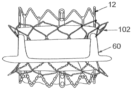
[049] FIG. 7 illustrates another exemplaty embodiment of an assembly 100
comprising a percutaneous prosthetic valve 12 and an adapter stent 102. The
adapter stent 102, like adapter stent 30, includes a radially compressible and
expandable frame 102 that mounts a flexible annular sealing member 106. FIG.
IS 8 illustrates the adapter stent 102 and the prosthetic valve 12 deployed
within a
previously implanted surgical valve 60. The adapter stent 102 has a length L
that is preferably greater than the length of the previously implanted valve
60
but need not be longer than the new valve 12. In certain embodiments, the
adapter stent 102 has a length L of about 10 mm and the new valve 12 has a
length of about 20 atm.
[0501 In view of the many possible embodiments to which the principles of the
disclosed invention may be applied, it should be recognized that the
illustrated
embodiments are only preferred examples of the invention and should not be
taken as limiting the scope of the invention.
CA 02678747 2014-08-12
-15-
[051] The scope of the claims should not be limited by the preferred
embodiments set
forth in the examples, but should be given the broadest interpretation
consistent with the
description as a whole.