Note: Descriptions are shown in the official language in which they were submitted.
CA 02678774 2009-08-19
WO 2008/106644 PCT/US2008/055498
TITLE OF THE INVENTION
TREATMENT OF DISEASES CHARACTERIZED BY INFLAMMATION
RELATED APPLICATIONS
[0001] This application claims the benefit of U.S. Provisional Application
Nos.
60/892,395, filed March 1, 2007 and 60/985,024, filed November 2, 2007, the
disclosures of
which are incorporated herein by reference in their entireties.
BACKGROUND
[0002] The complement system is a critical component of the innate and
adaptive
immune system (reviewed by Volanakis, 1998). Complement plays an important
role in
microbial killing, and is essential for the transport and clearance of immune
complexes.
Many of the activation products of the complement system are also associated
with
proinflammatory or immunoregulatory functions. The complement system consists
of
plasma and membrane-associated proteins that are organized in three enzymatic-
activation
cascades: the classical, the lectin, and the alternative pathways. All three
pathways can lead
to the formation of the terminal complement complex (TCC) and an array of
biologically
active products.
[0003] In some cases, complement activation is initiated either by specific
antibodies
recognizing and binding to a variety of pathogens and foreign molecules,
and/or by direct
interaction of complement proteins with foreign substances. On activation,
these pathways
result in the formation of unstable protease complexes, the C3-convertases.
The classical
pathway C3-convertase, C4b2a, and the alternative pathway C3-convertase,
C3bBb, are both
able to cleave the a chain of C3 generating C3b. C3b has the potential to bind
covalently to
biological surfaces. C3b binding leads to opsonization for phagocytosis by
polymorphonuclear cells and macrophages. When additional C3b is available, the
C3-
convertases can function as C5-convertases, cleaving C5 and initiating the
assembly of the
TCC, or membrane attack complex (MAC), which mediates cellular lysis by
insertion of
pore-forming protein complexes into targeted cell membranes.
[0004] The precise function of the complement system depends on tight
regulation, as
activation of the complement cascade leads to the production of a number of
proteins that
contribute to inflammation. This is beneficial when contributing to a host
defense, but can be
detrimental if activated on self tissue. Typically, activation of C3 in the
blood is kept at a low
level, and C3b deposition is limited to the surface of pathogens.
CA 02678774 2009-08-19
WO 2008/106644 PCT/US2008/055498
[0005] To regulate the complement system, a number of complement regulatory
proteins function to restrict complement activation. These proteins interact
with C3 or C4
derivatives and are encoded by closely linked genes that comprise the
Regulator of
Complement Activation (RCA) gene cluster on human chromosome 1 q32 (Diaz-
Guillen et
al., 1999).
[0006] Complement factor H (CFH or fH) a plasma protein encoded by one of the
RCA genes, is a soluble activation inhibitor of the alternative complement
pathway (Muller-
Eberhard et al., 1980; Zipfel et al., 2002; Rodriguez de Cordoba et al.,
2004). CFH prevents
binding of factor B to C3b, displays decay-accelerating activity for
dissociation of the C3bBb
complex, acts as a cofactor for the cleavage of C3b by factor I, and blocks
the generation of
C5b6-9, also known as membrane attack complex (MAC) (Whaley and Ruddy, 1976;
Weiler
et al., 1976; Pangburn et al., 1977). CFH binds to and interacts with multiple
ligands
including C3b, heparin, bacterial surface proteins, the acute phase protein, C-
reactive protein
(CRP), adrenomedullin and cell surface receptors (Zipfel et al., 2002).
[0007] Human CFH is a member of a protein family composed of seven
structurally
and immunologically related, multidomain, multifunctional serum proteins.
These include
CFH and factor H-like protein 1(FHL-1) and five factor H-related proteins (FHR-
1 to FHR-
5). Each of these proteins is composed exclusively of short consensus repeats
(SCRs) or
complement control modules, each encoded by a separate exon. CHF, 150 kDa, is
composed
of 20 SCRs. FHL-1, 43 kDa, and composed of seven SCR domains, is derived from
CFH by
alternative splicing (Estaller et al., 1991; Sim et al., 1993). FHL-1, like
CFH, functions as a
complement regulator and displays cofactor and decay-accelerating activity
(Zipfel and
Sherka, 1999). In addition, FHL-1 has unique functions including acting as an
adhesion
protein due to the presence of an exposed RGD domain in SCR4 (Hellwage et al.,
1997).
CFH is present in human plasma at 500 g/ml. In contrast, the FHL-1 plasma
concentration
is 10-50 g/ml. The five FHR proteins are each derived from a separate gene in
the RCA
gene cluster. Although members of this family differ in the number of SCRs,
the individual
SCR domains display a high degree of homology to each other.
[0008] The organization of CFH and related proteins into multiple,
individually
folded protein domains suggests a structure/function relationship. The
complement
regulatory domains of CFH and FHL-1 are located within the four amino terminal
SCRs.
Three C3-binding domains are located in CFH. The N-terminal domain (SCRs 1-4)
binds to
2
CA 02678774 2009-08-19
WO 2008/106644 PCT/US2008/055498
intact C3b (Gordon et al., 1995; Kuhn et al., 1995; 1996), the middle domain
(SCRs 12-14)
binds to the C3c fragment, and the C-terminal domain (SCR 19-20) binds to C3d
(Jokiranta et
al., 2000). The C-terminal domain (SCR 19-20) also blocks the lytic function
of the C5b6-9
TCC (Zipfel et al., 2002). This function is absent in FHL-1. CFH and FHL-1
contain
overlapping binding sites for heparin, C-reactive protein (CRP), and M-protein
located in
SCR7 (Giannakis et al., 2003).
[0009] Proteins in the family are predominantly synthesized in the liver,
although
CFH has also been demonstrated to be expressed in a wide variety of cell
types, such as
peripheral blood lymphocytes, myoblasts, fibroblasts, neurons, and glia cells
(Friese et al.,
1999). More recently, CFH sequences were identified in an expressed sequence
tag library
derived from human retinal pigment epithelial (RPE) cells and choroid (Wistow
et al., 2002),
and immunohistochemical staining identified CFH in choroid vessels and in an
area
bordering the RPE (Klein et al., 2005).
[0010] A CFH polymorphism is linked to an increased risk of age related
macular
degeneration (AMD) (Edwards et al., 2005; Klein et al., 2005; Haines et al.,
2005). This
polymorphism, a tyrosine to histidine change at amino acid 402 (tyr402his or
Y402H),
accounts for about 50% of the attributable risk of AMD. The Y--->H
polymorphism identified
in the recent Science articles is located in SCR7 (Edwards et al., 2005; Klein
et al., 2005;
Haines et al., 2005).
[0011 ] CRP is known to activate the classic complement pathway, and inhibits
the
deposition of C5b6-9 through the direct binding of CFH (Mold et al., 1999).
CFH binding to
heparin and/or CRP could potentially be altered by the replacement of a
neutral tyrosine with
a positively charged histidine (Rodriguez de Cordoba et al., 2004). Elevated
serum levels of
CRP were observed in AMD patients compared to controls in a clinical study
(Seddon et al.,
2004). Furthermore, nutritional supplementation with zinc slows down the
progression of
AMD and biochemical studies have shown that CFH function is sensitive to zinc
concentration (AREDS Research Group, 2001; Blom et al., 2003). Therefore,
altered binding
of CFH to CRP or heparin on retinal surfaces caused by the tyr402his
substitution could
affect the level of inflammation in the outer retina (Edwards et al., 2005;
Klein et al., 2005;
Haines et al., 2005).
[0012] CFH deficiencies have been described both in humans and animals. They
are
caused by mutations that result in truncations or amino acid substitutions
that impair CFH
3
CA 02678774 2009-08-19
WO 2008/106644 PCT/US2008/055498
function (Ault et al., 1997; Sanchez-Corral et al., 2002; Hegasy et al.,
2003). Lack of CFH in
plasma causes uncontrolled activation of the alternative complement pathway
with
consumption of C3 and other terminal complement components (Thompson &
Winterborn,
1981; Ault et al., 1997). Dysfunctional CFH molecules in humans have been
associated with
two different renal diseases, membranoproliferative glomerulonephritis (MPGN)
and atypical
hemolytic uremic syndrome (Wyatt et al., 1982; Ault, 2000; Zipfel, 2001).
Drusen of
identical composition to that found in AMD are found in the eyes of patients
with MPGN
type II. This drusen normally appears in early adulthood at the time of the
appearance of the
kidney disease, significantly earlier than in AMD (Mullins et al., 2001;
Colville et al., 2003).
Furthermore, animals with CFH deficiencies develop renal disease with features
of MPGN
(Hogasen et al., 1995; Pickering et al., 2002). In the pig, CFH deficiency
results in a
progressive glomerulonephritis, similar to human MPGN type II that leads to
renal failure
(Hogasen et al., 1995). Similarly, the CFH knockout mouse spontaneously
develops a
glomerulonephritis that also resembles human MPGN type II (Pickering et al.,
2002). Drusen
composed of complement and immunoglobulin deposition were detected in the Ccl-
2-
deficient (chemokine ligand 2) or Ccr-2-deficient (Ccl-2 receptor) mouse, a
possible model of
AMD, indicating that rodents can develop drusen (Ambati et al., 2003).
[0013] Complement activation has been implicated in several diverse human
diseases,
including atherosclerosis and Alzheimer's disease. Vitronectin, an abundant
component of
drusen, is also a component of extracellular deposits associated with
atherosclerosis
(Niculescu et al., 1989), amyloidosis (Dahlback et al., 1993), elastosis
(Dahlback et al.,
1988), and MPGN type II (Jansen et al., 1993). Vitronectin is a
multifunctional protein that
functions in cell adhesion, maintenance of hemostasis, and inhibition of
complement-induced
cell lysis (Preissner, 1991). Furthermore, atherosclerotic plaques share a
number of other
constituents with drusen, such as complement components and apoliproprotein E.
An
association between advanced AMD and atherosclerosis of carotid arteries was
reported in an
epidemiological study (Vingerling et al., 1995) and another study identified a
significant
correlation between elastotic degeneration of nonsolar-exposed dermis and
choroidal
neovascularization in AMD patients (Blumenkranz et al., 1986). Finally,
amyloid 0 peptide,
a major constituent of neuritic plaques in Alzheimer's disease, is also found
in drusen
(Johnson et al., 2002). Amyloid 0 peptide has been implicated as a primary
activator of
complement (Bradt et al., 1998).
4
CA 02678774 2009-08-19
WO 2008/106644 PCT/US2008/055498
[0014] While the complement system mediates such manifestations of
inflammation,
a number of stimuli can trigger activation of the complement system. For
example, TGF(3
molecules induce expression of certain complement factors, while suppressing
expression of
other complement factors.
[0015] Age-related macular degeneration (AMD) is the most common cause of
decreased vision in individuals over 65 years of age in the developed world.
Dry AMD is
characterized by a progressive degeneration of the macula causing central
field visual loss.
Approximately 25% of individuals between 65-74 have some degree of dry AMD,
while the
incidence increases to 40% between the ages of 75-84 (Hamdi & Kenney, 2003).
In the US,
an estimated 10 million people have decreased vision due to AMD, and with the
increasing
age of the population, 21 million people in the U.S. are at risk (Hamdi &
Kenney, 2003). A
more acute debilitating AMD includes florid neovascularization and
extravasation in the
retina, known as wet AMD. There is currently no effective therapy for AMD.
[0016] Like many other chronic diseases, AMD is caused by a combination of
genetic
and environmental risk factors. These risk factors include age, smoking, and
family history
(AREDS Research Group, 2000). A heritable component is manifest as an
autosomal
dominant trait in a significant proportion of affected individuals (Gorin et
al., 1999).
[0017] A characteristic of AMD is the accumulation of drusen, located between
the
basal lamina of the retinal pigment epithelium (RPE) and the inner layer of
Bruch's
membrane (Pauleikhoff et al., 1990; Bressler et al., 1990). Drusen, as well as
other age-
related changes that occur proximal to Bruch's membrane, contribute to the
dysfunction and
degeneration of the RPE and retina by inducing ischemia as well as restricting
the exchange
of nutrient and waste products between the retina and choroid (reviewed by
Bird, 1992).
Several studies have indicated immune-mediated processes in the development of
AMD.
Importantly, autoantibodies were detected in the sera of AMD patients (Penfold
et al., 1990),
as predicted by the hypothesis that immune and inflammatory-mediated processes
are
involved in the development and/or removal of drusen.
[0018] Comprehensive analysis of the molecular composition of human drusen, as
well as of the RPE cells that flank or overlie drusen, demonstrated
immunoreactivity to
immunoglobulins and components of the complement system that are associated
with
immune complex deposition (Johnson et al., 2000). Drusen also contains
multifunctional
proteins such as vitronectin (Hagemen et al., 1999) and apolipoprotein E
(Anderson et al.,
CA 02678774 2009-08-19
WO 2008/106644 PCT/US2008/055498
2001) that play a role in immune system modulation. In addition, molecules
involved in the
acute phase response to inflammation, such as amyloid P component and ai-
antitrypsin, have
also been identified in drusen (Mullins et al., 2000), as well as proteins
involved in
coagulation and fibrinolysis (factor X, thrombin, and fibrinogen) (Mullins et
al., 2000).
Drusen formation and associated RPE pathology were suggested to contribute to
a chronic
inflammatory response that activates the complement cascade (Hageman et al.,
2001;
Johnson et al., 2001).
[0019] One other form of an optic disorder arising from AMD and resulting in
perturbations of the retina is Geographic Atrophy, which leads to death of
patches of rod and
cone cells, as well as of the RPE cells.
[0020] Citation or discussion of a reference herein shall not be construed as
an
admission that such is prior art to the present invention.
SUMMARY OF THE INVENTION
[0021] The present invention, in part, provides methods and compositions for
modulating an immunological pathway. The present invention relates, in part,
to the delivery
of a molecule(s) to a cell. The cell may be in vitro or in vivo. Some
embodiments of the
invention relate to modulating a complement pathway, e.g., a classical, lectin
or alternative
pathway.
[0022] The present invention, in part, provides methods of modulating (e.g.,
inhibiting) complement pathways and/or complement related diseases. Inter
alia, the
inventors describe herein compositions and methods for modulating complement
pathways.
Modulation of complement pathways can be used to modulate a disease state in
an animal, to
study underlying mechanisms of a disease, as control arms of a study related
to complement,
and for the production of different components and/or end products in a
complement
pathway. Some embodiments of the invention can be used to study an
immunological
pathway, to study associated disease states, to develop treatments for a
disease state(s), to
create disease states in an animal (e.g., to develop a model in an animal such
as a mouse or
rat) or in vitro, or for screening drugs. Some embodiments of the invention
involve
modulating a classical complement pathway; an alternative complement pathway
or a lectin
complement pathway.
6
CA 02678774 2009-08-19
WO 2008/106644 PCT/US2008/055498
[0023] Provided herein are various compositions and/methods for modulating an
immunological pathway, such as a complement pathway.
[0024] The inventors provide herein, inter alia, compositions/molecules for
inhibiting
a complement pathway. For example, the inventors have found that certain
analogs of factor
B attenuate complement activation by maintaining the complex of C3bB with
factor D.
Accordingly, provided herein are examples of factor B analogs that are shown
to inhibit
complement activity. Also provided are analogs of factor D that would
similarly attenuate
complement activation. These analogs may provide an advantage of attenuating
but not
completely blocking complement activation.
[0025] In some embodiments, molecule(s) are delivered to determine their
effect, or
lack thereof, e.g., on a part of the eye. In some embodiments, molecules are
delivered to
provide a beneficial or therapeutic effect, e.g., in a human. In some
embodiments, molecules
(e.g., a protein) of the invention are used to study a biological pathway or
disease state, to
screen drugs, or as controls in studies and/or assays. In some embodiments, a
molecule(s)
modulates, enhances, mediates, or inhibits a complement pathway, e.g., a
classical pathway, a
lectin pathway, or an alternative pathway. In some embodiments, a molecule of
the invention
is a peptide, a protein, a complement inhibitory factor or a nucleic acid.
[0026] Additionally, the invention provides various methods for delivering
molecules
of the invention, e.g., to an eye or to particular parts/area of an eye. The
methods of the
invention also contemplate delivering a molecule(s) one or multiple times,
e.g., as described
herein.
[0027] Some embodiments of the invention provide methods and/or compositions
for
studying, inhibiting, stabilizing, exasperating (e.g., to produce a disease
model in an animal),
curing, treating, preventing, diminishing the severity of, shortening the
course of,
ameliorating, or altering the pathology, signs or symptoms of a disease or
condition. In some
embodiments, a disease or condition is a complement-mediated, complement-
associated,
complement-related or complement-dependent disease or condition. In some
embodiments, a
disease or condition is a blinding ocular disease, a disease arising from
inflammation, early
age related macular degeneration, age-related macular degeneration (AMD), wet
AMD,
glaucoma, uveitis, geography atrophy, diabetic proliferative retinopathy and
others as
described herein.
7
CA 02678774 2009-08-19
WO 2008/106644 PCT/US2008/055498
[0028] The invention also provides methods and compositions for blocking,
inhibiting, enhancing and/or modulating (i) a reaction involving C3b and
factor B; (ii) a
reaction involving C3bB and factor D; (iii) a reaction involving C3b, factor B
and factor D;
(iv) cleavage of factor B (e.g., by factor D); (v) cleavage of C3 (e.g., by
factor B); (vi)
dissociation of factor D from C3bBD and/or (vii) a complement pathway, e.g.,
alternative
complement pathway.
[0029] Some embodiments of the invention provide methods of treating,
ameliorating, or preventing a factor B-mediated disease in a subject, e.g., by
inhibiting the
synthesis, cleavage or activity of factor B.
[0030] The present invention also provides proteins including mutants of or
variants
of components of a complement pathway, such as factor B, e.g., as described
herein. Some
embodiments of the invention provide a factor B variant with one or more of
the following
characteristics: reduced ability to cleave C3, tighter binding to factor D,
tighter binding to
C3b or reduced ability to be cleaved by factor D.
[0031] The invention includes (i) molecules that bind to both factors C3b and
D e.g.,
fB3, a bispecific antibody, etc; (ii) complement protein analogs with
increased binding (as
compared to their native form) to both factors C3b and D; (iii) complement
protein analogs
with increased binding (as compared to their native form) to factor D; and
(iv) complement
protein analogs with increased binding (as compared to their native form) to
C3bB complex.
The invention also includes methods of inhibiting a complement pathway using
the molecules
of the invention, such as those of i-iv, described in this paragraph.
[0032] Some embodiments of the invention provide methods of treating a
complement-mediated disease comprising administering to a patient in need of
treatment a
pharmaceutical composition comprising a molecule that inhibits complement
activity and/or a
vector that comprises a transgene that codes for the molecule and/or a cell
comprising the
vector expressing the molecule. In some embodiments, the molecule is a
complement factor
analog.
[0033] In some embodiments, a complement factor comprises one or more altered
functions. In some embodiments, the altered complement factor comprises
diminished
protease activity. In some embodiments, the complement factor is factor B. In
some
embodiments, a complement factor B analog comprises increased C3b binding
affinity as
compared to the unaltered complement factor. In some cases this is
accomplished by an
8
CA 02678774 2009-08-19
WO 2008/106644 PCT/US2008/055498
alteration in the C3b binding domain such as a substitution of an aspartic
acid, an asparagine
or both. In some embodiments, the aspartic acid is replaced with glycine,
alanine or
asparagine. In some embodiments, the asparagine is replaced with glycine,
alanine, or
aspartic acid. In some embodiments, this aspartic acid corresponds to amino
acid 279 of SEQ
ID NO:2 and this asparagine corresponds to amino acid 285 of SEQ ID NO:2.
[0034] In some embodiments, the factor B comprises an alteration in the active
site
of the serine protease domain. In some embodiments, a serine protease domain
comprises or
consists of the amino acids corresponding to 739 to 746 of SEQ ID NO:2. In
some cases, an
alteration comprises substitution of an aspartic acid, e.g., with serine,
tyrosine, glycine,
alanine, glutamic acid or asparagine. In some embodiments, the substituted
aspartic acid
corresponds to amino acid 740 of SEQ ID NO:2. In some embodiments, a
complement factor
B analog comprises SEQ ID NO:4 or comprises amino acids 26-764 of SEQ ID NO:4.
[0035] In some embodiments, a complement factor B analog has diminished
ability to
be cleaved by factor D as compared to the native factor B. In some
embodiments, a factor B
analog comprises an alteration in the factor D cleavage site. In some
embodiments, the
alteration comprises substitution of at least one lysine, an arginine or both,
e.g., with alanine
for each. In some embodiments, the at least one lysine corresponds to amino
acid 258 or 260
of SEQ ID NO:2 and said arginine corresponds to amino acid 259 of SEQ ID NO:2.
[0036] The invention also provides factor D analogs with diminished
proteolytic
activity as compared to a native complement factor D and/or increased C3bBb
binding
affinity as compared to a native complement factor D. In some embodiments, a
complement
factor D analog comprises a reduced ability to cleave factor B as compared to
a native
complement factor D. In some embodiments, a complement factor D analog
comprises an
alteration in the serine protease catalytic domain of fD. In some embodiments,
an alteration
in the serine protease catalytic domain of fD comprises: (i) a substitution or
deletion of an
amino acid corresponding to His66, Aspl14, or Ser208 of SEQ ID NO:27 (human
fD); or (ii)
an insertion of at least one amino acid next to the His66, Asp 114, or Ser208
of SEQ ID
NO:27. In some embodiments, the amino acid corresponding to the His66 is
substituted with
at least one neutral amino acid, at least one negatively charged amino acid or
at least one
nonpolar amino acid. In some embodiments, the amino acid corresponding to the
Aspl14 is
substituted with at least one positively charged or at least one nonpolar
amino acid. In some
embodiments, the amino acid corresponding to the Ser 208 is substituted with
at least one
9
CA 02678774 2009-08-19
WO 2008/106644 PCT/US2008/055498
charged or at least one nonpolar amino acid. In some embodiments, a complement
factor D
analog comprises one or more additional amino acids at the N-terminus as
compared to a
wild-type factor D. In some embodiments, the one or more additional amino
acids comprise
glycine and arginine.
[0037] Some embodiments of the invention provide methods of treating a
complement-mediated disease comprising administering to a patient in need of
treatment a
pharmaceutical composition comprising: (i) a complement factor D analog that
inhibits or
reduces complement activity; (ii) a vector that encodes the complement factor
D analog; or
(iii) cells containing the vector that encodes the complement factor D analog.
[0038] In some embodiments, a molecule that inhibits complement activity is a
molecule that binds a complement factor. In some embodiments, this molecule
comprises at
least one complementary determining region of an antibody that binds a
complement factor.
In some embodiments, the molecule is an antibody or fragment thereof that
binds the
complement factor. In some embodiments, an antibody is a human, humanized,
chimeric,
murine, chicken or rabbit antibody. In some embodiments, a binding molecule is
an aptamer
that binds the complement factor. In some embodiments, a binding molecule of
the invention
binds factor B, factor C3b or factor D.
[0039] In some embodiments, a complement mediated disease is a disease of the
eye.
In some embodiments, a pharmaceutical composition of the invention is
delivered/administered to the eye, e. g. , via intravitreal injection,
subretinal injection, injection
into the anterior chamber of the eye, injection or application locally on the
cornea,
subconjunctival injection, subtenon injection, or by eyedrops.
[0040] The invention also provides a factor B analog comprising diminished
protease
activity and altered C3b binding affinity. Some embodiments of the invention
provide a
complement factor D analog comprising diminished protease activity.
[0041] Some embodiments of the invention provide a method of treating a
disease in
a mammal comprising administering to the mammal a pharmaceutical composition
comprising a molecule that inhibits or reduces complement activity. In some
instances, the
molecule is a protein or a nucleic acid. In some embodiments, the
pharmaceutical
composition comprises a vector that encodes the molecule. In some embodiments,
the
protein is an analog of a complement pathway component, e.g., a factor D or
factor B analog.
CA 02678774 2009-08-19
WO 2008/106644 PCT/US2008/055498
In some embodiments, an analog is a human factor Bl, B2 or B3 or a mouse
factor Bl, B2 or
B3.
[0042] Some methods of the invention relate to methods of treatment or
prevention of
a complement-mediated disease or disorder, for example, wherein the disease is
drusen
formation, macular degeneration, AMD, atherosclerosis, diabetic retinopathy,
vitreoretinopathy, comeal inflammation, airway hyperresponsiveness, immune-
related
diseases, autoimmune-related diseases, lupus nephritis, systemic lupus
erythematosus (SLE),
arthritis, rheumatologic diseases, anti-phospholipid antibody syndrome,
intestinal and renal
I/R injury, asthma, atypical hemolytic-uremic syndrome, Type II
membranoproliferative
glomerulonephritis, non-proliferative glomerulonephritis, fetal loss,
glaucoma, uveitis, ocular
hypertension, brain injury, stroke, post-traumatic organ damage, post
infarction organ
damage, vasculitis, ischemic-reperfusion injury, trauma of heart and lung
bypass procedures,
for example, as used in open heart surgery, cerebrovascular accident,
Alzheimer's disease,
transplant rejection, infections, sepsis, septic shock, Sj6gren's syndrome,
myasthenia gravis,
antibody-mediated skin diseases, Type I and Type II diabetes mellitus,
thyroiditis, idiopathic
thrombocytopenic purpura and hemolytic anemia, neuropathies, multiple
sclerosis,
cardiopulmonary bypass injury, polyarteritis nodosa, Henoch Schonlein purpura,
serum
sickness, Goodpasture's disease, systemic necrotizing vasculitis, post
streptococcal
glomerulonephritis, idiopathic pulmonary fibrosis, membranous
glomerulonephritis,
myocardial infarction, acute shock lung syndrome, adult respiratory distress
syndrome,
reperfusion, rejection and/or a complement mediated disease.
[0043] Some methods of the invention comprise administration of one or more of
Factor H, Factor H-like 1, MCP, DAF, CD59 or a soluble form of MCP either
alone or prior
to, subsequent to or concurrently with a complement factor analog of the
invention.
[0044] In some embodiments, a catalytic antibody is used to inhibit complement
activity. In some embodiments, a catalytic antibody acts as a protease, such
as by cleaving
factor B, factor D, factor Bb, factor C3 and/or factor C3b complement protein.
[0045] In some embodiments, an RNA that inhibits expression of a complement
protein is used, e.g., wherein the RNA is a ribozyme, an antisense
oligonucleotide, a siRNA,
a miRNA or an RNAi and, e.g., wherein the complement protein is C3, fB, fl),
C5, C6, C7,
C8, or C9. In some embodiments, a pharmaceutical composition comprises RNA
that
inhibits expression of at least two different complement proteins.
11
CA 02678774 2009-08-19
WO 2008/106644 PCT/US2008/055498
[0046] Some embodiments of the invention provide an isolated molecule that
binds to
both factors C3b and D, wherein the molecule is not a native factor B, an fBl,
fB2 or fB3.
Some embodiments of the invention provide a complement protein analog with
increased
binding to both factors C3b and D, as compared to their native form, wherein
the complement
protein analog is not an fBl, fB2 or fB3. Some embodiments of the invention
provide a
complement protein analog with increased binding to factor D, as compared to
their native
form, wherein the complement protein analog is not an fBl, fB2 or fB3. Some
embodiments
of the invention provide a complement protein analog with increased binding to
C3bB
complex, as compared to their native form, wherein the complement protein
analog is not an
fBl, fB2 or fB3. In some embodiments, a complement protein analog has
increased binding
of at least 2-fold, e.g., as measured by immunoprecipitation.
[0047] In some embodiments, a vector is a retroviral vector, a lentiviral
vector, an
adenoviral vector, an AAV vector, a Herpes viral vector, a Hepatitis viral
vector, such as a
Hepatitis B or Hepatitis D vector, an SV40 vector and an EBV vector. In some
embodiments, a lentivirus is HIV, EIAV, SIV, FIV or BIV. In some embodiments,
a viral
vector comprises decay accelerating factor. The invention also provides a cell
that produces
a viral vector of the invention.
[0048] In some embodiments of the invention, an anti-inflammatory is
administered
prior to, concurrently with, and/or after the administration of a
pharmaceutical composition of
the invention. In some embodiments, an anti-inflammatory is administered in
the same
solution and/or same syringe as the pharmaceutical composition. In some
embodiments, an
anti-inflammatory is administered to the eye. Anti-inflammatories include, but
are not
limited to, dexamethasone, dexamethasone sodium metasulfobenzoate,
dexamethasone
sodium phosphate, rapamycin, FK506, fluorometholone, bromfenac, pranoprofen,
RESTASISTM, a cyclosporine ophthalmic emulsion, naproxen, glucocorticoids,
ketorolac,
ibuprofen, tolmetin, non-steroidal anti-inflammatory drugs, steroidal anti-
inflammatory
drugs, diclofenac, flurbiprofen, indomethacin, and suprofen.
[0049] In some embodiments, complement activity is inhibited by administering
to a
mammal a first molecule that inhibits complement activity and a vector that
encodes a second
molecule that inhibits complement activity. In some case, the first and second
molecules are
different. In some cases, the first molecule is administered prior to,
concurrently with, and/or
after administration of the vector. In some cases, the first molecule and the
vector are
12
CA 02678774 2009-08-19
WO 2008/106644 PCT/US2008/055498
administered in the same solution and/or same syringe. Sometimes the first
molecule, the
vector or both may be administered to the eye.
[0050] Some embodiments of the invention provide, a complement factor D analog
comprising diminished proteolytic activity as compared to a native complement
factor D.
[0051 ] In some embodiments of the invention, a molecule of the invention or a
vector
encoding said molecule is administered about or at least once every: week;
month; 2 months;
3 months; 6 months; 9 months; year; 18 months; 2 years; 30 months; 3 years; 5
years; or 10
years to an individual, e.g., administered to the eye. In some embodiments, a
molecule of the
invention or a vector encoding said molecule is administered once and inhibits
complement
activity for a day or longer, for a month of longer, or for a protracted
period of time up to the
life of the individual. In other embodiments, the molecule of the invention or
vector
encoding said molecule is administered not more than once: a week; a month;
every 2
months; every 3 months; every 6 months; every 9 months; every year; every 18
months;
every 2 years; every 30 months; every 3 years; every 5 years; or every 10
years to an
individual, e.g., administered to the eye.
[0052] Some embodiments of the invention provide a vector construct or viral
vector
carrying a nucleic acid encoding a molecule of the invention (e.g., a
therapeutic molecule),
such as a complement inhibitory factor, such as decay accelerating factor
(DAF) or a
complement factor B analog that lacks or has less of a biological function as
compared to the
wild type molecule, or a binding molecule (e.g., an aptamer, antibody,
antibody-like or
antibody-derived molecule) that specifically binds to a molecule involved in a
complement
function, pathway or activity. In some embodiments, transformations utilizing
a vector
construct or viral vector of the invention provide sustained delivery to
and/or expression of a
molecule from a cell, e.g., in the retina.
[0053] The present invention additionally provides methods for transforming
(in vivo
or in vitro) a cell, a particular cell type(s) or a population of cells. Some
methods of the
invention can be used to transform cells including, but not limited to,
retinal cells and/or RPE
cells. Some methods of the invention can be used to transform cells of the
sclera, cornea, iris,
ciliary body, choroid, conjunctiva, tenons capsule, retina, subretinal tissue,
extraocular
adipose, muscle (e.g., extraocular muscle) and/or fascial tissue. However, the
invention is
not limited to transforming certain cell types. In some embodiments, the cell
is in a particular
organ or compartment within an animal, such as brain, an eye, spinal cord, a
joint, within the
13
CA 02678774 2009-08-19
WO 2008/106644 PCT/US2008/055498
circulatory system, and/or the blood. In some embodiments, a molecule is
delivered to a cell
and subsequently, the cell is introduced into an animal.
[0054] Some embodiments of the invention provide methods for detecting
efficacy or
measuring efficacy comprising detecting and/or measuring complement activity
and/or a
complement pathway component and/or its activity, e.g., after administration
of a
composition of the invention. In some embodiments, this can be measured
continually or
periodically to monitor a disease state and/or be used in the process of
determining further
treatment methods (related to those described herein or other methods).
[0055] It is contemplated that any method or composition described herein can
be
implemented with respect to any other method or composition described herein.
The use of
the word "a" or "an" when used in conjunction with the term "comprising" in
the claims
and/or the specification may mean "one," but it is also consistent with the
meaning of "one or
more," "at least one," and "one or more than one." The use of the term/phrase
"and/or" when
used with a list means one or more of the listed items may be utilized, e.g.,
it is not limited to
one or all of the elements.
[0056] Additional features and advantages are described in the following
Detailed
Description.
BRIEF DESCRIPTION OF THE DRAWINGS
[0057] For the purpose of illustrating the invention, there are depicted in
the drawings
certain embodiments of the invention. However, the invention is not limited to
the precise
arrangements and instrumentalities of embodiments depicted in the drawings.
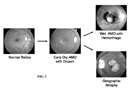
[0058] Figure 1 shows examples depicting the progression of AMD. Early AMD is
characterized by the deposition of drusen beneath the retinal pigment
epithelial (RPE) cell
layer. Drusen is visualized as pale or white spots in the middle panel.
[0059] Figure 2 shows examples of four constructs for making BIV (bovine
immunodeficiency virus)-based vectors. RSV is a Rous sarcoma virus (RSV)
promoter.
CMV is a cytomegalovirus (CMV) enhancer juxtaposed to the R region of the 5'
long
terminal repeat (LTR). SIN indicates deletions in the enhancer and promoter
from the U3
region of the 3' LTR to yield a self-inactivating vector. (p is the packaging
signal that directs
packaging of vector RNA into the viral particles. Heterologous Gene refers to
an expression
cassette that includes both a promoter and a coding region encoding a
molecule, e.g., a
therapeutic molecule. Gp64 is a baculovirus gp64 envelope gene.
14
CA 02678774 2009-08-19
WO 2008/106644 PCT/US2008/055498
[0060] Figure 3 shows expression of a green fluorescent protein (GFP) in BIV
vector-
transduced rat retina. 3x10⁵ transducing units (tu) in 3 1 were administered
via subretinal
injection. One month later, the rat was sacrificed, the retina was harvested,
and slits were cut
into the retina to prepare a flat whole mount. Figure 3A: The grey outline
depicts the edges
of the retina. The lighter area shows transduction and GFP expression at and
about the
injection site. Figure 3B shows that the immunohistochemical staining for GFP
is
predominantly in the RPE layer.
[0061] Figure 4 shows expression of GFP in BIV vector-transduced mouse
retinas.
Figure 4A depicts transduction and expression in an adult mouse retina two
weeks after
subretinal injection of l x 105 tu in 1 1. Figure 4B depicts transduction and
expression in a
newborn mouse two weeks after intravitreal injection of 1x10⁵ tu in 1 gl.
Figure 4C is a high
power view of Figure 4B.
[0062] Figure 5 shows a flat mount of rabbit retina one month after ocular
administration of a BIV GFP vector showing high level expression of GFP in the
RPE cells.
[0063] Figure 6 shows expression of GFP in monkey RPE cells ten weeks after
administration of a BIV vector encoding GFP.
[0064] Figure 7 shows that a BIV vector can efficiently transduce primary
human
RPE cells. The left panel shows staining for RPE65, a 65 KD RPE specific
protein. The
central and right panels show bright field and fluorescent views of the cells
transduced with
GFP vector.
[0065] Figure 8 shows that a BIV vector encoding an endostatin demonstrated
efficacy dampening angiogenesis in an animal model of florid
neovascularization. The top
left panel depicts an untreated eye. The bottom two left panels depict eyes
treated with the
control vector and those on the right depict eyes treated with the BIV
endostatin vector. The
top panels are fluorescein angiographs. The middle panels are histological
sections of the
retinas. The bottom panels are cross-sections of the entire eyes.
[0066] Figure 9 shows the curves of vector titer and BSA level in the elution
fractions
from a Sephacryl S 500-HR column. For this study, the culture medium from
which the
vector was purified was supplemented with 2% FBS. Details are described in
Example 30.
[0067] Figure 10 shows that a BIV vector expressing T2-TrpRS inhibited
neovascularization in the laser injury model. Figure 10 shows the average size
of the
neovascular areas from the two cohorts. The areas of neovascularization were
significantly
CA 02678774 2009-08-19
WO 2008/106644 PCT/US2008/055498
smaller in the T2-TrpRS (a carboxyl-terminal fragment of tryptophan tRNA
synthetase)
vector-treated eyes.
[0068] Figure 11 depicts the classical and lectin complement pathways. The
classical
pathway is initiated through C l while the lectin pathway is initiated through
mannose binding
lectin (MBL). C4bC2a is a protease that cleaves C3 to C3a and C3b and is
termed the C3
convertase. Similarly, C4bC2aC3b cleaves C5 to C5a and C5b and is termed the
C5
convertase. C3a, C4a, and C5a have inflammatory properties and attract
phagocytotic cells.
C5b6-9 forms the membrane attack complex (MAC), which creates membrane pores
that kill
infectious agents but can also damage host cells. MASP is mannan-binding
lectin associated
serine protease.
[0069] Figure 12 depicts the alternative complement pathway. This pathway is
constitutively active at a low level through spontaneous cleavage of C3. In
the presence of an
appropriate surface, C3b binds to complement factor B (fB). This complex is
then cleaved by
complement factor D(f)) to yield C3bBb. Spontaneous dissociation ("decay") of
this
complex within minutes leads to its inactivation, whereas stabilization by
properdin generates
a complex that cleaves C3; that is, a C3 convertase. Several of the factors
that attenuate the
complement pathways do so by accelerating the decay of the C3 and C5
convertases (see
Table One, below). Please note: C3b participates in the C3 convertase to
generate additional
C3b thereby creating a positive feedback loop as shown by the large arrow.
C3bBb is a C3
convertase. C3bBbC3b is a C5 convertase.
[0070] Figure 13 shows expression of vector-derived fB in human retinal cells.
ARPE cells (an RPE derived cell line) were transduced with BIV-based vectors
encoding fB
constructs, see Example 8. Medium from each of the transduced cell populations
was
subjected to Western analysis and probed for fB. Lane 1, 100 ng of purified
human plasma-
derived fB; Lanes 2-5, 40 1 of media from cells transduced with vectors
encoding human
wild type fB, fB3, fB2, and fBl, respectively. All of the lanes are from the
same gel.
[0071] Figure 14 shows structure of a single chain antibody that can be
subcloned,
e.g., as a Heterologous Gene in a transfer vector construct, e.g., of Figure
2, or expressed
from a cell. Leader is a leader sequence to direct secretion; VL and VH are
the variable light
and heavy chains or CDR containing portions thereof, respectively, connected
by a linker,
e.g., which are known in the art. In some embodiments, a heavy chain sequence
can be
placed upstream of the light chain sequence.
16
CA 02678774 2009-08-19
WO 2008/106644 PCT/US2008/055498
[0072] Figure 15 shows results from a hemolytic assay of vector-derived human
wild
type fB and fB dominant negatives. The positive control is Erab (rabbit
erythrocytes) mixed
with distilled water to produce 100% hemolysis. Purified fB Protein represents
hemolysis by
human fB-depleted serum supplemented with 500 ng of purified human plasma-
derived fB.
The Negative Control represents hemolysis by fB-depleted human serum alone.
The five
right-hand bars represent hemolysis by fB-depleted serum with the addition of
culture
medium from cells transduced with vectors encoding GFP, wild type human fB,
fBl, fB2, or
fB3.
[0073] Figure 16 shows lentiviral vector gene transfer to rat aorta and mouse
brain.
Figure 16A demonstrates transduction of a section of rat aorta with an HIV-
derived lentiviral
vector. Figure 16B demonstrates gene transfer to mouse brain using a BIV GFP
vector. The
related methods are described in Example 14.
[0074] Figure 17 shows results of a hemolytic activity assay to assess
alternative
complement pathway activity using various factor B mutants. The Y-axis
displays the
relative hemolytic activity as measured by the hemoglobin level released into
the supernatant
after lysis of the erythrocytes. X-axis from left to right: Positive control
with 100% lysis, red
blood cells (RBC) lysed in water; WT (wild-type) hfB, factor B-depleted human
serum
supplemented with 500 ng of purified plasma derived human factor B protein
(catalog#
A408, Quidel, San Diego, CA); Negative control, the RBCs were incubated in
isotonic saline
(no red blood cell lysis); Wild type hfB vector, factor B-depleted human serum
supplemented
with culture medium of the cells transduced with BIV vector encoding wild type
factor B;
GFP vector, factor B-depleted human serum supplemented with culture medium of
the cells
transduced with BIV vector encoding GFP; Mutant hfB 1 vector, factor B-
depleted human
serum supplemented with culture medium of the cells transduced with BIV vector
encoding
mutant fBl; Mutant hfB2 vector, factor B-depleted human serum supplemented
with culture
medium of the cells transduced with BIV vector encoding mutant fB2; Mutant
hfB3 vector,
factor B-depleted human serum supplemented with culture medium of the cells
transduced
with BIV vector encoding mutant fB3; GFP vector + WT fB 1:1, factor B-depleted
human
serum supplemented with a mixture of culture medium of the cells transduced
with BIV
vector encoding GFP and culture medium of the cells transduced with BIV vector
encoding
wild type factor B at 1 to 1 ratio; Mutant hfBl+ WT fB 1:1, factor B-depleted
human serum
supplemented with a mixture of culture medium of the cells transduced with BIV
vector
17
CA 02678774 2009-08-19
WO 2008/106644 PCT/US2008/055498
encoding mutant factor B 1 and culture medium of the cells transduced with BIV
vector
encoding wild type factor B at 1 to 1 ratio; Mutant hfB2 + WT fB l:l, factor B-
depleted
human serum supplemented with a mixture of culture medium of the cells
transduced with
BIV vector encoding mutant factor B2 and culture medium of the cells
transduced with BIV
vector encoding wild type factor B at 1 to 1 ratio; Mutant hfB3 + WT fB l:l,
factor B-
depleted human serum supplemented with a mixture of culture medium of the
cells
transduced with BIV vector encoding mutant factor B3 and culture medium of the
cells
transduced with BIV vector encoding wild type factor B at 1 to 1 ratio; GFP
vector + WT fB
2:1, factor B-depleted human serum supplemented with a mixture of culture
medium of the
cells transduced with BIV vector encoding GFP and culture medium of the cells
transduced
with BIV vector encoding wild type human factor B at 2 to 1 ratio; Mutant
hfBl+ WT fB 2:1,
factor B-depleted human serum supplemented with a mixture of culture medium of
the cells
transduced with BIV vector encoding mutant factor Bl and culture medium of the
cells
transduced with BIV vector encoding wild type factor B at 2 to 1 ratio; Mutant
hfB2 + WT fB
2:1, factor B-depleted human serum supplemented with a mixture of culture
medium of the
cells transduced with BIV vector encoding mutant factor B2 and culture medium
of the cells
transduced with BIV vector encoding wild type factor B at 2 to 1 ratio; Mutant
hfB3 + WT fB
2:1, factor B-depleted human serum supplemented with a mixture of culture
medium of the
cells transduced with BIV vector encoding mutant factor B3 and culture medium
of the cells
transduced with BIV vector encoding wild type factor B at 2 to 1 ratio.
[0075] Figure 18 shows results of a hemolytic activity assay to assess
alternative
complement pathway activity. Y-axis displays the relative hemolytic activity
as measured by
the hemoglobin level released to the supernatant after lysis of erythrocytes.
X-axis from left
to right: Positive control with 100% lysis, RBC lysed in water; Blank, the RBC
was
incubated in isotonic saline (no red blood cell lysis); Four-fold diluted
mouse serum (50 ul)
was added to the following samples (40 ul each): GFP vector, culture medium of
the cells
transduced with BIV vector encoding GFP mixed with culture medium of the cells
transduced with BIV vector encoding wild type mouse factor B at 1 to 1 ratio;
Mutant mfB 1
vector, culture medium of the cells transduced with BIV vector encoding mouse
mutant fB 1
mixed culture medium of the cells transduced with BIV vector encoding wild
type mouse
factor B at 1 to 1 ratio; Mutant mfB2 vector, culture medium of the cells
transduced with BIV
vector encoding mouse mutant fB2 mixed with culture medium of the cells
transduced with
18
CA 02678774 2009-08-19
WO 2008/106644 PCT/US2008/055498
BIV vector encoding wild type mouse factor B at 1 to 1 ratio; Mutant mfB3
vector, culture
medium of the cells transduced with BIV vector encoding mouse mutant fB3 mixed
with
culture medium of the cells transduced with BIV vector encoding wild type
mouse factor B at
1 to 1 ratio; GFP vector, culture medium of the cells transduced with BIV
vector encoding
GFP mixed with culture medium of the cells transduced with BIV vector encoding
wild type
mouse factor B at 2 to 1 ratio; Mutant mfB 1 vector, culture medium of the
cells transduced
with BIV vector encoding mouse mutant fB 1 mixed with culture medium of the
cells
transduced with BIV vector encoding wild type mouse factor B at 2 to 1 ratio;
Mutant mfB2
vector, culture medium of the cells transduced with BIV vector encoding mouse
mutant fB2
mixed with culture medium of the cells transduced with BIV vector encoding
wild type
mouse factor B at 2 to 1 ratio; Mutant mfB3 vector, culture medium of the
cells transduced
with BIV vector encoding mouse mutant fB3 mixed culture medium of the cells
transduced
with BIV vector encoding wild type mouse factor B at 2 to 1 ratio; GFP vector,
culture
medium of the cells transduced with BIV vector encoding GFP mixed with culture
medium
of the cells transduced with BIV vector encoding wild type mouse factor B at 1
to 1 ratio;
Mutant hfBl vector, culture medium of the cells transduced with BIV vector
encoding human
mutant fB 1 mixed with culture medium of the cells transduced with BIV vector
encoding
wild type mouse factor B at 1 to 1 ratio; Mutant hfB2 vector, culture medium
of the cells
transduced with BIV vector encoding human mutant fB2 mixed with culture medium
of the
cells transduced with BIV vector encoding wild type mouse factor B at 1 to 1
ratio; Mutant
hfB3 vector, culture medium of the cells transduced with BIV vector encoding
human mutant
fB3 mixed with culture medium of the cells transduced with BIV vector encoding
wild type
mouse factor B at 1 to 1 ratio; GFP vector, culture medium of the cells
transduced with BIV
vector encoding GFP mixed with culture medium of the cells transduced with BIV
vector
encoding wild type mouse factor B at 2 to 1 ratio; Mutant hfBl vector, culture
medium of
the cells transduced with BIV vector encoding human mutant fBl mixed with
culture
medium of the cells transduced with BIV vector encoding wild type mouse factor
B at 2 to 1
ratio; Mutant hfB2 vector, culture medium of the cells transduced with BIV
vector encoding
human mutant fB2 mixed with culture medium of the cells transduced with BIV
vector
encoding wild type mouse factor B at 2 to 1 ratio; Mutant hfB3 vector, culture
medium of
the cells transduced with BIV vector encoding human mutant fB3 mixed with
culture
19
CA 02678774 2009-08-19
WO 2008/106644 PCT/US2008/055498
medium of the cells transduced with BIV vector encoding wild type mouse factor
B at 2 to 1
ratio.
[0076] Figure 19 shows a hemolytic activity assay to assess alternative
complement
pathway activity. Y-axis displays the relative hemolytic activity as measured
by the
hemoglobin level released to the supernatant after lysis of erythrocytes. X-
axis from left to
right: Positive control with 100% lysis, RBC lysed in water; Blank, RBC
incubated in
isotonic buffer (no lysis of red blood cells); GFP, culture medium of the
cells transduced with
BIV GFP vector; WT hfB vector, factor B-depleted human serum supplemented with
culture
medium of the cells transduced with BIV vector encoding wild type factor B;
Mutant hfB 1
vector, factor B-depleted human serum supplemented with culture medium of
cells
transduced with BIV vector encoding mutant fBl; Mutant hfB2 vector, factor B-
depleted
human serum supplemented with culture medium of cells transduced with BIV
vector
encoding mutant fB2; Mutant hfB3 vector, factor B-depleted human serum
supplemented
with culture medium of cells transduced with BIV vector encoding mutant fB3;
GFP vector +
l:l wWT hfB, factor B-depleted human serum supplemented with a mixture of
culture
medium of the cells transduced with BIV vector encoding GFP and culture medium
of the
cells transduced with BIV vector encoding wild type human factor B at 1 to 1
ratio; Mutant
mfBl+ 1:1 wWT hfB, factor B-depleted human serum supplemented with a mixture
of
culture medium of the cells transduced with BIV vector encoding mouse mutant
factor Bl
and culture medium of the cells transduced with BIV vector encoding wild type
human factor
B at 1 to 1 ratio; Mutant mfB2 + 1:1 wWT hfB, factor B-depleted human serum
supplemented with a mixture of culture medium of the cells transduced with BIV
vector
encoding mouse mutant factor B2 and culture medium of the cells transduced
with BIV
vector encoding human wild type factor B at 1 to 1 ratio; Mutant mfB3 + 1:1
wWT hfB,
factor B-depleted human serum supplemented with a mixture of culture medium of
the cells
transduced with BIV vector encoding mouse mutant factor B3 and culture medium
of the
cells transduced with BIV vector encoding human wild type factor B at 1 to 1
ratio; GFP
vector + 2:1 wWT hfB, factor B-depleted human serum supplemented with a
mixture of
culture medium of the cells transduced with BIV vector encoding GFP and
culture medium of
the cells transduced with BIV vector encoding wild type human factor B at 2 to
1 ratio;
Mutant mfBl+ 2:1 wWT hfB, factor B-depleted human serum supplemented with a
mixture
of culture medium of the cells transduced with BIV vector encoding mouse
mutant factor B 1
CA 02678774 2009-08-19
WO 2008/106644 PCT/US2008/055498
and culture medium of the cells transduced with BIV vector encoding wild type
human factor
B at 2 to 1 ratio; Mutant mfB2 + 2:1 wWT hfB, factor B-depleted human serum
supplemented with a mixture of culture medium of the cells transduced with BIV
vector
encoding mouse mutant factor B2 and culture medium of the cells transduced
with BIV
vector encoding human wild type factor B at 2 to 1 ratio; Mutant mfB3 + 2:1
wWT hfB,
factor B-depleted human serum supplemented with a mixture of culture medium of
the cells
transduced with BIV vector encoding mouse mutant factor B3 and culture medium
of the
cells transduced with BIV vector encoding human wild type factor B at 2 to 1
ratio; GFP
vector + 4:1 wWT hfB, factor B-depleted human serum supplemented with a
mixture of
culture medium of the cells transduced with BIV vector encoding GFP and
culture medium
of the cells transduced with BIV vector encoding wild type human factor B at 4
to 1 ratio;
Mutant mfBl+ 4:1 wWT hfB, factor B-depleted human serum supplemented with a
mixture
of culture medium of the cells transduced with BIV vector encoding mouse
mutant factor B 1
and culture medium of the cells transduced with BIV vector encoding wild type
human factor
B at 4 to 1 ratio; Mutant mfB2 + 4:1 wWT hfB, factor B-depleted human serum
supplemented with a mixture of culture medium of the cells transduced with BIV
vector
encoding mouse mutant factor B2 and culture medium of the cells transduced
with BIV
vector encoding human wild type factor B at 4 to 1 ratio; Mutant mfB3 + 4:1
wWT hfB,
factor B-depleted human serum supplemented with a mixture of culture medium of
the cells
transduced with BIV vector encoding mouse mutant factor B3 and culture medium
of the
cells transduced with BIV vector encoding human wild type factor B at 4 to 1
ratio.
[0077] Figure 20 shows a hemolytic activity assay to assess alternative
complement
pathway activity. Y-axis displays the relative hemolytic activity as measured
by the
hemoglobin level released to the supernatant after lysis of erythrocytes. X-
axis from left to
right: Positive control with 100% lysis, RBC lysed in water; Negative control,
RBC
incubated in isotonic buffer (no lysis of red blood cells); GFP vector,
culture medium of the
cells transduced with BIV vector encoding GFP (40 ul) mixed with two-fold
diluted pig
serum (50 ul); Wild type hfB vector, culture medium of the cells transduced
with BIV vector
encoding wild type factor B (40 ul) mixed with two-fold diluted pig serum (50
ul); Mutant
hfBl vector, culture medium of the cells transduced with BIV vector encoding
human mutant
fBl (40 ul) mixed with two-fold diluted pig serum (50 ul); Mutant hfB2 vector,
culture
medium of the cells transduced with BIV vector encoding human mutant fB2 (40
ul) mixed
21
CA 02678774 2009-08-19
WO 2008/106644 PCT/US2008/055498
with two-fold diluted pig serum (50 ul); Mutant hfB3 vector, culture medium of
the cells
transduced with BIV vector encoding human mutant fB3 (40 ul) mixed with two-
fold diluted
pig serum (50 ul); GFP vector, culture medium of the cells transduced with BIV
vector
encoding GFP (40 ul) mixed with four-fold diluted pig serum (50 ul); Wild type
hfB vector,
culture medium of the cells transduced with BIV vector encoding wild type
factor B (40 ul)
mixed with four-fold diluted pig serum (50 ul); Mutant hfB1 vector, culture
medium of the
cells transduced with BIV vector encoding human mutant fBl (40 ul) mixed with
four-fold
diluted pig serum (50 ul); Mutant hfB2 vector, culture medium of the cells
transduced with
BIV vector encoding human mutant fB2 (40 ul) mixed with four-fold diluted pig
serum (50
ul); Mutant hfB3 vector, culture medium of the cells transduced with BIV
vector encoding
human mutant fB3 (40 ul) mixed with four-fold diluted pig serum (50 ul); GFP
vector,
culture medium of the cells transduced with BIV vector encoding GFP (40 ul)
mixed with
six-fold diluted pig serum (50 ul); Wild type hfB vector, culture medium of
the cells
transduced with BIV vector encoding wild type factor B (40 ul) mixed with six-
fold diluted
pig serum (50 ul); Mutant hfBl vector, culture medium of the cells transduced
with BIV
vector encoding human mutant fBl (40 ul) mixed with six-fold diluted pig serum
(50 ul);
Mutant hfB2 vector, culture medium of the cells transduced with BIV vector
encoding human
mutant fB2 (40 ul) mixed with six-fold diluted pig serum (50 ul); Mutant hfB3
vector,
culture medium of the cells transduced with BIV vector encoding human mutant
fB3 (40 ul)
mixed with six-fold diluted pig serum (50 ul).
[0078] Figure 21 shows C3b-dependent human factor B cleavage by factor D.
Western blot detection of full-length factor B or factor B cleavage products,
Bb and Ba. Lane
1, Purified human factor B protein as positive control; Lane 2, Culture medium
of the cells
transduced by BIV vector encoding GFP incubated with factor D; Lane 3, Culture
medium of
the cells transduced by BIV vector encoding human wild type factor B incubated
with factor
D; Lane 4, Culture medium of the cells transduced by BIV vector encoding human
mutant
factor Bl incubated with factor D; Lane 5, Culture medium of the cells
transduced by BIV
vector encoding human mutant factor B2 incubated with factor D; Lane 6,
Culture medium of
the cells transduced by BIV vector encoding human mutant factor B3 incubated
with factor
D; Lane 7, Molecular weight marker; Lane 8, Culture medium of the cells
transduced by BIV
vector encoding GFP incubated with C3b and factor D; Lane 9, Culture medium of
the cells
transduced by BIV vector encoding human wild type factor B incubated with C3b
and factor
22
CA 02678774 2009-08-19
WO 2008/106644 PCT/US2008/055498
D; Lane 10, Culture medium of the cells transduced by BIV vector encoding
human mutant
factor Bl incubated with C3b and factor D; Lane 11, Culture medium of the
cells transduced
by BIV vector encoding human mutant factor B2 incubated with C3b and factor D;
Lane 12,
Culture medium of the cells transduced by BIV vector encoding human mutant
factor B3
incubated with C3b and factor D. The reaction mixture for each sample is
described in Table
two.
[0079] Figure 22 shows a factor B and C3b binding assay. C3b, factor D, and
factor
B (wt or mutants) together were incubated in a reaction mixture. The reaction
mixture was
immunoprecipitated with polyclonal anti-factor B antiserum, separated by SDS-
PAGE, and
analyzed by Western Blot analysis with anti-C3b polyclonal antiserum. Lane 1,
purified C3b
protein as a positive control (the lower band is C3 beta-chain co-purified
with C3b); Lane 2,
culture medium of the cells transduced with BIV vector encoding GFP incubated
with C3b
and factor D; Lane 3, culture medium of the cells transduced with BIV vector
encoding
human wild type factor B incubated with C3b and factor D; Lane 4, culture
medium of the
cells transduced with BIV vector encoding human mutant factor B3 incubated
with C3b and
factor D; Lane 5, culture medium of the cells transduced with BIV vector
encoding human
mutant factor B2 incubated with C3b and factor D; and Lane 6, culture medium
of the cells
transduced with BIV vector encoding human mutant factor B 1 incubated with C3b
and factor
D.
[0080] Figure 23 shows an assay for human complement factor B and factor D
binding. Details of the experiment are found in Example 21, below. Lane 1,
negative
control, the reaction was performed in the absence of C3b, factor D, and
factor B; Lane 2,
culture medium of the cells transduced with BIV vector encoding GFP incubated
with C3b
and factor D; Lane 3, culture medium of the cells transduced with BIV vector
encoding
human wild type factor B incubated with C3b and factor D; Lane 4, molecular
weight
markers; Lane 5, culture medium of the cells transduced with BIV vector
encoding human
mutant factor B3 incubated with C3b and factor D; Lane 6, culture medium of
the cells
transduced with BIV vector encoding human mutant factor B2 incubated with C3b
and factor
D; Lane 7, culture medium of the cells transduced with BIV vector encoding
human mutant
factor Bl incubated with C3b and factor D; and Lane 8, purified human factor B
as a positive
control.
23
CA 02678774 2009-08-19
WO 2008/106644 PCT/US2008/055498
[0081 ] Figure 24 shows inhibition of human alternative complement pathway
activity
by an anti-human factor B monoclonal antibody using a hemolytic activity assay
to assess
alternative complement pathway activity. The Y-axis displays the relative
hemolytic activity
as measured by the hemoglobin level released to the supernatant after lysis of
erythrocytes.
The X-axis from left to right: Positive control with 100% lysis, RBC lysed in
water; Purified
hfB protein, factor B-depleted human serum supplemented with 500 ng of
purified human
factor B protein; Negative control, the RBC was incubated in isotonic saline
(no red blood
cell lysis); Anti-hfB mAb 0.4 g; factor B-depleted human serum supplemented
with a
mixture of anti-hfB mAb (0.4 g) and 500 ng of purified human factor B
protein; Anti-hfB
mAb 0.8 g; factor B-depleted human serum supplemented with a mixture of anti-
hfB mAb
(0.8 g) and 500 ng of purified human factor B protein; Anti-hfB mAb 1.6 g,
factor B-
depleted human serum supplemented with a mixture of anti-hfB mAb (1.6 g) and
500 ng of
purified human factor B protein; Anti-hfB mAb (2.4 g); factor B-depleted
human serum
supplemented with a mixture of anti-hfB mAb (2.4 g) and 500 ng of purified
human factor
B protein; Control mouse IgG 0.4 g, factor B-depleted human serum
supplemented with a
mixture of control mouse IgG (0.4 g) and 500 ng of purified human factor B
protein;
Control mouse IgG 0.8 g, factor B-depleted human serum supplemented with a
mixture of
control mouse IgG (0.8 g) and 500 ng of purified human factor B protein;
Control mouse
IgG 1.6 g, factor B-depleted human serum supplemented with a mixture of
control mouse
IgG (1.6 g) and 500 ng of purified human factor B protein; Control mouse IgG
2.4 g,
factor B-depleted human serum supplemented with a mixture of control mouse IgG
(2.4 g)
and 500 ng of purified human factor B protein.
[0082] Figure 25 shows analysis of human factor B3 protein. Panel A shows
silver
staining of affinity purified human factor B3 protein: Lane 1, molecular
weight marker; Lane
2, eluted sample from the first fraction; Lane 3, eluted sample from
combination of the
second and the third fractions. Panel B shows a Western blot analysis for
human factor B3
protein and the lane assignment is the same as in Panel A.
[0083] Figure 26 shows a hemolytic activity assay to assess alternative
complement
pathway activity. Y-axis displays the relative hemolytic activity as measured
by the
hemoglobin level released to the supernatant after lysis of erythrocytes. X-
axis from left to
right: Positive control with 100% lysis, RBC lysed in water; Blank, the RBCs
were incubated
in isotonic saline (no red blood cell lysis); Wild type factor B protein (50
ng, 100 ng, 200 ng,
24
CA 02678774 2009-08-19
WO 2008/106644 PCT/US2008/055498
and 500 ng), factor B depleted human serum supplemented with 50 ng, 100 ng,
200 ng, and
500 ng wild type human factor B (Quidel); Wild type factor B protein plus
factor B3 protein,
factor B depleted human serum supplemented with a mixture of 40 1 of affinity
purified
mutant factor B3 plus 0 ng, 200 ng, or 500 ng of wild type human factor B from
Quidel.
[0084] Figure 27 shows GFP expression and C3 staining in mouse retinas.
Figures
27A & 27B: GFP vector was administered as described in Example 27. Two weeks
later,
flat mounts were prepared and examined with a fluorescent microscope. Figures
27C & 27D:
Null and hfB3 vectors were administered as described in Example 27. Two weeks
later, laser
photocoagulation was performed near the center of the retinas. Twenty hours
later, the
retinas were harvested, stained for C3 deposition, and examined with a
fluorescent
microscope.
[0085] Figure 28 shows the vector titer in each of the elution fractions from
a
Sephacryl S 500-HR column. For this study, the culture medium from which the
vector was
purified was not supplemented with FBS. Details are described in Example 32.
[0086] Figure 29 depicts the following plasmids. Figure 29A is pAVTrGP038 (SEQ
ID NO:19. Figure 29B is pAVTrREV039 (SEQ ID NO:20). Figure 29C is pAVTrGP64-
040 (SEQ ID NO:21). Figure 29D is pAVT001 (SEQ ID NO:22). Figure 29E is
pAVTGFP006 (SEQ ID NO:23).
DETAILED DESCRIPTION OF THE INVENTION
[0087] The practice of the present invention will employ, unless otherwise
indicated,
conventional techniques of cell biology, molecular biology, cell culture,
virology and the like
which are in the skill of one in the art. These techniques are fully disclosed
in current
literature, for example, Sambrook, Fritsch and Maniatis eds., "Molecular
Cloning, A
Laboratory Manual", 2nd Ed., Cold Spring Harbor Laboratory Press (1989); Celis
J. E. "Cell
Biology, A Laboratory Handbook" Academic Press, Inc. (1994) and Bahnson et
al., J. of
Virol. Methods, 54:131-143 (1995).
[0088] The term "complement-mediated" refers to a process or disease that
involves
complement. Typically, a "complement-mediated" disease or condition is one
wherein
complement activity is one of the underlying causes of the disease or
condition and wherein
inhibition or blocking of the complement activity lessens the extent of the
disease or
CA 02678774 2009-08-19
WO 2008/106644 PCT/US2008/055498
condition. Examples of numerous complement-mediated diseases or conditions are
described
herein.
[0089] The term "complement protein" or "complement pathway component" is a
protein of the complement system or a receptor thereof. Complement proteins
are a group of
about 35 interacting proteins and glycoproteins found in all vertebrates. The
complement
proteins can be soluble or on the cell-surface. (Sim and Tsiftsoglou,
Biochemical Society
Transactions (2004) 32(1):21-27) In addition, there are regulatory membrane
proteins that
protect host cells from accidental or undesirable complement attack. A
complement protein
can be one that functions in the classical pathway, for example, C2 or one
that functions in
the alternative pathway, for example, Factor B. At least 6 complement proteins
are proteases.
For example, proteins included in the following exemplary list are complement
proteins: C l q,
Clr, Cls, C2-9, Factor B, Factor D, Factor H, Factor I, CRl, CR2, CR3, CR4,
properdin, Cl
(Inh), C4bp, MCP, DAF, CD59 (MIRL) and HRF.
[0090] The term wildtype (or wild-type), which is used interchangeably with
native,
as used herein relates to a naturally occurring protein encoded by a mammalian
genome, a
naturally occurring nucleic acid, a naturally occurring individual or animal
and so on.
[0091 ]"Complement protein variant", "complement protein mutant" or
"complement
protein analog" are used interchangeably and refer to a structural derivative
of the parent
protein that does not necessarily retain all of the properties of the native
(naturally-occurring)
parent protein or has at least one altered property as compared to the native
parent protein.
An analog or variant is produced by replacing, substituting, deleting, and/or
adding amino
acids with regard to the native amino acid sequence of the protein. The
substitutions or
insertions typically involve naturally occurring amino acids, but may also
include synthetic or
unconventional amino acids as well. In some embodiments, an analog or variant
is produced
by mutating a protein, e.g., mutating a nucleic acid encoding it. An analog
will typically
retain at least 70%, at least 75%, at least 80%, at least 85%, at least 90%,
at least 95%, at
least 96%, at least 97%, at least 98%, at least 99%, at least 99.5% of the
native parent
protein's amino acid sequence (e.g., have that percent amino acid sequence
identity with
respect to the naturally occurring parent protein as determined over the
length of the entire
parent protein or, in certain embodiments, over a specific domain or portion
of the parent
protein). Analogs also include fragments of full length analogs that comprise
a portion of the
amino acid sequence (for example, at least 30, 50, 70, 100, 150, 200, 300,
400, 500, 600 or
26
CA 02678774 2009-08-19
WO 2008/106644 PCT/US2008/055498
700 amino acids or comprise one or a subset of the domains of the analog) and
either retain
one or more biological activities of the parent protein or a full length
analog or inhibit one or
more of these biological activities.
[0092] The term "corresponds" when referring to an amino acid in a particular
protein
refers to the particular amino acid in that particular protein and also to an
amino acid in a
related or similar protein that provides a similar function to the protein.
For example, an
amino acid in a human complement factor B may be found to correspond with an
amino acid
in a murine complement factor B or in a human allelic variant of factor B,
usually determined
by aligning the two amino acid sequences. For example, one skilled in the art
can align two
related sequences, such as SEQ ID NO:2 and 16, to determine corresponding
amino acids,
e.g., using a BLAST program. Also, corresponding amino acids can be
determined, e.g., by
aligning motifs (e.g., a protease cleavage motif) within related or unrelated
proteins. Such an
alignment may also be used to derive consensus sequences of target protein or
domains
thereof.
[0093] As used herein, the term "gene" typically refers to a coding region for
a
protein. However, in some contexts herein it will be clear that the term
"gene" is also
referring to elements (e.g., regulatory elements) operatively linked to a
coding region such as
promoters, enhancers, splice sites (acceptors and/or donors), polyadenylation
signals, introns,
5' untranslated regions, 3' untranslated regions, etc.
[0094] As used herein, "gene of interest", sometimes referred to as a
transgene, is a
heterologous gene or a foreign gene, relative to the source of the nucleic
acid vector or vector
construct. Thus, for example, in the case of a BIV vector, a gene of interest
is generally not a
BIV gene. In some embodiments, a gene of interest is a therapeutic gene.
[0095] The term "packaging sequence" is a sequence necessary for packaging
viral
nucleic acids into virions, virus particles or virus-like particles. In BIV,
for example, a
packaging sequence(s) is generally located in the region between the 5' major
splice donor
and the upstream region of the gag gene. Vectors according to the invention
may include
packaging sequences corresponding to other viruses, e.g., lentiviruses such as
HIV-1, HIV-2
or SIV. Packaging sequences may include as many as 1000 nucleotides or a few
as 50
nucleotides. The size of a packaging sequence region can be easily determined
by one of
ordinary skill in the art.
27
CA 02678774 2009-08-19
WO 2008/106644 PCT/US2008/055498
[0096] The term "pharmaceutically acceptable" means approved by a regulatory
agency of the Federal or a state government or listed in the U.S. Pharmacopeia
or other
generally recognized pharmacopeia for use in humans.
[0097] A "therapeutic benefit" is not necessarily a cure for a particular
disease or
condition (including any disease or condition described herein), but rather,
encompasses a
result which most typically includes alleviation of the disease or condition,
elimination of the
disease or condition, reduction of one or more symptoms associated with the
disease or
condition, prevention or alleviation of a secondary disease or condition
resulting from the
occurrence of a primary disease or condition, diminishing the likelihood of
developing a
condition or disease, diminishing the severity of a disease or condition,
changing the
character of a disease or condition, shortening the course of a disease or
condition, slowing or
preventing the progression or worsening of a disease or condition, and/or
prevention of the
disease or condition.
[0098] "Transformed" is meant to include any means in which an exogenous,
heterologous or foreign nucleic acid is introduced into a cell. That can occur
by using a
nucleic acid per se, a plasmid (transfection), a virus (infection or
transduction), a synthetic
carrier molecule and so on, as known in the art. A cell that is transformed is
one which is
treated in any of a variety of ways to carry a foreign nucleic acid of
interest. Such cells can
be somatic cells, stem cells that are not embryonic stem cells, and embryonic
stem cells.
[0099] A "vector construct" or "vector sequence" or "transfer vector" refers
to an
assembly which is capable of directing the expression of a nucleotide
sequence, a transgene, a
protein or a therapeutic expression product. In some embodiments of the
invention, vector
construct can include a 5' sequence (comprising an operably linked promoter
region) which is
capable of initiating transcription; a DNA segment from a viral genome; and/or
a packaging
sequence from a virus such as a lentivirus or retrovirus. In one embodiment,
the present
invention provides a BIV vector construct comprising: a DNA segment from a BIV
genome,
a packaging sequence for packaging RNA into virions, a first promoter operably
linked to the
DNA segment, and a transgene operably linked to a second promoter. In one
embodiment, a
packaging sequence of the BIV vector construct is a BIV packaging sequence.
28
CA 02678774 2009-08-19
WO 2008/106644 PCT/US2008/055498
Tamet Molecules
[00100] "Target molecules" are molecules, which can be involved in a
pathway, which themselves or their activity can be modulated resulting in
modulation of the
pathway. A target molecule may not necessarily be involved directly in a
pathway, e.g., it
may have an activity that affects another, second molecule that in turn
affects a molecule
directly involved in the pathway. In some embodiments of the invention, the
quantity and/or
activity of a target molecule may be increased, decreased or maintained. This
may be
accomplished by, but is not limited to, modulating expression of the target
molecule or
destabilizing or eliminating at least some of the target molecule,
sequestering the molecule
and/or altering one or more biological activities of the target molecule. In
some
embodiments, modulating expression of the target molecule is accomplished by
transforming
a cell (e.g., in vitro or in vivo) with at least one nucleic acid coding for
the target molecule
when the target molecule is a protein or nucleic acid. In some embodiments, a
target protein
can be destabilized or its activity abrogated or reduced by utilizing a
binding molecule that
destabilizes or reduces the activity of the target protein. In some
embodiments, a binding
molecule is utilized that binds a target protein resulting in destabilization
of the target protein.
In some embodiments, a protease that cleaves the target protein can be used to
destabilize or
eliminate at least a part of the target molecules. In some embodiments, a
protease cleaves a
target molecule at 1, 2, 3, 4, 5, 6, 7, 8, 9, 10, or more sites within the
target molecule. Some
embodiments of the invention inhibit a target molecule(s). Inhibitors of the
invention
include, but are not limited to, binding molecules such as antibody molecules
(as well as
homologues, analogues and modified or derived forms thereof) including
immunoglobulin
fragments such as Fab, F(ab')2 and F,,, small molecules, peptides,
oligonucleotides, aptamers,
peptidomimetics, and organic compounds..
[00101] The complement system can mediate a chain reaction of proteolysis and
assembly of protein complexes, e.g., that result in the elimination of
invading
microorganisms. Three activation pathways (the classical, lectin, and
alternative pathways)
and a lytic pathway regulate these events.
[00102] In the classical pathway as shown in Figure 11, C l q, a collagenous
subcomponent of the first component (Cl), binds to immunoglobulins within
immune
complexes, and its associated serine proteases, Clr and Cls, become activated.
This
complement cascade is initiated by the subsequent cleavage of C4 and C2,
followed by C3
29
CA 02678774 2009-08-19
WO 2008/106644 PCT/US2008/055498
activation. The resulting C3b fragment not only acts as an opsonin but also
leads to the
membrane attack complex (MAC) formation in the lytic pathway. In innate
immunity, a
complex composed of a recognition molecule (lectin) and serine proteases,
termed the
mannose-binding lectin (MBL)-associated serine protease (MASP), activates C4
and C2 upon
binding to carbohydrates on the surface of microorganisms via the lectin
pathway. This
binding occurs in the absence of immunoglobulins. Recognition molecules of the
lectin
pathway found in jawed vertebrates are MBLs and ficolins, both of which are
characterized
by the presence of a collagen-like domain, like C l q, and a carbohydrate
binding domain
having a common binding specificity for G1cNAc. MASPs and Clr/Cls share the
same
domain organization and form a subfamily of serine proteases.
[00103] The lectin complement pathway in innate immunity is closely related to
the
classical complement pathway in adaptive immunity, e.g., with respect to the
structures and
functions of their components. Both pathways are typically initiated by
complexes consisting
of collagenous proteins and serine proteases of the mannose-binding lectin
(MBL)-associated
serine protease (MASP)/Clr/Cls family. It has been speculated that the
classical pathway
emerged after the lectin pathway.
[00104] Activation of the alternative complement pathway, shown in Figure 12,
begins when C3b (or C3i) binds to a cell and other surface components, e.g.,
of microbes.
C3b can also bind to IgG antibodies. Alternative pathway protein Factor B then
combines
with the C3b to form C3bB. Factor D then splits the bound Factor B into Bb and
Ba, forming
C3bBb. Properdin then binds to the Bb to form C3bBbP that functions as a C3
convertase
capable of enzymatically splitting typically hundreds of molecules of C3 into
C3a and C3b.
The alternative complement pathway is now activated. Some of the C3b
subsequently binds
to some of the C3bBb to form C3bBbC3b, a C5 convertase capable of splitting
molecules of
C5 into C5a and C5b
[00105] Since C3b is free in the plasma, it can bind to either a host cell or
pathogen
surface. To prevent complement activation from proceeding on the host cell,
there are several
different kinds of regulatory proteins that disrupt the complement activation
process.
Complement Receptor 1(CRl or CD35) and DAF (also known as CD55) compete with
Factor B in binding with C3b on the cell surface and can even remove Bb from
an already
formed C3bBb complex. The formation of a C3 convertase can also be prevented
when a
plasma protease called Factor I cleaves C3b into its inactive form, iC3b.
Factor I works with
CA 02678774 2009-08-19
WO 2008/106644 PCT/US2008/055498
C3b-binding protein cofactors such as CRl and Membrane Cofactor of Proteolysis
(MCP or
CD46). Another complement regulatory protein is Factor H which either competes
with
factor B, displaces Bb from the convertase, acts as a cofactor for Factor I,
or preferentially
binds to C3b bound to vertebrate cells.
[00106] Target molecules include, but are not limited to, components of
immunological pathways or variants thereof, such as components of the
classical complement
pathway; components of the alternative complement pathway or components of a
lectin
complement pathway. In some embodiments, more than one target molecules may be
modulated and/or acted on. In some embodiments, a component of the alternative
complement pathway is selected from the group consisting of C3, C3a, C3b,
C3bB, fB (factor
B), fD (factor D), C3bBb, C3bBbC3b, C5, C5a, C5b, C6, C7, C8, C9, C5b6-9, MAC
(membrane attack complex) and fragments thereof and components thereof. In
some
embodiments, a component of the classical complement pathway is selected from
the group
consisting of Clr, Clq, Cls, C4, C4b, C4a, MBL, MASP, C2, C2b, C4bC2a, C3,
C3b, C3a,
C4bC2aC3b, C5, C5a, C5b, C6, C7, C8, C9, C5b6-9, and MAC.
[00107] Target molecules that initiate or facilitate inflammation would
benefit from
reduced expression and/or activity, and include, properdin, TGF(3, complement
factor B
(human, accession no. NP 001701, Kavanaugh et al., Mol. Imm. 43(7)856, 2006;
mouse,
accession no. NP 032224m, Bora et al., J. Imm. 177(3)1872, 2006), complement
factor D and
other factors of the complement system, including, C2, C3, C4, C5, C6, C7, C8
and C9.
Thus, a delivery means of interest can express, for example, a specific
antigen-binding
polypeptide, a soluble receptor, an inhibitory nucleic acid, a ribozyme, an
aptamer, a catalytic
antibody, a molecule carrying amino acid substitutions that interfere with
function, such as
preventing docking to a receptor, or making such binding irreversible,
removing enzyme
activity and so on. Alternatively, a molecule which could compete with a
target molecule,
such as a receptor, but which is ineffective in activating or encouraging
inflammation, can be
used as a competitive inhibitor to attenuate inflammation. Also, a complement
factor with
altered or no (devoid) activity, which may be one or more particular
activities or functions of
any one molecule, can be used in the practice of the instant invention.
Altered means an
activity or function other than that obtained under normal, ambient condition,
such as
operating at a higher activity or presenting with a higher level of activity,
or a lower level of
31
CA 02678774 2009-08-19
WO 2008/106644 PCT/US2008/055498
activity. Other means of dampening expression or activity as known in the art
can be
practiced.
[00108] Complement factors often are polyfunctional. Thus, molecules may be
considered to have plural domains. For example, a complement factor may have a
domain
carrying a recognition and binding function or activity; a domain that is
recognized by
another molecule, which site or a region adjacent thereto, can be reacted,
changed or cleaved;
a domain that may have a biological activity, such as an enzymatic activity,
such as a
protease activity; and so on. A biological activity of a complement factor can
be diminished
or negated when any one or more of said domain functions is altered, and
alteration of one
domain may or may not have an impact on the normal functioning of another
domain.
Altering domains of a protein can be accomplished by, for example,
substituting, inserting
and/or deleting amino acids in the domain and testing for the desired
characteristic. Even if
the location of the domain is not known a systematic approach can be used to
locate and
obtain variants with altered function.
Pathway Modulators and Molecules of the Invention
[00109] A number of molecules can be targeted in the treatment of complement
mediated disease such as those related to ocular disease. With a goal of
mitigating or
ameliorating inflammation, or not allowing inflammation to continue, the
instant invention, in
part, relates to methods of having local or systemic delivery of a molecule to
achieve that
result. Thus in some embodiments of the invention, a molecule can be one which
inhibits
expression of or activity of a target molecule that initiates, contributes to
or facilitates
inflammation, or one that enhances the expression of or activity of a target
molecule that
dampens inflammation.
[00110] In some embodiments, the invention contemplates modulating a pathway
via modulation of the activity of any one or more components of the pathway as
described
herein and using any of the methods for modulating activity of a pathway
component as
described herein.
[00111 ] Immunological pathways and processes can contribute to the
progression,
maintenance, and/or inhibition of a condition (e.g., a disease) in an animal.
The present
invention provides, in part, methods of modulating (e.g., enhancing,
increasing, inhibiting,
decreasing or generally maintaining) an immunological pathway, e.g., in vitro
or in vivo.
Some embodiments of the invention can be used to study an immunological
pathway, to
32
CA 02678774 2009-08-19
WO 2008/106644 PCT/US2008/055498
study associated disease states, to develop treatments for a disease state(s),
to create disease
states in an animal (e.g., to develop a disease model in an animal such as a
mouse or rat), or
for screening drugs.
[00112] In some embodiments, a target molecule is a component of a complement
pathway, e.g., as described herein. Some embodiments of the invention involve
modulating a
classical complement pathway; an alternative complement pathway or a lectin
complement
pathway.
[00113] Some embodiments of the invention reduce, increase or prevent
inflammation and/or complement activity. Some embodiments of the invention
modulate
complement function or activity. Because complement is comprised of a number
of factors
which act in a synchronized manner, and a number of molecules and factors not
classically
identified as complement which regulate or control the presence and activity
of a complement
factor, any one of such complement factors or regulatory molecules of
complement can serve
as an entry point or as a target molecule of the instant invention as means to
impact
complement activity and function, such as enhancing or reducing complement
activity as
compared to baseline.
[00114] Inhibition of a complement pathway according to the present invention
can
be accomplished, for example, by directly affecting the expression (e.g.,
transcription and/or
translation) or biological activity of a protein in the complement pathway, or
by directly
affecting the ability of a protein to bind to a protein in the complement
pathway or to
otherwise contribute (positively or negatively) to the activation of
complement via the
alternative pathway. In some embodiments, expression of a protein refers to
either the
transcription of the protein or the translation of the protein. Therefore,
some methods of the
invention can inhibit the transcription and/or the translation of a protein
(e.g., in an animal)
that naturally expresses the protein (e.g., by administering an agent that
inhibits the
expression of the proteins and/or genetically modifying an animal to have
reduced protein
expression). In another embodiment, inhibition of a complement pathway is
defined herein
as any measurable (detectable) reduction (e.g., decrease, downregulation, or
inhibition) of the
activity of the pathway, such as by any measurable reduction in the expression
and/or
biological activity of a protein within the alternative complement pathway. In
yet another
embodiment, the invention relates to enhancing expression and/or function of a
factor that
attenuates complement activity, such as complement factor H, DAF and so on.
33
CA 02678774 2009-08-19
WO 2008/106644 PCT/US2008/055498
[00115] In some embodiments, vectors of interest can express known inhibitors
of
complement, such as C l esterase inhibitor (Kirschfink & Molines, Expert Opin.
Pharmacother. 2, 1073, 2001) or compstatin (Nilsson et al., Blood, 92, 1661,
1998). Other
target molecules that modulate the complement system and which are alternative
targets of
interest include HtrAl (Oka et al. Development 131, 1041, 2003; which inhibits
TGF(3
signaling, a known stimulus of C3 and factor B expression) and CRP.
[00116] In some embodiments, a target molecule's activity is diminished,
decreased,
increased, enhanced or maintained. This can be accomplished utilizing various
techniques.
In some embodiments, a binding molecule (e.g., a protein, ligand, receptor or
antibody) binds
a target molecule. In some embodiments, 1, 2, 3, 4, 5, 6, 7, 8, 9, 10 or more
binding
molecules bind a target molecule. In some embodiments, a binding molecule
binds a target
protein and inhibits a target protein by, for example, blocking, partially
blocking or
competing for binding of a site on a target molecule involved in an activity
of the target
molecule; by sequestering a target molecule; by competing for binding of a
target molecule
with a ligand (e.g. a native ligand); by causing a target molecule to be
degraded (e.g.,
macrophage targeted degradation utilizing a binding protein comprising a
macrophage
binding portion, such as an Fc portion, etc.).
[00117] As discussed herein in one approach in the practice of the instant
invention,
the coding sequences of a target molecule(s) (e.g., which participate in
inflammation
pathways or regulate the same), whether a nucleic acid or protein, can be
altered and/or
manipulated to yield less operative or inoperative expression products; to
yield dominant
negative expression products; to yield expression products that are more
operative or with a
greater activity; or to yield molecules where particular functions thereof are
manipulated to
have reduced or negated activity. Target molecules which have a binding or
docking
function, an enzymatic function and so on, such as those with domains or
segmented portions
with plural functions are suitable targets for selective diminution of an
activity. For example,
a target molecule can be altered not to bind to the natural receptor, or to
bind irreversibly
thereto. A target molecule can be manipulated so that the enzymatic activity
is lost or
diminished. Such changes can be made using materials and methods known in the
art, for
example, to introduce amino acid substitutions, such as, by site directed
mutagenesis. Some
disorders which are amenable to treatment by the instant invention may be
caused or have as
a symptom, inflammation, which as known in the art, is characterized by
release or activation
34
CA 02678774 2009-08-19
WO 2008/106644 PCT/US2008/055498
of inflammatory cytokines, vascular leakage, leukocyte infiltration, and/or
tissue damage.
Thus, for the purposes of the instant invention, a disease which presents with
inflammation is
one which has inflammation as an etiology or one which has as a symptom or
manifestation
during the course of the disease, a feature of inflammation.
[00118] In another embodiment, molecules with binding ability can be used to
entrap and to sequester target molecules to prevent them from interacting and
carrying out
their normal biological function. Thus, for example, soluble complement
receptors or
particular molecules that bind inducers of inflammation can be delivered or
expressed locally
using a vector construct of interest, e.g., to minimize having the bound
molecules exerting
their pro-inflammatory activities.
[00119] For example, target molecules that would dampen inflammation would
benefit from enhanced expression and/or activity, and include complement
receptor 1(CRl,
also known as CD35, binds to C3b, a soluble form of CRl is described in
Weisman et al.,
Science 249, 146, 1990); C4 binding protein and binding portions thereof;
clusterin, S protein
and homologous restriction factor (HRF), all three of which inhibit MAC
formation;
complement receptor 1-related protein/gene y (Crry); complement factor I
(breaks down
C3b); complement factor H (which inhibits binding of factor B to 3b); DAF
(also known as
CD55, dissociates C3 convertase); membrane cofactor protein (MCP, also known
as CD46, is
a cofactor for complement factor I); the membrane inhibitor of reactive lysis
(also known as
MIP or CD59) prevents formation of the membrane attack complex (MAC); and FHL-
l, for
example. Thus, a delivery means of interest can introduce into cells (e.g.,
eye cells) an
additional copy or an expressed copy of a nucleic acid expressing a protein
such as those
noted hereinabove; or can introduce a means to enhance expression of an
endogenous coding
sequence, such as, by inserting an operably linked enhancer or inducible
promoter, see, for
example, U.S. Pat. Nos. 5,272,071 and 6,303,379, for example. Other means and
methods
for obtaining expression can be practiced as known in the art. The present
invention also
contemplates the delivery of proteins. Therefore, any discussion of expressing
a protein or
peptide also contemplates the direct delivery of the protein itself, for
example, locally and/or
systemically.
[00120] In accordance with the present invention, complement protein analogs
are
provided which are structurally modified as compared to the naturally-
occurring complement
proteins, and which thereby have a functional modification in one or more
properties such as
CA 02678774 2009-08-19
WO 2008/106644 PCT/US2008/055498
proteolytic activity; stability; binding affinity; target specificity;
susceptibility to regulatory
proteins; susceptibility to proteolysis; and cofactor requirements.
Modification of these
properties in a complement protein analog can directly or indirectly affect
their complement-
mediated activity. These analogs can be used to modulate the complement
system. A
complement protein's proteolytic activity, stability, binding affinity for a
target, susceptibility
to regulatory proteins and/or susceptibility to proteolysis may be increased
or decreased.
Substrate specificity may be broadened or narrowed and/or a requirement for
cofactor may be
made more or less stringent or abolished. In some embodiments, modified
complement
proteins are produced by mutations in regions that control the above-mentioned
properties.
[00121] In some embodiments, the invention is directed to analogs and/or
inhibitors
of, for example, Clr, Cls, Factor B, Factor D, Factor I, C3b and/or C2. The
invention also
provides methods and encoding polynucleotides for preparing these analogs,
amino acid
sequences encompassing the mutations, pharmaceutical compositions of these
analogs as
therapeutic agents in the treatment of complement related disorders, and
diagnostic methods
using these analogs as reagents; for example, as standards for competitive
ELISA of
complement proteins in serum or tissue samples and the like.
[00122] Mutations to modify a complement protein(s) includes one or more amino
acid mutations (e.g., substitutions) such as in a short consensus repeat(s)
(SCR), a von
Willebrand Factor (vWF) domain(s) and/or a protease domain(s) or in any other
region(s)
that binds or associates with substrates, regulatory proteins or cofactors. An
amino acid
substitution comprises substituting at least one amino acid up to an entire
domain or more
than one domain (such as several SCRs); or a combination of the above. In
addition to
substitutions, additions and deletions of one or more amino acid residues or
domains may be
accomplished.
[00123] In some embodiments, a protease domain of a member of the complement
protease family is substituted with the protease domain of either (a) a second
member of the
complement family, or (b) a member of the serine protease superfamily. An
example of (a) is
the substitution of the protease domain of Factor I with the protease domain
of Factor B. An
example of (b) is the substitution with the protease domain of chymotrypsin or
elastase. Such
substitutions will alter substrate specificity of the complement protease. For
example, the
substrate specificity of a C3 convertase may be altered such that the C3
convertase is able to
cleave a toxin instead of its normal substrate. In some variations, an analog
will lack
36
CA 02678774 2009-08-19
WO 2008/106644 PCT/US2008/055498
substantial protease activity or have decreased protease activity but will
bind to a
complement component, e.g., with detectable binding affinity.
[00124] As discussed herein, the invention provides, inter alia, methods of
inhibiting complement activity using complement protein analogs, such as
factor B analogs.
As demonstrated in some of the examples below, in some cases a native factor B
from one
species may have activity in a complement reaction/pathway from another
species.
Therefore, the present invention also include complement protein analogs from
one species
for inhibiting complement activity in another species.
[00125] In some embodiments, a complement protein analog may comprise
glycosylation patterns which are distinct from glycosylation patterns on a
naturally-occurring
complement component, or may lack glycosylation altogether. Carbohydrates may
be added
to and/or removed from polypeptide analogs comprising glycosylation site
sequences for N-
and/or 0-linked glycosylation in vitro, such as with a canine pancreatic
microsome system
(e.g., see Mueckler and Lodish (1986) Cell 44: 629 and Walter, P. (1983) Meth.
Enzymol.
96:84) or the like. A complement protein analog of the invention may be
produced
comprising adding or deleting/mutating an amino acid sequence corresponding to
a
glycosylation site, e.g., changing the glycosylation pattern/status of a
protein can change
functional characteristics of a protein. For example, fb2 and fb3 (factor B
analogs) comprise
an N285D substitution which removes an N-glycosylation site. The loss of the
N-glycosylation site alters the characteristics of the protein. The same
effect may be
achieved by producing the protein in a cell that has an altered glycosylation
pattern or that
does not glycosylate this N285. For example, an fB protein or analog may be
produced in an
E. coli cell that does not glycosylate the N285. In some embodiments, a
protein comprising
the sequence of either fb2 or fb3, except that the corresponding amino acid
285 is an
asparagine, is produced in a cell (e.g., an E. coli) that does not glycosylate
amino acid 285.
[00126] In some embodiments, a composition or molecule of the invention is
PEGylated, e.g., see Roberts et al., Advanced Drug Delivery Reviews 54(4): 459-
476 (2002);
Veronese, Biomaterials 22(5): 405-417 (2001); Fee and AlstineChemical
Engineering
Science 61(3): 924-939 (2006); Kodera et ai., Progress in Polymer Science
23(7): 1233-1271
(1998); Morar, Biopharm International 19(4):34 (2006); and Veronese and Pasut,
Drug
Discovery Today 10(21): 1451-1458 (2005).
37
CA 02678774 2009-08-19
WO 2008/106644 PCT/US2008/055498
[00127] In certain embodiments, either the carboxy-terminus or the amino-
terminus,
or both, are chemically modified. Amino-terminal modifications such as
acylation (e.g.,
acetylation) or alkylation (e.g., methylation) and carboxy-terminal
modifications such as
amidation, as well as other terminal modifications, including cyclization, may
be
incorporated into various embodiments of the invention. Certain amino-terminal
and/or
carboxy-terminal modifications and/or peptide extensions (such as fused to a
heterologous
polypeptide, such as albumin, immunoglobulin or portion thereof, such as an
immunoglobulin Fc domain) to the core sequence can provide advantageous
physical,
chemical, biochemical, and pharmacological properties, such as: enhanced
stability, increased
potency and/or efficacy, resistance to serum proteases, desirable
pharmacokinetic properties,
and others.
[00128] The invention further provides analogs which are fragments of a
complement protein (including fragments of complement protein analogs) that
contain at
least a 10, 20, 50, 100, 200, 300, 500, or 600 amino acid portion of the
target protein and/or
comprises one, 2 or 3 domains of the protein and have one or more biological
activities of the
wild type complement protein or analog and/or acts as an inhibitor of an
aspect of the
complement system (either the classical pathway, alternative pathway or both).
[00129] Analogs of the invention can be prepared by various techniques,
including
but not limited to, chemical synthesis or by expression of the recombinant
analog.
[00130] For exemplary purposes, factor B is presented as an example of a
target
molecule whose activity can be modulated, e.g., inhibited. Also, for exemplary
purposes,
specific analogs of Factor B are described herein. Factor B can be manipulated
in a number
of ways, e.g., to inhibit or reduce activation of the alternative pathway. For
example, a factor
B binding molecule, such as an antibody-derived molecule or an aptamer, can
bind and/or
sequester factor B, e.g., so the molecule cannot contribute to forming the
convertase. An
inhibitory molecule, such as an RNAi molecule, a ribozyme or a catalytic
antibody can be
expressed locally to prevent expression of or to destroy factor B. In some
embodiments,
particular sites in factor B can be altered, for example, by site directed
mutagenesis, so that
the molecule no longer fully functions properly. In some embodiments, the
enzyme portion
or domain, (the protease, which is a serine protease) of the molecule can be
altered so that the
molecule no longer has enzymatic activity or has reduced enzymatic activity
(e.g., reduced by
at least 2 fold, 5 fold, 10 fold, 50 fold or 100 fold), such as by altering
the residue
38
CA 02678774 2009-08-19
WO 2008/106644 PCT/US2008/055498
corresponding to amino acid 740 of human factor B (as depicted in SEQ ID NO:2)
from D to
another amino acid such as N, A, E, S, Y, or G. The numbering of particular
factor B amino
acids herein relates to the entire polypeptide including the signal peptide
and is reflected in
SEQ ID NO:2. Other sites in factor B that can be altered include: 1) the
binding site for
properdin (the properdin binding domain) such that binding occurs with lower
affinity (for
example, such as 2 fold, 5 fold, 10 fold, 50 fold or 100 fold reduced affinity
as compared to
the wild type factor B) or with greater affinity (such as 2 fold, 5 fold, 10
fold, 50 fold or 100
fold increased affinity as compared to the wild type factor B); 2) the binding
site for C3b (the
C3b binding domain) such that binding occurs with lower affinity (such as 2
fold, 5 fold, 10
fold, 50 fold or 100 fold reduced affinity as compared to the wild type factor
B) or with
greater affinity (such as 2 fold, 5 fold, 10 fold, 50 fold or 100 fold
increased affinity as
compared to the wild type factor B, for example, this may be achieved by
substituting the
amino acid corresponding to position 279 and/or position 285 of SEQ ID NO:2
with other
amino acids, for example, wherein the amino acid at the position corresponding
to position
279 is substituted with G, A, or N, and/or the amino acid at the position
corresponding to
position 285 is substituted with D or A); 3) the site acted on by factor D
such that factor D
has reduced ability to cleave or no longer cleaves factor B to form Bb (for
example, at the
factor D cleavage site, at least one of the amino acids at the positions
corresponding to
position 258, 259 or 260 of SEQ ID NO:2, for example, can be altered to A or,
a combination
of 1, 2, and/or 3 above. Because factor B has a central role in complement
activation, factor
B is an attractive target molecule.
[00131 ] A "von Willebrand Factor (vWF)" domain (also called the A-type
domain)
averages about 200 amino acids. It is found 3 times in vWF and once in Factor
B, C2, CR3
(Mac-1), CR4 and other proteins (reviewed by Columbatti and Bonaldo (1991)
Blood
77:2305). The overall sequence similarities among the vWF domains typically
range from
18-64%.
[00132] The invention also provides modified human Factor B which exhibits
increased, decreased, or undetectable complement-mediated cell lysis activity.
[00133] Some modified factor B analogs of the invention comprise one or more
of
the amino acid alterations discussed herein and additionally have one or more
additional
amino acid substitutions, insertions or alterations (e.g., at least or no more
than 1, 2, 5, 8, 10,
15 20, 50, 100, or 200 alterations), which analogs retain the increased
binding to C3b and/or
39
CA 02678774 2009-08-19
WO 2008/106644 PCT/US2008/055498
factor D or other biological activity of the factor B analogs discussed
herein, which mediates
inhibition of the complement pathway. Such analogs may have at least 99.9%,
99%, 98%,
95%, 90%, 85%, 80%, 75% or 70% amino acid sequence identity with wild type
factor B, for
example, the amino acid sequence of SEQ ID NO:2 and retain the increased
binding to C3b
and/or factor D.
[00134] Some factor B analogs of the invention may have increased binding to
C3b
and/or factor D by a factor of 2 fold, 4 fold, 5 fold, 10 fold, 20 fold, 50
fold, 100 fold, 500
fold, 1000 fold as compared to binding of wild type factor B to C3b and/or
factor D.
[00135] Some embodiments of the invention are directed to polynucleotides and
host
cells (or host multicellular organisms) useful in the production of a modified
complement
pathway component(s), e.g., fB3. Methods of isolating and testing of
complement-mediated
activity of these modified complement pathway component(s) are also provided.
Some
aspects of the invention are directed to pharmaceutical compositions wherein
these modified
complement pathway component(s) are active ingredients in therapeutic and/or
prophylactic
contexts. Some embodiments of the invention are also directed to methods of
treating
complement-mediated disorders using a therapeutically effective amount of a
modified
complement protein.
[00136] Some embodiments of the invention utilize a binding molecule (e.g., an
antibody or fragment thereof) that binds a target molecule. In some
embodiments, a binding
molecule inhibits the activity of a target molecule, such as one involved in a
complement
related pathway.
[00137] In some embodiments, a binding molecule that binds factor B and/or
factor D is utilized, e.g., see U.S. Patent Application No. 20050260198 and
PCT Publication
No. W00021559. In some embodiments, a binding molecule binds factor B and
binds within
the third short consensus repeat domain.
[00138] Some embodiments of the invention may utilize a peptide, protein or
other
molecule (e.g., an antibody or an aptamer) that binds a component of a
complement related
pathway. In some embodiments, a peptide or other molecule binds a component
and blocks
or competes for binding by another component. In some embodiments, a peptide
or other
molecule binds a component and blocks or inhibits the component's activity
(e.g., blocks an
enzyme's catalytic domain) and/or blocks a site to be acted upon by, e.g.,
another component.
In these embodiments a site to be acted upon may be a cleavage site or another
site that is a
CA 02678774 2009-08-19
WO 2008/106644 PCT/US2008/055498
substrate for an enzymatic activity (e.g., a phosphorylation site or
dephosphorylation site). In
some embodiments, a peptide is utilized that mimics the binding properties of
factor B, but
lacks the ability to activate C3, e.g., see PCT Publication No. W00021559. In
some
embodiments, peptides or proteins described herein can be used or a nucleic
acid or vector
can be used to express them. In some embodiments, multiple peptides acting on
multiple
sites and/or components are utilized.
[00139] The invention includes (i) molecules that bind to both factors C3b and
D
e.g., fB3, a bispecific antibody, etc; (ii) complement protein analogs with
increased binding
(as compared to their native form) to both factors C3b and D; (iii) complement
protein
analogs with increased binding (as compared to their native form) to factor D;
and (iv)
complement protein analogs with increased binding (as compared to their native
form) to
C3bB complex. The invention also includes methods of inhibiting a complement
pathway
using the molecules of the invention, such as i-iv, above. In some
embodiments, a molecule
that binds to both factors C3b and D is not a wild-type fB. In some
embodiments, a molecule
that binds to both factors C3b and D is not fBl, fB2 or fB3.
[00140] In some embodiments, increased binding is increased by about 1.5 to
about
10,000, about 10 to about 10,000, about 100 to about 10,000, about 1,000 to
about 10,000,
about 1.5 to about 1,000, about 1.5 to about 100, about 1.5 to about 10, about
2 to about 5,
about 2 to about 10, about 5 to about 10, about 5 to about 20, about 10 to
about 20, about 10
to about 30, about 20 to about 30, about 30 to about 50, about 50 to about
100, about 100 to
about 500, about 500 to about 1,000, about 1,000 to about 5,000, or about
5,000 to about
10,000 fold. In some embodiments, increased binding is increased by greater
than 1.5, 2, 3,
4, 5, 10, 50, 100, 500, 1000, 5000 or 10,000 fold. In some embodiments,
increased binding
can be measured by immunoprecipitation, e.g., as compared to the wild type
protein. As an
example for (i) above, binding could be measured by immunoprecipitation of the
protein with
a binding molecule for C3b and then detecting D in the immunoprecipitate,
e.g., using an
immunoassay such as an ELISA or Western, for example, with increased binding
demonstrated as a band of increased intensity in a Western.
[00141] In some embodiments, a binding molecule binds to an epitope in the
third
SCR domain of factor B selected from: (a) an epitope of factor B that includes
at least a
portion of human factor B corresponding to the portion comprising from about
position 164
to about position 210 of SEQ ID NO:2, or equivalent positions thereto in a non-
human factor
41
CA 02678774 2009-08-19
WO 2008/106644 PCT/US2008/055498
B sequence; (b) an epitope of factor B that includes at least a portion of
human factor B
comprising the amino acid sequence corresponding to from about position 164 to
about
position 166 of SEQ ID NO:2, or equivalent positions thereto in a non-human
factor B
sequence; (c) an epitope of factor B that includes at least a portion of human
factor B
corresponding to the amino acid sequence comprising from about position 207 to
about
position 210 of SEQ ID NO:2, or equivalent positions thereto in a non-human
factor B
sequence; or (d) an epitope of factor B that includes at least a portion of
human factor B
comprising amino acids corresponding to any one or more of the following
positions or their
equivalent positions in a non-human factor B sequence: 164, 165, 166, 207,
209, or 210 of
SEQ ID NO:2. In yet another aspect, the antibody or antigen binding fragment
thereof
selectively binds to an epitope in the third SCR domain of factor B comprising
amino acids
corresponding to one or more of the following amino acid positions or their
equivalent
positions in a non-human factor B sequence: 162, 164, 166, 207, 210, 214, 215,
and 217 of
SEQ ID NO:2. In another aspect, the antibody or antigen binding fragment
thereof
selectively binds to an epitope in the third SCR domain of factor B comprising
amino acids
corresponding to the following amino acid positions or their equivalent
positions in a
non-human factor B sequence: 162, 164, 166, 207, 210, 214, 215, and 217 of SEQ
ID NO:2.
In yet another aspect, the antibody or antigen binding fragment thereof
selectively binds to an
epitope in the third SCR domain of factor B consisting of amino acids
corresponding to the
following amino acid positions or their equivalent positions in a non-human
factor B
sequence: 162, 164, 166, 207, 210, 214, 215, and 217 of SEQ ID NO:2. The
antibody or
antigen-binding fragment can bind to a non-linear epitope within the three-
dimensional
structure of a portion of the third SCR domain of factor B, wherein the
portion is defined by
at least the amino acid positions corresponding to positions 162-217 of SEQ ID
NO:2 or
equivalent positions in a non-human factor B sequence.
[00142] In some embodiments, a binding molecule binds factor B and inhibits,
prevents, reduces and/or ablates formation of a C3bBb complex. In some
embodiments, a
binding molecule binds factor B and inhibits, prevents, reduces and/or ablates
cleavage of
factor B by factor D. In some embodiments, a binding molecule competitively
inhibits
binding of the monoclonal antibody 1379 (produced by the hybridoma bearing
ATCC
Deposit No. PTA-6230, American Type Culture Collection, P.O. Box 1549,
Manassas, VA
20108) to human factor B. In some embodiments, a binding molecule is the
monoclonal
42
CA 02678774 2009-08-19
WO 2008/106644 PCT/US2008/055498
antibody 1379 (ATCC Deposit No. PTA-6230), a humanized or chimeric form of
monoclonal
antibody 1379, or any antigen binding fragment of antibody 1379 or a humanized
form
thereof, for example, an antibody comprising CDRl, CDR2, and CDR3 of the heavy
chain
variable domain of antibody 1379 and/or CDRl, CDR2, and CDR3 of the light
chain variable
domain (optionally with 1, 2, 3, 5, 10, 12, 15, or 20 amino acid deletions,
insertions and/or
substitutions in said CDRs which improve binding affinity or other kinetic
parameter); or an
antibody, preferably a humanized or human antibody, that competes for binding
with
monoclonal antibody 1379 as determined, for example, by ELISA or other
immunoassay..
[00143] In some embodiments of the invention, activity of factor D is
modulated
(e.g., inhibited). In some embodiments, this is done in combination with
modulation of
another pathway component's activity (e.g., factor B's, C3b's and/or C3bB's
activity).
Serum concentrations of factor D are believed to be relatively low. In some
embodiments,
factor D can be a target molecule, e.g., for an antibody, aptamer, an
inhibitory analog of a
pathway component or soluble receptor sequestering strategy. Local expression
of a factor D
"inhibiting" molecule, such as a neutralizing antibody or aptamer thereto,
into the space
between RPE cells and Bruch's membrane is obtainable by the practice of the
instant
invention. In some embodiments, a factor D`inhibiting" molecule such as a
protein can be
used (e.g., by injection to the eye). For example, U.S. Patent No. 6,956,107
relates to factor
D inhibitors. In some embodiments, an antibody or factor D binding fragment
thereof is
utilized, such as the monoclonal antibody 166-32 (Accession No.HB-12476, ATCC)
a
humanized or chimeric form of monoclonal antibody 166-32, or any antigen
binding
fragment of antibody 166-32 or a humanized form thereof, for example, an
antibody
comprising CDRl, CDR2, and CDR3 of the heavy chain variable domain of antibody
166-32
and/or CDRl, CDR2, and CDR3 of the light chain variable domain (optionally
with 1, 2, 3, 5,
10, 12, 15, or 20 amino acid deletions, insertions and/or substitutions in
said CDRs which
improve binding affinity or other kinetic parameter); or an antibody,
preferably a humanized
or human antibody, that competes for binding with monoclonal antibody 166-32
as
determined, for example, by ELISA or other immunoassay.
[00144] Another component of the alternative pathway is CFH. In some
embodiments of the invention, activity of CFH is modulated (e.g., inhibited).
In some
embodiments, this is done in combination w/modulation of another pathway
component's
activity (e.g., factor B's, factor D's, C3b's and/or C3bB's activity). For
example, tyrosine at
43
CA 02678774 2009-08-19
WO 2008/106644 PCT/US2008/055498
position 402 of CFH is associated with a low risk for AMD whereas a histidine
at that
position is associated with high risk. The Y402H polymorphism located in SCR7
of CFH is
also found in the CFH splice variant, FHL-1 (Estaller et al., 1991; Sim et
al., 1993). CFH
and FHL-1 display similar complement regulatory functions (Zipfel & Sherka,
1999). CFH
functions as an important inhibitor to prevent uncontrolled complement
activation. The
identified tyr402his polymorphism is potentially associated with inflammation
in the eye.
Thus, a therapeutic coding region can be a nucleic acid encoding tyrosine at
position 402 of a
CFH or FHL-1 molecule. Cells transformed to carry and to express said molecule
will have a
beneficial, therapeutic and/or prophylactic effect in the eye, e.g., with
regards to
inflammation.
[00145] It is understood that when introduction of a nucleic acid encoding a
protein
is discussed, that the invention also contemplates the introduction of the
protein itself. It is
understood that when introduction of a protein discussed, that the invention
also contemplates
the introduction of a nucleic acid encoding the protein. In some embodiments,
both a protein
and a nucleic acid encoding it are introduced.
[00146] Additionally, a nucleic acid or protein of the invention can be
delivered or
administered to an animal via a cell, e.g., as cell therapy. For example, if a
particular
protein(s) is to be administered or delivered, this can be accomplished by
administering or
delivering a cell(s) expressing the protein(s). In some embodiments, the
protein(s) is
expressed from the cell via a regulatable, inducible and/or repressible
promoter. In some
embodiments, encapsulated cells are delivered to an animal that express a
protein(s) and/or
nucleic acid(s) of interest, e.g., see PCT Publication No. W007078922. Cells
to be
administered to an animal can be autologous, allogeneic or xenogeneic. In some
embodiments, autologous cells are manipulated ex vivo to cause them to produce
a molecule
of the invention and then the cells are introduced back to the animal. In some
embodiments,
cells are administered locally (e.g., in a joint, intravitreal, intraretinal,
etc.) or systemically
(e.g., i.v.).
[00147] In some embodiments, target cells are mammalian cells such as primate
cells, and human cells. In some embodiments, target cells are cells of the
eye, such as RPE
cells, retinal cells, or pluripotential cells. Target cells can be in vitro,
ex vivo or in vivo. In
some embodiments, a gene delivery system contemplated herein can result in
stable
integration of a gene or coding region of interest in a host cell genome. In
some
44
CA 02678774 2009-08-19
WO 2008/106644 PCT/US2008/055498
embodiments, a cell is a stem cell. Stem cells include, but are not limited
to, pluripotent stem
cells, totipotent stem cells, hematopoietic stem cells, cancer stem cells and
embryonic stem
cells. In some embodiments, pluripotential cells contemplated herein are not
those for
propagating a living entity from a zygote or blastomere. The instant invention
also
contemplates the use of a partially undifferentiated cell for implantation
into the eye of a
patient in need of treatment, e.g., to regenerate cells of the eye.
[00148] Non-limiting examples of a gene of interest or a therapeutic gene
include
nucleic acids encoding expression products that enhance expression of an
inflammation
inhibitor, such as, DAF, that reduce expression of an inflammation inducer or
facilitator and
so on.
[00149] In some embodiments, a molecule of the present invention is an
aptamer.
Aptamers are nucleic acid sequences that, similar to antibodies, bind to a
target molecule.
The technology provides nucleic acid molecules, each having a unique sequence,
which have
the property of binding specifically to a desired target compound or molecule,
but not
necessarily by complementary base pairing of a single strand to another,
instead resembling
the reaction of a ligand to its receptor or partner or an antigen to an
antibody. A nucleic acid
molecule can be a specific ligand of a given target compound or molecule.
Nucleic acids
have sufficient capacity for forming a variety of two-dimensional and three-
dimensional
structures and have sufficient chemical versatility to act as ligands or
binders (form specific
binding pairs) with virtually any chemical compound, whether monomeric or
polymeric.
Molecules of any size can serve as targets, see, for example, U.S. Pat. Nos.
5,567,588;
5,270,163; and 5,756,291. Aptamers can be used similarly as described for
antibodies or
binding molecules as described herein. In some embodiments, an aptamer of the
invention is
a thioaptamer (e.g., see Volk et cil. Annals of the New York Academy of
Sciences 1082 (1),
116-119 (2006).
[00150] In some embodiments, a molecule or binding molecule of the invention
is an
antibody mimic such as an ADNECTINTM, also e.g., see U.S. Patent No.
7,115,396.
ADNECTINSTM are a class of targeted biologics that are derived from
fibronectin.
[00151] Generally, in vitro selection techniques for identifying aptamers
involve
having a pool of DNA molecules that contain at least some region that is
randomized or
mutagenized. Thus, an oligonucleotide pool for aptamer selection might contain
a region of
20-100 randomized nucleotides flanked by about 15-25 base regions of defined
sequence
CA 02678774 2009-08-19
WO 2008/106644 PCT/US2008/055498
useful, for example, for the binding of PCR primers. The oligonucleotide pool
can be
amplified using standard PCR techniques, although any means that provides
amplification of
the nucleic acid sequences can be employed. The pool can then be in vitro
transcribed to
produce RNA transcripts. The RNA transcripts may then be subjected to a
selection scheme
to isolate nucleic acids that bind specifically to another molecule, the
ligand (e.g., a protein or
any target molecule). The RNA molecules which bind the ligand can then be
reverse
transcribed and amplified. The selected sequences can be exposed to additional
selection
steps. The cDNA thereof then can be amplified, cloned, and sequenced to
further
characterize the candidate aptamers for the target ligand. Once an aptamer
sequence has been
successfully identified, the aptamer may be further optimized by additional
selection steps or
can be modified by mutagenesis to obtain molecules, with, for example, higher
specificity. It
may be beneficial for the aptamer to be selected for ligand binding in the
presence of salt
concentrations and temperatures which mimic normal physiological conditions.
[00152] As with many of the nucleic acids used herein, the nucleic acid of
interest
can be a DNA or an RNA, and may be a single-stranded molecule, or may be
partial or fully
double-stranded. In some embodiments, a nucleic acid may be, but is not
limited to, DNA,
RNA, miRNA or siRNA. In some embodiments, a nucleic acid encodes a protein. In
some
embodiments, a nucleic acid does not code for a protein. In some embodiments,
a nucleic
acid inhibits expression of a protein from a second nucleic acid, e.g., a
mRNA.
[00153] An aptamer, as with any therapeutic nucleic acid, can bind to a target
protein, or can bind to a target nucleic acid, whether in a coding region, or
in a non-coding
region, such as an intron or an upstream/downstream regulatory sequence.
[00154] There are a number of ways to modify a nucleic acid to obtain
beneficial
properties associated therewith, without diminishing the desired nucleic acid
binding property
thereof. These are known to those of skill in the art and include PEGylation,
sulfur backbone
modifications and methylation.
[00155] A "stabilized nucleic acid molecule" shall mean a nucleic acid
molecule that
is relatively resistant to in vivo degradation (e.g., via an exonuclease or
endonuclease).
Stabilization can be a function of length and/or secondary structure.
Stabilization can be
obtained by controlling, for example, secondary structure which can stabilize
a molecule. For
example, if the 3' end of a nucleic acid molecule is complementarily to an
upstream region,
that portion can fold back and form a "stem loop" structure which stabilizes
the molecule.
46
CA 02678774 2009-08-19
WO 2008/106644 PCT/US2008/055498
[00156] "Ribozyme" refers to a nucleic acid capable of cleaving a specific
nucleic
acid sequence. Within some embodiments, a ribozyme should be understood to
refer to RNA
molecules that contain anti-sense sequences for specific recognition, and an
RNA-cleaving
enzymatic activity, see, for example, U.S. Pat. No. 6,770,633.
[00157] Antisense oligonucleotides generally are small oligonucleotides
complementary to a part of a gene to impact expression of that gene. Gene
expression can be
inhibited through hybridization of an oligonucleotide to a specific gene or
messenger RNA
(mRNA) thereof. In some cases, a therapeutic strategy can be applied to dampen
expression
of one or several genes believed to initiate or to accelerate inflammation,
see, for example,
U.S. Pat. No. 6,822,087 and WO 2006/062716.
[00158] A "small interfering RNA" or "short interfering RNA" or "siRNA" or
"short
hairpin RNA" or "shRNA" are forms of RNA interference (RNAi). An interfering
RNA can
be a double-stranded RNA or partially double-stranded RNA molecule that is
complementary
to a target nucleic acid sequence, for example, VEGF. Micro interfering RNA's
(miRNA)
also fall in this category. A double-stranded RNA molecule is formed by the
complementary
pairing between a first RNA portion and a second RNA portion within the
molecule. The
length of each portion generally is less than 30 nucleotides in length (e.g.,
29, 28, 27, 26, 25,
24, 23, 22, 21, 20, 19, 18, 17, 16, 15, 14, 13, 12, 11, or 10 nucleotides). In
some
embodiments, the length of each portion is 19 to 25 nucleotides in length. In
some siRNA
molecules, the complementary first and second portions of the RNA molecule are
the "stem"
of a hairpin structure. The two portions can be joined by a linking sequence,
which can form
the "loop" in the hairpin structure. The linking sequence can vary in length.
In some
embodiments, the linking sequence can be 5, 6, 7, 8, 9, 10, 11, 12 or 13
nucleotides in length.
Linking sequences can be used to join the first and second portions, and are
known in the art.
The first and second portions are complementary but may not be completely
symmetrical, as
the hairpin structure may contain 3' or 5' overhang nucleotides (e.g., a 1, 2,
3, 4, or 5
nucleotide overhang). The RNA molecules of the invention can be expressed from
a vector
or produced chemically or synthetically.
[00159] miRNA's are small RNAs that regulate gene expression following
transcription through interaction with homologous mRNAs. miRNA's can control
expression
of genes by binding to complementary sites in target mRNAs from protein coding
genes.
miRNA's are processed from larger double-stranded precursor molecules. The
precursor
47
CA 02678774 2009-08-19
WO 2008/106644 PCT/US2008/055498
molecules are often hairpin structures of about 70 nucleotides in length, with
25 or more
nucleotides that are base-paired in the hairpin. The RNAse III-like enzymes,
Drosha and
Dicer, cleave the precursor to produce an miRNA. miRNA's generally are single-
stranded
and incorporate into a protein complex, termed an RNA-induced silencing
complex (RISC)
or miRNP. The RNA-protein complex targets a complementary mRNA. miRNA's
inhibit
translation or direct cleavage of target mRNAs. (Brennecke et al., Genome
Biology 4:228
(2003); Kim et al., Mol. Cells 19:1-15 (2005)).
[00160] RNAi-mediated suppression of nucleic acid expression is relatively
specific.
Some base pair mismatch between the RNAi molecule and the targeted nucleic
acid can be
tolerated without abolishing the action of RNA interference, e.g., see WO
2006/062716. An
RNAi of the invention generally does not elicit significant anti-viral
responses.
[00161] There are schemes for designing siRNAs (see, e.g., Elbashire et al.,
2001,
Nature, 411:494-8; Amarzguioui et al., 2004, Biochem. Biophys. Res. Commun.,
316(4):1050-8; and Reynolds et al., 2004, Nat. Biotech., 22(3):326-30) known
in the art.
Details for making siRNA's can be found in the websites of several commercial
vendors such
as Ambion, Dharmacon, GenScript, Invitrogen and OligoEngine. The sequence of
any
potential siRNA candidate generally can be checked for any possible matches to
other nucleic
acid sequences or polymorphisms of nucleic acid sequence using the BLAST
alignment
program (see the National Library of Medicine internet website). Typically, a
number of
siRNAs are generated and screened to obtain an effective drug candidate, see,
U.S. Pat. No.
7,078,196. siRNAs of the invention can be expressed from a vector and/or
produced
chemically or synthetically. Synthetic RNAi can be obtained from commercial
sources, for
example, Invitrogen (Carlsbad, CA). RNAi vectors can also be obtained from
commercial
sources, for example, Invitrogen.
[00162] Triplex molecules refer to single DNA strands that target duplex DNA,
forming co-linear triplexes by binding to the major groove, and thereby
preventing or altering
transcription (see, e.g., U.S. Pat. No. 5,176,996).
[00163] A number of target-binding molecules can be expressed by recombinant
means. For example, single chain antibodies, domain antibodies, receptors and
the like, and
the nucleic acids encoding same, can be used as therapeutic genes to make
therapeutic
proteins in the practice of the instant invention.
48
CA 02678774 2009-08-19
WO 2008/106644 PCT/US2008/055498
[00164] An "antibody" refers to an intact immunoglobulin, or to an antigen-
binding
portion thereof that competes with the intact antibody for specific binding,
that is, the
fragment retains cognate antigen-binding ability. In some embodiments, antigen-
binding
portions may be produced by recombinant DNA techniques or by enzymatic or
chemical
cleavage of intact antibodies. Antigen-binding portions include, inter alia,
Fab, Fab', F(ab')z, F,,,
dAb, and complementarity determining region (CDR) fragments, single-chain
antibodies
(scFõ), chimeric antibodies, diabodies and polypeptides that contain at least
a portion of an
immunoglobulin that is sufficient to confer specific antigen binding to the
polypeptide. An
Fab fragment is a monovalent fragment consisting of the VL, VH, CL and CHl
domains; an
F(ab')2 fragment is a bivalent fragment comprising two Fab fragments linked by
a disulfide
bridge at the hinge region; an Fd fragment consists of VH and CHl domains; an
Fõ fragment
consists of VL and VH domains of a single arm of an antibody; and a dAb
fragment (Ward et
al., Nature 341:544-546, 1989) consists of a VH domain. A single-chain
antibody (scFõ or
scAb) is an antibody derivative in which VL and VH regions are paired to form
a monovalent
molecule via a synthetic linker that enables the V regions to be made as a
single protein chain
(Bird et al., Science 242:423-426, 1988 and Huston et al., Proc. Natl. Acad.
Sci. USA
85:5879-5883, 1988). Diabodies are bivalent, bispecific antibodies in which VH
and VL
domains are expressed on a single polypeptide chain, but using a linker that
is too short to
allow for pairing between the two domains on the same chain, thereby forcing
the domains to
pair with complementary domains of another chain and creating two antigen
binding sites
(see e.g., Holliger, P., et al., Proc. Natl. Acad. Sci. USA 90:6444-6448,
1993, and Poljak, R.
J., et al., Structure 2:1121-1123, 1994). One or more CDRs may be incorporated
into a
molecule either covalently or noncovalently. As known in the art, a single CDR
can confer
antigen binding ability and specificity on a polypeptide carrying same. A
molecule that
specifically binds is one that carries the requisite focused reactivity to
recognize and to bind
the epitope to which the original antibody was generated, the cognate antigen,
from a
plurality of other molecules using standards for identifying and assessing
specificity and
cross reactivity as known in the immunology arts. Any of the antibodies or
fragments thereof
that retain at least some of their binding can be utilized in the present
invention. Various
methods are known in the art for preparing, purifying, administering and/or
utilizing
antibodies, e.g., see U.S. PatentNo. 6,884,879.
49
CA 02678774 2009-08-19
WO 2008/106644 PCT/US2008/055498
[00165] Chimeric antibodies are molecules containing different portions which
are
derived from different animal species, such as those having a variable region
derived from a
murine mAb and a human immunoglobulin constant region. Chimeric antibodies are
primarily made and used to reduce immunogenicity. Chimeric antibodies and
methods for
their production are known in the art (Morrison et al., Proc. Natl. Acad. Sci.
USA 81:6851-
6855 (1984); Boulianne et al., Nature 312:643-646 (1984); Europe Patent
Application
125023; Neuberger et al., Nature 314:268-270 (1985); Europe Patent Application
171496;
Europe Patent Application 184187; Sahagan et al., J. Immunol. 137:1066-1074
(1986); Liu et
al., Proc. Natl. Acad. Sci. USA 84:3439-3443 (1987); Sun et al., Proc. Natl.
Acad. Sci. USA
84:214-218 (1987); Better et al., Science 240:1041-1043 (1988); and Harlow &
Lane
Antibodies: a Laboratory Manual Cold Spring Harbor Laboratory (1988)).
[00166] The term "humanized immunoglobulin" as used herein refers to an
immunoglobulin comprising portions of immunoglobulins of different origin,
wherein at least
one portion is of human origin. For example, the humanized antibody can
comprise portions
derived from an immunoglobulin of nonhuman origin with the requisite
specificity (e.g., from
a mouse) and from immunoglobulin sequences of human origin (e.g., chimeric
immunoglobulin), joined together chemically by conventional techniques (e.g.,
synthetic) or
prepared as a contiguous polypeptide using genetic engineering techniques
(e.g., DNA
encoding the protein portions of the chimeric antibody can be expressed to
produce a
contiguous polypeptide chain). Another example of a humanized immunoglobulin
of the
present invention is an immunoglobulin containing one or more immunoglobulin
chains
comprising a CDR derived from an antibody of nonhuman origin and a framework
region
derived from a light and/or heavy chain of human origin (e.g., CDR-grafted
antibodies with
or without framework changes to improve antibody stability and/or binding
affinity and,
optionally, one or more changes in one or more CDRs to improve antibody
stability and/or,
preferably, binding affinity). Chimeric or CDR-grafted single chain antibodies
are also
encompassed by the term humanized immunoglobulin, see, e.g., Cabilly et al.,
U.S. Pat. No.
4,816,567; Boss et al., U.S. Pat. No. 4,816,397; Neuberger et al., WO
86/01533; Winter, U.S.
Pat. No. 5,225,539; Padlan et al., Europe Patent Application No. 0,519,596,
Ladner et al.,
U.S. Pat. No. 4,946,778; Huston, U.S. Pat. No. 5,476,786; Studnicka et al.,
U.S. Pat. No.
5,766,886; Queen et al., U.S. Pat. No. 7,022,500 and Bird et al., Science,
242:423-426
(1988).
CA 02678774 2009-08-19
WO 2008/106644 PCT/US2008/055498
[00167] Catalytic antibodies are those that can effect a chemical reaction or
change,
such as cleavage of a peptide bond, in the bound antigen. Catalytic antibodies
can be induced
by immunizing an animal with a transition state analogue (TSA) rendered
immunogenic
(Pollack et al., J. Am. Chem. Soc. (1988) 110:8713, Jackson et al., PNAS
(1988) 85:4953,
Shokat et al., Chem. Int. Ed. Engl. (1988) 27:1172) and are capable of
catalyzing different
types of chemical reactions, see, for example, U.S. Pat. Nos. 5,401,641;
6,590,080; 7,109,291
and 7,205,136. In some embodiments, a catalytic antibody causes
destabilization of a target
protein. In some embodiments, a catalytic antibody acts as a protease that
cleaves a target
molecule, e.g., factor B, factor D, factor Bb, factor C3 and/or factor C3b.
[00168] Also, camelid antibodies that naturally lack a light chain can be
used.
Structures known as nanobodies and domain antibodies can be used, including
polypeptides
comprising a single CDR of an antibody known to bind the cognate antigen so
long as the
antigen, determinant or epitope binding ability is retained.
[00169] Single chain antibody ("SCA"), and other forms of recombinantly
produced
binding molecules are useful, e.g., for gene delivery. The relevant coding
sequences of a
particular antibody, and antigen-binding portions thereof, can be isolated and
cloned for
preparation of polypeptides with the requisite cognate antigen binding
ability. Methods of
making these fragments are known in the art (see, for example, Harlow & Lane,
Antibodies:
A Laboratory Manual, Cold Spring Harbor Laboratory, New York, 1988).
[00170] In some embodiments of the invention, antibody sequences are humanized
or human, e.g., to reduce the risk of generating an immune response thereto.
Various steps
can be taken, as known in the art, to modify the amino acids of an antigen-
binding
polypeptide or any therapeutic protein of interest to retain the binding
activity while making
that molecule less antigenic, by substituting amino acids at particular sites
of interest, as
determined as a design choice.
[00171] In some embodiments, amino acid substitutions include, but are not
limited
to, those which: (1) reduce susceptibility to proteolysis, (2) reduce
susceptibility to
oxidation, (3) alter binding affinity for forming protein complexes, (4) alter
(e.g., increase or
decrease) binding affinities, and (5) reduce immunogenicity.
[00172] The present invention also includes the use of analogs. Analogs can
include
various muteins of a sequence other than the naturally-occurring peptide
sequence. For
example, single or multiple amino acid substitutions (e.g., conservative amino
acid
51
CA 02678774 2009-08-19
WO 2008/106644 PCT/US2008/055498
substitutions) may be made in the naturally-occurring sequence (e.g., in the
portion of the
polypeptide outside the domain(s) forming intermolecular contacts). A
conservative amino
acid substitution should not substantially change the structural
characteristics of the parent
sequence (e.g., a replacement amino acid should not tend to break a helix that
occurs in the
parent sequence, or disrupt other types of secondary structure that
characterizes the parent
sequence). Examples of art-recognized polypeptide secondary and tertiary
structures are
described in Proteins, Structures and Molecular Principles (Creighton, Ed., W.
H. Freeman
and Company, New York (1984)); Introduction to Protein Structure (C. Branden
and J.
Tooze, eds., Garland Publishing, New York, N.Y. (1991)); and Thornton et at.
Nature
354:105 (1991). In some embodiments, a non-conservative substitution is used.
[00173] In the context of gene therapy, alterations will be reflected at the
level of the
encoding nucleic acid. Thus, modifications also can be made to the nucleic
acid to enhance
expression. For example, certain codons may be preferred by a particular host
cell. Thus,
recoding can occur where certain codons are preferred in, for example,
mammalian
expression systems and cells. Recoding of nucleic acids is a design choice
available to the
artisan.
[00174] Methods for controlling transcription and/or translation can be used
in the
practice of the instant invention. In some embodiments, these methods are
amenable to local
gene delivery, for example, to eye cells. For example, one can use peptide-
nucleic acid
oligomers (PNA's) (Eglon et al. Nature 365, 566, 1993) or nucleic acid binding
polypeptides,
such as transcription factors, which can be designed to bind any genomic DNA
sequence
through the incorporation of engineered zinc fingers. Such engineered
transcription factors
can be designed to either up regulate or down regulate any endogenous gene.
[00175] Various methods for gene therapy and gene transfer are known and any
can
be used to practice the instant invention. General reviews of the methods of
gene transfer
include Goldspiel et al., Clin. Pharm. 12:488-505 (1993); Wu & Wu, Biotherapy
3:87-95
(1991); Tolstoshev, Ann. Rev. Pharmacol. Toxicol. 32:573-596 (1993); Mulligan,
Science
260:926-932 (1993); Morgan & Anderson, Ann. Rev. Biochem. 62:191-217 (1993);
and
May, TIBTECH 11(5):155-215 (1993). Methods of recombinant DNA technology are
known and reference can be made to Ausubel et al. (eds.), Current Protocols in
Molecular
Biology, John Wiley & Sons, NY (1993); and Kriegler, Gene Transfer and
Expression, A
Laboratory Manual, Stockton Press, NY (1990).
52
CA 02678774 2009-08-19
WO 2008/106644 PCT/US2008/055498
[00176] In one aspect, an expression vector or vector construct encodes an
antibody
derivative, such as a single chain antibody. In some embodiments, an
expression vector of
interest expresses an antibody derivative in a human eye cell.
[00177] Some embodiments of the invention involve delivery of a vector
construct
into an animal, e.g., a human. Delivery of a vector or even a protein into a
human may be
either direct, in which case the human is directly exposed to the vector or
protein, such as by
injection (e.g., into the eye such as intravitreally or subretinally), or
indirect, in which case,
cells are first transformed with the vector in vitro, and then the transformed
cells are
transplanted into the patient. In some embodiments, these transformed cells
are autologous.
In some embodiments, these transformed cells allogeneic. Transferring a
nucleic acid
comprised of a coding region to cells in tissue culture can be by any method,
such as,
electroporation, microinjection, cell fusion, chromosome-mediated gene
transfer, microcell-
mediated gene transfer, spheroplast fusion, lipofection, microparticle
bombardment, calcium
phosphate mediated transfection, viral infection and so on. Optionally, a
selectable marker
also can be introduced into the cells. If a selectable marker is utilized, the
cells can be then
placed under selection, e.g., to enhance expression and/or to isolate those
cells that express
the transferred coding region (see, e.g., Loeffler & Behr, Meth. Enzymol.
217:599-618
(1993); Cohen et al., Meth. Enzymol. 217:618-644 (1993); and Cline, Pharmac.
Ther.
29:69-92 (1985)).
[00178] Recombinant cells (e.g., autologous or allogeneic cells transformed in
vitro)
can be delivered to a patient by various methods known in the art. For
example, cells can be
encapsulated prior to administration, as known in the art. In some
embodiments, when
encapsulated, the cells are not autologous. In some embodiments, recombinant
blood cells
(e.g., hematopoietic stem and/or progenitor cells) are administered
intravenously. In some
embodiments, eye cells and/or pluripotential cells can be injected directly
into the eye. The
amount of cells needed depends on the desired effect, the animal's state,
etc., and can be
determined by one skilled in the art practicing methods\known in the art.
[00179] An amount of a composition, such as comprising a protein, nucleic acid
or
vector particle of the invention that will be effective in the treatment,
inhibition and
prevention of a disease, pathway or disorder, e.g., associated with blinding
ocular disease,
can be determined by standard clinical techniques. In addition, in vitro
assays may optionally
be employed to help identify optimal dosage ranges. The precise dose to be
employed in a
53
CA 02678774 2009-08-19
WO 2008/106644 PCT/US2008/055498
formulation will also depend on the route of administration, and the
seriousness of the disease
or disorder. In some embodiments, this should be decided according to the
judgment of the
practitioner and/or the individual circumstances of the patient. Effective
doses may be
extrapolated from dose-response curves derived from in vitro or animal model
test systems.
[00180] Some molecules of interest need not be delivered by a gene transfer
means,
and may be administered by any drug delivery means known in the art, such as
by injection,
drops and so on. Thus, an aptamer, a spiegelmer, an antibody-type or antibody-
derived
molecule, a complement regulator protein, a dominant negative complement
protein, a
complement binding peptide or protein, a peptide nucleic acid (PNA), and so
on, which, for
example, dampen, neutralize, reduce expression of and so on of, for example, a
complement
factor or complement regulatory molecule, are included in the present
invention and can be
formulated as known in the art. Thus, a molecule of interest may be
lyophilized for
reconstitution when used, or may be presented in liquid form containing
pharmaceutically
acceptable diluents, such as water, saline or buffer, and can contain optional
buffers,
preservatives, stabilizers and the like. A formulation may be stable at room
temperature,
reduced or refrigerator temperatures or may be frozen. A formulation can also
take other
forms, such as being tableted or stored in a capsule or depot and so on.
Complement Pathways and a Disease Model for an Etiology of AMD in Humans
[00181] This section provides background information on the complement system
as
well as a novel disease model to facilitate an understanding of the instant
invention. There
are three pathways of complement activation, the classical pathway, the
alternative pathway,
and the lectin pathway. Examples 8 and 9 describe examples of proteins of the
instant
invention. In some embodiments, these proteins can attenuate the alternative
pathway of
complement activation. However, based on the way all three complement pathways
intersect,
these proteins will diminish inflammation caused by any of the three
complement pathways
and thereby provide therapy for any illness whose etiology involves, at least
in part,
complement activation. These include, but are not limited to, early AMD, wet
AMD, and
Geographic Atrophy.
[00182] Complement pathways are a part of the immune system known as the
innate
immune system that provides immediate protection from infection prior to
activation of the
humoral and cell mediated branches of the immune system. These pathways are
composed of
approximately 35 factors. They are activated and inactivated through cascading
reactions
54
CA 02678774 2009-08-19
WO 2008/106644 PCT/US2008/055498
that exhibit high order kinetics but are remarkably well-regulated. The
alternative
complement pathway, in particular, has evolved to cycle up with great rapidity
through a
positive feedback loop. The inventors are not aware of any developed
therapeutic
complement inhibitors that are both efficacious and safe for the treatment of
chronic diseases
possibly because persistent systemic blockade or inhibition of complement
activity may
predispose a patient to infection.
[00183] Proteins of the invention and/or the vectors that express them may
advantageously be used for systemic administration to a mammal and/or to treat
chronic
diseases. For example, complement protein analogs, such as factor B or factor
D analogs of
the invention inhibit the complement pathway by competing with the binding of
the native
protein. This can allow attenuation of complement activity as opposed to
complete blockade
of the pathway. Therefore, it may be possible to downregulate complement
activity to a level
that is therapeutic (e.g., alleviates some symptoms or their severity) without
completely
blocking complement activity. Thus, avoiding or decreasing the risks
associated with
blockage of complement activity, such as increased risk of infection.
Therefore, the present
invention provides methods for treating a complement related disease (e.g., a
chronic disease)
by systemic administration (e.g., i.v., intraperitoneal or oral) of a protein
or composition of
the invention.
[00184] Complement-mediated activity not specifically directed at the
offending
infectious agent can cause significant collateral damage to normal cells. To
protect itself
from this collateral damage, several components of the complement system are
specifically
designed to rein in complement activity and protect nearby normal cells. These
complement
inhibitors are usually found either in the fluid (plasma) phase or as integral
membrane
proteins on normal cells. One complement factor in particular, CFH, is a major
inhibitor of
the alternative pathway that normally circulates at high levels in plasma but
has the capability
of binding to cell membranes, intercellular matrix components, and some plasma
proteins.
[00185] Figures 11 and 12 outline complement pathways, note that they
intersect at
C3b.
[00186] Table One outlines some of the regulators of complement activation.
CA 02678774 2009-08-19
WO 2008/106644 PCT/US2008/055498
Table 1
Regulators of Complement Activation
Protein Pathway Function
Cl inh Classical Blocks initiation
C4bp Classical Decay acceleration and CFI cofactor
DAF Both Decay acceleration of C3 convertases
MCP Both Cofactor for CFI (for fluid phase C3b)
CR1 Both Decay acceleration and CFI cofactor
CFH Alternative Decay acceleration, CFI cofactor, and MAC inhibitor
CFI Both Degrades C3b and C4b
CD59 Both Inhibits MAC assembly
Clusterin Both Inhibits MAC assembly
S Protein Both Inhibits MAC assembly
HRF Both Inhibits MAC assembly
[00187] The distribution of each inhibitor is relevant to its function. For
example,
DAF, MCP, CD59 and others are integral membrane proteins on the host cells.
CFH, on the
other hand, is in the fluid phase, but can bind to certain solid structures.
Model to Describe One Etiology for AMD
[00188] Whereas the instant invention is not meant to be limited by a specific
mechanistic model, the inventors speculate that the following model describes
a major
etiology for most cases of AMD. Many factors contribute to the development and
course of
AMD in an individual. The following model depicts one set of factors that can
accelerate the
course of the illness, e.g., in a majority of patients. Moreover, these
factors can lead to a
particularly early or rapid course in those individuals who, for a variety of
reasons, are
genotypically predisposed to this etiology.
[00189] Early AMD is characterized by the accumulation of drusen between the
RPE cells and the underlying membrane, termed Bruch's membrane (Figure 1).
Drusen
contains many components including some complement factors as well as a plasma
protein
called C Reactive Protein (CRP), which is generally viewed as a marker for
inflammation.
CRP is a pentomeric protein that binds to various lipids and nuclear
components. CRP also
serves to: 1) bind to the Fc receptors on phagocytotic cells; 2) activate the
early steps in the
classical complement pathway; 3) up-regulate the membrane bound complement
inhibitory
56
CA 02678774 2009-08-19
WO 2008/106644 PCT/US2008/055498
factors DAF, MCP, and CD59; and 4) bind CFH (e.g., see Johnson et al. 2006,
Black et al.
2004, Mold et al. 1999, and Li et al. 2004).
[00190] Not wishing to be bound by theory, the inventors speculate that, in
early
AMD, as drusen accumulates, CRP binds to and is immobilized in the drusen. The
immobilized CRP would then serve to attract phagocytotic cells and stimulate
them to engulf
and to remove the drusen. CRP would do so through its ability to bind the Fc
receptor and
through generation of the biologically active fragments C4a and C3a. However,
activation of
the classical pathway to yield these fragments also yields C3b, which is then
able to enter the
alternative pathway and cycle up through the positive feedback loop. Thus,
unless the
alternative pathway is controlled, drusen may become a repository for
increasing
inflammation. Whereas CRP does up-regulate DAF, MCP, and CD59, these are cell
surface
proteins and may not be able to effectively control the alternative pathway
inside the drusen.
The inventors speculate that, to prevent runaway activation of the alternative
pathway and
significant inflammation, the CRP may bind and immobilize CFH within the
drusen.
According to this non-limiting model, if the CFH is not able to bind
efficiently to CRP or if it
is simply not effective in attenuating the alternative pathway, then there
could be significant
inflammation under the retina. This could lead to increased tissue damage with
increased
binding of CRP. Thus, the whole process would cycle up and eventually lead to
progressive
AMD including both end-stage courses, wet AMD and Geographic Atrophy (GA).
Whether
or not a patient develops wet AMD or GA would be determined by additional
acquired and
genetic factors including the balance of pro- and anti-angiogenic factors as
well as the
expression levels, e.g., of the matrix protease and TGF-0 regulator HtrAl
(e.g., see Oka et al.
2004, Yang et al. 2006, and DeWan et al. 2006).
[00191 ] In this model, CRP is part of the natural clearing process for debris
in the
back of the eye. However, in carrying out this role, CRP can potentially
provoke tissue
injury from activation of the alternative complement pathway. Ineffective
control of the
alternative pathway, over the years, may lead to progressive AMD. The instant
invention
provides various embodiments which inhibit the alternative pathway.
Desi~4n of Proteins that Can Attenuate Complement Activation
[00192] The instant invention includes proteins, e.g., that can be delivered
as
proteins and/or via gene transfer (e.g., vectors comprises of genes and/or
coding regions that
code for the protein(s)) to attenuate the alternative pathway of complement
activation. These
57
CA 02678774 2009-08-19
WO 2008/106644 PCT/US2008/055498
proteins may overcome hurdles that impede the development of complement
inhibitors (e.g.,
for eye disease) including, for example: 1) avoiding long term systemic immune
suppression;
2) achieving efficacy in the face of otherwise prohibitively high levels of
complement factors
in the blood; 3) achieving sufficient levels and distribution of the
therapeutic protein in the
proximity of the retina and Bruch's membrane for efficacy; 4) achieving
activity of the
therapeutic protein within drusen; 5) achieving sufficient duration of
therapeutic delivery to
treat a chronic disease; 6) achieving efficacy without interfering with the
classical
complement pathway activities in the back of the eye; and 7) avoiding or
diminishing an
immune reaction (e.g., a local immune reaction) to the therapeutic.
[00193] The inventors have determined that attenuating the positive feedback
loop
in the alternative pathway is a means of down-regulating the entire pathway.
Such
attenuation could be achieved by interfering with the function and/or level of
fB, fD, or
properdin. In some embodiments, pathway attenuation could be achieved by up-
regulating or
normalizing the function of natural regulators such as DAF, MCP, CRl, or CFH.
In some
embodiments, expressing the extracellular domains thereof may enable
penetration into the
drusen.
[00194] The inventors have determined that a suitable means of attenuating the
alternative pathway feedback loop is to interfere with complement factor B
(fB) function or
levels. Some embodiments of the invention use a dominant negative strategy for
attenuating
fB function. The following describes as examples three specific dominant
negative fB
moieties, each with unique attributes.
[00195] Complement factor B circulates as an inactive protease and functions
via a
two-step process. First, it binds factor C3b to form a transient complex.
Second, it is cleaved
by fD to yield Bb and to activate the fB serine protease. The C3bBb complex is
then
stabilized by properdin.
[00196] Nucleotide sequences for genes and coding regions encoding human
factor
B and other complement proteins, as well as the amino acid sequence of such
proteins, are
known in the art. For example, a gene encoding human factor B and other
complement
proteins is found in NCBI Database Accession No. NG000013. A coding sequence
for
factor B is found in NCBI Database Accession No. NM_001710 and the amino acid
sequence
for factor B preproprotein is found in NCBI Database Accession No. NP_001701
or P0075 1.
NCBI Database Accession No. P00751 is a human preproprotein factor B sequence.
58
CA 02678774 2009-08-19
WO 2008/106644 PCT/US2008/055498
Sequences from other animal species are also known in the art. By way of
comparison, in the
mouse factor B sequence (e.g., see NCBI Database Accession No. P04186), the
third SCR
domain is located at positions 160-217 of this 761 amino acid preprotein, and
the mature
murine factor B protein spans positions 23-761. The first 22 amino acids of
mouse factor B
is a signal sequence (SEQ ID NO: 16).
[00197] Human factor B preprotein is a 764 amino acid protein (SEQ ID NO:2)
with
a signal peptide spanning amino acid positions 1-25. The mature chain of
factor B
corresponds to positions 26-764. The three SCR regions of human factor B are
SCRl, also
known as Sushi 1, spanning from about position 35 to about position 100, SCR2,
also known
as Sushi 2, spanning from about position 101 to about position 160 and SCR3,
also known as
Sushi 3, spanning from about position 163 to about position 220.
[00198] The first of the three dominant negatives, termed fBl, alters one
amino acid
in the fB protease site. This fB moiety binds C3b with normal affinity and
kinetics, but when
acted upon by fD and stabilized by properdin, does not function as a protease
and does not
form a C3 convertase e.g., a substation with N at an amino acid corresponding
to position 740
of SEQ ID NO:2 (e.g., D740N).
[00199] The second dominant negative, termed fB2, alters the same amino acid
as
fBl, but in addition, alters two additional amino acids in the C3b binding
domain
(substitutions at amino acids corresponding to positions 279 and 285 of SEQ ID
NO:2) to
increase the binding affinity of fB2 to C3b, e.g., D279G, N285D and D740N
changes. The
N285D substitution removes a putative N-glycosylation site.
[00200] The third dominant negative, termed fB3, combines the mutations that
increase C3b binding from fB2 with a mutation that knocks out the binding site
for cleavage
by factor D, particularly with substitutions at positions corresponding to
residues 258, 259
and 260 of SEQ ID NO:2 as well as substitutions at 279 and 285, e.g., K258A,
R259A,
K260A, D279G and N285D changes. Cleavage by factor D of wild type fB activates
the fB
protease. Thus, fB3, with its five amino acid changes, efficiently binds C3b
but has minimal
protease activity.
[00201] FBl, fB2 and fB3 are examples of fB analogs that can be used in the
practice of the invention, but the invention is not limited to these specific
analogs. Some
embodiments of the invention include any fB analog that inhibits a complement
pathway. In
some embodiments, an fB analog is not fBl. In some embodiments, an fB analog
is not fB2.
59
CA 02678774 2009-08-19
WO 2008/106644 PCT/US2008/055498
In some embodiments, an fB analog is not fB3. In some embodiments, an fB
analog
comprises one or more mutations of amino acids corresponding to one ore more
of the
following amino acids in SEQ ID NO:2: amino acid 258, 259, 260, 279, 285, 739,
740, 741,
742, 743, 744, 745 and 746. These one or more mutations can be a substitution
or deletion of
the amino acid or an addition of at least one amino acid next to or within 1,
2, 3, 4, 5, 6, 7, 8,
9 or 10 amino acids. In some embodiments, this addition disrupts, changes,
enhances or
inhibits the function of the listed amino acid, e.g., disrupts its role (i) in
cleavage of another
protein (e.g., 740), (ii) as a site of cleavage by another protein (e.g., 258,
259 and/or 260), or
(iii) its role in binding another protein (e.g., 279 or 285).
[00202] Some embodiments of the invention comprise a substitution of the amino
acid corresponding to one or more of the 258, 259 and/or 260 amino acids with
an amino acid
selected from the group consisting of alanine, glycine, valine, leucine and
isoleucine. Some
embodiments of the invention comprise a deletion of the amino acid
corresponding to one,
two or three of the following amino acids: 258, 259 and/or 260. Some
embodiments of the
invention comprise at least one addition of 1, 2, 3, 4, 5, 6, 7, 8, 9, 10 or
more amino acids
immediately next to an amino corresponding to the 258, 259 and/or 260 amino
acids.
[00203] Some embodiments of the invention comprise a substitution of the amino
acid corresponding to position 739 of SEQ ID NO:2 with an alanine. Some
embodiments of
the invention comprise a substitution of the amino acid corresponding to
position 739 of SEQ
ID NO:2 with an amino acid selected from the group consisting of alanine,
glycine, valine,
leucine and isoleucine. Some embodiments of the invention comprise deletion
the amino
acid corresponding to the 739 amino acid.
[00204] Some embodiments of the invention comprise a substitution of the amino
acid corresponding to position 740 of SEQ ID NO:2 with an amino acid selected
from the
group consisting of glutamic acid, asparagine, alanine, serine, glycine and
tyrosine. Some
embodiments of the invention comprise a substitution of the amino acid
corresponding to
position 740 with an amino acid selected from the group consisting of valine,
leucine,
isoleucine, threonine, cysteine, methionine, aspartic acid, glutamine,
phenylalanine, tyrosine,
tryptophan, glutamic acid, asparagine, alanine, serine, glycine and tyrosine.
Some
embodiments of the invention comprise a deletion of the amino acid
corresponding to the 740
amino acid.
CA 02678774 2009-08-19
WO 2008/106644 PCT/US2008/055498
[00205] Some embodiments of the invention comprise a substitution of the amino
acid corresponding to position 741 of SEQ ID NO:2 with an amino acid selected
from the
group consisting of tryptophan and alanine. Some embodiments of the invention
comprise a
substitution of the 741 amino acid with an amino acid selected from the group
consisting of
alanine, glycine, valine, leucine and isoleucine. Some embodiments of the
invention
comprise a substitution of the 741 amino acid with an amino acid selected from
the group
consisting of tryptophan, tyrosine and phenylalanine. Some embodiments of the
invention
comprise a deletion of the 741 amino acid.
[00206] Some embodiments of the invention comprise a substitution of the amino
acid corresponding to position 742 of SEQ ID NO:2 with a glutamine. Some
embodiments of
the invention comprise a substitution of the 742 amino acid with an amino acid
selected from
the group consisting of glutamine, glutamic acid, asparagine, and aspartic
acid. Some
embodiments of the invention comprise a deletion of the 742 amino acid.
[00207] Some embodiments of the invention comprise a substitution of the amino
acids corresponding to positions 743 and/or 745 of SEQ ID NO:2 with a
phenylalanine.
Some embodiments of the invention comprise a substitution of the 743 and/or
745 amino acid
with an amino acid selected from the group consisting of phenylalanine,
tyrosine and
tryptophan. Some embodiments of the invention comprise a deletion of one or
more of the
743, 744 and/or 745 amino acids.
[00208] Some embodiments of the invention comprise a substitution of the amino
acid corresponding to position 746 of SEQ ID NO:2 with an amino acid selected
from the
group consisting of tryptophan and alanine. Some embodiments of the invention
comprise a
substitution of the 746 amino acid with an amino acid selected from the group
consisting of
alanine, glycine, valine, leucine and isoleucine. Some embodiments of the
invention
comprise a substitution of the 746 amino acid with an amino acid selected from
the group
consisting of tryptophan, tyrosine and phenylalanine. Some embodiments of the
invention
comprise a deletion of the 746 amino acid.
[00209] Some embodiments of the invention comprise the insertion or
substitution
of 1, 2, 3, 4, 5, 6, 7, 8, 9, 10 or more amino acids immediately next to or in
place of positions
739, 740, 741, 742, 743, 744, 745 and/or 746 amino acids.
[00210] Some embodiments of the invention comprise a substitution of the amino
acid corresponding to position 279 of SEQ ID NO:2 with an amino acid selected
from the
61
CA 02678774 2009-08-19
WO 2008/106644 PCT/US2008/055498
group consisting of glycine, alanine and asparagine. Some embodiments of the
invention
comprise a substitution of the 279 amino acid with an amino acid selected from
the group
consisting of glycine, alanine, valine, leucine and isoleucine. Some
embodiments of the
invention comprise a substitution of the 279 amino acid with an amino acid
selected from the
group consisting of aspartic acid, asparagine, glutamic acid and glutamine.
Some
embodiments of the invention comprise a deletion of the 279 amino acid. Some
embodiments of the invention comprise the insertion or substitution of 1, 2,
3, 4, 5, 6, 7, 8, 9,
or more amino acids immediately next to or in place of 279.
[00211 ] Some embodiments of the invention comprise a substitution of the
amino
acid corresponding to position 285 of SEQ ID NO:2 with an amino acid selected
from the
group consisting of alanine and aspartic acid. Some embodiments of the
invention comprise a
substitution of the 285 amino acid with an amino acid selected from the group
consisting of
glycine, alanine, valine, leucine and isoleucine. Some embodiments of the
invention
comprise a substitution of the 285 amino acid with an amino acid selected from
the group
consisting of aspartic acid, asparagine, glutamic acid and glutamine. Some
embodiments of
the invention comprise a deletion of the N285 amino acid. Some embodiments of
the
invention comprise the insertion or substitution of 1, 2, 3, 4, 5, 6, 7, 8, 9,
10 or more amino
acids immediately next to or in place of 285.
[00212] Some embodiments of the invention comprise a substitution of the one
or
more of the amino acids corresponding to positions 279, 282, 283, 284 and 285
of SEQ ID
NO:2. In some embodiments, these amino acids are replaced with glycine,
isoleucine,
proline, histidine and aspartic acid, respectively.
[00213] Some embodiments of the invention comprise mutations of the amino
acids
corresponding to 258, 259, 260, 279 and 285 as described herein.
[00214] In specific embodiments, factor B analogs can be used in methods of
the invention that comprise the amino acid sequence of one of SEQ ID NO:4, 6
or 8
(optionally without any signal sequence contained therein).
[00215] Analogs of the invention include factor B analogs that comprise a
combination of the substitutions discussed herein and retain one or more of
the attributes of
the fBl, fB2 and/or fB3 analogs or any other fB analog discussed herein. The
invention
further provides analogs that are fragments (for example comprising one or
more domains of
a factor B protein having one or more of the amino acid alterations set forth
herein) of these
62
CA 02678774 2009-08-19
WO 2008/106644 PCT/US2008/055498
analogs that have one or more of the attributes of the analogs discussed
above. In addition,
the analogs may comprise additional amino acid substitutions, deletions or
insertions (for
example, conservative amino acid substitutions, truncations of the N-terminus
or C-terminus,
etc.) such that the analog is at least 99.5%, 99%, 98%, 95%, 90%, 85%, 80%,
75% or 75%
identity.
[00216] In some embodiments, a dominant negative fB moiety is produced or
delivered at levels that approximate or exceed the levels of native fB and C3b
found locally,
e.g., in the retina. In this regard, it is noteworthy that the high levels of
fB and C3 in the
plasma, which are 200 and 1,000 g/ml, respectively, may not reflect the local
levels of fB
and C3b in other areas, such as the retina. In some embodiments, a dominant
negative fB
moiety is produced or delivered at levels that are from about 1% to about
100,000%; about
1% to about 10%; about 1% to about 20%; about 1% to about 30%; about 1% to
about 40%;
about 1% to about 50%; about 1% to about 60%; about 1% to about 70%; about 1%
to about
80%; about 1% to about 90%; about 1% to about 100%; 90% to about 100%; 80% to
about
100%; 70% to about 100%; 60% to about 100%; 50% to about 100%; 40% to about
100%;
30% to about 100%; 20% to about 100%; 10% to about 100%; 10% to about 20%;
about 20%
to about 30%; about 30% to about 40%; about 40% to about 50%; about 50% to
about 60%;
about 60% to about 70%; about 70% to about 80%; 80% to about 90%; about 100%
to about
125%; about 100% to about 150%; about 100% to about 175%; about 100% to about
200%;
about 100% to about 250%; about 100% to about 300%; about 100% to about 400%;
about
100% to about 500%; about 100% to about 700%; about 100% to about 850%; about
100% to
about 1000%; about 200% to about 300%; about 300% to about 400%; about 400% to
about
500%; about 500% to about 600%; about 600% to about 700%; about 700% to about
800%;
about 800% to about 900%; about 900% to about 1000%; about 250% to about 500%;
about
500% to about 750%; about 750% to about 1000%; about 1000% to about 2000%;
about
2000% to about 3000%; about 3000% to about 4000%; about 4000% to about 5000%;
about
5000% to about 6000%; about 6000% to about 7000%; about 7000% to about 8000%;
about
8000% to about 9000%; about 9000% to about 10,000%; about 10,000% to about
20,00%;
about 10,000% to about 50,000%; or about 50,000% to about 100,000% of the
levels of
native fB and C3b found locally, e.g., in the retina of an animal such as a
human.
[00217] To perform rodent experiments, analogous mutations for fBl, fB2, and
fB3
were introduced into a mouse fB to yield mfBl, mfB2, and mfB3. At the amino
acid level,
63
CA 02678774 2009-08-19
WO 2008/106644 PCT/US2008/055498
human and mouse fB have 83% sequence identity. The amino acids that were
modified in
the human fB are completely conserved in the mouse fB. Despite this sequence
identity,
complement factors generally function in a species-specific manner. All in
vitro studies are
performed with both human and mouse specific assays, and the in vivo rodent
studies are
performed initially with the human and mouse fB mutants.
[00218] Exemplary procedures for generating eight cDNAs (human wild type fB
and the three dominant negatives as well as the four analogous murine
sequences) and their
incorporation into vectors are detailed below in Example 8.
[00219] The inventors have concluded that another suitable means of
attenuating the
alternative pathway feedback loop is to interfere with complement factor D(fD)
function or
levels. Some embodiments of the invention use a dominant negative strategy for
attenuating
fD function. The following describes as examples of factor D analogs for
inhibiting
alternative complement pathway activity.
[00220] Complement factor D(fD) is a protease that cleaves and activates fB in
the
C3bB complex (Figure 12). Factor D is typically present in plasma at low
levels of
approximately 2 ug/ml and serves as a catalyst in the alternative pathway.
That is, a single
fD moiety binds C3bB, cleaves fB to form the complex C3bBb, dissociates from
the
complex, and then goes on to repeat these steps. A dominant negative version
of fD, which
does not have proteolytic activity, would not be expected to inhibit the
Alternative Pathway.
In the presence of wild type fD, the dominant negative would simply bind and
release,
leaving the C3bB complex to be cleaved by the wild type fD. However, the
discovery
outlined in Example 21 showed that, in the absence of fB cleavage, fD does not
release or at
least releases from the complex at a lower frequency/rate. Therefore, it is
possible that, in
some embodiments, a dominant negative fD would, in fact, function as an
inhibitor of the
alternative pathway. In some embodiments, a dominant negative fD would bind
C3bB and
not dissociate or dissociate at a slower rate than wild-type fD. The complex
would remain
unavailable for fB cleavage by wild type fD and would not continue through the
process of
complement activation.
[00221] A wild-type human factor D is provided in SEQ ID NO:27. This is a fD
preprotein with a signal peptide spanning amino acid positions 1-20.
[00222] In some embodiments, a fD analog binds the C3bB complex but does not
cleave the fB or has a reduced ability to cleave fB. In some embodiments, a fD
analog of the
64
CA 02678774 2009-08-19
WO 2008/106644 PCT/US2008/055498
invention comprises additional amino acids to the N-terminus as compared to a
wild-type fD.
For example, it has been shown that there is a variant of fD with two
additional amino acids,
Gly-Arg, at the N-terminus, which circulates in the plasma at less than 1% of
the level of
wild type fD. This variant has no protease activity and can only be activated
by a very high
concentration of trypsin (Yamauchi et al., 1994 J Immunol. 152(7):3645-53).
This dominant
negative variant should compete with wild type fD by binding to C3bB and
preventing it
from becoming a functional C3bBb, a C3 convertase. Therefore, the present
invention
provides fD analogs comprising one or more additional amino acids on the N-
terminus as
compared to wild-type fD, wherein the fD analog has a reduced or ablated
ability or rate of
cleaving fB. In some embodiments, this fD analog comprises more than two
additional N-
terminus amino acids. In some embodiments, this fD analog comprises 2, 3, 4,
5, 6, 7, 8, 9 or
additional N-terminus amino acids. In some embodiments, the additional N-
terminus
amino acids comprise glycine and arginine.
[00223] In some embodiments, a factor D analog has a mutation in the catalytic
domain of the wild-type factor D. Three amino acids of fD are believed to be
important
components for the serine protease catalytic domain of fD. These three amino
acids
correspond to amino acids 66, 114, and 208 of SEQ ID NO:27, which also
corresponds to
amino acids 57, 102, and 195 as described in Volanakis & Narayana et al. (1996
Protein
Science 5:553-564). In some embodiments, substitutions in one, two or all of
these three
amino acids can diminish or eliminate the serine protease activity but still
enable binding to
C3B. In some embodiments, the amino acid corresponding to the amino acid at
position 66
of SEQ ID NO:27 is substituted with at least one neutral amino acid, at least
one negatively
charged amino acid or at least one nonpolar amino acid. In some embodiments,
the amino
acid corresponding to the amino acid at position 114 of SEQ ID NO:27 is
substituted with at
least one charged amino acid or at least one nonpolar amino acid. In some
embodiments, the
amino acid corresponding to the amino acid at position 208 is substituted with
at least one
charged amino acid or at least one nonpolar amino acid. In some embodiments,
the amino
acid corresponding to the amino acid at position 66, 114, or 208 of SEQ ID
NO:27 is
substituted with at least one amino acid selected from the group consisting of
alanine,
arginine, asparagine, aspartic acid, cysteine, glutamic acid, glutamine,
glycine, histidine,
isoleucine, leucine, lysine, methionine, phenylalanine, proline, serine,
threonine, tryptophan,
tyrosine and valine.
CA 02678774 2009-08-19
WO 2008/106644 PCT/US2008/055498
[00224] These fD analogs will compete with wild type fD by binding to C3bB and
prevent or slow the rate at which it becomes a functional C3 convertase.
Therefore, some
embodiments of the invention include a fD analog comprising one or more
mutations of
amino acids corresponding to amino acids His66, Asp 114, and Ser208 of SEQ ID
NO:27.
These three amino acids also correspond to His57, Asp102, and Ser195 amino
acids using the
numbering as used in Volanakis & Narayana et al. (1996 Protein Science 5:553-
564). These
one or more mutations can be a substitution or deletion of the amino acid or
an addition of at
least one amino acid next to or within 1, 2, 3, 4, 5, 6, 7, 8, 9 or 10 amino
acids. Some
embodiments of the invention include fD analogs with a mutation resulting in a
change to the
fD serine protease catalytic domain, wherein the fD analog still has the
capability to bind
C3B, but the fD analog has decreased ability to cleave fB. Analogs of the
invention include
factor D analogs that comprise a combination of the substitutions discussed
above and retain
one or more of the attributes of the fD analogs discussed above (such as
decreased ability to
cleave fB). The invention further provides analogs that are fragments (for
example
comprising one or more domains of a factor D protein having one or more of the
amino acid
alterations set forth herein) of these analogs that have one or more of the
attributes of the
analogs discussed herein. In addition, the analogs may comprise additional
amino acid
substitutions, deletions or insertions (for example, conservative amino acid
substitutions,
truncations of the N-terminus or C-terminus, etc.) such that the analog is at
least 99.5%, 99%,
98%, 95%, 90%, 85%, 80%, 75% or 75% identity.
[00225] fD analogs as described herein can be used as a stand-alone
therapeutic to
block alternative pathway activation or in combination with other inhibitors
of the alternative
complement pathway, such as fD analogs as described herein. fD analogs as
described
herein, can be used as described for or in place of uses for fB as described
herein.
[00226] Factor H, Factor H-like 1, MCP, DAF and CD59 typically act by
inhibiting
complement activity. Some embodiments of the invention include using Factor H,
Factor H-
like 1, CRl, MCP, DAF, and/or CD59 (alone or in combinations) to regulate
complement
activity. These proteins can be used in their protein form or be delivered via
nucleic acids
that encode the proteins, e.g., using a vector. In some embodiments, a MCP,
DAF or CD59 is
a soluble form or soluble fragment (e.g., wherein the transmembrane region is
missing or
replaced) capable of modulating (e.g., inhibiting) complement activity.
66
CA 02678774 2009-08-19
WO 2008/106644 PCT/US2008/055498
Modulating _ Complement Related Pathways or Proteins Related to Various
Complement-
Associated Conditions
[00227] In some embodiments, the present invention provides compositions and
methods for modulating, regulating, inhibiting and/or enhancing a complement-
related,
complement-associated pathway(s). Complement-related pathways include, but are
not
limited to, the classical and lectin complement pathways and the alternative
complement
pathway. In some cases, a complement-related pathway may play a role in a
particular
condition, disease or diseases. Therefore, some embodiments of the invention
provide
methods of regulating, modifying, curing, inhibiting, preventing,
ameliorating, slowing
progression of and/or treating a disease state mediated by one or more
complement-related
pathways. Such disease states or conditions include, but are not limited to,
drusen formation,
macular degeneration, AMD, dry eye, corneal ulcers, atherosclerosis, diabetic
retinopathy,
vitreoretinopathy (Grisanti et ad. Invest. Ophthalmol. Vis. Sci. 32:2711-
2717), corneal
inflammation, airway hyperresponsiveness, immune-related diseases, autoimmune-
related
diseases, lupus nephritis, systemic lupus erythematosus (SLE), arthritis
(e.g., rheumatoid
arthritis), rheumatologic diseases, anti-phospholipid antibody syndrome,
intestinal and renal
I/R injury, asthma, atypical hemolytic-uremic syndrome, Type II
membranoproliferative
glomerulonephritis, non-proliferative glomerulonephritis, fetal loss (e.g.,
spontaneous fetal
loss), glaucoma, uveitis, ocular hypertension, brain injury (e.g., traumatic
brain injury), stroke
(e.g., see Arumugam et eil. PNAS 93(12):5872-6 (1996)), post-traumatic organ
damage, post
infarction organ damage (e.g., cardiac, neurological), vasculitis, ischemic-
reperfusion injury,
cerebrovascular accident, Alzheimer's disease, transplant rejection (e.g.,
xeno and allo),
infections, sepsis, septic shock, Sj6gren's syndrome, myasthenia gravis,
antibody-mediated
skin diseases, all antibody-mediated organ-specific diseases (including Type I
and Type II
diabetes mellitus, thyroiditis, idiopathic thrombocytopenic purpura and
hemolytic anemia,
and neuropathies), multiple sclerosis, cardiopulmonary bypass injury,
polyarteritis nodosa,
Henoch Schonlein purpura, serum sickness, Goodpasture's disease, systemic
necrotizing
vasculitis, post streptococcal glomerulonephritis, idiopathic pulmonary
fibrosis (usual
interstitial pneumonitis), membranous glomerulonephritis, myocarditis (e.g.,
autoimmune
myocarditis) (Kaya et al. Nat Immunol. 2001;2(8):739-45), myocardial
infarction, muscular
dystrophy (e.g., associated with dystrophin-deficiency), acute shock lung
syndrome, adult
67
CA 02678774 2009-08-19
WO 2008/106644 PCT/US2008/055498
respiratory distress syndrome, reperfusion, rejection and/or or a complement
mediated
disease.
[00228] The formation of drusen in the eye can be associated with various
diseases
such as macular degeneration. In some cases, drusen formation and/or its
association with a
disease has been implicated to be related to complement activity (e.g., see
Figure 1). Some
embodiments of the invention provide coi-npositions a~td methods for
modulating, regulating,
inhibiting, reducitig, retarding and/or reversing the formatioti or growth of
drusun in an
animal, such as a human. For example, compositions or molecules of the
invention may be
delivered to drusen (e.g., by direct bnlJection into drusen (intradrusen
injection) or intravitreal
injection). Some embodiments of the invention can be, utilized to slow the
progression of
macular degeneration, e.g., via. inhibiting drusen forrnation.
[00229] Atherosclerosis has been shown to typically involve complement related
pathways, e.g., see Niculescu et al. Immunologic Research, 30(1):73a80(8)
(20OZI.) and
Niculescu and I-lorea, Immunologic Research 306.1):73-80 (2004). Complement
activatioti
and C15b-9 deposition typically occurs both in human and experimental
atherosclerosis. C5b-
9 rnay be responsible for cell lysis, and sublytic assembly of ('15b-9
itidtices smooth muscle
cell (SMC) and endothelial cell (EC) activation and proliferation. C'omplement
C6
deficiency has a protective effeck ori diet-induced atherosclerosis,
suggesting that C5b-9
assembly is rc;quired for, or at least plays a significant role, in the
progression of
atherosclerotic lesions, e.g., see Nictilescii and Horea, Ininiunologic
Research 30(1):73-80
(2004). Some embodiments of the invention may be used to inhibit the formation
of C5b-9
a~td/or iiihibit atherosclerosis. This cait be done by inhibiting the
forrnation directly or
iiihibiting a step in a pathway, that thereby inhibits the fomiation and/or
activatioii of C_'5b-9.
In some embodiments, a factor B variant(s) as described herein is administered
to a site or
potential site of atherosclerosis. This factor 13 variant(s) inhibits a
pathway (e.g., the classical
and/or alte,rnative complement pathway) which in turn inhibits the formation
or activation of
C'5b-9 or another complement pathway rela:ted cornpound involved in
atherosclerosis. There
may be other con-ipleme,nt related proteins iiivolved in atherosclerosis whose
formation
and/or activation may be inhibited or blocked in a siniilar manner.
[00230] Airway hyperresponsiveness (AHR) is characteristic of various diseases
including, but not limited to, asthma (e.g., allergic asthma). AHR has been
shown to
typically involve complement related pathways, e.g., see Taube et at., 2006
I'NAS
68
CA 02678774 2009-08-19
WO 2008/106644 PCT/US2008/055498
103(21).8084-8089, l3ark et ti1., American .lourtial of Respiratory and
Critical C'-are Medicine
1690726-732, (2004); Thurrnan and Holers, J Inimunologv 176;1305-1310 (2006)
and U.S.
Patent I"ublication No. 20050260198. Park et ul. showed that C:rry-Ig
administered by
inkraperitoneal injection had an effect on A1-1R. Sorne ern_bodiments of the
invention provide
compositioiis atid methods for modulating, regulating, inhibiting, reducing,
activating atid/or
increasing AHR in an animalo such as a human. Specific AHR related diseases
that may be
treated, alleviated, inhibited and%or ameliorated iiichjde, but are not
limited to, asthma,
chronic obstructive pulmonai-,r disease (COPD), allergic bronchopulmonary
aspergillosis,
hypersensitivity pneumonia, eosinophilic pneurn_onia, ernphylsema, bronchitis,
allergic
bronchitis bronchiectasis, cystic fibrosis, tuberculosis, hypersensitivity
pneumonitis,
occiipatlonal asthma, sarcoid, reactive aijivay disease syndrome, interstikial
lung disease,
hy-per-eosinophilic syndrome, rhinitis, sinusltis; exercise-induced asthma,
pollution-induced
asthma, cough variaitt asthma, parasitic lung disease, respiratorv svncvtlal
vii-Lis (RSV)
infection, parainfluetiza vinis (EI=V) infectioti, rhinovira.s (RV)
infectioti, llantaan_ virits (e.g.,
four-corners straiii) and adeitovirus iitfection
[00231] Immune-related diseases such as autoimmune-related diseases, HLA-B27
associated inflammatory diseases, lupus nephritis and systemic lupus
erythematosus (SLE)
have been shown to typically involve complement related pathways, e.g., see
'fhurngal'i and
Holers, J Immunology 176:1305-1310 (2006). Lupus nephritis is one complication
of SLE.
It is related to the autoimmiine process of lupiis, where the imniune systeni
produces
antibodies (antinuclear antibody and others) against body componeiits.
Complexes of these
aittibodies and complement typically accumulate in the kidneys and result in
an inflammatory
response. Some embodiments of the invention provide niuthods and compositiotis
for
regulating, modifying, curing, inhibiting, preventing, ameliorating and/or
treating an
immune-related disease, e.g., involving or related to a complement pathway
such as SLE.
[00232] Arthritis has been shown to typically involve complement related
pathways,
e.g., see Thurman and I-Iolers, J Immunologv 176:1305-1310 (2006) and Banda ~l
a,!o J
Iminunol. 177(3):1904-12 62006). 'I'he alternative, complement pathwaV plaVs a
signiflcant
role in the induction of a:rthritis and the alter-native cornplement pathway
l'Tlay even be
required. Some embodiments of the invention provide methods and compositions
for
regulating, modifying, curing, inhibiting, preventing, ameliorating and/or
treating arthritis,
e.g., rheumatoid arthritis or inflammatory arthritis.
69
CA 02678774 2009-08-19
WO 2008/106644 PCT/US2008/055498
[00233] Glaucoma is a group of diseases of the optic nerve involving loss of
retinal
ganglion cells in a characteristic pattern of optic neuropathy. Approximately
25% of
glaucoma patients with retinal ganglion cell loss have normal ocular pressure.
Ocular
hypertension (OHT) is a significant risk factor for developing glaucoma and
lowering it via
pharmaceuticals or surgery is currently the mainstay of glaucoma treatment.
Ocular
hypertension and glaucoma have been shown to typically involve complement
related
pathways, e.g., see Khalyfa et a.l., Molecular Vision, 13:293-308 (2007);
Stasi et at. IOVS
47(3):1024-1029 (2007); and Kuehn et cil., Experimental Eye Research 83:620-
628 (2006).
Expression and/or the presence of Clq and C3 have been shown to be higher in
retinae
subjected to OHT. Some embodiments of the iiivention provide methods and
compositions
for regulating, modifying, curing, inhibiting, preventing, ameliorating and/or
treating
glaucoma.
[00234] Uveitis has been shown to typically be associated with the complement
pathway, e.g., see Mondino and Rao, Investigative Ophthalmology & Visual
Science 24:380-
384 (1983) and Jha et ad. Molecular Immunology 44:3901-3908 (2007). Mondino
and Rao
found that mean values of all tested complement components in aqueous humor to
serum
measurements were increased in patients with a history of previous eye
surgeries and were
highest in patients with anterior uveitis. Sorne embodiments of the bnvention
provide
methods and compositions for regulating, modifying, curing, inhibiting,
preventing,
ameliorating and/or treating uveitis.
[00235] Diabetic retinopathy is one of the leading causes of vision loss in
middle-
aged individuals. Activation of the complement system is believed to play an
important role
in the pathogenesis of diabetic retinopathy (e.g., see Jha et at. Molecular
Immunology
44:3901-3908 (2007)). Some embodiments of the invention provide methods and
compositions for regulating, modifying, curing, inhibiting, preventing,
ameliorating and/or
treating diabetic retinopathy.
[00236] Proliferative vitreoretinopathy (PV) is one of the most common
complications of retinal detachment. PV has been linked to complement
activity, e.g., see
Grisante et al. Invest Ophthalmol Vis Sci. 1991;32(10):2711-7 and Grisante et
al.
Ophthalmologe. 1992;89(1):50-4. Some embodiments of the itivetition provide
methods and
compositions for regulating, modifying, curing, inhibiting, preventing,
ameliorating and/or
treating PV.
CA 02678774 2009-08-19
WO 2008/106644 PCT/US2008/055498
[00237] Anti-phospholipid antibody syndrome, intestinal and renal ischemic
reperfusion I/R injury, atypical hemolytic-uremic syndrome, Type II
membranoproliferative
glomerulonephritis, and fetal loss (e.g., spontaneous fetal loss), have been
shown to typically
involve complement related pathways, e.g., see Thu.rrnan and I-lolers, J
Inununology
176:1305-1310 (2006).
[00238] Brain injury (e.g., traumatic brain injury) has been shown to
typically
involve complement related pathways, e.g., see Leinhase et al., J
Neuroinflammation 4:13
(2007) and BMC Neurosci.7:55 (2006). Leinhase 2006, showed that after
experimental
traumatic brain injury in wild-type (fB+/+) mice, there was a time-dependent
systemic
complement activation. In contrast, the extent of systemic complement
activation was
significantly attenuated in fB-/- mice. Songe ernbodiments of the invention
provide rnethod.s
and compositions for regulating, modifying, curing, inhibiting, preventing,
ameliorating
and/or treating neuronal cell death, traumatic neural injury (e.g. brain),
complement-mediated
neuroinflammation and/or neuropathology.
[00239] Ischemia-reperfusion injury can cause increases in the production of
or
oxidation of various potentially harmful compounds produced by cells and
tissues, which can
lead to oxidative damage to or death of cells and tissues. For example, renal
ischemia-
reperfusion injury can result in histological damage to the kidneys, including
kidney tubular
damage and changes characteristic of acute tubular necrosis. The resultant
renal dysfunction
permits the accumulation of nitrogenous wastes ordinarily excreted by the
kidney, such as
serum urea nitrogen (SUN). Ischemia-reperfusion may also cause injury to
remote organs,
such as the lung. Some embodiments of the invention utilize modulators, such
as inhibitors,
of a complement pathway (e.g., inhibitors of factor B activity), e.g., when
administered to an
animal that has, or is at risk of experiencing or developing, ischemia-
reperfusion. In some
embodiments, these modulators, prevent, reduce or inhibit at least one symptom
of injury due
to ischemia-reperfusion. Other types of ischemia-reperfusion injury, that can
be prevented or
reduced using methods and compositions of the invention, include, but are not
limited to,
cardiac ischemia-reperfusion injury such as myocardial infarction or coronary
bypass
surgery, central nervous system ischemia-reperfusion injury, ischemia-
reperfusion injury of
the limbs or digits, ischemia-reperfusion of internal organs such as the lung,
liver or intestine,
or ischemia-reperfusion injury of any transplanted organ or tissue. See, e.g.,
PCT Publication
No. W003/061765 which discusses myocardial infarction and complement pathways.
71
CA 02678774 2009-08-19
WO 2008/106644 PCT/US2008/055498
[00240] Inflammation is a major etiologic determinant of myocardial infarction
(Ridker, 2007 Nutr. Rev. 65(12 Pt 2):S253-9). It has also been shown that
delivery (e.g.,
intracoronary) of bone marrow (stem) cells leads to an improvement in systolic
function after
acute myocardial infarction (Wollert, 2008, Curr. Opin. Pharmacol. Jan 31
[Epub]). Also,
bone marrow stem cells can regenerate infarcted myocardium (Orlic et aL 2003
Pediatr.
Translpant. 7 Suppl 3:86-88). Mesenchymal stem cells have been shown to
provide a
cardiac protective effect in ischemic heart disease (Guo et al. 2007
Inflammation 30(3-4):97-
104). In the present invention, delivery of the stem cells can be by any
means, such as
intracoronary injection, injection directly into myocardium (e.g., into
diseased and/or healthy
myocardium (e.g., adjacent to the injured area)). In some embodiments, a
mammal is treated
with cytokines to mobilize their bone marrow stem cells in the circulation
allowing the stem
cells to traffic to the myocardial infarct.
[00241] Various stem cells have been used in vivo for various applications.
One
issue with the use of stem cells in vivo is the lower than desired survival
and/or seeding of the
stem cells, e.g., in the area of interest. The inventors believe that one
significant reason for
low seeding and survival of stems cells can be inflammation at the site.
Therefore, the present
invention provides a method of treatment and/or a method of improving stem
cell survival
and/or seeding, the methods comprising administering a composition of the
invention before,
during and/or after administration or mobilization of stem cells. In some
embodiments,
complement inhibitors of the invention act as anti-inflammatory agents that
will create a
favorable environment for stem cells to home in and survive in the area of
desired seeding
(e.g., damaged heart) and therefore repair or replace the damaged tissue. Stem
cells may be
administered in a solution that also contains a molecule of the present
invention, such as a fB
analog or a fD analog. Stem cells may be, but are not limited to,
hematopoietic stem cells,
embryonic stem cells, mesenchymal stem cells, neural stem cells, mammary stem
cells,
olfactory stem cells, pancreatic islet stem cells, totipotent stem cells,
multipotent stem cells or
pluripotent stem cells. The stem cells may be autologous, allogeneic, or
syngeneic.
[00242] It appears that complement activity is involved in muscular dystrophy
(e.g.,
associated with dystrophin-deficiency). For example, see PCT Publication No.
W02007130031, Spuler & Engel 1998 Neurology 50:41-46, and Selcen et al. 2001
Neurology 56:1472-1481. Therefore, some embodiments of the invention provide
methods
72
CA 02678774 2009-08-19
WO 2008/106644 PCT/US2008/055498
and compositions for regulating, modifying, curing, inhibiting, preventing,
ameliorating
and/or treating muscular dystrophy.
[00243] Complement activity may contribute to comeal inflammation. Therefore,
some embodiments of the invention provide rnethod.s and cornpositions for
regulating,
modifying, curing, inhibiting, preventing, ameliorating and/or treating comeal
inflammation,
e.g., after surgery. In some embodiments, a molecule of the invention is
administered via eye
drops or as otherwise described herein.
[00244] Some embodiments of the invention provide methods for enhancing the
efficacy of post-coronary or peripheral artery bypass grafting or angioplasty.
In some
embodiments, a vector of the invention encoding a protein of the invention
(e.g., fB1 and/or
fB3) is used to transduce cells of a blood vessel (e.g., endothelial cells).
In some
embodiments, cells of a blood vessel are transduced prior to implantation in
an animal. In
some embodiments, cells of a blood vessel are in vivo.
[00245] Alleviating pain and suffering and inflammation in postoperative
patients is
an area of special focus in clinical medicine, especially with the growing
number of out-
patient operations performed each year. Compositions of the present invention
can be
utilized to inhibit inflammation, e.g., by inhibiting a complement activity.
Therefore,
compositions of the invention can be used to reduce inflammation, e.g., in
postoperative
patients. In some embodiments, a composition of the invention is delivered
locally (e.g.,
perioperative delivery) to a site of surgery to inhibit inflammation, which in
some cases will
reduce pain and suffering. In some embodiments, the composition of the
invention is
administered in a solution, e.g., in a physiologic electrolyte carrier fluid.
In some
embodiments, the composition is delivered via perioperative delivery directly
to a surgical
site of an irrigation solution containing the composition. In some
embodiments, due to the
local perioperative delivery method of the present invention, a desired
therapeutic effect may
be achieved with lower doses of agents than are necessary when employing other
methods of
delivery, such as intravenous, intramuscular, subcutaneous and oral. In some
embodiments,
when used perioperatively, the solution will result in a clinically
significant decrease in
operative site pain and/or inflammation, thereby allowing a decrease in the
patient's
postoperative analgesic (e.g., opiate) requirement and, where appropriate,
allowing earlier
patient mobilization of the operative site. In some embodiments, no extra
effort on the part of
the surgeon and operating room personnel is required to use the present
solution relative to
73
CA 02678774 2009-08-19
WO 2008/106644 PCT/US2008/055498
conventional irrigation fluids. In some embodiments, a composition of the
invention is used
(e.g., in irrigation fluid) for arthroscopy, cardiovascular and general
vascular therapeutic and
diagnostic procedures, urologic procedures, general surgical wounds and wounds
in general.
Compositions of the invention may be delivered by, but not limited to,
injection (e.g., via
syringe), via irrigation fluid, as part of a bandage over a wound, or in a
topical application
such as a solution, cream, gel or the like.
[00246] In another embodiment, a composition or molecule of the invention is
administered in combination with LUCENTIS or a molecule(s) that binds VEGF or
that
inhibits angiogenesis. LUCENTIS is used to treat wet AMD. Some embodiments of
the
invention can also be used to treat wet AMD. Therefore, the present invention
provides
methods and compositions for treating wet AMD comprising administering
LUCENTIS and
a composition of the invention, wherein they can be administered separately or
together.
Additionally, intraocular inflammation is one of the most common adverse
reactions reported
after administration of LUCENTIS , e.g., see the "Full Prescribing
Information" for
LUCENTIS . Therefore, the present invention provides a method for inhibiting
or reducing
intraocular inflammation (e.g., resulting from the administration of LUCENTIS)
comprising
administering a molecule or composition of the invention prior to, at the same
time, and/or
after the administration of LUCENTIS .
[00247] Complement pathways contributing to and/or causing a disease can be
modulated, regulated, inhibited and/or activated using various methods and/or
compositions
that are part of the present invention. Some embodiments of the invention
utilize a protein(s)
to modulate a pathway.
Administration to an Animal and/or a Cell
[00248] Compositions and methods are provided herein relating to regulating a
complement related pathway. This can be done in vitro, ex vivo, or in vivo.
Administration
to an animal (e.g., to a site of interest) can be accomplished any number of
ways, e.g., as
described herein or known in the art. In some embodiments, a protein(s) is
administered
"indirectly" through the administration of a nucleic acid(s) that encodes the
protein(s). In
some embodiments, a protein itself is administered to an animal. One of skill
in the art is
aware of delivery methods that are compatible with delivering a composition(s)
of the
invention to a desired site in an animal.
74
CA 02678774 2009-08-19
WO 2008/106644 PCT/US2008/055498
[00249] In some embodiments, compositions (e.g., comprising an analog(s)) can
be
administered locally or systemically. Useful routes of administration are
described herein
and known in the art. Methods of introduction or administration include, but
are not limited
to, intradermal, intramuscular, intraperitoneal, intravenous, subcutaneous,
intranasal,
intratracheal, topical, inhaled, transdermal, rectal, parenteral routes,
epidural, intracranial,
into the brain, intraventricular, subdural, intraarticular, intrathecal,
intracardiac,
intracoronary, intravitreal, subretinal, intraanterior chamber of the eye,
particular, locally on
the cornea, subconjunctival, subtenon injection, by applying eyedrops, oral
routes, via
balloon catheter, via stent or any combinations thereof. In some embodiments,
a composition
or molecule of the invention is administered to a drusen, e.g., by injecting
directly into a
drusen. Systemic administration may be, but is not limited to, by injection or
by
transmucosal and/or transdermal delivery. In some embodiments, a composition
of the
invention may be initially directed to a site other that a site of, for
example, disease. For
example regarding AHR which occurs in the lungs of an animal, an
intraperitoneal injection
of a protein or nucleic acid of the invention may result in a change in AHR in
the lungs, e.g.,
see 1'ark el ai.t American .Iourtial of Respiratory atid (7t=ltical Care
Medicitie 169:726-732,
(2004). In some embodiments, a dosage level and/or mode of administration of a
composition may depend on the nature of the composition, the nature of a
condition(s) to be
treated, and/or a history of an individual patient. In some embodiments, cells
expressing a
protein of the invention are administered. These cells can be a cell line,
xenogeneic,
allogeneic or autologous.
[00250] In some embodiments, e.g., comprising administration to the eye, the
molecule or vector of the invention is administered about once every week,
month, 2 months,
3 months, 6 months, 9 months, year, 18 months, 2 years, 30 months, 3 years, 5
years, 10
years or as needed. In some embodiments, e.g., comprising administration to
the eye, the
molecule or vector of the invention is administered from about every 1 to 4
weeks, about
every 4 to 8 weeks, about every 1 to 4 months, about every 3 to 6 months,
about every 4 to 8
months, about every 6 to 12 months, about every 9 to 15 months, about every 12
to 18
months, about every 15 to 21 months, about every 18 to 24 months, about every
1 to 2 years,
about every 1.5 to 3 years, about every 2 to 4 years, about every 3 to 5
years, about every 5 to
7 years, about every 7 to 10 years or about every 10 to 20 years.
CA 02678774 2009-08-19
WO 2008/106644 PCT/US2008/055498
[00251] In some embodiments, e.g., comprising administration to the eye, the
molecule or vector of the invention is administered 1, 2, 3, 4, 5, 6, 7, 8, 9,
10 or more times to
a patient in their lifetime. In some embodiments, e.g., comprising
administration to the eye, a
lentiviral vector of the invention is administered 1, 2, 3, 4, 5, 6, 7, 8, 9,
10 or more times to a
patient in their lifetime.
[00252] In some embodiments, an anti-inflammatory may be delivered in
combination with a molecule or vector (e.g., fB3) of the invention. An anti-
inflammatory
may be delivered prior to, concurrently with, and/or after administration of a
molecule or
vector of the invention. In some embodiments, an anti-inflammatory is
administered in the
same solution and/or same syringe as a molecule or vector of the invention. In
some
embodiments, a molecule or vector of the invention and an anti-inflammatory
are co-
administered to the eye, e.g., as described herein.
[00253] Many anti-inflammatory drugs are known in the art and include, but are
not
limited to, dexamethasone, dexamethasone sodium metasulfobenzoate,
dexamethasone
sodium phosphate, fluorometholone, bromfenac, pranoprofen, RESTASISTM, a
cyclosporine
ophthalmic emulsion, naproxen, glucocorticoids, ketorolac, ibuprofen,
tolmetin, non-steroidal
anti-inflammatory drugs, steroidal anti-inflammatory drugs, diclofenac,
flurbiprofen,
indomethacin, and suprofen.
[00254] Some embodiments of the invention include proteins that inhibit
complement activity and/or vectors that code for a protein that inhibits
complement activity.
Some embodiments of the invention include administration of both a protein and
a vector
encoding it or encoding another protein of the invention. A protein of the
invention may be
delivered prior to, concurrently with, and/or after administration of a vector
of the invention.
In some embodiments, a protein of the invention is administered in the same
solution and/or
same syringe as a vector of the invention. In some embodiments, a protein of
the invention
and a vector of the invention are co-administered to the eye, e.g., as
described herein.
Nucleic Acids
[00255] To ensure local and long term expression of a nucleic acid of
interest, some
embodiments of the instant invention contemplate the transformation of a cell
using a nucleic
acid or vector. The instant invention is not to be construed as limited to any
one particular
nucleic delivery method, and any available nucleic acid delivery vehicle with
either an in vivo
or in vitro nucleic acid delivery strategy, or the use of manipulated cells
(such as the
76
CA 02678774 2009-08-19
WO 2008/106644 PCT/US2008/055498
technology of Neurotech, Lincoln, RI, e.g., see U.S. Patent Nos. 6,231,879;
6,262,034;
6,264,941; 6,303,136; 6,322,804; 6,436,427; 6,878,544) as well as nucleic
acids encoding a
therapeutic protein per se (e.g., "naked DNA"), can be used in the practice of
the instant
invention. Various delivery vehicles, such as vectors, can be use with the
present invention.
For example, lentiviruses, adenoviral vectors (see, for example, U.S. Pat. No.
7,045,344),
AAV vectors (see, for example, U.S. Pat. No. 7,105,345), plasmids (see, for
example, U.S.
Pat. No. 6,936,465), other viral vectors (for example, Herpes, U.S. Pat. Nos.
5,830,727 and
6,040,172; Hepatitis D, U.S. Pat. No. 5,225,347; and EBV, U.S. Pat. No.
6,521,449),
amphitrophic lipids, cationic polymers, such as polyethyleneimine (PEI) and
polylysine,
dendrimers, such as combburst molecules and starburst molecules, nonionic
lipids, anionic
lipids, vesicles, liposomes and other synthetic nucleic acid means of gene
delivery (see, for
example, U.S. Pat. Nos. 6,958,325 and 7,098,030; and Langer, Science 249:1527-
1533
(1990); Treat et al., in "Liposomes" in "The Therapy of Infectious Disease and
Cancer"; and
Lopez-Berestein & Fidler (eds.), Liss, New York, pp. 317-327 and 353-365
(1989); "naked"
nucleic acids and so on can be used in the practice of the instant invention.
Solely for the
purpose of exemplification, some discussions herein will focus on some
particular vector
types, including lentiviral vectors such as those developed and obtained from
bovine
immunodeficiency virus (BIV), a lentivirus distantly related to HIV.
[00256] A vector is a means by which a nucleic acid of interest (e.g., a
therapeutic
nucleic acid, e.g., that can encode a therapeutic protein) is introduced into
a target cell of
interest. A vector is typically constructed or obtained from a starting
material, such as a
nucleic acid capable of carrying a foreign gene or transgene and which is
capable of entering
into and being expressed in a target cell. Suitable starting materials from
which a vector can
be obtained include transposons, plasmids, viruses, PCR products, cDNAs, mRNAs
and so
on, as known in the art. Methods for obtaining or constructing a vector of
interest include,
but are not limited to, standard gene manipulation techniques, sequencing
reactions,
restriction enzymes, polymerase, PCR, PCR soeing, ligations, recombinase
reactions (e.g.,
Invitrogen's GATEWAY technology) other enzymes active on nucleic acids,
bacteria and
virus propagation materials and methods, chemicals and reagents, site directed
mutagenesis
protocols and so on, as known in the art, see, for example, the Maniatis et
al. text, "Molecular
Cloning."
77
CA 02678774 2009-08-19
WO 2008/106644 PCT/US2008/055498
[00257] A wide variety of nucleotide sequences generally referred to as
transgenes
may be carried by a vector of the present invention, in addition to an
optional marker gene,
which may be used, for example, as a selection means or for enhancing
expression. In some
embodiments, a foreign nucleotide sequence should be of sufficient size to
allow production
of viable virus particles. For example, certain virus particles will only
package nucleic acids
of a particular size range. A non-exhaustive list of these transgenes
(heterologous genes)
includes sequences which encode proteins, such as single chain antibodies,
antibodies and
various antigen-binding forms thereof, ribozymes, inhibitory RNA molecules
such as siRNA,
catalytic antibodies, as well as antisense sequences, for example.
[00258] A protein may be a therapeutic protein or a protein that impacts a
therapeutic
protein. Further, a protein may be an entire protein or a functionally active
fragment thereof.
The protein may be, for example, one that participates in or regulates
inflammation, e.g.,
wherein the protein is a therapeutic protein or may be a protein that
regulates inflammation
by acting on a complement factor, for example, or acting on a gene expressing
a complement
factor or a regulator thereof, or acting on another gene capable of regulating
elements
responsible for inflammation.
[00259] In some embodiments, expressed sequences will be operably linked to a
promoter. In some embodiments, a transgene insert will be operably linked to a
second
promoter.
[00260] With respect to constructs as disclosed herein, the choice of promoter
is
well within the skill of one in the art and extends to any eukaryotic,
prokaryotic or viral
promoter capable of directing gene transcription in a target or host cell
transformed with a
construct using a first promoter (e.g., functional in the producer cell) or
construct using a
second promoter (e.g., functional in the ultimate target cell) according to
the invention. A
promoter may be a tissue specific promoter, a cell specific promoter, an
inducible promoter, a
repressible promoter, a synthetic promoter or a hybrid promoter, for example.
More than one
promoter may be placed in a construct of the invention. Examples of promoters
useful in the
constructs of the invention include, but are not limited to, a phage lambda
(PL) promoter; an
SV40 early promoter; a herpes simplex viral (HSV) promoter; a cytomegalovirus
(CMV)
promoter, such as the human CMV immediate early promoter; a tetracycline-
controlled trans-
activator-responsive promoter (tet) system; a long terminal repeat (LTR)
promoter, such as a
MoMLV LTR, BIV LTR or an HIV LTR; a U3 region promoter of Moloney murine
sarcoma
78
CA 02678774 2009-08-19
WO 2008/106644 PCT/US2008/055498
virus; a Granzyme A promoter; a regulatory sequence(s) of the metallothionein
gene; a CD34
promoter; a CD8 promoter; a thymidine kinase (TK) promoter; a B 19 parvovirus
promoter; a
PGK promoter; a glucocorticoid promoter; a heat shock protein (HSP) promoter,
such as
HSP65 and HSP70 promoters; an immunoglobulin promoter; an MMTV promoter; a
Rous
sarcoma virus (RSV) promoter; a lac promoter; a CaMV 35S promoter; and a
nopaline
synthetase promoter. Numerous promoters are available from commercial sources,
such as,
Stratagene (La Jolla, Calif.) and Invitrogen (Carlsbad, CA).
[00261] In some embodiments, promoters include the promoter region of LTRs,
such as a 5' LTR promoter of HIV or BIV. In some embodiments, promoters
include CMV
promoters and PGK promoters. In some embodiments, a promoter is an MND
promoter
(Robbins et al., 1997, J. Virol. 71:9466-9474), or an MNC promoter, which is a
derivative of
the MND promoter in which the LTR enhancers are combined with a minimal CMV
promoter (Haberman et al., J. Virol. 74(18):8732-8739, 2000).
[00262] Heterologous introns are known and non-limiting examples include a
human 0-globin gene intron. In some embodiments, a vector of the invention
comprises an
intron, e.g., as part of the gene coding for a protein or transgene of
interest. In some
embodiments, introns used in some constructs of the invention may be obtained
from an
SV40 virus or a human insulin gene. In some embodiments, in retroviral
constructs, an intron
will be located upstream of gag and/or pol coding region(s).
[00263] Signal sequences or leader sequences are known and can be used in
expression constructs, e.g., to express proteins of the invention such as an
fB analog or fD
analog. Signal sequences are translated in frame as a peptide attached to the
amino-terminal
end of a polypeptide of choice, the secretory signal sequence will cause the
secretion of the
polypeptide by interacting with the machinery of the host cell. As part of the
secretory
process, this secretory signal sequence will be cleaved off. The human
placental alkaline
phosphatase secretory signal sequence is an example of a signal sequence. The
present
invention is not limited by specific secretory signal sequences and others are
known to those
skilled in the art. The term "signal sequence" also refers to a nucleic acid
sequence encoding
the secretory peptide. If a signal sequence is included, it can either be the
native sequence,
homologous sequence, or a heterologous sequence.
[00264] Expression of a viral gene or a transgene typically involves an
adequate
promoter being operably linked to the coding nucleic acid sequence. The terms
"operably
79
CA 02678774 2009-08-19
WO 2008/106644 PCT/US2008/055498
linked" or "operatively linked" are interchangeable and refer to an
arrangement of elements
in a construct wherein the components are configured so as to perform their
usual, expected
or stated function. A promoter or other control elements need not be
contiguous with the
coding sequence. For example, there may be intervening residues between a
promoter or
control elements and the coding region so long as the functional relationship
is maintained.
[00265] As disclosed herein, one or more constructs according to the invention
may
further include a polyadenylation signal (polyA) that is positioned 3' of a
coding sequence. A
polyA tail may be of any size which is sufficient to promote stability, e.g.,
in the cytoplasm.
A polyA signal may be derived from a lentivirus such as BIV, HIV and SIV.
However, a
polyA sequence may be derived from other cells or viruses as well, such as
from SV40. A
number of polyA sites are known and can be used as a design choice, or a
synthetic polyA
can be used, as known in the art.
[00266] In some embodiments, a therapeutic gene may be one that expresses a
complement factor or regulator thereof, or that antagonizes production or
function of an
element that contributes to inflammation, such as a ribozyme, catalytic
antibody, inhibitory
RNA, antisense molecule and so on.
Viral Vectors
[00267] The present invention is not limited to a particular viral vector.
Viral vectors
include, but are not limited to, retroviral vectors, lentiviral vectors,
adenoviral vectors (see,
for example, U.S. Pat. No. 7,045,344), AAV vectors (see, for example, U.S.
Pat. No.
7,105,345), Herpes viral vectors (e.g., U.S. Pat. Nos. 5,830,727 and
6,040,172), Hepatitis D
viral vectors (e.g., U.S. Pat. No. 5,225,347), SV40 vectors and EBV vectors
(e.g., U.S. Pat.
No. 6,521,449).
[00268] Virions of the invention (e.g., BIV-based vectors) may be administered
in
vivo or in vitro to cells (e.g., mammalian cells). Vectors (viral and
nonviral) can be used to
transduce or transform cells including, but not limited to, undifferentiated
cells, differentiated
cells, somatic cells, primitive cells and/or stem cells. In some embodiments,
stem cells are
intended for administration to a human and not for implantation in a suitably
pseudopregnant
woman for differentiation and development into an infant.
[00269] In some embodiments, a virion encodes a heterologous (as compared to
the
virus and/or to the target cell) coding region or transgene. In some
embodiments, the
heterologous coding region encodes a therapeutic product. Some virions
produced according
CA 02678774 2009-08-19
WO 2008/106644 PCT/US2008/055498
to the invention are, but are not limited to, recombinant particles,
recombinant virus particles,
recombinant BIV particles, recombinant vector particles or virions. A
"recombinant particle
or virion" refers to a virus particle that contains a viral based vector
nucleic acid. In some
instances, a vector construct may be contained in a particle derived from
viruses (e.g., other
than BIV), for example, retroviruses or lentiviruses, such as FIV, HIV, SIV,
BIV, and EIAV,
of which type strains are publicly available to serve as starting materials
from which vectors
of interest can be obtained.
[00270] Generally, the concentration of some viral particles of the invention
can be
increased by, first, collecting the virus particles via centrifugation, (e.g.,
of the virus-
containing medium), and then removing the supematant. In some embodiments,
most or
essentially all of the supematant is removed and the virus particles are
suspended in a smaller
volume. Some embodiments, comprise concentrating the virus 10-fold by
suspending the
collected virus in a volume of liquid 10-fold smaller than the original volume
before
centrifugation. In some cases, this can result in 3-fold to 10-fold increase
in transduction
efficiency. As the person skilled in the art will readily appreciate, a
further concentration of
the virus can result in even higher increases in transduction efficiency.
Other forms of
concentration that can be used alone or in combination with others described
herein include,
but are not limited to, tangential flow purification, diafiltration, ion
exchange
chromatography, affinity chromatography, size exclusion chromatography,
immunoaffinity
chromatography, reverse phase chromatography, heparin sepharose affinity
chromatography
and other known forms of separation and concentration. In some embodiments,
viral
particles are concentrated from about 1.5 to about 1000 fold; about 2 to about
90 fold; about 2
to about 80 fold; about 2 to about 70 fold; about 2 to about 60 fold; about 2
to about 50 fold;
about 2 to about 40 fold; about 2 to about 30 fold; about 2 to about 20 fold;
about 2 to about
15 fold; about 2 to about 10 fold; about 2 to about 7 fold; about 2 to about 5
fold; about 5 to
about 500 fold; about 10 to about 500 fold; about 50 to about 500 fold; about
100 to about
500 fold; about 200 to about 500 fold; about 300 to about 500 fold; about 400
to about 500
fold; about 450 to about 500 fold; about 500 to about 1000 fold; about 600 to
about 1000
fold; about 700 to about 1000 fold; about 800 to about 1000 fold; about 900 to
about 1000
fold; about 10 to about 200 fold; about 50 to about 300 fold; about 50 to
about 150 fold;
about 50 to about 100 fold; about 100 to about 200 fold; about 100 to about
300 fold; or
about 300 to about 500 fold.
81
CA 02678774 2009-08-19
WO 2008/106644 PCT/US2008/055498
[00271] BIV vectors are presented herein as an exemplary vector type and as an
example of nucleic acid delivery methods/compositions and as an example of a
retroviral or
lentiviral vector that can be utilized in the present invention. However, the
present invention
is not limited to BIV vectors or even lentiviral or retroviral vectors. BIV
vectors and the
systems that produce them are but some examples of vectors that can be used in
accordance
with the current invention. Other vectors can be used with the present
invention. For
features described herein for BIV vectors, corresponding features can be
designed into other
vectors such as other lentiviruses and HIV viruses and in some cases other
retroviruses.
Therefore, BIV is provided as an exemplary embodiment.
[00272] BIV is not known to cause human disease. Nevertheless, BIV vectors do
transduce a variety of human cells, including cells of the eye, such as RPE
cells. In some
embodiments of the invention, a vector contains only the minimal of BIV
elements required
for transfer of the therapeutic gene (Molina et al., 2004). Essentially all of
the viral genes,
accounting for more than 90% of the BIV genome, can be removed in such
vectors. Features
of some BIV vector systems include, but are not limited to, high titers, e.g.,
at least 106 and
up to 3 x 10⁹ or more transducing units/ml; efficient gene transfer in vitro
in a broad spectrum
of human cells; efficient gene transfer to retinal cells in vivo; extensive
safety features that in
some embodiments equal or exceed those of other lentiviral vector systems; and
technology
for scale-up and manufacturing. Examples of BIV systems are described, for
example, in
Matukonis et al., 2002; Molina et al., 2002; 2004; U.S. Pat. Nos. 6,864,085,
7,125,712 and
7,153,512.
[00273] A number of different combinations of DNA constructs can be used to
obtain BIV particles carrying a viral-based genome housing a therapeutic gene
of interest. In
one system, four DNA components are used for BIV vector production. These
include the
expression construct encoding a BIV vector sequence; an expression construct
encoding a
BIV rev sequence; an expression construct encoding a BIV gag/pol sequence; and
an
expression construct encoding an envelope. In some embodiments, an envelope
coding
region is not derived from BIV. An expression construct encoding a vector
sequence may
generate RNA carrying a desired transgene that is packaged into vector/viral
particles.
Expression constructs for gag/pol and envelope produce the BIV capsid proteins
that form the
vector particle. An expression construct for rev produces a protein which,
inter alia, assists
with transport of vector RNA out of the cell nucleus. Rev can be placed on the
gag/pol
82
CA 02678774 2009-08-19
WO 2008/106644 PCT/US2008/055498
and/or env constructs. In some embodiments, rev is not part of a gag, pol, or
env expression
construct. In some embodiments, a cell line(s) carrying one or more BIV genes
integrated
into the host cell line genome can be used, e.g., to minimize the number of
transformation
events at the time the BIV vector is introduced. In some embodiments, a cell
line which
incorporates all of the components necessary to generate the vector can be
used to eliminate
the need for transformation at the time vector is prepared. Some methods of
the invention for
generating vector particles involve co-transformation of the expression
constructs into cells in
tissue culture. After packaging of the vector RNA and assembly of the
particles in the
cytoplasm, vector particles bud through the cell membrane, acquiring a lipid
bilayer coat, and
accumulate in the tissue culture medium from which they may optionally be
purified and/or
optionally concentrated. Methods of collecting virions produced by transformed
cells are
described, for example, in Rigg et al., Virology 218:290-295 (1996).
[00274] In some embodiments, for example regarding lentiviral or retroviral
vectors,
a portion of a gag gene may be incorporated into a DNA segment from a viral
genome. As an
example a BIV gag coding sequence is typically approximately 1431 nucleotides.
In some
embodiments, a portion of a gag coding region used in a vector of interest
will include no
more than about 102 nucleotides of the gag coding region. In some embodiments,
a portion
of a gag coding region used in a vector of interest will include between from
about 76 to
about 500, about 76 to about 200, about 76 to about 102, about 76 to about
100, about 76 to
about 95, about 76 to about 90, about 76 to about 85, about 76 to about 80,
about 80 to about
90, or about 90 to about 100 nucleotides of the gag coding region. In some
embodiments,
this RNA sequence can enhance packaging of the vector RNA into the vector
particles. In
some embodiments, a DNA segment may comprise a gene or coding region for a
protein
selected from the group consisting of vif, vpw, vpy, tat, vpu, vpr, nef, tmx,
or rev such as
from BIV, HIV or another lentivirus may be used to enhance gene transfer.
[00275] A BIV vector construct may further comprise one or more regulatory
elements such as an RNA transport element (e.g., a rev response element
(RRE)), a
constitutive transport element (CTE), such as a Mason-Pfizer Monkey Virus CTE
or an
Avian Leukemia Virus CTE, sequences that enhance translation, signal
sequences, copy
number control elements, integration compatible sequences, termination
sequences, mRNA
leader sequences, a scaffold attachment region (SAR), e.g., of human origin, a
polypurine
tract, generally upstream of the 3' LTR, a posttranscriptional regulatory
element, such as that
83
CA 02678774 2009-08-19
WO 2008/106644 PCT/US2008/055498
of a woodchuck hepatitis virus (WPRE; Zufferey et ad., Virol. 73(4):2886-92
(1999)), an
internal ribosome entry site (IRES), a ribosome binding site (RBS), enhancers
and other
regulatory sequences as known in the art. In some embodiments, a transgene
(e.g., operably
linked to a second promoter) is located downstream of a putative BIV RRE. An
RRE may be
from a lentivirus other than BIV. As with most genetic elements included in a
construct of
interest, said elements are configured and joined in a fashion that results in
operable
expression products, that is, the elements are operably linked. The elements
can be
synthesized, purchased, and/or subcloned from other nucleic acids or from
natural sources
and so on.
[00276] In some embodiments, a retroviral or lentiviral vector construct of
the
invention is a self-inactivating vector. For example, when an LTR is present,
a portion of the
U3 region of the 3' LTR of the BIV vector construct may be deleted or replaced
by a
heterologous sequence. In such a situation, a transgene may be operably linked
to an internal
promoter. In some embodiments, the U3 element may further contain a sequence
that
enhances polyadenylation. For example, a portion of the U3 region of the 3'
LTR can be
replaced with the SV40 late polyadenylation signal enhancer element (e.g., see
Sehet et al.,
Mol. Cell Biol., 12:5386-5393 (1992)).
[00277] A "packaging construct," also sometimes referred to as a helper
construct,
refers to an assembly which is capable of directing expression of at least a
gag and/or pol
coding region and a promoter operably linked thereto and optionally, a
polyadenylation
sequence located downstream of the nucleotide sequence encoding the gag and/or
pol. The
polyadenylation sequence can be, for example, derived from Simian virus 40
(SV40) or a
bovine growth hormone gene. Numerous polyadenylation signals are known in the
art.
[00278] A packaging construct may include other coding regions in addition to
the
specific genes mentioned above. Other genes include vif, vpw, vpy, tat and rev
genes. In
some embodiments, a rev gene can be obtained from BIV or from a different
lentivirus, such
as, from HIV. Constructs can also include a sufficient number of nucleotides
corresponding
to a functional tat gene.
[00279] In some embodiments, a splice site, such as the major splice donor
site, may
be inactivated or eliminated to reduce or eliminate aberrant splicing.
Sequence changes, such
as to the packaging sequence and/or the splice donor site, may be accomplished
by standard
techniques as known in the art.
84
CA 02678774 2009-08-19
WO 2008/106644 PCT/US2008/055498
[00280] Some embodiments of the invention also provide a minimal retroviral or
lentiviral packaging construct. In some embodiments, a construct comprises a
promoter
operatively linked to a gag/pol coding sequence and a polyadenylation signal
at the 3' end of
the gag/pol coding sequence. In some embodiments, the packaging construct
comprises a
heterologous intron upstream (i.e., 5') of the gag/pol coding sequence. In
addition, a
packaging construct may contain an RNA transport element. In some embodiments,
this
element may be a lentiviral RRE, a BIV RRE, or it may be a CTE as described
herein. In
some embodiments, a packaging construct may also contain a Rev coding
sequence. In some
embodiments, a gag/pol coding region can be altered without changing the amino
acid
sequence, but eliminates the need for an RRE, e.g., by recoding a gag/pol
coding region, such
as with optimized codons.
[00281] In some embodiments, a virus cell surface protein expression construct
of
the invention, an expression construct carrying an envelope coding sequence,
includes a
VSV-G env coding region. In some embodiments, VSV-G protein is a desirable env
gene
because VSV-G confers broad host range on a recombinant virus. However, in
some cases a
VSV-G env can have deleterious effects on a host cell. Thus in some
embodiments, when a
coding region such as that for VSV-G env is used, a controlled gene expression
mechanism is
employed, such as an inducible promoter system, so that VSV-G expression can
be regulated
to minimize host cell toxicity when VSV-G expression is not required. For
example, the
tetracycline-regulatable gene expression system of Gossen & Bujard (Proc.
Natl. Acad. Sci.
(1992) 89:5547-5551) can be employed to provide for inducible expression of
VSV-G. In
some embodiments, a tet/VP16 transactivator may be present on a first vector
and the VSV-G
coding sequence may be cloned downstream from a promoter controlled by tet
operator
sequences on another vector. Other non-limiting examples of regulatable
expression systems
are described in PCT Publications WO 01/30843 and WO 02/06463.
[00282] In some embodiments, an envelope-encoding construct of the invention
includes an LCMV mutant env coding region (Beyer, et al., J. Virol., 76:1488-
1495, 2002), a
Thogoto envelope (e.g., see PCT Publication No. W003066810) and/or a
baculovirus gp64
env coding region (Monsma et al., J. Virol. 70(7):4607-4616, 1996). In one
embodiment, an
LCMV mutant env and/or the gp64 env coding region is constitutively expressed.
In another
embodiment, an LCMV mutant env and/or a gp64 env coding region is expressed
from an
inducible promoter. Inducible promoter systems and constitutive promoter
systems are
CA 02678774 2009-08-19
WO 2008/106644 PCT/US2008/055498
known in the art (see e.g., W003066810) and some are also described herein.
Other
envelopes can be used in the practice of the invention and include, but are
not limited to, a
4070A env, a murine leukemia viruses (MuLV) env, a Moloney murine leukemia
virus env,
an amphotropic env, a xenotropic env, an ecotropic env, a polytropic env, a GP
120 env from
an HIV, a HTLV I env, HTLV II env, hepatitis B virus (HBV) env, influenza env
such as
HA, a Lyssavirus glycoprotein (GP), an alphavirus GP, a Ross River virus GP, a
Semliki
Forest virus GP, a Sindbis GP, a Filovirus GP, an Ebola virus (e.g., Zaire
strain) env, a
Marburg virus env, a gammoretrovirus GP, and an EBV env, also see, e.g.,
Reiser, Gene
Therapy 7:910-913 (2000) and Cronin et al., Curr Gene Ther 5(4):387-398
(2005). These
envelopes are not limited to the use in BIV or lentiviral vectors, but can be
used with any
appropriate or compatible enveloped virus or enveloped vector.
[00283] In some embodiments, a viral vector of the invention comprises a decay
accelerating factor (DAF). For example, an enveloped viral vector includes a
DAF on the
viral membrane. In some embodiments, a DAF is a wild-type DAF. In some
embodiments, a
DAF is part of a fusion protein with an envelope protein, e.g., see Guibinga
et al'. Mol Ther.
2005 11(4):645-51. The invention also includes a BIV producer cell that
expressed a DAF.
[00284] Some vector constructs (e.g., BIV constructs) according to the
invention
may be used to transform virtually any cell line or host cell, which will
serve as the
packaging cell or the producer cell. Such cells can be prokaryotic or
eukaryotic host cells.
The cells can be bacterial, yeast, insect and so on. Transformation generally
is by transfection
or transduction. Transfection is the transformation of target or host cells
with isolated DNA
genome, such as a plasmid, see, for example, Kriegler, M., Gene Transfer and
Expression: A
Laboratory Manual, W.H. Freman & Company NY (1990). Reference is made to
commercially available kits, such as CalPhos kit, (Clontech Inc. Palo Alto,
Calif.) and
Profection kit, (Promega, Madison Wis.). Also, see Kotani et al., Human Gene
Ther. 5:19-28
(1994) and Forstell et al., J. Virol. Methods 60:171-178 (1996) for examples
of methods of
spinoculation. In some embodiments, cells are mammalian cells, such as primate
cells or
human cells. Examples include, but are not limited to, human embryonic kidney
cells (e.g.,
293 or 293T), EREp rabbit cells, Cf2Th (ATCC No. CRL 1430), CHO, SW480, CV-1,
the
human T cell line CEM-SS, Jurkat, the MDCK and D17 dog cell line, HT1090,
LINA, WES
and a murine cell line such as NIH3T3. A cell line or cell culture denotes
eukaryotic cells
grown or maintained in vitro. It is understood that descendants of a cell may
not be
86
CA 02678774 2009-08-19
WO 2008/106644 PCT/US2008/055498
completely identical (e.g., morphologically, genotypically or phenotypically)
to the parent
cell.
[00285] Because a packaging cell and/or producer cell may be human, and target
cells can be human, various elements of a vector construct of interest may be
selected to
provide optimized expression in the specific host and target cells, e.g.,
human derived
promoters, recoding of the coding region with human optimized codons, etc.
[00286] Once a packaging cell and/or producer cell is transfected or
transduced with
a vector construct of the invention, genes will be expressed and new virions
will be made by
the cell. As a result, virions may be collected and used to infect or to
transduce a target cell,
thereby transferring the gene or genes of interest in the vector to the target
cell. Methods of
transduction using virus include direct co-culture of cells (Bregni et al.,
Blood 80:1418-1422
(1992)) or culturing with viral supernatant alone or with appropriate growth
factors (Xu et al.,
Exp. Hemat., 22:223-230 (1994). In some embodiments, viral virions are
purified (partially
or nearly completely) and/or concentrated.
[00287] Some embodiments of the present invention are also concerned with
establishing stable packaging cell lines and producer cell lines for making
virus. Mutations
in the active site of a respective lentiviral protease can enable the
construction of lentiviral
packaging vectors which are useful to establish stable packaging cell lines
for the production
of lentiviral vectors, e.g., see U.S. Pat. No. 7,070,993. In brief, the
catalytic center of HIV
protease includes a three amino acid motif, Asp-Thr-Gly (e.g., see Konvalinka,
J. et al., J.
Virol. 69:7180-7186, 1995). These three amino acids are typically conserved
among HIV,
BIV, EIAV, FIV and SIV isolates documented so far (Korber B et al., Science
(1998)
280:5371). A mutation altering the Thr residue (corresponding to amino acid
number 26
from the start of the protease gene in HIV isolate HXB2) to, for example, a
Ser, yields a
functional protease which can enable sustained, constitutive expression in a
host cell.
[00288] Accordingly, in one embodiment, the invention provides for a mutation
of
the Thr to Ser in the Asp-Thr-Gly motif of a protease, e.g. a BIV or HIV
protease. Some
embodiments of the invention employ a BIV-based stable packaging cell line,
for BIV-based
lentiviral vector production, expressing BIV gag/pol with this point mutant in
the protease
coding region. Such a stable packaging cell line can allow for the production
of a BIV
lentiviral vector producing cell line that generates high titers of viral
particles.
87
CA 02678774 2009-08-19
WO 2008/106644 PCT/US2008/055498
[00289] A mutation "corresponding to" a T--->S substitution in the encoded
lentiviral
protease may be either the T->S substitution itself or a substitution having
an equivalent
biologic effect at the same or analogous motif in the protease. One skilled in
the art can
readily evaluate other substitutions to see if they result in an equivalent
biologic effect.
"Equivalent biologic effect" means a substitution resulting in constitutive
and sustained
expression in the host cell, retaining a similar level of viral protease
activity as the T->S
substitution or the wildtype protease, Konvalinka et al., J. Virol. 69:7180-
7186, 1995. "Viral
protease activity" may be measured as described in Konvalinka J. et al., J.
Virol.
69:7180-7186, 1995. Activities and cytotoxicities are "similar" within the
meaning of the
invention when the difference between that measured for the T->S substitution
and with
another amino acid at that site under essentially the same experimental
conditions is less than
2-fold, less than 1.5-fold or even less than 1.2-fold. In some embodiments, a
substitution at
the same or analogous motif in the protease, when used in a production system,
will produce
similar viral particle yields, e.g. within about 3-fold, within about 4-fold,
within about 5-fold,
within about 2-fold, within about 1.5-fold or within about 1.2-fold difference
as compared to
a production system using the corresponding wild-type protease, for example in
a transient
transfection system. The fold difference may be less and/or more than the
yields with the
corresponding wild-type protease.
[00290] In some embodiments of the invention, safety features have been
engineered into a lentiviral vector system, such as HIV or BIV. One of the
major directives
in developing a virus-based vector system can be to eliminate or minimize the
possibility that
the cells, which produce the vector, can generate a "live" virus, e.g., one
that can propagate
outside of producer cells or can propagate in the target cells. To that end,
some vector
systems (e.g., BIV) of the present invention incorporate safety features that
equal or exceed
those of other currently available vector systems, e.g., other lentiviral
vectors. Some features
that can be incorporated into a BIV, retroviral or lentiviral production
system of the invention
include, but are not limited to, gag and/or RRE sequences in the vector being
minimized; the
gag ATG in the vector being mutated; a vector backbone can be self-
inactivating (SIN); a tat
gene can be eliminated from the system; and/or all or some accessory genes can
be
eliminated. In some embodiments, a virus-based vector system is used which is
designed to
minimize, reduce or eliminate the chances that the viral vector will propagate
or replicate in
the target cells. In some embodiments, a virus-based vector system is used
which is designed
88
CA 02678774 2009-08-19
WO 2008/106644 PCT/US2008/055498
to allow a viral vector to propagate, spread and/or replicate in the target
cells. This includes
controlled replication which may limit the spread of the virus, e.g., limited
to one round of
replication in the target cell such as the initial transformation results in
the transformed cells
producing viral vector particles which in turn are "released" and transform a
second
population of cells. In some embodiments, this second population of cells does
not produce
viral vector particles. A replication competent viral vector or limited
replication competent
viral vector can result in an increased quantity of transformed cells.
[00291] A BIV vector system and its use for nucleic acid delivery (e.g.,
ocular
delivery), can contribute to an excellent safety profile. For example, BIV
does not cause
human disease; typically BIV vectors do not share significant sequence
identity with the
human pathogen, HIV; BIV is the most distant virus from HIV in the lentivirus
phylogenetic
tree; and HIV does not package a BIV vector into viral particles. Thus,
infection with HIV
will not mobilize and spread a therapeutic BIV vector.
Proteins of the Invention
[00292] The invention provides methods of expressing and producing proteins of
the
invention, e.g., an antibody or protein analog as described herein. The
invention also
provides isolated nucleic acid encoding a protein(s) of the invention, vectors
and host cells
comprising the nucleic acid, and recombinant techniques for the production of
the proteins(s).
[00293] For recombinant production of a protein, a nucleic acid encoding it
may be
inserted into a vector (e.g., replicable) for further cloning (e.g.,
amplification of the DNA) or
for expression. In some embodiments, a sequence coding for a protein of the
invention may
be a coding sequence containing codons optimized for the cell that it is
expressed in. In
another embodiment, a protein may be produced by homologous recombination,
e.g., as
described in US Patent 5,204,244.
[00294] Many vectors are available. Vector components generally include, but
are
not limited to, one or more of the following: a signal sequence, an origin of
replication, one
or more marker genes, an enhancer element, a promoter, and a transcription
termination
sequence, e.g., as described in US Patent No. 5,534,615.
[00295] Suitable host cells for cloning or expressing a coding region in a
vector are
prokaryote, yeast, or higher eukaryote cells. Suitable prokaryotes for this
purpose include,
but are not limited to, eubacteria, such as Gram-negative or Gram-positive
organisms, for
example, Enterobacteriaceae such as Escherichia, e.g., E. coli, Enterobacter,
Erwinia,
89
CA 02678774 2009-08-19
WO 2008/106644 PCT/US2008/055498
Klebsiella, Proteus, Salmonella, e.g., Salmonella typhimurium, Serratia, e.g.,
Serratia
marcescans, and Shigella, as well as Bacilli such as B. subtilis and B.
licheniformis (e.g., B.
licheniformis 41P), Pseudomonas such as P. aeruginosa, and Streptomyces. In
some
embodiments, an E. coli cloning host is E. coli 294 (e.g., ATCC 31,446),
although other
strains such as E. coli B, E. coli X1776 (e.g., ATCC 31,537), and E. coli
W3110 (e.g., ATCC
27,325) may be suitable. These examples are illustrative rather than limiting.
[00296] In addition to prokaryotes, eukaryotic microbes such as filamentous
fungi or
yeast are suitable cloning or expression hosts. Saccharomyces cerevisiae, or
common baker's
yeast, is commonly used among lower eukaryotic host microorganisms. However, a
number
of other genera, species, and strains are commonly available and useful
herein, such as
Schizosaccharomyces pombe; Kluyveromyces hosts such as, e.g., K. lactis, K.
fragilis (e.g.,
ATCC 12,424), K. bulgaricus (e.g., ATCC 16,045), K. wickeramii (e.g., ATCC
24,178), K.
waltii (e.g., ATCC 56,500), K. drosophilarum (e.g., ATCC 36,906), K.
thermotolerans, and
K. marxiamis; yarrowia (e.g., EP402,226); Pichia pastoris (e.g., EP183,070);
Candida;
Trichoderma reesia (e.g., EP244,234); Neurospora crassa; Schwanniomyces such
as
Schwanniomyces occidentalis; and filamentous fungi such as, e.g., Neurospora,
Penicillium,
Tolypocladium, and Aspergillus hosts such as A. nidulans and A. niger.
[00297] Suitable host cells for the expression of glycosylated proteins can be
derived from multicellular organisms. Examples of invertebrate cells include
plant and insect
cells. Numerous baculoviral strains and variants and corresponding permissive
insect host
cells from hosts such as Spodoptera frugiperda (caterpillar), Aedes aegypti
(mosquito), Aedes
albopictus (mosquito), Drosophila melanogaster (fruitfly), and Bombyx mori
have been
identified and can be used for expressing proteins. A variety of viral strains
for transfection
can be used for protein expression and are publicly available, e.g., the L-1
variant of
Autographa californica NPV and the Bm-5 strain of Bombyx mori NPV, and such
viruses
may be used as the virus herein according to the present invention, for
example, for
transfection of Spodoptera frugiperda cells. Plant cell cultures of cotton,
corn, potato,
soybean, petunia, tomato, and tobacco can also be utilized as hosts.
[00298] Some embodiments of the invention utilize vertebrate cells, and
propagation
of vertebrate cells in culture (tissue culture) can be a routine procedure.
Examples of useful
mammalian host cell lines are monkey kidney CVI line transformed by SV40
(e.g., COS-7,
ATCC CRL 1651); human embryonic kidney line (e.g., 293 or 293T cells including
either
CA 02678774 2009-08-19
WO 2008/106644 PCT/US2008/055498
cell line subcloned for growth in suspension culture, Graham et al., J. Gen
Virol. 36:59
(1977) such as 293 Freestyle (Invitrogen, Carlsbad, CA)) or 293FT; baby
hamster kidney
cells (e.g., BHK, ATCC CCL 10); Chinese hamster ovary cells; Chinese hamster
ovary cells/-
DHFR (e.g., CHO, Urlaub et al., Proc. Natl. Acad. Sci. USA 77:4216 (1980));
mouse sertoli
cells (e.g., TM4, Mather, Biol. Reprod. 23:243-251 (1980)); monkey kidney
cells (e.g., CVI
ATCC CCL 70); African green monkey kidney cells (e.g., VERO-76, ATCC CRL-
1587);
human cervical carcinoma cells (e.g., HELA, ATCC CCL 2); canine kidney cells
(e.g.,
MOCK, ATCC CCL 34); CF2TH cells; buffalo rat liver cells (e.g., BRL 3A, ATCC
CRL
1442); human lung cells (e.g., W138, ATCC CCL 75); human liver cells (e.g.,
Hep G2, HB
8065); mouse mammary tumor (e.g., MMT 060562, ATCC CCL51); TRI cells (Mather
et al.,
Annals N.Y. Acad. Sci. 383:44-68 (1983)); MRC 5 cells; FS4 cells; and a human
hepatoma
line (Hep G2).
[00299] Host cells are typically transformed with the expression or cloning
vectors
for protein production and cultured in conventional nutrient media modified as
appropriate
for inducing promoters, selecting transformants, amplifying the genes encoding
the desired
sequences or for downstream purification and/or concentration procedures.
[00300] In some instances, a host cell may be modified to decrease or
eliminate
expression of an endogenous protein. For example, if a factor B analog is to
be produced in a
particular host cell (e.g., a CHO cell), then the host cell could be modified
so as expression of
the host cell's native factor B (e.g., hamster factor B), is reduced or
eliminated. Therefore,
the invention provides a method of producing a complement protein analog
comprising
reducing or eliminating the expression of the corresponding native complement
protein in the
host cell. Methods for reducing, eliminating or knocking out expression of a
host cell protein
are known in the art. For example, a protein's expression level may be reduced
or eliminated
by engineering the host cell to express inhibitory RNA (e.g., RNAi) specific
for the RNA
coding for the protein. For example, Clontech (Mountain View, CA) sells
various vectors
and kits, such as those referred to as part of the KNOCKOUTTM RNAi Systems,
for knocking
down expression of proteins in a host cell. Other methods include gene
targeting by
homologous recombination which allows the introduction of specific mutations
into any
cloned gene, e.g., see Current Protocols in Molecular Biology, John Wiley &
Sons, Inc.,
1994-1998, Sections 9.16 and 9.17. This can be used to knockout the gene
expressing the
host cell protein.
91
CA 02678774 2009-08-19
WO 2008/106644 PCT/US2008/055498
[00301] Another method which may be utilized to reduce expression of an
endogenous protein, involves using a targeted transcription factor that
represses expression of
the endogenous protein. For example, a repressor domain from a transcription
factor may be
attached or fused to a DNA binding domain such as a zinc finger polypeptide.
One skilled in
the art can design zinc finger polypeptides that bind specific DNA sequences,
e.g., see U.S.
Patent Nos. 6,140,081; and 7,067,617; and U.S. Published Patent Applications
20060078880;
20040224385; and 20070213269. One skilled in the art can associate designed
zinc finger
polypeptides with a transcriptional repressor domain (e.g., a KRAB (Kruppel-
associated box)
domain). Examples of such molecules and techniques are described in Beerli et
al. (Proc
Natl Acad Sci U S A. 2000 97(4): 14951500) and U.S. Published Patent
Application
2007002Ã1627. In some embodiments of the invention, a host cell wotild be
transduced with a
vector expressing the transcriptional repressor. '1'his approach has an
advantage over
knocking out the gene of interest using homologous recombinatioil because, in
most cases, a
host cell wllI be diploid and it would be desirable to knock out both gene
copies. Whereas,
expression of a transcriptional repressor should repress expression of both
gene copies.
[00302] The expression of particular endogenous protein may also be reduced
using
compounds that will directly or indirectly reduce the expression of the
particular endogenous
protein. Using fB as an example, various compounds can be used to reduce the
expression of
endogenous fB expression. For example, fB expression has been shown to be
inhibited by
histamine (Falus & Meretey, Immunology 1987 60:547-551 and Falus & Meretey,
Mol
Immunol 1988 25(11):1093-97), sodium butyrate (Andoh et al. Clin Exp Immuno
1999
118:23-29), a glucocorticoid such as dexamethasone (Dauchel et al. Eur J
Immunol 1990
20(8):1669-75), platelet derived growth factor (Circolo et al. 1990 The
Journal of Biol Chem
265(9):5066-5071), epidermal growth factor (Circolo et al. 1990), and
fibroblast growth
factor (Circolo et a.l. 1990). A host cell of the invention may be cultured in
the presence of
any one or combination of these molecules to reduce the endogenous expression
of fB and
possibly fD. This is useful for the production of fB analogs or fD analogs in
a host cell.
Therefore, in some embodiments of the invention, a host cell expressing an fB
analog or fD
analog is cultured in the presence of any one or more compounds selected from
the group
consisting of a histamine, a sodium butyrate, a glucocorticoid (e.g.,
dexamethasone), a
platelet derived growth factor, an epidermal growth factor, or a fibroblast
growth factor.
92
CA 02678774 2009-08-19
WO 2008/106644 PCT/US2008/055498
[00303] Various compounds and proteins have been shown to upregulate or
maintain expression of fB. For example, fB expression has been shown to be
upregulated or
maintained by TNF (Andoh et ad. Clin Exp Immuno 1999 118:23-29), estrogen
(Sheng-
Hsiang et al. Biology of Reproduction 2002 66:322-332), Interleukin-1 (Dauchel
et al. Eur J
Immunol 1990 20(8):1669-75), dexamethasone (Lappin & Whaley, Biochem J 1991
280:117-
123), prednisolone (Lappin & Whaley 1991), cortical (Lappin & Whaley 1991),
and
Interferon-gamma (Huang et aL 2001 Eur J Immunol 31:3676-3686). A host cell of
the
invention may be cultured in the absence of any one or combination of these
molecules to
reduce the endogenous expression of fB and possibly fD. Additionally, a host
cell may be
cultured in the presence of an inhibitor of any one or more of these
compounds. This can be
useful for the production of fB analogs or fD analogs in a host cell.
Therefore, in some
embodiments of the invention, a host cell expressing an fB analog or fD analog
is cultured in
the presence of any one or more compounds that inhibit a compound selected
from the group
consisting of a TNF, estrogen, interleukin-l, dexamethasone, prednisolone,
cortical, and
interferon-gamma. In some embodiments, expression by the host cell of one or
more of these
compounds is reduced, e.g., using methods as described herein. Examples of
inhibitors of
estrogen include, but are not limited to, tamoxifen. Inhibitors also include
antibodies that
bind and reduce the activity of the compound. For example, various antibodies
that bind and
inactivate TNF are know in the art.
[00304] Host cells used to produce proteins of the invention may be cultured
in a
variety of media. Commercially available media such as Ham's Fl0 (e.g.,
Sigma), Minimal
Essential Medium ((MEM), (e.g., Sigma), RPMl-1640 (e.g., Sigma), and
Dulbecco's
Modified Eagle's Medium ((DMEM), e.g., Sigma) can be suitable for culturing
the host cells.
In addition, any of the media described in Ham et al., Meth. Enz. 58:44
(1979), Barnes et al.,
Anal. Biochem.102:255 (1980), U.S. Pat. Nos. 4,767,704; 4,657,866; 4,927,762;
4,560,655;
or 5,122,469; WO 90/03430; WO 87/00195; or U.S. Patent Re. No. 30,985 may be
used as
culture media for the host cells. In some embodiments, medium can be
completely defined
(e.g., CD-CHO medium (Invitrogen)), serum-free or serum containing. Any of
these media
may be supplemented as necessary with hormones and/or other growth factors
(such as
insulin, transferrin, or epidermal growth factor), salts (such as sodium
chloride, calcium,
magnesium, and phosphate), buffers (such as HEPES), nucleotides (such as
adenosine and
thymidine), antibiotics (such as GENTAMYCINTM drug), trace elements (defined
as
93
CA 02678774 2009-08-19
WO 2008/106644 PCT/US2008/055498
inorganic compounds usually present at final concentrations in the micromolar
range), and
glucose or an equivalent energy source. Any other necessary supplements may
also be
included at appropriate concentrations that would be known to those skilled in
the art. The
culture conditions, such as temperature, pH, and the like, are those
previously used with the
host cell selected for expression, or will be within the skill of the
ordinarily skilled artisan to
develop.
[00305] When using recombinant techniques, a protein can be produced
intracellularly, in the periplasmic space and/or can be directly secreted into
the medium. In
some embodiments, if a protein is produced intracellularly, as a first step,
the particulate
debris, either host cells or lysed fragments, is removed, for example, by
centrifugation or
ultrafiltration. Carter et al., Bio/Technology 10:163-167 (1992) describe a
procedure for
isolating a protein (an antibody) which is secreted to the periplasmic space
of E. coli. In
some embodiments, a protein is secreted into a medium. In some embodiments,
supematant
from such expression systems are generally first concentrated, e.g., using a
commercially
available protein concentration filter, for example, an Amicon or Millipore
Pellicon
ultrafiltration unit. In some embodiments, a protease inhibitor such as PMSF
may be
included in any of the foregoing steps to inhibit proteolysis and/or
antibiotics may be
included to prevent the growth of adventitious contaminants.
[00306] A protein composition prepared from cells can be purified using, for
example, hydroxylapatite chromatography, gel electrophoresis, dialysis, size
exclusion
chromatography, affinity chromatography, immunoaffinity chromatography,
tangential flow
purification, diafiltration, ion exchange chromatography, reverse phase
chromatography,
heparin sepharose affinity chromatography and other known forms of separation
and
concentration.
[00307] When the protein is an antibody, protein A can be utilized for
purification.
The suitability of protein A as an affinity ligand can depend on the species
and isotype of any
immunoglobulin Fc domain that is present in the antibody. Protein A can be
used to purify
antibodies that are based on human yl, y2, or y4 heavy chains (Lindmark et
al., J. Immunol.
Meth. 62:1-12 (1983)). Protein G is recommended for all mouse isotypes and for
human y 3
(Guss et al., EMBO J. 5:1567-1575 (1986)). In some embodiments, a matrix to
which an
affinity ligand is attached is agarose, but other matrices are available.
Mechanically stable
matrices such as controlled pore glass or poly(styrenedivinyl)benzene can
allow for faster
94
CA 02678774 2009-08-19
WO 2008/106644 PCT/US2008/055498
flow rates and shorter processing times than can be achieved with agarose.
Where the
antibody comprises a CH3 domain, the BAKERBOND ABXTM resin (J. T. Baker,
Phillipsburg, NJ) is useful for purification. Other techniques for protein
purification such as
fractionation on an ion-exchange column, ethanol precipitation, Reverse Phase
HPLC,
chromatography on silica, chromatography on heparin SEPHAROSETM,
chromatography on
an anion or cation exchange resin (such as a polyaspartic acid column), SDS-
PAGE, and
ammonium sulfate precipitation are also available depending on the protein or
antibody to be
recovered.
[00308] Following any preliminary purification step(s), a mixture comprising
the
protein of interest and contaminants, if any, may be subjected to low pH
hydrophobic
interaction chromatography, e.g., using an elution buffer at a pH between
about 2.5-4.5, in
some cases performed at low salt concentrations (e.g., from about 0-0.25M
salt) or other
procedures for further purification.
Ocular Delivery or Therapy
[00309] The choice of an ocular gene therapy use is relevant to the safety of
the
therapy. Vectors that achieve sustained expression generally do so by
integrating their
transgene payloads into a target cell genomic DNA. An integration event could
cause a local
disruption in the target cell DNA known as "insertional mutagenesis." In some
embodiments, a BIV vector can specifically target a cell type(s), for example,
RPE cells.
RPE cells generally do not undergo division; RPE cells very rarely give rise
to tumors; in
some cases only a limited number of RPE cells need be transduced; and
transduced RPE cells
will remain localized at the injection site, which in some cases can be
repeatedly visualized
via non-invasive methods, e.g., ophthalmoscopy. In some embodiments, a non-
invasive
method for visualization is ophthalmoscopy and RPE cells (e.g., transduced RPE
cells) can be
eliminated via laser treatment. Thus, RPE cells as target cells for producing
products (e.g.,
therapeutic products such as proteins or siRNA) are particularly well suited
for genetic
modification. Finally, a vector can be engineered to achieve site-directed
integration. One
means for obtaining such directed integration is through the use of particular
nucleic acid
binding molecules, such as a molecule containing a zinc finger motif e.g.
associated with an
integrase. Another way to address this issue is to use a vector such as AAV
which, for the
most part, remains episomal.
CA 02678774 2009-08-19
WO 2008/106644 PCT/US2008/055498
[00310] Figure 1 is an example of the progression of AMD. Early AMD is
typically
characterized by drusen, small deposits between the retinal pigment epithelial
(RPE) layer
and Bruch's membrane. Drusen can act to block the flow of nutrients, oxygen,
and/or waste
between the retina and the underlying choroidal capillary bed. Importantly,
drusen contains
many inflammatory factors and complement factors. The course of AMD primarily
proceeds
in one of two directions. In wet AMD, new blood vessels grow out of the
choroid by a
process known as neovascularization. The new vessels are defective. They leak
and bleed,
which rapidly leads to blindness. The second disease course is Geographic
Atrophy. In this
case, the retina, RPE layer, and choroid all die leading to expanding areas in
the back of the
eye with no retina. The expanding areas are associated with blind spots, which
eventually
can knock out central vision and lead to blindness. Importantly, some
embodiments of the
instant invention will treat all stages of AMD; that is, early AMD and both
forms of late
AMD. The instant invention can be used as a general treatment of AMD.
Compositions, Formulations and Preparations
[00311] Some embodiments of the invention provide compositions, e.g.,
pharmaceutical compositions such as for therapeutic uses. In some embodiments,
a
composition comprises a complement analog(s) as described herein. In some
embodiments, a
"therapeutically effective amount" or appropriate dosage is determined by
comparing the in
vitro activity of the naturally occurring protein with that of the analog
and/or comparing the
in vitro activity of the naturally occurring protein with the in vivo activity
of the naturally
occurring protein (e.g., in an animal model), then calculating or
extrapolating the expected
and/or desired in vivo activity of the analog, in some cases adjusting for any
differences in
half-life.
[00312] Formulations (e.g., for injection) are generally, but not necessarily,
biocompatible solutions of the active ingredient, e.g., comprising Hank's
solution or Ringer's
solution. Formulations for transdermal or transmucosal administration
generally include, but
are not limited, penetrants such as fusidic acid or bile salts in combination
with detergents or
surface-active agents. In some embodiments, formulations can be manufactured
as aerosols,
suppositories, or patches. In some embodiments, oral administration may not be
favored for
protein or peptide active ingredients; however, this type of composition may
be suitably
formulated, e.g., in an enteric coated form, in a depot, in a capsule and so
on, so as to be
protected from the digestive enzymes, so that oral administration can also be
employed.
96
CA 02678774 2009-08-19
WO 2008/106644 PCT/US2008/055498
Some formulations of the invention comprise balanced salt solution (Alcon
Laboratories,
Inc., Fort Worth, Texas) or balanced salt solution plus (Alcon Laboratories,
Inc.). In some
embodiments, a formulation comprises one or more of the following: citrate,
NaC1 (e.g.,
0.64%), potassium chloride (KC1) (e.g., 0.075%), calcium chloride dihydrate
(CaCl2=2H2O)
(e.g., 0.048%), magnesium chloride hexahydrate (MgC12=6H2O) (e.g., 0.03%),
sodium acetate
trihydrate (CH3CO2Na=3H2O) (e.g., 0.39%), sodium citrate dihydrate
(C6H5O7Na3=2H2O)
(e.g., 0.17%), and sodium hydroxide and/or hydrochloric acid (to adjust pH)
and water. The
preceding list includes some molecules that are listed as particular hydrates,
e.g., dihydrate,
trihydrate, hexahydrate, etc. It is understood that various hydrates of these
compounds can be
used in the present invention and the invention is not limited to these
particular hydrate forms
of the listed molecules. In some embodiments, a formulation comprises one or
more of the
following: NaC1, monobasic phosphate monohydrate, dibasic sodium phosphate
heptahydrate
and hydrochloric acid and/or sodium hydroxide to adjust pH and water. In some
embodiments, a formulation comprises one or more of the following: histidine
(e.g., about 10
mM), a, a-trehalose dehydrate (e.g., about 10% or about 50mM), MgC1z (e.g.,
about lOmM),
a polysorbate such as polysorbate 20 (e.g., about 0.01%) and NaC12 (e.g.,
about 0.1%). In
some embodiments, a formulation comprises or consists of a molecule(s) of the
present
invention, lOmM histidine, lOmM MgC1z, 50mM trehalose and 0.01% polysorbate
20. In
some embodiments, a formulation comprises or consists of a molecule(s) of the
present
invention, 1.0% NaC1 and 10mM MgC12. In some embodiments, a formulation or
composition is at a pH of about 5.5. In some embodiments, a formulation or
composition is
at a pH of between from about 5.0 to 9.0, about 5.0 to 5.5, about 5.3 to 5.7,
about 5.5 to 6.0,
about 5.8 to 6.2, about 6.0 to 6.5, about 6.3 to 6.7, about 6.5 to 7.0, about
6.8 to 7.2, about 7.0
to 7.5, about 7.3 to 7.7, about 7.5 to 8.0, about 7.8 to 8.2, about 8.0 to
8.5, about 8.3 to 8.7
and about 8.5 to 9.0, whatever is suitable to retain the biological activity
and stability of the
active ingredient(s).
[00313] Examples of suitable formulations and formulatory methods for a
desired
mode of administration may be found in Remington's Pharmaceutical Sciences,
latest edition,
Mack Publishing Co., Easton, PA and in U.S. Patent No. 7,208,577. Dosage
levels and
precise formulations may also be determined by routine optimization procedures
as is
generally known in the art.
97
CA 02678774 2009-08-19
WO 2008/106644 PCT/US2008/055498
[00314] Some embodiments of the invention provide pharmaceutical compositions
of
complement analogs (e.g., factor B analogs). In some embodiments, a
therapeutically
effective amount or appropriate dosage is determined by, e.g., comparing the
in vitro activity
of the naturally occurring protein with that of the analog, comparing the in
vitro activity of
the naturally occurring protein with the in vivo activity of the naturally
occurring protein,
then calculating the expected in vivo activity of the analog, adjusting for
any measured
differences in half-life. In some embodiments, a therapeutically effective
amount is
determined based on previous studies, such as clinical trials. In some
embodiments, it is
determined by varying the dosage, e.g., in a patient, until a desired effect
is achieved or a
therapeutic benefit is achieved. Guidance as to particular dosages and methods
of delivery is
provided in the literature; see, for example, U.S. Pat. No. 4,657,760;
5,206,344; or 5,225,212.
[00315] In some embodiments, a composition for use in vivo contains a
"carrier" or
a "pharmaceutically acceptable carrier". The term "carrier" refers to a
diluent, adjuvant,
excipient, or vehicle with which the vector of interest is administered. The
term "carrier'
includes, but is not limited to, either solid or liquid material, which may be
inorganic or
organic and of synthetic or natural origin, with which an active component(s)
of the
composition is mixed or formulated to facilitate administration to a subject.
Any other
materials customarily employed in formulating pharmaceuticals may be suitable.
Solid
carriers include, but are not limited to, natural and synthetic cloisonne
silicates, for example
natural silicates such as diatomaceous earths; magnesium silicates, for
example talcs;
magnesium aluminum silicates, for example attapulgites and vermiculites;
aluminum
silicates, for example kaolinites, montmorillonites, and micas; calcium
carbonate; calcium
sulfate; synthetic hydrated silicone oxides and synthetic calcium or aluminum
silicates;
elements such as carbon or sulfur; natural and synthetic resins such as
polyvinyl alcohol; and
waxes such as paraffin and beeswax. Examples of suitable liquid carriers
include water;
aqueous solutions containing oxygenated organic compounds such as ethanol and
oils,
including those of petroleum, animal, vegetable or synthetic origin, such as
peanut oil (e.g.,
hypoallergenic), soybean oil, mineral oil, sesame oil and the like. In some
embodiments,
water, physiological saline, dextrose and glycerol solutions or a buffer can
be a carrier. In
some embodiments, a composition comprises one or more pharmaceutically
acceptable
carriers such as saline, phosphate buffered saline, and/or a controlled
release formulation.
Buffers and other materials normally present in pharmaceutical preparations,
such as
98
CA 02678774 2009-08-19
WO 2008/106644 PCT/US2008/055498
flavoring, odoring, coloring and suspending agents, can also be present.
Pharmaceutical
carriers can differ from typical solutions and suspensions in that they are
specifically
prepared for use in vivo to exclude and/or minimize the amount or availability
of substances
that may be harmful to the host to whom the composition is administered (e.g.,
removal of
bacterial toxins).
[00316] In general, water, a suitable oil(s), saline, aqueous dextrose
(glucose), and
related sugar solutions and glycols such as propylene glycol or polyethylene
glycols are
typically suitable carriers for parenteral solutions. In some embodiments,
solutions for
parenteral administration contain a water soluble salt of the active
ingredient, suitable
stabilizing agents, and if desirable or necessary, buffer substances.
Antioxidizing agents such
as sodium bisulfite, sodium sulfite, or ascorbic acid, either alone or
combined, are suitable
stabilizing agents. Also used are citric acid and its salts and sodium EDTA.
In addition,
parenteral solutions can contain preservatives, such as benzalkonium chloride,
methyl- or
propyl-paraben, and chlorobutanol.
[00317] Suitable pharmaceutical excipients include, but are not limited to,
starch,
glucose, lactose, sucrose, gelatin, antibiotics, preservatives, malt, rice,
flour, chalk, silica gel,
sodium stearate, glycerol monostearate, talc, sodium chloride, dried skim
milk, glycerol,
propylene, glycol, water, ethanol and the like. A composition, if desired, can
also contain
wetting and/or emulsifying agents, and/or pH buffering agents. Where
necessary, a
composition may also include a solubilizing agent and/or a local anesthetic
such as lignocaine
to ease pain at the site of the injection.
[00318] Suitable pharmaceutical carriers are described in Remington's
Pharmaceutical Sciences, Mack Publishing Co., a standard reference text in
this field. Some
embodiments of the invention include the use of l , 2, 3, 4, 5, 6, 7, 8, 9,
10, 1 l, 12, 13, 14, 15
or more carriers and/or excipients.
[00319] Active ingredients may also be entrapped in microcapsules prepared,
for
example, by coascervation techniques or by interfacial polymerization, for
example,
hydroxymethylcellulose or gelatin microcapsule and poly-(methylmethacylate)
microcapsule,
respectively, in colloidal drug delivery systems (for example, liposomes,
albumin
microspheres, microemulsions, nanoparticles and nanocapsules) or in
macroemulsions. Such
techniques are disclosed in Remington's Pharmaceutical Sciences, Mack
Publishing Co.
99
CA 02678774 2009-08-19
WO 2008/106644 PCT/US2008/055498
[00320] Stabilizers can be used in the present invention. Stabilizers refer to
a broad
category of excipients which can range in function from a bulking agent to an
additive which
solubilizes an agent or helps to prevent denaturation or adherence to the
container wall.
Typical stabilizers can be polyhydric sugar alcohols; amino acids, such as
arginine, lysine,
glycine, glutamine, asparagine, histidine, alanine, omithine, L-leucine, 2-
phenylalanine,
glutamic acid, threonine etc., organic sugars or sugar alcohols, such as
lactose, trehalose,
stachyose, arabitol, erythritol, mannitol, sorbitol, xylitol, ribitol,
myoinisitol, galactitol,
glycerol and the like, including cyclitols such as inositol; polyethylene
glycol; amino acid
polymers; sulfur containing reducing agents, such as urea, glutathione,
thioctic acid, sodium
thioglycolate, thioglycerol, a-monothioglycerol and sodium thiosulfate; low
molecular
weight polypeptides (e.g., <10 residues); proteins, such as human serum
albumin, bovine
serum albumin, gelatin or immunoglobulins; hydrophilic polymers, such as
polyvinylpyrrolidone, saccharides, monosaccharides, such as xylose, mannose,
fructose,
glucose; disaccharides, such as lactose, maltose and sucrose; trisaccharides
such as raffinose;
polysaccharides such as dextran and so on. Stabilizers are typically present
in the range from
0.1 to 10,000 w/w per part of active agent (e.g., an fB analog such as fb3 or
an antibody or
fragment thereof that binds fB).
[00321] Disintegrants may be included in the formulation of a therapeutic into
a
solid dosage form. Materials used as disintegrates include, but are not
limited to, starch
including the commercial disintegrant based on starch, Explotab. Sodium starch
glycolate,
Amberlite, sodium carboxymethylcellulose, ultramylopectin, sodium alginate,
gelatin, orange
peel, carboxymethyl cellulose, natural sponge and bentonite may all be used.
Another form of
the disintegrants is the insoluble cationic exchange resins. Powdered gums may
be used as
disintegrants and as binders and these can include powdered gums such as agar,
Karaya or
tragacanth. Alginic acid and its sodium salt are also useful as disintegrants.
[00322] Binders may be used to hold an agent together to form a tablet and
include
materials from natural products such as acacia, tragacanth, starch and
gelatin. Others include
methyl cellulose (MC), ethyl cellulose (EC), carboxymethyl cellulose (CMC) and
other
celluloses. Polyvinyl pyrrolidone (PVP) and hydroxypropylmethyl cellulose
(HPMC) could
both be used in alcoholic solutions to granulate the therapeutic.
[00323] An antifrictional agent may be included in the formulation of the
therapeutic to prevent sticking during the formulation process. Lubricants may
be used as a
100
CA 02678774 2009-08-19
WO 2008/106644 PCT/US2008/055498
layer between the therapeutic and the die wall and/or can be added to the
formulation, and
these can include but are not limited to: stearic acid including its magnesium
and calcium
salts, polytetrafluoroethylene (PTFE), liquid paraffin, vegetable oils and
waxes. Soluble
lubricants may also be used such as sodium lauryl sulfate, magnesium lauryl
sulfate,
polyethylene glycol of various molecular weights, e.g., Carbowax 4000 and 6000
and so on.
[00324] Glidants that might improve the flow properties of the agent during
formulation and aid rearrangement during compression might be added. Glidants
may
include starch, talc, pyrogenic silica and hydrated silicoaluminate.
[00325] To aid dissolution of an agent into an aqueous environment, a
surfactant
might be added as a wetting agent. Surfactants may include anionic detergents
such as
sodium lauryl sulfate, dioctyl sodium sulfosuccinate and dioctyl sodium
sulfonate. Cationic
detergents might be used and could include benzalkonium chloride or
benzethomium
chloride. Nonionic detergents that could be included in the formulation as
surfactants
include, but are not limited to, lauromacrogol 400, polyoxyl 40 stearate,
polyoxyethylene
hydrogenated castor oil 10, 50 and 60, glycerol monostearate, polysorbate 20,
40, 60, 65 and
80, sucrose fatty acid ester, methyl cellulose, carboxymethyl cellulose and
any of the pluronic
detergents such as Pluronic F68 and/or Pluronic F127 (e.g., see Strappe et ad.
European
Journal of Pharmaceutics and Biopharmaceutics 61:126-133 (2005)). Surfactants
could be
present in the formulation of a protein or derivative either alone or as a
mixture in different
ratios.
[00326] Additives which potentially enhance uptake of a protein (or
derivative)
include, but are not limited to, fatty acids, oleic acid, linoleic acid and
linolenic acid.
[00327] In some embodiments, a controlled release formulation may be
desirable.
An agent (e.g., a protein) could be incorporated into an inert matrix which
permits release by
either diffusion, swelling or leaching mechanisms, e.g., gums and cellulosic
compounds.
Slowly degenerating matrices may also be incorporated into a formulation.
Another form of
a controlled release is by a method based on the Oros therapeutic system (Alza
Corp.), e.g., a
drug is enclosed in a semipermeable membrane which allows water to enter and
push drug
out through a single small opening due to osmotic effects. Entric coatings
have a delayed
release effect, but may also have a sustained release effect.
[00328] Other coatings may be used for the formulation. These include a
variety of
sugars which could be applied in a coating pan. An agent could also be given
in a film coated
101
CA 02678774 2009-08-19
WO 2008/106644 PCT/US2008/055498
tablet and the materials used in this instance can be divided into 2 groups.
The first are the
nonenteric materials and include methyl cellulose, ethyl cellulose,
hydroxyethyl cellulose,
methylhydroxy-ethyl cellulose, hydroxypropyl cellulose, hydroxypropyl-methyl
cellulose,
sodium carboxy-methyl cellulose, providone and the prolyethylene glycols. The
second
group consists of the enteric materials that are commonly esters of phthalic
acid.
[00329] A mix of materials might be used to provide an optimum film coating.
Film
coating may be carried out in a pan coater or in a fluidized bed or by
compression coating.
[00330] In some embodiments, nucleic acids and particles of the invention can
be
formulated as neutral or salt forms. Pharmaceutically acceptable salts
include, but are not
limited to, those formed with anions such as those derived from hydrochloric,
phosphoric,
acetic, citric, oxalic and/or tartaric acids, etc., and those formed with
cations such as those
derived from sodium, potassium, ammonium, calcium, ferric hydroxides,
isopropylamine,
triethylamine, 2-ethylamino ethanol, histidine, procaine, etc. Other
stabilizing agents include
proteins, such as albumin, emulsifiers, such as, Pluronics, Tweens and so on.
In addition,
amino acids, saccharides, such as trehalose, mannose, sucrose and other
compounds with
stabilizing properties can be included as known in the art.
[00331] Also contemplated herein is pulmonary delivery of an agent or protein
(or
derivative thereof) of the present invention. A protein (derivative) is
delivered to the lungs of
a mammal while inhaling and in some embodiments traverses across the lung
epithelial lining
to the blood stream. (e.g., see Adjei et al., Pharmaceutical Research 7: 565-
569 (1990); Adjei
et al., International Journal of Pharmaceutics 63: 135-144 (1990); Braquet et
al., Journal of
Cardiovascular Pharmacology 13(suppl. 5):s.143-146 (1989); Hubbard et al.,
Annals of
Internal Medicine 3:206-212 (1989); Smith et al., J. Clin. Invest. 84:1145-
1146 (1989);
Oswein et al., Proceedings of Symposium on Respiratory Drug Delivery II,
Keystone, Colo.,
March, 1990; Debs et al., The Journal of Immunology 140:3482-3488 (1988) and
Platz et al.,
U.S. Pat. No. 5,284,656).
[00332] Contemplated for use in the practice of this invention are a wide
range of
mechanical devices designed for pulmonary delivery of therapeutic products,
including but
not limited to nebulizers, metered dose inhalers, and powder inhalers.
[00333] Some specific examples of commercially available devices suitable for
the
practice of some embodiments of the invention are the Ultravent nebulizer,
manufactured by
Mallinckrodt, Inc., St. Louis, Mo.; the Acorn II nebulizer, manufactured by
Marquest
102
CA 02678774 2009-08-19
WO 2008/106644 PCT/US2008/055498
Medical Products, Englewood, Colo.; the Ventolin metered dose inhaler,
manufactured by
Glaxo Inc., Research Triangle Park, N.C.; and the Spinhaler powder inhaler,
manufactured by
Fisons Corp., Bedford, Mass.
[00334] In some embodiments, these devices use formulations suitable for the
dispensing of protein. Typically, each formulation is specific to the type of
device employed
and may involve the use of an appropriate propellant material, in addition to
diluents,
adjuvants and/or carriers useful in therapy.
[00335] In some embodiments, a protein is prepared in particulate form. In
some
embodiments, this particulate form has an average particle size of less than
10 m (or
microns), most preferably 0.5 to 5 m, for delivery to the distal lung.
[00336] Carriers can include carbohydrates such as trehalose, mannitol,
glutathione,
xylitol, sucrose, lactose, and sorbitol. Other ingredients for use in
formulations may include,
for example, DPPC, DOPE, DSPC and DOPC. Natural or synthetic surfactants may
be used.
Polyethylene glycol may be used (even apart from its use in derivatizing a
protein).
Dextrans, such as cyclodextran, may be used. In some embodiments,
cyclodextrin, tertiary
amines and/or beta-cyclodextrin may be used. Bile salts and other related
enhancers may be
used. Cellulose and cellulose derivatives may be used. Amino acids may be
used, such as
use in a buffer formulation. Also, the use of liposomes, microcapsules or
microspheres,
inclusion complexes, or other types of carriers is contemplated.
[00337] Formulations suitable for use with a nebulizer (e.g., jet or
ultrasonic) will
typically comprise protein dissolved in water, in some embodiments, at a
concentration of
about 0.1 to about 25 mg of biologically active protein per mL of solution. A
formulation
may also include a buffer and/or a simple sugar (e.g., for protein
stabilization and regulation
of osmotic pressure). A nebulizer formulation may also contain a surfactant,
to reduce or
prevent surface induced aggregation of a protein(s) caused by atomization of
the solution in
forming the aerosol.
[00338] Formulations for use with a metered-dose inhaler device will generally
comprise a finely divided powder containing a protein or agent of the
invention suspended in
a propellant, e.g., with the aid of a surfactant. A propellant may be any
conventional material
employed for this purpose, such as a chlorofluorocarbon, a
hydrochlorofluorocarbon, a
hydrofluorocarbon, or a hydrocarbon, including trichlorofluoromethane,
dichlorodifluoromethane, dichlorotetrafluoroethanol, and 1,1,1,2-
tetrafluoroethane, or
103
CA 02678774 2009-08-19
WO 2008/106644 PCT/US2008/055498
combinations thereof. Suitable surfactants include sorbitan trioleate and soya
lecithin. Oleic
acid may also be useful as a surfactant.
[00339] In some embodiments, formulations for dispensing from a powder inhaler
device will comprise a finely divided dry powder containing an agent(s) or
protein(s) of the
invention and may also include a bulking agent, such as lactose, sorbitol,
sucrose, mannitol,
trehalose, or xylitol in amounts which facilitate dispersal of the powder from
the device, e.g.,
50 to 90% by weight of the formulation.
[00340] Nasal delivery of a protein is also contemplated. In some embodiments,
nasal delivery allows the passage of the protein to the blood stream, e.g.,
directly after
administering an agent(s) to the nose. In some embodiments, this is
accomplished without
the necessity for deposition or minimal deposition of the agent(s) in the
lung. Formulations
for nasal delivery include those with dextran or cyclodextran. Delivery via
transport across
other mucus membranes is also contemplated.
[00341] In some embodiments, the formulation of an agent such as a protein
will be
such that between about 0.10 g/kg/day and 10 mg/kg/day will yield a desired
(e.g.,
therapeutic) effect. Methods and routes of administration are described
herein.
Compositions and formulations of the invention may be administered to an
animal by, for
example, infusion (e.g., slow infusion) or bolus injection. A molecule or
vector of interest
may be administered by infusion (e.g., slow infusion) or bolus injection, by
absorption
through epithelial or mucocutaneous linings and may be administered together
with other
biologically active agents. Administration can be systemic (e.g., I.V.) or
local.
[00342] In some embodiments, administration is by ocular injection. Various
types
of ocular injections are described herein. In some embodiments, an ocular
injection of a
protein of the invention is between about 0.05 mg to about 10 mg, about 0.1 mg
to about
mg, about 0.5 mg to about 10 mg, about 1 mg to about 10 mg, about 5 mg to
about 10 mg,
about 0.05 mg to about 5 mg, about 0.5 mg to about 3 mg, about 0.5 mg to about
1 mg, about
0.05 mg to about 0.5 mg, about 0.05 mg to about 0.1 mg, about 0.1 mg to about
5 mg, about
1 mg to about 5 mg, about 1 mg to about 3 mg, and 0.5 to about 2 mg of the
protein per
injection. In some embodiments, an ocular injection of between from about 5 ul
to about
150 ul, about 25 ul to about 150 ul, about 50 ul to about 150 ul, about 100 ul
to about 150 ul,
about 5 ul to about 100 ul, about 50 ul to about 150 ul, about 25 ul to about
150 ul, about
104
CA 02678774 2009-08-19
WO 2008/106644 PCT/US2008/055498
25 ul to about 100 ul, or about 35 ul to about 70 ul is performed. In some
embodiments, an
ocular injection of about 50 ul is performed.
[00343] Some formulations of the invention can then be manufactured as
aerosols,
suppositories, eye drops or patches.
[00344] In some embodiments, ingredients are supplied either separately or
mixed
together in unit dosage form, for example, as a dry lyophilized powder or
water free
concentrate which may be in a hermetically sealed container such as an ampoule
or sachet
typically indicating the quantity of active agent. When a pharmaceutical
composition is
administered by injection, an ampoule of sterile water for injection or saline
can be provided,
e.g., so that the ingredients may be mixed prior to administration.
Ingredients also can be
supplied in frozen form or in liquid form. Various ingredients (e.g., inert
ingredients) to
stabilize the active ingredients and/or to enhance shelf life can be included,
e.g., as known in
the art.
[00345] To prolong the serum circulation in vivo of some composition of the
invention (e.g., an fb3 or an antibody), various techniques can be used. For
example, inert
polymer molecules, such as high molecular weight polyethylene glycol (PEG),
can be
attached (e.g., to an antibody) with or without a multifunctional linker
either through site-
specific conjugation of the PEG (e.g., to the N-terminus or to the C terminus
of an antibody)
or via epsilon amino groups present on lysine residues. In some embodiments,
linear or
branched polymer derivatization that results in minimal loss of biological
activity can be
used. The degree of conjugation can be closely monitored by SDS-PAGE and mass
spectrometry to ensure proper conjugation of PEG molecules to the composition
(e.g., a
protein such as an antibody or an fB analog). In some embodiments, unreacted
PEG can be
separated from PEG conjugates by size-exclusion and/or by ion exchange
chromatography.
PEG-derivatized compositions can be tested for activity as well as for in vivo
efficacy using
methods known to those of skilled in the art.
Articles of Manufacture
[00346] In some embodiments, an article of manufacture contains materials,
e.g.,
useful for the treatment of the disorders or diseases as described herein. In
some
embodiments, an article of manufacture comprises a container and a label.
Suitable
containers include, for example, bottles, vials, syringes, and test tubes.
Containers may be
formed from a variety of materials such as glass or plastic. A container may
hold a
105
CA 02678774 2009-08-19
WO 2008/106644 PCT/US2008/055498
composition of the invention, e.g., which is effective for treating a
condition. A container
may have an access port (such as a sterile port), for example, the container
may be an
intravenous solution bag or a vial having a stopper pierceable by a hypodermic
injection
needle. In some embodiments, a container may have an access port, e.g., having
a sealing
means that can be traversed to allow removal of the contents such as a
pierceable and/or
pliable material. An active agent in the composition can be any of those
described herein. In
some embodiments, a label on or associated with the container indicates that
the composition
is used for treating a condition of choice. In some embodiments, an article of
manufacture
may comprise a second container comprising a pharmaceutically-acceptable
buffer, such as
phosphate-buffered saline, Ringer's solution and/or dextrose solution. In some
embodiments,
an article of manufacture comprises a container of water. It may further
include other
materials desirable from a commercial and user standpoint, including other
buffers, diluents,
filters, needles, syringes, and package inserts with instructions for use.
Trans4enic Animals
[00347] Some embodiments of the invention provide a transgenic animal (e.g.,
nonhuman) expressing a variant or mutant of at least one complement pathway
component as
described herein. Methods for making a transgenic animal are known in the art.
Some
embodiments of the invention provide a transgenic animal expressing a nucleic
acid and/or
protein of the invention, e.g. a factor B analog such as fB3. In some
embodiment, a
transgenic animal (such as a mouse) will also comprise a mutation, deletion or
disruption in
the Fas gene, e.g., see Macmicking et al. Cell. 81:641-650 (1995).
[00348] The instant invention now will be exemplified in the following non-
limiting
examples.
EXAMPLES
[00349] The invention is now described with reference to the following
examples. These examples are provided for the purpose of illustration only and
the invention
should in no way be construed as being limited to these examples but rather
should be
construed to encompass any and all variations which become evident as a result
of the
teachings provided herein.
[00350] The inventors, inter alia, have determined that gene delivery provides
an
efficacious means of achieving sustained and continuous delivery of
therapeutics to the eye.
106
CA 02678774 2009-08-19
WO 2008/106644 PCT/US2008/055498
Embodiments of the invention will achieve sufficient gene transfer, sufficient
gene
expression, appropriate timing and distribution of expression and of the
expressed protein
therapeutic, negligible systemic distribution of the expressed therapeutic
protein, appropriate
biological activity of the expressed protein therapeutic, and/or an absence of
or diminished
immune response. The examples will outline vector delivery with BIV-based
vectors,
derived from a bovine lentivirus and in some embodiments in combination with
proteins that
attenuate complement activation (e.g., alternative pathway complement
activation). One
purpose is to treat and/or study age related macular degeneration (AMD) as
well as both
types of end stage AMD, Geographic Atrophy and wet AMD. These diseases are the
leading
causes of blindness in the developed world and affect more than 25% of people
over the age
of 65. The inventors chose the alternative pathway of complement activation as
a therapeutic
target based on their conclusion that this pathway is an important and general
underlying
cause for all forms of AMD. This conclusion is consistent with the statistical
association of
certain polymorphisms in the gene encoding the complement inhibitory protein,
Complement
factor H, with an increased risk for the development of all forms of AMD
(Klein et al. 2005,
Haines et al. 2005, Edwards et al. 2005, Hageman et al. 2005, Li et al. 2006,
Maller et al.
2006, Magnusson et al. 2006, Sepp et al. 2006, and Postel et al. 2006).
[00351] Example one outlines some production methods for BIV vectors by a
process that can be utilized for most if not all lentiviral vectors. Examples
two through seven
demonstrate the efficiency with which BIV vectors genetically modify animal
retinal cells in
vivo and primary human retinal cells in vitro. These examples also include two
studies of
efficacy in mouse models relevant to the treatment of human eye disease.
Example eight
describes novel therapeutic proteins that attenuate activation of a complement
pathway by
impeding a positive feedback loop. These include three dominant negative
variants of
complement factor B designated fBl, fB2, and fB3. Example nine describes the
generation
of antibodies to inhibit a complement pathway. Examples ten and eleven discuss
in vitro
evaluations of complement-inhibiting proteins. Example twelve outlines the
strategy for an
in vivo evaluation of vectors in a mouse model relevant to complement-mediated
AMD.
Example thirteen utilizes a vector system that is not based on a lentivirus to
deliver proteins.
Example 14 is included to demonstrate that the instant invention has utility
for diseases or
conditions other than those of the eye. Examples 15 through 18 provide
additional details
and data to demonstrate that dominant negative factor B moieties inhibit human
and other
107
CA 02678774 2009-08-19
WO 2008/106644 PCT/US2008/055498
non-human complement pathways. Example 19 provides data on the cleavage of
dominant
negative factor B moieties by factor D. Example 20 examines the affinity with
which
dominant negative factor B variants form a complex with complement factor C3b.
Example
21 shows that the dominant negative variant, fB3 forms a stable complex with
complement
factor D. Examples 22 through 24 demonstrate that antibodies against
complement factors B
and D can inhibit a complement pathway. Examples 25 and 26 provide data to
verify the
biological activity of purified human fB3 protein and describe the generation
of cell lines for
the production of human fB3. Examples 27 and 28 describe the evaluation of a
BIV vector
encoding human fB3 in a mouse laser injury model of complement activation as
well as the
evaluation of human fB3 protein injection in the same animal model. Example 29
provides a
detailed and scaleable protocol for the concentration and purification of BIV
vectors.
Examples 30 through 32 provide modifications to the protocol in Example 29 for
vector
production.
Example One: Production of BIV Vectors
[00352] Some general production methods for BIV vectors are described in the
literature, e.g., see Matukonis et al., 2002; Molina et al., 2004 as well as
in U.S. Pat. No.
6,864,085 and PCT Publ. No. WO 03/066810. In some methods, four components can
be
used for vector production. These components, e.g., shown in Figure 2,
include: 1) the BIV
transfer vector construct; 2) an expression construct encoding the BIV gag/pol
polyproteins;
3) an expression construct encoding an envelope protein such as the VSV-G
protein or
baculovirus gp64 envelope protein; and 4) an expression construct encoding the
BIV rev
protein. The transfer vector construct contains the heterologous (therapeutic)
gene and
generates an RNA transcript that is packaged into the vector particles. The
gag/pol and
envelope constructs produce the capsid proteins that form the vector particle.
The rev
construct produces a protein that is required to transport the vector RNA out
of the cell
nucleus.
[00353] To generate vector, cells in culture are co-transfected with the four
expression constructs via calcium phosphate co-precipitation. One to three
days later, after
assembly of the particles in the cytoplasm and packaging of the vector RNA,
the vector
particles bud through the cell membrane, acquire a lipid bilayer coat, and
accumulate in the
tissue culture medium from which they can be purified and/or concentrated.
108
CA 02678774 2009-08-19
WO 2008/106644 PCT/US2008/055498
[00354] Specifically, 1 x 10' 293T (ATCC) or 293 FT (Invitrogen) cells are
seeded
into 150 mm dishes in DMEM plus 10% FBS and incubated at 37 C in a 5% COz
incubator.
The cells are transfected the following day when the plates are approximately
85-90%
confluence. Typically, 45 g of the transfer vector construct, 45 g of the
gag/pol construct,
15 g of the envelope construct (which, in this example, encodes the
baculovirus gp64
envelope protein), and 30 g of the rev expression construct are used for each
dish. Methods
for calcium phosphate transfection are well known to those skilled in the art,
and kits for
calcium phosphate transfection are commercially available (e.g. Promega).
After 24 hr, the
medium is aspirated and replaced with fresh medium plus 5 mM sodium butyrate.
The viral
vector supematant is harvested 24 hours later, filtered through a 0.45 m
filter, and stored
frozen in aliquots at -80 C for in vitro use. Titers of vector in cell
supematants are generally
2-6 x 10⁶ transducing units per ml (tu/ml).
[00355] For in vivo use, viral vector can be readily purified and concentrated
100-
fold with an ultracentrifugation procedure well known to those skilled in the
art or with the
following chromatographic method. Vector supematant is incubated with 50
units/ml of
Benzonase at 37 C for 30 minutes and then filtered through a 0.2 m aPES
filter. Three
hundred mls of vector supematant are diluted l:l with loading buffer (2X PBS
containing a
total of 1M NaC1) and then loaded onto a Sartobind Q75 membrane adsorber unit
(Sartorius)
at a rate of 5 ml/min using a peristaltic pump. The Q75 membrane adsorber unit
is then
washed with 50-75 mls of wash buffer (1X PBS containing a total of 500 mM
NaC1) at 5
ml/min. After the wash step, the vector is eluted as follows. The Q75 unit is
disconnected
from the peristaltic pump and carefully attached to a 5 ml syringe containing
elution buffer
(1X PBS containing a total of 1.3 M NaC1). At a rate of approximately 5
drops/min, three
one ml fractions are collected with a fifteen-minute incubation between each
fraction. The
concentrated vector is eluted in fractions two and three. A further two-fold
concentration
along with a buffer exchange into a storage buffer is achieved via
diafiltration with a
Vivaspin20 (1 million dalton mwco) (Sartorius) centrifugation apparatus. The
concentrated
vector is placed into the Vivaspin20 unit, a dialysis cup is inserted, and 12
mls of appropriate
storage buffer are added. The unit is centrifuged at 800g for approximately 40
minutes or
until the volume of concentrated vector is reduced to 0.75-1.0 ml.
Concentrated vector is
filter sterilized with a 0.2 m PES syringe filter and then aliquoted and
stored at -80 C. If the
storage buffer contains protein, e.g. BSA, then the BSA is added after the
diafiltration step
109
CA 02678774 2009-08-19
WO 2008/106644 PCT/US2008/055498
but prior to sterile filtration. This procedure typically leads to about a 100-
fold increase in
vector titer typically with about a 30% yield. Unless otherwise noted, the
storage buffer was
PBS supplemented with 2.5 mM KC1 and 1.0 mM MgC12.
[00356] In the following examples, when lentiviral vectors are used to
transduce
cells in culture, either Polybrene at 8 g/ml or protamine at 1 or 5 g/ml was
added to the
tissue culture medium. All of these polycation additions typically can be used
interchangeably. Unless otherwise noted, polycations were not co-administered
with the
lentiviral vectors when they were injected directly into animals.
Example Two: Transduction of Rodent Retinal Cells In vivo
[00357] A BIV vector efficiently transduced ocular cells in vivo in both rats
and
mice. For the rat and the mouse studies shown in Figures 3 and 4A, the GFP
vector was
concentrated by ultracentrifugation and formulated in PBS supplemented with 2%
BSA.
Polybrene at 8 g/ml was added to the vector at the time of injection. For the
mouse study
shown in Figures 4B and 4C, the vector was concentrated by the chromatographic
method of
Example 1 and formulated in PBS. Polybrene was not co-administered with the
vector. One
to three microliters of vector encoding Green Fluorescent Protein (GFP) with a
titer of
approximately 1 x 10⁸ tu (transducing units)/ml were injected under the
retinas, and the
retinas were subsequently examined for GFP expression. The rats were followed
for up to
nine months and the mice were followed for up to five months. In both animal
models, high
level GFP expression was seen in the retinal cells at the injection site.
Additionally, there
was no noticeable loss of expression for the duration of each study. An
example of GFP
expression in the rat retina is shown in Figure 3, and examples of GFP
expression in mice
retinas are shown in Figure 4. Immunocytochemical analysis of retinal cross
sections
demonstrated that the vector expressed predominantly in retinal pigment
epithelial (RPE)
cells, the retinal cell type that underlies the photoreceptor layer.
Example Three: Transduction of Rabbit Retinal Cells In vivo
[00358] A BIV vector encoding GFP was prepared by the chromatographic method
of Example 1 and formulated in the PBS/KCl/MgC1z buffer of Example 1
supplemented with
50 mM trehalose and 0.1% BSA. New Zealand white rabbits, weighing
approximately 3 kg
each, were anesthetized, and the pupils were dilated with trapicamide 1% and
AK-dilate 1%.
A drop of alcaine 0.5% was instilled in each eye. A lid speculum was placed,
and the eyelids
and conjunctiva cul-de-sac were swabbed with povidone iodine. Under a Zeiss
operating
110
CA 02678774 2009-08-19
WO 2008/106644 PCT/US2008/055498
microscope, paracentesis was performed with 30 g needle to reduce intraocular
pressure. A
contact lens was placed over the cornea to facilitate viewing the retina. A 25
g needle
(SurModics) was pushed through the conjunctiva and sclera 3-4 mm posterior to
the limbus
in the superior temporal quadrant at a steep angle to avoid the lens. A 39 g
cannula was
guided through the 25 g needle until slightly touching the retinal surface. A
pulse of 100 1 of
GFP vector solution, with a titer of 1 x 10⁸ tu/ml, was injected to create a
retinotomy. A
subretinal bleb was clearly formed. The needle and cannula were then slowly
withdrawn from
the eye, which resulted in a self-sealing sclerotomy. Following the surgery,
animals received
a subconjunctival injection of 0.5 ml Kenalog-G, and a corneal application of
ointment
containing neomycin, polymixin B, and dexamethasone (Alcon).
[00359] Four weeks later, the rabbits were sacrificed, the retinas were
harvested, and
retinal whole mounts were prepared. As shown in Figure 5, there was robust
expression of
GFP in the RPE cells of the rabbit retina. Expression was also demonstrated in
the neural
retina in a variety of cell types (data not shown).
Example Four: Transduction of Monkey Retinal Cells In vivo
[00360] A BIV vector encoding GFP was prepared by the chromatographic method
of Example 1 and formulated in HEPES buffered saline (20 mM HEPES pH 7.4, 130
mM
NaC1, 1 mM MgC1z, 50 mM trehalose, 0.1% BSA). The vector titer was 1.5 xl0g
tu/ml. Two
cynomolgus monkeys each received 75 1 of the BIV GFP vector in one eye via
subretinal
injection with a surgical procedure similar to that described for the rabbits
(Example 3). One
monkey was sacrificed ten weeks later, and a flat mount of the retina was
prepared. Figure 6
shows expression of GFP in the RPE layer of the retina.
Example Five: Expression of GFP in Primary Human RPE Cells
[00361] Human eyes from a 75-year old female, a 73-year old male, and a 43-
year
old male were procured from the Lions Eye Bank of Oregon. The eyes were
dissected by
removing the anterior segment, vitreous, and neurosensory retina. The eyecups
were rinsed
with PBS and incubated in 2 ml of 0.05% trypsin-EDTA for 10-20 min depending
on the
condition of the RPE layer. RPE cells were gently scraped off with a spatula
and collected in
15 ml conical tubes. The cells were centrifuged 5 min at 100 rpm and washed
with PBS.
Finally, RPE cells from each eye were resuspended in 1 ml of DMEM+15% FBS
prior to
seeding in a 12-well culture plate.
111
CA 02678774 2009-08-19
WO 2008/106644 PCT/US2008/055498
[00362] The RPE cells grew slowly and took 2-3 weeks to become confluent in
the
wells. To ensure that the pigmented cells were RPE, they were assayed for
expression of the
RPE-specific protein, RPE65, by immunofluorescent staining. As shown in Figure
7, the cells
exhibited strong staining with a monoclonal antibody to RPE65 (Novus
Biologicals). In
contrast, only background staining was observed in controls from which the
primary antibody
was omitted (data not shown).
[00363] The cells were transduced with 5 1 of BIV GFP vector in the presence
of
2 g/ml protamine. The vector was formulated in PBS and had a titer of 1 x 10⁸
tu/ml. As
shown in Figure 7, the cells exhibited marked GFP expression 48 hrs after
transduction.
Example Six: The Inducible VEGF Model
[00364] As shown in the examples above, BIV vectors are able to transduce
retinal
cells in vivo in both small and large animal models including non-human
primates.
Moreover, a BIV vector can efficiently transduce primary human retinal cells
in vitro.
Additional studies in mice and rats in which the vector was administered via
intravitreal,
subtenon, and periocular injections as well as via application of the vector
directly onto the
cornea demonstrated that the vector can transduce other ocular cell types
including corneal
cells, conjunctival cells, and scleral fibroblasts (data not shown). These
data support a gene
transfer strategy to cells of the eye, e.g., to ameliorate or stabilize human
ocular disorders.
[00365] Several studies were performed to verify that BIV vectors are capable
of
achieving efficacy in disease models relevant to the treatment of human eye
disease. In the
following two examples, the vectors encoded genes for anti-angiogenic factors
to block or
inhibit new blood vessel growth and leakage in mouse models of retinal
neovascularization.
Following injection into the mouse eyes, the vectors genetically modified the
retinal cells.
The modified cells then secreted the anti-angiogenic factor, which diffused
throughout the
entire eye and blocked and/or inhibited neovascularization.
[00366] A BIV vector encoding the anti-angiogenic protein endostatin (the
endostatin cDNA was purchased from InvivoGen, catalog#pbla-hendol8) was
evaluated in a
very aggressive mouse model of ocular neovascularization (e.g., see Okamoto et
al. 1997).
The mice were engineered such that, when they were treated with doxycycline,
their
photoreceptors began to secrete VEGF. Within three days, the VEGF led to
significant
vascular leakage and new vessel formation. Within one week, the pathology was
so severe
that the retinas detached from the backs of the eyes.
112
CA 02678774 2009-08-19
WO 2008/106644 PCT/US2008/055498
[00367] In each animal, one eye was treated via subretinal injection with a
BIV
endostatin vector and the other eye was treated with a control vector that did
not encode a
therapeutic protein (Takahashi et al., The FASEB Journal, published online
March 28, 2003).
Three weeks later, doxycycline was administered. Five days later, vascular
leakage was
evaluated in the live animals by fluorescein angiography.
[00368] In this evaluation, fluorescein is administered via intravenous
injection, and
vascular leakage in the retina is visualized by a diffuse fluorescence pattern
in the back of the
eye.
[00369] A separate cohort of 10 mice was sacrificed seven days after
doxycycline
treatment. Histological sections of the retinas were examined for the
thickening that results
from vascular leakage, and cross-sections of the entire eyes were examined for
the severe
consequence of retinal detachment.
[00370] The results are depicted in Figure 8 and are provided in Takahashi et
al.
(2003). Fluorescein angiography revealed extensive vascular leakage in the
control eyes but
normal or nearly normal vascular patterns in the endostatin vector-treated
eyes. The
histological evaluations confirmed these results. Retinas from the control
eyes exhibited
severe thickening from vascular leakage, whereas retinas from the endostatin
vector-treated
eyes appeared normal or minimally thickened. Finally, the control eyes
suffered from partial
or complete retinal detachment, whereas the endostatin vector-treated eyes
demonstrated
much less and, in some cases, no retinal detachment. Overall, the BIV
endostatin vector
protected the treated eyes in 80% of the mice. Moreover, the therapeutic
benefit extended
throughout each retina and was not limited to the injection site.
[00371] These data indicate that a BIV vector has the potential to prevent
ocular
neovascularization and leakage.
Example Seven: The Laser Injury Model
[00372] BIV vectors with two different transgenes were evaluated in the laser
injury
model of ocular neovascularization. This model, which is well accepted for the
development
of ocular therapeutics, uses a laser burn to the retina to stimulate
neovascularization (e.g., see
Gehlbach et al. I-lum Gene Ther. 2003 14(2);129-41; Mor-i et al. Invest
Ophthali'Tiol Vis Scio
2002 43(6):1994-2000, and Mori et al. J Cell I'hysiol. 2001 188(2):253-63). In
brief, the
burn creates a hole in the retina allowing new capillaries to grow into the
retinal from the
underlying choroidal capillary bed. The vessels are usually defective and are
leaky.
113
CA 02678774 2009-08-19
WO 2008/106644 PCT/US2008/055498
[00373] In the first study, a BIV vector encoding the anti-angiogenic protein
Pigment Epithelial-Derived Factor (PEDF) (the cDNA for PEDF was purchased from
InvivoGen, catalog# pbla-hpedf) was administered via subretinal injection into
rat eyes. In
each rat, one eye received the PEDF vector and the other received a control
BIV vector that
did not encode a therapeutic transgene. Two weeks later, neovascularization
was induced by
laser injury. After an additional two weeks, the rats were treated with FITC-
dextran, which
outlines the new vessels. The retinas were harvested and examined for vessel
structure. The
results are shown in Figure 9. As expected, the eyes treated with the control
vector
demonstrated pathological neovascularization. In contrast, the new capillaries
in the eyes
treated with the PEDF vector had an appearance that was characteristic of
resolving
neovascularization.
[00374] In the second study, a BIV vector encoding the anti-angiogenic protein
T2-
TrpRS (the cDNA for T2-TrpRS was purchased from InvivoGen, catalog# pbla-
htrprs) was
evaluated in mice with the laser injury model. One eye of each mouse received
the T2-
TrpRS vector, the other received the control vector. On the same day,
neovascularization
was induced by laser injury. Two weeks later, the size of the neovascular
areas was
evaluated with FITC-dextran and serial sectioning. The data in Figure 10 show
that the
average neovascular area was significantly smaller in the eyes treated with
the T2-TrpRS
vector indicating that this vector was highly efficacious in inhibiting
neovascularization.
[00375] The data from these PEDF and T2-TrpRS animal studies support, inter
alia,
the use of gene transfer for the amelioration and/or stabilization of eye
disease.
Example Eight: Design of Therapeutic Proteins that Can Attenuate Complement
Activation
[00376] A wild type human fB cDNA with its native flanking sequences at both
the
5' and 3' ends was obtained by PCR from a human liver cDNA library (Origene
Technologies, Inc., Catalog# CH1005). The two primers used for the PCR were:
5'-CTAGCTAGCTCCTGCCCCAGGCCCAGCTTCTCTCC-3' (Forward primer) (SEQ ID
NO:17) and 5'-CTAGCTAGCTCAATCCCACGCCCCTGTCC-3' (Reverse primer) (SEQ
ID NO: 18). Both primers contained Nhe I sites. The amplified PCR products
were digested
with Nhe I and ligated into a BIV transfer vector plasmid previously digested
with Nhe I (see
Example 15). In this construct, the MNC promoter is used to drive fB
transcription. The
sequences of the vector and fB were confirmed. The DNA sequence for a wild
type human
114
CA 02678774 2009-08-19
WO 2008/106644 PCT/US2008/055498
fB is shown in SEQ ID NO:l, and the corresponding amino acid sequence is shown
in SEQ
ID NO:2. The schematic illustration for the lentiviral transfer vector
construct encoding fB is
shown in Figure 2. In this case, the section of the construct labeled
Heterologous Gene is the
fB sequence. It should be noted that the nucleic acid sequences of the
Sequence Listing
include flanking sequences.
[00377] Subsequently, three DNA sequences encoding human dominant negative fB
mutants/analogs (fBl, fB2, and fB3) were directly synthesized (Geneart, Inc.).
These three
DNA sequences were identical to SEQ ID NO:l with the exception of specific
mutations in
the fB coding region. At the amino acid level, fBl contains the change D740N.
The DNA
and amino acid sequences of fBl are shown in SEQ ID NOs:3 and 4, respectively.
At the
amino acid level, fB2 contains the changes D279G, N285D, and D740N. The DNA
and
amino acid sequences of fB2 are shown in SEQ ID NOs:5 and 6, respectively. At
the amino
acid level, fB3 contains the changes K258A, R259A, K260A, D279G, and N285D.
The
DNA and amino acid sequences of fB3 are shown in SEQ ID NOs:7 and 8,
respectively.
Each dominant negative human fB construct was subcloned into a BIV transfer
vector
plasmid as an Nhe I fragment. All of the constructs were sequenced to verify
their integrity.
[00378] The three analogous mouse dominant negative fB mutants/analogs were
also directly synthesized (Geneart, Inc.) and subcloned into a BIV transfer
vector plasmid as
Nhel fragments. Mouse fB1 contains a D737N change. The sequences are shown in
SEQ ID
NOs:9 and 10, respectively. Mouse fB2 contains D276G, N282D, and D737N
changes. The
sequences are shown in SEQ ID NOs:l1 and 12, respectively. Mouse fB3 contains
K255A,
R256A, K257A, D276G, and N282D changes. The sequences are shown in SEQ ID NOs:
13
and 14, respectively. The mouse wild type fB was obtained by reverse
engineering mfBl.
Specifically, the N at position 737 was converted to a D by site-directed
mutagenesis with the
Quick Change kit (Stratagene Inc.). The sequences for mouse wild type fB are
shown in SEQ
ID NOs: 15 and 16, respectively. All of the constructs were sequenced to
verify their
integrity.
[00379] Herein, human factor B wild type and mutants/analogs are designated as
such with the letter h (e.g. hfBl) and the mouse analogues are designated with
an m (e.g.
mfBl).
[00380] Lentiviral vector preps were generated as outlined in Example 1, and
the
vectors were evaluated in vitro for expression. Vector supematants were used
to transduce
115
CA 02678774 2009-08-19
WO 2008/106644 PCT/US2008/055498
ARPE cells, a human retinal pigment epithelial cell line (ATCC, CRL-2302).
Transduction
of cells with BIV vectors has been described previously (e.g., Matukonis et
al., 2002; Molina
et al., 2004). Briefly, the ARPE cells were plated in 6-well plates at a
density of 1 x 10⁵ cells
per well. The following day, the cells were transduced with 3 ml of vector
supernatant in the
presence of 5 g/ml protamine sulfate (Sigma) at 37 C in a 5% COz incubator.
Five hours
later, the medium was replaced with fresh cell culture medium (DMEM with 10%
FBS). The
transduced cells were cultured for 72 hours at which time the culture medium
was subjected
to SDS-PAGE and Western-blot analysis for fB expression.
[00381] Forty 1 of cell culture medium was mixed with 10 1 of 5X SDS-sample
buffer and heated to 95 C for 3 minutes. The sample was then separated on a
7.5% SDS-
polyacrylamide gel. The separated proteins were transferred onto a
nitrocellulose membrane,
which was probed with a goat-anti human fB serum (Nordic Immunological
Laboratories,
Catalog# GAHu/PFB). The membrane was then incubated with biotinylated rabbit
anti-goat
IgG (Vector Laboratories, Catalog# BA-5000) followed by an avidin-biotinylated
alkaline
phosphatase complex. Finally, the membrane was incubated with alkaline
phosphatase
substrate (Vector Laboratories) to visualize the bands. Representative data
for human wild
type fB, fBl, fB2, and fB3 are shown in Figure 13. fB proteins with the
correct molecular
weight were secreted from the vector-transduced cells at levels of 2.5 g/ml.
Similar data
were obtained with vectors encoding the mouse wild type and dominant negative
fB proteins
(data not shown). These data indicate that BIV lentiviral vectors can mediate
efficient
expression of fB proteins in retinal cells and support the potential for
clinical application.
Example Nine: Binding Molecule Strategy to Inhibit Complement fB Activity
[00382] A second strategy to inhibit the alternative complement pathway
involves
generating antibodies (e.g., monoclonal) or fragments thereof that neutralize
or inhibit fB
activity. Monoclonal antibodies are routinely generated in mice, rabbits, and
chickens. For
this example, the inventors used rabbits. The use of rabbits optimizes the
likelihood of
obtaining a monoclonal antibody that cross-reacts with mouse fB for rodent
studies.
Obviously this simplifies studies in rodents, but is not a requirement for
inhibiting human fB.
An exemplary strategy further involves cloning antibody sequences from the
hybridoma, e.g.,
via RT-PCR, to generate a recombinant single chain antibody. Techniques for
preparing
single chain antibodies are well known to those skilled in the art (Carolina
et al. 1994). If
necessary, the antibody can be further optimized for therapeutic use by
humanizing the
116
CA 02678774 2009-08-19
WO 2008/106644 PCT/US2008/055498
framework sequences, again by procedures well known to those skilled in the
art (Adams et
al. 2005). Additionally, binding properties of the antibody or antibody
fragment can be
altered, for examples see U.S. Patent Nos. 7,175,996 and 6,656,467. Finally,
an antibody(s)
(e.g., a single chain antibody that neutralizes fB can be encoded in the
vector and delivered
via gene transfer. In some embodiments, an antibody or fragment thereof can be
delivered.
[00383] The process of generating rabbit monoclonal antibodies against human
fB is
as follows. Purified (>95%) plasma-derived human fB is obtained from Quidel
(San Diego,
CA). Each rabbit is immunized up to three times with a total of 2.5 mg of
human fB. Six
weeks to three months after the initiation of the immunization protocol, serum
samples from
the rabbits are checked by ELISA for antibody titers against fB. The serum
samples are also
assayed for neutralizing titers in the hemolytic assay (see Example Eleven).
Both human-
specific and mouse-specific hemolytic assays are performed to reveal
antibodies that cross-
react with human and mouse fB. Rabbits with high titer serum are used for
monoclonal
antibody generation. The spleens are removed for cell fusion. Hybridomas are
isolated and
monoclonal antibodies from each are screened by ELISA and for human and mouse
fB
neutralizing titers. The hybridomas that secrete monoclonal antibodies with
the highest
neutralizing titers are subcloned one to three times to insure the stability
of antibody
production. With each subcloning, the clones are screened for neutralizing
titers. The best
clones are then chosen for RT-PCR amplification of the antibody sequences to
produce
recombinant single chain antibodies. Briefly, primers are designed that flank
the antibody
variable regions, which confer antigen binding specificity. The variable
regions are then PCR
amplified and the resulting DNA sequences are used to construct single chain
antibodies with
the general structure shown in Figure 14. The recombinant single chain
antibodies are re-
evaluated to insure that they still exhibit fB binding and neutralizing
activities. The single
chain antibodies are then subcloned into the transfer vector construct of
Figure 2 to generate
BIV vectors for in vitro and in vivo evaluations. A single chain antibody
chosen for clinical
application in humans can be further modified to humanize the framework
regions. Finally,
if necessary, directed evolution techniques are employed to further increase
the affinity of the
antibody for fB and to further improve the efficacy of fB neutralization
(e.g., see Broder et al.
2000). These techniques include, but are not limited to, CDR grafting,
framework shuffling,
and resurfacing technologies.
117
CA 02678774 2009-08-19
WO 2008/106644 PCT/US2008/055498
[00384] In some embodiments, individual CDRs are PCR amplified from the
hybridomas and combined into DNA constructs that encode polypeptides with
antigen-
binding capacity. Although such polypeptides do not have the structure of
single chain
antibodies, they efficiently neutralize fB and can serve as proteins (e.g.,
therapeutic) to be
delivered via BIV vectors or delivered as proteins.
[00385] Whereas the antibodies in this example are generated against plasma-
derived human fB, equally effective antibodies can also be generated from
recombinant fB or
even from manufactured fB polypeptides (e.g., 20 amino acid epitopes).
Finally, an identical
or similar strategy can be used to generate antibodies that neutralize any
component(s) of the
complement pathway. Complement factor D (fD) is an excellent target for an
antibody
strategy to attenuate the alternative complement pathway since fD is found in
plasma and in
the eye at very low levels (approximately 1-2 g/ml) and mediates a rate
limiting step in the
alternative pathway (Volanakis & Narayana et al. 1996 Protein Science 5:553-
564).
Moreover, recombinant single chain antibodies that neutralize fD can be used
in combination
with the technologies described above that attenuate fB activity.
[00386] The steps of rabbit immunization, hybridoma generation, subcloning,
and
antibody collection can be performed by commercial entities such as Genesis
Biotech, Inc. in
Taiwan. Additionally, numerous companies provide the service of generating
mouse
monoclonal antibodies (e.g. Covance or Charles River Laboratories).
Example Ten: Hemolytic Assays to Evaluate Examples of fB Dominant Negatives
[00387] BIV vectors encoding the human wild type fB and the three dominant
negatives were used to transduce ARPE cells, and the functional activities of
the secreted fB
proteins were evaluated with a hemolytic assay of the alternative complement
pathway. This
assay utilizes rabbit erythrocytes, which spontaneously activate the human and
mouse
alternative pathways (Sohn et al. 2000). First, a cell suspension of
unsensitized rabbit
erythrocytes (Erab) at 2x10g cells/ml was prepared in gelatin/veronal buffered
saline plus
Mg++ and EGTA, pH 7.35 (GVB-EGTA). Second, 20 1 of fB-depleted human serum
(Quidel) was diluted 1:5 with GVB-EGTA containing: (1) no additive; (2) 500 ng
of plasma-
derived human fB (Quidel); or (3) 40 1 of culture medium from ARPE cells that
had been
transduced with vectors encoding either GFP, wild type human fB, fBl, fB2, or
fB3. The
culture media had been concentrated ten-fold with an Amicon Ultra Centrifugal
Filter
(Millipore, catalog# UFC803008). Then 100 1 of each serum mixture was added
to 100 1
118
CA 02678774 2009-08-19
WO 2008/106644 PCT/US2008/055498
of Erab and incubated for 60 min at 37 C in a shaker water bath. Ice-cold
NaC1(0.15 M) was
used to stop the reactions. The tubes were centrifuged at 1250g for 10 minutes
at 4 C to
pellet the cells, and the OD405 of each supernatant was determined. For the
positive control,
distilled water was added to the Erab suspension, which resulted in osmotic
lysis of 100% of
the cells.
[00388] The data are presented in Figure 15. When added to the fB-depleted
serum,
500 ng of purified plasma-derived human fB yielded 80% hemolysis (compared to
the 100%
obtained through osmotic lysis). The fB-depleted serum without the addition of
any fB
yielded no hemolysis. Tissue culture media from cells transduced with a GFP
vector did not
restore the hemolytic capacity of the serum. In contrast, tissue culture media
from cells
transduced with the wild type human fB vector yielded 40% hemolysis. As
expected, tissue
culture media from cells transduced with vectors encoding each of the three
human dominant
negative fB moieties did not restore the hemolytic capacity of the serum.
These data verify
that vector-derived human wild type fB is biologically active and that the
dominant negatives
do not activate the alternative complement pathway.
Example Eleven: Use of Hemolytic Assays to Evaluate Proteins that Inhibit a
Complement Pathway
[00389] Competition assays can be used to evaluate the potency of vectors
encoding
dominant negative fB moieties. fB dominant negative proteins are prepared in
tissue culture
by vector transduction of ARPE cells. The fB proteins are secreted into the
tissue culture
medium and quantified by Western analysis as shown in Example Eight.
Competition assays
are performed in which varying ratios of a dominant negative fB and wild type
fB are added
to the hemolytic assay. The potency of each dominant negative fB is determined
by its
ability to compete with the wild type fB protein and attenuate the wild type
protein's capacity
to reconstitute the hemolytic activity of fB-depleted serum. Vectors encoding
the proteins
can be also evaluated in animal models and in some cases ones with a desired
potency (e.g.,
the most potent) are further developed, such as for studying complement
pathways and/or for
application in animal (e.g., humans).
[00390] As noted herein, fBl is believed to bind C3b with normal affinity and
kinetics, but when acted upon by fD and stabilized by properdin, fBl does not
function as a
protease and does not form a C3 convertase. fB2 has an increased binding
affinity for C3b
while inactivating the protease function. fB3 has an increased binding
affinity for C3b but
119
CA 02678774 2009-08-19
WO 2008/106644 PCT/US2008/055498
cannot be cleaved by factor D and should therefore have minimal protease
activity. fBl, fB2,
and fB3 are tested in the competition assay with wild type fB.
[00391] The potency of the anti-fB antibodies is evaluated in a similar
hemolytic
assay, as described in Example Ten, using fB-depleted serum. Specifically, the
assay
measures the potency with which dilutions/concentrations of each antibody
block or inhibit
the capacity of purified fB to reconstitute hemolytic activity. The initial in
vitro evaluations
of the anti-fB antibodies in the hemolytic assay are performed with antibody
proteins without
using a vector for the antibody's production. That is, purified antibody is
added to the assay
to evaluate the potency with which each antibody diminishes hemolysis.
Subsequent in vitro
evaluations with the hemolytic assays are performed with tissue culture media
from ARPE
cells that are transduced with vectors encoding single chain versions of the
antibodies, e.g., of
the most potent antibodies.
Example Twelve: In vivo Analyses of Vectors Encoding fB Dominant Negatives and
fB
Neutralizing Antibodies
[00392] In vivo evaluations in mice are performed initially with the mouse fB
dominant negatives and with those antibodies that neutralize mouse fB. The
human fB
dominant negatives are also evaluated in vivo in mice, although species
specificity may
interfere with the reliability of these evaluations.
[00393] A mouse model used to evaluate the vectors encoding the potential
therapeutic proteins is the laser injury model (Campochiaro and Hackett 2003).
As described
in Example Seven, a laser pulse is used to create a hole in Bruch's membrane
through which
new blood vessels grow from the choroidal capillary bed. The extent of blood
vessel growth
is quantified by FITC-dextran infusion one to two weeks after the laser
treatment.
Interestingly, the new blood vessel growth is dependent upon activation of the
alternative
complement pathway. Recent data demonstrate that when the alternative pathway
is
inhibited or blocked in mice, laser-induced neovascularization is
substantially diminished
(Bora, N. et al. 2006, Bora, P. et al. 2006, and Bora, N. et al. 2007).
Therefore, this model
provides a facile method for assessing the effectiveness with which the gene
transfer vectors
of the instant invention inhibit complement activation in vivo. Since the
model assays
complement inhibition, it has predictive value for the development of
therapeutics to treat
human diseases whose etiology involves complement activation including all
forms of AMD
(early dry AMD, wet AMD, and Geographic Atrophy). Finally, it is noteworthy
that the
120
CA 02678774 2009-08-19
WO 2008/106644 PCT/US2008/055498
complement components C3a and C5a found in human drusen have been shown to
induce
VEGF expression in vitro suggesting that the mechanism of neovascularization
in mice may
be very similar to that in humans (Nozaki et al. 2006).
[00394] Briefly, vectors encoding fB dominant negatives or a control vector
that
encodes an irrelevant protein (e.g., does not encode a therapeutic protein)
are injected via
subretinal and/or intravitreal injection into mice, e.g., neonatal (p5) or
adolescent
(approximately 6 week old) C57B1/6 mice. When the animals are approximately
eight weeks
old, laser injury is performed generating three spots per retina. Many
different laser injury
procedures are known to those skilled in the art. The inventors typically use
an Iris Oculight
SLX laser that contains a red diode and emits light with a wavelength of 810
nm. The laser
parameters are typically set for a beam diameter of 75 m, energy level of 100
mwatts, and
pulse duration of 100 msec. Seven days after the laser injury, the animals are
perfused with
FITC-dextran, the retinas are harvested, and the extent of neovascularization
is determined by
confocal microscopy.
[00395] To test an antibody strategy, laser injury is performed in adolescent
C57B1/6
mice. The same day, the animals are treated with an intravitreal injection of
monoclonal anti-
fB antibody. In each case, the negative control cohort is treated with an
irrelevant antibody.
[00396] As with the in vitro analyses, typically, but not necessarily always,
hybridomas expressing the most potent antibodies are used to generate
recombinant single
chain antibodies. These single chain antibodies are then encoded in a gene
transfer vector
and tested in the mice in a manner identical to that described for the fB
dominant negatives.
For human applications, a recombinant single chain anti-fB antibodies can be
optionally
further modified to humanize the framework regions.
[00397] Optionally, directed evolution techniques are employed to further
increase
the affinity of the antibody for fB and to further improve the efficacy of fB
neutralization
(Broder et al. 2000).
Example Thirteen: Delivery of fB Dominant Negatives and Anti-fB Antibodies
with a
Vector System That Is Not Based on a Lentivirus
[00398] Whereas the previous examples have focused on lentiviral vector gene
transfer systems, and, in particular, a BIV-based vector system, the proteins
or therapeutics
can be delivered to the eye via other vector systems. For example, dominant
negative fB
moieties and single chain antibodies described above are easily encoded in AAV
vectors.
121
CA 02678774 2009-08-19
WO 2008/106644 PCT/US2008/055498
The use of AAV vectors is quite facile and is well known to those skilled in
the art (e.g., see
Lu 2004; U.S. Patent Nos. 7,037,713; 6,953,575; 6,897,063; 6,764,845;
6,759,050;
6,710,036; 6,610,290; 6,593,123; 6,582,692; 6,531,456; 6,416,992; 6,207,457;
and
6,156,303). There are at least eight AAV serotypes with varying gene transfer
efficiencies in
vivo and varying onsets of expression. Most serotypes of AAV vectors work in
the eye (e.g.,
see Aurricchio et al. 2001 and Yang et al. 2002). An AAV vector system is
commercially
available through Stratagene (La Jolla, CA) along with a detailed instruction
manual. The
steps of subcloning the therapeutic proteins into the vectors, generating the
vector preps by
transient transfection, and purifying the vector by density gradient
ultracentrifugation or
column chromatography are facile and well known to those skilled in the art.
AAV vectors
encoding the proteins, e.g., described in the previous Examples, are evaluated
in vitro and
injected into animal models via the same or similar procedures described for
the BIV vectors.
Example Fourteen: Application of the Instant Invention to the Treatment of
Human
Diseases Such as Atherosclerotic Cardiovascular Disease
[00399] Whereas the previous Examples have focused on ocular diseases, it is
noteworthy that many different diseases in humans have complement activation
as an
etiology. These include, among others, rheumatologic, neurologic, and
cardiovascular
diseases (e.g., see Niculescu and Rus 2004, Kardys et al. 2006, and Rus et al.
2006).
Therefore, some vectors of the instant invention have immediate application to
the inhibition,
stabilization and/or treatment of diseases other than eye diseases, e.g., via
direct injection of
vectors or proteins of the invention into a diseased organ. In particular,
atherosclerotic
plaques may grow, at least in part, via the same etiology as that described
for drusen in the
model of AMD provided herein. Local expression and/or delivery of complement
inhibitors
will slow the development of the plaques and slow progression of the disease.
In some
embodiments to treat coronary artery or peripheral artery diseases, vector is
administered to
blood vessels. If a treatment involves angioplasty, a vector and/or protein
can be
administered via catheter, e.g., to a site of the angioplasty. If a treatment
involves vascular
grafting, a vector and/or protein can be infused through a vessel prior to
grafting. In some
embodiments, vector and/or protein can attenuate local inflammation and slow
the recurrence
or progression of atherosclerotic plaques. Figure 16 provides support for non-
ocular
applications by demonstrating the efficiency with which lentiviral vectors
transfer genes to
blood vessels and brain.
122
CA 02678774 2009-08-19
WO 2008/106644 PCT/US2008/055498
[00400] Figure 16 shows lentiviral vector gene transfer to rat aorta and mouse
brain.
Figure 16A demonstrates transduction of a section of rat aorta. In this case,
the vessel was
infused with a lentiviral vector derived from HIV that encoded 0-
galactosidase. The 0-
galactosidase reporter protein was engineered to localize in the cell nucleus.
The vessel was
subsequently sectioned and stained for 0-galactosidase, which produces a blue
color. The
blue nuclei shown in Figure 16A indicate efficient gene transfer to
endothelial and smooth
muscle cells along the luminal surface and, to some extent, throughout the
vessel wall. The
generation and use of HIV vectors is well known to those skilled in the art,
and an HIV
vector system is commercially available from Invitrogen (Carlsbad, CA). Figure
16B
demonstrates gene transfer to mouse brain using a BIV GFP vector. One l of
vector was
administered via stereotactic injection to the substantia nigra. Seventeen
days later the mouse
was sacrificed and the brain was sectioned. GFP expression is seen in both the
neurons and
glial cells. Interestingly, GFP expression is noted on both sides of the brain
even though only
one side was injected. The brain was stained for neurons with NeuN, shown in
red. The
inset shows, at high power, the yellow co-incidence of green and red staining
verifying the
transduction of neurons.
[00401] The combination of in vitro and in vivo analyses described above
support,
inter alia, a gene transfer strategy for the treatment of human eye diseases
with gene transfer
vectors encoding anti-inflammatory therapeutic proteins. The studies of this
example also
demonstrates the potential of the instant invention to treat other human
diseases such as
cardiovascular and neurologic diseases.
Example Fifteen: Inhibition of Human Alternative Complement Pathway Activity
by
Human Factor B Mutants
Materials, Methods and Equipment
[00402] Beckman Allegra 6KR Centrifuge, C76 Water bath shaker (New Brunswick
Scientific Classic Series), Fisher Vortex Genie 2, Molecular Device Spectra
Max 190
microplate reader, Coming 96 well plate white, 14 ml polystyrene round bottom
tubes
(Fisher), Amico Ultra filter device with molecular weight cutoff 30K
(Millipore).
Rea4ents and Buffers
[00403] GVB++ (Sigma) contained 5 mM Barbital buffer, 0.15 mM CaC12, 141 mM
NaC1, 0.5 mM MgC12. Mg2+-EGTA buffer was prepared fresh for each use and
contained
100 mM EGTA, 100 mM MgC1z, GVB++ and 5% Glucose. Rabbit Erythrocytes were
123
CA 02678774 2009-08-19
WO 2008/106644 PCT/US2008/055498
purchase from Innovative Research. Human factor B depleted serum was purchased
from
Quidel (Catalog # A506).
Production of human factor B wild type protein and three dominant ne~4ative
human mutant
proteins, fBl, fB2, and fB3 encoded by BIV vectors
[00404] Plasmids were constructed for the production of BIV based vectors.
pAVTrGP038 (SEQ ID NO:19; Figure 29A) has an RSV promoter operatively linked
to a
BIV gag/pol coding region (that codes for a threonine to serine mutation in
the DTGAD
motif of the protease) followed by a synthetic polyA signal, e.g., see U.S.
Patent No.
7,070,993. The codons of the gag/pol coding sequence have also been optimized
for
expression, e.g., see Molina et al. Hum Gene Ther. 2004 15(9):865-77.
pAVTrREV039
(SEQ ID NO:20; Figure 29B) contains an RSV promoter operatively linked to a
BIV rev
coding region (that was recoded with optimal codons) followed by a synthetic
polyA signal.
pAVTrGP64-040 (SEQ ID NO:21; Figure 29C) codes for a GP64 envelope. BIV-based
transfer vectors coding for human wild-type factor B, fBl, fB2 and fB3 (SEQ ID
NOs:2, 4, 6
and 8, respectively) were all prepared by cloning their respective coding
regions (see the
coding regions of SEQ ID NOs:l, 3, 5 and 7, respectively and Example 8) into
the Nhe I site
of pAVT00l (SEQ ID NO:22; Figure 29D).
[00405] To generate BIV vector particles encoding wild type human factor B and
three dominant negative human factor B mutants, 293FT cells were plated in 150-
mm dishes
at l.lxl0' cells/dish. The following day the cells were transfected as
described in Example 1
with 45 g of the BIV-based packaging construct, pAVTrGP038, 45 g of the BIV-
based
transfer vector construct encoding wild type human factor B protein, fBl, fB2,
or fB3, 30 g
of Rev expression construct, pAVTrREV039 and 15 g of GP64 envelope expression
construct, pAVTrGP64-040. A control BIV vector encoding eGFP was similarly
prepared
using pAVTGFP006 (SEQ ID NO:23; Figure 29E). Thirty-six hours post-
transfection, the
vector supematants were harvested and centrifuged at 2000 rpm for 10 min at 4
C to clear
cell debris.
[00406] To generate wild type human factor B and three dominant negative human
factor B mutant proteins, 2x10⁵ ARPE cells or Cf2Th cells were transduced with
3 ml of
tissue culture media containing the appropriate vector. As a control, cells
were transduced
with 3 ml of vector encoding eGFP. To enhance transduction, protamine sulfate
was added
to the wells at a final concentration of 8 g/ml. After 6 hours, vector
supematant was
124
CA 02678774 2009-08-19
WO 2008/106644 PCT/US2008/055498
aspirated and replaced with 3 ml of fresh cell DMEM culture medium containing
10% heat-
inactivated fetal bovine serum. After the cells reached confluence, the medium
was replaced
with 1.5 ml of medium without phenol red containing 2% heat-inactivated fetal
bovine
serum. Ninety-six hours later, the cell culture medium was collected and
centrifuged at 2000
rpm for 10 min to clear the cell debris and then filtered through a 0.2 m
filter. The
harvested cell culture medium was then concentrated five fold with a Millipore
Amico Ultra
filter device with a molecular weight cutoff of 30K. The protein expression
levels of the wild
type human factor B and the three human dominant negative factor B mutants
were assessed
by Western blot and were found to be essentially equivalent, e.g., within 2-
fold by visual
observation. The concentrated media containing the wild type and the dominant
negative
mutant human factor B proteins were filter sterilized and stored as aliquots
at -80 C until use.
Procedure for the Alternative Complement Pathway Hemolytic Activity AssX
[00407] The following assay is a competition assay that utilizes human serum
that is
depleted of factor B. This factor B-depleted serum, by itself, is devoid of
detectable
complement-mediated hemolytic activity. The addition of human wild type factor
B to the
assay reconstitutes the serum hemolytic activity. The concurrent addition of
culture
supernatant containing a dominant negative factor B protein (fBl, fB2, or fB3)
is performed
to demonstrate whether a dominant negative competes with wild type factor B
and attenuates
the reconstitution of hemolytic activity.
[00408] One ml of rabbit erythrocyte suspension (Erab) was transferred into a
50 ml
conical centrifuge tube and washed with 30 ml of freshly made cold Mg2+-EGTA
buffer. The
Erab were centrifuged in the cold Mg2+-EGTA buffer with a Beckman Allegra 6KR
centrifuge at 1200 rpm at 4 C for 5 min. with the brake turned off. The Erab
were washed 2
more times and resuspended in 2 ml of ice-cold Mg2+-EGTA buffer. Cell counts
were
performed with a hemocytometer. It should be noted that the EGTA functions to
inhibit the
classic complement pathway without affecting the alternative complement
pathway.
[00409] The first arm of the study was designed to demonstrate that fBl, fB2,
and
fB3, by themselves, do not reconstitute the hemolytic activity. The second arm
of the study
was designed to show that fBl, fB2, and fB3 block the capacity of wild type fB
to
reconstitute hemolytic activity. The hemolytic reaction mixture was set up on
ice in 14 ml
polystyrene round bottom tubes. For the first arm of the study, 40 1 of
culture medium
containing wild type fB, fBl, fB2, or fB3, prepared as described above, was
added to each
125
CA 02678774 2009-08-19
WO 2008/106644 PCT/US2008/055498
tube. For the second arm of the study, 40 l of a mixture of culture media
containing wild
type factor B and either fBl, fB2, or fB3 at the indicated ratios was added to
each tube.
Then, 50 1 of 25-fold diluted human factor B-depleted human serum was added
to each tube.
The factor B-depleted human serum was diluted in the freshly made ice-cold
Mg2+-EGTA
buffer. The tubes were vortexed thoroughly with a Fisher Vortex Genie 2
device. Then, 10
1 of Mg2+-EGTA washed Erab containing 5x10' erythrocytes was added to each
tube
followed by gentle mixing without vortexing. The tubes were incubated in a 37
C water bath
with orbital shaking at 110 rpm per min for 40 min. Then, each tube was placed
on ice and
150 1 of ice-cold 0.9% saline was added to stop the reaction. The tubes were
gently mixed
and centrifuged at 2000 rpm in a Beckman Centrifuge for 5 min at 4 C with the
brake turned
off. Without disturbing the pellet, 180 1 of supernatant was transferred into
a flat-bottom
96-well plate and the OD 405 was determined in a 96 well microplate reader.
[00410] Additionally, two control tubes were prepared to establish the OD
readings
for 100% and 0% Erab lysis. For 100% lysis, the tube contained 40 1 of
culture medium
from cells transduced with the GFP vector and 10 l of Mg2+-EGTA washed Erab
containing
5x10' erythrocytes. After the 37 C incubation in the orbital shaker, 200 l of
ice-cold water
was added to osmotically lyse the red blood cells. For the 0% lysis (blank),
the tube
contained 90 1 of culture medium from cells transduced with the GFP vector
and 10 1 of
Mg2+-EGTA washed Erab containing 5x10⁷ erythrocytes. After the 37 C incubation
in the
orbital shaker, 150 1 of ice-cold 0.9% saline was added to prevent red blood
cell lysis.
Results
[00411] As noted, human wild type fB and three human dominant negative fB
moieties were made in tissue culture medium from BIV vector-transduced cells.
The
potencies with which the fBl, fB2, and fB3-containing culture supernatants
competed with
the wild type fB to inhibit alternative complement pathway hemolytic activity
are shown in
Figure 17. The data indicate, inter alia, that: (1) human wild type fB encoded
by a vector
can functionally substitute for endogenous human complement factor B and did
reconstitute
the alternative pathway hemolytic activity in factor B-depleted human serum;
(2) human
fBl, fB2, and fB3 did not, by themselves, function like wild type fB and did
not reconstitute
the alternative pathway hemolytic activity in factor B-depleted human serum;
(3) fBl, at the
ratios shown, demonstrated no significant inhibitory activity in blocking
alternative
complement pathway activity (it is noteworthy that, at a higher fBl to wild
type fB ratio of
126
CA 02678774 2009-08-19
WO 2008/106644 PCT/US2008/055498
1:6, fB 1 did demonstrate inhibitory activity (data not shown)); (4) fB2
displayed some
inhibitory activity; and (5) fB3 demonstrated potent inhibition of alternative
complement
pathway activity. When mixed at a 1 to 1 ratio with wild type fB, fB3
inhibited the hemolytic
activity by approximately 90%. At a 2 to 1 ratio, fB3 completely inhibited
alternative
pathway complement activity (Figure 17).
Example Sixteen: Inhibition of Mouse Alternative Complement Pathway Activity
by
Mouse Factor B Mutants and Human Factor B Mutants
Materials and Methods
[00412] The equipment, reagents and buffers were essentially the same as
described
in Example 15 except fresh normal mouse serum was purchased from Innovative
Research or
freshly drawn from mice.
[00413] In addition to BIV vectors encoding human wild type factor B and three
human factor B mutants, BIV vectors encoding the wild type mouse factor B and
three mouse
factor B mutants were also prepared as described in Example 15. The human
factor B
mutants are designated hfBl, hfB2, and hfB3, and the mouse factor B mutants
are designated
mfBl, mfB2, and mfB3. The vectors were then used to transduce ARPE cells or
Cf2Th cells
to produce mouse wild type factor B protein and mouse mutant factor B proteins
as described
in Example 15. Expression levels of wild type mouse factor B and three mouse
factor B
mutants were assessed by Western blot analysis and were found to be
essentially equivalent,
e.g., within 2-fold by visual observation.
Alternative Complement Pathway Hemolytic Activity Assy
[00414] There is no commercially available complement factor B-depleted mouse
serum. We found that, for alternative complement pathway activation in mouse
serum, factor
B is a rate limiting factor. Therefore, by diluting the serum appropriately,
which for this
study was four fold, the assay could be carried out similarly to the study in
Example 15. A
competition assay was set up by mixing diluted whole mouse serum with culture
medium
from the cells transduced with BIV vectors encoding GFP or mfBl, mfB2, or mfB3
respectively at 1 to 1 or at 1 to 2 ratios by volume. In parallel, the three
human factor B
mutants were also used in this study to determine if the human dominant factor
B mutants can
compete with the endogenous wild type mouse factor B; that is, to determine if
the human
factor B mutant(s) could be evaluated in an appropriate mouse model. (In this
regard, it is
noteworthy that complement factors frequently function in a species specific
manner (e.g.,
127
CA 02678774 2009-08-19
WO 2008/106644 PCT/US2008/055498
see Horstmann et al. J Immunol (1985) 134:11401-4) and will not support
complement
activation in serum from a different species.)
[00415] Hemolytic activity reactions were set up in 14 ml polystyrene round
bottom
tubes on ice. 40 1 of the mixtures described above were added to each tube
along with 50 l
of freshly made ice-cold Mg2+-EGTA buffer. The tubes were mixed by vortexing
thoroughly
with a Fisher Vortex Genie 2 device. Then, 10 1 of Mg2+-EGTA washed Erab
containing
5x10' erythrocytes were added to each tube followed by gentle mixing without
vortexing.
The tubes were then incubated in a 39 C water bath with orbital shaking at 110
rpm for 1
hour. Note these incubation conditions were optimized for the assay with mouse
serum and
differed from those used for the assay with human serum. The tubes were then
returned to
ice and 150 1 of ice-cold 0.9% saline was added to stop the reaction. After
gentle mixing,
the tubes were centrifuged at 2000 rpm with a Beckman Centrifuge for 5 min at
4 C with the
brake turned off. Without disturbing the pellet, 180 1 of each supernatant
was transferred
into a flat-bottom 96-well plate and the OD 405 was determined in a microplate
reader. The
100% lysis and the negative control (blank) samples were set up as described
in Example 15.
Results
[00416] The potencies with which the mouse and human factor B mutants competed
with endogenous mouse factor B to inhibit the alternative complement pathway
in mouse
serum are shown in Figure 18. The data indicate, inter alia, that: (1) mfB3
inhibited the
mouse alternative complement pathway while mfBl and mfB2 displayed less
inhibitory
activity in this study; (2) surprisingly, hfB2 and hfB3 inhibited the mouse
alternative
complement pathway while hfB 1 did not inhibit the mouse alternative
complement pathway
in this study. This result, which was not expected due to expected species
specificity of
complement factors, enables the testing of hfB3 in vivo in mouse models.
Example Seventeen: Inhibition of Human Alternative Complement Pathway Activity
By
Mouse Factor B Mutants
Materials and Methods
[00417] The equipment, reagents and buffers were essentially the same as
described
in Examples 15 and 16.
[00418] The generation of BIV vectors encoding mouse wild type factor B and
dominant negative factor B mutants was essentially the same as described in
Examples 15
and 16. Production of the wild type mouse factor B and dominant negative
factor B mutants
128
CA 02678774 2009-08-19
WO 2008/106644 PCT/US2008/055498
from BIV vector-transduced cells was essentially the same as described in
Examples 15 and
16. The hemolytic assay was performed with human factor B-depleted serum
according to
the procedure in Example 15.
Results
[00419] Examples 15 and 16 show, inter alia, that the human dominant negative
mutant factors B2 and B3 can inhibit both the human and mouse alternative
complement
pathways and that mouse dominant negative mutant factor B3 can inhibit mouse
alternative
complement pathway. This Example is designed to determine if the mouse factor
B mutants
are capable of inhibiting the human alternative complement pathway. As shown
in Figure 19,
the data indicate that: (1) mfB3 inhibited the human alternative complement
pathway while
(2) mfB 1 and mfB2 did not show substantial inhibitory activity in this study.
Also included
were wild type human factor B and the human factor B mutants as assay
controls.
Example Eighteen: Inhibition of Porcine Alternative Complement Pathway
Activity by
Human Factor B Mutants.
Materials and Methods
[00420] The equipment, reagents and buffers were essentially the same as
described
in Example 15 except that fresh porcine serum was drawn from Yucatan Mini-
pigs.
[00421] The generation of BIV vectors encoding human wild type factor B and
the
human dominant negative factor B mutants was essentially as described in
Example 15. The
production of wild type human factor B and the human factor B mutants from BIV
vector-
transduced cells was essentially the same as described in Example 15.
[00422] The porcine alternative complement pathway hemolytic activity assay
was
essentially the same as described in Example 16 except that diluted fresh
porcine serum was
used instead of the mouse serum. The serum dilutions in this study were 1:2,
1:4, and 1:6.
Results
[00423] Pigs are useful as a large animal ocular model since the size and
vasculature
of pig eyes are similar to those of human eyes. We determined if the human
dominant
negative factor B mutants could inhibit the pig alternative complement pathway
and thereby
potentially allow future in vivo modeling in pigs. As shown in Figure 20, the
results indicate
that hfB3 efficiently inhibited the pig alternative complement pathway. hfB2
also
demonstrated inhibitory activity, although hfB2 was not as potent as hfB3.
129
CA 02678774 2009-08-19
WO 2008/106644 PCT/US2008/055498
[00424] As evidenced in Figure 20, wild type human factor B appears to be
functional in the pig alternative complement pathway. Specifically, as the pig
serum was
diluted, the overall potency of hemolytic activity declined. However, at each
dilution, the
addition of human wild type complement factor B boosted the hemolytic
activity. In addition
to the observation that human wild type factor B has biological activity in
pig serum, this
finding also supports the assay supposition that factor B is the limiting
compound for porcine
and probably human complement-mediated hemolytic activity, at least under
these
experimental conditions. The results further strengthen the strategies of the
present
invention, inter alia, that blocking or decreasing factor B function will
potently inhibit the
alternative complement pathway.
Example Nineteen: C3b Dependent Factor B Cleavage
Rea4ents and Buffers and Materials and Methods
[00425] Purified human complement factor D protein and purified human
complement factor C3b protein were purchased from Quidel (Catalog numbers A409
and
A413, respectively). Anti-human complement factor B polyclonal antibodies were
purchased
from Quidel (Catalog# A311).
[00426] The VECTASTAIN ABC-Amp Western Blotting Immunodetection Kit was
purchased from Vector Laboratories (Catalog# AK-6000). Phosphate buffer
solution (PB)
contained 8 mM NazHPO4, 2mM NaH2PO4, pH 7.4
[00427] The generation of BIV vectors encoding the human factor B wild type
protein and three human dominant-negative mutant proteins was essentially as
described in
Example 15 except the factor B proteins harvested from the BIV transduced
cells were used
without any further concentration step.
C3b Dependent Factor B Cleavage Assay
[00428] Wild type human factor B protein and three human factor B mutant
proteins
from transduced supernatant, at a final concentration of approximately 500
ng/ml, were
incubated with human factor D, at a final concentration of 200 ng/ml, and
human factor C3b,
at a final concentration of 2000 ng/ml, in PB plus 25 mM NaC1 and 10 mM MgC1z
(PB+) for
30 min at 37 C. The amount of each component in the reaction mixture is listed
in Table two
below. As a control, one set of reactions was performed without C3b.
[00429] Cleavage of wild type factor B by factor D is C3b dependent. Upon
binding
of factor B to C3b, factor B undergoes a conformation change, exposing a
factor D cleavage
130
CA 02678774 2009-08-19
WO 2008/106644 PCT/US2008/055498
site and allowing factor D to cleave factor B (93 Kda) to Bb (63 Kda) and Ba
(30 Kda),
thereby activating the C3 convertase, C3bBb. Factor B cleavage by factor D can
not occur,
or occurs at very minimal levels, in the absence of C3b. It should be noted
that the
concentration of each BIV-encoded human factor B protein from transduced-cell
supernatant
was estimated to be approximately 10 g/ml based on Western Blot analysis.
[00430] After the reaction, 25 1 reaction mixture was mixed with reducing
protein
sample buffer incubated at 90 C for 30 min, cooled, and subjected to SDS-PAGE
electrophoresis in a 7.5% Tris-HC1 PAGE gel (Bio-RAD). The gel was blotted
onto a
nitrocellulose membrane via a semi-gel transferring system. The membrane was
then probed
with a 1:8000 dilution of goat anti-human factor B polyclonal antibody
(Quidel, Catalog#
8000), followed by a 1:5000 dilution of rabbit anti-goat biotinylated IgG(H+L)
antibody
(Vector Laboratories, Catalog# BA-5000). Detection was performed with the
VECTASTAIN
ABC-Amp Western Blotting Immunodetection Kit (Vector Laboratories).
Table 2
Samples( l) GFP hfB WT hfBl hfB2 hfB3
Transduced
25 25 25 25 25
Supernatant
hfD protein
1 1 1 1 1
(0.1 mg/ml)
hfC3b protein
0 1 0 1 0 1 0 1 0 1
(1.0 mg/ml)
mM PB+ 474 473 474 473 474 473 474 473 474 473
Total Volume 500 l 500 l 500 l 500 l 500 l
Mix, 30 mins at 37 C
Results
[00431] The wild type human complement factor B and three factor B mutants
were
examined for cleavage by factor D. As shown in Figure 21, in the absence of
C3b, there was
no efficient cleavage of wild type factor B or any of the three factor B
mutants. In the
presence of C3b, factor D efficiently cleaved wild type factor B. It also
cleaved fBl and fB2
to varying degrees. However, there was little or no or cleavage of fB3,
verifying that the
131
CA 02678774 2009-08-19
WO 2008/106644 PCT/US2008/055498
mutation introduced into the factor D cleavage site of fB3 effectively blocked
factor D
cleavage. The significance of this finding is that, in the absence of this
proteolytic cleavage,
the C3 convertase, C3bBb, cannot be formed and activation of the alternative
complement
pathway will be blocked or inhibited. It should be noted that, in Figure 21,
the Bb from fB2
was smaller in molecular mass than Bb from wild type factor B or fB 1 because
the mutation
engineered into fB2 removed an N-glycosylation site.
Example Twenty: Binding of Factor B to C3b
Rea4ents and Buffers and Materials and Methods
[00432] Purified human complement factor D protein and purified human
complement C3b protein were purchased from Quidel (Catalog# A409 andA4l3,
respectively). Anti-human complement factor B and C3 polyclonal antibodies
were
purchased from Quidel (Catalog# A311 and A413). Biotinylated rabbit anti-goat
IgG Fc
Fragment Antibody was purchased from Jackson ImmunoLaboratory (Catalog# 305-
065-
046).
[00433] Phosphate buffer solution (PB) contained 8mM NazHPO4, 2mM NaH2PO4,
pH 7.4. Wash buffer contained 20 mM Tris-HCL pH 8.0, 0.15 M NaC1, 1% NP-40, 2
mM
EDTA. Phenylmethanesulfonyl fluoride (PMSF) was purchased from Sigma (Cat#
P7626).
Proteinase Inhibitor Cocktail was purchased from Roche (Cat# 11836170001).
Protein A
Agarose Beads were purchased from Invitrogen (Cat# 15918-014). Normal Rabbit
IgG was
purchased from R & D Systems (Cat# AB-105-C). VECTASTAIN ABC-Amp Western
Blotting Immunodetection Kit was purchased from Vector Laboratories. The
generation of
BIV vectors encoding the human factor B wild type protein and the three human
dominant-
negative mutant proteins was essentially as described in Example 15 except the
factor B
proteins harvested from the BIV transduced cells were used without any further
concentration
step.
Assay for Binding of Factor B to C3b
[00434] Wild type human factor B protein and three human factor B mutant
proteins
from transduced supernatants, at a final concentration of approximately 500
ng/ml, were
incubated with human factor D, at a final concentration of 200 ng/ml, and
human factor C3b,
at a final concentration of 2000 ng/ml, in PB plus 25 mM NaC1 and 10 mM MgC1z
(PB+) for
30 min at 37 C. The amount of each component in the reaction mixture is listed
in Table
three below. As a control, one set of reactions was performed without C3b.
132
CA 02678774 2009-08-19
WO 2008/106644 PCT/US2008/055498
Table 3
Samples(ul) GFP hfB WT hfBl hfB2 hfB3
Transduced supernatant 25 25 25 25 25
hfD protein 1 1 1 1 1
(0.1 mg/ml)
hfC3b 1 1 1 1 1
(1 mg/ml)
mM PB+ 473 473 473 473 473
Total Volume 500 l 500 l 500 l 500 l 500 l
Mix, 30 mins at 37 C
[00435] After the cleavage assay, the reaction tubes were put on ice and the
samples
were subjected to immunoprecipitation. Each sample (500 l) was mixed with 1
ml of ice-
cold wash buffer containing PMSF and the proteinase inhibitor cocktail tablet.
Then 2 1 of
normal Rabbit IgG (1 mg/ml) were added, the tubes were rocked at 4 C for 1 hr,
and 100 1
Protein A beads were added. After mixing, the tubes were again rocked at 4 C
for 1 hr and
then centrifuged at 14,000 rpm for 5 min. Each supernatant was transferred to
a new tube, 2
1 of anti-human complement factor B polyclonal antibody was added, and the
tubes were
rocked overnight at 4 C. Then 100 1 Protein A beads in ice cold wash buffer
were added to
each tube and the tubes were rocked at 4 C for 1 hr. The tubes were then
centrifuged at
14,000 rpm for 1 min, the supernatants were discarded, and, without disturbing
the pellet, the
beads were washed 3 times with ice-cold wash buffer plus PMSF and the protease
inhibitor
cocktail tablet. The beads were then resuspended and centrifuged at 14,000 rpm
for 1 min.
Finally, the beads were washed with 1 ml of 500 mM MgClz buffer to remove any
proteins
that had bound nonspecifically. After an additional spin at 14,000 rpm for 1
min, the
supernatant was discarded, 100 l of the reducing protein sample buffer was
added, and the
beads were mixed well by vortexing. The beads were then heated to 95 C for 3
min to
release the proteins. The beads were removed by centrifugation at 14,000 rpm
for 2 min and
90 l of the supernatants were transferred into new tubes.
133
CA 02678774 2009-08-19
WO 2008/106644 PCT/US2008/055498
[00436] Forty 1 of each sample were loaded onto a 7.5% Tris-HC1 PAGE gel (Bio-
RAD) and subjected to SDS-PAGE followed by Western Blot analysis. The gel was
blotted
onto a nitrocellulose membrane via a semi-gel transferring system. The
membrane was
probed with a 1:5000 dilution of goat anti-human factor C3 polyclonal antibody
followed by
a 1:20,000 dilution of rabbit anti-goat biotinylated IgG Fc Fragment antibody.
The protein
band was visualized with the VECTASTAIN ABC-Amp Western Blotting
Immunodetection
Kit (Vector Laboratories).
Results
[00437] This experiment examined the C3b binding characteristics of the human
factor B mutants compared to the wild type factor B. As shown in Figure 22, an
in vitro
binding assay was performed using C3b, factor B and factor D. The reaction
complex was
then immunoprecipitated with a polyclonal anti-factor B antiserum, and the
complex was
evaluated by Western analysis with a C3b polyclonal antibody probe under
denaturing
conditions. The extent of C3b immunoprecipitation was greatest with fB3
(Figure 22, Lane
4). Significantly smaller amounts of C3b were immunoprecipitated with fB2, and
even small
amounts of C3b were immunoprecipitated with wild type factor B and fBl (Figure
22, Lanes
3, 5, and 6). It is noteworthy that, in this study, the amount of
immunoprecipitated C3b was
anticipated to be small since the complex with wild type factor B is short-
lived with a known
half-life of less than two minutes. The observation that the binding of C3b to
fB3 was
significantly greater than the binding of C3b to fB2 is quite surprising since
the amino acid
changes designed to improve C3b binding were identical in both fB2 and fB3.
The reason
that the particular combination of mutations in fB3 was so effective to result
in tighter
binding to C3b remains unknown. Most importantly, the strong binding of fB3 to
C3b may
be one explanation for the finding that fB3 is the most potent of the factor B
dominant
negatives at inhibiting the alternative complement pathway. Specifically,
sustained binding
of fB3 to C3b would lead to sustained sequestration of C3b in a nonfunctional
C3 convertase.
Example Twenty-one: Binding of Factor D to the C3bB Complex.
Materials and Methods
[00438] The reagents and buffers were essentially the same as described in
Example
20 except that polyclonal goat anti-human factor D antiserum was purchased
from Quidel
(R&D Systems, catalog# AF-1824).
134
CA 02678774 2009-08-19
WO 2008/106644 PCT/US2008/055498
[00439] The generation of BIV vectors encoding the human wild type factor B
protein and three dominant negative human factor B proteins was essentially
the same as
described as in Example 15.
[00440] The assay for the binding of factor D to the C3bB complex was
essentially
the same as the assay for the binding of fB to C3b described in Example 20
except that the
reaction complex was immunoprecipitated with anti-factor D antiserum and the
Western blot
was probed with polyclonal goat anti-human factor B antiserum.
Results
[00441] Examples 19 and 20 showed that the human fB3 is resistant to cleavage
by
factor D and that it binds to C3b more tightly than either wild type factor B,
fBl, or fB2.
This Example was designed to evaluate the binding characteristics of factor D
to different
C3bB complexes, with each different C3bB complex having a different human
dominant
negative factor B analog. Factor D is normally found at very low levels and
functions as a
catalyst to cleave factor B in the C3bB complex to Ba and Bb. Therefore, very
little factor D
would be expected to be bound to the C3bB complex at any point in time. As
expected, and
as shown in Figure 23, only small amounts of factor B were co-precipitated
with factor D for
the wild type factor B, fBl, and fB2. Interestingly, and very unexpectedly,
with fB3, much
more co-precipitated with the factor D (Figure 23, Lane 5). Thus, when C3b
complexes with
fB3, it appears factor D binds much more tightly to the complex. The mechanism
by which
this sustained binding occurs remains unknown. Most importantly, the sustained
factor D
binding provides a further explanation for why fB3 is more potent than fBl or
fB2 at
inhibiting the alternate complement pathway. Specifically, after complexing
with C3b, fB3
can achieve sustained binding of factor D and thereby sequester factor D.
Since factor D is
only available in small amounts and factor D protein is called upon to mediate
many
proteolytic cleavages, removal of factor D from the alternative complement
pathway would
serve to have a potent effect at blocking the pathway.
[00442] The data in this example and the previous ones demonstrate that, while
all
three dominant negative factor B moieties function to attenuate the
alternative complement
pathway, fB3 unexpectedly stood out as the most potent. Furthermore,
mechanistic studies
indicate that the unique potency of fB3 may be due to two attributes: 1) its
tight binding to
C3b and 2) its tight binding to factor D. Thus, upon entering the alternative
complement
pathway, fB3 achieves sustained binding to both C3b and factor D and removes
both of these
135
CA 02678774 2009-08-19
WO 2008/106644 PCT/US2008/055498
components from the pathway without forming a functional C3 convertase (or C5
convertase). The end result is a blockade of the positive feedback
amplification loop of the
alternative complement pathway and potent inhibition of the pathway.
[00443] Not wishing to be bound by theory, the finding that factor D binds
tightly to
a complex of C3b and fB3 may be related to its inability to cleave fB3. This
supports the use
of a dominant negative factor D (e.g., as described herein), designed to have
debilitated
protease activity, as an additional means of efficiently inhibiting the
alternative complement
pathway. Due to its inability to cleave factor B, such dominant negative
factor D would be
expected to bind a C3bB complex tighter than wild type factor D. The bound
dominant
negative factor D would prevent wild type factor D from entering the complex,
cleaving the
factor B, and activating the C3 convertase. The structure of factor D has been
characterized
(Volanakis JE and Narayana SVL, 1996, Protein Science 5:553-564) allowing the
design of
a dominant negative with debilitated protease activity. Finally, a dominant
negative factor D
moiety may be used alone or in combination with a dominant negative factor B
moiety.
Example Twenty-two: Inhibition of the Human Alternative Complement Pathway by
Anti-human Factor B Monoclonal Antibodies.
Materials and Methods
[00444] The equipment, reagents, and buffers were essentially the same as
described
in Example 15 except that the mouse monoclonal antibody against human factor B
was
purchased from Quidel (Catalog# A227) and isotype matched control mouse IgG
was
purchased from eBioscience (Catalog# 14-4714).
Alternative Complement Pathway Hemolytic Activity AssqX
[00445] Rabbit red blood cells were handled in the same way as described in
Example 15. Before hemolytic reaction, 12.5 ng/ l of purified recombinant
human factor B
protein was pre-incubated with anti-human factor B monoclonal antibody at
1:0.5, 1:1, 1:2,
and 1:3 molar ratios in Mg-EGTA buffer in each tube with 40 1 of total volume
for 30 min
at 4 C. Then, 50 1 of human factor B depleted serum diluted 25 fold in
freshly prepared ice
cold Mg-EGTA buffer was added into each tube. For the positive control tube,
500 ng of
purified human complement factor B protein was prepared in 50 l of the 25-
fold diluted
human factor B depleted serum and the volume was increased to 90 l with Mg2+-
EGTA
buffer. Then 10 l of Mg2+-EGTA washed Erab containing 5x10⁷ erythrocytes was
added to
each tube. The reactions were gently mixed without a vortex. The tubes were
placed in a
136
CA 02678774 2009-08-19
WO 2008/106644 PCT/US2008/055498
37 C water bath with orbital shaking at 110 rpm for 40 min. After 40 min, the
tubes were
placed back on ice and 150 1 of ice cold 0.9% saline was added to stop the
reaction. Each
tube was gently mixed and then centrifuged at 2000 rpm for 5 min at 4 C in a
Beckman
centrifuge with the brake turned off. Without disturbing the pellet, 180 1 of
each
supernatant was gently transferred to a flat-bottom 96-well plate, and the OD
405 was
determined in a microplate reader.
Results
[00446] Another aspect of the invention for inhibiting the alternative
complement
pathway is to use a binding molecule, such as a monoclonal antibody (mAb) or
binding
fragment thereof against a component(s) of the pathway including, but not
limited to, factor
B, factor D, C3b, C3 convertase, etc. A binding molecule could be either
delivered directly
(e.g., to the retina) or expressed by a gene transfer vector (such as a
lentiviral vector). A
monoclonal antibody could be delivered as a fragment such as a Fab fragment or
a single
chain antibody. This experiment examined if a binding molecule, such as a
monoclonal
antibody, could effectively inhibit the alternative pathway. As shown in
Figure 24, an anti-
human factor B mAb essentially shut down alternative complement pathway
activity at all
four doses tested. The inhibition was specific as the control mouse IgG did
not inhibit or
block the pathway activity at similar doses.
Example Twenty-three: Inhibition of the Human Alternative Complement Pathway
Activity by Anti-human Complement Factor D.
Materials and Methods
[00447] The equipment, reagents and buffers were essentially the same as
described
in Example 15 except that the monoclonal anti-human factor D antibody was
purchased from
Affinity Bioreagents (Golden, CO, Catalog# GAU008-01-02) and the isotype
matched mouse
control IgG was obtained from eBioscience (Catalog# 14-4732). The alternative
complement
pathway activity hemolytic assay was essentially the same as described in
Example 22 except
the anti-factor D mAb was used instead of the anti-factor B mAb.
Results
[00448] Factor D represents an important component of the alternative
complement
pathway, and is therefore an ideal target to inhibit the pathway. This
experiment was
designed to show that a binding protein, such as a mAb, against human factor D
could inhibit
the pathway. As shown in Table Four, anti-factor D mAb inhibited alternative
complement
137
CA 02678774 2009-08-19
WO 2008/106644 PCT/US2008/055498
pathway hemolytic activity in a dose-dependent manner. The inhibition is
factor D specific
as the isotype matched control mouse IgG did not score any inhibition.
Table 4: Inhibition of human alternative complement pathway activity by an
anti-
human factor D monoclonal antibody.
138
CA 02678774 2009-08-19
WO 2008/106644 PCT/US2008/055498
OD OD
Sample 405 nm Sample 405nm
Anti-hfD mAb 5 ng + 0.373 Control IgG 5 ng + 0.428
Anti-hfD mAb 10 ng + 0.432 Control IgG 10 ng + 0.361
Anti-hfD mAb 20 ng + 0.369 Control IgG 20 ng + 0.441
Anti-hfD mAb 30 ng + 0.235 Control IgG 30 ng + 0.392
Anti-hfD mAb 40 ng + 0.227 Control IgG 40 ng + 0.426
Anti-hfD mAb 50 ng + 0.165 Control IgG 50 ng + 0.430
Anti-hfD mAb 60 ng + 0.126 Control IgG 60 ng + 0.416
Anti-hfD mAb 70 ng + 0.103 Control IgG 70 ng + 0.395
Anti-hfD mAb 80 ng + 0.078 Control IgG 80 ng + 0.387
Anti-hfD mAb 90 ng + 0.058 Control IgG 90 ng + 0.347
Anti-hfD mAb 100 ng + 0.061 Control IgG 100 ng + 0.397
Anti-hfD mAbl50 ng + 0.035 Control IgG 150 ng + 0.426
Anti-hfD mAb 200 ng + 0.029 Control IgG 200 ng + 0.347
Anti-hfD mAb 400 ng + 0.022 Control IgG 400 ng + 0.506
Anti-hfD mAb 800 ng + 0.015 Control IgG 800 ng + 0.538
Anti-hfD mAb1600 ng + 0.014 Control IgG 1600 ng + 0.618
Anti-hfD mAb 2400 ng + 0.049 Control IgG 2400 ng + 0.588
Positive (100% Lysis) 1.453
Negative Control 0
Purified hfB Protein 0.5 ug 0.399
[00449] Activity of the alternate complement pathway was assessed with a
hemolytic assay. The relative hemolytic activity is measured by the amount of
hemoglobin
139
CA 02678774 2009-08-19
WO 2008/106644 PCT/US2008/055498
released into the supernatant after lysis of rabbit erythrocytes. Positive
control with 100%
lysis, RBC lysed in water; Purified hfB protein, factor B-depleted human serum
supplemented with 500 ng of purified human factor B protein; Negative control,
the
erythrocytes were incubated in isotonic saline (no red blood cell lysis);
Factor B-depleted
human serum supplemented with a mixture of 500 ng of purified human factor B
protein and
either anti-human factor D mAb (from 5 ng to 2400 ng) or control mouse IgG
(from 5 ng to
2400 ng).
Example Twenty-four: Inhibition of the Human and Mouse Alternative Complement
Pathways by Rabbit Anti-factor B Monoclonal Antibodies
Materials and Methods
[00450] The alternative complement pathway hemolytic assay was essentially the
same as described in Example 22 except that the rabbit anti-factor B mAb
described in this
Example was used instead of mouse anti-factor B mAb.
[00451] We generated mAbs in rabbits to increase the likelihood that
antibodies
generated against human factor B would cross-react with mouse factor B thereby
enabling
animal modeling in mice. Purified human factor B was purchased from Quidel
(Catalog#
A408) and used as an antigen to immunize rabbits. The antibodies were
generated by
Genesis Biotech, Inc. (Taiwan) according to their standard procedure. Two
rabbits were
immunized, spleen cells were fused with a fusion partner, hybridomas were
identified, and
mAb secretion was screened by ELISA. Twenty hybridoma supernatants were
further
analyzed for their ability to block the alternative complement pathway with
the hemolytic
assay described in Example 22. Lyophilized hybridoma medium (from 1 ml of
hybridoma
culture medium) was dissolved in a total of 250 1 of lx PBS per vial. The PBS
re-
suspended hybridoma solution was stored in aliquots at -80 C.
[00452] Prior to the hemolytic reaction, 20 l of each hybridoma culture
medium
was pre-incubated with 500 ng of purified human factor B protein in Mg-EGTA
buffer in a
total of 40 l for 30 min at 4 C. For the antibody positive control, 500 ng of
purified human
factor B protein was pre-incubated with 400 or 800 ng of anti-human factor B
monoclonal
antibody (Quidel) in Mg-EGTA buffer in 40 1 for 30 min at 4 C. Then, 50 1 of
human
factor B depleted serum diluted 25 fold in freshly prepared ice cold Mg-EGTA
buffer was
added into each tube. For the positive control tube, 500 ng of purified human
complement
factor B protein was prepared in 50 1 of the 25-fold diluted human factor B
depleted serum
140
CA 02678774 2009-08-19
WO 2008/106644 PCT/US2008/055498
and the volume was increased to 90 l with Mg2+-EGTA buffer. Then 10 1 of
Mg2+-EGTA
washed Erab containing 5x10' erythrocytes was added to each tube. The
reactions were
gently mixed without a vortex. The tubes were placed in a 37 C water bath with
orbital
shaking at 110 rpm for 40 min. After 40 min, the tubes were placed back on ice
and 150 1
of ice cold 0.9% saline was added to stop the reaction. Each tube was gently
mixed and then
centrifuged at 2000 rpm for 5 min at 4 C in a Beckman centrifuge with the
brake turned off.
Without disturbing the pellet, 180 1 of each supernatant was gently
transferred to a flat-
bottom 96-well plate, and the OD 405 was determined in a microplate reader.
[00453] To examine the inhibition of the 20 rabbit mAbs against mouse
alternative
complement pathway activity, the same experiment was performed except it was
done with
four-fold diluted whole mouse serum instead of factor B-depleted human serum.
Results
[00454] Human and mouse factor B are approximately 83% homologous at the
amino acid level. It would be desirable to generate a mAb that can be used for
both animal
modeling in rodents and for therapy in human. As shown in Tables 5 and 6,
twenty positive
rabbit hybridomas were generated. Monoclonal antibodies produced by some of
these clones
inhibited both the human (Table 5) and mouse (Table 6) alternative complement
pathways.
These positive hybridomas can be used as sources to generated single chain
antibodies, which
can then be humanized, e.g., for human therapies. Such single chain antibodies
can be
delivered either via injection of protein or with a vector encoding the single
chain antibody.
Alternatively, a rabbit mAb can be humanized and delivered as an Fab fragment
or as a
whole protein, e.g., for therapeutic uses. The strategy in this example can be
used to generate
rabbit monoclonal antibodies with therapeutic utility against other
alternative complement
pathway critical components, e.g. factor D, C3b, or C3 convertase.
141
CA 02678774 2009-08-19
WO 2008/106644 PCT/US2008/055498
Table 5: Inhibition of Complement Activity using an anti-hfB mAb
OD OD
Sample 405 nm Sample 405nm
Hybridoma No.7 + Hybridoma No.25 +
0.036 0.135
Purified hfB protein 0.5ug Purified hfB protein 0.5ug
Hybridoma No.9 + Hybridoma No.27 +
0.112 0.067
Purified hfB protein 0.5ug Purified hfB protein 0.5ug
Hybridoma No.l 1+ Hybridoma No.28 +
0.09 0.037
Purified hfB protein 0.5ug Purified hfB protein 0.5ug
Hybridoma No.l3 + Hybridoma No.34 +
0.071 0.103
Purified hfB protein 0.5ug Purified hfB protein 0.5ug
Hybridoma No.l6 + Hybridoma No.36 +
0.203 0.056
Purified hfB protein 0.5ug Purified hfB protein 0.5ug
Hybridoma No.l8 + Hybridoma No.37 +
0.061 0.2
Purified hfB protein 0.5ug Purified hfB protein 0.5ug
Hybridoma No.l9 + Hybridoma No.38 +
0.112 0.041
Purified hfB protein 0.5ug Purified hfB protein 0.5ug
Hybridoma No.21+ Hybridoma No.41 +
0.262 0.125
Purified hfB protein 0.5ug Purified hfB protein 0.5ug
Hybridoma No.22 + Hybridoma No.43 +
0.164 0.093
Purified hfB protein 0.5ug Purified hfB protein 0.5ug
Hybridoma No.23 + Hybridoma No.44 +
0.142 0.085
Purified hfB protein 0.5ug Purified hfB protein 0.5ug
Positive
1.146
(100% Lysis)
Negative Control
0
Purified hfB Protein 0.5 ug
0.388
AntihfBmAb0.4ug+
0.051
Purified hfB Protein 0.5 ug
AntihfBmAb0.8ug+ 0
142
CA 02678774 2009-08-19
WO 2008/106644 PCT/US2008/055498
Purified hfB Protein 0.5 ug
Table 6: Inhibition of Alternative Complement Pathway Activity with an anti-
hfB mAb
OD OD
Sample 405 nm Sample 405nm
Hybridoma No.7 + Hybridoma No.25 +
0.143 0.073
Mouse serum 50u1(1:6 Diluted) Mouse serum 50u1(1:6 Diluted)
Hybridoma No.9 + Hybridoma No.27 +
0.05 0.131
Mouse serum 50u1(1:6 Diluted) Mouse serum 50u1(1:6 Diluted)
Hybridoma No.l 1+ Hybridoma No.28 +
0.095 0.134
Mouse serum 50u1(1:6 Diluted) Mouse serum 50u1(1:6 Diluted)
Hybridoma No.l3 + Hybridoma No.34 +
0.055 0.112
Mouse serum 50u1(1:6 Diluted) Mouse serum 50u1(1:6 Diluted)
Hybridoma No.l6 + Hybridoma No.36 +
0.074 0.143
Mouse serum 50u1(1:6 Diluted) Mouse serum 50u1(1:6 Diluted)
Hybridoma No.l8 + Hybridoma No.37 +
0.107 0.065
Mouse serum 50u1(1:6 Diluted) Mouse serum 50u1(1:6 Diluted)
Hybridoma No.l9 + Hybridoma No.38 +
0.073 0.075
Mouse serum 50u1(1:6 Diluted) Mouse serum 50u1(1:6 Diluted)
Hybridoma No.21+ Hybridoma No.41 +
0.22 0.067
Mouse serum 50u1(1:6 Diluted) Mouse serum 50u1(1:6 Diluted)
Hybridoma No.22 + Hybridoma No.43 +
0.077 0.09
Mouse serum 50u1(1:6 Diluted) Mouse serum 50u1(1:6 Diluted)
Hybridoma No.23 + Hybridoma No.44 +
0.165 0.104
Mouse serum 50u1(1:6 Diluted) Mouse serum 50u1(1:6 Diluted)
Positive
1.164
(100% Lysis)
Negative Control
0
Mouse serum50u1
0.981
(1:6 Diluted)
143
CA 02678774 2009-08-19
WO 2008/106644 PCT/US2008/055498
Anti hfB mAb 0.4 ug +
0.074
Mouse serum50u1(1:6 Diluted)
Example Twenty-five: Purified Human fB3 Protein Inhibits the Alternative
Complement Pathway
[00455] Some embodiments of the present invention include delivering a protein
that inhibits or blocks a pathway such as the alternative complement pathway.
For exemplary
purposes, this example describes the use of fB3 protein, e.g., to inhibit the
alternative
complement pathway.
[00456] fB3 protein, as described herein, could be made in mammalian cells, in
bacteria, in yeast, in insect, or in other living organisms. This study was
designed to
determine if we could purify fB3 protein from cell culture medium and still
preserve its
biological activity. Cf2Th cells were transduced with a BIV vector encoding
human fB3 (see
Example 15). The transduced cells were maintained in DMEM medium containing 2%
FBS.
Seventy-two hours post transduction, the cell culture medium was harvested and
cleared of
cell debris by filtering through a 0.2 m sterile filter. The cleared cell
culture medium was
loaded onto an affinity column (PIERCE, Catalog# 44894) conjugated with a
monoclonal
antibody against human factor B (R&D Systems, Catalog# MAB2739). The column
was
washed and the sample was eluted according to the manufacture's instructions.
The eluted
fractions were evaluated by SDS-PAGE with a silver stain. The fractions were
also
examined by Western blot analysis to verify the identity of protein. As shown
in Figures 25A
and 25B, the affinity column yielded relatively pure fB3 of the expected size.
Figure 25A
shows silver staining of affinity purified human factor B3 protein: Lane 1,
molecular weight
marker; Lane 2, eluted sample from the first fraction; Lane 3, eluted sample
from
combination of the second and the third fractions. Figure 25B shows a Western
blot analysis
for human factor B3 protein and the lane assignment is the same as in Panel A.
[00457] Furthermore, there is no obvious degradation product, suggesting that
the
purification process was well tolerated by the mutant factor B3. There is an
additional band
at a molecular size equivalent to BSA at approximately 65 Kda (Figure 25,
Panel A, lanes 2
and 3).
[00458] To determine if the purified mutant factor B3 was biologically active,
the
alternative complement pathway hemolytic assay was performed as described in
Example 15.
144
CA 02678774 2009-08-19
WO 2008/106644 PCT/US2008/055498
As shown in Figure 26, the affinity purified human fB3 protein efficiently
inhibited the
human alternative complement pathway, showing that the purification process
did not cause
any obvious damage to the protein's biological function. The fB3 protein can
also be purified
by other means or in combination with other means, e.g. ion-exchange
chromatography, size
exclusion chromatography, ammonia sulfate precipitation, HPLC.
Example Twenty-six: Cell Lines that Constitutively Express Human fB3
[00459] To manufacture and purify fB3 more readily, we generated a cell line
that
constitutively expresses fB3 in serum-free, suspension culture. Specifically,
293 Freestyle
cells (Invitrogen) were transduced with a BIV vector encoding fB3 (e.g., see
Example 15).
The cells were grown in 293 Freestyle Serum-Free Medium (Invitrogen) in
suspension cell
culture. The tissue culture medium was subjected to SDS-PAGE and Western blot
analyses
as described in Example 25. fB3 protein was found to be expressed and secreted
into the
tissue culture medium (data not shown). The biological activity of the fB3 was
evaluated
with the hemolytic assay as described in Example 25 and the fB3 was shown to
be
biologically active (data not shown). fB3 protein has also been expressed from
CHO cells
and Cf2Th cells.
[00460] fB3 protein or other proteins described herein could be further
modified to
increase potency, e.g., for therapeutic purposes. For example, a protein could
be conjugated
to polyethylene glycol (PEGylated) to increase its half-life in vivo; could be
delivered by a
device that releases the protein when the device is implanted in vivo ; could
be made as a
fusion protein to increase its potency or half-life in vivo; could be mixed
with a carrier to
increase its distribution in vivo; could be delivered via a cell that
expresses the said protein;
could be further modified to improve its pharmacokinetics profile; and/or
could be truncated
to make a variant that still preserves its biological function.
Example Twenty-seven: A BIV Vector Encoding Human FB3 Inhibits Retinal
Inflammation In vivo
[00461] This example evaluated a BIV vector encoding human fB3 in a mouse
model of laser injury to the retina. In this aggressive model of inflammation,
a laser is used
to burn the retina resulting, within hours, in a rapid activation of the
alternative complement
pathway.
[00462] In the study, each mouse received a subretinal injection at the
periphery of
the retina with a vector encoding either fB3 or no transgene. Two weeks later,
three laser
145
CA 02678774 2009-08-19
WO 2008/106644 PCT/US2008/055498
bums were made near the central part of the retina. Twenty hours later, the
retinas were
harvested and stained for deposition of complement factor C3 or Membrane
Attack Complex
(MAC), which are markers of complement activation.
[00463] To demonstrate the size and location of the vector injection, a
separate
cohort of mice received an injection of a vector encoding GFP. This cohort was
not subject
to laser injury.
Methods
[00464] Vectors encoding GFP, human fB3, or no transgene (null) were prepared
and concentrated via anion exchange chromatography as described in Example 15.
The
storage buffer was PBS plus 1mM MgC1z, 2.5 mM KC1, and 0.1% BSA. The vector
was
stored at -80 C until use.
[00465] The GFP vector was titered as follows: On day 1, 2x10⁵ Cf2TH cells
were
seeded per well in a 6 well plate in 3 mls of DMEM supplemented with 2 mM
glutamine,
Pen/Strep, and 10% FBS (complete DMEM). The cells were incubated overnight at
37 C in
8.5% COz. The following day, the medium was replaced with 1.5 mls of complete
DMEM
plus 8 ug/ml Polybrene. Vector (0.2 or 1 ul) was added to each well and the
plates were
incubated for 15-18 hours. The medium was then replaced with 3 mls of fresh
complete
DMEM. After an additiona149-52 hours, the cells were subjected to flow
cytometry, and the
percentage of cells that were fluorescent (i.e. more fluorescent than
untransduced cells) was
assessed. The titer was mathematically determined from the vector input
volume, the number
of cells in the well, and the percentage of fluorescent cells.
[00466] To determine the titer of the hfB3 and null vectors, all three vectors
were
used to transduce cells as above. However, instead of flow cytometry, the
transduced cells
were subjected to Real-Time PCR to determine their vector DNA copy number. DNA
was
prepared from the cells by routine procedures. PCR primers were designed to
amplify the
RRE region of the BIV vectors:
Probe: 5'-FAM-ACACCACCATCCCTCCGCATCCGA-BHQ-1-3' (SEQ ID NO:24)
Sense Primer: 5'-TGGGTTTGTGGTAGTAAATGACAC-3' (SEQ ID NO:25)
Anti-Sense Primer: 5'-TGGTTCACGAGCGTTGTAGC-3' (SEQ ID NO:26)
[00467] PCR amplification was performed with an IQ5 Multicolor Real-Time PCR
Detection system (BioRad) with the following conditions: 100 nM probe, 600 nM
sense
primer, 600 nM anti-sense primer, and 1X SuperMix (BioRad). Reactions were
incubated at
146
CA 02678774 2009-08-19
WO 2008/106644 PCT/US2008/055498
95 C for 3 min followed by 40 cycles of 95 for 10 sec and 55 for 30 sec. The
titer of the
hFB3 and null vectors in transducing units (tu)/ml was determined by comparing
their vector
DNA copy numbers to that of the GFP vector.
[00468] For this study, the titers in tu/ml were as follows: GFP vector, 9x10⁷
; hfB3
vector, 3.2x10⁷ ; and null vector, 7.3x10⁷.
[00469] The subretinal injection procedure was as follows. Each mouse
(C57BL/6)
received ketamine (100 ug/gm) via intramuscular injection. Dosages were
adjusted to
achieve a deep plane of anesthesia. The eyes were treated with topical 0.55
praparacaine
immediately prior to the procedure. Under anesthesia, the eye was gently
protruded
manually. A small incision was made through the sclera just posterior to the
limbus with the
edge of a 30 gauge needle. A 33 gauge blunt-tipped needle was inserted
tangentially toward
the posterior pole of the eye and placed between the retina and retinal
pigment epithelial
layers. 0.5 to 1.0 ul of vector suspension was injected, and the success of
the injection was
verified by observing the retina lift from the surface at the injection site.
(It is noteworthy
that the injected fluid was absorbed and the retina was reattached within one
day.) The
needle was withdrawn and pressure maintained to prevent back leakage. The
animals were
observed until awake and ambulatory before being returned to their cages.
[00470] Two weeks after vector injection, retinas from the animals that
received the
GFP vector were harvested; retinal flat mounts were prepared; and GFP
expression was
observed (Figures 27A & 27B).
[00471] Two weeks after vector injection, the eyes that received the hfB3 and
null
vectors were subjected to laser injury as follows. Laser photocoagulation was
performed
using a diode laser (810 nm, OcuLight Six from IRIS Medical) and a Zeiss slit
lamp system
with a handheld cover glass as a contact lens. The laser parameters were set
at 100 mW
intensity, 75 micron spot size, 0.1 sec duration, and single pulse. Three
burns (at 3, 12, and 9
o'clock) were made in each eye with each spot at 2 to 3 disc diameters from
the optic nerve.
The success of the burn procedure; that is, the rupture of Bruch's membrane,
was verified by
the identification of bubble formation at the site of the burn. These laser
parameters had been
previously optimized to consistently provide for a successful burn without
causing excessive
damage; that is, hemorrhage at the burn site.
[00472] Twenty hours after the laser injury, the retinas were harvested and
stained
for deposition of complement factor C3 or MAC at the burn sites. Animals were
sacrificed
147
CA 02678774 2009-08-19
WO 2008/106644 PCT/US2008/055498
by overdose of ketamine and xylazine, and the eyes were immediately
enucleated. After
overnight fixation in 4% paraformaldehyde at 4 C, each eye was carefully
dissected under a
Nikon dissecting microscope with removal of the anterior segment and vitreous.
The
neurosensory retina was carefully separated from the RPE layer, and the
remaining RPE-
choroid-sclera complex was cut radially to form a flat mount. The complexes
were subjected
to immunohistochemical staining.
[00473] The immunohistochemical staining procedure for C3 deposition was as
follows. (The MAC staining procedure was done similarly.) Each RPE-choroid-
sclera
complex flat mount was washed for 5 minutes with TBS (Tris buffered saline:
Tris/Tris-HC1
25 mM, NaC1 0.13 M, KC1 0.0027 M, pH 7.4 0.13; Fisher Scientific, Catalog
# BP2471-100) times to remove the paraformaldehyde. Each complex was then
blocked with
2% BSA and 1% Triton X 100 in TBS for 1 hr. The complexes were subsequently
washed
for 5 minutes in TBS 3 times. The complexes were then blocked with 10% Normal
Rabbit
Serum (NRS) and 1% Triton in TBS for 2 hours at room temperature followed by 3
five
minute washes in TBS. The complexes were then incubated overnight at 4 C in
the primary
antibody (Polyclonal Goat anti-human C3 from Calbiochem, cat. # 204869)
diluted 1:100 in
TBS plus 10% NRS and 0.5% Triton. The following morning the complexes were
washed 3
times for 10 minutes each with TBS plus 0.2% Tween 20 followed by 1 wash for
10 minutes
with TBS. The complexes were then incubated for 2 hours at room temperature
with the
secondary antibody (Alexa fluor 594 conjugated Rabbit anti-goat IgG from
Invitrogen, cat. #
A-11080) diluted 1:300 in TBS. The complexes were washed 3 times for 10
minutes each
with TBS plus 0.2% Tween 20 followed by 1 wash for 5 minutes with TBS. The
complexes
were mounted on slides under coverslips with Vectashield mounting medium. The
complexes were then examined by fluorescence microscopy. Control stains
omitted the
primary antibody. It is noteworthy that, although the primary antibody was
generated against
human C3, it also stained mouse C3.
Results
[00474] Figures 27A & 27B show GFP staining from two representative retinal
flat
mounts. In each case, the injection transduced an area of the peripheral
retina. Figures 27C
& 27D show C3 staining of a representative laser burn from an eye treated with
the null
vector and an eye treated with the hfB3-encoding vector. The area and
intensity of C3
staining was substantially greater in the null vector treated eyes than in the
hfB3 vector
148
CA 02678774 2009-08-19
WO 2008/106644 PCT/US2008/055498
treated eyes. The results with the MAC staining were similar to those with C3
staining (data
not shown).
Conclusions
[00475] The vector encoding hfB3 was effective at inhibiting complement
activation
in this laser injury model. The results were particularly impressive for at
least two reasons.
First, this is a very aggressive model of complement activation in which the
complement
activation results from an acute bum. Second, efficacy was achieved with a
very small
number of transduced cells that were located distant from the laser injury
sites. Efficacy in
this relevant animal model of ocular inflammation predicts efficacy in the
treatment of human
disease.
Example Twenty-eight: Evaluation of Human FB3 Protein Delivery as a Means of
Blocking Complement Activation In vivo
[00476] This experiment is designed to demonstrate that hfB3 protein, when
delivered by direct intraocular injection is able to inhibit complement
activation. The study is
performed similarly to the one in the example above. In this case, human fB3
protein, e.g., as
prepared by the procedure in Examples 25 or 26, is administered via
intravitreal and/or
subretinal injection. The intravitreal injection procedure differs from the
subretinal injection
procedure only in that the needle is placed in the vitreous rather than
beneath the retina. In
each case, the injected volume is approximately 1 1 with concentrations of
fB3 ranging from
1 ng/ l to 20 g/ l. Control eyes are injected with formulation without hfB3
protein. Laser
bums are performed immediately prior to the injections. Twenty hours after the
laser injury
the retinas are harvested and stained for C3 or MAC and compared to injections
of
formulation alone.
Example Twenty-nine: Vector Concentration and Purification Procedure: Scale-up
Using a Sartobind SingleSep Mini Q Membrane Adsorber Capsule
[00477] The following is a detailed procedure for scaling up the purification
process
for BIV-based lentiviral vectors.
[00478] Reagents and Materials
a. Benzonase, ultrapure, Sigma Cat# E8263 or equivalent
b. 1X PBS pH 7.4, Invitrogen, Cat# 10010-023 or equivalent
c. lOX PBS, pH 7.4, Invitrogen, Cat# 70011-044 or equivalent
149
CA 02678774 2009-08-19
WO 2008/106644 PCT/US2008/055498
d. Distilled Water, sterile, DNase, RNase-free, Invitrogen Cat# 10977015 or
equivalent
e. 5M NaC1, Cambrex Cat#51202 or equivalent
f. Sartobind SingleSep Mini Q, Sartorius Cat# 92IEXQ42D4-OO
g. Vivaspin 20, 1 million MWCO, Sartorius Cat# VS2061
h. Diafiltration cups for Vivaspin 20, Sartorius Cat# VSA005
i. Nalgene Tubing, 180PVC, FDA/USPVI, 1/8"ID x 1/4" OD s 1/16" wall or
equivalent
j. EGTA, BioChemika Ultra >99%, Sigma Cat# 03778 or equivalent
k. Storage bottles, disposable, various sizes., Coming or equivalent
1. Centrifuge tubes, 250 ml, Coming
m. Centrifuge tube, 50 ml, Falcon
n. MF75 aPES 0.2 m filter unit, 1 liter, Nalgene Item# 567-0020 or equivalent
o. 0.2 m PES, 26 mm, sterile syringe filter. Coming item# 431229 or
equivalent
p. Syringe, 3 ml, Becton-Dickinson, BD item#309585 or equivalent
q. Syringe, 60 ml, Becton-Dickinson, BD item# 301627 or equivalent
r. Serological pipets, various sizes, sterile, individually wrapped
s. Ring stand and clamp
t. Peristaltic pump and pumphead, Masterflex L/S or equivalent
u. Centrifuge, Beckman Allegra 6KR with GH3.8 rotor or equivalent
[00479] Sample Preparation
a. Start with 1250 mls of cell culture medium containing vector prepared by
calcium phosphate plasmid transfection as described in Example 1
(unconcentrated vector).
b. Divide the unconcentrated vector into 250 ml centrifuge tubes.
c. Centrifuge for approximately 10 minutes at 2800 RPM, in Beckman Allegra
6KR centrifuge with GH3.8 rotor, to remove cell debris.
d. Place 600 mls of unconcentrated vector into each of two Coming 1 liter
bottles.
e. To each bottle, add 12 mls of 500 mM EGTA (10mM final concentration), pH
8.0 and 30,000 units of Benzonase (50 units/ml final concentration). Incubate
at 37 C for 30-40 minutes.
150
CA 02678774 2009-08-19
WO 2008/106644 PCT/US2008/055498
f. Filter each 600 mls of unconcentrated vector through a Nalgene 0.2 m PES
filter unit.
[00480] Loading
a. Dilute the 1,200 mls of vector 1:1 with chilled Loading Buffer. Place on
ice.
Loading Buffer (2X PBS, 1.0 M NaC1):*
240.0 mls l OX PBS, pH 7.4
165.6 mis 5M NaCl
794.4 mls sterile, DNase and RNase - free water (chilled)
1,200 mls
b. Place tubing in head of peristaltic pump as per manufacturer's directions.
c. Place end of tubing into vessel containing 1 X PBS. Attach to SingleSep
Mini Q and purge air from tubing and capsule unit using approximately 250
mls of 1X PBS. Make sure all air is removed from the Sartobind Mini Q
Capsule.
d. Carefully place the end of the feed tube into the unconcentrated vector
solution.
e. Pass sample solution through the unit at a rate of approximately 12 ml/min.
Continue until a minimum of sample remains in the feed vessel. Do not draw
air into the tube.
[00481] Washing
a. Carefully remove feed tube from sample vessel and place into chilled Wash
Buffer. Do not allow air to enter the SingleSep Mini Q unit.
b. Wash SingleSep Mini Q with approximately 200 mls of Wash Buffer at a rate
of 12 ml/min.
Wash Buffer (1X PBS, 500mM NaC1):
40.0 mls l OX PBS, pH 7.4
27.6 mls 5M NaCI
332.4 mls sterile, DNase and RNase - free water (chilled)
400 mls total
[00482] Elution
a. Fill a 60 ml syringe with 40 mls of chilled Elution Buffer.
Elution Buffer (lX PBS, 1.3 M NaC1):
151
CA 02678774 2009-08-19
WO 2008/106644 PCT/US2008/055498
20.0 mis l OX PBS, pH 7.4
45.8 mis 5M NaCI
134.2 mis sterile, DNase and RNase - free water (chilled)
200 mis total
b. Attach to SingleSep Mini Q, making sure no air is introduced.
c. Holding the unit vertically, push through 9 ml of Elution Buffer, very
slowly,
drop by drop. Discard.
d. Allow to stand for 10-15 minutes.
e. Place fresh 50 ml centrifuge tube under SingleSep Mini Q and collect
approximately 20 mis.
[00483] Further Concentration and Diafiltration
a. Pre-cool Beckman Allegra 6KR with GH3.8 rotor to 10 C.
b. Carefully place eluted vector into two Vivaspin 20 units (1 million MWCO).
Centrifuge in the Beckman Allegra 6KR with GH3.8 rotor at 2200 RPM for 20
minutes, check volume and recentrifuge until approximately 2 mis remain
(liquid is fully contained within the V of the device).
c. Place diafiltration cup into unit, push cup all the way down.
d. Add 12 mis Diafiltration Buffer (1X PBS supplemented with 1 mM MgClz and
2.5 mM KCl) to diafiltration cup.
e. Centrifuge Vivaspin 20 in the Beckman Allegra 6KR with GH3.8 rotor at
2200 RPM for 15 minutes at 10 C.
f. Check volume remaining and respin as needed until vector volume is
approximately 750 1 in each of the two Vivaspin 20 diafiltration units.
g. Remove the diafiltration cup. Collect the vector by pipeting up and down
several times (about 10 times).
h. Pool the two diafiltrates to obtain a final volume of 1.5 ml of
concentrated
vector.
i. Sterile filter through a 0.2 M PES syringe filter.
j. Store at -80 C.
[00484] This procedure routinely yielded 2 mis of vector preparation with
titers in
the low to mid 108 tu/ml range with less than 50 pg/ml plasmid DNA
contamination, less than
4 pg/ml host cell DNA contamination, and undetectable (less than 0.1 ng/ml)
Benzonase
152
CA 02678774 2009-08-19
WO 2008/106644 PCT/US2008/055498
contamination. The procedure did not completely eliminate BSA. When the
unconcentrated
vector starting material contained 10% FBS, the BSA levels in the concentrated
vector ranged
from 7 to 21 g/ml (BSA ELISA from Cygnus Technologies, Southport, NC, Cat.#
F030).
This procedure is further scaleable to meet manufacturing requirements.
Example Thirty: Addition of a Size Exclusion Chromatography Polishing Step to
the
Vector Concentration and Purification Procedure of Example Twenty-nine
[00485] The following is a procedure to further purify the vector of Example
29 and
diminish the BSA level. Following the diafiltration step of Example 29, but
prior to sterile
filtration, the concentrated vector is applied to a Sephacryl S 500-HR column
(Sigma).
[00486] Pack an empty 20 ml Econo-Pac Chromatography column (BioRad) with
Sephacryl S 500-HR under gravity with a bed volume of 20 mls.
a. Equilibrate with five volumes of storage buffer (PBS supplemented with 2.5
mM
KC1 and 1.0 mM MgC12).
b. Carefully apply approximately 1.5 ml of concentrated vector to the top of
the
Sephacryl and allow the vector to enter the Sephracryl under gravity.
c. Add additional storage buffer to the top of the column, drain the column
under
gravity, and collect 0.5 to 1.0 ml fractions. Vector is found in the elution
volume from 6.5 to
9.5 ml.
d. Pool the fractions containing vector, sterile filter, and store at -80 C.
[00487] When compared to the diafiltrate that was applied to the column, the
vector
titer in the eluate was diluted two-fold and the yield was approximately 90%.
In the study
shown in Figure 28, the unconcentrated vector starting material was in tissue
culture medium
containing 2% FBS (see Example 31, below). The BSA content of the diafiltrate
that was
applied to the column was 311 ng/ml. As shown in Figure 9, the addition of
this size
exclusion chromatography step effectively separated the BSA from the vector.
This
procedure is further scaleable to meet manufacturing requirements.
Example Thirty-one: Collection of Vector in Low Serum or Serum-Free Defined
Medium
[00488] The upstream process described in Example 1 involves calcium phosphate
mediated transfection of four plasmids into 293, 293T, or 293FT cells followed
by a media
change with the addition of butyrate and then collection of the vector-
containing medium.
The examples above used, for the most part, tissue culture media containing
10% FBS. The
153
CA 02678774 2009-08-19
WO 2008/106644 PCT/US2008/055498
inventors have found that it is possible to collect vector in media with less
FBS or to use
completely defined medium and eliminate the FBS all together without suffering
any loss in
titer. In this example, all of the cells were plated in DMEM plus 10% FBS. The
next day,
the media was changed to DMEM plus either 10% or 2% FBS. Three hours later the
cells
were transfected. Eighteen hours after transfection, the media was changed to
DMEM plus
either 10% FBS or 2% FBS each containing 5 mM sodium butyrate, or the medium
was
changed to CD CHO Medium (Invitrogen) without any FBS but containing 5 mM
sodium
butyrate. Twenty-four hours later the media was collected and the vector titer
was
determined. The results, shown in Table 7, indicate that calcium phosphate
transfection can
be performed in 2% FBS and high titer vector can be obtained in serum-free,
completely
defined medium. The inventors have also determined that the upstream processes
of
transfection and vector collection can be scaled up in Nunc Cell Factories
with yields that
approximate those achieved in tissue culture dishes (titers are within two-
fold).
Table 7
Transfection Media Collection Media Titer
DMEM plus 10% FBS DMEM plus 10% FBS 4 x 10⁶ tu/ml
DMEM plus 10% FBS DMEM plus 2% FBS 4 x 10⁶ tu/ml
DMEM plus 10% FBS CD CHO Media (no FBS) 5 x 10⁶ tu/ml
DMEM plus 2% FBS CD CHO Media (no FBS) 7 x 10⁶ tu/ml
Example Thirty-two: Combining the Vector Concentration and Purification
Technologies of Examples 1 and 29 through 31
[00489] 293 FT cells (Invitrogen) were plated in DMEM plus 10% FBS as in
Example 1. The following day, the media was changed on each plate to DMEM plus
2%
FBS. Calcium phosphate transfection of the four vector-generating plasmids was
performed
as in Example 1. Eighteen hours later the media was changed to CD CHO Medium
(Invitrogen) without any FBS but containing 5 mM sodium butyrate. Twenty-four
hours
later, the media was collected and subjected to the concentration and
purification procedure
of Example 29 in combination with the size exclusion chromatography procedure
of Example
30. Figure 28 shows the vector titer in each of the fractions from the
Sephacryl column.
There was no BSA detectable in any of the fractions from the Sephacryl column.
The limit of
154
CA 02678774 2009-08-19
WO 2008/106644 PCT/US2008/055498
detection for the BSA ELISA (Cygnus Technologies, Southport, NC, Cat. # F030)
was 500
pg/ml. This scaleable procedure provides highly purified, high titer vector.
Example Thirty-three: Purification of human fB3 by AKTA purifier
[00490] Purification of soluble secreted fB3 from culture supernatant of cells
(e.g.,
CHO or 293 cells) is conducted by a combination of anion exchange (IEXQ),
hydrophobic
interaction (HIC) and size exclusion chromatography (SEC) for capturing,
intermediate
purification and polishing steps. The fB3-containing cell culture supernatant
is concentrated
40 to 50-fold on a 30 kDa cut-off ultrafiltration membrane and adjusted to pH
9.0 with
50 mM Tris-HC1 before loading onto a pre-pack anion exchange column (HiTrap
Capto Q,
GE Healthcare). The column is previously equilibrated with buffer containing
50 mM Tris-
HC1, pH 9.0 (buffer A, conductivity 5 mS/cm), at 60 ml/h linear flow rate and
the effluent
monitored by UV detection at 280 nm. After elution of unbound material,
retained materials
are eluted by mixing with buffer B (50 mM Tris-HC1 and 1 M NaC1, pH 9.0) using
a non-
linear gradient to raise the conductivity of the mobile phase stepwise to 16
mS/cm (10% of
buffer B for 10 CV), 34 mS/cm (27% of buffer B for 10 CV), 54 mS/cm (50% of
buffer B for
CV) and then 101 mS/cm (100% of buffer B for 10 CV). Fractions from the anion-
exchange column are analyzed by means of SDS-PAGE under reducing conditions
and by
ELISA. The majority of fB3 is detected in the material eluted at the second
step (27% of
buffer B) between conductivity 18-30 mS/cm which contains about 80% of total
input fB3.
These fractions will typically contain a main species (50-70% of total loading
material) at
about 93 kDa, corresponding to the completely reduced fB3. However, other
protein
contaminants may also be present within these fractions. Optionally, a
hemolytic assay can
be applied as a functional assay to determine if the fB3 purified under this
condition retained
its dominant-negative activity or not. Positive results will show that
increasing doses of fB3
at this purification step can suppress alternative complement activation
pathway-mediated
hemolysis, indicating that the anion exchange purified fB3 maintains its
dominant negative
activity over wild-type fB.
[00491] To facilitate removal of protein contaminants, an intermediate step
may be
utilized. This intermediate step can use a hydrophobic interaction
chromatography which
purifies and separates proteins mainly based on differences in their surface
hydrophobicity.
The major fB3-containing fractions from anion exchange chromatography are
pooled and
adjusted to a final concentration 1.5 M ammonium sulfate and 50 mM phosphate
buffer,
155
CA 02678774 2009-08-19
WO 2008/106644 PCT/US2008/055498
pH 7.0 (conductivity 216 mS/cm) by adding 2 M ammonium sulfate and 50 mM
phosphate
buffer. The sample is then applied to a hydrophobic interaction column (e.g.,
HiTrap Phenyl
HP, GE Healthcare) which is pre-equilibrated with 1.5 M ammonium sulfate and
50 mM
phosphate buffer, pH 7.0 at the flow rate of 60 ml/h. After sample loading,
the retained
material is eluted by decreasing the ammonium sulfate concentration in a
linear fashion (from
1.5 M to 0 M by 35 CV). The presence of fB3 in fractions can be determined,
e.g., by
SDS-PAGE, Western Blot and/or ELISA.
[00492] The fB3-containing peaks are then concentrated to a final volume of 1
ml
and subjected to gel filtration on a Sephacryl S300 16/26 HR column and
equilibrated in PBS
buffer (50 mM phosphate, 150 mM NaC1, pH 7.0) respectively. The elution of fB3
is
performed at a constant linear flow rate of 30 cm/h and the effluent is
monitored by UV
detection at 280 nm. Purity of fractions can be confirmed, e.g., by SDS-PAGE
using silver
staining. Activity can be performed using a hemolytic assay in the presence of
wild-type fB.
[00493] Additionally, a PD-10 desalting column can be used to exchange
fractions
or pooled fractions of HIC-purified fB3 into GVB buffer. The hemolytic assay
can be
performed by adding increasing doses of HIC-purified fB3 to test for
inhibitory activity.
156
CA 02678774 2009-08-19
WO 2008/106644 PCT/US2008/055498
REFERENCES CITED
[00494] Adams, C.W., et al. (2005). Humanization of a recombinant monoclonal
antibody to produce a therapeutic HER dimerization inhibitor, pertuzumab.
Cancer Immunol.
Imunotherapy 55, 717-727.
[00495] Age-Related Eye Disease Study (AREDS) Research Group. (2000). Risk
factors associated with age-related macular degeneration. A case-control study
in the age-
related eye disease study: Age-Related Eye Disease Study Report Number 3.
Ophthalmology, 107, 2224-2232.
[00496] AREDS Research Group. (2001). A randomized, placebo-controlled,
clinical trial of high-dose supplementation with vitamins C and E and 0
carotene for age-
related cataract and vision loss, AREDS report no. 9. Arch. Ophthalmol. 119,
1439-1452.
[00497] Ambati, J., Anand, A., Fernandez, S., Sakurai, E., Lynn B.C., Kuziel.
W.A.,
Rollins, B.J., and Ambati, B.K. (2003). An animal model of age-related macular
degeneration in senescent Ccl-2 or Ccr-2 deficient mice. Nat. Med. 9, 1390-
1397.
[00498] Anderson, D.H., Ozaki, S., Nealon, M., Neitz, J., Mullins, R.F.,
Hageman,
G.S., and Johnson, L.V. (2001). Local cellular sources of apolipoprotein E in
the human
retina and retinal pigmented epithelium: implications for the process of
drusen formation.
Am. J. Ophthalmol. 131, 767-78 1.
[00499] Ault, B.H. (2000). Factor H and the pathogenesis of renal diseases.
Pediatr. Nephrol. 14, 145-1053.
[00500] Ault, B.H., Schmidt, B.Z., Fowler, N.L., Kaashtan, C.E., Ahmed, A.E.,
Vogt, B.A., and Colten, H.R. (1997). Human factor H deficiency. Mutations in
framework
cysteine residues and block in H protein secretion and intracellular
catabolism. J. Biol.
Chem. 272, 25168-25175.
[00501] Auricchio, A., et al. (2001). Exchange of surface proteins impacts on
viral
vector cellular specificity and transduction characteristics: the retina as a
model. Hum. Mol.
Genet. 10, 3075-3081.
[00502] Bradt, B.M., Kolb, W.P., and Cooper, N.R. (1998). Complement-
dependent pro-inflammatory properties of the Alzheimers's disease 0-peptide.
J. Exp. Med.
188, 431-438.
157
CA 02678774 2009-08-19
WO 2008/106644 PCT/US2008/055498
[00503] Bennett, J., Duan, D., Engelhardt, J.F., and Maguire, A.M. (1997).
Real-
time, noninvasive in vivo assessment of adeno-associated virus-mediated
retinal transduction.
Invest. Ophthalmol. Vis. Sci. 38, 2857-2863.
[00504] Bird, A. (1992). Pathophysiology of AMD. In Age-Related Macular
Degeneration: Principles and Practice (Hampton, G., and Nelsen, P.T., eds.)
Chap. 3,
Raven Press, New York.
[00505] Black, S., Kushner, I. and Samols, D. (2004) C-reactive protein. J.
Bio.
Chem. 279, 48487-48490.
[00506] Blom, A.M., Kask, L., Ramesh, B., and Hillarp, A. (2003). Effects of
zinc
on factor I cofactor activity of C4b-binding protein and factor H. Arch.
Biochem. Biophys.
418, 108-118.
[00507] Blumenkranz, M.S., Russell, S.R., Robey, M.G., Kott-Blumenkranz, R.,
and
Penneys, N. (1986). Risk factors in age-related maculopathy complicated by
choroidal
neovascularization. Ophthalmology, 93, 552-558.
[00508] Broder, E.T. Midelfort, K.S., and Wittrup, K.D. (2000). Directed
evolution
of antibody fragments with monovalent femtomolar antigen-binding affinity.
PNAS 97,
10701-10705.
[00509] Bora, P.S., et al. (2005) Role of complement and complement membrane
attack complex in laser-induced choroidal neovascularization. J. Imm. 174, 491-
497.
[00510] Bora, N. et al. (2006) Complement activation via alternative pathway
is
critical in the development of laser-induced choroidal neovascularization:
role of factor B
and factor H. J. Imm. 177:1872-1878.
[00511] Bora, N.S. et al. (2007) CD59, a complement regulatory protein,
controls
choroidal neovascularization in a mouse model of wet-type age-related macular
degeneration.
J. Imm. 178, 1783-1790.
[00512] Bressler, S.B., Maguire, M.G., Bressler, N.M., and Fine, S.L. (1990).
Relationship of drusen abnormalities of the retinal pigment epithelium to the
prognosis of
neovascular macular degeneration. The Macular Photocoagulation Study Group.
Arch.
Ophthalmol. 108, 1442-1447.
[00513] Campochiaro, P.A. & Hackett, S.F. (2003) Ocular neovascularization: a
valuable model system. Oncogene 22, 6537-6548.
158
CA 02678774 2009-08-19
WO 2008/106644 PCT/US2008/055498
[00514] Carolina, R. et al. (1994) Mammalian expression and secretion of
functional single chain Fv molecule. J. Biol. Chem. 269, 26267-26273.
[00515] Colville, D., Guymer, R., Sinclair, R.A., and Savige, J. (2003).
Visual
impairment caused by retinal abnormalities in mesangiocapillary
(membranoproliferative)
glomerulonephritis type II ("dense deposit disease"). Am. J. Kidney Dis. 42,
E2-E5.
[00516] Connelly, S., Smith, T.A.G., Dhir, G., Gardner, J., Mehaffey, M.,
Zaret, K.,
McClelland, A., and Kaleko, M. (1995). In vivo Gene Delivery and Expression of
Physiological Levels of Functional Human Factor VIII in Mice. Human Gene
Therapy 6,
185-193.
[00517] Dahlback, K., Lofberg, H., and Dahlback, B. (1988).
Immunohistochemical studies on vitronectin in elastic tissue disorders,
cutaneous
amyloidosis, lichen ruber planus and porphyria. Acta Derm. Venereol. 68, 107-
115.
[00518] Dahlback, K., Wulf, H. and Dahlback, B. (1993). Vitronectin in mouse
skin: immunohistochemical demonstration of its association with cutaneous
amyloid. J.
Invest. Dermatol. 100, 166-170.
[00519] DeWan, A. et al. (2006) HTRAl promoter polymorphism in wet age-
related macular degeneration. Science 314, 989.
[00520] Diaz-Guillen, M.A., Rodriguez de Cordoba, S., and Heine-Suner, D.
(1999). A radiation hybrid map of complement factor H and factor H-related
genes.
Immunogenetics, 49, 549-552.
[00521] Edwards, A.O., Ritter, R., Abel, K.J., Manning, A., Panhuysen, C., and
Farrer, L.A. (2005). Complement factor H polymorphism and age-related macular
degeneration. Sciencexpress, 10 March 2005.
[00522] Esparza-Gordillo, J., Soria, J.M., Buil, A., Almasy, L., Blangero, J.,
Fontcuberta, J. and Rodriguez de Cordoba, S. (2004). Genetic and environmental
factors
influencing the human factor H plasma levels. Immunogenetics, 56, 77-82.
[00523] Estaller, C., Schwaeble, W., Dierich, M., and Weiss, E.H. (1991).
Human
complement factor H: two factor H proteins are derived from alternatively
spliced
transcripts. Eur. J. Immunol. 21, 799-802.
[00524] Friese, M.A., Hellwage, J., Jokiranta, T.S., Meri, S., Peter, H.H.,
Eibel, H.,
and Zipfel, P.F. (1999). FHL-1/reconectin and factor H: two human complement
regulators
159
CA 02678774 2009-08-19
WO 2008/106644 PCT/US2008/055498
which are encoded by the same gene are differently expressed and regulated.
Mol. Immunol.
36, 809-818.
[00525] Giannakis, E., Jokiranta, T.S., Male, D.A., Ranganathan, S., Ormsby,
F.J.,
Fischetti, V.A., Mold, C., and Gordon, D.L. (2003). A common site within
factor H SCR 7
responsible for binding heparin, C-reactive protein and streptococcal M
protein. Eur. J.
Immunol. 33, 962-969.
[00526] Gordon, D.L., Kaufman, R.M., Blackmore, T.K., Kwong, J., and Lublin,
D.M. (1995). Identification of complement regulatory domains in human factor
H. J.
Immunol. 155, 348-356.
[00527] Gorin, M.B., Breitner, J.C., De Jong, P.T., Hageman, G.S., Klaver,
C.C.,
Kuehn, M.H., and Seddon, J.M. (1999). The genetics of age-related macular
degeneration.
Mol. Vis. 5,29.
[00528] Hageman, G.S., Luthert, P.J., Victor Chong, N.H., Johnson, L.V.,
Anderson, D.H., and Mullins, R.F. (2001). An integrated hypothesis that
considers drusen as
biomarkers of immune-mediated processes at the RPE-Bruch's membrane interface
in aging
and age-related macular degeneration. Prog. Retin, Eye Res. 20, 705-732.
[00529] Hageman, G.S., Mullins, R.F., Russell, S.R., Johnson, L.V., and
Anderson,
D.H. (1999). Vitronectin is a constituent of ocular drusen and the vitronectin
gene is
expressed in human retinal pigmented epithelial cells. FASEB J. 13, 477-484.
[00530] Hageman, G.S. et al. (2005) A common haplotype in the complement
regulatory gene factor H(HFl/CFH) predisposes individuals to age-related
macular
degeneration. PNAS 102, 7227-7232.
[00531] Haines, J.L., Hauser, M.A., Schmidt, S., Scott, W.K., Olson, L.M.,
Gallins,
P., Spencer, K.L., Kwan, S.Y., Noureddine, M., Gilbert, J.R., Schnetz-Boutaud,
N., Agarwal,
A., Postel, E.A., Pericak-Vance, M.A. (2005). Complement factor H variant
increases the
risk of age-related macular degeneration. Sciencexpress, 10 March 2005.
[00532] Hamdi, H.K. and Kenney, C. (2003). Age-related macular degeneration:
A new viewpoint. Frontiers in Bioscience, 8, 305-314.
[00533] Hegasy, G.A., Willhoeft, U., Majno, S.A., Seeberger, H., and Zipfel,
P.F.
(2003). Pig complement regulator factor H: molecular cloning and functional
characterization. Immunogenetics 55, 462-471.
160
CA 02678774 2009-08-19
WO 2008/106644 PCT/US2008/055498
[00534] Hellwage, J., Skerka, C., Zipfel, P.F. (1997). Biochemical and
functional
characterization of the factor-H-related protein 4 (FHR-4).
Immunopharmacology. 38, 149-
157.
[00535] Hogasen, K., Jansen, J.H., Mollnes, T.E., Hovdenes J., Harboe, M
(1995).
Hereditary porcine membranoproliferative glomerulonephritis type II is caused
by factor H
deficiency. J. Clin. Invest. 95, 1054-1061.
[00536] Jansen, J.H., Hogasen, K., and Mollnes, T.E. (1993). Extensive
complement activation in hereditary porcine membranoproliferative
glomerulonephritis type
II (porcine dense deposit disease). Am. J. Pathol. 143, 1356-1365.
[00537] Johnson, L.V., Leitner, W.P., Staples, M.K., and Anderson, D.H.
(2001).
Complement activation and inflammatory processes in drusen formation and age
related
macular degeneration. Exp. Eye Res. 73, 887-896.
[00538] Johnson, L.V., Leitner, W.P., Rivest, A.J., Staples, M.K., Radeke,
M.J., and
Anderson, D.H. (2002). The Alzheimer's AB-peptide is deposited at sites of
complement
activation in pathologic deposits associated with aging and age-related
macular degeneration.
Proc. Natl. Acad. Sci. USA, 99, 11830-11835.
[00539] Johnson, L.V., Ozaki, S., Staples, M.K., Erickson, P.A., and Anderson,
D.H. (2000). A potential role for immune complex pathogenesis in drusen
formation. Exp.
Eye Res. 70, 441-449.
[00540] Johnson, P.T., Betts, K.E., Radeke, M.J., Hageman, G.S., Anderson,
D.H.
and Johnson, L.V. (2006) Individuals homozygous for the age-related macular
degeneration
risk-conferring variant of complement factor H have elevated levels of CRP in
the choroid.
PNAS 103, 17456-17461.
[00541] Jokiranta, T.S., Hellwage, J., Koistinen, V., Zipfel, P.F., and Meri,
S.
(2000). Each of the three binding sites on complement factor H interacts with
a distinct site
on C3b. J. Biol. Chem. 275, 27657-27662.
[00542] Kardys, I. et al. (2006) A common polymorphism in the complement
factor
H gene is associated with increased risk of myocardial infarction. J. Am.
Coll. Cardiol. 47,
1568-1575.
[00543] Klein, R.J., Zeiss, C., Chew, E.Y., Tsai, J-Y., Sackler, R.S., Haynes,
C.,
Henning, A.K., SanGiovanni, J.P., Mane, S.M., Mayne, S.T., Bracken, M.B.,
Ferris, F.L.,
161
CA 02678774 2009-08-19
WO 2008/106644 PCT/US2008/055498
Ott, J., Bamstable, C., and Hoh, J. (2005). Complement factor H polymorphism
in age-
related macular degeneration. Sciencexpress, 10 March 2005.
[00544] Kuhn, S., Skerka, C., and Zipfel, P.F. (1995). Mapping of the
complement
regulatory domains in the human factor H-like protein 1 and in factor Hl. J.
Immunol. 155,
5663-56670.
[00545] Kuhn, S, and Zipfel, P.F. (1996). Mapping of the domains required for
decay acceleration activity of the human factor H-like protein 1 and factor H.
Eur. J.
Immunol. 26, 2383-2387.
[00546] Li, M. et al. (2006) CFH haplotypes without the Y402H coding variant
show strong association with susceptibility to age-related macular
degeneration. Nature
Genetics 38, 1049.
[00547] Li, S. et al. (2004) C-reactive protein upregulates complement-
inhibitory
factors in endothelial cells. Circulation 109, 833-836.
[00548] Lu, Y (2004) Recombinant adeno-associated virus as delivery vector for
gene therapy-a review. Stem Cells Develop, 13, 133-145.
[00549] Magnusson, K.P. et al. (2006) CFH Y402H confers similar risk of soft
drusen and both forms of advanced AMD. PLoS Med. 3, e5.
[00550] Maller, J., George, S., Purcell, S., Fagemess, J., Altshuler, D.,
Daly, M.J.
and Seddon, J.M. (2006) Common variation in three genes, including a noncoding
variant in
CFH, strongly influences risk of age-related macular degeneration. Nature
Genetics 38,
1055-1059.
[00551] Matukonis, M, Li, M., Molina, R., Paszkiet, B., Kaleko, M., and Luo,
T.
(2002). Development of second- and third-generation bovine immunodeficiency
virus-based
gene transfer systems. Hum. Gene Ther. 13, 1293-1303.
[00552] Mold, C., Gewurz, H., and Du Clos, T.W. (1999). Regulation of
complement activation by C-reactive protein. Immunopharmacology, 42, 23-30.
[00553] Molina, R., Matukonis, M., Paszkiet, B., Zhang, J., Kaleko, M., and
Luo, T.
(2002). Mapping of the bovine immunodeficiency virus packaging signal and RRE
and
incorporation into a minimal gene transfer vector. Virology. 304, 10-23.
[00554] Molina, R.P., Ye, H.Q., Brady, J., Zhang, J., Zimmerman, H., Kaleko,
M.,
and Luo, T. (2004). A Synthetic Rev-Independent Bovine Immunodeficiency Virus
Based
Packaging Construct. Hum. Gene Ther., 15, 65-877.
162
CA 02678774 2009-08-19
WO 2008/106644 PCT/US2008/055498
[00555] Muller-Eberhard, H.J. and Schreiber, R.D. (1980). Molecular biology
and
chemistry of the alternative pathway of complement. Adv. Immunol. 29, 1-53.
[00556] Mullins, R.F., Aptsiauri, N., and Hageman, G.S. (2001). Structure and
composition of drusen associated with glomerulonephritis: implications for the
role of
complement activation in drusen biogenesis. Eye 15, 390-395.
[00557] Mullins, R.F., Russell, S.R., Anderson, D.H., and Hagemen, G.S.
(2000).
Drusen associated with aging and age-related macular degeneration contain
proteins common
to extracellular deposits associated with atherosclerosis, elastosis,
amyloidosis, and dense
deposit disease. The FASEB Journal, 14, 835-846.
[00558] Niculescu, F., Rus, H.G., Porutiu, D., Ghiurca, V., and Vlaicu, R.
(1989).
Immunoelectron-microscopic localization of S-protein/vitronectin in human
atherosclerotic
wall. Atherosclerosis, 78, 197-203.
[00559] Niculescu, F. and Rus, H. (2004) The role of complement activation in
atherosclerosis. Imm. Res. 30, 73-80.
[00560] Nozaki, M. et al. (2006) Drusen complement components C3a and C5a
promote choroidal neovascularization. PNAS 103, 2328-2333.
[00561] Ohno-Matsui, K., Hirose, A., Yamamoto, S., Saikia, J., Okamoto, N.,
Gehlback, P., Duh, E.J., Hackett, S.F., Chang, M., Bok, D., and Campochiaro,
P.A. (2002).
Inducible expression of vascular endothelial growth factor in photoreceptors
of adult mice
causes severe proliferative retinopathy and retinal detachment. Am. J. Pathol.
160, 711-719.
[00562] Oka et al. (2004) HtrAl serine protease inhibits signaling mediated by
Tgfo family proteins. Develop. 131:1041-1053.
[00563] Okamoto, N., Tobe, T., Hackett, S.F., Ozaki, H., Vinores, M.A.,
LaRochelle, W., Zack, D.J. and Campochiaro, P.A. (1997) Transgenic mice with
increased
expression of vascular endothelial growth factor in the retina: a new model of
intraretinal
and subretinal neovascularization. Am. J. Pathol. 151, 281-291.
[00564] Pangburn, M.K., Schreiber, R.D., and Muller-Eberhard, H.J. (1977).
Human complement C3b inactivator: isolation, characterization, and
demonstration of an
absolute requirement for the serum protein betalH for cleavage of C3b and C4b
in solution.
J. Exp. Med. 146, 257-270.
163
CA 02678774 2009-08-19
WO 2008/106644 PCT/US2008/055498
[00565] Pauleikhoff, D., Barondes, M.J., Minassian, D., Chisholm, I.H., and
Bird,
A.C. (1990). Drusen as risk factors in age-related macular degeneration. Am.
J.
Ophthalmol. 109, 38-43.
[00566] Penfold, K.L., Killingsworth, M.C., and Sarks, S.H. (1985). Senile
macular degeneration: the involvement of immunocompetent cells. Graefe's Arch.
Clin.
Exp. Ophthalmol. 223, 69-76.
[00567] Penfold, P.L., Provis, J.M., Furby, J.H., Gatenby, P.A., and Billson,
F.A.
(1990). Autoantibodies to retinal astrocytes associated with age-related
macular
degeneration. Graefes Arch. Clin. Exp. Ophthalmol. 228, 270-274.
[00568] Pickering, M.C., Cook, H.T., Warren, J., Bygrave, A.E., Moss, J.,
Walport,
M.J., and Botto, M. (2002). Uncontrolled C3 activation causes
membranoproliferative
glomerulonephritis in mice deficient in complement factor H. Nat. Genet. 31,
424-428.
[00569] Postel, E.A. et al. (2006). Complement factor H increases risk for
atrophic
age-related macular degeneration. Ophthalmol. 113, 1504-1507.
[00570] Preissner, K.T. (1991). Structure and biological role of vitronectin.
Ann.
Rev. Cell Biol. 7, 275-3 10.
[00571] Provost, N., Le Meur, G., Weber, M., Mendes-Madeira, A., Podevin, G.,
Cherel, Y., Colle, M-A., Deschamps, J-Y., Moulier, P., and Rolling, F. (2005).
Biodistribution of rAAV vectors following intraocular administration: Evidence
for the
presence and persistence of vector DNA in the optic nerve and the brain. Mol.
Ther. 11, 275-
283.
[00572] Rodriguez de Cordoba, S., Esparza-Gordillo, Goicoechea de Jorge, E.,
Lopez-Trascasa, M., Sanchez-Corral, P. (2004). The human complement factor H:
functional roles, genetic variations and disease associations. Molecular
Immunology, 41,
355-367.
[00573] Rus, H., Cudrici, C., Savid, S and Niculescu, F. (2006) The complement
system in central nervous system diseases. Autoimm. 39, 395-402.
[00574] Sanchez-Corral, P., Perez-Caballero, D., Huarte, 0., Simckes, A.M.,
Goicoechea, E., Lopez-Trascasa, M., de Cordoba, S.R. (2002). Structural and
functional
characterization of factor H mutations associated with atypical hemolytic
uremic syndrome.
Am. J. Hum. Genet. 71, 1285-1295.
164
CA 02678774 2009-08-19
WO 2008/106644 PCT/US2008/055498
[00575] Seddon, J.M., Gensler, G., Milton, R.C., Klein, M.L., and Rifai, N.
(2004).
Association between C-reactive protein and age-related macular degeneration.
JAMA 291,
704-710.
[00576] Sepp, T. et al. (2006) Complement factor H variant Y402H is a major
risk
determinant for Geographic Atrophy and choroidal neovascularization in smokers
and
nonsmokers. Invest. Ophthalmol. 47, 536-540.
[00577] Sim, R.B., Kolble, K., McAleer, M.A., Dominguez, 0., and Dee, V.M.
(1993). Genetics and deficiencies of the soluble regulatory proteins of the
complement
system. Int. Rev. Immunol. 10, 65-86.
[00578] Sohn, J.H., Kaplan, H.J., Suk, H.J., Bora, P.S. and Bora, N.S. (2000)
Complement regulatory activity of normal human intraocular fluid is mediated
by MCP, DAF
and CD59. Invest. Ophthalmol. Vis. Sci. 41, 4195-4202.
[00579] Takahashi, K., Luo, T., Saishin, Y., Saishin, Y., Sung, J., Hackett,
S.,
Brazzell, P.K., Kaleko, M., and Campochiaro, P. (2002). Sustained transduction
of ocular
cells with a bovine immunodeficiency viral vector. Hum. Gene Ther. 13, 1305-
1316.
[00580] Takahashi, K., Saishin, Y., Saishin, Y., Lima e Silva, R., Oshima, Y.,
Oshima, S., Paszkiet, B., Zerby, D., Kadan, M., Liau, G., Kaleko, M.,
Connelly, S., Luo, T.,
and Campochiaro, P.A. (2003). Intraocular expression of endostatin reduces
VEGF-induced
retinal vascular permeability, neovascularization, and retinal detachment.
FASEB, 17,:896-
899.
[00581] Thompson, R.A., and Winterbom, M.H. (1981). Hypocomplementaemia
due to a genetic deficiency of beta 1H globulin. Clin. Exp. Immunol. 46, 110-
119.
[00582] Vingerling, J.R., Dielemans, I., Bots, M.L., Hofinan, A., Grobbee,
D.E, and
de Jong, P.T. (1995). Age-related macular degeneration is associated with
atherosclerosis.
The Rotterdam Study. Am. J. Epidemiol. 142, 404-409.
[00583] Volanakis, J.E., 1998. Overview of the complement system. Chapter 2.
In
The Human Complement System in Health and Disease. Edited by J. E. Volanakis,
and M.M.
Frank. Marcel Dekker, Inc., New York pp 9-32.
[00584] Weiler, J.M, Daha, M.R., Austen, K.F., and Fearon, D.T. (1976).
Control
of the amplification convertase of complement by the plasma protein betalH.
Proc. Natl.
Acad. Sci. USA. 73, 3268-3272.
165
CA 02678774 2009-08-19
WO 2008/106644 PCT/US2008/055498
[00585] Whaley, K., and Ruddy, S. (1976). Modulation of the alternative
complement pathways by beta 1 H globulin. J. Exp. Med. 144, 1147-1163.
[00586] Wistow, G., Bernstein, S.L., Wyatt, K., Fariss, R.N., Behal, A.,
Touchman,
J.W., Bouffard, G., Smith, D., Peterson, K. (2002). Expressed sequence tag
analysis of
human RPE/choroid for the NEIBank Project: over 6000 non-redundant
transcripts, novel
genes and splice variants. Molecular Vision, 8, 205-220.
[00587] Wyatt, R.J., Julian, B.A., Weinstein, A., Rothfield, N.F., and McLean,
R.H.
(1982). Partial H (beta 1H) deficiency and glomerulonephritis in two families.
J. Clin.
Immunol. 2, 110-117.
[00588] Yang, G.S. et al. (2002) Virus-mediated transduction of murine retina
with
adeno-associated virus: effects of viral capsid and genome size. J. Virol. 76,
7651-7660.
[00589] Yang, Z. et al. (2006) A variant of the HTRAl gene increases
susceptibility
to age-related macular degeneration. Science 314, 992.
[00590] Zipfel, P.F. (2001). Complement factor H: physiology and
pathophysiology. Semin. Thromb Haemost. 27, 191-199.
[00591] Zipfel, P.F., and Skerka, C. (1999). FHL-1/rreconectin: a human
complement and immune regulator with cell-adhesive function. Immunol. Today.
20, 135-
140.
[00592] Zipfel, P.F., Skerka, C., Hellwage, J., Jokiranta, S.T., Meri, S.,
Brade, V.,
Kraiczy, P., Noris, M., and Remuzzi, G. (2002). Structure-function studies of
the
complement system. Biochemical Society Transactions, 30, 971-978.
[00593] It should be understood that various changes and modifications to the
embodiments described herein will be apparent to those skilled in the art.
Such changes and
modifications can be made without departing from the spirit and scope of the
present subject
matter and without diminishing its intended advantages.
[00594] All publications, patents and patent applications mentioned in this
specification are herein incorporated by reference in their entirety into the
specification to the
same extent as if each individual publication, patent or patent application
was specifically and
individually indicated to be incorporated herein by reference.
166