Note: Descriptions are shown in the official language in which they were submitted.
CA 02678821 2014-11-21
IN VIVO HYDRAULIC FIXATION INCLUDING BIO-RIVETS USING
BIOCOMPATIBLE EXPANDABLE FIBERS
FIELD OF THE INVENTION
[0002] The invention relates to implantable constructs.
BACKGROUND OF THE INVENTION
[0003] The use of an implanted internal prosthetic device to repair
dysfunctional
tissues in the skeletal system poses complex biomechanical challenges. One
challenge is
achieving a mechanically competent fixation of the device to the biological
tissue at the
reconstruction site. Fixation strength should be adequate to withstand loads
encountered in
vivo during the immediate post-operative period as well as during long-term
progressive
rehabilitation. Post-operative loads are generally managed by immobilization
protocols in
order to allow fixation strength to develop coordinately with the repair
process. Rehabilitative
loads are typically applied once the repaired structure attains sufficient
mechanical
competence. An effective fixation strategy should be able to achieve immediate
fixation
during the surgical procedure to maintain the proper positioning of the device
during the
repair phase and should be able to promote effective integration into the
repairing tissue with
sufficient fixation strength and functional longevity to allow for tissue
ingrowth, such as, for
example, neo-tendon or neo-ligament growth.
[0004] Current methods for attachment of a graft or bioprosthesis to
bone
CA 02678821 2009-08-19
WO 2008/103377
PCT/US2008/002230
involve drilling, insertion and fixation with adhesives or mechanical
fasteners such as
interference screws, anchors or buttons. Surgical repair of avulsed tendons
and
ruptured ligaments often requires joining fibrous biomaterials to bone.
Sutures can be
used to join the ends of avulsed tendons to bone, and they are fixed in place
with bone
anchors or buttons, both of which typically require drilling bone tunnels.
Tendon
autografts are used for anterior cruciate ligament repair, and these are fixed
within
bone tunnels with interference screws. These fixation approaches have
limitations
due to one or more of a variety of factors, including invasiveness, the use of
non-
biological materials, and a propensity of the device to fail with time that is
thought to
be associated with micro-motion of the bioprosthesis in the bone insertion
site. See,
e.g., Silva et al, The insertion site of the canine flexor digitorum profundus
tendon
heals slowly following injury and suture repair. J. Orthop. Res. 20:447-453,
2002;
Rodeo et al, Tendon healing in a bone tunnel. A biomechanical and histological
study
in a dog. J. Bone Joint Surg. (AM) 75:1795-1803, 1993; and Greis et al, The
influence
of tendon length andfit on the strength of the tendon-bone tunnel complex, Am.
J.
Sports Med. 29:493-497, 2001. In addition, fixation of biomaterials for tendon
and
ligament repair in children presents an additional challenge in that these
fixation
strategies utilize bone tunnels that may traverse the growth plate, creating
potential
problems for skeletal growth.
SUMMARY OF EMBODIMENTS OF THE INVENTION
[0005] Embodiments of the present invention are directed to providing
medical implants that allow for hydraulic fixation in vivo.
[0006] Embodiments of the invention can include biologically-based
fibrous materials configured for insertion into bone tunnels that can swell to
frictionally engage local structure and which may eliminate or reduce the need
for
supplemental conventional fixation devices.
[0007] Some embodiments are directed to implantable medical products
that include a dry or partially hydrated biocompatible construct comprising
collagen
fibers configured to expand in situ after implantation to frictionally engage
a bone
tunnel wall to thereby affix the construct in the bone tunnel.
[0008] The collagen fibers can be arranged in an array of substantially
parallel polymerized collagen fibers. The collagen fibers may comprise
nordihydroguaiaretic acid (NDGA) polymerized collagen fibers. The dry or
partially
2
--- =
CA 02678821 2009-08-19
WO 2008/103377 PCT/US2008/002230
hydrated construct can have a cross-sectional area that is between about 70-
98% of
that of the bone tunnel before implantation.
[0009] In some embodiments, in a fully hydrated unconstrained state,
when measured ex vivo, the construct is configured to increase in cross-
sectional area,
on average, at least about 100%, typically between about 200-300%. The
construct
may have a substantially constant length, whether in the dry or partially
hydrated
configuration or the fully hydrated configuration.
[0010] In some embodiments, the array of substantially parallel fibers
comprise between about 5-200 elongate fibers compressed together so that
adjacent
fibers snugly contact each other to define the construct.
[0011] Other embodiments are directed to implantable ligament or tendon
bioprostheses that include a dry or partially hydrated flexible implantable
biocompatible construct having a primary body comprising polymerized collagen
fibers having opposing first and second end portions. At least one of the end
portions
is configured to expand in a direction that is substantially orthogonal to an
axial
direction of the fibers in vivo to frictionally engage a wall of a bone tunnel
while the
construct retains a substantially constant unconstrained length in a dry or
partially
hydrated state and in a fully hydrated state.
[0012] Still other embodiments are directed to medical rivets that include
an implantable partially hydrated or dry rivet comprising a plurality of
elongate
biologically compatible fibers configured to expand when exposed to liquid to
frictionally engage targeted local structure.
[0013] The rivet can be sized and configured to be slidably inserted into
two aligned adjacent bone tunnels, then expand to frictionally engage
respective walls
thereof whereby bones housing the respective bone tunnels are held in
alignment.
The biologically compatible fibers may include polymerized collagen. The rivet
can
have a dry or partially hydrated cross-sectional area that is between about
80%-98%
of that of a bone tunnel configured to hold the rivet. The rivet can be
configured to
withstand a pull-out force that is at least about 10 N after 24 hours after
implantation.
[0014] The rivet can be sized and configured for pediatric fracture repairs
to extend through bone tunnels in bone (growth) plates.
[0015] Yet other embodiments are directed to medical kits that include: (a)
an implantable dry or partially hydrated construct having a hydraulic fixation
portion
3
CA 02678821 2014-11-21
comprising collagen fibers; and (b) a sterile package sealably enclosing the
hydraulic
fixation member therein.
[0016] The kits may include a hemostat having an axially-extending
center
channel configured and sized to snugly hold a leading edge portion of the dry
or partially
hydrated construct for insertion into a bone tunnel.
[0017] Still other embodiments are directed to methods of making a
medical
construct. The methods include: (a) arranging a plurality of collagen fibers
into a prosthesis;
(b) dehydrating the collagen fibers forming the prosthesis to a desired dry or
partially
hydrated state; and (c) enclosing the dry or partially hydrated prosthesis in
a sterile package.
[0018] The collagen fibers may be polymerized collagen fibers and may
be held
in tension during the dehydrating step.
[0018a] According to an aspect, there is provided an implantable medical
product,
comprising:
a flexible dry or partially hydrated biocompatible construct comprising a
plurality of
elongate synthetic collagen fibers configured to expand in situ after
implantation to
frictionally engage a bone tunnel wall to affix the construct in the bone
tunnel, wherein the
elongate fibers are substantially parallel over at least a major portion of a
length of the
construct, wherein the flexible dry or partially hydrated biocompatible
construct has a cross-
sectional area that is between about 85% to about 95% that of the bone tunnel
before
implantation, and wherein after no more than 24 hours after implantation, the
construct is
configured to swell to a sufficient degree to frictionally engage the bone
tunnel wall to
withstand a pull out force that is at least about 15 N.
[0018b] According to an aspect, there is provided an implantable medical
product,
comprising:
a dry or partially hydrated biocompatible construct having a length and
comprising
polymerized collagen fibers configured to expand in situ after implantation to
frictionally
engage a bone tunnel wall to hydraulically affix the construct in the bone
tunnel,
wherein the polymerized collagen fibers are configured as an array of discrete
polymerized collagen fibers, wherein the discrete polymerized collagen fibers
are
substantially parallel over at least a major portion of the length of the
construct, and wherein
the dry or partially hydrated construct is flexible,
wherein the dry or partially hydrated construct has a cross-sectional area
that is
between about 85% to about 95% that of the bone tunnel before implantation,
and wherein
after no more than 24 hours after implantation, the construct is configured to
swell to a
4
CA 02678821 2014-11-21
sufficient degree to frictionally engage the bone tunnel wall to withstand a
pull out force that
is at least about 15 N.
10018c] According to another aspect, there is provided an implantable medical
product, comprising:
a dry or partially hydrated biocompatible construct comprising collagen fibers
that
expand in situ after implantation to frictionally engage a bone tunnel wall to
thereby affix the
construct in the bone tunnel,
wherein the construct has between 5 to 200 elongate substantially parallel
fibers with
a diameter, on average, of between 0.01 mm to 10 mm and defines a flexible bio-
rivet that is
(a) used to reside in bone tunnels in two adjacent bone plates or bone
segments to thereby
hold the two bone plates or segments in alignment (b) used to connect a first
bone with the
bone tunnel wall to an adjacent plate with an aperture to align fractured
bones or (c) used to
engage a first bone with the bone tunnel wall and define a ligament or tendon
bioprosthesis
implant, wherein the dry or partially hydrated construct has a cross-sectional
area that is
between 85% to 95% that of the bone tunnel before implantation, and wherein
after no more
than 24 hours after implantation, the construct swells to a sufficient degree
to frictionally
engage the bone tunnel wall to withstand a pull out force that is at least 15
N.
[0018d] According to an aspect, there is provided an implantable medical
product,
comprising:
a dry or partially hydrated flexible biocompatible array of between about 5 to
about
200 elongate substantially parallel discrete polymerized collagen fibers with
a diameter, on
average, of between about 0.01 mm to about 10 mm that are substantially
parallel over a
length of the fibers, wherein the array has a cross-sectional area that is
between about 85% to
about 95% that of a predefined size bone tunnel cross-sectional area before
implantation, and
wherein after no more than 24 hours after implantation, the array is
configured to swell to a
sufficient degree to frictionally engage a wall of the defined size bone
tunnel to withstand a
pull out force that is at least about 15 N.
10018e]
According to an aspect, there is provided an implantable medical product,
comprising:
a dry or partially hydrated biocompatible construct comprising collagen fibers
that
expand in situ after implantation to frictionally engage a bone tunnel wall to
thereby affix the
construct in the bone tunnel,
wherein the construct has between 5 to 200 elongate substantially parallel
fibers with
a diameter, on average, of between 0.01mm to 1 Omm and defines a bio-rivet
that is (a) used
4a
CA 02678821 2014-11-21
to reside in bone tunnels in two adjacent bone plates or bone segments to
thereby hold the
two bone plates or segments in alignment (b) used to connect a first bone with
the bone
tunnel wall to an adjacent plate with an aperture to align fractured bones or
(c) used to engage
a first bone with the bone tunnel wall and define a ligament or tendon
bioprosthesis implant.
10018f] According to an aspect, there is provided a medical rivet,
comprising:
an implantable partially hydrated or dry rivet comprising a plurality of
elongate
biologically compatible fibers that expand in situ when exposed to liquid to
frictionally
engage a bone tunnel wall, wherein the rivet comprises at least 100 NDGA
polymerized
parallel collagen fibers defining a substantially cylindrical body and wherein
the rivet
comprises at least one head portion that has a size that is greater than that
of the bone tunnel.
[0018g] According to an aspect, there is provided a method of making a medical
construct, comprising:
arranging a plurality of collagen fibers into a prosthesis;
dehydrating the collagen fibers forming the prosthesis to a desired dry or
partially
hydrated state wherein the dry or partially hydrated construct has a cross-
sectional area that is
between 70-98% that of a target bone tunnel before implantation; and
enclosing the dry or partially hydrated prosthesis in a sterile package.
[001811] In another aspect, the construct has at least about one hundred
elongate
substantially parallel fibers. In another aspect, the construct has at least
100 NDGA
polymerized substantially parallel collagen fibers.
[0019] Further features, advantages and details of the present
invention will be
appreciated by those of ordinary skill in the art from a reading of the
figures and the detailed
description of the embodiments that follow, such description being merely
illustrative of the
present invention.
BRIEF DESCRIPTION OF THE DRAWINGS
[0020] Figure IA is a top view of a partially hydrated or
substantially dry array or
bundle of fibers used to form an implantable biocompatible hydraulic fixation
construct
according to embodiments of the present invention.
[0021] Figure 1B is a cross-sectional view of the construct shown in
Figure 1A.
[0022] Figure 2A is a top view of the construct shown in Figure lA
illustrated in
a fully hydrated configuration.
[0023] Figure 28 is a cross-sectional view of the construct shown in
Figure 2A.
4b
CA 02678821 2014-11-21
[0023a1 Figure 2C illustrates the construct shown in Figure 2B inside a bone
tunnel according to embodiments of the invention.
100241 Figures 3A-3C are exemplary schematic illustrations of bone
tunnels
according to embodiments of the present invention.
[0025] Figure 4A is a schematic illustration of the construct shown in
Figure 2A
in an exemplary use position hydraulically affixed in bone tunnels.
[0026] Figure 4B is a section view of the device in the bone tunnels
shown in
Figure 4A according to embodiments of the present invention.
4c
CA 02678821 2009-08-19
WO 2008/103377 PCT/US2008/002230
[0027] Figure 5 is a schematic illustration of the construct shown in
Figure 2A with one end portion hydraulically affixed in a bone tunnel and the
other
end portion affixed to local structure such as tendon, ligament or other
muscle or soft
tissue.
[0028] Figure 6A is a schematic illustration of a hydraulically fixed bio-
rivet in an exemplary in vivo position according to embodiments of the
invention.
[0029] Figure 6B is a schematic illustration of a hydraulically fixed bio-
rivet in an exemplary in vivo position similar to the embodiment shown in
Figure 6A
but using mechanical holding members that cooperate with the bio-rivet
according to
embodiments of the invention.
[0030] Figure 6C is an exploded schematic illustration of a holding
member that can engage an end portion of a hydraulic fixation member according
to
embodiments of the present invention.
[0031] Figure 6D is an exploded schematic illustration of a holding
member that can engage an end portion of a hydraulic fixation member according
to
other embodiments of the present invention.
[0032] Figure 7 is a schematic illustration of hydraulic fixation members
used to engage a plate and align bone segments according to embodiments of the
present invention.
[0033] Figure 8 is a schematic illustration of hydraulic fixation members
used as a "pin" substitute to align bone segments according to embodiments of
the
present invention.
[0034] Figure 9A is an exploded schematic illustration of a construct that
can be rolled at one end portion to a desired shape for insertion into a bone
tunnel
according to embodiments of the present invention.
[0035] Figure 9B is a schematic illustration of a substantially
cylindrically
shaped construct suitable for insertion in the bone tunnel shown in Figure 9A.
[0036] Figure 10 is an exploded schematic illustration of a substantially
flat construct and corresponding bone tunnel according to other embodiments of
the
present invention.
[0037] Figure 11 is a schematic illustration of a construct with a braided
segment according to embodiments of the present invention.
[0038] Figure 12 is a flow chart of operations that can be used to carry
out
embodiments of the invention.
CA 02678821 2009-08-19
WO 2008/103377 PCT/13S2008/002230
[0039] Figure 13 is a schematic illustration of a medical kit according to
embodiments of the present invention.
[0040] Figure 14A is a graph of tensile strength of NDGA fibers of
different fibers showing strength (MPa) versus test rate in mmisee.
[0041] Figure 14B is a graph of stiffness of NDGA fibers of different
fibers showing modulus versus test rate in mm/sec.
[0042] Figure 14C is a graph of strain at failure of NDGA fibers of
different fibers showing strain versus test rate in mm/sec.
[0043] Figure 15 is a schematic illustration of a construct with biological
fibers potted in cyanoacrylate adhesive on the inside of a nylon spacer.
[0044] Figure 16 is a schematic illustration of a text fixture used to
mount
and orient the construct in-bone in a MTS (Material Testing System).
[0045] Figure 17 is a graph of force (N) versus deformation (mm) for a
10-fiber construct hydraulically fixed in a 0.95 mm diameter by 5 mm bone
tunnel.
Maximum fixation force is interpreted as the peak of the force curve.
[0046] Figures 18A-18C are graphs of fixation force (N) versus tunnel
depth (mm) for constructs made of 10-fiber NDGA cross-linked fibers (the
number of
samples "n" is 5 for each bar and error bars in the graph show the standard
deviation).
Figures 18A and 18B are for cortical bone at a 0.95 mm and 1.0 mm tunnel
diameter,
respectively, and Figure 18C is for cancellous bone at a 0.95 mm bone tunnel
diameter.
[0047] Figures 19A and 19B are graphs of fixation force (N) versus tunnel
diameter (mm) for 10-fiber constructs (the number of samples "n" is 5 for each
bar
and error bars in the graph show the standard deviation). Diameters were
varied
while the depth was constant at 5 mm for cortical bone (Figure 19A) and 7 mm
for
cancellous bone (Figure 19B).
[0048] Figure 20A is a graph of estimated swelling pressure (MPa) of
NDGA fibers calculated from pressures associated with a change in diameter of
PTFE
tubing versus change in diameter (mm).
[0049] Figure 20B is a graph of fixation force/cross-sectional area of hole
in MPa as a function of percentage fill.
[0050] Figure 20C is a graph of calculated pressure versus percent fill.
[0051] Figure 21A is a graph of fixation force (N) versus a number of
fibers in the construct using a 5 mm long tunnel with the tunnel diameters
designed to
6
CA 02678821 2009-08-19
WO 2008/103377 PCT/US2008/002230
provide a constant fit with the construct diameters (sample size n=5, error
bars show
the standard deviation).
[0052] Figure 21B is a graph of fixation force (N) versus tunnel diameter
(mm). The number of fibers in the construct at each diameter is shown by the
numbers directly above the x-axis.
[0053] Figure 22 is a graph of fixation force (N) of two different
biological fibers, NDGA and glutaraldehyde cross-linked fibers, both used to
form
10-fiber constructs. Both were hydrated overnight in saline to cause the
hydraulic
fixation. Tunnel diameters were 1.0 mm, tunnel depths were 5 mm, n=5, and the
error
bars in the graph show the standard deviation.
DETAILED DESCRIPTION
[0054] The present invention now is described more fully hereinafter with
reference to the accompanying drawings, in which embodiments of the invention
are
shown. This invention may, however, be embodied in many different forms and
should not be construed as limited to the embodiments set forth herein;
rather, these
embodiments are provided so that this disclosure will be thorough and
complete, and
will fully convey the scope of the invention to those skilled in the art.
[0055] Like numbers refer to like elements throughout. In the figures, the
thickness of certain lines, layers, components, elements or features may be
exaggerated for clarity. Broken lines illustrate optional features or
operations unless
specified otherwise.
[0056] The terminology used herein is for the purpose of describing
particular embodiments only and is not intended to be limiting of the
invention. As
used herein, the singular forms "a", "an" and "the" are intended to include
the plural
forms as well, unless the context clearly indicates otherwise. It will be
further
understood that the terms "comprises" and/or "comprising," when used in this
specification, specify the presence of stated features, integers, steps,
operations,
elements, and/or components, but do not preclude the presence or addition of
one or
more other features, integers, steps, operations, elements, components, and/or
groups
thereof. As used herein, the term "and/or" includes any and all combinations
of one
or more of the associated listed items. As used herein, phrases such as
"between X
and Y" and "between about X and Y" should be interpreted to include X and Y.
As
used herein, phrases such as "between about X and Y" mean "between about X and
7
CA 02678821 2009-08-19
WO 2008/103377
PCT/US2008/002230
about Y." As used herein, phrases such as "from about X to Y" mean "from about
X
to about Y."
[0057] Unless otherwise defined, all terms (including technical and
scientific terms) used herein have the same meaning as commonly understood by
one
of ordinary skill in the art to which this invention belongs. It will be
further
understood that terms, such as those defined in commonly used dictionaries,
should be
interpreted as having a meaning that is consistent with their meaning in the
context of
the specification and relevant art and should not be interpreted in an
idealized or
overly formal sense unless expressly so defined herein. Well-known functions
or
constructions may not be described in detail for brevity and/or clarity.
[0058] It will be understood that when an element is referred to as
being
"on", "attached" to, "connected" to, "coupled" with, "contacting", etc.,
another
element, it can be directly on, attached to, connected to, coupled with or
contacting
the other element or intervening elements may also be present. In contrast,
when an
element is referred to as being, for example, "directly on", "directly
attached" to,
"directly connected" to, "directly coupled" with or "directly contacting"
another
element, there are no intervening elements present. It will also be
appreciated by
those of skill in the art that references to a structure or feature that is
disposed
"adjacent" another feature may have portions that overlap or underlie the
adjacent
feature.
[0059] It will be understood that, although the terms first, second,
etc.
may be used herein to describe various elements, components, regions, layers
and/or
sections, these elements, components, regions, layers and/or sections should
not be
limited by these terms. These terms are only used to distinguish one element,
component, region, layer or section from another region, layer or section.
Thus, a
first element, component, region, layer or section discussed below could be
termed a
second element, component, region, layer or section without departing from the
teachings of the present invention. The sequence of operations (or steps) is
not
limited to the order presented in the claims or figures unless specifically
indicated
otherwise.
[0060] The terms "implant" and "prosthesis" are used interchangeably
herein to designate a product configured to repair or replace (at least a
portion of) a
natural tendon, ligament or other tissue of a mammalian subject (for
veterinary or
medical (human) applications). The term "implantable" means the device can be
8
CA 02678821 2009-08-19
WO 2008/103377 PCT/US2008/002230
inserted, embedded, grafted or otherwise chronically attached or placed on or
in a
patient. The term "tissue" means skin, muscle, bone or other group of cells.
[0061] The term "array" means an arrangement of fibers in rows and/or
columns that are held together as in a matrix.
[0062] Collagen "microfibrils," "fibrils," "fibers," and "natural fibers"
refer
to naturally-occurring structures found in a tendon. Microfibrils are about
3.5 to 50
rim in diameter. Fibrils are about 50 nm to 50 gm in diameter. Natural fibers
are
above 50 gm in diameter. A "synthetic fiber" refers to any fiber-like material
that has
been formed and/or chemically or physically created or altered from its
naturally-
occurring state. For example, an extruded fiber of fibrils formed from a
digested
tendon is a synthetic fiber but a tendon fiber newly harvested from a mammal
is a
natural fiber. Of course, synthetic collagen fibers can include non-
collagenous
components, such as particulates, hydroxyapatite and other mineral phases, or
drugs
that facilitate tissue growth. For example, the compositions can contain
carbon nano-
tubes, zinc nano-wires, nano-crystalline diamond, or other nano-scale
particulates;
larger crystalline and non-crystalline particulates such as calcium phosphate,
calcium
sulfate, and apatite minerals. For example, the compositions can contain
therapeutic
agents such as bisphosphonates, anti-inflammatory steroids, growth factors
such as
basic fibroblast growth factor, tumor growth factor beta, bone morphogenic
proteins,
platelet-derived growth factor, and insulin-like growth factors; chemotactic
factors
such fibronectin and hyaluronan; and extracellular matrix molecules such as
aggrecan,
biglycan, and decorin.
[0063] The term "suture" refers to a flexible elongate material that is
used
to attach the bioprosthesis to a target anatomical structure to help hold the
bioprosthesis in location in the body. The suture may be resorbable or non-
resorbable, synthetic or natural. The suture can be configured to hold the
implant in
location for at least an initial post-implantation period of at least about 1
week, but
may reside permanently in the body or, as noted above, may be substantially
resorbable over time. The suture can be a single filament or multi-filament
thread,
floss, gut or wire, or combinations thereof that can be used to hold a portion
of an
implant against or attached to target structures, typically to bone and/or
tissue. The
suture may comprise a resorbable or non-resorbable biocompatible material.
Examples of suture materials include elastomeric materials, such as, for
example,
polymers, copolymers and/or derivatives thereof, including Vicryl , as well as
other
9
_._ =
CA 02678821 2009-08-19
WO 2008/103377
PCT/CS2008/002230
materials including, for example, NITINOL, and combinations thereof. The
suture
may be used with a suture anchor (bone or tissue anchor), staple, screw, plate
or
other bio-compatible fixation member to affix the implant in the desired
location
and/or orientation.
[0064] The term "atraumatic" with respect to suture needles with thread
refers to an atraumatic or eyeless needle attached to a specific length of
suture
material (thread or filament). The suture and needle are preformed and
purchased as a
unit, as the suture needle manufacturer swages or binds the suture thread to
the
eyeless atraumatic needle at the factory. In a conventional traumatic needle
with
suture, the thread comes out of the needle's hole or eye on both sides. When
passing
through the tissues, this type of suture may rip tissue, at least to a certain
extent. In
contrast to the conventional "trauma"-type needle with suture, the atraumatic
needle
with suture does not cause trauma (hence the name "atraumatic"). Because of
these
advantages, atraumatic needles with sutures are today very widely used.
[0065] As with conventional sutures, the sutures of atraumatic needles
can
be absorable or non-absorbable. As is well known, there are several shapes of
atraumatic needles, including straight, half curved, one-third curved and
others. The
body of the needle is available also in different makes, like circular, with
edge on the
outer side, with edge on the inner side, and others.
[0066] The term "flexible" means that the so-called member can be flexed
or bent.
[0067] The array of fibers can be held together in any suitable manner
including by their natural affinity to stick together upon compression or
extrusion, by
using a sticky coating or adhesive, such as a gelatinous coating, or by
otherwise
attaching the fibers to form the array. The fibers may also optionally
comprise
braided segments. The term "braided" and derivatives thereof mean to
(inter)weave
and/or interlock, in any manner, three or more fibers or bundles of fibers
together,
including knitting and knotting and combinations of these or other
interlocking
constructions.
[0068] The term "dry" means the construct has a moisture content
substantially less than the amount present when fully hydrated. The term
"partially
hydrated" means that the construct and/or fibers thereof have a moisture
content that
is less than about 50%, typically less than about 75% of the moisture content
at full
hydration, measured ex vivo after 24 hours in a saline bath at ambient
conditions.
CA 02678821 2014-11-21
[0069] In some embodiments, biocompatible constructs can be placed in
bone
tunnels or other, typically substantially rigid, target structures. A suitably
sized and
configured array or bundle of dry or partially hydrated fibers can be inserted
into a respective
bone tunnel. When exposed to a hydrating environment, the fibers respond by
increasing in
cross-sectional area and fill and pressurize the bone tunnel, thereby
providing an effective
frictional restraint. The moisture-induced increase in size to cause the
frictional restraint or
engagement is referred to as "hydraulic fixation" and a member or construct
that provides the
hydraulic fixation is a "hydraulic fixation member" or "hydraulic fixation
construct".
[0070] Generally stated, it is contemplated that hydraulic fixation
will be
particularly suitable for use in tendon and ligament repairs. Hydraulic
fixation constructs can
eliminate or reduce the need for peripheral anchoring means such as bone
anchors, screws,
buttons, sutures, glues or resins or may improve the fixation with such
devices. The hydraulic
fixation can have an advantage of short insertion and fixation times.
Hydraulic fixation may
simplify surgery and allow increased, and potentially "normal", joint
mobilization within
hours after surgery.
[0071] The hydraulic fixation technique can be used for any suitable
fixation.
Non-limiting examples include repair modalities such as, for example, fixation
of ligament
bioprostheses to bone, fixation of avulsed tendons to bone, and attachment for
tendon
transfer. The hydraulic fixation member can be packaged in a medical kit as a
"fixation" kit.
Alternatively, in some embodiments, the hydraulic fixation member can be
configured to
attach a bioprosthesis alone, or the hydraulic fixation member may cooperate
with other
attachment members to allow attachment of bioprostheses to allograft bone
and/or attachment
of bioprostheses to bone anchors. The hydraulic fixation member can be
configured to have
one or more end portions that hydraulically engage bone. The hydraulic
fixation member can
be configured to have a flexible primary body portion that can approximate the
stiffness and
flexibility of tendon and/or ligament. Alternatively, the hydraulic fixation
member may be
substantially rigid or have increased rigidity in situ (typically with more
fibers over the more
flexible versions) and function similar to a pin, rivet or other mechanical
fixation device.
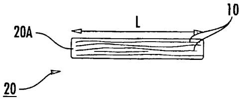
[00721 Figure IA is a schematic illustration of an implantable
construct 20 with
multiple fibers 10 that can be held together to form an array of fibers. As
shown in Figure
1A, the multiple fibers 10 can be axially arranged so that at least a
11
CA 02678821 2014-11-21
majority of the fibers are substantially parallel to each other over at least
a major portion of
the length of the construct 20, typically over substantially the entire length
of the construct
20. Some of the fibers may not run the entire length of the construct 20. The
construct 20
shown in Figures 1A and 1B is dry or partially hydrated. The construct 20
illustrated in
Figures 2A and 2B is shown fully hydrated and in a "hydraulic" fixation
configuration 20B.
As shown in Figure 1B, the cross-sectional shape 20A of the construct 20 may
be
substantially round during placement or ex vivo. As shown in Figure 2B, the
shape 20B of the
construct 20 can swell to a larger height "H2" and width ("W2") configuration
(which may,
for a substantially circular, oval or elliptical shape, have an associated
diameter "D2"), to take
on the shape of the adjacent local structure to frictionally engage therewith,
such as, for
example, a bone tunnel configuration. That is, as shown in Figure 2C, if the
local structure is
a bone tunnel 100t that is formed as a round tunnel, the construct 20 will
swell to fill the
tunnel and take the corresponding shape 20B as it swells and impart frictional
forces against
the wall of the bone tunnel to hydraulically affix itself therein. In typical
embodiments, the
length "L" of the construct is substantially constant between the dry or
partially hydrated and
hydrated configurations, typically changing less than about 3%.
[0073] In some embodiments, the cross-sectional area of the construct
20 is sized
to be between about 60%-98% of that of the bone tunnel 100t at insertion,
typically between
about 75%-90%, to allow for an insertion tool to cooperate with the construct
20 to slide the
construct 20 into the tunnel 100t and to allow for sufficient expansion after
placement to
cause a desired frictional engagement (higher pull-out forces). Measured
outside the body,
after 24 hours in a saline bath at ambient conditions, the construct 20 can be
configured to
expand to an increased hydrated unconstrained equilibrium cross-sectional area
of between
about 50% to about 250%, typically between about 150-220%.
[0074] The hydraulic fixation construct 20 may be particularly
suitable for
ligament and/or tendon repairs, replacements or treatments. The constructs 20
can be
configured to have at least about 60% of the tensile strength of natural
ligament or tendon at
implantation or within about 1-2 days thereof, which strength can increase
with neo-tissue in-
growth over time, and may have tensile strength and stiffness similar or even
greater than the
tensile strength, stiffness and/or dynamic flexibility of corresponding
natural tissue, e.g.,
natural ligament or tendon fibers. Thus,
12
CA 02678821 2014-11-21
embodiments of the invention may be particularly suitable for augmenting,
repairing or
replacing tendons and ligaments.
[0075] The construct 20 can be configured with a substantially planar
flexible
body for a ligament prosthesis, such as for an ACL repair or replacement. In
other
embodiments, the construct 20 may be configured as a substantially cylindrical
body for a
tendon-prosthesis, such as, for example, the flexor tendon. In some
embodiments, the
construct 20 can have between about 20-75 fibers and be used as a digital
flexor tendon.
Other configurations may also be used as suitable for the target treatment
site/prosthesis.
[0076] In some embodiments, the plurality of fibers 10 in a respective
construct
20 can be between about five to about two hundred. In some embodiments, the
number of
fibers 10 forming the construct 20 is between about ten (10) to about fifty
(50). For bio-rivet
configurations, the number of fibers may be more to add additional structural
rigidity, such as
between about 100 to about 250, typically between about 100 to about 150.
Lesser and
greater numbers of fibers may be used depending on the desired tensile
strength, rigidity, or
other mechanical parameter of the construct at the target implant site.
[0077] In some embodiments, the construct 20 is configured to have
substantially
the same physical thickness and/or configuration as the replaced or repaired
tissue so as to
not cause discomfort or physical abnormalities in structure when in position.
[0078] The array can be a relatively tightly compressed array of
fibers or a
relatively loosely compressed or attached arrangement having voids between
some adjacent
fibers depending on the target location and the desired mechanical properties
and
configuration and to allow for neo-tissue in-growth.
[0079] In some embodiments, the construct 20 is between about 0.5-50
cm long,
typically between about 1-25 cm, and in some embodiments between about 1 cm to
about 10
cm long. The construct 20 may have a width that is between about 0.05 mm to 8
cm, and is
typically between about 0.5 mm to about 3 cm. The constructs 20 may have a
cross-sectional
thickness of about 0.01 to about 30 mm. For the flat construct, the thickness
may be more
typically between about 0.1 to about 10 mm, while the tubular construct may
have a thicker
cross-section, such as between about 0.75 mm to about 30 mm.
13
CA 02678821 2014-11-21
[0080] As shown in Figure 3A, the bone 100 with the bone tunnel(s)
100t that
receives the construct 20 may be substantially straight (vertical or
horizontal). Alternatively,
the tunnel 100t may angle as shown in Figure 3B depending on the target
repair/implant site.
In some embodiments, the bone tunnel 100t can include different path
trajectories as shown
for example in Figure 3C. The construct 20 can bend to follow the tunnel
trajectory during
insertion. Having the end portion orthogonal or angled with respect to the
other tunnel
portion may inhibit pull-out.
[0081] The bone tunnel 100t can vary in width (diameter) and length
depending
on the target application. The length of the bone tunnels 100t is typically
between about 3
mm to about 12 mm, with bone tunnels 100t in pediatric bone plates being
between about 1
mm to about 4 mm. However, in some particular embodiments the bone tunnel 100t
can be
configured to have a length that is at least about 5 mm in cortical bone (for
adults), and at
least about 7 mm in cancellous bone (for adults).
[0082] In some embodiments, the biocompatible material 20 is inserted
in a dry
state and the interstitial fluid environment mediates a hydration process that
proceeds until
equilibrium is reached. The hydration causes an increase in the cross
sectional area of the
fibers until they fill the tunnel and cause a build up in internal pressure.
The pressure causes a
large frictional force, which effectively fixes the prosthesis in the bone
tunnel.
[0083] The construct 20 and/or fibers 10 can incorporate anti-
inflammatory agents
or other pharmaceutically suitable agents. The construct 20 and/or fibers 10
can be
configured with an anti-swelling inhibitor to control the time or rate of
hydration induced-
swelling to allow enough time for a clinician to properly orient and adjust
the fixation
member 20 in situ. For example, the anti-swelling inhibitor may be a heat or
light sensitive
coating or matrix and/or hydrogel coating or matrix that can dissolve or
resorb when in the
body over a relatively short period (such as to allow the swelling to occur
about 20-60
minutes after placement). In some embodiments, natural body heat may be
sufficient to
release the coating and initiate the swelling or a clinician may locally apply
increased heat.
Other swelling-inhibitor removal techniques may be used depending on the
inhibitor, such as,
for example, applying laser or infrared light, RF heat, heated and/or solvent
liquid or fluid
irrigation materials, and the like, to release the swelling inhibitor to allow
the
14
CA 02678821 2009-08-19
WO 2008/103377 PCT/US2008/002230
hydration-induced swelling. The swelling-inhibitor may also be lubricious so
as to
facilitate slidable insertion as appropriate.
[0084] The construct 20 may also or alternatively be coated or
impregnated with a thin film of polylactic acid (PLA) or other suitable
substance to
promote strength and/or ease of handling. For example, the construct 20 can be
dipped, painted or sprayed with a 3% solution of PLA in chloroform or other
suitable
solution.
[0085] Figures 4A and 4B illustrate the use of the construct 20 for joining
opposing end portions 20e of the biologically based fibrous materials to bone
tunnels
100t that may reduce or eliminate the need for supplemental mechanical
fixation
devices or serve as an adjunct fixation strategy. As shown, no additional
fixation
devices are required. However, surgical adhesives (glue), staples, buttons,
sleeves or
other devices may be used with the construct 20 depending on the repair site
and
needs thereof. Figure 5 illustrates that one end portion of the construct 20e
can
reside in a bone tunnel 100t while the other end portion is attached to a
repair site of a
ligament or tendon 110, typically using a suture(s) 105. The integration
technique can
include drilling at least one bone tunnel 100t to a defined size and inserting
an end
portion 20e of the construct 20 with a defined size and swelling capacity. As
discussed above, at least one end portion 20e of the construct 20 may be
configured as
a substantially round cross-sectional shape with the elongate body formed by
an array
of substantially parallel fibers 10. The cross-sectional configuration (shape
and/or
size) of the array or bundle 20 can be designed to achieve mechanical
competence
(frictional engagement) for the target application. The size (e.g.,
width/height or
diameter) of the bone tunnel 100t can be designed to provide for a snug fit
during
initial insertion.
[0086] One contemplated use of the construct 20 is a bioprosthesis with
one end portion hydraulically engaged in a bone tunnel to bridge gaps in
tendon and
ligaments by providing the construct 20 in a matching length and suturing into
the
patient's own remaining tendon or ligament end portions using a suitable
surgical
tying technique, such as, but not limited to, a double Kessler technique or
similar
methodology.
[0087] Figure 5 illustrates that a suture 105 can be attached to each end
portion of the construct 20e and used to affix the construct 20 to local
tendon or
ligament, in the embodiment shown in Figure 5, the suture 105 is tied to the
CA 02678821 2014-11-21
construct 20 so that opposing legs 311, 312 extend from a looped portion 32 of
the suture
having one or more loops 32 encasing the construct 20 that is tied to form one
or more knots
33. The sutures 105 may be resorbable or non-resorbable. Adhesive may be used
to help
secure one or both of the end portions 20e during an initial healing phase for
additional
stabilization.
[00881 The knot 33 can be configured to provide a secure attachment to
the array
or fiber bundle 20 and organize the parallel array of fibers into a desired
cross-sectional
configuration. The knot configuration can position the suture 105 to reach out
into adjacent
tissue for anchorage at about 180 to about 360 degrees from each other The
suture legs 31,
312 can extend substantially parallel to each other from opposing outer
lateral edges of the
construct 20 in the direction of the target anchoring-site. In the embodiment
shown, the
sutures 105 are oriented to exit the construct body outside the bounds of the
construct itself at
opposing side locations and extend substantially parallel to the anchoring
site. The looped
portion 32 and the knot(s) 33 are configured to improve tensile/compression
force
distribution and/or cancel unwanted torque. For additional discussion of
exemplary knot
configurations see, co-pending U.S. Provisional Application Serial No.
60/890,660, identified
by Attorney Docket No. 9624-5PR.
[0089] Figure 6A illustrates the construct 20 can be a hydraulic bio-
rivet 20r that,
in position, can hold adjacent bones, bone fragments or pieces 1001, 1002 in a
desired
alignment. The bones can be bone plates and the bone tunnels 100t may extend
all the way
through at least one of the bones 1001, 1002 (as shown, the bio-rivet 20r
extends through both
bone plates). One or both of the exposed end portions 20e can be configured to
define a head
20h. One or both of the heads 20h can be formed after the construct 20 is in
position.
[0090] In some embodiments, the construct has one head 20h formed
prior to
insertion into the bone tunnel that extends through both bones. The construct
20 can bundle
the fibers 10 as a single grouping of fibers that form the head 20h when
hydrated to cause
sufficient swelling to obtain a size that is greater than that of the adjacent
tunnel 100t. The
head 20h can include a metallic or polymeric substantially rigid member.
[00911 As shown in Figure 6B, at least one of the heads 20h (shown as
both
heads) can be formed by trapping the exiting portion of the fibers 10 in a
holding
16
CA 02678821 2009-08-19
WO 2008/103377 PCT/US2008/002230
member 25. The holding member 25 can be a resorbable or permanent component,
such as, but not limited to, a cap, staple, screw, pin, suture and the like.
For example,
the exiting portion of the fibers 10 can be captured in a cap 25 and or staple
(not
shown).
[0092] In some embodiments, as shown in Figure 6C, the construct 20 can
be separated into discrete separate fibers 20s or bundles of fibers, which can
be
formed into the respective head 20h using a mechanical member such as a cap
25.
The exiting strands can be braided, folded (Figure 6D) or otherwise formed and
trapped or held against the local bone using a holding member 25.
[0093] As shown in Figures 6B and 6C, in some embodiments, the
holding member comprises a cap 25 or staple (not shown) that can engage the
exiting
portion of the construct 20e and be used to trap the fibers and define the
head 20h.
The holding member 25 can be attached to the exiting portion of the fibers 10
in any
suitable manner, including being pressed against the fibers 10, adhesively
attached
thereto, anchored or screwed into the bone, or the like to trap the fibers 10
therebetween.
100941 Figure 6C illustrates that the holding member 25 can include a
serrated edge 26 that can engage the exposed bone to trap the fibers 10
against the
bone to help hold the-construct in position. Figure 6D illustrates the end
portion of
the construct 20e can be folded over and the holding member 25 used to trap
the
folded end portion of the construct for additional anchoring stability.
[0095] Figure 7 illustrates that the hydraulic fixation construct 20 can
engage a plate 125 to hold bone segments 100 in position. Figure 8 illustrates
that
the construct 20 can function as a bio-pin that engages an internal bone
tunnel 100t
and holds separated portions of bone segments 1001, 1002 in axial alignment
and
promote tissue ingrowth. Such a configuration may be particularly suitable for
collar-
bone fractures, finger fractures, and foot fractures.
[0096] Figure 9A illustrates that at least one end portion 20e of the
construct 20 may be rolled, wrapped or folded (see construct 201 in Figure 9A)
for
insertion into a substantially round bone tunnel 100t, while Figure 9B
illustrates that
the construct 20 can be arranged in a substantially cylindrical shape. Figure
10
illustrates that the construct may be substantially planar and the bone tunnel
configured with a substantially rectangular (or square) shape. Figure 11
illustrates
that the construct may include a braided segment 20br with bundles of fibers
braided
17
_ ,
CA 02678821 2009-08-19
WO 2008/103377
PCT/US2008/002230
together. The braided segment 20br may reside at an end portion, a medial
portion,
or segments therebetween, or may extend substantially the entire length of the
construct. Combinations of these shapes may be used. For example, a
substantially
flat construct 20 can be wrapped about a cylindrical construct body and both
(with
reduced moisture content) inserted into the target structure. Where two
constructs are
used, different fiber sizes and numbers may be used to form the constructs.
[0097] Figure 12 illustrates some operations that can be used to carry
out
embodiments of the invention. As shown, a multi-fiber construct is inserted
into a
bone tunnel (block 150). The construct is expanded by exposing the construct
to
liquid to cause the construct to frictionally engage the wall of the bone
tunnel (block
160). The dry or partially hydrated construct can be configured with a cross-
sectional
area that is between about 70-98%, typically 85-95%, of that of the receiving
bone
tunnel, and the expanding is done to create sufficient frictional engagement
to define a
pull-out tensile strength of at least about 5 N, typically at least about 10 N
(block
156).
[0098] The fibers may comprise NDGA polymerized collagen fibers
(block 120). The construct can have between about 10-250 fibers, with an
average
fiber width (diameter) of between about 0.01 mm to about 0.10 mm, typically
between about 0.1 and 0.5 mm (block 155). The length of the construct can be
substantially constant (during the insertion step and after the expanding
step) (block
157). The construct can have a flat shape and may be used for a ligament
repair or
replacement (block 152). The construct can have a substantially solid core
tubular
configuration or substantially circular cross-section and can be used for a
tendon
repair or replacement (block 154).
[0099] Optionally, the construct can be implanted in a patient using the
hydraulic fixation and one or more of a suture, suture anchor, staple, cap,
bone anchor
and the like. The suture, where used, can be a suture with an atraumatic
needle and
may be pre-applied to the construct and packaged in a medical kit for
subsequent use.
[0100] Also, the construct can optionally include (e.g., be coated,
impregnated ancUor amalgamated with) a gel or other material. The coating may
be to
promote fibroblasts, and/or may comprise one or more of an anti-inflammatory
agent,
an antibiotic or other therapeutic agent.
[0101] Figure 13 is a schematic illustration of a medical kit 200 that
includes the hydraulic fixation construct 20. The construct 20 can be held in
a sealant
18
CA 02678821 2014-11-21
210 that holds the construct in a dry or partially hydrated state. The package
200 may include
a desiccant to help maintain the desired dry or partially hydrated state of
the construct 20.
The sealant 210 may be a flexible, sealed sterile bag that is substantially
impermeable at
normal atmospheric conditions. The kit 200 may optionally include a modified
hemostat tool
225 that includes an axially extending aperture 225a through the centerline of
the tool 225
that is sized and configured to snugly hold the leading edge of the construct
20 to slidably
insert the construct in position in the bone tunnel 100t.
101021 The construct 20 can be preformed in different lengths for
selection by a
clinician during a surgical procedure or can be cut to length in situ by a
clinician. The
construct 20 can be preformed with a suture(s) 105 attached to one end of the
construct and
provided in the medical kit to reduce on-site preparation time. This latter
embodiment may be
particularly suitable where the construct 20 is provided in predetermined
lengths. The
construct 20 can be configured to have a strength and stiffness similar to
natural tendon or
ligament and can provide an effective scaffold for neo-tendon and ligament to
grow into and
further enhance some repairs. The kit 200 may include a temperature warning so
that the
construct 20 is not exposed to unduly hot temperatures that may degrade the
implant. A
temperature sensor may optionally be included on the package of the kit (not
shown) to alert
the clinician as to any excessive or undue temperature exposure prior to
implantation.
[0103] The fibers 10 can be any biologically compatible fibers formed
in any
suitable manner that can function as a biomedical product (implant/construct).
The construct
20 is suitable for chronic implantation and may optionally be absorbed,
resorbed and/or
biodegradable over time.
[0104] In particular embodiments, the fibers can comprise collagen
fibers such as
glutaraldehyde cross-linked collagen fibers and/or NDGA-treated collagen.
Suitable ways of
forming NDGA polymerized and/or treated fibers are described in U.S. Patent
Nos.
6,565,960 and 6,821,530. Generally stated, bulk collagen can be solubilized by
digestion with
a protease, then extruded into a synthetic fiber. Properly processed NDGA
polymerized fibers
are biocompatible. After the polymerization process, the fibers can be washed
in ethanol and
phosphate buffered saline to remove cytotoxins due to leachable reaction
products.
19
CA 02678821 2014-11-21
[0105] NDGA-treated collagen fibers are biocompatible and have
desirable
mechanical properties. Figures 14A-14C illustrate exemplary properties of NDGA-
treated
collagen fibers of different sizes (0.01, 0.1, 1 and 10 mm). The diameter of
the fibers was
measured with a dial caliper to the nearest 0.1 mm. The fibers were mounted in
clamps with 2
cm nominal tested length. Fibers were deformed to failure. The linear portion
of the
stress/strain curve was used to calculate the elastic modulus (stiffness) and
the force at which
the fibers failed was normalized to cross sectional area yielding tensile
strength. Values
shown are means +/- S.D. for six specimens. For additional discussion of the
NDGA
polymerized fibers, see, Thomas J. Koob, Biomimetic approaches to Tendon
Repair,
Comparative Biochemistry and Physiology Part A 133 (2002) 1171-1192. See also,
co-
pending U.S. Provisional Application Serial No. 60/883,408, Filed January 4,
2007 to Koob
et al., entitled, Methods of Making High Strength NDGA Polymerized Collagen
Fibers and
Related Collagen-Prep Methods, Medical Devices and Constructs.
[0106] The array or bundle 20 can be formed with fibers 10 having
widths or
diameters in any suitable range, typically in the range of between about 0.01-
10 mm. One or
more of the fibers 10 may be continuous or discontinuous over the length of
the construct 20.
Fibers 10 of different widths or diameters may be used in a particular
construct 20.
[01071 The present invention is explained in greater detail in the
following non-
limiting Examples.
EXAMPLES
[0108] The following discussion describes a study of mechanical
properties of
hydraulically fixed polymerized collagen fibers in bone tunnels.
Materials and Methods
101091 Biological fibers: NDGA polymerized collagen fibers were
prepared
substantially as previously described in Koob et al., Material properties of
NDGA-collagen
composite fibers: development of biologically based tendon constructs,
Biomaterials, 23:202-
212 (2002); and/or as described in co-pending U.S. Provisional Application
Serial No.
60/883,408, titled, Methods of Making High Strength NDGA Polymerized Collagen
Fibers
and Related Collagen-Prep Methods, Medical Devices and Construct.
CA 02678821 2014-11-21
Dried fibers averaged 0.26 .04 mm in diameter. Hydrated fibers had a mean
tensile strength
of 90 15 MPa or 10 1N breaking force. Dry fiber constructs were made by
arranging
fibers in a round parallel array with one end glued (Loctite-rm 454
cyanoacrylate) to round
nylon spacers (4.8 OD x 2.0 ID x 3.2 mm) used for clamping for mechanical
testing. The
other end of the construct was left intact for insertion into the bone tunnel
(Figure 15). The
length of the construct was such that there was a 5 mm unconstrained length
(the distance
between the bone and the clamped spacer). Ten-fiber constructs were used on
all tests except
for the scale-up series described later.
[0110] Glutaraldehyde crosslinked fibers were prepared by cross-
linking collagen
fibers overnight in a solution of 2.5 % glutaraldehyde in 0.1 M NaH2PO4, pH
7Ø These
collagen fiber constructs were prepared following the procedure described
above for the
NDGA fibers. After cross-linking, the fibers were washed twice with 70 %
ethanol for one
hour and dried for at least 2 hours under 0.06 N tension to keep them
straight.
[0111] Bone Tunnels: Bovine metatarsus was obtained fresh from 14 week
old
calves from a local slaughterhouse. Cortical bone specimens were cut from the
mid-shaft
using a band saw, and the cancellous specimens were taken from the proximal
phalanx of the
metatarsalphalangeal joint. Sections were made perpendicular to the long axis.
Specimens
were brought to final dimensions by polishing on an # 220 pit abrasive sheet
on a flat
surface. Through tunnels were drilled parallel to the long axis of the bone
using high-speed
steel drill bits (Small Parts Inc., Miami Lakes, Florida, USA). To reduce the
effects of
frictional heat, the drilling was done on a small milling machine at
approximately 70 rpm and
2 mm per minute feed rate (Sherline Model 4000, Sherline Manufacturing Inc,
San Marcos,
California, USA). Phosphate buffered saline (PBS: 0.1 M NaH2PO4, 0.15 M NaCl,
pH 7.0)
was used to lubricate the drill bit. The height of the bone corresponded to
the insertion depth.
During the cutting and polishing, the specimens were irrigated with PBS and
were stored for
up to 2 hrs at 4 C, pending insertion. For ease of clamping, the bones were
further cut into
sections not less than 5 mm square in cross section.
[01121 Insertion of fiber constructs: A tool to insert the fiber
bundles was
fabricated by drilling a hole through the centerline of the jaws of a
hemostat. The hole size
approximated the diameter of the dry fiber bundle. This allowed gripping the
21
CA 02678821 2014-11-21
fiber bundle into a tight circle for ease of insertion. The fibers were
inserted into the bone
tunnels until flush with the distal end. The specimens were then submerged in
PBS and
incubated at 4 C for 16 hrs. Preliminary experiments determined that this time
was sufficient
to attain full hydration of the fiber construct.
[01131 Swelling: Changes in diameter as a result of hydration to
equilibrium were
measured on ten fiber constructs by gauging with a template consisting of
holes of varying
diameters drilled into a 5 mm thick Lexanerm plate. Hole diameters were
verified using a
Super Hole GageTM, Bacor Inc., Deerfield, II, USA. Changes in length were
measured with a -
dial caliper.
101141 Mechanical Tests: The force required to pull out the
hydraulically fixed
fiber constructs from the bone tunnels was measured with uniaxial tests on an
MTS materials
testing apparatus (858 MinibionixTM II, Eden Prairie, MN, USA). The
displacement rate of
the piston was set at 1 mm per second. Force and displacement were recorded at
20 Hz. The
bone portion of the specimen was clamped to a specially designed ball-jointed
device fitted
with a vacuum plenum (Figure 16). This fixture allowed precise parallel
alignment of the
bone tunnel and construct with the piston transit. The nylon spacer holding
the fiber construct
was clamped to the piston with standard compression clamps. The pullout force
was taken as
the peak of the force/deformation curve (Figure 17). Five replicate specimens
were tested for
each experimental group.
101151 Effect of tunnel dimensions: To assess sensitivity of varying
the prosthesis
to tunnel clearance, 10-fiber constructs averaging 0.89 0.01 mm were tested
in 5 mm long
tunnels with varying bone tunnel diameters (0.95, 1.0, 1.1, 1.2, 1.65 and 2.35
mm). Tunnel
depth sensitivity was determined using 0.95 and 1.0 mm diameter tunnels, with
depths of 1,
2, 3, 5, 6 and 9 mm in cortical bone and 0.95 mm diameter and depths of 1, 2,
3, 5, 7, 9, and
11 mm in cancellous bone. The additional 11 mm depth in the cancellous bone
was chosen to
illustrate a plateau in the fixation force progression beyond a critical
maximum depth.
101161 Friction Coefficient: Hydrated fibers were wound 10 times
around a
stainless steel plate measuring 20 x 20 x 5 mm and placed on top of a flat
section of cortical
bone of the same dimensions, which was fixed to a tilt platform (N = 5). The
platform was
slowly tilted until the top plate started sliding. The difference in height of
the tilted edges was
measured and the angle of repose was obtained with
22
CA 02678821 2009-08-19
WO 2008/103377 PCT/US2008/002230
trigonometry. The Measured Friction Coefficient was defined as the tangent of
the
angle of repose. See, e.g., pp. 3-24, Avallone et al., Marcs' Standard
Handbook for
Mechanical Engineers,(Mc-Graw-Hill Book Co., 1987).
[0117] As a comparison, the Calculated Friction Factor was obtained
starting with the formula:
Fp = F, FN (Equation 1)
where Fp is the pull-out or fixation force, Fc is the friction factor and FN
is the normal
force produced by the pressure (P). Since, FN = AP and A = L 70, where "A" is
the
area of the tunnel, "D" is the internal diameter and "L" is the depth.
Combining
equations and rearranging yields:
Fc = Fp/(PL7tD) Equation (2)
Equation (2) was used to obtain the Calculated Friction Coefficient.
[0118] Pressure: In order to approximate the swelling pressure generated
by the fibers as they hydrate in a confined space, the change in dimensions of
Polytetrafluorethylene (PTFE) tubing (Small Parts Inc., Miami Lakes, Florida,
USA)
vs. the pressure generated by hydraulically loading the tubing was measured.
The
inner diameter of the tubing was 0.95 mm, matching the optimal bone tunnel
diameter
as established in Figures 19A and 19B. The wall thickness was 0.15 mm. Bundles
of
parallel dry fibers were placed inside the PTFE tubing. The fiber-in-tubing
units
were placed in PBS and allowed to hydrate for 16 hrs. The changes in outside
diameter resulting from the internal pressure exerted by the fibers were
measured with
a micrometer. An identical section of tubing was connected to a 3 ml syringe
filled
with water that was progressively compressed by the MTS machine, thereby
subjecting the tubing to progressively higher pressure. The outside diameter
of the
tubing was measured at sequential pressure points. The pressure was calculated
by
dividing the force measured by the load cell of the MTS machine by the
internal cross
sectional area of the syringe. This method neglects the internal deformation
of the
tubing thickness; therefore, the results represent the minimum pressure
generated by
the fibers in a rigid tunnel. This effect was not expected to be significant.
[0119] Fill percentage of the dry construct in the bone tunnel was
calculated by taking the total cross sectional area of the dry fibers and
dividing by the
cross sectional area of the bone tunnel. The largest and smallest diameter of
each fiber
was measured with a dial caliper and the formula for the area of an ellipse
was used to
23
CA 02678821 2009-08-19
WO 2008/103377 PCT/US2008/002230
calculate the area. The calculated pressure was defined as the fixation force
divided
by the product of the internal surface area of the tunnel area and measured
friction
factor, P = Fp/(FFA).
[0120] Scale up: To assess the potential for scaling up to different sized
constructs, 6, 10, 13, 16, 32, and 48 fiber constructs were tested. The tunnel
diameters
were designed to follow a linear progression of the aggregate areas of the
bundles
using the 10 fiber construct as the basis. This yielded tunnel diameters of
0.76, 0.95,
1.07, 1.16, 1.65, and 2.35 mm, respectively, which included adjustments within
a 3 %
margin to allow for the use of commercially available drill bits.
[0121] Statistical Analysis - The student T-Test was used to determine
statistical significance (p <0.05). N = 5 on all tests.
Results
[0122] Fiber swelling
As the 10-fiber constructs swelled from their dry state to a state of
unconstrained hydrated equilibrium, they increased an average of 45.8 3.0 %
in
diameter and 213 8.8 % in cross sectional area. Changes in length were
negligible
(<1%) therefore the volume change was also 213 8.8 %
[0123] Effects of tunnel depth
Pullout tests of the constructs inserted into bone with a 5 mm unconstrained
fiber length produced smooth force/displacement curves (Figure 17). The force
increased sharply with small piston displacement as expected with an effective
fixation. Bone tunnel depth directly influenced fixation strength. In cortical
bone, the
fibers showed a trend of increasing fixation strength with increasing length
of
insertion for a 0.95 nun diameter bone tunnel (Figure 18A). For a tunnel
diameter of
1.0 mm, the force of pull-out followed a similar trend but with slightly lower
forces in
the shorter tunnels (Figure 18B). Both curves reached an apparent maximum
plateau
at 5 mm tunnel length. The force at 5, 6, and 9 mm lengths averaged 18.7 3.9
and
17.6 2.0 N for the 0.95 and 1.0 mm diameters, respectively. At 5 mm depth, 3
of 5
of the specimens broke at the bone or spacer interface; at 7 and 9 mm all of
the
specimens broke rather than pulled out. The fiber bundle inserted into
cancellous
bone tunnels showed a similar trend to that of fibers in cortical bone, but
the plateau
was reached at 7 nun and the average pullout force for 7, 9, and 11 mm was
16.8
24
CA 02678821 2009-08-19
WO 2008/103377 PCT/US2008/002230
3.1 N (Figure 18C). At 5, 7 and 9 mm insertion depths the fibers pulled out of
the
bone tunnel; at II mm depth, all the fiber constructs broke before pullout.
[0124] Effects of tunnel diameter
The effect of bone tunnel diameter on fixation strength was examined using
the 10-fiber constructs and optimal tunnel depths established above (5 mm for
cortical
bone; 7 mm for cancellous bone). The fixation strength of the constructs
showed
significant sensitivity to tunnel diameter (Figures 19A and 19B). The pullout
force
decreased as the diameter increased until it reached zero at 1.65 mm diameter
for
cortical bone (Figure 19A) and 0.6 0.16 N for cancellous bone (Figure 19B).
At
1.1 mm diameter the average pullout force was 30.3 % of that at 0.95 mm
diameter
for cortical bone and for cancellous bone it reduced to 45.3 % for the same
diameters.
[0125] Pressure and friction
Plotting pressure vs. change in tubing dimensions yielded a linear regression
with r2= 0.96 (Figure 20A). A 10-fiber construct was then loaded into the same
tubing and allowed to swell to equilibrium; the change in diameter was
measured.
Inserting the value obtained into the regression equation yielded an
approximate
swelling pressure of 1.05 MPa. Inserting this pressure into the friction
factor equation
described in the methods yielded a Calculated Friction Factor of 1.16 0.35.
Fiber to
bone friction measured by the angle of repose method averaged 0.907 0.056.
The
measured friction factor was used in calculations below for the hydraulic
pressure
exerted on the wall of the tunnel.
[0126] Pullout force vs. percent fill
The pull-out force was directly related to the percent of the tunnel cross-
sectional area filled by the dried fiber construct (expressed as % fill). The
greatest
fixation was achieved at a % fill greater than 85% (Figure 20B). This
relationship
correlated with the calculated pressure vs. % fill (Figure 20C), indicating
that the
magnitude of the pull-out force depended on the swelling pressure exerted on
the
bone tunnel by the fibers in the construct causing a corresponding amount of
friction.
[0127] Scale-up to larger bundles
At constant tunnel depth, fixation strength scaled up as a linear function of
the
diameter of the tunnel and, therefore, of the diameter of the fiber bundle
(Figures 21A
and 21B). For the 48 fiber constructs in a 2.10 mm diameter tunnel, the
fixation
strength averaged 63.3 6.9 N.
CA 02678821 2009-08-19
WO 2008/103377
PCT/US2008/002230
[0128] Glutaraldehyde cross-linked fibers
Constructs made with glutaraldehyde cross-linked fibers showed similar
hydraulic locking forces to those of NDGA cross-linked fiber constructs
(Figure 22).
Discussion
[0129] Embodiments of the present invention can provide a novel and
effective strategy for joining biologically-based fibrous materials to bone
tunnels.
Hydraulic fixation may serve as an adjunct fixation strategy or optimally
eliminate the
need for mechanical fixation devices.
[0130] The basis for hydraulic fixation is swelling of the fibers as
they
imbibe liquid (water) and cause an increase in pressure inside the bone
tunnel. The
pressure acts along the internal surface area of the tunnel and produces a
"locking
force" as a result of friction. One simple model of friction force applied to
this
situation describes the friction or fixation force as equal to the normal
force times the
friction factor, Fp = FN FF. The normal force equals the pressure times the
internal
surface area of the bone tunnel, FN = PA. The above model is validated by the
correlation of Measured Friction Coefficient between bone and fibers with the
Calculated Friction Coefficient. This correlation also suggests that the
effect of any
deformation of the interior of the PTFE tubing used to measure swelling
pressure had
a minimal impact on the results. 'Since A = nDL, where D is the diameter and L
is the
depth of the bone tunnel, the above equations can be combined to yield Pp =
PrEDLFF.
Therefore, Fp (fixation force) should increase linearly as either the diameter
(D) or the
depth (L) of the bone tunnel are increased, as long as a substantially
constant fit
(percent fill) between the construct and tunnel is maintained. Increasing the
construct
size while maintaining a constant fit and depth produced a linear relationship
as
predicted above (Figures 21A, 21B). While it may be postulated that load
sharing
with larger constructs between the fibers might be an issue, this was not
evidenced
since the results of the scale-up were linear as predicted. This might be
accounted for
by fiber-to-fiber engagement, e.g., frictional engagement, since the internal
fibers
were likely subjected to a similar pressure of those in contact with the bone
tunnel
wall.
[0131] Varying the diameter of the tunnel while maintaining a constant
construct size showed that the highest fixation force and presumably the
highest
pressure resulted from the tightest initial fit between the construct and
tunnel. Pull-out
26
CA 02678821 2009-08-19
WO 2008/103377
PCT/11S2008/002230
strength deteriorated rapidly as the tunnel diameter to construct fit was made
looser.
There was a linear relationship between calculated pressure and percent fill,
further
supporting the likelihood of a correlation of fill or tunnel size to initial
prosthesis size
(and/or swelling).
101321 Assuming constant pressure and friction factor, the total
fixation
force can be represented as the sum of the forces at each plane and can be
modeled by
integrating force over the length of the insertion using the equation:
F= PTEDdx Equation (3)
Bringing out the constants and integrating yields:
F = 13701 Equation (4)
where F is the cumulative force at the depth 1. This equation describes a
linear
progression of accumulated force starting at zero at depth 1 to a maximum
equal to the
pull force F at the surface of the bone facing the source of tension. Seen
another way,
force will act upon the fibers to a layer deep enough to generate the required
total
fixation force. The force required for pullout or breaking of the construct
limits the
total force generated. For deep enough insertions the construct will break
rather than
pull out (Figures 18A-18C). This was evident in cortical bone tunnels
measuring 5
mm or more in depth. For forces less than those required for breaking or pull-
out, the
fibers should be stressed up to the depth necessary to withstand the applied
force. At
levels deeper than those required to generate the necessary force to resist
pull-out
there should be no force acting on the fibers, and no deformation or movement
should
occur in the deeper levels of the fibers.
[0133] For constructs of 10 fibers in tunnels of 0.95 mm diameter, the
integration by hydraulic fixation of NDGA-crosslinked fiber constructs in bone
is
stronger than the construct itself at tunnel depths exceeding 5 mm in cortical
bone and
7 mm or longer in cancellous bone. The integration of biological fibers into a
bone
tunnel via hydraulic fixation could be useful for some repairs of avulsed
tendons and
tom ligaments where the desired fixation forces are within the capabilities of
this
method. In some applications, hydraulic fixation may not require the use of
peripheral devices such as bone anchors, screws, buttons, sutures, glues or
resins. In
some embodiments, the techniques described herein may advantageously not add
27
CA 02678821 2009-08-19
WO 2008/103377 PCT/US2008/002230
additional materials to the surgical placement and anchoring of a fibrous
bioprosthesis, thereby reducing or eliminating the need for harvesting
autografts.
Additionally, maximum fixation force may be obtained within hours of
insertion,
allowing early mobilization and resultant therapeutic advantages.
[0134] Potential applications include the development of prostheses for
tendon transfers, repair of tendon avulsions and supplementing anterior
cruciate
ligament (ACL) repairs. Pediatric applications are especially attractive since
it may
allow for relatively short bone tunnels that would avoid interfering with the
growth
plates. The number of fibers, aggregate diameter and tunnel depth can be
designed
taking into consideration the strength of the fibers and the required strength
of the
application, which can be a function of the strength of normal tendons and
ligaments,
anatomical site, and patient size and age. The design of the tunnel diameter
should be
considered since the integration can, in some embodiments, be sensitive to the
fit
between the construct and the tunnel. It is contemplated that hydraulic
fixation of
fiber constructs potentially operates effectively at larger construct sizes
than those
tested and therefore can be designed to fit various applications, and is not
limited to
those described herein.
[0135] It is contemplated that the rate of hydration in the bone tunnel may
be controlled for some applications to allow sufficient time for surgical
placement of
a bioprosthesis and, if needed, adjustment of length and tension. The amount
of time
after the fibers are exposed to a hydrating environment and the speed of
fixation can
be coordinated so as to avoid premature locking. A means of fast insertion
and/or
controlling the rate of hydraulic swelling in vivo may be used, for example,
hydrogel
matrices are potential hydration retardants.
[0136] Another advantage of the swelling properties of the fiber constructs
is that swelling occurs substantially only perpendicular to the long axis of
the fiber.
The constructs do not substantially lengthen or shorten. Appling the proper
tension in
the re-attachment of tendons or ligaments to bone would not suffer from
problematic
lengthening of the construct due to hydration.
[0137] While these data establish that hydraulic fixation results in
mechanical coupling of biological fibers to bone, other factors may influence
the
ultimate performance of a fibrous bioprosthesis. The objective of using such a
bioprosthesis to connect tendons or ligaments to bone is to provide immediate
and
sufficient stabilization to allow early rehabilitative mobilization. However,
the
28
CA 02678821 2009-08-19
WO 2008/103377 PCT/US2008/002230
ultimate fixation strength will rely on the mechanobiology of the bone. The
best
outcome would be complete osteointegration of the fiber construct without
producing
untoward stress concentration at the fiber-bone interface.
[0138] NDGA polymerized collagen fibers may be particularly suitable for
implementing the hydraulic fixation. They can provide the swelling properties
for
effective hydraulic fixation, they are not cytotoxic, they do not harbor
diffusible
cytotoxic reaction products, they are biocompatible with cells in vitro, and
they are
biocompatible and can be configured so that they do not get degraded for six
weeks in
vivo. See, Koob, Biomimetic approaches to tendon repair, Comp. Biochem.
Physiol.
A Mol. Integr. Phys. 133: 1171-1192 (2002). The biocompatiblility of these
fibers
combined with biomechanics similar to natural tendon and ligament offer a
potential
of serving as effective scaffolding for new tissue growth. In a functional
context, the
optimal mechanical performance of hydraulic fixation is only as good as the
tensile
properties of the anchored bioprosthesis. This was the case in the scale-up
experiment
in which the 6-, 10-, 13- and 16- fiber prostheses broke before pulling out of
the
tunnel. NDGA-polymerized fibers used in this study exhibit material properties
in
uniaxial tensile tests that are comparable to those of tendons and ligaments.
However,
when bundled in linear arrays and hydraulically fixed in bone, stress
concentration at
the prosthesis bone junction apparently lowers the aggregate tensile strength
of the
bioprosthesis. For example, the 10-fiber prosthesis failed at 18.6 N, in
contrast to the
theoretical tensile strength of 100 N. Despite this lowered force at failure,
the stress
at the failure site, calculated by dividing the force by the cross sectional
area of the
tunnel, averages 24.4 +/- 3.6 MPa. The tensile strength of tendons ranges from
40 to
100 MPa. Although this performance is lower than that of natural tissue it
could be
enough to maintain the integrity of the repair until neo-tendon and neo-
ligament
growth augments the total strength of the repair.
[0139] One potential application is the repair of digital flexor tendons,
where the median forces of active and passive mobilization were found to be 27
N.
Flexion. Flexion under a 500 g load produced a median force of 48 N. See,
Trail et
al., Forces transmitted along human flexor tendons during passive and active
movements of the fingers, J. Hand Surg., 29:4:386-389 (2004). As tested,
constructs
of 48 NDGA polymerized collagen fibers in a bone tunnel of 2.1 mm diameter and
5
mm depth withstood an average force of 63.3 6.87 N. A prosthesis of similar
size
29
CA 02678821 2009-08-19
WO 2008/103377
PCT/US2008/002230
and fixation strength could be developed for flexor tendon repair that could
allow
early passive and active motion and light initial use.
[0140] In conclusion, hydraulic fixation can provide a simple and
effective
means for joining biological fibers to bone either alone or in conjunction
with another
fixation device. Bioprosthesis could be developed using a parallel array of
NDGA
polymerized fibers that relies on hydraulic fixation as the coupling modality
in bone.
NDGA-crosslinked fibers are mechanically competent as compared to native
tendons
and ligaments and they have excellent biocompatibility. Such a medical device
could
provide the surgeon the advantages of simple and effective placement,
adjustment and
fixation. The device could deliver cytokines for enhanced osteointegration.
Additionally, hydraulic fixation may allow passive motion within hours of
surgery
and could potentially be used in a variety of applications such as, for
example, digital
flexor tendon repair. In addition to being strongly fixed within hours it
could allow
for osteointegration of the fibers into the regenerated bone tissue and serve
as a
scaffold for neo tendon and neo ligament growth and a corresponding
augmentation
of strength and reliability.
[0141] The foregoing is illustrative of the present invention and is not
to
be construed as limiting thereof. Although a few exemplary embodiments of this
invention have been described, those skilled in the art will readily
appreciate that
many modifications are possible in the exemplary embodiments without
materially
departing from the novel teachings and advantages of this invention.
Accordingly, all
such modifications are intended to be included within the scope of this
invention as
defined in the claims. The invention is defined by the following claims, with
equivalents of the claims to be included therein.