Note: Descriptions are shown in the official language in which they were submitted.
CA 02679172 2009-08-25
WO 2008/104564
PCT/EP2008/052371
1
FIXATION OF A BIOLOGICAL MATERIAL
The present invention relates to a method for fixation of a biological
material, the
biological material resulting from this method, kits for carrying out said
method
as well as the use of a composition for said method.
For a long time the emphasis has been solely the pathological or histological
ex-
aminations of biological materials. If at all, the samples for these types of
exami-
nation were usually conserved or stabilised by storing the samples in formalde-
hyde solutions and/or by embedding them in paraffin. However, the conservation
of biological materials by cooling or freezing has also been standard practice
for a
long time.
It was first recognised that the determination of particular components of
biologi-
cal materials, such as for example nucleic acids or proteins, particularly in
the
field of medical and clinical diagnosis, is of great benefit, and also the
need for
new, more effective and more economical conservation and stabilisation
reagents
and or methods became apparent.
In the course of these developments it was recognised that precisely the
status
(gene expression profile or proteome) of the important components of the fresh
samples for molecular biological examination can change rapidly, immediately
after removing the sample from its natural environment, such that already a
longer
storage of the samples in a non-treated state, e.g. due to an unintentionally
delayed
transport to a laboratory etc., can invalidate molecular analyses or even make
them quite impossible.
Precisely the nucleic acid status of a biological material alters that much
more, the
longer the elapsed time between the sample and its analysis. The ribonucleic
acids
(RNA) degrade particularly quickly due to ubiquitous RNases. Also, besides the
degradation of nucleic acids, stress genes, for example, are induced and lead
to the
CA 02679172 2009-08-25
WO 2008/104564
PCT/EP2008/052371
2
synthesis of new mRNA molecules that likewise strongly alter the transcription
pattern of the sample. Accordingly, in order to retain the gene expression
profile,
the sample under examination has to be stabilised immediately.
An immediate stabilisation of the sample is needed, not only for the analysis
of
the nucleic acids but also for detailed examinations of the proteome of a
biologi-
cal material, because the protein pattern is also immediately altered after
removal
of the sample. This occurs firstly by degradation or new syntheses, secondly ¨
particularly quickly ¨ by changes to the protein modifications, such as e.g.
phos-
1 0 phorylation/dephosphorylation.
As chemical analyses of proteins and molecular analyses are also increasingly
used in areas other than in the field of medical and clinical diagnostics,
such as
forensics, pharmacy, food analysis, agriculture, environmental analyses as
well as
in many research areas, the retention of the integrity of the molecular
structure of
samples and consequently their immediate stabilisation, is therefore a
necessary
requirement in all these fields.
Thus, over the years, a great number of the most varied stabilisation reagents
and
stabilisation methods have been developed for the stabilisation of a wide
range of
the most varied biological materials.
As mentioned before, stabilisation by means of formalin and the subsequent em-
bedding of the stabilised samples for the histological examination of tissue
has
been known for a long time. However, this type of stabilisation is mostly
unsuit-
able for use in molecular biological processes, as only a very insufficient
stabilisa-
tion of the nucleic acids results, and so at most, a qualitative, but not
quantitative
indication of the nucleic acids or nucleic acid fragments present is possible.
Moreover, the stabilisation with crosslinking stabilisers such as formalin
leads to a
reduced extractability of the nucleic acids or proteins from the tissue.
Formalin is
also toxicologically not harmless.
CA 02679172 2009-08-25
WO 2008/104564
PCT/EP2008/052371
3
Stabilisation reagents, such as cationic detergents are described in US
5,010,184,
US 5,300,545, WO-A-02/00599 and WO-A-02/00600, with which very good
qualitative identifications of nucleic acids can be achieved. However, these
types
of stabilisers do not sufficiently stabilise nucleic acids in compact pieces
of tissue.
Other stabilisers that comprise, for example, high concentrations of ammonium
sulfate (see e.g. US 6,204,375), are very suitable for stabilising nucleic
acids in
various tissues. However, they are extremely unsuited for use in stabilising
cell-
containing or cell-free body fluids, such as, for example, blood, serum or
plasma,
and in addition demonstrate poorer stabilising properties with some types of
tis-
sue, such as, for example, fatty tissue.
Moreover, those reagents and methods, with which nucleic acids can be
stabilised
for quantitative identification, are generally not suitable for histological
examina-
tions, because these stabilisers conserve the nucleic acids but not the
cellular or
tissue structures.
This demonstrates that it is particularly difficult to simultaneously
stabilise RNA,
DNA and proteins in tissue samples and also histologically conserve the tissue
samples. Moreover, work with cells or other biological materials cannot
necessar-
ily be applied to compact tissue. The stabilisation of nucleic acids in
compact tis-
sue samples is particularly difficult in comparison with other biological
materials.
Due to their composition, their components and structure, tissues are complex
and
heterogeneous. In order to stabilise nucleic acids in compact tissue samples,
the
effect of the stabilising reagent must develop not only on the surface of the
cells
and/or inside a layer of cells, but also deep within the complex sample
material. In
addition, often within one and the same biological material, very different
tissue
and/or cell types have to be addressed, which differ, for example, in the cell
struc-
ture, the membrane constitution, the compartmentalisation and the
biomolecules,
e.g. with regard to the proteins, the carbohydrates and/or the fat content.
CA 02679172 2009-08-25
WO 2008/104564
PCT/EP2008/052371
4
A frequently used form of stabilisation for tissue samples, including all the
com-
ponents, and known from the prior art, is freezing or deep freezing the
samples.
The sample, directly after removal from its natural environment, is deep
frozen
below -80 C in liquid nitrogen. Samples treated in this way can then be
stored at
about -70 C for an almost unlimited time without loss of integrity. However,
these types of processes consistently require very laborious logistical
conditions,
as a thawing of the samples has to be prevented during transport, storage or
during
the most varied applications and uses. Besides the additional costs for
special
sample holders and the permanent cooling of the samples, in addition the use
of
HI liquid nitrogen is not only very cumbersome, but also only
practicable under spe-
cific safety measures.
Moreover, a subsequent analysis of the deep-frozen sample mostly proves to be
very difficult, particularly for individual components of the sample. Thus,
the
thawing or melting of the sample during transport or processing leads to
degrada-
tion, particularly of the RNA. This means that such melted or thawed samples
no
longer yield reproducible results. Also, precisely such pieces of tissue in
the fro-
zen state are processable only with great difficulty, cut up by hand for
example, or
with an increased apparatus cost.
So-called transition solutions have also been described that reduce the
disadvan-
tages associated with processing deep-frozen samples, especially for isolating
RNA. This entails the deep-frozen sample being firstly transferred into a
solution
precooled to -70 C to -80 C, and then stored in it for a few hours (at least
16
hours) at about -20 C. Then the sample, soaked with the transition solution,
can
be warmed up to a working temperature of -4 C to room temperature, but only
for a short period, for example enough for cutting up the sample, without the
status of the nucleic acid being altered. However, further analyses and
storage of
the sample - especially at room temperature - are not possible. These types of
transition solutions, known for example from WO-A-2004/72270, consist primar-
ily of monohydric alcohols.
CA 02679172 2009-08-25
WO 2008/104564
PCT/EP2008/052371
Disadvantageously, the samples treated with current transition solutions only
re-
main stable at room temperature for a very short time, with the result that
the
processing time is very short and is very easily exceeded, especially for
cutting
and weighing steps. Moreover, the transition occurs only very slowly, such
that no
5 direct experiments may follow and mostly result in waiting periods of one
day.
Similarly, an undamaged transport at room temperature of the samples treated
in
this way is not possible as not only the transition but also the subsequent
stable
sample storage must be carried out at < -20 C. Also, transport of the sample
is
only possible at < -20 C, requiring the use of coolants, for example dry ice.
Although the use of conventional transition solutions brings improvements in
sample processing, such as e.g. the weighing or cutting, neither the equipment
expense is reduced (as the solution for the transition has to be cooled down
to -70
to -80 C and therefore a suitable cooling device has to be available anyway),
nor
can the samples treated with the transition solution be stabilised for a
longer pe-
riod at room temperature.
Another disadvantage of all methods including snap freezing of fresh tissue
sam-
ples is, that due to ice crystal artefacts morphological details are not
retained accu-
rately. This may impair diagnosis based on histological stainings.
Tissue fixatives for preservation of morphological structures can be divided
into
two groups. Group one contains crosslinking agents like formaldehyde, parafor-
maldehyde or glyoxal. From these 4% buffered formaldehyde is the most widely
employed fixative. The only change made since its introduction in 1896 by F.
Blum is neutral buffering of formalin at a physiological pH (NBF, neutral buff-
ered formalin). Although not fully understood it is thought that the aldehydes
form cross-links between proteins. By this enzymatic activity is ceased and
solu-
ble proteins are fixed to structural proteins. Since formaldehyde also reacts
with
nucleic acids, NBF fixation leads to low yield and degradation of nucleic
acids.
CA 02679172 2009-08-25
WO 2008/104564
PCT/EP2008/052371
6
The second group does not contain crosslinking agents, but alcohols as major
components. Noncrosslinking, alcoholic fixatives were introduced to avoid haz-
ardous formaldehyde fixation and to better preserve tissue macromolecules and
especially protein epitops for immunohistochemical methods. Alcohols have to
penetrate the tissue and to reach a certain local concentration for
precipitating and
denaturation of proteins. In recent times some of the alcoholic fixatives like
Car-
noy's (60% ethanol, 30% chloroform, 10% acetic acid) or Methacarn (Carnoy's
with the substitution of ethanol with methanol) have been found to yield
superior
results as nucleic acids fixatives compared to aldehydes (Cox et al.,
Experimental
and Molecular Pathology 2006; 80: 183-191). Other more recently published fixa-
tives based on alcohol consist of 70% ethanol (Gillespie et al., Am. J.
Pathol.
2002; 160(2):449-457), 56% ethanol and 20% PEG (polyethylene glycol, Bost-
wick et al.; Arch.Pathol. Lab. Med. 1994; 118:298-302) or 90% methanol and
10% PEG300 (Vinvek et al., Lab. Investigation 2003; 83(10): 1427-35).
In addition several recently published patent applications claim for improved
non-
crosslinking fixatives. In DE 199 28820 a fixative is described containing a
mix-
ture of different alcohols, aceton, PEG and acetic acid. In US 2003/0211452 Al
a
fixative commercialised as "Umfix" is described containing at least 80%
methanol
and up to 20% PEG300 for preservation of RNA, DNA and morphology. Fixa-
tives based on ethanol for preservation of molecular content and morphology
are
described in US 2005/0074422 Al ("Finefix"), WO 2004/083369 ("RCL2") and
WO 05/121747 Al ("Boonfix"). In US 2003/119049 a universal collection me-
dium is described which is water based and comprises a buffer component, one
alcohol, a fixative component and an agent to inhibit degradation.
However all these above mentioned alcoholic fixatives show significant
disadvan-
tages. Fixatives based on ethanol or mixtures of different alcohols balanced
with
water do not prevent RNA from degradation. Fixatives without acid and high
amount of alcohol may preserve RNA for short times but show artefacts like tis-
sue hardening or shrinkage which lead to disruption of morphological details.
CA 02679172 2009-08-25
WO 2008/104564
PCT/EP2008/052371
7
Neither of the described fixatives is able to preserve RNA integrity over
longer
time periods, e.g. up to three days at ambient temperature with simultaneous
stabi-
lisation of morphology.
Beside crosslinking and alcoholic fixatives there are a number of agents known
which combine both groups of fixatives. One example is "Genofix", described in
US 5,976,829 A for preservation of morphology, immunogenicity as well as RNA
and DNA integrity. Since formaldehyde is one component "Genofix" also shows
all the disadvantages of crosslinking agents.
A different approach is described in WO 03/029783 Al. The so-called HOPE-
technique (Hepes-Glutamic acid buffer mediated Organic solvent Protection Ef-
fect) comprises a protection-solution with an organic buffer, acetone as the
only
dehydrating agent, and pure paraffin of 52-54 C melting temperature. The
disad-
vantage of HOPE is that the tissue must be kept at 4 C over night until
processing.
"Liforlab" from OncoSience described in W02004/099393A1 is an oxygen en-
riched solution which contains inorganic salts, amino acids, vitamins,
cholesterol
and adenosine. Since neither HOPE nor Liforlab fix tissue they do not conserve
the molecular content of the cells as if they were still in the patient's
body. Instead
it is highly likely that the expression profile of e.g. RNA will change during
stor-
age and/or transportation.
The object of the present invention was to provide a method to stabilise
biological
material in an effective, fast and easy way allowing isolation of nucleic
acids
and/or proteins as well as histological analysis.
In particular, the present invention was based on the object of specifying a
method
for stabilising a biological material, which leads to an acceptable
stabilisation of
the biological material if possible without adding crosslinking, carcinogenic
or
foetus damaging substances.
CA 02679172 2009-08-25
WO 2008/104564
PCT/EP2008/052371
8
Moreover, the present invention was based on the object of specifying a method
for stabilising a biological material, with which both frozen and also fresh
bio-
logical materials can be stabilised under the most moderate temperature condi-
tions possible, for example also at room temperature, without impairing the ex-
pression profile of the genome and/or proteome of the biological material.
Moreover, the method for stabilising a biological material was intended to
enable
both a histological analysis of the stabilised biological material as well as
an
analysis of the biomolecules comprised in the biological material. In this
context,
the stabilisation method should particularly enable both proteins as well as
nucleic
acids in the stabilised biological material to be qualitatively and
quantitatively
analysed. Therefore by stabilising the biological material, the quality and
quantity
of the nucleic acids that for example can be determined by gel analysis or by
the
number of PCR cycles until a given amount of nucleic acid is obtained, and the
quality and quantity of the proteins that can be determined by polyacrylamide
gel
electrophoresis or for example in the case of an enzyme can be determined by
suitable activity tests, should not be impaired.
Furthermore, the method for stabilising a biological material was intended to
af-
ford a stabilised biological material that can be analysed not only at
moderate
temperatures, for example at room temperature, but can be optionally stored
prior
to or after such an analysis for the longest possible time under such moderate
con-
ditions of temperature.
In the case of biomolecules, the term "stabilisation" is intended to mean
prefera-
bly the inhibition of the degradation, the modification, the induction or the
change
in the activity of biomolecules. In the case of histological analyses of
biological
materials, the term "stabilisation" is intended to mean preferably the
prevention of
a significant change of the morphology of the samples.
A contribution to achieving the objects cited above is provided by a method
for
treating a biological material, wherein the method comprises the steps of
CA 02679172 2009-08-25
WO 2008/104564
PCT/EP2008/052371
9
i) providing a biological material, and
ii) contacting the biological material with a first non-aqueous composi-
tion comprising:
(al) 10 to 90 vol.% methanol, and
(a2) at least one additional additive, and
(a3) optionally an acid.
iii) transferring the biological material into a second composition (B)
comprising up to 99 vol.% ethanol.
Particularly useful as a first composition in step ii) is a non-aqueous
composition
(A) for preservation of biological material, the non-aqueous composition (A)
comprising
(al) 10 to less than 80 vol.%, methanol, and
(a2) at least one additional additive, and
(a3) an acid.
It was surprisingly found that in particular freshly isolated biological
materials can
be stabilised using the methanol containing composition (A), and biological
mate-
rials, which preferably can be fresh or frozen, for example deep frozen in
liquid
nitrogen, can be prepared for a histological, molecular biological and/or
microbi-
ological analysis. It is not required for the biological material that had
been
brought into contact with the composition to be cooled below 0 C and to
analyse
or store it at such low temperatures, and therefore the inventive method can
be
carried out without any costly apparatus, especially without cooling devices
or
coolants.
CA 02679172 2009-08-25
WO 2008/104564
PCT/EP2008/052371
Compositions described herein were developed for their chemical simplicity,
abil-
ity to preserve morphologic and genetic characteristics of tissue, and
convenience
and practicality of usage at ambient temperature. A cell or tissue may be
stored
therein and serve as an archiva source for cytology, histology, and/or genetic
5 analysis. It may be preserved and/or stored for prospective or
retrospective study.
Although not preferred, storage in the composition of the present invention
may
also follow contact of the cell or tissue with other preservatives and/or
fixatives.
The biological material prepared in step i) of the method can be a frozen or
pre-
HI ferred a non-frozen biological material, wherein all biological
materials known to
the person skilled in the art can be used as the biological material.
Preferred bio-
logical materials are selected from such comprising biomolecules, for example
natural, preferably isolated linear, branched or circular nucleic acids, such
as
RNA, especially mRNA, siRNA, miRNA, snRNA, tRNA, hnRNA or Ribozymes,
DNA and the like, synthetic or modified nucleic acids, for example oligonucleo-
tides, particularly for the primer, probes or standards used for PCR, nucleic
acids
or PNAs (peptide nucleic acids) marked with digoxigenin, biotin or fluorescent
dyes, preferably isolated proteins or oligopeptides, synthetic or modified
proteins
or oligopeptides, for example antibodies with fluorescence markers or coupled
with enzymes, hormones, growth factors, lipids, oligosaccharides, polysaccha-
rides, proteoglucanes, bodily fluids such as blood, sperm, cerebrospinal
liquids,
saliva, sputum or urine, liquids that are obtained when processing blood, such
as
serum or plasma, leucocyte fractions or "buffy coat", saliva, fecal matter,
smears,
aspirates, scurf, hair, skin fragments, forensic samples, food or
environmental
samples that comprise free or bonded biomolecules, particularly free or bonded
nucleic acids, metabolic products, whole organisms, preferably non-living
organ-
isms, tissues of metazoa, preferably of insects and mammals, especially from
hu-
mans, for example in the form of tissue sections or organs, isolated cells,
for ex-
ample in the form of adhering or suspended cell cultures, organella, for
example
chloroplasts or mitochondria, vesicles, cell nuclei or chromosomes, plants,
plant
parts, plant issue or plant cells, bacteria, viruses, viroids, prions, yeasts
and fungi.
CA 02679172 2009-08-25
WO 2008/104564
PCT/EP2008/052371
11
Cells may be pellets or suspensions, preferably isolated cells from a
biological
fluid (e. g. ascites, blood, cerebrospinal fluid, lymph, pleural exudate),
cell sus-
pensions from the aspiration of organs or lavage of body cavities, or cell
smears
(e. g. , cervix). Cells may be isolated by enzymatic and/or mechanical
disaggrega-
tion. They may be cultured as living cells for maintenance or propagation
before
preservation and/or storage. Cells may be washed and collected by
centrifugation
into a pellet ; they may be collected on a slide or other substrate.
For blood and other single-cell suspensions, cells may be isolated by
sedimenta-
tion or density gradient centrifugation, panning on a coated or uncoated
plastic
plate, passage through glass wool or other chromatographic matrix, rosetting,
sort-
ing by light scatter or fluorescently-labeled antibody, binding to antibody
coated
magnetic particles, or a combination thereof. Cells may be cancerous (benign
or
malignant) or precancerous, obtained from an animal or human subject affected
by disease or suspected of same (normal or diseased), or be affected by other
pa-
thology. lt may be obtained by autopsy or biopsy (e.g., catheterization or
phlebot-
omy) or other fluid collection. Cells preferably are placed in contact with
the
composition (A) within one to 30 min after removal from the body or in vitro
cul-
ture, but this time may be extended by cooling them on ice. Cells may be pre-
served and/or stored.
Cells may be processed for cytology. They may be smeared on a slide and exam-
ined with a microscope. Antigen or antibody may be directly or indirectly
labelled
with a colorimetric, enzymatic, fluorescent, luminescent, magnetic, or
radioactive
moiety which is detectable. Cells may be identified and/or isolated in
accordance
with antigen expression by antibody panning or sorting, or other affinity
chroma-
tography. A cytometer may analyze or a cell sorter may separate such cells by
DNA/RNA content, size, viability, binding of fluorescent-labeled antibody, or
a
combination thereof. A magnet may affinity purify cells that bind an antibody-
coated magnetic bead. Cells may be characterized by cell cycle, division,
growth,
or organelles. Negative or positive selection (e. g. , affinity or sorting
techniques)
may be used to isolate cell populations.
CA 02679172 2009-08-25
WO 2008/104564
PCT/EP2008/052371
12
The tissue may be obtained from autopsy, biopsy or from surgery. It may be a
solid tissue such as, for example, parenchyme, connective or fatty tissue,
heart or
skeletal muscle, smooth muscle, skin, brain, kidney, liver, spleen, breast,
carci-
noma (e.g. bowel, nasopharynx, breast, lung, stomach etc.), cartilage,
lymphoma,
meningioma, placenta, prostate, thymus, tonsil, umbilical cord or uterus.
Option-
ally, calcified tissue like bone or teeth may need to be demineralized before
fur-
ther processing. "Tissue" does not usually refer to single cells from a
biological
fluid (e. g. , ascites, blood, pleural exudate), cell suspensions from the
aspiration
of organs or lavage of body cavities, or cell smears. The tissue may be a
tumor
(benign or malignant), cancerous or precancerous, obtained from an animal or
human subject affected by disease or suspected of same (normal or diseased),
or
be affected by other pathology. It may be obtained by autopsy, biopsy (e. g.,
en-
doscopy or laparoscopy), or surgical resection. Tissue preferably is placed in
con-
tact with the composition (A) within one to 30 min after death or removal from
the body but this time may be extended by cooling it on ice. A piece of tissue
(e.
g., a slice or block) may be preserved with and/or stored in the composition
of the
invention; tissue that has been preserved and/or stored may also be embedded
in a
medium. Tissue may be analysed by serial reconstruction with different
analyses
applied to adjacent sections. Negative or positive selection (e. g.
microdissection
with optical tweezers or laser ablation) may be used to isolate cell
populations.
A freshly prepared biological material is preferably used as a non-frozen
biologi-
cal material in step i) of the inventive method, for example a fresh tissue
sample
or freshly isolated blood cells from a living or dead organism, or in the case
of a
synthetic biomolecule as the biological material, freshly synthesised nucleic
acids
or proteins. According to the invention, a "fresh" biological material is
preferably
understood to mean a sample that prior to being contacted with the composition
in
step ii) was removed not more than 96 hours, preferably not more than 48
hours,
particularly preferably not more than 24 hours, further preferably not more
than
10 hours, further even more preferably not more than 60 minutes and most pref-
erably not more than 10 minutes previously, or in the case of a synthetic bio-
CA 02679172 2009-08-25
WO 2008/104564
PCT/EP2008/052371
13
molecule after having been synthesised. However, the designation "fresh" bio-
logical material also includes such samples that have been removed within the
previously mentioned periods, but which prior to having been contacted with
the
composition had not been pre-treated, for example with conventional fixatives,
such as for example formalin, with dyes, such as eosin, with antibodies and
the
like. The preparation of fresh cell samples or tissue samples can result from
all
methods of preparation known to the person skilled in the art for this
purpose, for
example in the case of a tissue sample by means of a scalpel, for example
during
an autopsy, in the case of a blood cell sample by centrifugation of freshly
removed
blood and the like. In the case of the use of a fresh biological material, the
first
composition used in step ii) principally serves as the stabilisation
composition.
Said first composition preferably is composition (A).
A biological material may be used as a frozen biological material in step i)
of the
inventive method, which, after having been isolated according to the
previously
described technique, is firstly cooled down to temperatures of 0 C or less,
pref-
erably to temperatures of -20 C or less and most preferably to temperatures
of -
70 C or less, for example by contact with liquid nitrogen, before being
contacted
with the first composition in step ii). In the case that a frozen biological
material is
used in the inventive method, then the first composition used in step ii)
principally
serves as the transition composition. The first composition used in step ii)
pref-
erably is composition (A).
Component (al) of composition (A) is methanol. Methanol is contained in com-
position (A) in an amount of 10 to less than 80%, preferably from 20 % to less
than 80 vol.%, more preferably from 30 to less than 80% and most preferably
from 50 to less than 80 vol.%. This means that methanol can be comprised in
composition (A) in an amount of up to 79, 78, 77, 76, 75, 74, 73, 72, 71, 70,
69,
68, 67, 66, 65, 64, 63, 62, 61, 60, 59, 58, 57, 56, 55, 54, 53, 52, 51, 50,
49, 48, 47,
46, 45, 44, 43, 42, 41, 40, 39, 38, 37, 36, 35, 34, 33, 32, 31, 30, 29, 28,
27, 26, 25,
24, 23, 22, 21, 20, 19, 18, 17, 17, 16, 15, 14, 13, 12, 11 vol.% or with 10
vol.%.
CA 02679172 2009-08-25
WO 2008/104564
PCT/EP2008/052371
14
Preferably methanol is comprised with about 70 vol.%, with about 60 vol.% or
with about 50 vol.%.
The at least one additive (a2) of composition (A) or (a2) of the first
composition
of step (ii) of the method above, respectively, can be an additional solvent
that
differs from methanol, or an additive that is selected from the group
comprising
detergents, inhibitors that inhibit the degradation of nucleic acids or
proteins, such
as, for example the protease inhibitor PMSF or the commercially available prod-
ucts ANTI-RNase (Ambion, St. Austin, USA), RNAsecure (Ambion) or DEPC,
alkylation agents, acetylation agents, halogenation agents, nucleotides,
nucleotide
analogous compounds, amino acids, amino acid analogous compounds, viscosity
regulators, dyes, particularly dyes for the specific coloration of certain
cellular
structures, buffers, for example HEPES, MOPS, TRIS or a phosphate buffer, con-
servation agents, complexants, such as for example EDTA or EGTA, reducing
agents, such as for example 2-mercaptoethanol, dithiothreitol (DTT), pterin,
hy-
drogen sulfide, ascorbic acid, NADPH, tricarboxyethyl phosphine (TCEP) and
hexamethylphosphoramide (Me2N)3P, oxidising agents such as 5,5'-dithio-bis(2-
nitrobenzoic acid) (DTNB), substances that improve the permeability of cells,
for
example DMSO or DOPE, chaotropic substances, such as for example guanidin-
ium isothiocyanate or guanidinium hydrochloride or chaotropic salts with
anions
like F, P043- , 5042-, cH3coo-, a, Br-, I-, NO3-, C104-, SCN-, C13CC00-, and
cations like NH4 Rb K', Li Mg2+,
Ca2+5 Ba2+ as well as mixtures of at
least two, at least three, at least four, at least five or at least six of
these additives.
Preferred additional components are C2 to C12 polyols belonging to but not re-
stricted to the group comprising 1,2-ethanediol, 1,2-propanediol, 1,3-
propanediol,
2-methyl- 1,3 -prop anedio 1, 2-methyl-1 ,2-propanedio1, 2,2-
diemthyl- 1,3 -
prop anedio 1, 2,2-diethyl- 1,3 -prop anedio 1, 2-methyl-2-propyl- 1,3 -prop
anedio 1, 2-
butyl-2-ethyl- 1,3 -prop anedio 1, dihydroxyaceton, 2,2-dibutyl- 1,3 -prop
anedio 1, 3-
methoxy- 1,3 -prop anedio 1, 3 -methoxy- 1
52-prop anedio 1, 3 -methoxy-2,3 -
prop anedio 1, 2-methoxymethyl- 1,3 -prop anedio 1, 3 -ethoxy- 1,3 -prop
anedio 1, 3 -
ethoxy- 1 52-prop anedio 1, 3 -ethoxy-2,3 -prop anediol, 3 -allylo xy- 1 52-
prop anedio 1,
CA 02679172 2009-08-25
WO 2008/104564
PCT/EP2008/052371
2,3 -butanedio1, 2,3 -dimethy1-2,3 -butanedio1, 3,3 -dimethy1-1,2-butanedio1,
1,2-
pentanedio1, 1,3-pentanedio1, 1,4-pentanedio1, 1,5-pentanedio1, 2,3-
pentanedio1,
2,4-pentanedio1, 2-methy1-2,4-pentanedio1, 2,4-dimethy1-2,4-pentanedio1, 2,2,4-
Trimethy1-1,3-pentanedio1, 1,2-hexanedio1, 1,3-hexanedio1, 1,4-hexanedio1, 1,5
-
5 hexanedio1, 1,6-hexanedio1, 2,3-hexanedio1, 2,4-hexanedio1, 2,5-
hexanedio1, 3,4-
hexanedio1, 2,5 -dimethy1-2,5 -hexanedio1, 2-ethyl- 1,3 -hexanedio1, 1,2-
heptanedio1,
1,3 -heptanediol, 1,4-heptanediol, 1,5 -heptane diol, 1,6-
heptanediol, 1,7-
heptanediol, 1,8-o ctanediol, 1,2-o ctanediol, 1,3 -o ctanediol 1,4-o
ctanediol, 1,5 -
octanedio1, 1,6-octanedio1, 1,7-octanedio1, 1,2-nonanedio1, 1,9-nonanedio1,
1,10-
10 decanedio1, 1,2-decanedio1, 1,2-undecanedio1, 1,11-undecanedio1, 1,12-
dodecanedio1, 1,2-dodecanedio1, diethyleneglycol, dipropyleneglycol,
triethylene-
glycol, tripropyleneglycol, tetraethyleneglycol, tetrapropyleneglycol,
pentaethyle-
neglyco1, pentapropyleneglycol, hexaethylenglycol, hexaapropylenglycol, hep-
taethylen-glyco1, heptapropyleneglyco1, octaethyleneglyco1,
octapropyleneglyco1,
15 nona-ethyleneglyco1, nonapropyleneglyco1, decaethyleneglyco1,
decapropylene-
glycol, cis- or trans-1,2-cylopentanediol, cis- or trans-1,3-cylopentanediol,
cis- or
trans-1,2-cylohexanedio1, cis- or trans-1,3-cylohexanedio1, cis- or trans-1,4-
cylo hexanediol, cis- or trans-1,2-cyloheptanediol, cis- or
trans-1,3 -
cyloheptanedio1, cis- or trans-1,4-cyloheptanedio1, 1,2,3-cyclopentanetrio1,
1,2,4-
cyc lop entanetriol, 1,2,3 -cyc lo hex anetriol, 1,2 ,4-
cyclo hexanetriol, 1,2,3 -
cylo heptanetrio1, 1,2,4-cyloheptanetrio1, 1,2,3 -
prop anetriol, 3 -ethy1-2-
hydroxymethyl-1,3 -prop anedio1, 2-hydroxymethy1-2-methy1-1,3 -prop anedio1, 2-
hydroxymethy1-2-methy1-1,3-propanedio1, 1,2,3-butanetrio1, 1,2,4-butanetrio1,
2-
methyl-1,2,3 -butanetriol, 2-methyl-1,2,4-butanetriol, 1,2,3 -p entanetriol,
1,2,4-
pentanetrio1, 1,2,5-pentanetrio1, 2,3,4-pentanetriol, 1,3,5-pentanetrio1, 3-
methyl-
1,3 ,5 -p entanetriol, 1,2,3 -hexanetriol, 1,2,4-hexanetriol, 1,2,5-
hexanetriol, 1,2,6-
hexanetrio1, 2,3 ,4-hexanetrio1, 2,3,5 -hexanetriol, 1,2,3 -heptanetrio1,
1,2,7-
heptanetriol, 1,2,3 -o ctanetriol, 1,2,8-o ctanetriol, 1,2,3 -
nonanetriol, 1,2,9-
nonanetriol, 1,2,3-decanetriol, 1,2,10-decanetriol, 1,2,3-undecanetriol,
1,2,11 -
undecanetriol, 1,2,3-dodecanetriol, 1,1,12-
dodecanetriol, 2,2,-
bis(hydroxymethyl)-1,3-propanedio1, 1,2,3 ,4-butanetetraol, 1,2,3 ,4-
pentanetetraol,
1,2,3,5-pentanetetraol, 1,2,3,4-hexanetetraol, 1,2,3,6-hexanetetraol, 1,2,3,4-
CA 02679172 2009-08-25
WO 2008/104564
PCT/EP2008/052371
16
heptanetetraol, 1,2,3 ,7-heptanetetraol, 1,2,3 ,4-o ctanetetraol, 1,2,3,8-o
ctanetetraol,
1,2,3 ,4-nonanetetraol, 1,2,3 ,9-nonanetetraol, 1,2,3 ,4-decanetetraol,
1,2,3,10-
decanetetraol, trimethylolpropanol, pentaerythritol, sugar like mannite,
sorbitol or
arabitol, hexanehexol, 1,2,3,4,5-pentanepentol and 1,2,3,4,5,6-hexanehexao1.
Most preferred additional components are diols and/or triols like 1,3-
butanediol,
1,4-butanedio1, 1,3 -prop anediol, 1,2-prop anediol, 3 -methyl-1,3 ,5 -p
entanetriol,
1,2,6-hexanetriol, glycerin, glycol; polyethylene glycol (PEG) and
diethylenegly-
col monoethylether acetate (DEGMEA) and chloroform. It is preferred according
to the present invention that the additional component in composition A or the
non-aqueous composition in step ii), respectively, do not comprise a
halogenated
hydrocarbon, in particular a chlorinated hydrocarbon, and especially
chloroform
and/or trichloroethane. The PEG preferably has a melting point below ambient
temperature. It may have an average molecular weight of about 800 daltons or
less, preferably about 600 daltons or less, more preferably about 400 daltons
or
less, and even more preferably about 300 daltons or less ; the average
molecular
weight may be between 0 to about 800 daltons, between about 100 to about 600
daltons, or between about 200 daltons to about 400 daltons. The term "about"
when referring to the average molecular weight of PEG means that a variation
of
10, 25 or 50 daltons is permissible. The higher molecular weight PEG (e. g.
1000
average molecular weight or more) are not preferred although they may be
present
in amounts of less than 5%, 10% or 20% of the molecular weight distribution.
The
melting point of PEG 400 is about 4 C to about 8 C and PEG 600 is about 20 C
to about 25 C. The melting point of PEG used in the composition may be 37 C or
less, 32 C or less, 27 C or less, 22 C or less, 15 C or less, 10 C or less, or
5 C or
less; the lower melting points are preferred for tissues that are refrigerated
or
chilled during storage. PEG has a density of about 1.1 to 1.2mg/m1 depending
on
its molecular weight so the concentrations given herein may be converted
between
weight and volume measurements using 1.1 as the specific gravity.
The solvent that is different from methanol can be an organic solvent that is
dif-
ferent from methanol, which is preferably selected from the group comprising
monohydric alcohols (monools), C2-C12 polyols, ketones, dimethylsulfoxide, aro-
CA 02679172 2009-08-25
WO 2008/104564
PCT/EP2008/052371
17
matic hydrocarbons, halogenated hydrocarbons, ethers, carboxylic acids, carbox-
ylic acid amides, nitriles, nitroalkanes and esters, wherein suitable solvents
for
example, can be selected from the group ethanol, 1-propanol, 2-propanol, 1,3-
butanediol, 1,4-butanediol, acetonitrile, acetone, anisole, benzonitrile, 1-
methoxy-
2-propanol, quino line, cyclohexanone, diacetin, dichloromethane, chloroform,
xylene, diethyl ether, dimethyl ether, toluene, dimethyl ketone, diethyl
ketone,
dimethyl adipate, dimethyl carbonate, dimethyl sulfite, dioxane,
dimethylsulfox-
ide, methyl acetate, ethyl acetate, benzoic acid, methyl benzoate, ethyl
benzoate,
ethylbenzene, formamide, glycerin triacetate, ethyl acetoacetate, methyl
acetoace-
tate, N,N-diethylacetamide, N-methyl-N-ethylacetamide, N,N-dimethylacetamide,
N,N-dimethylformamide, N-methyl-N-ethylformamide, N,N-diethylformamide,
N,N-dimethylthioformamide, N,N-diethylthioformamide, N-methyl-N-ethylthio-
formamide, N,N-dimethylacetamide, N-methyl-N-ethylacetamide, N,N-
diethylacetamide, nitroethane, nitromethyltoluene and triethyl phosphate.
Prefera-
bly compositon A or the non-aqueous composition in step ii), respectively, do
not
comprise a halogenated hydrocarbon, in particular a chlorinated hydrocarbon,
and
especially chloroform and/or trichloroethane.
The concentration of component (a2) and (a2) in the present invention may be
about 50% (v/v), preferably 40% (v/v) or less, more preferably about 30% (v/v)
or
less, even more preferably about 20% (v/v) or less, about 1% (v/v) or more, 5%
(v/v) or more, about 10% (v/v) or more, and any intermediate range thereof.
The
term "about" refers to concentrations with a variation of 1 % (v/v) or 2.5%
(v/v) .
Component (a3) of composition (A) and optional component (a3) of the first
composition used in step (ii) of the method above, respectively, is an acid,
i.e.,
organic or inorganic acid, preferably a weak acid. With weak acid according to
the
present invention is meant preferably an acid having a pKs value of from 2 to
12,
more preferably from 3.5 to 8, most preferably from 4 to 7.5. Preferably said
compound is a weak organic acid. More preferably said organic acid belongs to
the group of amino acids, or carboxylic (mono-, bi-, tri-, polycarboxylic)
acids,
CA 02679172 2009-08-25
WO 2008/104564
PCT/EP2008/052371
18
e.g. formic acid, fumaric acid, maleic acid, tartaric acid, citric acid, most
prefera-
bly acetic acid or propionic acid..
Component (a3) or (a3), respectively, is present in composition (A) in an
amount
from 0.5% to 30%, preferably from 1 to 15%, more preferably from 5 to 10%,
which means that the amount can be 1,2, 3,4, 5, 6, 7, 8, 9, 10, 11, 12, 13,
14, 15,
16, 17, 18, 19 or 20%, particularly preferred 5, 6, 7, 8, 9, 10%.
The composition (A) or the first composition in step ii) is preferably
prepared
from the components (al) to (a3) or (al) to (a3), respectively, by simple
mixing
of the components. Should any of the components have a melting point above
room temperature, then it can be preferred to heat it to its melting point and
then
mix it with the additive. However, for the preparation, it is also possible if
one of
the components (a2) to (a3) or (a2) to (a3), respectively, of the composition
is
solid and the other component(s) is/are liquid, to dissolve the solid
component in
the liquid component, at least in the methanol. Thus, for example, a solid com-
pound can be dissolved in a liquid additive or in component (al) or (al),
respec-
tively.
Particularly suitable compositions usable as composition (A) are e.g. the
composi-
tions used in the examples, falling under the definition of component (A)
above,
independent from the conditions shown there. This means that all the composi-
tions used in examples, falling under the above definition of this application
rep-
resent the most preferred embodiments of composition (A) and can be used as
said composition independent from the other conditions of the example shown.
Composition (A) can be used as the first composition in step ii) of the
inventive
method. However, it is particularly pointed out, that composition (A) as well
can
be used in a method for treatment or preservation of biological material
without
the "transfer step" iii). Further, the first composition of step ii) in the
inventive
method can be a composition different from composition (A), as long as the
first
CA 02679172 2009-08-25
WO 2008/104564
PCT/EP2008/052371
19
composition of step ii) comprises methanol as the main ingredient as defined
above.
Contacting the biological material with composition (A) or with the first
composi-
tion in step ii) is preferably carried out by dipping the biological material
into the
composition that is preferably in liquid form during the contacting, such that
the
complete sample can be saturated with the composition. If a liquid or isolated
cells or e.g. a granular sample is used as the biological material, then the
contact-
ing is carried out by mixing the biological material with the composition or
by
suspending the biological material in the composition.
Thus, in accordance with a particular embodiment of the inventive method, it
is
preferred that the contacting of the biological material with the composition
is
effected at a temperature in a range ¨80 C to +80 C, preferably in a range 0
C
to +80 C, even more preferably in a range 2, 3, 4, 5, 6, 7 or 8 C to +80 C
and
further preferably in a range 18 C to +80 C, for example at a temperature of
at
least -20 C, -19 C, -18 C, -17 C, -16 C, -15 C, -14 C, -13 C, -12 C, -
11
C, -10 C, -9 C, -8 C, -7 C, -6 C, -5 C, -4 C, -3 C, -2 C, -1 C, 0
C, 1 C,
2 C, 3 C, 4 C, 5 C, 6 C, 7 C, 8 C, 9 C, 10 C, 11 C, 12 C, 13 C, 14 C,
15 C, 16 C, 17 C, 18 C, 19 C, 20 C, 21 C, 22 C, room temperature, 23
C,
24 C, 25 C, 26 C, 27 C, 28 C, 29 C, 30 C, 31 C, 32 C, 33 C, 34 C,
35
C, 36 C, 37 C, 38 C, 39 C, 40 C, 41 C, 42 C, 43 C, 44 C, 45 C, 46
C,
47 C, 48 C, 49 C, 50 C, 51 C, 52 C, 53 C, 54 C, 55 C, 56 C, 57 C,
58
C, 59 C or 60 C.
Here, the statement that "the contacting of the biological material with the
compo-
sition is effected at a temperature in a range ¨80 C to +80 C" or is
effected at
one of the other previously cited temperatures, means that after the
contacting of
the biological material with the composition, the temperature of the mixture
ob-
tamed in this way is within the previously cited temperatures. Thus, for
example,
a deep-frozen sample at temperatures of less than -20 C, for example a sample
stored in liquid nitrogen, can be used as the biological material, wherein in
this
CA 02679172 2009-08-25
WO 2008/104564
PCT/EP2008/052371
case such a quantity of composition or a composition is used with such a
tempera-
ture that after contacting the biological material with the composition, the
tem-
perature of the mixture (and therefore also the temperature of the biological
mate-
rial) is in the above mentioned temperature range.
5
According to the present invention the method for treatment of the biological
ma-
terial comprises a "transfer step" iii), wherein the biological material is
transferred
into a second composition (B) comprising up to 99 vol.% ethanol. The transfer
step particularly is suitable to store the biological material, e.g. if
further process-
10 ing can not be carried out within the next one to three days.
Said transfer can be carried out by taking out the biological material from
the
composition according to step ii) or composition (A), respectively, and
immersing
said material into composition (B), or by transferring or combining the whole
15 sample, which means the biological material together with the
composition ac-
cording to step ii) or composition (A), respectively, into/with composition
(B). In
the last mentioned case e.g. composition (B) can be added to the sample
(biologi-
cal material and composition according to step ii) or composition (A)), or the
sample can be poured into composition (B). Said step of transferring the
biologi-
20 cal material or combining the sample with composition (B) can be carried
out by
using e.g. two different containers/tubes/dishes, one with the composition
accord-
ing to step ii) or composition (A), respectively, one with composition (B) and
transferring the biological material from one to the other, or by pouring the
whole
sample (biological material and composition according to step ii) or
composition
(A)) into composition (B), or by adding composition (B) to the sample. On the
other hand for said transfer step iii) a particularly designed device can be
used,
e.g. a device having two chambers, one containing the composition according to
step ii) or composition (A), one containing composition (B). The device is de-
signed to provide the option to carry out said transfer step by any
manipulation,
e.g. by turning the device, by opening a barrier or by any other suitable step
al-
lowing the combination of the sample (biological material and composition ac-
cording to step ii) or composition (A)) with composition (B).
CA 02679172 2009-08-25
WO 2008/104564
PCT/EP2008/052371
21
If the transfer step is carried out by combining the sample with composition
(B),
the composition according to step ii) or composition (A) may be mixed with com-
position (B) at every ratio. It is preferred, that composition (B) is used at
least in
an equivalent ratio compared to the composition according to step ii) or
composi-
tion (A), preferably composition (B) is used in excess. Thus, the ratio of the
com-
position according to step ii) or composition (A) to composition (B) is in the
range
of from 20:1 to 1:50, preferably from 1:1 to 1:20, more preferably from 1:5 to
1:10.
The transfer according to step iii) preferably is carried out within 10
minutes to 72
h, preferably 48 h, most preferably 24 h after contact of the biological
material
with the first composition of step ii).
Composition (B) comprises as the main component ethanol. Ethanol content can
be up to 99% and preferably is in the range of 20 to 90%, more preferred in
the
range of 40 to 85 %, even more preferred in the range of 50 to 80% and most
pre-
ferred in the range of 60 to 70%. Composition (B) further can comprise an acid
corresponding to component (a3) or (a3) of composition (A) in an amount of 0
to
30%, preferably from 1 to 15%, more preferably from 5 to 10%, bi- or trivalent
alcohols and an additional component corresponding to component (a2) or (a2)
of
composition (A) in amounts cited above for composition (A). Composition (B),
however, contains in a highly preferred embodiment no water, which means that
it
is as well a non-aqueous composition.
In addition, in accordance with a particular embodiment of the inventive
method,
it can also be preferred that the biological material, after having been
contacted
with the composition in step ii), preferably under the above mentioned tempera-
ture conditions, is stored after step ii) or preferably after step iii) at a
temperature
in a range -80 C to +80 C, preferably in a range 0 C to +80 C, even more
pref-
erably in a range 2, 3, 4, 5, 6, 7 or 8 C to +80 C and further preferably in
a range
18 C to +80 C, for example at a temperature of at least -20 C, -19 C, -18
C, -
CA 02679172 2009-08-25
WO 2008/104564
PCT/EP2008/052371
22
17 C, -16 C, -15 C, -14 C, -13 C, -12 C, -11 C, -10 C, -9 C, -8 C, -7
C,
-6 C, -5 C, -4 C, -3 C, -2 C, -1 C, 0 C, 1 C, 2 C, 3 C, 4 C, 5 C,
6 C, 7
C, 8 C, 9 C, 10 C, 11 C, 12 C, 13 C, 14 C, 15 C, 16 C, 17 C, 18 C, 19
C, 20 C, 21 C, 22 C, room temperature, 23 C, 24 C, 25 C, 26 C, 27 C,
28
C, 29 C, 30 C, 31 C, 32 C, 33 C, 34 C, 35 C, 36 C, 37 C, 38 C, 39
C,
40 C, 41 C, 42 C, 43 C, 44 C, 45 C, 46 C, 47 C, 48 C, 49 C, 50 C,
51
C, 52 C, 53 C, 54 C, 55 C, 56 C, 57 C, 58 C, 59 C or 60 C, wherein
this
storage can occur for a period of at least one day, preferably at least 2
days, fur-
ther preferably at least 3 days, optionally for at least one week, at least
two weeks,
at least one month, at least three months, at least six months or also at
least 12
months.
The method according to the present invention enables a treated biological
mate-
rial to be stored at room temperature, at refrigerator temperatures or at even
higher
temperatures, without the occurrence of an observable degradation of the bio-
molecules such as nucleic acids or proteins in the biological material. This
repre-
sents a significant advantage over conventional fixation with neutral buffered
formalin, which requires further processing of the specimen within 24 hours to
avoid overfixation. Compared to cryofixation which compromises tissue mor-
phology the presented method has the advantage, that is does not compromise
morphology and that it is carried out without the use of liquid nitrogen or
deep-
freezing devices and the stabilised sample can also be stored without the use
of
liquid nitrogen or deep-freezing devices.
After the inventive treatment and optionally before or also after a possible
storage
step, the treated biological material, particularly in case of tissue
material, might
be further processed. Tissues are taken through a series of reagents and are
finally
infiltrated and embedded in a stable medium which when hardened, provides the
necessary support for microtomy. The first step in the processing is
dehydration.
Tissues are processed to the embedding medium by removing some or all of the
free water. This is performed by transferring the specimens through increasing
concentrations of hydrophilic or water miscible fluids. Examples of
dehydrating
CA 02679172 2009-08-25
WO 2008/104564
PCT/EP2008/052371
23
agents can be alcohols like ethanol, methanol, isopropanol, straight-chain and
ter-
tiary butanols, glycol-ethers like 2-ethoxyethanol, dioxane or polyethylene
glycols
as well as other dehydrants like acetone, tetrahydrofurane or 2,2-
dimethoxypropane. Next step of processing is clearing with a solvent, which fa-
cilitates the transition between dehydration and infiltration steps. This step
is nec-
essary whenever the dehydrant and the embedding medium are immiscible. Ex-
amples of clearants include xylene, limonene, benzene, toluene, chloroform, pe-
troleum ether, carbon bisulfide, carbon tetrachloride, dioxane, clove oil or
cedar
oil or other commercially available xylene substitutes.
After treatment and optional clearance the biological material can be
infiltrated
and embedded in a suitable embedding material (C), for example in paraffin,
min-
eral oil, non-water soluble waxes, diethylene glycol, ester wax, polyester
wax,
celloidin, polyethylene glycols, polyethylene glycols monostearate, polyvinyl
al-
cohol, agar, gelatine(s), nitrocelluloses, methacrylate resins, epoxy resins,
other
plastic media or the like, in order to be able to more easily produce tissue
sections
of the biological material suitable for histological examinations. Said
infiltrating
and embedding steps preferably are carried out under mild conditions, e.g.
with
low melting paraffin (wax), e.g. lower than 60 C, and as fast as possible,
how-
ever, doesn't restrict the present inventive method. Said steps can be carried
out
manually or by any automated system. Examples of tissue processors are the
Shandon Excelsior, Shandon Pathcentre or Shandon Citadel from Thermo Elec-
tron Corporation as well as Leica TP1020, Leica ASP200S or Leica ASP300S
from Leica Microsystems or any other tissue processor device.
In addition, according to a particular embodiment of the inventive method, it
can
be preferred that after step iii), there follows a further step
iv) additional processing step
selected from manual processing of the biological material, processing of the
bio-
logical material by microwave energy or processing of the biological material
by
any tissue processor device.
CA 02679172 2009-08-25
WO 2008/104564
PCT/EP2008/052371
24
The processing of biological material with microwave energy is a known method.
Said method and suitable devices for best results are described for examples
in US
6,207,408 Bl, W099/09390, W001/44783 and W001/44784. The devices de-
scribed in said documents preferably are used in the present inventive method.
In this step the biological material is subjected to microwave radiation.
Preferably
it is subjected to the radiation while it is agitated in 100% ethanol for
dehydration,
optionally followed by 100% isopropanol as the intermedium reagent, followed
by the embedding material (C) in a suitable microwave device (i.e. a microwave
which should be designed for laboratory use, strictly controlling the
temperature;
e.g. RHS-1 from Milestone, Pelco BioWave from Pelco or Shando TissueWave
from Thermo). Alternatively the intermedium step can be omitted and agitating
can be performed directly in composition B or mixtures from different
alcohols,
followed by the embedding material (C). Preferably the biological materials
are
processed under heating conditions by microwave radiation of 40 to 85 C, more
preferably 40 to 75 C, most preferably 40 to 65 C, effected for up to 60
minutes,
preferably up to 30 minutes.
According to a standard protocol using the RHS-1 microwave histoprocessor with
vacuum from Milestone Inc. the biological material is agitated in 100% ethanol
under radiation for 18min reaching in the end 65 C, followed by 100% isopropa-
nol under same conditions. After drying of tissues for 30sec at 70 C under
500mbar vacuum, the samples are agitated in liquid paraffin up to 30min at 70
C
under vacuum, reaching 150mbar.
Microwave processing of the biological material has two main advantages: first
the whole procedure of treatment of the biological material up to the finally
pre-
pared sample can be markedly shortened. Second, it has been found out, that
shortening the processing time by microwave energy increases the yield of unde-
generated nucleic acid, particularly RNA, significant.
CA 02679172 2009-08-25
WO 2008/104564
PCT/EP2008/052371
Thus, in a preferred embodiment the method of the present invention comprises
the steps i) providing a sample, ii) fixation/dehydrating in a first
composition,
which might be composition (A), iii) transfer into a second composition (B),
iv)
processing the sample manually, by microwave energy or processing device(s)
5 and embedding.
The present compositions and method provide samples which are treated in a way
that isolation of most of the biological components originally contained in
the
sample is still possible. The biomolecules have a very low degree of
degradation,
10 thus still RNA isolation (RNA is the molecule which usually has the
highest de-
gree of degradation for reason of ubiquitanous RNAses) is possible with a very
high yield. Comparison with prior compositions, developed for RNA maintenance
only, e.g. RNAlater (Ambion), shows that the samples give nearly the same RNA
yield, however, different to RNAlater the morphology of the sample is
perfectly
15 maintained when the present compositions and method is used. Thus, the
same
samples can be used for molecular biological as well as for histological
analysis.
In addition present compositions and methods enable extraction of biomolecules
from tiny samples or single cells even after histological analysis e.g. with
the use
of a laser microdissection device.
A histological examination is preferably understood to mean each examination
method that is suitable for analysing the morphological state of a tissue, a
tissue
section, a cell or sub-cellular structures, for example by microscopy and
option-
ally with the use of dyeing and marking techniques known to the person skilled
in
the art.
The biomolecules that can be analysed include all biomolecules known to the
per-
son skilled in the art, especially natural, modified or synthetic nucleic
acids, natu-
ral, modified or synthetic proteins or oligopeptides, hormones, growth
factors,
metabolic substrates, lipids, oligosaccharides or proteoglucanes. The nucleic
acids
include all nucleic acids known to the person skilled in the art, especially
ribonu-
cleic acids (RNA), for example mRNA, siRNA, miRNA, snRNA, t-RNA, hnRNA
CA 02679172 2009-08-25
WO 2008/104564
PCT/EP2008/052371
26
or ribozymes, or deoxyribonucleic acids (DNA). Fundamentally all types of
polynucleotides are concerned that include a N-glycoside or C-glycoside of a
purine base or pyrimidine base. The nucleic acid can be single, double or
multi-
stranded, linear, branched or circular. It can correspond to a molecule
occurring in
cells, such as for example genomic DNA or messenger RNA (mRNA), or be pro-
duced in vitro such as complementary DNA (cDNA), reverse strand RNA
(aRNA), or synthetic nucleic acids. The nucleic acid can consist of a few
subunits,
at least two subunits, preferably eight or more units, such as for example
oligonu-
cleotides, several hundred units up to several thousand subunits, such as for
ex-
ample expression vectors, or significantly more subunits such as genomic DNA.
Preferably, the nucleic acid comprises the coding information for a
polypeptide in
functional connexion with regulatory sequences, which enable the expression of
the polypeptide in the cell, into which the nucleic acid is brought in or is
naturally
present. In a preferred embodiment, the nucleic acid is therefore an
expression
vector. In another embodiment it is a pDNA (plasmid DNA), an siRNA, an
siRNA duplication or an siRNA heteroduplication, wherein the term "siRNA" is
understood to mean ribonucleic acids with a length of about 22 nucleotides,
which
are formed from the splitting of a double stranded RNA (dsRNA) by the enzyme
"Dicer" and are built into the enzyme complex "RISC" (RNA-induced silencing
complex).
The statement "analysis of biomolecules in or of the biological material that
was
brought into contact with the composition" means that the analysis can be
carried
out both in situ and ex situ, thus, for example after isolation of the
biomolecule
from the biological material. If, for the purposes of the analysis,
biomolecules are
intended to be isolated, then it can be advantageous, especially in the case
of cells,
tissue or other complexes or compact samples, first of all to homogenise the
sam-
ples, wherein this homogenisation can be carried out mechanically, for example
by means of canulae, mortars, rotor-stator homogenisers, a ball mill or the
like,
chemically, by adding suitable lysis buffers that usually contain detergents
and/or
chaotropic substances, enzymatically, for example by adding proteases, or by a
combination of these techniques.
CA 02679172 2009-08-25
WO 2008/104564
PCT/EP2008/052371
27
For the histological analysis or for the analysis of biomolecules in or of the
bio-
logical material, all the analytical methods that are known and appear
suitable to
the person skilled in the art can be employed, preferably methods selected
from
the group comprising staining methods like Cresyl violet, Fuchsine - methylene
blue - azur, Gallocyanin, Giemsa, Green Masson Trichrome, Heidenhain's azan,
Heidenheim iron hematoxyline, Hematoxyline-Eosine, Hematoxyline-Eosine +
alcian blue, Impregnation of reticuline by silver, Luxol fast blue, Methylene
Blue,
Staining by Pappenheim, Toluidine blue, Weigert resorcine-fuchsine, Weigert
Van Gieson, Yellow Masson trichrome, optical microscopy, electron microscopy,
confocal laser scanning microscopy, laser micro-dissection, scanning electron
microscopy, in situ hybridisation, fluorescence in situ hybridisation,
chromogene
in situ hybridisation, immuno-histo-chemistry, Western blotting, Southern blot-
ting, Northern blotting, enzyme linked immonosorbent assay (ELISA), immune
precipitation, affinity chromatography, mutation analysis, polyacrylamide gel
electrophoresis (PAGE), especially the two-dimensional PAGE, nucleic acid am-
plification technologies including, but not limited to polymerase chain
reaction
(PCR), transcription-mediated amplification (TMA), NASBA, SDA, branched
DNA analysis, RFLP analysis (Restriction Fragment Length Polymorphism-
Analysis), SAGE analysis (Serial Analysis of Gene Expression), FPLC analysis
(Fast Protein Liquid Chromatography), mass spectrometry, for example MALDI-
TOFF mass spectrometry or SELDI mass spectrometry, microarray analysis,
LiquiChip analysis, analysis of the activity of enzymes, HLA-Typing,
sequencing,
WGA (Whole Genome Amplification), RT-PCR, Real-Time-PCR or -RT-PCR,
RNase protection analysis or primer extension analysis.
According to a particular embodiment of the inventive method, the analysis in-
cludes both a histological analysis of the biological material as well as an
analysis
of biomolecules in or of the biological material. According to a further
particular
embodiment of the inventive method, the analysis if biomolecules includes
both,
an analysis of nucleic acids in or of the biological material as well as an
analysis
of proteins in or of the biological material.
CA 02679172 2009-08-25
WO 2008/104564
PCT/EP2008/052371
28
A contribution to achieving the objects cited above is also provided by the
bio-
logical material treated by means of the inventive method.
A contribution to achieving the objects cited above is also provided by a kit,
com-
prising
()1) a composition usable as the first composition in step ii) described in
the con-
text of the inventive method above, and
(b2) composition (B)
(b3) optionally embedding material (C) and/or reagents for the analysis of bio-
molecules in or of a biological material or for the analysis of the morphol-
ogy of a biological material
A further contribution to achieving the objects cited above is also provided
by a
kit, comprising
()1) composition (A)
(b2) optionally composition (B)
(b3) optionally embedding material (C) and/or reagents for the analysis of bio-
molecules in or of a biological material or for the analysis of the morphol-
ogy of a biological material.
The reagents for the analysis of biomolecules in or of a biological material
or for
the analysis of the morphology of a biological material can be basically all
re-
agents known to the person skilled in the art, which can be used for or during
the
morphological analysis of a biological material or for or during the analysis
of
biomolecules in a or of a biological material. These reagents particularly
include
dyes for staining cells or cell components, antibodies, optionally marked with
fluorescent dyes or enzymes, an absorption matrix, such as for example DEAE
cellulose or a silica membrane, substrates for enzymes, agarose gels,
polyacryla-
CA 02679172 2015-05-20
52362-24
29
mide gels, solvents such as ethanol or phenol, aqueous buffer solutions, RNA-
free water, lysis
reagents, alcoholic solutions and the like.
A contribution to achieving the objects cited above is also provided by the
use of composition
(A) or one of the previously described kits for the treatment of a biological
material, the use of
the combination of a first composition of step ii) of the inventive method
with composition
(B) or one of the previously described kits in the inventive method for the
treatment of a
biological material, especially for the stabilization of a biological
material.
A further contribution to achieving the objects cited above is provided by a
method for
diagnosing an illness outside of a living body, comprising the following
process steps:
(c 1 ) carrying out the inventive method comprising the process steps i), ii),
iii) and optionally
iv), and
(c2) analysing the treated biological material molecular biologically and/or
histologically.
The present invention as claimed relates to:
- method for treating a biological material comprising the steps of: i)
providing
a biological material; ii) contacting the biological material with a first non-
aqueous
composition comprising: (al) 10 to 90 vol.% methanol, (a2) at least one
additional additive
not comprising chloroform and/or trichloroethane, and (a3) optionally an acid;
and iii)
transferring the biological material into a second composition (B) comprising
up to
99 vol.% ethanol;
- non-aqeuous composition (A) for preservation of biological material for use
in the method as described herein comprising: (al) 10 to less than 80 vol.%
methanol, (a2) at
least one additive which either does not comprise chloroform and/or
trichloroethane and/or
formalin, or is selected from C2 to C12 polyols, and (a3) an acid;
CA 02679172 2015-05-20
52362-24
29a
- a kit, comprising: (bl) a composition usable as the first composition in
step ii) in
the method as described herein; and (b2) the second composition (B) in step
iii) in the method as
described herein;
- a kit, comprising: (bp a composition (A) comprising: (al) 10 to less than
80 vol.% methanol, (a2) at least one additional additive not comprising
chloroform and/or
trichloroethane, and (a3) optionally an acid; and (b2) optionally a
composition (B) comprising up
to 99 vol.% ethanol;
- use of the composition (A) as described herein, or of the kit as
described herein
for the treatment of a biological material;
- use of the composition (A) as described herein, or the kit as described
herein in
the method as described herein;
- method for the production of a treated biological material, comprising
the
composition (A) as described herein or the kit as described herein to treat a
biological material;
- method for the analysis of a biological material outside of a living body,
comprising using the method as described herein, the composition (A) as
described herein or the
kit as described herein to treat a biological material, and analysing the
biological material
molecular biologically and/or histologically; and
- treated biological material, obtained by the method as described herein
or by
contacting a biological material with the composition (A) as described herein.
Figures
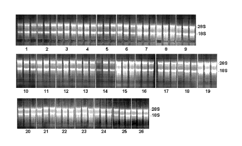
Figure 1 shows preparations of RNA from liver tissue on a gel as explained in
example 1. Lanes
on the gel correspond to sample numbers of table 1.
Figure 2 shows preparations of RNA from liver tissue on a gel as explained in
example 2. Lanes
on the gel correspond to sample numbers of table 2.
Figure 3 shows preparations of RNA from liver tissue on a gel as explained in
example 3. Lanes
on the gel correspond to sample numbers of table 3.
CA 02679172 2009-08-25
WO 2008/104564
PCT/EP2008/052371
Figure 4 shows preparations of RNA from intestine tissue on a gel as explained
in
example 4. Lanes on the gel correspond to sample numbers of table 4.
Figure 5 shows preparations of RNA from liver tissue on a gel as explained in
5 example 5. Lanes on the gel correspond to sample numbers of table 5.
Figure 6 shows preparations of RNA from liver tissue on a gel as explained in
example 6. Lanes on the gel correspond to sample numbers of table 6.
10 Figure 7 shows preparations of RNA from liver tissue on a gel as
explained in
example 7. Lanes on the gel correspond to sample numbers of table 7.
Figure 8 shows preparations of RNA from spleen tissue on a gel as explained in
example 8. Lanes on the gel correspond to sample numbers of table 8.
Figure 9 shows preparations of RNA from spleen tissue on a gel as explained in
example 9. Lanes on the gel correspond to sample numbers of table 9.
Figure 10 shows a fixed and stained intestine tissue, prepared according to
exam-
ple 10.
Figure 11 shows fixed and stained spleen tissue, prepared according to example
11.
Figure 12 shows fixed and stained spleen and kidney tissue, prepared according
to
example 11.
Figure 13 shows DNA, isolated from fixed spleen tissue an a gel, prepared and
isolated according to example 12.
CA 02679172 2009-08-25
WO 2008/104564
PCT/EP2008/052371
31
The invention is now described in more detail by the following examples. The
examples are provided for illustration only and should not be considered as
limit-
ing the invention to the shown embodiments.
Example 1
RNA isolation from tissue samples stabilised in different reagents according
to
composition A
Liver tissue from rat was cut into pieces of approximately 5x4x4mm directly
after
dissection. The samples were completely immersed into 2 to 4m1 of a fixation
solution according to composition A (table 1) in a 5m1 collection vessel made
of
polypropylene. Tissue samples were stored for 24h at ambient temperature.
RNA extraction was performed with a commercially available kit (RNeasy Mini,
QIAGEN) as described in the RNeasy Mini protocol for isolation of total RNA
from animal tissue. Tissue sample were cut into small pieces and placed into
2m1
microcentrifuge tubes. The weight of each piece of tissue was determined and
lysis buffer (Buffer RLT, QIAGEN) containing guanidine isothiocyanate (GITC)
with a volume of 350 1 per 10mg tissue was added along with a steel ball (5mm
).
Disruption and simultaneous homogenization were performed on a Mixer-Mill
(Tissue-Lyser, QIAGEN) with 20Hz for 2min. According to the state of the art
GITC lyses cells and precipitates proteins. Lysates were centrifuged using
14.000
rpm for 3min. 350 1 supernatant, representing approximately 10mg tissue were
transferred into a new tube, mixed with 1 volume (350 1) 70% ethanol, and ap-
plied on a silica membrane containing spin-column (RNeasy-Mini column). Lys-
ates were transferred through the membrane by centrifugation, thereby
adsorbing
the RNA to the membrane. Contaminants were removed by washing the mem-
brane twice with 350 1 GITC containing washing buffer RW1 (QIAGEN). Be-
tween the two washing steps residual DNA was removed from the membrane by
pipetting 10 1 DNase (approximately 30 Kunitz units) mixed with 70 1 buffer
RDD (QIAGEN) onto the membrane and incubating for 15min at ambient tem-
perature. After two more washing steps with 500 1 washing buffer RPE
(QIAGEN), containing Tris-Cl buffer and alcohol, the membrane was dried by
full speed centrifugation for lmin at 14.000 rpm. Finally the RNA was eluted
by
CA 02679172 2009-08-25
WO 2008/104564
PCT/EP2008/052371
32
pipetting 40 1 water followed by lmin incubation at ambient temperature and
centrifugation for lmin at 10.000 rpm. This elution step was repeated with
addi-
tional 40 1 water and both eluates were combined. All extractions were
performed
in triplicates.
The concentration of RNA was determined by measuring the absorbance at
260nm (A260) in a spectrophotometer. To ensure significance, eluates were di-
luted with 10mM Tris-Cl pH7.5 to show an absorbance A260 between 1 and 0.15.
Under these conditions an absorbance of 1 unit at 260nm corresponds to 44 iLig
RNA.
The integrity and size distribution of total RNA was analysed by denaturing
aga-
rose gel electrophoresis. For example 15 1 of eluates were mixed with 3 1
sample
buffer containing formaldehyde (FA) and bromophenol blue, incubated 10min at
70 C, chilled on ice and loaded on a 1.0% formaldehyde-agarose-MOPS gel
equilibrated with lx FA gel running buffer. Electrophoresis was performed for
90min and approximately 3 volts per cm length of the electrophoresis chamber.
RNA was visualized by ethidium bromide staining. The gel is shown as figure 1,
lanes on the gel correspond to sample numbers shown in table 1.
As shown in table 1, treating a tissue sample with a composition according to
composition A leads to high RNA yield comparable to tissue fixation with
RNAlater, if tissue is stored for no longer than 24h. Shown are examples with
different concentrations of component al and different additional components
and
concentrations of components a2 and a3 (table 1, 1-18 and 20-26) and RNAlater
as a reference (table 1, 19).
Analysis of integrity and size distribution showed, that the ribosomal bands
for
18S- and 28S rRNA appeared as sharp bands on the stained gel (figure 1). Since
the 28S rRNA band was approximately twice as intense as the 18S RNA band, no
smaller sized RNA and almost no smear were visible on the stained gel, one can
conclude that the RNA sample did not suffer from degradation during fixation
or
preparation.
CA 02679172 2009-08-25
WO 2008/104564
PCT/EP2008/052371
33
Table 1:
no. reagent composition RNA yield
from 10mg
tissue [jig]
1 70% Methanol, 10% glacial acetic acid, 10% PEG300, 10% triol-mixture
(25% 3 -methy1-1,3,5-pentanetriol, 75% 1,2,6-hexanetriol) 10% glacial
acetic acid 13,79
2 70% Methanol, 10% glacial acetic acid, 10% PEG300, 10% diethylene
glycol monoethyl ether acetate 15,20
3 70% Methanol, 10% glacial acetic acid, 10% PEG300, 10% 1,3-
butanediol 19,13
4 70% Methanol, 10% glacial acetic acid, 10% PEG300, 10% 1,4-
butanediol 20,26
70% Methanol, 10% glacial acetic acid, 20% PEG300 14,73
6 70% Methanol, 10% glacial acetic acid, 20% triol-mixture (25% 3-
methy1-1,3,5-pentanetriol, 75% 1,2,6-hexanetriol), 10% glacial acetic
acid 19,17
7 70% Methanol, 10% glacial acetic acid, 20% diethylene glycol mono-
ethyl ether acetate 29,14
8 70% Methanol, 10% glacial acetic acid, 20% 1,3-butanediol 29,90
9 70% Methanol, 10% glacial acetic acid, 20% 1,4-butanediol 28,08
60% Methanol, 10% glacial acetic acid, 20% triol-mixture (25% 3-
methy1-1,3,5-pentanetriol, 75% 1,2,6-hexanetriol) 10% glacial acetic
acid, 10% PEG300 17,53
11 60% Methanol, 10% glacial acetic acid, 20% Diethylene glycol mono-
ethyl ether acetate, 10% PEG300 17,64
12 60% Methanol, 10% glacial acetic acid, 20% 1,3-butanediol, 10%
PEG300 18,66
13 60% Methanol, 10% glacial acetic acid, 20% 1,4-butanediol, 10%
PEG300 17,97
14 60% Methanol, 10% glacial acetic acid, 30% PEG300 9,60
60% Methanol, 10% glacial acetic acid, 30% triol-mixture (25% 3-
methy1-1,3,5-pentanetriol, 75% 1,2,6-hexanetriol) 10% glacial acetic
acid 35,03
16 60% Methanol, 10% glacial acetic acid,30% diethylene glycol mono-
ethyl ether acetate 28,66
17 60% Methanol, 10% glacial acetic acid, 30% 1,3-butanediol 26,92
18 60% Methanol, 10% glacial acetic acid, 30% 1,4-butanediol 18,26
19 RNAlater 39,97
70% Methanol, 10% propionic acid, 10% PEG300, 10% LiC1 (1M) 35,57
CA 02679172 2009-08-25
WO 2008/104564
PCT/EP2008/052371
34
no. reagent composition RNA yield
from 10mg
tissue [jig]
21 70% Methanol, 10% propionic acid, 10% PEG300, 10% LiC1 (100mM)
32,88
22 70% Methanol, 10% propionic acid, 10% PEG300, 10% 1,3-butanediol
40,37
23 70% Methanol, 10% propionic acid, 10% LiC1 (1M), 10% 1,3-butanediol
36,19
24 70% Methanol, 10% propionic acid, 10% LiC1 (100mM), 10% 1,3-
butanediol 33,14
25 60% Methanol, 10% propionic acid, 10% PEG300, 10% LiC1 (1M), 10%
1,3 -butanediol 29,61
26 60% Methanol, 10% propionic acid, 10% PEG300, 10% LiC1 (100mM),
10% 1,3 -butanediol 31,79
Example 2
RNA isolation from tissue stabilised with different reagents according to
composi-
tion A as well as reagents containing ethanol as major component
Liver tissue from rat was cut into pieces of 5x4x4mm directly after
dissection.
The samples were completely immersed into 2 to 4m1 of a fixation solution con-
taining ethanol (table 2, 4-6) or methanol according to composition A (table
2, 1-
3), in a 5m1 collection vessel made of polypropylene. Tissue samples were
stored
for 24h at ambient temperature.
RNA extraction and analysis of yield and integrity was performed as described
in
example 1. All extractions were performed in triplicates. The gel is shown as
fig-
ure 2, lanes on the gel correspond to sample numbers shown in table 2.
As shown in table 2, RNA yield was exceptionally high when tissue samples were
stabilised with reagents containing ethanol as major component. Despite the
high
yield, agarose gel electrophoresis showed that the RNA from ethanol containing
stabilisation reagents suffered various degree of degradation during storage.
In
contrast to compositions 1-3 18S and/or 28S ribosomal RNA could not be stained
as sharp distinct bands. Instead a smear of different degree and bands of
smaller
sized RNAs became visible (figure 2). In case of the ethanol containing
reagents it
can be assumed that the yield determination was artificially high due to the
fact
that smaller fragments of ribonucleic acids show higher absorbance at 260nm.
CA 02679172 2009-08-25
WO 2008/104564
PCT/EP2008/052371
Table 2:
no. reagent composition RNA yield
from 10mg
tissue [jig]
1 60% Methanol, 10% PEG300, 25% Diethylene glycol monoethyl ether
acetate, 5% Propionic acid 30,66
2 70% Methanol, 25% Diethylene glycol monoethyl ether acetate, 5%
Propionic acid 38,26
3 70% Methanol, 20% Diethylene glycol monoethyl ether acetate, 10%
Propionic acid 26,55
4 70% Ethanol 47,29
5 Boonfix (primary ingredient ethanol ¨ according to the supplier)
50,27
6 Finefix (working solution contains 70% ethanol) 52,96
Example 3
5 RNA isolation from tissue stabilised with different reagents containing
methanol
as major component with or without water
Liver tissue from rat was cut into pieces of approximately 5x4x4mm directly af-
ter dissection. The samples were completely immersed into 2 to 4m1 of a
fixation
solution containing methanol in an aqueous solution (table 3, 1) or methanol
in a
10 non-aqueous reagent according to composition A (table 3, 2). Tissue
samples
were stored for an extensive time period of 4 days at ambient temperature.
RNA extraction and analysis of yield and integrity was performed as described
in
example 1. All extractions were performed in triplicates. The gel is shown as
fig-
ure 3, lanes on the gel correspond to sample numbers shown in table 3.
As shown in table 3 and figure 3 (no. 2) even after 4 days storage intact RNA
could be isolated from tissue samples stored in non-aqueous reagents according
to
composition A. The yield was decreased compared to the results shown in exam-
ple 1 and 2, but 28S- and 18S rRNA were still visible as intact bands without
smear or smaller, distinct RNA bands. In contrast the RNA from a tissue sample
stabilised with an aqueous reagent was highly degraded and low concentrated (
no. 1 table 3 and figure 3).
CA 02679172 2009-08-25
WO 2008/104564
PCT/EP2008/052371
36
Table 3:
no. reagent composition RNA yield
from 10mg
tissue [jig]
1 60% Methanol, 30% water, 10% glacial acetic acid 2,25
2 60% Methanol, 30% triol-mixture (25% 3-methyl-1,3,5-pentanetriol,
75% 1,2,6-hexanetriol) 10% glacial acetic acid 10,98
Example 4
RNA isolation from tissue stabilised with different reagents with or without
trans-
fer into a second reagent with composition B according to the invention
Intestine tissue from rat was cut into pieces of approximately 5x4x4mm
directly
after dissection. The samples were completely immersed into 5m1 of different
reagents according to composition A and stored at ambient temperature. Samples
were either stored for 7 days (table 4, 1-4) or transferred after 4h hours
into 5m1 of
a reagent according to composition B and stored within this reagent for 7 days
(table 4, 5-8).
RNA extraction and analysis of yield and integrity was performed as described
in
example 1. All extractions were performed in triplicates. The gel is shown as
fig-
ure 4, lanes on the gel correspond to sample numbers shown in table 4.
As shown in table 4 RNA yield dropped down significantly when samples were
not transferred into the second reagent compared to those samples, which were
transferred into a reagent according to composition B according to the
invention.
Agarose gel electrophoresis showed that even after 7 days storage intact RNA
could be isolated from tissue samples stored in reagents according to
composition
A and B and treated according to the invention. The gel confirmed the low
yield
when the transfer was omitted (figure 4).
CA 02679172 2009-08-25
WO 2008/104564
PCT/EP2008/052371
37
Table 4:
no. reagent composition A reagent composition B RNA yield
from 10mg
tissue [jig]
1 70% Methanol, 25% Diethylene
glycol monoethyl ether acetate,
5% Propionic acid - 3,7
2 70% Methanol, 25% 1,3-
Butanediol, 5% Propionic acid - 3,5
3 90% Methanol, 10% PEG300 - 4,4
4 70% Methanol, 30% PEG300 - 2,4
70% Methanol, 25% Diethylene 70% Ethanol, 30% 1,3-Butanediol
glycol monoethyl ether acetate,
5% Propionic acid 8,7
6 70% Methanol, 25% 1,3- 70% Ethanol, 30% 1,3-Butanediol
Butanediol, 5% Propionic acid 6,8
7 90% Methanol, 10% PEG300 70% Ethanol, 30% 1,3-Butanediol 8,7
8 70% Methanol, 30% PEG300 70% Ethanol, 30% 1,3-Butanediol 12,6
Example 5:
5 RNA isolation from tissue stabilised with reagents according to
composition A
and B according to the invention
Liver tissue from rat was cut into pieces of approximately 5x4x4mm directly
after
dissection. The samples were completely immersed into 10m1 of a reagent accord-
ing to composition A and stored at ambient temperature. One sample was stored
for 3 days (table 5, 10), the others were transferred after 2h hours into 10m1
of
different reagents according to composition B and stored within these reagents
for
3 days (table 5, 1-8). As a reference one sample was stored in RNAlater (table
5,
9).
RNA extraction and analysis of yield and integrity was performed as described
in
example 1. All extractions were performed in triplicates. The gel is shown as
fig-
ure 5, lanes on the gel correspond to sample numbers shown in table 5
This example shows once more the effect of tissue treatment on RNA stability
according to the invention, i.e. the transfer from a reagent according to
composi-
CA 02679172 2009-08-25
WO 2008/104564
PCT/EP2008/052371
38
tion A to a reagent according to composition B. In samples where transfer took
place the RNA yield remained high in a range comparable to the reference with
RNAlater (table 5, 1-7 and 9). No visible RNA degradation could be observed
even after 3 days of storage (figure 5, 1-7 and 9). In contrast, when the
transfer
was omitted, yield dropped down significantly and RNA degradation became
visible by increased smear and a diminished 28S rRNA band (table 5, 10 and fig-
ure 5, 10). A similar effect could be observed when the sample was transferred
into Boonfix (see above), which according to the supplier is a reagent
consisting
mainly of ethanol but is balanced with water (table 5, 8 and figure 5, 8).
CA 02679172 2009-08-25
WO 2008/104564
PCT/EP2008/052371
39
Table 5:
no. reagent composition A reagent composition B RNA yield
from 10mg
tissue [jig]
1 70% Methanol, 10% Glacial acetic
acid/ 10% PEG300, 10% 1,3-
Butanediol 70% Ethanol, 30% 1,3-Butanediol 35,90
2 70% Methanol, 10% Glacial acetic
acid/ 10% PEG300, 10% 1,3- 70% Ethanol, 1% 1% Glacial acetic
Butanediol acid, 29% 1,3-Butanediol 33,97
3 70% Methanol, 10% Glacial acetic
acid/ 10% PEG300, 10% 1,3- 70% Ethanol, 5% 1% Glacial acetic
Butanediol acid, 25% 1,3-Butanediol 19,93
4 70% Methanol, 10% Glacial acetic
acid/ 10% PEG300, 10% 1,3- 70% Ethanol, 10% 1% Glacial ace-
Butanediol tic acid, 20% 1,3-Butanediol 18,44
70% Methanol, 10% Glacial acetic
acid/ 10% PEG300, 10% 1,3-
Butanediol 60% Ethanol, 40% 1,3-Butanediol 41,68
6 70% Methanol, 10% Glacial acetic
acid/ 10% PEG300, 10% 1,3- 60% Ethanol, 1% 1% Glacial acetic
Butanediol acid, 39% 1,3-Butanediol 33,86
7 70% Methanol, 10% Glacial acetic
acid/ 10% PEG300, 10% 1,3- 60% Ethanol, 5% 1% Glacial acetic
Butanediol acid, 35% 1,3-Butanediol 21,02
8 70% Methanol, 10% Glacial acetic
acid/ 10% PEG300, 10% 1,3-
Butanediol Boonfix 14,59
9 RNAlater - 37,25
70% Methanol, 10% Glacial acetic -
acid/ 10% PEG300, 10% 1,3-
Butanediol 6,00
Example 6:
RNA isolation from tissue stabilised according to the invention
5 Liver tissue from rat was cut into pieces of approximately 5x4x4mm
directly after
dissection. The samples were completely immersed into 2m1 of a reagent accord-
ing to a composition of step ii), stored at ambient temperature for 4 hours,
trans-
ferred into different reagents according to composition B and stored for
additional
2 days (table 6, 3-9). In addition one sample was not transferred but stored
within
CA 02679172 2009-08-25
WO 2008/104564
PCT/EP2008/052371
the reagent composition according to step ii) (table 6, 10), one sample was
trans-
ferred into 100% ethanol (table 6, 1) and one sample was transferred into 70%
ethanol (table 6, 2).
RNA extraction and analysis of yield and integrity was performed as described
in
5 example 1. All extractions were performed in triplicates. The gel is
shown as fig-
ure 6, lanes on the gel correspond to sample numbers shown in table 6.
This example demonstrates the stabilising effect according to the invention of
transfer into different reagents according to composition B. Shown are
different
10 reagents with different additional components (a2) and
concentrations of organic
acids (a3) (table 6, 3-9 and figure 6, 3-9). Balancing of the second reagent
with
water led to decreased RNA yield and increased smear on the agarose gel,
indicat-
ing advanced degradation (table and figure 6, 2). Omitting the transfer led to
de-
creased RNA yield (table 6 and figure 6, 10). Transfer into 100% ethanol did
not
15 lead to any degradation and resulted in high RNA yield (table 6 and
figure 6, 1).
However it is commonly accepted that tissue samples stored in 100% ethanol un-
dergo hardening and shrinkage. As a consequence the specimens become brittle
which leads to artefacts when subsequent histological work methods are con-
ducted.
25
CA 02679172 2009-08-25
WO 2008/104564
PCT/EP2008/052371
41
Table 6:
no. reagent composition according to reagent composition B RNA yield
ii) from 10mg
tissue [jig]
1 60% methanol, 30% chloroform,
10% glacial acetic acid 100% ethanol 36,51
2 60% methanol, 30% chloroform,
10% glacial acetic acid 70% ethanol, 30% water 8,16
3 60% methanol, 30% chloroform,
10% glacial acetic acid 70% ethanol, 30% 1,3-ethanediol 29,50
4 60% methanol, 30% chloroform, 70% ethanol, 30% diethyleneglycol
10% glacial acetic acid monoethyl etheracetate 33,22
60% methanol, 30% chloroform,
10% glacial acetic acid 70% ethanol, 30% PEG300 24,29
6 60% methanol, 30% chloroform,
10% glacial acetic acid 70% ethanol, 30% 1,2,6-hexanetriol 29,22
7 60% methanol, 30% chloroform, 70% ethanol, 5% glacial acetic acid,
10% glacial acetic acid 25% 1,3-butanediol 9,41
8 70% ethanol, 5% glacial acetic acid,
60% methanol, 30% chloroform, 25% diethyleneglycol monoethyl
10% glacial acetic acid ether acetate 16,74
9 60% methanol, 30% chloroform, 70% ethanol, 5% glacial acetic acid,
10% glacial acetic acid 25% 1,2,6-hexanetriol 14,24
60% methanol, 30% chloroform,
10% glacial acetic acid - 4,48
Example 7:
5 RNA isolation from tissue stabilized according to the invention,
replacement of
chloroform
Liver tissue from rat was cut into pieces of approximately 5x4x4mm directly
after
dissection. The samples were completely immersed into 5m1 of either a chloro-
form containing reagent, a reagent in which chloroform is replaced by
correspond-
10 ing amounts of water or methanol or a reagent according to a composition
of step
ii), respectively, and stored at ambient temperature for 48 hours (table 7, 1-
4). In
addition one sample was transferred into a reagent according to composition B
after 3 hours and stored for additional 45 hours (table 7, 5).
CA 02679172 2009-08-25
WO 2008/104564
PCT/EP2008/052371
42
RNA extraction and analysis of yield and integrity was performed as described
in
example 1. All extractions were performed in triplicates. The gel is shown as
fig-
ure 7; lanes on the gel correspond to sample numbers shown in table 7.
This example demonstrates that chloroform within a fixation reagent can not be
replaced by corresponding amounts of methanol or water (table 7 and figure 7,
1-
3). Replacing chloroform with methanol or water led to decreased RNA yields
and increased smear on the agarose gel, indicating advanced degradation (table
and figure 7, 2 and 3). In order to achieve a similar stabilizing effect
chloroform
has to be replaced by at least one additional additive according to (a2)
(table and
figure 7, 4). However the best stabilizing effect with the highest RNA yield
and
least RNA degradation could be achieved by transferring the tissue sample
after 3
hours into a reagent according to composition B (table and figure 7, 5).
Table 7:
no. reagent composition reagent composition B RNA yield
from 10mg
tissue [jig]
1 60% methanol, 30% chloroform,
10% glacial acetic acid - 8,73
2 60% methanol, 30% water, 10%
glacial acetic acid - 3,04
3 90% methanol, 10% glacial acetic
acid - 0,96
4 60% methanol, 30% Diethylene-
glycol monoethyl ether acetate,
10% glacial acetic acid - 9,42
5 60% methanol, 30% Diethylene-
glycol monoethyl ether acetate,
10% glacial acetic acid 70% Ethanol, 30% 1,3-Butandiol 25,13
CA 02679172 2009-08-25
WO 2008/104564
PCT/EP2008/052371
43
Example 8:
RNA isolation from paraffin embedded tissue stabilised with reagents according
to composition A and B according to the invention, and processed by a conven-
tional method
Spleen tissue from rat was cut into pieces of approximately 4x4x4mm directly
after dissection. The samples were completely immersed into 5m1 of a reagent
according to composition A and stored at ambient temperature for 24 hours.
After
this incubation period samples were either directly processed (table 8, 1 and
4) or
transferred into a reagent according to composition B, stored for additional 4
days
at ambient temperature (table 8, 2 and 5) or at 4 C (table 8, 3 and 6) and
finally
processed.
Processing comprising of dehydration, clearing, infiltration and embedding,
was
performed manually following standard protocols. The specimens were placed
into standard processing cassettes (histosettes) and dehydrated by transfer
through
increasing concentrations of ethanol, i.e. incubation in 70, 80, two times 96%
ethanol, 60min each. Clearing as the transition step between dehydration and
infil-
tration with the embedding medium was performed by incubation twice for 60min
in 100% xylene. Tissue cavities and cells were saturated in liquid paraffin
(low
melting Paraplast XTRA, Roth Inc.) at 56 C for approximately 12 hours. To pro-
vide the necessary support for microtomy specimen were embedded into the same
paraffin used for infiltration.
Freshly cut sections of the paraffin blocks were used as starting material for
RNA
extraction. Paraffin blocks were trimmed with a rotary microtome (Leica
R1V12245) and 10 slices with a thickness of 10 m each where cut off from each
specimen and collected in a microcentrifuge tube. Deparaffination was
performed
by adding lml of xylene, vortexing and centrifugation for 2min at 14.000 rpm.
The supernatant was removed and the pellet was dissolved with lml of 100%
ethanol. After centrifugation for 2min at 14.000 rpm the supernatant was
removed
and the ethanol washing step was repeated. After centrifugation and removal of
the ethanol the pellet was dissolved within 350 1 Buffer RLT (QIAGEN) contain-
ing 0.143 M B-mercaptoethanol. For homogenisation the lysate was loaded on a
QIAshredder spin column (QIAGEN) and centrifuged for 3min at 14.000 rpm.
CA 02679172 2009-08-25
WO 2008/104564
PCT/EP2008/052371
44
The flowthrough was mixed with 1 volume of 70% ethanol (350 1) and loaded on
a RNeasy MinElute spin column (QIAGEN). The lysates were transferred through
the membrane by centrifugation, thereby absorbing the RNA to the membrane.
Washing steps with buffer RW1 and RPE as well as on membrane DNase diges-
tion were performed as described in example 1. For elution 15 1 of water were
pipetted on the silica membrane. RNA was eluted after lmin incubation at ambi-
ent temperature by lmin centrifugation at 14.000rpm.
All extractions were performed in triplicates and RNA analysis of yield and
integ-
rity was performed as described in example 1. The gel is shown as figure 8,
lanes
on the gel correspond to sample numbers shown in table 8.
Analysis of RNA integrity by agarose gel electrophoresis (figure 8) revealed
that
from paraffin embedded tissue samples treated according to the invention full
length RNA can be isolated. Even incubation for up to 12 hours in liquid
paraffin,
necessary for specimen embedding did not lead to major RNA degradation. Bands
for 28S- and 18S- rRNA were still visible as sharp and distinct bands (figure
8).
Variation of RNA yield (table 8) can be explained by different amount of
starting
material. In contrast to the RNA extractions from soft tissue, starting
material
within the slices could not be determined and normalized to e.g. 10mg per
sample.
Significant differences between storage of tissue in reagent according to
composi-
tion B at 4 C or ambient temperature could not be observed.
30
CA 02679172 2009-08-25
WO 2008/104564
PCT/EP2008/052371
Table 8:
no. reagent composition A reagent composition B RNA
yield from
10 slices a
101.tm [jig]
1 60% Methanol, 5% Propionic acid,
10% PEG300, 25% Diethylene-
glycol monoethyl ether acetate - 3,53
2 60% Methanol, 5% Propionic acid,
10% PEG300, 25% Diethylene-
glycol monoethyl ether acetate 70% Ethanol, 30% 1,3-Butanediol 1,79
3 60% Methanol, 5% Propionic acid,
10% PEG300, 25% Diethylene-
glycol monoethyl ether acetate 70% Ethanol, 30% 1,3-Butanediol 1,25
4 70% Methanol, 10% Propionic
acid, 20% Diethyleneglycol mono-
ethyl ether acetate - 2,70
5 70% Methanol, 10% Propionic
acid, 20% Diethyleneglycol mono-
ethyl ether acetate 70% Ethanol, 30% 1,3-Butanediol 1,53
6 70% Methanol, 10% Propionic
acid, 20% Diethyleneglycol mono-
ethyl ether acetate 70% Ethanol, 30% 1,3-Butanediol 1,39
Example 9:
RNA isolation from paraffin embedded tissue stabilised according to the inven-
5 tion, processed by microwave energy
Spleen tissue from rat was cut into pieces of approximately 4x4x4mm directly
after dissection. The samples were completely immersed into 5m1 of a reagent
according to composition A. Samples were stored at ambient temperature for 24
hours (table 9, 2 and 3) or transferred after 30min into a reagent according
to
10 composition B and stored for 24 hours at ambient temperature (table 9, 4
and 5).
In parallel one sample was immersed into 5m1 of Boonfix (table 9, 1).
After 24 hours samples were placed into standard processing cassettes (histo-
settes) and processed on a RHS-1 microwave histoprocessor (Milestone).
For processing a four step standard protocol was applied, involving dewatering
15 samples in 100% ethanol and heating to 65 C by microwave energy. The
ethanol
was replaced by isopropanol and after air drying by vacuum and heat, specimens
were infiltrated in liquid paraffin (low melting Paraplast XTRA, Roth Inc.) in
a
CA 02679172 2009-08-25
WO 2008/104564
PCT/EP2008/052371
46
final step with heat by microwave energy and simultaneous vacuum (protocol
steps see table 10). To provide the necessary support for microtomy specimen
were manually embedded into the same paraffin used for infiltration.
Paraffin embedded tissue blocks were stored at ambient temperature for 5 weeks
prior to RNA extraction. RNA was extracted from 10 freshly prepared slices of
m thickness. Deparaffination, RNA extraction and analysis of yield and integ-
rity were performed as described in example 8 (and 1 respectively). All extrac-
tions were performed in duplicates. The gel is shown as figure 9, lanes on the
gel
correspond to sample numbers shown in table 9.
As shown in figure 9, RNA from tissue samples stabilised according to the
inven-
tion and processed by microwave energy was of high quality with no signs of
deg-
radation (figure 9, 2-5). Ribosomal bands of 28S- and 18S rRNA were sharp,
with
neither smear nor smaller bands detectable, in contrast to the sample treated
with
Boonfix (figure 9, 1).
Table 9:
no. reagent composition reagent composition B RNA yield
from 10 slices
a 101.tm [jig]
1 Boonfix (primary ingredient etha-
nol ¨ according to the supplier) - 2,43
2 70% Methanol, 5% Propionic acid,
25% Diethylene glycol monoethyl
ether acetate - 4,54
3 60% Methanol, 5% Propionic acid,
25% Diethylene glycol monoethyl
ether acetate, 10% PEG(300) - 4,55
4 70% Methanol, 5% Propionic acid, 70% Ethanol, 30% 1,3-Butanediol
25% Diethylene glycol monoethyl
ether acetate 2,11
5 60% Methanol, 5% Propionic acid, 70% Ethanol, 30% 1,3-Butanediol
25% Diethylene glycol monoethyl
ether acetate, 10% PEG(300) 3,55
CA 02679172 2009-08-25
WO 2008/104564
PCT/EP2008/052371
47
Table 10:
Step Incubation medium Duration Microwave Vacuum Stirring
[min] heating [mbar]
1 Ethanol 18 up to 65 C no yes
2 Is oprop anol 19 up to 68 C no yes
3 Airdry 0.5 70 C 500 no
4 Liquid paraffin 22 70 C down to 100 yes
Example 10:
Histological analysis of tissue samples stabilised with reagents according to
com-
position A and B according to the invention; processed by a conventional
method
Small intestine tissue from rat was cut into pieces with approximately 6mm in
length directly after dissection. The sample was completely immersed into 5m1
of
a reagent according to composition A. After 4 hours at ambient temperature the
sample was transferred into a reagent according to composition B and stored
for
hours at ambient temperature (table 11, A). In parallel one sample was im-
mersed into 5m1 of a 10% solution of neutral buffer formalin (NBF; table 11,
B).
After 24 hours samples were placed into standard processing cassettes (histo-
settes) and processed manually following a standard protocol starting with
incuba-
15 tion twice in 100% ethanol for 180min each. Clearing was performed by
incuba-
tion twice for 60min in 100% xylene. Infiltration in liquid paraffin (low
melting
Paraplast XTRA, Roth Inc.) was performed at 65 C for approximately 12 hours
followed by embedding into the same paraffin.
Sections of 6iLtm thickness were sliced with a rotary microtome (Leica RM2245)
20 and mounted on slides. Haematoxylin and eosin staining was performed
manually
with dyes from Sigma Inc., following a standard protocol (table 12).
As shown in figure 10 (100-fold magnification) the section from a tissue
sample
stabilised according to the invention (figure 10, A) displayed a similar
morpho-
logical preservation as the section preserved in 10% neutral buffered formalin
(figure 10, B).
CA 02679172 2009-08-25
WO 2008/104564
PCT/EP2008/052371
48
Table 11:
no. reagent composition reagent composition B
A 70% Methanol, 10% Propionic 70% Ethanol, 30% 1,3-Butanediol
acid, 20% Diethylene glycol
monoethyl ether acetate
B NBF, 10% of neutral buffered _
formalin
Table 12:
Incubation/Medium Duration [min]
Incubation at 70 C 10
Rotihistol (Xylene substitute, Roth Inc.) 10
Rotihistol 10
96% Ethanol 5
80% Ethanol 5
70% Ethanol 5
60% Ethanol 5
water 3
Mayer's Haematoxylin 5
water 0.5
70% Ethanol containg 1% HC1 0.5
water 5
Eosin 5
water 1
96% Ethanol 3
96% Ethanol 5
100% Isopropanol 10
Rotihistol 10
Rotihistol 10
Example 11:
Histological analysis of tissue samples stabilised with different reagents
according
to composition A according to the invention; processed by a conventional
method
Spleen and kidney from rat were cut into pieces of approximately 3x5x5 mm di-
rectly after dissection. The samples were completely immersed into 10m1 of a
CA 02679172 2009-08-25
WO 2008/104564
PCT/EP2008/052371
49
reagent according to composition A or in 10% neutral buffered formalin (table
13). After 24 hours at ambient temperature the samples were processed and
stain-
ed. Processing of the samples was performed approximately 30 hours after
dissec-
tion in a Leica TP1020 Tissue Processor. Paraffin embedded tissue samples were
cut in 6 m slices. Only the kidney sample preserved in 10% formalin was cut in
4 m slices. Haematoxylin and eosin staining was performed on a Leica
Autostainer following standard protocols identical or similar to the
procedures
described in detail in examples 8 and 10.
Figure 11 shows sections of spleen stained with haematoxylin and eosin in a
100-
fold magnification. The results demonstrate similar morphological preservation
between the tissue sample preserved in a reagent according to composition A
(fig-
ure 11, A) and the sample preserved in 10% neutral buffered formalin (figure
11,
B). The same result holds true for kidney, depicted in figure 12 (A) or (B);
respec-
tively, with 200-fold magnification. The only difference observed addresses
eryth-
rocytes. In case of the fixation reagent according to composition A the
erythro-
cytes do not contain haemoglobin.
Table 13:
no. reagent composition
A 70% Methanol, 10% Glacial acetic acid, 10% Diethylene glycol
monoethyl ether ace-
tate, 10% PEG300
B NBF, 10% of neutral buffered formalin
Example 12:
DNA isolation from paraffin embedded tissue, stabilised with reagents
according
to composition A according to the invention, processed by a conventional
method
Spleen tissue from rat was cut into pieces of approximately 2x5x5mm directly
after dissection. The samples were completely immersed into 5m1 of different
reagents according to composition A (table 14) and stored at ambient
temperature
for 24 hours. After this incubation period samples were manually processed as
CA 02679172 2009-08-25
WO 2008/104564
PCT/EP2008/052371
described in example 7. Paraffin embedded tissue blocks were stored at ambient
temperature for 5 weeks prior to DNA extraction.
For DNA extraction, paraffin blocks were cut into half Pure paraffin was re-
moved and the tissue deparaffinized with xylene and ethanol (see also example
8).
5 DNA extraction was performed with a commercially available kit
(DNeasy Tissue
kit, QIAGEN) as described in the DNeasy Tissue protocol for 'purification of
to-
tal DNA from animal tissues'. The pellet resulting from deparaffination was
dis-
solved in 180 1 buffer ATL and a steel ball (5mm ) was added. Disruption and
simultaneous homogenization was performed on a Mixer-Mill (Tissue-Lyser,
10 QIAGEN) with 20Hz for 15seconds. The lysates were frozen at -20 C
and further
processed after 24 hours by adding 40 1 proteinase K (activity 600 mAU/m1).
Digestion was performed for one hour at 55 C with constant gentle mixing of
the
samples. RNA was removed from the samples by adding 4 1 RNase A
(100mg/m1) and incubation for 2min at ambient temperature. After adding 200 1
15 lysis buffer AL (QIAGEN), incubation for another 10min at 70 C and
adding
200 1 ethanol (100%) the lysates were applied on a silica membrane containing
DNeasy Mini spin column. Lysates were transferred through the membrane by
centrifugation (lmin, 8000 rpm), thereby absorbing the DNA to the membrane.
Contaminants were removed by washing the membrane with 500 1 AW1 and
20 500 1 AW2 (QIAGEN). After the last washing step the membrane was
dried by
full speed centrifugation for 3min at 14.000 rpm. Finally the DNA was eluted
by
pipetting 100 1 water followed by lmin incubation at ambient temperature and
centrifugation for lmin at 10.000 rpm. This elution step was repeated with
another
100 1 water and both eluates were combined.
25 The concentration of DNA was determined by measuring the absorbance
at
260nm (A260) in a spectrophotometer. For DNA an absorbance of 1 unit at
260nm corresponds to 50 iLig DNA.
The integrity and size of total DNA was analysed by agarose gel
electrophoresis.
400ng DNA in 15 1 volume were mixed with 5 1 loading buffer (containing 50%
30 glycerol and bromophenol blue). The samples were applied to 0.8%
agarose gels
in lx TBE buffer. Electrophoresis was run for 120min and approximately 3.3
CA 02679172 2009-08-25
WO 2008/104564
PCT/EP2008/052371
51
Volts per cm length of the electrophoresis chamber. DNA was visualised by
ethidium bromide staining.
The DNA extracted from tissue samples preserved in a reagent according to com-
position A, processed, embedded into paraffin blocks and stored for 5 weeks
was
of high molecular weight when extracted with the QIAamp DNeasy procedure.
Figure 13 shows a gel electrophoresis with 400ng DNA from each sample (lanes
correspond to sample numbers of table 14). Typical yields are listed in table
14.
Table 14:
no. reagent composition A DNA
yield [jig]
1 70% Methanol, 10% Propionic acid, 20% PEG300 50,10
2 70% Methanol, 10% Propionic acid, 20% triol-mixture (25% 3-Methy1-
1,3,5-
pentanetriol, 75% 1,2,6-Hexanetriol) 41,90
3 70% Methanol, 10% Propionic acid, 20% Diethylene glycol monoethyl
ether
acetate 17,80
4 70% Methanol, 10% Propionic acid, 20% 1,3-Butanediol 33,10
5 70% Methanol, 10% Propionic acid, 20% 1,4-Butanediol 61,30