Note: Descriptions are shown in the official language in which they were submitted.
CA 02680070 2009-09-04
WO 2008/121649
PCT/US2008/058260
1
INSERTION SYSTEM FOR CORNEAL IMPLANTS
FIELD OF THE INVENTION
[0001] The field of the invention relates generally to corneal implants,
and more
particular, to insertion systems for corneal implants.
BACKGROUND INFORMATION
[0002] As is well known, abnormalities in the human eye can lead to
vision
impairment. Some typical abnormalities include variations in the shape of the
eye, which
can lead to myopia (near-sightedness), hyperopia (far-sightedness) and
astigmatism as
well as variations in the tissue present throughout the eye, such as a
reduction in the
elasticity of the lens, which can lead to presbyopia. A variety of
technologies have been
developed to try and address these abnormalities, including corneal implants.
[0003] Corneal implants can correct vision impairment by altering the
shape of the
cornea. Corneal implants can be classified as an onlay or an inlay. An onlay
is an implant
that is placed over the cornea such that the outer layer of the cornea, e.g.,
the epithelium,
can grow over and encompass the implant. An inlay is an implant that is
surgically
implanted into the cornea beneath a portion of the corneal tissue by, for
example, cutting a
flap in the cornea and inserting the inlay beneath the flap. Both inlays and
outlays can
alter the refractive power of the cornea by changing the shape of the anterior
cornea, by
having a different index of refraction than the cornea, or both. Since the
cornea is the
strongest refracting optical element in the human ocular system, altering the
cornea's
anterior surface is a particularly useful method for correcting vision
impairments caused
by refractive errors.
[0004] There is a need for improved apparatuses, systems and methods for
storing
a corneal implant prior to use and for retrieving the corneal implant from
storage during a
surgical procedure. There is also a need for improved apparatuses, systems and
methods
for delivering a corneal implant to the cornea and for precisely depositing
the corneal
implant at a desired location in or on the cornea without damaging the corneal
implant.
SUMMARY
[0005] Provided herein are apparatuses, systems and methods for storing
and
retrieving a corneal implant and for delivering the corneal implant in or on
the cornea.
CA 02680070 2013-11-19
2
[0006] In an embodiment, an insertion system comprises an inserter for
delivering a
corneal implant to a desired location in or on the cornea. The inserter
comprises an elongated
body having a distal end and a proximal end. The elongated body has a holding
space at its distal
end for holding the corneal implant to be delivered. The holding space is
formed between a top
distal portion and a bottom distal portion of the elongated body. In a
preferred embodiment, a
solution, e.g., saline, substantially fills the holding space with the corneal
implant to keep the
implant hydrated and to hold the implant in the holding space by the surface
tension of the
solution. The elongated body of the inserter may also have a curved portion
that follows the
curvature of the cornea and a clearance bend that provides clearance between
the inserter and a
facial feature, e.g., nose, of the patient.
[0007] In an embodiment, the corneal implant is preloaded in the holding
space of the
inserter and the preloaded inserter is stored in a storage container filled
with storage fluid, e.g.,
saline, until use. In one embodiment, a cap is placed on the distal end of the
inserter after the
implant is preloaded. The cap encloses the holding space of the inserter to
prevent the corneal
implant from moving out of the holding space in the storage fluid during
storage. By preloading
the implant in the inserter, the surgeon does not have to separately retrieve
the implant and place
the implant in the inserter, which is difficult due to the small size and
delicate nature of the
implant.
[0008] A method of delivering a corneal implant according to an
embodiment includes
positioning an inserter with the corneal implant at a desired location in or
on the cornea. At the
desired location, the corneal implant is held down in the holding space of the
inserter by a
surgical tool, e.g., cannula. The surgical tool accesses the implant in the
holding space through a
slot in the inserter. While the corneal implant is held down by the surgical
tool, the inserter is
retracted to release the corneal implant from the inserter and deposit the
corneal implant at the
desired location. By holding down the implant at the desired location and
retracting the inserter
to release the implant, the surgeon is able to precisely deposit the implant
at the desired location.
[0009] Other systems, methods, features and advantages of the invention
will be or will
become apparent to one with skill in the art upon examination of the following
figures and
detailed description. The scope of the claims should not be limited by
particular embodiments set
forth herein, but should be construed in a manner consistent with the
specification as a whole.
CA 02680070 2009-09-04
WO 2008/121649
PCT/US2008/058260
3
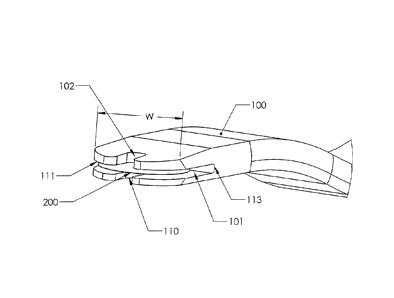
BRIEF DESCRIPTION OF THE FIGURES
[0010] FIG. 1 shows a perspective view of an insertion system comprising
an
inserter and a cap according to an embodiment of the present invention.
[0011] FIG. 2 shows a perspective view of the cap placed on the inserter
according
to an embodiment of the present invention.
[0012] FIG. 3 shows a side view of the distal end of the inserter
according to an
embodiment of the present invention.
[0013] FIG. 4 shows a close-up perspective view of the distal end of the
inserter
according to an embodiment of the present invention.
[0014] FIG. 5A shows the inserter depositing a corneal implant on the
cornea
according to an embodiment of the present invention.
[0015] FIG. 5B shows a close-up of the inserter depositing the corneal
implant on
the cornea.
[0016] FIG. 5C shows the inserter depositing a corneal implant on an
interior
surface of the cornea exposed by forming a flap in the cornea according to an
embodiment
of the invention.
[0017] FIG. 5D shows the inserter depositing a corneal implant within a
pocket
formed in the cornea according to an embodiment of the present invention.
[0018] FIG. 6 shows the inserter and cap stored in a container filled
with storage
fluid according to an embodiment of the present invention.
[0019] FIG. 7 shows a perspective view of the inserter with a luer lock
attached to
the proximal end of the inserter according to an embodiment of the present
invention.
[0020] FIG. 8 shows a perspective view of the inserter with a syringe
connected to
the proximal end of the inserter according to an embodiment of the present
invention.
[0021] FIG. 9 shows a perspective view of an inserter according to
another
embodiment of the present invention.
[0022] FIG. 10 shows a back view of the distal end of the inserter
according to an
embodiment of the present invention.
[0023] FIG. 11 shows the inserter depositing a corneal implant on the
cornea
according to an embodiment of the present invention.
DETAILED DESCRIPTION
[0024] Figures 1-5 show an insertion system according to an embodiment
that is
particular suited for delivering a corneal implant, e.g., inlay, in or on the
cornea. The
CA 02680070 2009-09-04
WO 2008/121649
PCT/US2008/058260
4
insertion system is also suited for storing the implant prior to its use. The
insertion system
includes an inserter 100 having an elongated body, which may be made of
titanium,
stainless steel, plastic, or other biocompatible material. The inserter 100
comprises a
distal portion having generally flat top and bottom surfaces. The distal
portion of the
inserter 100 includes a clearance bend 104 where the inserter is bent to
provide clearance
between the inserter and a patient's facial features (e.g., nose, cheeks,
etc.) as explained
further below. The distal portion of the inserter 100 also includes a curved
portion 103
that is contoured to follow the shape of a patient's cornea as explained
further below. The
curved portion 103 is concaved on the bottom surface of the inserter 100.
[0025] The
inserter 100 further includes a holding space 101 for holding a corneal
implant 200 to be delivered by the inserter. Preferably, saline, BSS or other
solution (not
shown) is placed in the holding space 101 to hold the implant 200 therein due
to surface
tension of the saline. The saline stays in the holding space 101 due to
capillary forces,
thereby keeping the implant hydrated. The inserter also includes top and
bottom inserter
slots 102 and 110 as shown in Figure 4. As explained below, the inserter slots
102 and
110 allow a surgeon to view the patient's cornea through the slots for precise
placement of
the implant 200. In addition, the top inserter slot 102 allows the surgeon to
hold down the
implant 200 in the holding space 101 at a desired position while the surgeon
retracts the
inserter 100 to release the implant 200. The surgeon may hold down the implant
200 with
a surgical tool, such as a cannula, Sinskey hook or other tool that can fit
through the top
inserter slot 102. The top inserter slot 102 extends to the leading edge 111
of the inserter
100 so that the tool can hold down the implant 200 as the inserter 100 is
retracted. The
leading edge 111 of the inserter is preferably rounded to prevent damage to
the cornea.
[0026] In the preferred embodiment, the width "w" of the holding space 101
is
slightly larger than the diameter of the implant 200 to be delivered by the
inserter 100 as
shown in Figure 3. In an exemplary embodiment, the implant 200 has a diameter
of about
1.5 mm and the width "w" of the holding space 101 is between 1.6 and 1.7 mm.
The
rounded leading edge 111 of the inserter 100 follows the perimeter of the
implant 200.
The center length "1" of the holding space 101 is slightly larger than the
diameter of the
implant 200. As shown in Figure 3, the center length "1" extends from the
center of the
leading edge 111 to the back wall 113 of the holding space 101. The geometry
of the
holding space 101 and the surface tension of the saline in the holding space
101 keep the
implant 200 substantially centered in the inserter 100. The height of the
holding space 101
CA 02680070 2009-09-04
WO 2008/121649
PCT/US2008/058260
may be several times larger than the center thickness of the implant 200 to
ensure that
enough saline is in the holding space 101 to keep the implant sufficiently
hydrated.
[0027] The inserter 100 may be manufactured from a rod that is cut and
bent to
form the inserter 100. In one embodiment, a cylindrical titanium rod is cut
and bent to
form the inserter 100. In this embodiment, the proximal portion of the
inserter 100 is
generally cylindrical with angled portions that taper down to the distal
portion of the
inserter 100.
[0028] The inserter system further includes an inserter cap 300, which
may be
made of Teflon (PTFE). In an embodiment, the inserter cap 300 is generally
cylindrical
and can be fitted snugly on the distal end of the inserter 100 by engaging the
sides of the
inserter 100 as shown in Figure 2.
[0029] In a preferred embodiment, the implant 200 is preloaded in the
inserter 100
and packaged for later use by the surgeon during an implantation procedure. In
this
embodiment, the implant is 200 preloaded into the holding space 101 of the
inserter 100
with the top surface of the implant 200 orientated to face the top surface of
the inserter
100. The implant 200 may be preloaded by submerging both the implant 200 and
the
holding space 101 of the inserter 100 in a solution, e.g., saline, and
inserting the implant
200 into the holding space 101 while they are both submerged. After the
implant 200 is
preloaded in the inserter 100, the inserter cap 300 is placed on the distal
end of the inserter
100. The cap 300 may be placed on the inserter 100 while the holding space 101
is still
submerged in the solution. The preloaded inserter 100 assembled with the
inserter cap 300
is placed into a vial 400 or other storage container filled with saline 410 or
other suitable
solution as shown in Figure 6. The inserter cap 300 prevents the implant 200
from moving
out of the inserter 100 when placed in the vial 400 filled with saline 410.
The vial 400 is
capped and placed in an outer package 420, which is sterilized to store the
insertion
system until use.
[0030] An implantation procedure using an insertion system according to
an
embodiment will now be given. In this embodiment, the preloaded inserter 100
is
removed from the outer package 420 and the vial 400 filled with saline 410.
The saline
within the space between the inserter cap 300 and the inserter 101 is then
removed by
placing a sterile surgical sponge (not shown) or other absorbent material on
the open end
on the inserter cap 300. The sponge draws out the saline from the interior of
the cap 300
by capillary action through the opening between the cap 300 and the inserter
101. In the
embodiment in which the cap 300 has a generally cylindrical shape, the opening
is formed
CA 02680070 2013-11-19
6
between the cylindrical cap 300 and the flat top and bottom surfaces of the
inserter 100. The
saline is removed from the spaced between the cap 300 and the inserter 100
while the cap 300 is
still on the inserter 100. This is done to prevent the cap 300 from pulling
the implant 200 out of
the inserter 100 by capillary action when the cap 300 is removed from the
inserter 100. After the
saline is removed, the cap 300 is removed from the inserter 100. At this
point, a small amount of
saline or BSS may be applied to the holding space 101 of the inserter 100 to
keep the implant
200 hydrated. The saline stays in the holding space 101 due to capillary
forces, thereby keeping
the implant 200 hydrated during the procedure. Further, the surface tension of
the saline holds
the implant 200 in the holding space 101 of the inserter 100 so that the
implant 200 does not fall
out of the inserter 100 during the procedure. This surface tension and the
geometry of the
holding space 101 keep the implant 200 centered in the inserter 100. To enable
a surgeon to
better hold the inserter 100, a handle 500 may be attached to the proximal end
of the inserter 100
as shown in Figure 5A. The handle may be similar to handles that attach to
disposable blades.
Further, the surgeon may determine the proper orientation of the implant based
on features of the
inserter 100. For example, when the top of the inserter 100, and hence the
implant 200, are
facing upward, the concaved bottom surface of the curved portion 103 of the
inserter 100 is
facing downward.
100311
The surgeon may then implant the corneal implant 200 in the patient's cornea.
To
access the interior of the cornea, a flap may be cut into the cornea and
lifted to expose the
cornea's interior, e.g., stroma bed of the cornea. An example of this is shown
in Figure 5C, in
which a flap 1120 is cut into the cornea 600 and pulled backed to expose the
stroma bed 1100 of
the cornea. The flap 1120 is attached to the cornea 600 by a flap hinge 1110.
The flap 1120 may
be cut using a laser, e.g., femtosecond laser, a mechanical keratome or
manually. Several
methods for forming flaps in corneal tissue, and other related information,
are described in
further detail in co-pending U.S. Patent Application Serial No. 10/924,152,
filed August 23,
2004, entitled "Method for Keratophakia Surgery". Once the interior is
exposed, the surgeon
positions the inserter 100 so that implant 200 is at the desired location on
the cornea 600, e.g.,
the patient's pupil or visual axis as shown in Figure 5A. Prior to positioning
the inserter 100, the
surgeon may use a surgical sponge to remove excess fluid on the outer surface
of the inserter 100
being careful not to remove the saline from the holding space 101. The
clearance bend 104
allows the inserter to clear the patient's facial features (e.g., nose) as the
surgeon manipulates the
inserter 100. To precisely position the implant 200 the surgeon may view the
cornea 600 through
CA 02680070 2013-11-19
7
the inserter slots 102 and 110 and the implant 200, which is transparent. When
the implant 200 is
at the desired location, the surgeon holds down the implant 200 on the cornea
600 using a
surgical carmula, Sinskey Hook or other tool 610 such that implant 200 gently
touches the stroma
bed of the cornea 600 through the bottom slot 110. This tool 610 holds down
the implant 200
through the top inserter slot 102 as shown in Figure 5B. The surgeon then
retracts the inserter
100 from the cornea 600 to release the implant 200 from the inserter 100 and
deposit the implant
200 at the desired location. If the implant 200 is not precisely at the
desired location, then the
surgeon may gently move the implant 200 into position using a surgical sponge,
rounded-tip tool,
or other tool. In the example shown in Figure 5C, the implant 200 is centered
on the patient's
pupil 1130. After the implant 200 is correctly positioned, the surgeon places
the flap 1120 over
the implant 200.
[0032] The implant 200 may be implanted concurrent with a LASIK procedure
or post-
LASIK. Since a flap is cut into the cornea during a LASIK procedure, the same
flap may be used
to implant the implant 200. If the implant 200 is implanted post-LASIK, then
the LASIK flap
may be re-opened or the inserter 100 may be advanced between the flap and the
underlying
corneal tissue to the desired position. In this example, the LASIK procedure
may be used to
correct distance vision while the implant is used to provide near vision.
Additional details can be
found, for example, in U.S. Patent Application Serial No. 11/554,544, entitled
"Small Diameter
Inlays, "filed on October 30, 2006.
[0033] The implant 200 may also be implanted through a closed flap
instead of an open
flap. In this embodiment, the distal portion of the inserter 100 may be
inserted between the flap
and the underlying corneal tissue and advanced between the flap and underlying
corneal tissue to
the desired position in the cornea. The distal portion of the inserter 100
preferably has a thin
cross-section so that the inserter 100 does not induce corneal wound
stretching. The curved
portion 103 of the inserter 100 follows the curvature of the cornea allowing
the inserter to more
easily move between the flap and underlying corneal tissue while minimizing
stress on the
cornea. Further, the top surface of the inserter 100 preferably a downward
slopping portion 115
that slopes downward to the leading edge 111 of the inserter 100 as shown in
Figure 3. In this
embodiment, a surgical cannula or other tool may also be inserted between the
flap and the
underlying corneal tissue to hold down the implant 200 at the desired location
and release the
implant 200 from the inserter 100.
CA 02680070 2013-11-19
8
100341 The implant 200 may also be implanted using different methods to
access the
interior of the cornea. For example, the interior of the cornea may be
accessed through a lamellar
pocket, channel, or pathway cut into the cornea. Additional details may be
found, for example, in
U.S. Patent Application Serial No. 11/421,597, entitled "Ocular Tissue
Separation Areas With
Barrier Regions For Inlays Or Other Refractive Procedures, " filed on June 1,
2006. Methods for
creating pockets in the cornea are described in United States Patent
Application Publication No.
2003/0014042, published January 16, 2003, entitled "Method of Creating Stromal
Pockets for
Corneal Implants". For example, the inserter may be inserted into a channel or
pocket cut into
the cornea and advanced through the channel to position the implant at the
desired location in the
cornea. A second channel may also be cut into the cornea to provide access for
the surgical
cannula or other tool used to hold down the implant at the desired location. A
pocket is a recess
formed within the corneal tissue for receiving the corneal implant and may be
accessed through a
channel formed in the cornea. Figure 5D shows an example of the inserter 100
placing the
implant 200 within a pocket 700 in formed in the cornea 600 through an opening
710.
100351 In another embodiment, the inserter 100 may include a channel
running through
the inserter 100 and extending from the proximal end of the inserter 100 to
the holding space
101. The proximal end of the inserter 100 may be connected to a syringe filled
with fluid, e.g.,
saline, for delivering fluid to the holding space 101 through the channel. In
this embodiment, the
channel may deliver fluid at the back of the holding space 101. This allows a
surgeon to deliver a
small amount of fluid into the holding space 101 to hydrate the implant 200
and/or gently push
the implant 200 out of the holding space 101 for releasing the implant 200
from the inserter 100.
For example, when the implant 200 is at the desired location on the cornea,
the surgeon may
deliver fluid through the channel to help release the implant 200 from the
inserter 101. This may
be done instead of or in conjunction with the tool used to hold down the
implant 200. Figure 7
shows an inserter 100 according one embodiment comprising a luer lock 810 at
the proximal end
of the inserter 100 that is configured to mate with a corresponding luer lock
of a syringe or other
fluid delivering device. Figure 8 shows an embodiment in which a syringe 820
is connected to
the proximal end of the inserter 100 via the luer lock 810 for delivering
fluid through the
channel.
CA 02680070 2009-09-04
WO 2008/121649
PCT/US2008/058260
9
[0036] Figures 9 and 10 show a distal portion of an inserter 900 according
to
another embodiment. In this embodiment, the inserter 900 comprises a cannula
910 or
tube configured to hold the implant 1000 therein for delivery to the cornea.
The cannula
910 preferably has a width slightly larger than the width of the implant 1000
to be
delivered by the inserter 900. The cannula 910 also preferably has a height
that is slightly
larger than the thickness of the implant 1000. The distal end 920 of the
cannula 910 is
preferably shaped to hold the implant 1000 in an unstressed state. The cannula
910 may
be slightly curved along its width and/or length to follow the curvature of
the cornea.
Fluid, e.g., saline or BSS, may be delivered to the implant 1000 through a
channel in the
inserter 900 to ensure that the implant 1000 is hydrated prior to use and/or
to release the
implant 1000 from the inserter 900.
[0037] The inserter 900 also includes a top inserter slot 930 through
which a
surgical cannula, Sinskey Hook or other tool can be used to hold down the
implant 1000 at
the desired location in the cornea. The inserter 900 also includes a bottom
opening 940
through which the implant 1000 can contact the cornea when the implant is held
down as
shown in Figure 10. Preferably, the edges and corners at the tip of the
cannula 910 are
smooth and rounded to prevent cutting by the cannula 910 and damage to the
cornea or
implant from the tip of the cannula. A handle may be attached to the proximal
end of the
inserter for easier handling by the surgeon. Further, a syringe or other fluid
delivering
device may be connected to the inserter 900 for delivering fluid to the
implant through the
channel in the inserter 900. Figure 11 shows the entire inerter 910, which
includes a
clearance bend 945 and an elongated portion 950 with an optional luer lock 960
at the
proximal end of the inserter 910 for connecting, e.g., a fluid delivering
device to the
inserter 910.
[0038] The implant 1000 may be implanted in the cornea using procedures
similar
to the ones discussed above. For example, a flap may be cut into the cornea
and lifted to
expose a stroma bed of the cornea. The surgeon may then position the implant
1000 at the
desired location using the inserter 900. When the implant 1000 is at the
desired position,
the surgeon may use a surgical cannula or other tool to hold the implant 1000
through the
top inserter slot 930. The surgeon may hold down the implant 1000 such that
the bottom
surface of the implant 1000 contacts the cornea through the bottom opening 940
of the
inserter 900. While the implant 1000 is held down at the desired location, the
surgeon
retracts the inserter 900 to deposit the implant 1000 on the cornea. The
surgeon may also
deliver fluid to the implant 1000 through the channel in the inserter to
release the implant
CA 02680070 2013-04-11
1000 from the inserter 900. After the implant 1000 is correctly positioned,
the surgeon
places the flap over the implant 1000. Figure 11 shows an example of the
inserter 900
positioned over the desired location of the cornea for depositing the implant
1000 at the
desired location.
[0039] The implant 1000 may also be implanted using other procedures
including
implantation through a channel, pocket or pathway cut into the cornea for
access to the
desired position in the cornea. In these procedures, the inserter 900 may be
moved to the
desired position through the channel, pocket or pathway. The thin cross
section of the
inserter 900 minimizes stress on the cornea as the inserter 900 is advanced
through the
channel, pocket or pathway. A second channel may also be cut into the cornea
to provide
access for the surgical tool used to hold down the implant 1000 at the desired
location.
[0040] The inserter systems described herein may to used to implant various
types
of corneal implant. For example, the inserter systems may be used to implant
corneal
implants deep within the cornea such as intraocular lenses or at lower depths
such as
inlays. The inserter systems may also be used to place an onlay on the surface
of the
cornea. Thus, the inserter systems may be used to implant corneal implants of
various
rigidity, sizes and properties at various depths in the cornea. The corneal
implant may be
an inlay, lens, or the like.
[0041] In the foregoing specification, the invention has been described
with
reference to specific embodiments thereof. It will, however, be evident that
various
modifications and changes may be made thereto. The scope of the claims should
not be
limited by particular embodiments set forth herein, but should be construed in
a manner
consistent with the specification as a whole. As another example, each feature
of one
embodiment can be mixed and matched with other features shown in other
embodiments.
As yet another example, the order of steps of method embodiments may be
changed.
Features and processes known to those of ordinary skill may similarly be
incorporated as
desired. Additionally and obviously, features may be added or subtracted as
desired.
Accordingly, the invention is not to be restricted except in light of the
attached claims
construed in light of the specification as a whole.