Note: Descriptions are shown in the official language in which they were submitted.
CA 02680373 2009-09-09
WO 2008/110581
PCT/EP2008/052962
1
MASS SPECTROMETRIC QUANTITATION
This invention relates to mass spectrometry methods of assaying for an analyte
by labelling
test samples and calibration samples with isobaric mass labels. Relative
and/or absolute
quantitation of the analytes of interest is particularly facilitated by the
invention,
Various methods of labelling molecules of interest are known in the art,
including radioactive
atoms, fluorescent dyes, luminescent reagents, electron capture reagents and
light absorbing
dyes. Each of these labelling systems has features which make it suitable for
certain
applications and not others. For reasons of safety, interest in non-
radioactive labelling
systems lead to the widespread commercial development of fluorescent labelling
schemes
particularly for genetic analysis. Fluorescent labelling schemes permit the
labelling of a
relatively small number of molecules simultaneously, typically four labels can
be used
simultaneously and possibly up to eight. However the costs of the detection
apparatus and the
difficulties of analysing the resultant signals limit the number of labels
that can be used
simultaneously in a fluorescence detection scheme.
More recently there has been development in the area of mass spectrometry as a
method of
detecting labels that are cleavably attached to their associated molecule of
interest. In many
molecular biology applications one needs to be able to perform separations of
the molecules
of interest prior to analysis. These are generally liquid phase separations.
Mass spectrometry
in recent years has developed a number of interfaces for liquid phase
separations which make
mass spectrometry particularly effective as a detection system for these kinds
of applications.
Until recently Liquid Chromatography Mass Spectrometry was used to detect
analyte ions or
their fragment ions directly, however for many applications such as nucleic
acid analysis, the
structure of the analyte can be determined from indirect labelling. This is
advantageous
particularly with respect to the use of mass spectrometry because complex
biomolecules such
as DNA have complex mass spectra and are detected with relatively poor
sensitivity. Indirect
detection means that an associated label molecule can be used to identify the
original analyte,
where the label is designed for sensitive detection and a simple mass
spectrum. Simple mass
spectra mean that multiple labels can be used to analyse multiple analytes
simultaneously.
CA 02680373 2009-09-09
WO 2008/110581 PCT/EP2008/052962
2
PCT/GB98/00127 describes arrays of nucleic acid probes covalently attached to
cleavable
labels that are detectable by mass spectrometry which identify the sequence of
the covalently
linked nucleic acid probe. The labelled probes of this application have the
structure Nu-L-M
where Nu is a nucleic acid covalently linked to L, a cleavable linker,
covalently linked to M,
a mass label. Preferred cleavable linkers in this application cleave within
the ion source of the
mass spectrometer. Preferred mass labels are substituted poly-aryl ethers.
This application
discloses a variety of ionisation methods and analysis by quadrupole mass
analysers, TOF
analysers and magnetic sector instruments as specific methods of analysing
mass labels by
mass spectrometry.
PCT/GB94/01675 discloses ligands, and specifically nucleic acids, cleavably
linked to mass
tag molecules. Preferred cleavable linkers are photo-cleavable. This
application discloses
Matrix Assisted Laser Desorption Ionisation (MALDI) Time of Flight (TOF) mass
spectrometry as a specific method of analysing mass labels by mass
spectrometry.
PCT/US97/22639 discloses releasable non-volatile mass-label molecules. In
preferred
embodiments these labels comprise polymers, typically biopolymers which are
cleavably
attached to a reactive group or ligand, i.e. a probe. Preferred cleavable
linkers appear to be
chemically or enzymatically cleavable. This application discloses MALD1 TOF
mass
spectrometry as a specific method of analysing mass labels by mass
spectrometry.
PCT/US97/01070, PCT/US97/01046, and PCT/US97/01304 disclose ligands, and
specifically nucleic acids, cleavably linked to mass tag molecules. Preferred
cleavable linkers
appear to be chemically or photo-cleavable. These applications disclose a
variety of
ionisation methods and analysis by quadrupole mass analysers, TOF analysers
and magnetic
sector instruments as specific methods of analysing mass labels by mass
spectrometry.
None of these prior art applications mention the use of tandem or serial mass
analysis for use
in analysing mass labels.
Gygi et al. (Nature Biotechnology 17: 994-999, "Quantitative analysis of
complex protein
mixtures using isotope-coded affinity tags" 1999) disclose the use of 'isotope
encoded affinity
CA 02680373 2009-09-09
WO 2008/110581 PCT/EP2008/052962
3
tags' for the capture of peptides from proteins, to allow protein expression
analysis. In this
article, the authors describe the use of a biotin linker, which is reactive to
thiols, for the
capture peptides with cysteine in them. A sample of protein from one source is
reacted with
the biotin linker and cleaved with an endopeptidase. The biotinylated cysteine-
containing
peptides can then be isolated on avidinated beads for subsequent analysis by
mass
spectrometry. Two samples can be compared quantitatively by labelling one
sample with the
biotin linker and labelling the second sample with a deuterated form of the
biotin linker.
Each peptide in the samples is then represented as a pair of peaks in the mass
spectrum.
Integration of the peaks in the mass spectrum corresponding to each tag
indicates the relative
expression levels of the peptide linked to the tags.
PCT/GB01/01122 discloses a set of two or more mass labels, each label in the
set comprising
a mass marker moiety attached via a cleavable linker to a mass normalisation
moiety, the
mass marker moiety being fragmentation resistant. The aggregate mass of each
label in the
set may be the same or different and the mass of the mass marker moiety of
each label in the
set may be the same or different. In any group of labels within the set having
a mass marker
moiety of a common mass each label has an aggregate mass different from all
other labels in
that group, and in any group of labels within the set having a common
aggregate mass each
label has a mass marker moiety having a mass different from that of all other
mass marker
moieties in that group, such that all of the mass labels in the set are
distinguishable from each
other by mass spectrometry. This application also discloses an array of mass
labels,
comprising two or more sets of mass labels as defined above. The aggregate
mass of each of
the mass labels in any one set is different from the aggregate mass of each of
the mass labels
in every other set in the array. This application further discloses methods of
analysis
comprising detecting an analyte by identifying by mass spectrometry a mass
label or a
combination of mass labels unique to the analyte. This application discloses a
vast number of
different specific mass labels. Preferred mass labels have the structure M-L-
X, where M is
the mass normalization group, L is the cleavable linker and X is the mass
marker moiety.
The nature of M and X is not particularly limited.
PCT/GB02/04240 discloses a set of two or more mass labels, each label in the
set comprising
i
a mass marker moiety attached via at least one amide bond to a mass
normalisation moiety.
CA 02680373 2009-09-09
WO 2008/110581 PCT/EP2008/052962
4
The mass marker moiety comprises an amino acid and the mass normalisation
moiety
comprises an amino acid. As for PCT/GB01/01122 the aggregate mass of each
label in the
set may be the same or different and the mass of the mass marker moiety of
each label in the
set may be the same or different such that all of the mass labels in the set
are distinguishable
from each other by mass spectrometry. As for PCT/GB01/01122 this application
also
discloses an array of mass labels and a method of analysis. This application
is specifically
directed to the analysis of peptides and mass labels with mass normalisation
moieties and
mass marker moieties comprising at least one amino acid.
Whilst the mass labels and methods of analysis disclosed in PCT/GB01/01122 and
PCT/GB02/04240 are by and large successful, there is still a requirement to
provide
improved reagents and methods of relatively or absolutely quantifying an
analyte by
providing a mass labelled reference corresponding to the said analyte, which
labelled
reference material can be added to the sample containing the analyte and
wherein the analyte
and the reference material can be simultaneously quantified and identified by
tandem mass
spectrometry.
The development of isobaric mass tags in the late 1990's has revolutionised
biomarker
discovery. The ability to analyse multiple samples in theoretically unlimited
numbers in a
single LC-MS/MS workflow increases throughput whilst at the same time reducing
analytical
variability. Whilst application of these methodologies provides enhanced
biomarker
discovery there remains a significant bottleneck in biomarker validation and
development of
routine assays capable of analysing large numbers of samples. This bottleneck
is created by
the need to obtain high specificity reagents, typically in the form of
antibodies, against each
candidate biomarker. The production of antibodies is laborious, costly and
takes several
months with no guarantee of success.
In addition to cost and time constraints, use of antibody based methods for
biomarker
validation are also hampered by the limit of such methods to detect analytes
with widely
different normal and regulated concentration ranges. For example it is seldom
possible to
measure more than 10 up to 20 different analytes in a single multiplex assay
using antibody
arrays. Where the normal concentration of such proteins is separated by more
than one log
CA 02680373 2009-09-09
WO 2008/110581 PCT/EP2008/052962
(i.e. micromolar to nanomolar) it is even less likely that such multiplex
antibody arrays can
accurately quantify each analyte and multiplexing rates are consequently
significantly lower.
There remains therefore a need for improved methods of quantitatively
detecting and
routinely measuring analytes by mass spectrometry in a wide range of samples.
The majority of protein biomarker discovery is performed using mass
spectrometry linked to
various methods of protein separation. More recently a number of groups have
proposed
using mass spectrometers to provide absolute quantitation of proteins based on
one or more
isotopically labelled reference peptides. WO 03/016861 discloses one such
embodiment
termed 'AQUA' which uses synthetic peptides incorporating one or more stable
isotope
labelled amino acids as a reference standard. Such peptides are normally
selected based on a
number of criteria including their ionisation behaviour, physicochemical
properties, and ease
and cost of manufacture. In an Aqua experiment the reference peptides are
spiked into the
sample of interest at a defined concentration. Because they are labelled with
stable isotopes
the reference peptide will produce a distinct peak from the naturally
occurring form of the
peptide in the sample of interest. Typically the AQUA peptide mass will be
separated by an
increased mass of about 5 ¨ 50 daltons compared to the natural peptide. By
comparing the
relative peak intensities of the natural peptide and its AQUA equivalent the
absolute
concentration of the parent peptide in the sample can be determined.
Whilst AQUA is able to measure absolute quantities of multiple proteins in a
single
experiment, it is not suitable for development of reference standard curves to
cover a range of
naturally occurring concentrations. For biomarker validation studies this may
be problematic
since regulation of protein expression may result in a ten-fold or greater
range of
concentrations for a given protein. Using a single reference standard may lead
to inaccurate
quantitation of natural peptide levels at the extremes of regulation and it
would be desirable
to provide a means of including readily distinguishable reference peptides at
several different
concentrations to cover the physiological range and which provide an
appropriate standard
curve against which the level of the target peptide in a sample can be read.
Producing such
curves using AQUA would be difficult since each added peptide increases the
complexity of
4
the MS profile. In addition, to ensure that the standard curve is built only
on the reference
CA 02680373 2009-09-09
WO 2008/110581 PCT/EP2008/052962
6
peptides it would be advisable if not essential to perform sequence
confirmation by MS/MS
of each reference peptide as well as for the target peptide in the sample.
It is an aim of the present invention to solve one or more of the problems
with the prior art
described above. Specifically, it is an aim or the present invention to
provide an improved
mass spectrometric method of assaying for an analyte.
To overcome the limitations of the art the inventors have developed a method
of quantifying
molecules of interest using isobarically tagged reference biomolecules or
complex biological
materials, for example peptides, that allow construction of multi-point
standard curves for
each analyte without increasing MS complexity,
Accordingly, the present invention provides a method of assaying for an
analyte, which
method comprises:
a) combining a test sample, which may comprise the analyte, with a calibration
sample
comprising at least two different aliquots of the analyte, each aliquot having
a
different known quantity of the analyte, wherein the sample and each aliquot
are
differentially labelled with one or more isobaric mass labels each with a mass
spectrometrically distinct mass marker group, such that the test sample and
each
aliquot of the calibration sample can be distinguished by mass spectrometry;
b) determining by mass spectrometry the quantity of the analyte in the test
sample and
the quantity of analyte in each aliquot in the calibration sample, and
calibrating the
quantity of the analyte in the test sample against the known and determined
quantities of the analytes in the aliquots in the calibration sample.
The different aliquots each have a known quantity of the analyte. The term
"known quantity"
means that the absolute quantity, or a qualitative quantity of the analyte in
each aliquot of the
calibration sample is known. A qualitative quantity in the present context
means a quantity
which is not known absolutely, but may be a range of quantities that are
expected in a subject
having a particular state, for example a subject in a healthy or diseased
state, or some other
CA 02680373 2009-09-09
WO 2008/110581
PCT/EP2008/052962
7
expected range depending on the type of test sample under investigation. Each
aliquot is
"different" since it contains a different quantity of the analyte. Typically
this is achieved by
taking different volumes from a standard sample, especially for qualitative
quantities where
taking different volumes will ensure that different quantities are present in
each aliquot in a
desired ratio, without needing to know the absolute quantities.
Preferably, step (b) comprises:
i) in a mass spectrometer selecting and fragmenting ions of a mass to charge
ratio
corresponding to the analyte labelled with the mass label, detecting and
producing a
mass spectrum of fragment ions, and identifying the fragment ions
corresponding to
the mass marker groups of the mass labels;
ii) determining the quantity of the analyte in each test sample on the basis
of the
quantity of their mass marker groups in a mass spectrum relative to the
quantities of
the mass marker groups from the aliquots of the calibration sample in the same
mass
spectrum.
Typically, the fragmentation is caused by Collision Induced Dissociation
(CID), Surface
Induced Dissociation (SID), Electron Capture Dissociation (ECD), Electron
Tranfer
Dissociation (ETD), or Fast Atom Bombardment..
Electron capture dissociation (ECD) is a method of fragmenting multiply
charged
(protonated) peptide or proteins ions for tandem mass spectrometric analysis
(structural
elucidation). In this method multiply protonated peptide or proteins are
confined in the
Penning trap of a Fourier transform ion cyclotron resonance (FTICR) mass
spectrometer and
exposed to electrons with near-thermal energies. The capture of a thermal
electron by a
protonated peptide is exothermic (--,-,' 6 eV; 1 eV = 1.602 x 10⁻¹⁹ J), and
causes the peptide
backbone to fragment by a nonergodic process (i.e., a process that does not
involve
intramolecular vibrational energy redistribution).
[M --I- niI] -I- e --- [-I- rt [M I11(1-1 m
)+I .¨ fragent.5
In addition, one or more protein cations can be neutralized with low energy
electrons to cause
specific cleavage of bonds to form c, z products, in contrast to b, y products
formed by other
CA 02680373 2009-09-09
WO 2008/110581
PCT/EP2008/052962
=
8
techniques such as collisionally activated dissociation (CAD; also known as
collision-induced
dissociation, CID). Since thermal electrons introduced into the RF fields of
RF 3D
quadrupole ion trap (QIT), quadrupole time-of-flight, or RF linear 2D
quadrupole ion trap
(QLT) instruments maintain their thermal energy only for a fraction of a
microsecond and are
not trapped in these devices, ECD remains a technique exclusively used with
FTICR, the
most expensive type of MS instrumentation.
Electron transfer dissociation (ETD) is a method of fragmenting multiply
protonated peptide
or proteins ions for tandem mass spectrometric analysis (structural
elucidation). Similar to
electron capture dissociation (ECD), ETD induces fragmentation of cations
(e.g. multiple
charged peptide or proteins) by transferring electrons to them. In contrast to
ECD, ETD does
not use free electrons but employs radical anions for this purpose (e.g.
anthracene or
azobenzene anions which possess sufficiently low electron affinities to act as
electron
donors).
[M HP2-1- + A- [EM ni/]0.-1)+ + A fragments
After the electron transfer, ETD results in a similar fragmentation pattern as
ECD, i.e. the
formation of so called c and z ions. Based on the different way of electron
transfer, ETD can
be implemented on various "lower cost" mass spectrometers like quadrupole ion
trap (QIT)
or RF linear 2D quadrupole ion trap (QLT) instruments which are not
appropriate for ECD.
For an appropriate reference see John E. P. Syka, Joshua J. Coon, Melanie J.
Schroeder,
Jeffrey Shabanowitz, and Donald F. Hunt, PNAS, Vol. 101, no. 26, pp. 9528¨
9533.
The most preferred embodiment is where the fragmentation is caused by
collision-induced
dissociation. Collision-induced dissociation occurs during an MS/MS
experiment. The term
'MS/MS' in the context of mass spectrometry refers to an experiment which
involves
selecting ions, subjecting selected ions to CID and subjecting the fragment
ions to further
analysis.
This method enables multi-point calibration of the quantity of each analyte
without increasing
MS complexity. Analyte quantitation is obtained in the MS/MS profile, and the
analyte in the
sample and in the calibration sample can be simultaneously quantified and
identified by
tandem mass spectrometry. This method provides means for the measurement of up
to 10, up
to 20, up to 50 or more analytes in a single LC-MS/MS experiment.
CA 02680373 2009-09-09
WO 2008/110581 PCT/EP2008/052962
9
The method may comprise a further step prior to step (a) of differentially
labelling each test
sample or each aliquot of the calibration sample with one or more isobaric
mass labels. In a
preferred embodiment the method also comprises a further step of combining the
differentially labelled aliquots to produce a calibration sample prior to step
(a).
The test sample may comprise a plurality of different analytes, and in this
case a calibration
sample is provided for each different analyte, and step (b) is repeated for
each different
analyte. In one embodiment the plurality of analytes are peptide fragments of
a protein or
polypeptide which are produced by chemical or enzymatic processing of the
protein or
polypeptide prior to step (a). In a particular embodiment, the plurality of
analytes are peptides
from the same protein or polypeptide.
In one embodiment, a plurality of test samples is assayed for an analyte. In a
particular
embodiment, each of the plurality of test samples is assayed for the same
analyte. In this case,
each of the test samples may be differentially labelled with one or more
isobaric mass labels
and combined with a single calibration sample in step (a), and the quantity of
the analyte in
each sample is determined simultaneously in step (b). In another embodiment,
each test
sample is labelled with the same mass label, and steps (a) and (b) are
repeated for each
different sample. The same calibration sample can be used for each test sample
to be assayed.
Typically, the same known volume of the calibration sample comprising at least
two aliquots
of the analyte is added to each different test sample. This method is
particularly useful in
clinical studies involving multiple samples from patients. If a large quantity
of the calibration
sample is prepared and fractions taken, the same calibration sample can be
used by multiple
laboratories, facilitating cross-study and cross-laboratory comparisons.
In a method according to the invention, the quantity of analyte in each
aliquot in the
calibration sample is a known absolute quantity. This allows for the absolute
quantity of an
analyte in a test sample to be determined in step (b).
In an alternative method, the absolute quantity of an analyte in each aliquot
in the calibration
sample is not known. In this embodiment, the quantity of analyte in each
aliquot in the
o
calibration sample is a known qualitative quantity. The calibrating step
comprises calibrating
CA 02680373 2009-09-09
WO 2008/110581 PCT/EP2008/052962
the quantity of the analyte in the test sample against the qualitative and
determined quantities
of the analytes in the aliquots of the calibration sample. In a particular
embodiment, the
qualitative quantity is an expected range of quantities of analyte in a
subject having a
particular state, such as a healthy or diseased state.
In a preferred embodiment, the quantity of analyte in each different aliquot
is selected to
reflect the known or suspected variation in the quantity of the analyte in the
test sample. In a
yet further preferred embodiment, aliquots are provided which correspond to
the upper and
lower limits, and optionally intermediate points within a range of the known
or suspected
quantities of the analyte found in test samples of healthy or diseased
subjects. The different
quantities of analyte present in the different aliquots may correspond to the
known or
suspected quantity of analyte present in a test sample which has been
incubated for different
periods of time.
The calibration sample may comprise an analyte in a quantity that indicates
the presence
and/or stage of a particular disease. The calibration sample may also comprise
the analyte in a
quantity which indicates the efficacy and/or toxicity of a therapy.
The method according to the present invention may comprise a further step of
separating the
components of the samples prior to step (a). The method may also comprise a
step of
digesting each sample with at least one enzyme to digest components of the
samples prior to
step (a). In one embodiment the samples are labelled with the isobaric mass
labels prior to
digestion. In another embodiment, the labeling step occurs after the digestion
step. The
enzyme digestion step may also occur after step (a) but before step (b).
In another embodiment, the mass labels used in the method further comprise an
affinity
capture ligand. The affinity capture ligand of the mass label binds to a
counter-ligand so as to
separate the isobarically labeled analytes from the unlabelled analytes after
step (a) but before
step (b). The affinity capture ligand provides a means of enrichment of the
analytes of
interest, thereby increasing analytical sensitivity.
The method according to the invention may further include the step of
separating the
CA 02680373 2009-09-09
WO 2008/110581
PCT/EP2008/052962
11
isobarically labeled analytes electrophoretically or chromatographically after
step (a) but
before step (b).
Although the structure of the mass labels used in the present invention is not
especially
limited, providing that they are isobaric and have mass spectrometrically
distinct mass
marker groups (moieties), in preferred embodiments the mass label comprises
the following
structure:
X-L-M
wherein X is a mass marker moiety, L is a cleavable linker and M is a mass
normalisation
moiety. L may be a single bond, or part of X, or part of M. These mass labels
may be
attached at any point to the analyte in the test or calibration samples, e.g.
through M, L or X.
Preferably, they are attached through M, e.g. the label would comprise the
structure:
(X-L-M)-
This is typically effected by including a reactive functionality in the mass
label to allow it to
bind to the analyte, e.g:
X-L-M-reactive functionality
When the labels comprise a reactive functionality these are termed reactive
mass labels.
In preferred embodiments X is a mass marker moiety comprising the following
group:
R1
R1
(CR1 ) _____________________________________________
R X 2 Y
wherein the cyclic unit is aromatic or aliphatic and comprises from 0-3 double
bonds
independently between any two adjacent atoms; each Z is independently N,
N(RI), C(R.1),
CO, CO(RI) (i.e. ¨0-C(R1)- or ¨C(RI)-0-), C(RI)2, 0 or S; X is N, 'C or C(RI);
each RI is
CA 02680373 2009-09-09
WO 2008/110581 PCT/EP2008/052962
12
independently H, a substituted or unsubstituted straight or branched C1-C6
alkyl group, a
substituted or unsubstituted aliphatic cyclic group, a substituted or
unsubstituted aromatic
group or a substituted or unsubstituted heterocyclic group; and y is an
integer from 0-10.
The reactive functionality for attaching the mass label to the analyte is not
especially limited
and may comprise any appropriate reactive group.
The term mass label used in the present context is intended to refer to a
moiety suitable to
label an analyte for determination. The term label is synonymous with the term
tag.
The term mass marker moiety used in the present context is intended to refer
to a moiety that
is to be detected by mass spectrometry. The term mass marker moiety is
synonymous with
the term mass marker group or the term reporter group.
The term mass normalisation moiety used in the present context is intended to
refer to a
moiety that is not necessarily to be detected by mass spectrometry, but is
present to ensure
that a mass label has a desired aggregate mass. The mass normalisation moiety
is not
particularly limited structurally, but merely serves to vary the overall mass
of the mass label.
In the above general formula, when Z is C(R1)2, each R1 on the carbon atom may
be the same
or different (i.e. each RI is independent). Thus the C(RI)2 group includes
groups such as
CH(RI), wherein one Rt is H and the other RI is another group selected from
the above
definition of R'.
In the above general formula, the bond between X and the non-cyclic Z may be
single bond
or a double bond depending upon the selected X and Z groups in this position.
For example,
when X is N or C(R1) the bond from X to the non-cyclic Z must be a single
bond. When X is
C, the bond from X to the non-cyclic Z may be a single bond or a double bond
depending
upon the selected non-cyclic Z group and cyclic Z groups. When the non-cyclic
Z group is N
or C(RI) the bond from non-cyclic Z to X is a single bond or if y is 0 may be
a double bond
depending on the selected X group and the group to which the non-cyclic Z ;is
attached.
When the non-cyclic Z is N(R1), CO(RI), CO, C(RI)2, 0 or S the bond to X must
be a single
CA 02680373 2009-09-09
WO 2008/110581 PCT/EP2008/052962
13
bond. The person skilled in the art may easily select suitable X, Z and
(CR12)y groups with
the correct valencies (single or double bond links) according to the above
formula.
The present inventors have discovered that the mass labels defined above can
be easily
identified in a mass spectrometer and also allow sensitive quantification.
In a preferred embodiment the aggregate molecular weight of the mass label is
600 Daltons
or less, more preferably 500 Daltons or less, still more preferably 400
Daltons or less, most
preferably from 300 to 400 Daltons. Particularly preferred molecular weights
of the mass
labels are 324, 338, 339 and 380 Daltons. These preferred embodiments are
particularly
advantageous because the small size of the mass labels means that the size of
the peptide to
be detected is minimally increased when labelled with the mass label.
Therefore, the peptide
labelled with the mass label may be viewed in the same mass spectrum window as
unlabelled
peptide when analysed by mass spectroscopy. This facilitates identification of
peaks from the
mass label itself.
In a preferred embodiment, the molecular weight of the mass marker moiety is
300 Daltons
or less, preferably 250 Daltons or less, more preferably 100 to 250 Daltons,
most preferably
100-200 Daltons. These preferred embodiments are particularly advantageous
because the
small size of the mass marker moiety means that it produces a peak in the
silent region of a
mass spectrum, which allows the mass marker to be easily identified from the
mass spectrum
and also allows sensitive quantification.
The term silent region of a mass spectrum (such as an MS/MS spectrum) used in
the present
context is intended to refer to the region of a mass spectrum with low
background "noise"
caused by peaks relating to the presence of fragments generated by
fragmentation of the
labelled peptides. An MS/MS spectrum is obtained by the fragmentation of one
peak in MS-
mode, such that no contaminants, such as buffering reagents, denaturants and
detergent
should appear in the MS/MS spectrum. In this way, quantification in MS/MS mode
is
advantageous. Thus, the term silent region is intended to refer to the region
of the mass
spectrum with low ."noise" caused by peaks relating to the peptide to be
detected. For a,
peptide or protein, the silent region of the mass spectrum is less than 200
Daltons,
CA 02680373 2009-09-09
WO 2008/110581 PCT/EP2008/052962
14
,
The present inventors have also discovered that the reactive mass labels
defined above are
easily and quickly reacted with a protein to form a labelled protein.
In the present invention a set of two or more mass labels is employed. The
labels in the sets
are isobaric mass labels each having a mass marker of a different mass. Thus,
each label in
the set is as defined above and wherein each mass normalisation moiety ensures
that a mass
label has a desired aggregate mass, and wherein the set comprises:
mass labels having a mass marker moiety, each mass marker moiety having a mass
different
from that of all other mass marker moieties in the set, and each label in the
set having a
common aggregate mass; and wherein all the mass labels in the set are
distinguishable from
each other by mass spectroscopy.
The term "isobaric" means that the mass labels have substantially the same
aggregate mass as
determined by mass spectrometry. Typically, the average molecular masses of
the isobaric
mass labels will fall within a range of 0.5 Da of each other. The term
"labels" shall be
synonymous with the term "tags". In the context of the present invention, the
skilled
addressee will understand that the term "mass marker moiety" and the term
"reporter group"
can be used interchangeably.
The number of labels in the set is not especially limited, provided that the
set comprises a
plurality of labels. However, it is preferred if the set comprises two or
more, three or more,
four or more, or five or more labels, more preferably six or more labels, most
preferably eight
or more labels.
The term aggregate mass in the present context refers to the total mass of the
mass label, i.e.
the sum of the masses of the mass marker moiety, the cleavable linker, the
mass
normalisation moiety and any other components of the mass label.
The mass of the mass normalisation moiety will be different in each mass label
in the set.
The mass of the mass normalisation moiety in each individual mass label will
be equal to the
common aggregate mass minus the mass of the particular mass marker moiety in
that mass
CA 02680373 2009-09-09
WO 2008/110581 PCT/EP2008/052962
label and minus the mass of the cleavable linker.
All mass labels in the set are distinguishable from each other by mass
spectroscopy.
Therefore, a mass spectrometer can discriminate between the mass labels, i.e.
the peaks
derived from individual mass labels can be clearly separated from one another.
The
difference in mass between the mass marker moieties means that a mass
spectrometer can
discriminate between ions derived from different mass labels or mass marker
moieties.
The present invention may also employ an array of mass labels, comprising two
or more sets
of mass labels as defined above, wherein the aggregate mass of each of the
mass labels in any
one set is different from the aggregate mass of each of the mass labels in
every other set in
the array.
In preferred embodiments of the invention, the array of mass labels are
preferably all
chemically identical (substantially chemically identical). The term
"substantially chemically
identical" means that the mass labels have the same chemical structure, into
which particular
isotopic substitutions may be introduced or to which particular substituents
may be attached.
In further preferred embodiments of this invention, the mass labels may
comprise a
sensitivity enhancing group. The mass labels are preferably of the form:
sensitivity enhancing group - X-L-M - reactive functionality
In this example the sensitivity enhancing group is usually attached to the
mass marker
moiety, since it is intended to increase the sensitivity of the detection of
this moiety in the
mass spectrometer. The reactive functionality is shown as being present and
attached to a
different moiety than the sensitivity enhancing group. However, the mass
labels need not be
limited in this way and in some cases the sensitivity enhancing group may be
attached to the
same moiety as the reactive functionality.
In a further aspect, the present invention provides a method of assaying a low
abundance
analyte in a sample, which method comprises the method of mass spectrometric
analysis as
CA 02680373 2009-09-09
WO 2008/110581
PCT/EP2008/052962
16
defined above, wherein the calibration sample comprises a large quantity of
the analyte to be
assayed, and the sample may comprise the analyte in low abundance. In this
method, the
analyte is present in the calibration sample in a quantity such that it can be
readily detected
and separated together with the analyte in the sample by a method such as one
or two-
dimensional gel electrophoresis, free-flow electrophoresisõcapillary
electrophoresis, off-gel
isoelectric focusing or liquid chromatography mass spectrometry prior to step
(b). Preferably,
the analyte in the sample is a protein, and the analyte in the calibration
sample is a
recombinant form of the protein in the sample.
The present invention will now be described farther by way of example only
with reference
to the accompanying drawings, in which:
Figure 1 shows a schematic of a method according to the present invention.
Figure 2 shows the MS/MS profile of one BSA tryptic peptide. Upper panel shows
the full
MS/MS spectrum. Lower panel shows an expansion of the isobaric mass marker
moiety
region, the different intensities reflecting different abundances of the same
peptide in the
study sample (126) and calibration sample (128,129, 130, 131).
Figure 3 shows the four point calibration curve for a set of isobaiically-
labelled bovine serum
albumin aliquots in a calibration sample.
Figure 4 shows a schematic of a method of preparing a plasma sample for use in
the present
invention.
Figure 5 shows a schematic of a method according to the present invention
wherein prior to
mass spectrometry analysis a mixture comprising a sample and a calibration
sample is run on
a 1D PAGE gel, and an appropriate spot on the gel is picked and digested.
Figure 6 shows a schematic of a method according to the present invention for
assaying an
analyte in a plurality of samples.
CA 02680373 2009-09-09
WO 2008/110581 PCT/EP2008/052962
17
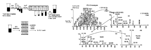
Figure 7 shows an accumulated MS from the retention profile of a labelled
peptide from
clusterin as described in Example 3 below. Inset ¨ zoom view of m/z region 915-
935 showing
the peptide of interest.
Figure 8 shows an accumulated MS/MS spectrum of the labelled peptide from
clusterin.
Insert: Zoom view at the mass marker region.
Figure 9 shows a calibration curve of the chosen clusterin peptide.
Figure 10 shows a MALDI MS/MS spectrum of peptide FQVDNNNR as described in
Example 4 below.
Figure 11 shows a MALDI MS/MS spectrum of peptide GAYPLSIEPIGVR as described
in
Example 4 below.
Figure 12 shows a MALDI MS/MS spectrum of peptide GQYCYELDEK as described in
Example 4 below.
The present invention will now be described in detail.
This invention provides useful reagents for determining relative and/or
absolute quantities of
analytes such as peptides, proteins, nucleotides and nucleic acids and means
of their
production. Specifically this invention relates to isobarically labelled
analytes and/or
calibration samples for detection by tandem mass spectrometry and associated
methods of
analysing test samples into which such calibration samples have been added.
Relative and/or
absolute quantitation of the analytes is particularly facilitated by the
invention.
This invention provides new methods for assaying analytes by mass spectrometry
in a variety
of settings including measurement of protein, lipid, carbohydrate and nucleic
acid changes in
cells, tissues and fluids in human, veterinary, plant, microbial,
pharmaceutical, environmental
and security sciences.
CA 02680373 2009-09-09
WO 2008/110581
PCT/EP2008/052962
18
In the methods according to the present invention the quantity of the analyte
in the test
sample and in each aliquot of the calibration sample is determined by mass
spectrometry. A
calibration function is used to relate the quantity of the analyte in the test
sample as measured
by mass spectrometry to the actual quantity of the analyte in the test sample.
This calibration
function uses the quantities of the analyte in each aliquot of the calibration
sample (both the
actual quantities in the aliquots prior to analysis and the corresponding
quantities as measured
by mass spectrometry) as variables.
In a preferred embodiment, the method comprises a step of plotting a graph of
the quantity of
the analyte in each aliquot versus the quantity of the analyte in each aliquot
as determined by
mass spectrometry. This step may instead simply involve calculation. The
quantity of the
analyte in the sample is then calculated by measuring the quantity in the
sample as
determined by mass spectrometry against the calibration graph. In the context
of this
invention, a reference to "a quantity as measured by mass spectrometry" is
typically an ion
abundance, ion intensity, or other signal measured by mass spectrometry which
relates to the
quantity of an analyte.
Typically, the method comprises:
i) in a mass spectrometer selecting and fragmenting ions of a mass to charge
ratio
corresponding to the analyte labelled with the mass label, detecting and
producing a
mass spectrum of fragment ions, and identifying the fragment ions
corresponding to
the mass marker groups of the mass labels;
ii) determining the quantity of the analyte in each test sample on the basis
of the
quantity of their mass marker groups in a mass spectrum relative to the
quantities of
the mass marker groups from the aliquots of the calibration sample in the same
mass
spectrum.
In a particular embodiment, the method comprises the steps of:
1. Optionally preparing the isobarically labelled reference material
containing a
reference biomolecule or mixture of reference biomolecules by reacting with a
set of mass labels according to this invention;
2. Labelling a sample in which the quantity of the biomolecule or mixture
of
CA 02680373 2009-09-09
WO 2008/110581
PCT/EP2008/052962
19
biomolecules is to be quantified by reacting with one of the same set of mass
labels as used in step 1 above according to this invention;
3. Adding a known amount of the isobarically labelled reference material
into the
isobarically labelled test sample prepared in step 2;
4. Optionally separating the isobarically labelled biomolecules
electrophoretically
or chromatographically;
5. Ionising the labelled biomolecules in a mass spectrometer;
6. Selecting ions of a predetermined mass to charge ratio corresponding to
the
mass to charge ratio of the preferred ions of the labelled biomolecule in a
mass
analyser;
7. Inducing dissociation of these selected ions by collision or electron
transfer;
8. Detecting the collision products to identify collision product ions that
are
indicative of the mass labels;
9. Producing a standard curve of ion intensity versus biomolecule amount
based on
intensity of the collision product ions that are indicative of the mass
labels;
10. Calculating the absolute or relative abundance of the biomolecule or
mixture of
biomolecules.
In relation to this invention the term "mass spectrometry" shall include any
type of mass
spectrometry capable of fragmentation analysis. The mass spectrometers
suitable for use in
the present invention include instruments that comprise any form of MS/MS
analyser such as
a triple quadropole mass spectrometer equipped with a collision chamber, an
ion trap mass
spectrometer capable of fragmenting selected precursor ions by fast atom
bombardment,
collision induced dissociation, electron transfer dissociation or any other
form of parent ion
fragmentation, and matrix assisted laser desorption/ionisation (MALDI) mass
spectrometers
fitted with a dual time of flight (TOF/TOF) analyser and a means of parent ion
fragmentation.
In certain embodiments the step of selecting the ions of a predetermined mass
to charge ratio
is performed in the first mass analyser of a serial instrument. The selected
ions are then
channelled into a separate collision cell where they are collided with a gas
or a solid surface.
The collision products are then channelled into a further mass analyser of a
serial instrument
to detect collision products. Typical serial instruments include triple
quadrupole mass
CA 02680373 2009-09-09
WO 2008/110581 PCT/EP2008/052962
spectrometers, tandem sector instruments and quadrupole time of flight mass
spectrometers.
In other embodiments, the step of selecting the ions of a predetermined mass
to charge ratio,
the step of colliding the selected ions with a gas and the step of detecting
the collision
products are performed in the same zone of the mass spectrometer. This may be
effected in
ion trap mass analysers and Fourier Transform Ion Cyclotron Resonance mass
spectrometers,
for example.
In the present invention, matrix assisted laser desorption/ionisation (MALDI)
techniques may
be employed. MALDI requires that the biomolecule solution be embedded in a
large molar
excess of a photo-excitable 'matrix'. The application of laser light of the
appropriate
frequency results in the excitation of the matrix which in turn leads to rapid
evaporation of
the matrix along with its entrapped biomolecule. Proton transfer from the
acidic matrix to the
biomolecule gives rise to protonated forms of the biomolecule which can be
detected by
positive ion mass spectrometry, particularly by Time-Of-Flight (TOF) mass
spectrometry.
Negative ion mass spectrometry is also possible by MALDI TOF. This technique
imparts a
significant quantity of translational energy to ions, but tends not to induce
excessive
fragmentation despite this. The laser energy and the timing of the application
of the potential
difference used to accelerate the ions from the source can be used to control
fragmentation
with this technique. This technique is highly favoured due to its large mass
range, due to the
prevalence of singly charged ions in its spectra and due to the ability to
analyse multiple
peptides simultaneously. The TOF/TOF technique may be employed in the present
invention.
The photo-excitable matrix comprises a 'dye' , i.e. a compound that strongly
absorbs light of
a particular frequency, and which preferably does not radiate that energy by
fluorescence or
phosphorescence but rather dissipates the energy thermally, i.e. through
vibrational modes. It
is the vibration of the matrix caused by laser excitation that results in
rapid sublimation of the
dye, which simultaneously takes the embedded analyte into the gas phase.
Although MALDI techniques are useful in the context of the present invention,
the invention
is not limited to this type of technique, and other techniques common to the
art can be
CA 02680373 2015-03-26
21
employed by the skilled person in the present invention, if desired. For
example electrospray
or nanoelectrospray mass spectrometry may be employed.
The term "analyte" is not particularly limiting, and the methods according to
the present
invention may be employed to assay any type of molecule provided that it can
be analysed by
mass spectrometry, and is capable of being labelled by an isobaric mass label
with a mass
spectrometrically distinct mass marker group. Analytes include amino acids,
peptides,
polypeptides, proteins, glycoprote ins, lipoproteins, nucleic acids,
polynucleotides,
oligonucleotides, DNA, RNA, peptide-nucleic acids, sugars, starches and
complex
carbohydrates, fats and complex lipids, polymers and small organic molecules
such as drugs
and drug-like molecules. Preferably the analyte is a peptide, protein,
nucleotide or nucleic
acid.
In relation to this invention the term protein shall encompass any molecule
comprising two or
more amino acids including di-peptides, tri-peptides, peptides, polypeptides
and proteins.
In relation to this invention the term nucleic acid shall encompass any
molecule comprising
two or more nucleotide bases including di-nucleotides, tri-nucleotides,
oligonucleotides,
deoxyribonucleic acids, ribonucleic acids and peptide nucleic acids.
In relation to this invention the term analyte shall be synonymous with the
term biomolecule.
The mass labels employed to tag the analytes in the present invention will now
be described
in more detail.
The skilled artisan will understand that the nature of the isobaric mass label
is not particularly
limiting. Various suitable isobaric mass labels are known in the art such as
Tandem Mass
Tags (Thompson et al., 2003, Anal. Chem. 75(8): 1895 ¨ 1904 disclosed in WO
01/68664
and WO 03/025576, iPROT tags disclosed in US 6824981 and iTRAQ tags (Pappin et
al.,
2004, Methods in Clinical Proteomics Manuscript M400129-MCP200). Any of these
isobaric
CA 02680373 2009-09-09
WO 2008/110581
PCT/EP2008/052962
22
mass labels are suitable for preparation of the samples and calibration
samples and
performing the methods of the current invention.
Mass Marker Moiety
In a preferred embodiment, the present invention uses a mass label as defined
above wherein
the molecular weight of the mass marker moiety is 300 Daltons or less,
preferably 250
Daltons or less, more preferably 100 to 250 Daltons, most preferably 100-200
Daltons.
Particularly preferred molecular weights of the mass marker moiety are 125,
126, 153 and
154 Daltons, or weights in which one or more or all of the 12C atoms are
replaced by 13C
atoms, e.g. for a non-substituted mass marker moiety having a weight of 125,
masses for its
substituted counterparts would be 126, 127, 128, 129, 130 and 131 Daltons for
substitution
with 1, 2, 3, 4, 5 and 6 13C atoms respectively and/or one or more or all of
the 14N atoms are
replaced by 15N atoms.
The components of the mass marker moiety of this invention are preferably
fragmentation
resistant so that the site of fragmentation of the markers can be controlled
by the introduction
of a linkage that is easily broken by Collision Induced Dissociation (CID),
Surface Induced
Dissociation, Electron Capture Dissociation (ECD), Electron Tranfer
Dissociation (ETD), or
Fast Atom Bombardment. In the most preferred embodiment, the linkage is easily
broken by
CID.
The mass marker moiety used in the present invention typically comprises the
following
group:
R1
R1
X (CR12)y __
RIZZ
wherein the cyclic unit is aromatic or aliphatic and comprises from 0-3 double
bonds
CA 02680373 2009-09-09
WO 2008/110581
PCT/EP2008/052962
23
independently between any two adjacent atoms; each Z is independently N,
N(RI), C(RI),
CO, CO(RI) (i.e. -0-C(R1)- or -C(R1)-0-), C(RI)2, 0 or S; X is N, C or C(RI);
each R1 is
independently H, a substituted or unsubstituted straight or branched C1-C6
alkyl group, a
substituted or unsubstituted aliphatic cyclic group, a substituted or
unsubstituted aromatic
group or a substituted or unsubstituted heterocyclic group; and y is an
integer from 0-10.
The substituents of the mass marker moiety are not particularly limited and
may comprise
any organic group and/or one or more atoms from any of groups IIIA, IVA, VA,
VIA or
VITA of the Periodic Table, such as a B, Si, N, P, 0, or S atom or a halogen
atom (e.g. F, Cl,
Br or I).
When the substituent comprises an organic group, the organic group preferably
comprises a
hydrocarbon group. The hydrocarbon group may comprise a straight chain, a
branched chain
or a cyclic group. Independently, the hydrocarbon group may comprise an
aliphatic or an
aromatic group. Also independently, the hydrocarbon group may comprise a
saturated or
unsaturated group.
When the hydrocarbon comprises an unsaturated group, it may comprise one or
more alkene
functionalities and/or one or more alkyne functionalities. When the
hydrocarbon comprises a
straight or branched chain group, it may comprise one or more primary,
secondary and/or
tertiary alkyl groups. When the hydrocarbon comprises a cyclic group it may
comprise an
aromatic ring, an aliphatic ring, a heterocyclic group, and/or fused ring
derivatives of these
groups. The cyclic group may thus comprise a benzene, naphthalene, anthracene,
indene,
fluorene, pyridine, quinoline, thiophene, benzothiophene, furan, benzofiiran,
pyrrole, indole,
imidazole, thiazole, and/or an oxazole group, as well as regioisomers of the
above groups.
The number of carbon atoms in the hydrocarbon group is not especially limited,
but
preferably the hydrocarbon group comprises from 1-40 C atoms. The hydrocarbon
group may
thus be a lower hydrocarbon (1-6 C atoms) or a higher hydrocarbon (7 C atoms
or more, e.g.
7-40 C atoms). The number of atoms in the ring of the cyclic group is not
especially limited,
but preferably the ring of the cyclic group comprises from 3-19 atoms, such as
3, 4, 5, 6 or 7
atoms.
CA 02680373 2009-09-09
WO 2008/110581 PCT/EP2008/052962
24
The groups comprising heteroatoms described above, as well as any of the other
groups
defined above, may comprise one or more heteroatoms from any of groups IIIA,
IVA, VA,
VIA or VITA of the Periodic Table, such as a B, Si, N, P, 0, or S atom or a
halogen atom (e.g.
F, Cl, Br or I). Thus the substituent may comprise one or more of any of the
common
functional groups in organic chemistry, such as hydroxy groups, carboxylic
acid groups, ester
groups, ether groups, aldehyde groups, ketone groups, amine groups, amide
groups, imine
groups, thiol groups, thioether groups, sulphate groups, sulphonic acid
groups, and phosphate
groups etc. The substituent may also comprise derivatives of these groups,
such as carboxylic
acid anhydrydes and carboxylic acid halides.
In addition, any substituent may comprise a combination of two or more of the
substituents
and/or functional groups defined above.
The cleavable linker of the mass label used in the present invention is not
especially limited.
Preferably, the cleavable linker is a linker cleavable by Collision Induced
Dissociation,
Surface Induced Dissociation, Electron Capture Dissociation (ECD), Electron
Transfer
Dissociation (ETD), or Fast Atom Bombardment. In the most preferred
embodiment, the
linkage is cleavable by CID. The linker may comprise an amide bond.
Linker
In the discussion above and below reference is made to linker groups which may
be used to
connect molecules of interest to the mass label compounds used in this
invention. A variety
of linkers is known in the art which may be introduced between the mass labels
of this
invention and their covalently attached biological molecule. Some of these
linkers may be
cleavable. Oligo- or poly-ethylene glycols or their derivatives may be used as
linkers, such
as those disclosed in Maskos, U. & Southern, E.M. Nucleic Acids Research 20:
1679 -1684,
1992. Succinic acid based linkers are also widely used, although these are
less preferred for
applications involving the labelling of oligonucleotides as they are generally
base labile and
are thus incompatible with the base mediated de-protection steps used in a
number of
oligonucleotide synthesisers.
Propargylic alcohol is a bifunctional linker that provides a linkage that is
stable under the
CA 02680373 2015-03-26
conditions of oligonucleotide synthesis and is a preferred linker for use with
this invention in
relation to oligonucleotide applications. Similarly 6-aminohexanol is a useful
bifunctional
reagent to link appropriately functionalised molecules and is also a preferred
linker.
A variety of known cleavable linker groups may be used in conjunction with the
compounds
employed in this invention, such as photocleavable linkers. Ortho-nitrobenzyl
groups are
known as photocleavable linkers, particularly 2-nitrobenzyl esters and 2-
nitrobenzylamines,
which cleave at the benzylamine bond. For a review on cleavable linkers see
Lloyd-Williams
et al., Tetrahedron 49, 11065-11133, 1993, which covers a variety of
photocleavable and
chemically cleavable linkers.
WO 00/02895 discloses the vinyl sulphone compounds as cleavable linkers, which
are also
applicable for use with this invention, particularly in applications involving
the labelling of
polypeptides, peptides and amino acids.
WO 00/02895 discloses the use of silicon compounds as linkers that are
cleavable by base in
the gas phase. These linkers are also applicable for use with this invention,
particularly in
applications involving the labelling of oligonucleotides.
Mass Normalisation Moiety
The structure of the mass normalization moiety of the mass label used in the
present
invention is not particularly limited provided that it is suitable for
ensuring that the mass label
has a desired aggregate mass. However, the mass normalization moiety
preferably comprises
a straight or branched C1-C20 substituted or unsubstituted aliphatic group
and/or one or more
substituted or unsubstituted amino acids.
Preferably, the mass normalization moiety comprises a C1-C6 substituted or
unsubstituted
aliphatic group, more preferably a C1, C2, C3, C4, C5 substituted or
unsubstituted aliphatic
group, still more preferably a C1, C2, or C5 substituted or unsubstituted
aliphatic group or a C1
methyl substituted group.
CA 02680373 2009-09-09
WO 2008/110581
PCT/EP2008/052962
26
The one or more substituted or unsubstituted amino acids may be any essential
or non-
essential naturally occurring amino acids or non-naturally occurring amino
acids. Preferred
amino acids are alanine, P-alanine and glycine.
The substituents of the mass normalisation moiety are not particularly limited
and may
comprise any organic group and/or one or more atoms from any of groups IIIA,
IVA, VA,
VIA or VIIA of the Periodic Table, such as a B, Si, N, P, 0, or S atom or a
halogen atom (e.g.
F, Cl, Br or I).
When the substituent comprises an organic group, the organic group preferably
comprises a
hydrocarbon group. The hydrocarbon group may comprise a straight chain, a
branched chain
or a cyclic group. Independently, the hydrocarbon group may comprise an
aliphatic or an
aromatic group. Also independently, the hydrocarbon group may comprise a
saturated or
unsaturated group.
When the hydrocarbon comprises an unsaturated group, it may comprise one or
more alkene
functionalities and/or one or more alkyne functionalities. When the
hydrocarbon comprises a
straight or branched chain group, it may comprise one or more primary,
secondary and/or
tertiary alkyl groups. When the hydrocarbon comprises a cyclic group it may
comprise an
aromatic ring, an aliphatic ring, a heterocyclic group, and/or fused ring
derivatives of these
groups. The cyclic group may thus comprise a benzene, naphthalene, anthracene,
indene,
fluorene, pyridine, quinoline, thiophene, benzothiophene, furan, benzofuran,
pyrrole,
imidazole, thiazole, and/or an oxazole group, as well as regioisomers of the
above groups.
The number of carbon atoms in the hydrocarbon group is not especially limited,
but
preferably the hydrocarbon group comprises from 1-40 C atoms. The hydrocarbon
group may
thus be a lower hydrocarbon (1-6 C atoms) or a higher hydrocarbon (7 C atoms
or more, e.g.
7-40 C atoms), The number of atoms in the ring of the cyclic group is not
especially limited,
but preferably the ring of the cyclic group comprises from 3-10 atoms, such as
3, 4, 5, 6 or 7
atoms.
CA 02680373 2009-09-09
WO 2008/110581
PCT/EP2008/052962
27
The groups comprising heteroatoms described above, as well as any of the other
groups
defined above, may comprise one or more heteroatoms from any of groups IIIA,
IVA, VA,
VIA or VITA of the Periodic Table, such as a B, Si, N, P, 0, or S atom or a
halogen atom (e.g.
F, Cl, Br or I). Thus the substituent may comprise one or more of any of the
common
functional groups in organic chemistry, such as hydroxy groups, carboxylic
acid groups, ester
groups, ether groups, aldehyde groups, ketone groups, amine groups, amide
groups, imine
groups, thiol groups, thioether groups, sulphate groups, sulphonic acid
groups, and phosphate
groups etc. The substituent may also comprise derivatives of these groups,
such as carboxylic
acid anhydrydes and carboxylic acid halides.
In addition, any substituent may comprise a combination of two or more of the
substituents
and/or functional groups defined above.
Reactive Mass Label
The reactive mass labels typically used in the present invention for labelling
and detecting a
biological molecule by mass spectroscopy comprise a reactive functionality for
facilitating
attachment of or for attaching the mass label to a biological molecule and a
mass label as
defined above. In preferred embodiments of the present invention, the reactive
functionality
allows the mass label to be reacted covalently to an analyte, preferably an
amino acid, peptide
or polypeptide. The reactive functionality may be attached to the mass labels
via a linker
which may or may not be cleavable. The reactive functionality may be attached
to the mass
marker moiety of the mass label or the mass normalization moiety of the mass
label.
A variety of reactive functionalities may be introduced into the mass labels
used in this
invention. The structure of the reactive functionality is not particularly
limited provided that
it is capable of reacting with one or more reactive sites on the biological
molecule to be
labelled. The reactive functionality is preferably a nucleophile or an
electrophile.
In the simplest embodiments this may be an N-hydroxysuccinimide ester. An
N-hydroxysuccinimide activated mass label could also be reacted with hydrazine
to give a
hydrazide reactive functionality, which can be used to label periodate
oxidised sugar
moieties, for example. Amino-groups or thiols can be used as reactive
functionalities in some
CA 02680373 2009-09-09
WO 2008/110581
PCT/EP2008/052962
28
applications. Lysine can be used to couple mass labels to free carboxyl
functionalities using
a carbodiimide as a coupling reagent. Lysine can also be used as the starting
point for the
introduction of other reactive functionalities into the mass labels of this
invention. The thiol-
reactive maleimide functionality can be introduced by reaction of the lysine
epsilon amino
group with maleic anhydride. The cysteine thiol group can be used as the
starting point for
the synthesis of a variety of alkenyl sulphone compounds, which are useful
protein labelling
reagents that react with thiols and amines. Compounds such as aminohexanoic
acid can be
used to provide a spacer between the mass modified mass marker moiety or mass
normalization moiety and the reactive functionality.
Table 1 below lists some reactive functionalities that may be reacted with
nucleophilic
functionalities which are found in biomolecules to generate a covalent linkage
between the
two entities. Any of the functionalities listed below could be introduced into
the compounds
of this invention to permit the mass markers to be attached to a biological
molecule of
interest. A reactive functionality can be used to introduce a further linker
groups with a
further reactive functionality if that is desired. Table 1 is not intended to
be exhaustive and
the present invention is not limited to the use of only the listed
functionalities.
,.
CA 02680373 2009-09-09
WO 2008/110581 PCT/EP2008/052962
29
Nucleophilic Functionality Reactive Functionality Resultant Linking Group
-SH -S 02- CH¨CR2 -S-CR2-C112-S02-
-NH2 -S02-CH¨CR2 -N(CR2-CH2-S02-)2 or
-NH-CR2-CH2-S02-
-NH2 0 -CO-NH-
0
II )
)r\----
¨C¨O¨N
0
-NH2 0 -CO-NH-
II
¨C¨O¨N N
-NH2 -NCO -NH-CO-NH-
-NH2 -NCS -NH-CS-NH-
-NH2 -CHO ' -CH2-NH-
-NH2 -S02C1 -S02-NH-
-NH2 -CH¨CH- -NH-CH2-CH2-
-OH -0P(NCH(CH3)2)2 -0P(-
0)(0)0-
Table 1
In a preferred embodiment of the present invention the reactive functionality
comprises the
following group:
0
0
¨N
>--------\i'i2
0
- -
CA 02680373 2009-09-09
WO 2008/110581
PCT/EP2008/052962
wherein each R2 is independently H, a substituted or unsubstituted straight or
branched C1-C6
alkyl group, a substituted or unsubstituted aliphatic cyclic group, a
substituted or
unsubstituted aromatic group or a substituted or unsubstituted heterocyclic
group.
The substituents of the reactive functionality are not particularly limited
and may comprise
any organic group and/or one or more atoms from any of groups IIIA, IVA, VA,
VIA or
VIIA of the Periodic Table, such as a B, Si, N, P, 0, or S atom or a halogen
atom (e.g. F, Cl,
Br or I).
When the substituent comprises an organic group, the organic group preferably
comprises a
hydrocarbon group. The hydrocarbon group may comprise a straight chain, a
branched chain
or a cyclic group. Independently, the hydrocarbon group may comprise an
aliphatic or an
aromatic group. Also independently, the hydrocarbon group may comprise a
saturated or
unsaturated group.
When the hydrocarbon comprises an unsaturated group, it may comprise one or
more alkene
functionalities and/or one or more alkyne functionalities. When the
hydrocarbon comprises a
straight or branched chain group, it may comprise one or more primary,
secondary and/or
tertiary alkyl groups. When the hydrocarbon comprises a cyclic group it may
comprise an
aromatic ring, an aliphatic ring, a heterocyclic group, and/or fused ring
derivatives of these
groups. The cyclic group may thus comprise a benzene, naphthalene, anthracene,
indene,
fluorene, pyridine, quinoline, thiophene, benzothiophene, furan, benzofuran,
pyrrole, indole,
imidazole, thiazole, and/or an oxazole group, as well as regioisomers of the
above groups.
The number of carbon atoms in the hydrocarbon group is not especially limited,
but
preferably the hydrocarbon group comprises from 1-40 C atoms. The hydrocarbon
group may
thus be a lower hydrocarbon (1-6 C atoms) or a higher hydrocarbon (7 C atoms
or more, e.g.
7-40 C atoms). The number of atoms in the ring of the cyclic group is not
especially limited,
but preferably the ring of the cyclic group comprises from 3-10 atoms, such as
3, 4, 5, 6 or 7
atoms.
CA 02680373 2009-09-09
WO 2008/110581
PCT/EP2008/052962
31
The groups comprising heteroatoms described above, as well as any of the other
groups
defined above, may comprise one or more heteroatoms from any of groups IIIA,
IVA, VA,
VIA or VITA of the Periodic Table, such as a B, Si, N, P, 0, or S atom or a
halogen atom (e.g.
F, Cl, Br or I). Thus the substituent may comprise one or more of any of the
common
functional groups in organic chemistry, such as hydroxy groups, carboxylic
acid groups, ester
groups, ether groups, aldehyde groups, ketone groups, amine groups, amide
groups, imine
groups, thiol groups, thioether groups, sulphate groups, sulphonic acid
groups, and phosphate
groups etc. The substituent may also comprise derivatives of these groups,
such as carboxylic
acid anhydrydes and carboxylic acid halides.
In addition, any substituent may comprise a combination of two or more of the
substituents
and/or functional groups defined above.
In a more preferred embodiment the reactive functionality comprises the
following group:
0
0
0¨N
0
In a preferred embodiment of the present invention the reactive mass label has
one of the
following structures:
CA 02680373 2009-09-09
WO 2008/110581
PCT/EP2008/052962
32
0 0
0
H
)r---
0
342-(2,6-Dimethy1-piperidin-1-y1)-acetylamino]-propanoic acid-(2,5-dioxo-
pyrrolidine-1-
y1)-ester (DMPip-BAla-OSu)
0
N
Y
1 H
N.õ........,.....,õ 0 ¨ N
N S
)7------
0
0
3 [2-(Pyrimidin-2-ylsulfany1)-acetylamino]-propanoic acid-(2,5-dioxo-
pyrrolidine-1-y1)-ester
(Pyrni-BAla-OSu)
0
N 0
1 H
Y----
NN
0
)r ---
0
6-[( Pyrimidin-2-ylsulfany1)-acetylamino]-hexanoic acid-(2,5-dioxo-pyrrolidine-
1-y1)-ester
(Pyrrn-C6-0Su)
CA 02680373 2009-09-09
WO 2008/110581
PCT/EP2008/052962
33
0
0
Y------
-....õ....,,,,,,N.NN.....õ7õ.......õ...õvõ....õ..,....õ.....õ0¨Ny
H
0
0
2 42,-(2,6-Dirnethyl-piperidin- 1 -y1)-acetyl amino] -propanoic acid-
(2, 5 -di oxo -pyrrolidine- 1 -
y1)-ester (DMPip-Ala-OSu)
0
0
)\---"----
NO¨Ny0
0
[2-(2,6-Dimethyl-piperidin-1 -y1)-acetylamino]-acetic acid-(2,5-dioxo-
pyrro1idine- 1 -y1)-ester
(Pyrm-Gly-OSu).
In the method according to the present invention, each label in the set has a
common
aggregate mass and each label in the set has a mass marker moiety of a unique
mass.
It is preferred that, each mass marker moiety in the set has a common basic
structure and each
mass normalisation moiety in the set has a common basic structure, and each
mass label in
the set comprises one or more mass adjuster moieties, the mass adjuster
moieties being
attached to or situated within the basic structure of the mass marker moiety
and/or the basic
structure of the mass normalisation moiety. In this embodiment, every mass
marker moiety
in the set comprises a different number of mass adjuster moieties and every
mass label in the
set has the same number of mass adjuster moieties.
Throughout this description, by common basic structure, it is meant that two
or more moieties
share a structure which has substantially the same structural skeleton,
backbone or core. The
CA 02680373 2009-09-09
WO 2008/110581
PCT/EP2008/052962
34
skeleton comprises the mass marker moiety of the formula given above or the
mass
normalisation moiety as defined above. The skeleton may additionally comprise
a number of
amino acids linked by amide bonds. Other units such as aryl ether units may
also be present.
The skeleton or backbone may comprise substituents pendent from it, or atomic
or isotopic
replacements within it, without changing the common basic structure.
In a preferred embodiment the set of mass labels or reactive mass labels
according to the
invention comprise mass labels having the following structure:
M(A)y-L-X(A)z
wherein M is a mass normalisation moiety, X is a mass marker moiety, A is a
mass adjuster
moiety, L is a cleavable linker, y and z are integers of 0 or greater, and y+z
is an integer of 1
or greater. Preferably M is a fragmentation resistant group, L is a linker
that is susceptible to
fragmentation on collision with another molecule or atom and X is preferably a
pre-ionised,
fragmentation resistant group.
The sum of the masses of M and X is the same for all members of the set.
Preferably M and
X have the same basic structure or core structure, this structure being
modified by the mass
adjuster moieties. The mass adjuster moiety ensures that the sum of the masses
of M and X
is the same for all mass labels in a set, but ensures that each X has a
distinct (unique) mass.
The mass adjuster moiety (A) is preferably selected from:
(a) an isotopic substituent situated within the mass marker moiety and/or
within
the mass normalisation moiety, and
(b) substituent atoms or groups attached to the mass marker moiety and/or
attached to the mass normalisation moiety.
Preferably the mass adjuster moiety is selected from a halogen atom
substituent, a methyl
group substituent, and 2H, 15N, 180, or 13C isotopic substituents.
In one preferred embodiment the present invention, each mass label in the set
of mass labels
CA 02680373 2009-09-09
WO 2008/110581
PCT/EP2008/052962
as defined above has the following structure:
X(*)"-L-MMin
wherein X is the mass marker moiety, L is the cleavable linker, M is the mass
normalisation
moiety, * is an isotopic mass adjuster moiety, and n and m are integers of 0
or greater such
that each label in the set comprises a mass marker moiety having a unique mass
and each
label in the set has a common aggregate mass.
It is preferred that X comprises the following group:
i*
R'
,*
R.
* **
X *(CR12)y ____
wherein RI, Z, X and y are as defined above and each label in the set
comprises 0, 1 or more
* such that each label in the set comprises a mass marker moiety having a
unique mass and
each label in the set has a common aggregate mass.
In a further preferred embodiment, the reactive mass labels of the present
invention comprise
the following reactive functionality group:
0
0
* * 4> 2
O-N
R2
0*
CA 02680373 2009-09-09
WO 2008/110581
PCT/EP2008/052962
36
wherein R2 is as defined above and the set comprises 0, 1 or more * such that
each label in
the set comprises a mass marker moiety having a unique mass and each label in
the set has a
common aggregate mass.
In all of the above preferred formulae, it is particularly preferred that the
isotopic species * is
situated within the mass marker moiety and/or the linker and/or the mass
adjuster moiety,
rather than on any reactive moiety that is present to facilitate attaching the
label to an analyte.
The number of isotopic substituents is not especially limited and can be
determined
depending on the number of labels in the set. Typically, the number of *
species is from 0-20,
more preferably from 0-15 and most preferably from 1-10, e.g. 1, 2, 3, 4, 5,
6, 7 or 8. In a set
of two labels, it is preferred that the number of species * is 1, whilst in a
set of 5 labels, it is
preferred that the number is 4, whilst in a set of 6 labels it is preferred
that the number is 5.
However, the number may be varied depending upon the chemical structure of the
label.
If desired, isotopic variants of S may also be employed as mass adjuster
moieties, if the labels
contain one or more sulphur atoms.
In a particularly preferred embodiment wherein the mass adjuster moiety is 15N
or 13C the set
of reactive mass labels comprises two mass labels having the following
structures:
0 0 0
N N0¨NY-
H
)7.-------
0
CA 02680373 2009-09-09
WO 2008/110581
PCT/EP2008/052962
37
0
0 0
N15 0 N
0
In an alternative particularly preferred embodiment wherein the mass adjuster
moiety is 15N
or 13C the set of reactive mass labels comprises the set comprises five mass
labels having the
following structures:
0 13
0 _________________________________________________ N
N 1
0
0
0 13
0 N
N 3 y0
0
0 13
0 ¨ N
13
0
0
CA 02680373 2009-09-09
WO 2008/110581 PCT/EP2008/052962
38
0 13
11\j5N()¨N),r,,,
13 H
0
0
0
"--------
13
13 H
0
0 .
In an alternative particularly preferred embodiment wherein the mass adjuster
moiety is 15N
or 13C the set of reactive mass labels comprises six mass labels I-VI having
the following
structures, or stereoisomers of these structures:
o o
1)1 o
I I 1311 13
N 13C. 13C. õ.13C ,N
I-1 H
0 0
H3 H3
13C 0 13c 0
35 0 9 o
1.--
N 15N
1 C. 13C.....1.....,o N N13 15N
-- ,--
H H
"CH, 0 13CH3 0
III IV
H3 H,
13n
0 0C13C" 0 0
I
, 141..,..,.õ,...
13C' Ni5NO"N--R 13 1,
IC' N"---'''-"..---LOIR
31 H 1 H
CH3 0 13CH3 0
V VI
CA 02680373 2009-09-09
WO 2008/110581
PCT/EP2008/052962
39
The method according to the present invention may comprise a further step of
separating the
components of the samples prior to step (a). The method may also comprise a
step of
digesting each sample with at least one enzyme to digest components of the
samples prior to
step (a). The enzyme digestion step may also occur after step (a) but before
step (b).
In a further embodiment, the mass labels used in the method further comprise
an affinity
capture ligand. The affinity capture ligand of the mass label binds to a
counter-ligand so as to
separate the isobarically labeled analytes from the unlabelled analytes after
step (a) but before
step (b).
Affinity capture ligands are ligands which have highly specific binding
partners. These
binding partners allow molecules tagged with the ligand to be selectively
captured by the
binding partner. Preferably a solid support is derivitised with the binding
partner so that
affinity ligand tagged molecules can be selectively captured onto the solid
phase support. A
preferred affinity capture ligand is biotin, which can be introduced into the
mass labels of this
invention by standard methods known in the art. In particular a lysine residue
may be
incorporated after the mass marker moiety or mass normalization moiety through
which an
amine-reactive biotin can be linked to the mass labels ( see for example
Geahlen R.L. et al.,
Anal Biochem 202(1): 68-67, "A general method for preparation of peptides
biotinylated at
the carboxy terminus." 1992; Sawutz D.G. et al., Peptides 12(5): 1019-1012,
"Synthesis and
molecular characterization of a biotinylated analogue of [Lys]bradykinin."
1991; Natarajan S.
et al., Int J Pept Protein Res 40(6): 567-567, "Site-specific biotinylation. A
novel approach
and its application to endothelin-1 analogues and PTH-analogue.", 1992).
lminobiotin is also
applicable. A variety of avidin counter-ligands for biotin are available,
which include
monomeric and tetrameric avidin and streptavidin, all of which are available
on a number of
solid supports.
Other affinity capture ligands include digoxigenin, fluorescein, nitrophenyl
moieties and a
number of peptide epitopes, such as the c-myc epitope, for which selective
monoclonal
antibodies exist as counter-ligands. Metal ion binding ligands such as
hexahistidine, which
readily binds Ni2+ ions, are also applicable. Chromatographic resins, which
present
iminodiacetic acid chelated Ni2+ ions are commercially available, for example.
These
CA 02680373 2009-09-09
WO 2008/110581 PCT/EP2008/052962
immobilised nickel columns may be used to capture mass labels. As a further
alternative, an
affinity capture functionality may be selectively reactive with an
appropriately derivatised
solid phase support. Boronic acid, for example, is known to selectively react
with vicinal
cis-
diols and chemically similar ligands, such as salicylhydroxamic acid.
The method according to the invention may further include the step of
separating the
isobarically labeled analytes electrophoretically or chromatographically after
step (a) but
before step (b). In a preferred embodiment, strong cation exchange
chromatography is used.
The term "test sample" refers to any specimen in which an analyte may be
present. The test
sample may comprise only one analyte. Alternatively, the test sample may
comprise a
plurality of different analytes. In this embodiment, a calibration sample is
provided for each
different analyte. The test sample may be from a natural source or may be
produced
synthetically. An example of a synthetic sample is a mixture of recombinant
proteins. In one
embodiment, the test sample is a complex mixture, for example a sample from a
plant or an
animal. In a preferred embodiment the sample is from a human.
Examples of test samples assayed in the present invention include: mammalian
tissue, fluids
such as blood, plasma, serum cerebrospinal fluid, synovial fluid, ocular
fluid, urine, tears and
tear duct exudate, lung aspirates including bronchoalveolar lavage fluid,
breast milk, nipple
aspirate, semen, lavage fluids, cell extracts, cell lines and sub-cellular
organelles, tissues such
as solid organ tissues, cell culture supernatants or preparations derived from
mammals, fish,
birds, insects, annelids, protozoa and bacteria, tissue culture extracts,
plant tissues, plant
fluids, plant cell culture extracts, bacteria, viruses, fungi, fermentation
broths, foodstuffs,
pharmaceuticals and any intermediary products.
In a preferred embodiment the test sample is blood plasma. In a particularly
preferred
embodiment the test sample is depleted blood plasma. This is blood plasma
which has been
purified to remove the most abundant plasma proteins, such as albumin, so as
to reduce the
protein load in the sample, hence reducing the number of analytes in the
sample.
The term "calibration sample" refers to a sample which comprises at least two
different
CA 02680373 2009-09-09
WO 2008/110581
PCT/EP2008/052962
41
aliquots of the analyte. The different aliquots each have a known quantity of
the analyte. The
term "known quantity" means that the absolute quantity, or a qualitative
quantity of the
analyte in each aliquot of the calibration sample is known. A qualitative
quantity in the
present context means a quantity which is not known absolutely, but may be a
range of
quantities that are expected in a subject having a particular state, for
example a subject in a
healthy or diseased state, or some other expected range depending on the type
of test sample
under investigation.
Each aliquot is "different" since it contains a different quantity of the
analyte. Typically this
is achieved by taking different volumes from a standard sample, especially for
qualitative
quantities where taking different volumes will ensure that different
quantities are present in
each aliquot in a desired ratio, without needing to know the absolute
quantities. As an
alternative, each aliquot is prepared separately and is not taken from the
same sample. In one
embodiment, each different aliquot has the same volume, but comprises a
different quantity
of the analyte.
The calibration sample may be a natural sample such as a body fluid or a
tissue extract or
may be synthetic, as for the sample to be assayed. The calibration sample may
comprise a
recombinantly expressed protein, synthetically manufactured peptide or
oligonucleotide. In
addition it is possible to produce a number of different peptides by
recombinant protein
expression in a concatenated sequence. European patent application EP 1736480
discloses
methods of producing multiple reference peptides as a concatenated recombinant
protein for
use in multiple reaction monitoring experiments in a mariner analogous to the
AQUA
methodology. Such methods of production may be combined with isobaric mass
labels to
provide the calibration samples according to any of the various aspects of
this invention.
The calibration sample may be a standardised form of the sample to be assayed.
The
calibration sample may comprise all of the components of the sample to be
assayed but in
particular quantities. For example, the calibration sample may comprise a
standardised
preparation of mammalian tissue, fluids such as blood, plasma, serum
cerebrospinal fluid,
synovial fluid, ocular fluid, urine, tears and tear duct exudate, lung
aspirates including
bronchoalveolar lavage fluid, breast milk, nipple aspirate, semen, lavage
fluids, cell extracts,
CA 02680373 2009-09-09
WO 2008/110581
PCT/EP2008/052962
42
cell lines and sub-cellular organelles, tissues such as solid organ tissues,
cell culture
supernatants or preparations derived from mammals, fish, birds, insects,
annelids, protozoa
and bacteria, tissue culture extracts, plant tissues, plant fluids, plant cell
culture extracts,
bacteria, viruses, fungi, fermentation broths, foodstuffs, pharmaceuticals and
any
intermediary products. If the analytes of interest are proteins, since all
proteins in the
calibration sample are labelled, the entire proteome of such a sample may be
used as a
reference for all proteins of the study sample.
Alternatively, the calibration sample may comprise only analytes to be assayed
in the sample,
and not any other components of the sample. The calibration sample comprising
one or more
analytes may be produced and isobarically labelled exogenously and added to
the complex
mixture containing the analyte. For example, if the sample is a plasma sample,
but only a
particular protein is to be assayed in the plasma sample, a calibration sample
can be prepared
which comprises different aliquots of the recombinant form of the protein.
In a method according to the invention, the quantity of analyte in each
aliquot in the
calibration sample is a known absolute quantity. This allows for the absolute
quantity of an
analyte in a test sample to be determined in step (b).
In an alternative method, the absolute quantity of an analyte in each aliquot
in the calibration
sample is not known. In this embodiment, the quantity of analyte in each
aliquot in the
calibration sample is a known qualitative quantity. The calibrating step
comprises calibrating
the quantity of the analyte in the test sample against the qualitative and
determined quantities
of the analytes in the aliquots of the calibration sample. In a particular
embodiment, the
qualitative quantity is an expected range of quantities of analyte in a
subject having a
particular state, such as a healthy or diseased state. Assays which provide
such calibration
samples for relative quantitation have wide range of applications including
biomarker
discovery, industrial microbiology, pharmaceutical and food manufacture and
the diagnosis
and management of human and veterinary disease
Relative quantitation experiments are often useful when analysing complex
biological
samples such as blood plasma. In a specific embodiment, a large amount of
entire human
CA 02680373 2009-09-09
WO 2008/110581
PCT/EP2008/052962
43
blood plasma is split into several (i.e, four) aliquots and individually
labelled with different
isobaric mass labels. For instance, one could utilise the 6-plex Tandem Mass
Tag reagents
(see above) to produce four labelled aliquots of blood plasma. 6TMT-128, 6TMT-
129,
6TMT-130, 6TMT-131 would be used for labelling. All individual samples of a
blood plasma
study are labelled with one further different version of this isobaric mass
tag, i.e. 6TMT-126.
The aliquots of blood plasma can now be used to generate a calibration curve,
for instance by
mixing the 4 aliquots in a 0.5 to 1 to 2 to 5 p,L ratio to produce a
calibration sample, and then
adding 1 ul of the study sample. By combining the sample with the calibration
sample
comprising four differentially labelled aliquots, virtually all MS/MS
experiments performed
with this material will result in groups of five reporter-ions ¨ four from the
calibration sample
and one from the sample. Thus, the entire proteome can be used in a 4-point
calibration
curve. If all plasma samples of the study are spiked with the identical amount
of the
calibration sample, relative quantification across all study samples becomes
possible. Since
the calibration sample can be used by multiple laboratories, cross-study and
cross-laboratory
comparisons are possible.
Whereas the absolute quantity of an analyte might not be known, the % change
in quantity
can be calculated from the calibration curve. Depending on the application,
the ratio and
width of the calibration curve can be adjusted.
In a preferred embodiment, the quantity of analyte in each different aliquot
of the calibration
sample is selected to reflect the known or suspected variation of the analyte
in the test
sample. In a still further preferred embodiment, aliquots are provided which
comprise the
analyte in quantities which correspond to the upper and lower limits, and
optionally
intermediate points within a range of the known or suspected quantities of the
analyte found
in test samples of healthy or diseased subjects,
Because each analyte is quantified independently of all other analytes in the
sample it is
conceivable to prepare multiple sets of calibration samples each at widely
different
concentrations to all other calibration samples, so enhancing the dynamic
range of the
experiment. It is also possible to prepare a number of reference biomolecules
for each analyte
wherein each biomolecule is provided in a range of overlapping quantities
thereby extending
CA 02680373 2009-09-09
WO 2008/110581
PCT/EP2008/052962
44
the total range of the standard curve for a given analyte. As an example a
number of different
tryptic peptides from a target protein may be selected for use as reference
standards based on
their performance in a tandem mass spectrometer. The reference peptides may be
selected on
the basis of the ion intensity of the ion corresponding to the peptide in a
mass spectrum or on
the basis of the signal-to-noise ratio in the area of the spectrum in which
the ion
corresponding to the peptide appears. Alternatively the reference peptides may
be selected so
as to avoid peptides which have isobaric species. The selection of proteotypic
peptides, i.e.
peptides which are only present in a particular protein is particularly
favoured.
If each standard peptide is independently labelled with up to five different
members of a six-
plex set of isobaric mass tags these may be mixed in different ratios to
provide a five-point
standard curve. The same isobaric mass labels may be used to label second,
third, fourth or
more standard peptides each of which may be mixed in different ratios covering
a range of
concentrations different to that covered by each of the other reference
peptides for the same
analyte.
A different calibration curve is produced for each peptide derived from the
target protein,
each calibration curve covering a different range of concentrations. The
concentration of each
peptide is then determined from their respective calibration curve, and this
can be related
back to the concentration of the target protein. For some of the calibration
curves, the
quantity of the peptide in the test sample may fall in the middle of the
calibration curve,
providing an accurate determination of its actual quantity in the sample. For
other calibration
curves covering a different range in concentrations, the quantity of the
peptide in the test
sample may fall outside the range of the calibration curve. By using multiple
peptides which
are each derived from a single analyte of interest, we can produce multiple
calibration curves
which can be related to the same analyte and then choose the most accurate
calibration to
determine the concentration of the analyte in the test sample from the
concentration of one or
more of the peptides. In this way a broad dynamic range may be covered without
compromising assay sensitivity.
The calibration sample may comprise a normal quantity of an analyte. The
quantity of the
analyte in the calibration sample may indicate that a plant, animal, or
preferably a human is
CA 02680373 2009-09-09
WO 2008/110581
PCT/EP2008/052962
healthy. Alternatively, the calibration sample may comprise an analyte in a
quantity that
indicates the presence and/or stage of a particular disease. In a further
embodiment, the
calibration sample comprises an analyte in a quantity that indicates the
efficacy and/or
toxicity of a therapy. Standard panels of known markers of a particular trait
such as presence
and/or stage of disease, response to therapy, and/or toxicity are prepared.
Calibration samples
comprising body fluids or tissue extracts labelled with an isobaric mass tag
could be prepared
from patients with well characterised disease including but not limited to
tumours,
neurodegeneration, cardiovascular, renal, hepatic, respiratory, metabolic,
inflammatory, and
infectious diseases. Known amounts of such samples are added to multiple test
samples in
such a manner that for a series of analyses ion intensities in the MS/MS scan
can be
normalised based on the ion intensity of the common calibration sample,
thereby providing
more accurate comparisons between the separate analyses, reducing the
analytical variability
of the study.
In the case of coronary medicine a series of peptides derived from the tryptic
digestion of
known heart disease markers such as myoglobin, troponin-I, CK-MB, BNP, pro-BNP
and
NT-pro-BNP are produced synthetically and split into three aliquots. Each
aliquot of each
reference peptide is labelled with one of three isobaric mass tags from a set
of such isobaric
mass tags wherein all tags in the set have substantially the same aggregate
mass as
determined by mass spectrometry and wherein each tag in the set releases a
mass reporter ion
of unique mass on collision induced dissociation in a mass spectrometer. Each
unique
reference peptide-mass tag molecule is then added to a carrier solution such
as a MS-
compatible buffer at a known concentration such that the concentration of the
three
differentially labelled aliquots of the same reference peptide are different,
and that the
differences span the normal biological concentrations of the parent protein in
patients with
cardiac disease. The resultant reference peptide panel is added at a defined
volumetric ratio
with a test sample that has been labelled with a fourth isobaric mass tag from
the same set of
isobaric mass tags used to label the reference peptides. The spiked sample is
then subjected to
tandem mass spectrometry wherein the survey scan is performed in a directed
manner to only
identify those precursor ions of characteristic retention time and mass
correlating to each of
the isobarically labelled reference peptides. For each selected ion the MS/MS
scan will
contain reporter ions derived from the high, medium and low concentration
reference
CA 02680373 2009-09-09
WO 2008/110581
PCT/EP2008/052962
46
peptides and the test sample.
A simple standard curve is easily constructed from the reference peptide
reporter ion
intensities and the fourth reporter ion from the same peptide in the test
sample can be read
against the calibration curve. By this means the absolute concentration of
multiple
biologically relevant proteins can be determined in a single MS/MS experiment.
The skilled
artisan will be aware that the number of different proteins for which
reference peptides are
prepared need not be particularly limited and will be in the range of 1 ¨ 100
and most
preferably 1 - 50. Similarly the number of representative peptides may be in
the range of 1 ¨
20, preferably 1 ¨ 10, more preferably 1 ¨ 5 and most preferably 1 ¨ 3. It
would be
understood by the skilled artisan that the example described above is a
general examplar and
the principles described therein may be applied to known markers of any
disease and applied
for disease diagnosis, monitoring of disease progression or monitoring the
response of a
patient to treatment.
A further application is in the use of these calibration samples in time
course experiments.
The "Status" of a sample with respect to time course can be established if the
(4) different
aliquots are from 4 different time points, such as time zero, 1 hour, 8 hours,
and 24 hours into
an experiment (drug challenge in mice and man, induction of fermentation in
Rcoli and
yeast), also on a longer time scale of weeks and months for development or
treatment
response of chronic diseases.
In a further aspect of the invention, one of the aliquots of the calibration
sample comprises an
analyte in a quantity which serves as a trigger during an MS scan or during
non-scanning
MS/MS to initiate an MS/MS scan.
Non-scanning MS/MS is when you do not select for any particular ion with a
given m/z ratio
in a mass analyser in a mass spectrometer, but instead essentially all of the
analytes are
fragmented to produce an unspecific fragment spectrum. Typically, this
involves allowing all
ions to pass from a first mass analyser into a collision cell, where CID
occurs on all of the
analytes in the sample instead of a particular selected ion as in conventional
MS/MS.
Although the MS/MS spectrum will not be specific to a particular analyte, the
reporter ion
CA 02680373 2009-09-09
WO 2008/110581
PCT/EP2008/052962
47
from the trigger can be used as an indicator that an analyte of interest is
right now entering
the mass spectrometer. In a preferred embodiment, the presence of a reporter
ion from the
trigger indicates that an analyte of interest is eluting from an LC column
during LC-MS. This
would "trigger" the execution of a pre-defined MS/MS experiment.
This trigger may not necessarily be an analyte labelled with an isobaric mass
label. The
trigger may be any other labelled analyte which co-elutes, or substantially co-
elutes with the
labelled analyte of interest during LC-MS. The label of the trigger analyte
may have a
different mass to that of the isobaric mass labels of the calibration sample.
For example, in
one embodiment, the calibration sample comprises aliquots of an analyte
differentially
labelled with isobaric mass labels, and further comprises an aliquot of the
analyte which is
labelled with a chemically identical but isotopically distinct mass label,
preferably with a
mass difference of 5 Da from that of the isobaric mass labels. The
isotopically distinct mass
label could then serve as the "trigger". During the MS phase of the analysis
each analyte
present in the calibration sample bearing the isotopically distinct and
isobaric labels will
appear as a pair of peaks separated by the mass difference between the
isobaric and
isotopically distinct labels and wherein the analyte bearing the isotopically
distinct label is
present in a readily detectable amount. The mass spectrometer is programmed to
perform a
dedicated MS/MS experiment on the isobarically labelled analyte in such pairs,
thereby
triggering the quantitative analysis of the analytes of interest.
In a preferred embodiment, the isotopically distinct mass label trigger
comprises no isotopic
substituents, and the isobaric mass labels comprise a plurality of isotopic
substituents,
preferably 2H, 15N, 180, or 13C isotopic substituents. This provides a mass
difference between
the analytes of the calibration sample labelled with isobaric mass labels and
the analyte
labelled with the trigger label. Since the trigger label comprises no isotopic
substituents, this
label can be used in large quantities if required without the need for costly
isotope labelling.
The present invention also provides a method for increasing the detectability
of low
abundance analytes in a sample. For a low abundance protein of interest a
recombinant
reference protein may be expressed and then labelled with an isobaric mass
tag. A test sample
is then labelled with a second member of the same set of isobaric mass labels
and a large
CA 02680373 2009-09-09
WO 2008/110581
PCT/EP2008/052962
48
amount of the isobarically labelled recombinant reference protein is added to
the test sample
at such a concentration as to be readily detectable by any chosen method which
might include
one- or two-dimensional gel electrophoresis, free-flow electrophoresis,
capillary
electrophoresis, off-gel isoelectric focussing and LC-MS/MS, LC-MS" and/or LC-
TOF/TOF.
Subsequent to detection of the isobarically labelled reference material a
MS/MS scan or
TOF/TOF analysis is performed and the reporter masses of the reference
material and test
sample are quantified. By using several members of a set of isobaric mass tags
it is possible
to provide a multipoint calibration curve with more physiologically relevant
concentrations
and so improving the overall accuracy of the analysis. A non-isobaric label
can also be used
to label the trigger analyte in this embodiment if it can be detected together
with the labelled
analytes of interest, for example if the trigger analyte and isobarically
labelled analytes
appear at the same spot on a gel or co-elute during LC-MS.
The Invention is described by the following non-limiting examples.
Example 1. Preparation of a four point absolute quantitative standard for
bovine serum
albumin
To demonstrate the principle of the invention a set of reference reagents for
bovine serum
albumin (BSA) were prepared. One milligram of BSA was dissolved in buffer,
reduced,
alkylated and then digested by trypsin. The skilled artisan would appreciate
that any method
suitable of preparing tryptic peptides compatible for analysis by tandem mass
spectrometry
may be used.
The tryptic digest was split into four aliquots and each aliquot was labelled
with a different
member of the sixplex TMT labels of WO 2007/012849 whereby the first aliquot
was
labelled with the TMT label whose mass marker moiety has a mass of 128 Da, the
second
aliquot with the TMT label whose mass marker moiety has a mass of 129 Da , the
third
aliquot with the TMT label whose mass marker moiety has a mass of 130 Da and
the final
aliquot with the TMT label whose mass marker moiety has a mass of 131 Da.
Labelling was
performed by adding each respective TMT label reagent stock solutions (60mM in
acetonitrile) to the respective sample to give a final concentration of 15mM
TMT reagent.
CA 02680373 2009-09-09
WO 2008/110581
PCT/EP2008/052962
49
Samples were then incubated at room temperature for 1hour. Finally, each
sample was treated
to reverse partial side reactions and pooling of labelled samples by adding
hydroxylamine
stock (50% w/v in water) to each protein sample (to reach a final
concentration of 0.25 %
[w/v] hydroxylamine) and incubated at room temperature for 15min.
To provide the BSA reference standard the different aliquots of TMT-labelled
digests were
mixed to give the following final concentrations:
128-TMT 15.6 [tg
129-TMT 46.9 [tg m1-1
130-TMT 140.61.tg
131-TMT 421.9 lig m1-1
For each analysis 10 1 of reference material is spiked into the analytical
sample thereby
providing reference amounts of 0.156, 0.469, 1.406 and 4.219 ps.
Example 2. Analysis of bovine serum albumin solutions by tandem mass
spectrometry
The accuracy of quantitation of the BSA standard prepared in Example 1 was
determined by
analyzing a series of solutions of known BSA concentration into which the BSA
standard
solution was spiked.
Individual solutions of BSA were prepared in [Buffer] and treated as described
above to
prepare tryptic digests. Each tryptic digest was labelled essentially as
described above using
the TMT label whose mass marker moiety has a mass of 126 Da.
Prior to analysis by tandem mass spectrometry 10 p.1 of BSA standard stock
solution was
added to 10 p.1 of each 126-TMT labelled BSA solution and the total volume
injected into the
ionisation source of the QTOF II electrospray mass spectrometer.
LC-ESI-MS protocol
MS/MS data were generated via our pipeline consisting of a Waters Cap-LC with
75p.m,
150inm RP-C18 column with 3pin particle size, flow rate 300 1/min coupled to
Micromass
CA 02680373 2009-09-09
WO 2008/110581
PCT/EP2008/052962
QToF II. MS/MS experiments were performed by data dependent acquisition (DDA).
During
the run, MS/MS experiments were done using acquisition time of 1.0 sec. for an
MS-scan
followed by 4 consecutive MS/MS scans of the four most abundant ion species of
1.4 sec,
each. Figure 2 shows an MS/MS profile for BSA tryptic peptide AEFVEVTK. Upper
panel
shows the full MS/MS spectrum. Lower panel shows zoom of isobaric mass marker
moiety
region and the different intensities reflecting different abundance of the
same peptide in the
study sample (126) and reference material (128, 129, 130 & 131).
MS/MS spectra are then analysed by SequestTM and matched to an actual release
of IPI
database. Protein ID (accession number plus partial sequence as extracted from
MS/MS
scan), retention times, as well as reporter ion intensities of all reporters
(126, 128, 129, 130 &
131 Da) are exported into an excel spreadsheet.
Depending on experimental conditions and the individual behaviour of a peptide
during
analysis, the selection of 1, 4, 10 or more peptides to participate in
quantification is done.
Preferably, peptides with low intensity reporters are excluded if their
analytical precision and
quality are questionable, as well as peptides where reporters are outside of
defined intensity
thresholds.
The results of the analysis of 10 pi of a BSA solution containing 100 jig m1-1
126-TMT
labelled BSA spiked with 1c) ill of the four point BSA reference standard
described above are
shown in Table 2.
The standard curve for this experiment was calculated by adding all the TMT
mass marker
moiety intensities of the BSA-derived tryptic peptides (128, 129, 130 & 131 Da
respectively)
for each reference amount of BSA and plotting the summed ion intensity against
absolute
BSA amount injected. The standard curve is shown in Figure 3. To calculate the
amount of
BSA in the analytical sample the ion intensities of all of the 126 Da TMT mass
marker
moiety intensities were added and this value read against the standard curve.
This gave a
calculated BSA amount injected of 0.892 jig (one outlier peptide discarded for
analysis). If
data for individual peptides were used the calculated range was 0.751 to 1.016
pig BSA.
51
0
t..)
o
o
BSA peptide Sequence Peak retention Sample
Reference ion intensity Slope characteristics Absolute
peptide Tf
,-,
time ion
(y-mx+b) concentration a
,0
(min) intensity
(Actual input 1 jig) '--
start end 126 128 129 130 131 m b r2
Da Da Da Da Da
04
K.A]EFVEVTK.L 36,16 36,16 489,39 84,34 225,73 690,79 1971,28
464,4 17,3 1,000 1,016
K.A1FDEK.L 28,66 28,66 222,49 46,29 101,51 396,07 1079,19
256,3 5,3 0,998 0,847 n
K.A]WSVAR.L 30,11 30,11 807,32 142,32 354,13 1285,76 3567,41
847,2 13,7 0,999 0,937 0
I.)
0,
K.K]VPQVSTPTLVEVSR.N 35,92 35,92 29,43 14,26 45,00 148,96 421,26
100,1 1.0 0,999 0,284 0
0
UJ
K.L]GEYGFQNALIVR.Y 49,69 49,69 14,22 0,00 8,16 23,76
64,49 -- -- -- -- UJ
IV
0
K.L]GEYGFQNALIVR.Y 53,12 53,12 2,10 0,00 0,00 0,00
19,12 -- -- -- -- 0
ko
,
i
0
K.L]VNELTEFAK.T 52,92 52,92 19,45 0,00 5,11
34,80 103,88 -- -- -- ko
,
0
ko
K.L]VTDLTK.V 39,48 39,48 410,41 77,59 241,53 715,05 2075,43
490,3 11,3 1,000 0,814
K.Q]NCDQFEK.L 26,67 26,67 214,62 31,42 112,63 285,43 1013,20
241,8 -17,1 0,997 0,958
K.Q]TALVELLK.H 50,03 50,03 144,95 29,74 77,77 288,61 810,59 193,2
-0,2 0,999 0,751
K.QJTALVELLK.H 51,63 51,63
12,19 0,00 0,00 29,65 87,27 -- -- -- -- 1-d
n
K.V]LTSSAR.Q 23,30 23,30 259,29 57,48 154,79 412,19 1249,77
293,1 10,7 1,000 0,848
m
1-d
K.Y]LYEIAR.R 40,62 40,62 221,01 31,09 101,50 332,90 955,92
227,5 -0,0 1,000 0,972 t..)
=
o
Table 2 Relative ion intensity, slope characteristics and calculation of
absolute peptide amount in test sample - four-point reference of BSA Go
u,
t..)
o
o
t..)
CA 02680373 2009-09-09
WO 2008/110581
PCT/EP2008/052962
52
Example 3. Analysis of a plasma sample to detect a specific protein biomarker
candidate,
crude human plasma samples were used.
The analyte of interest to be quantified by using the invention was the
protein
clusterin. One clusterin peptide with the amino acid sequence depicted below
was
used as a reference:
VITVASHTSDSDVPSGVTEVVVK
This peptide has a molecular weight of 2313.17 Da (monoisotopic) or 2314.53 Da
(average). This peptide corresponds to residues 386-408 within the SwissProt
entry
(CLUS HUMAN), and is a part of the a chain of matured clusterin that has a
molecular weight of 25878 Da. The peptide is estimated to contain 3x TFA
counter
ions, causing an increase of the molecular weight to 2655 Wmol when generating
the
peptide stock.
Generation of the calibration sample from the peptide
A lug/uL peptide stock was obtained from 1.66 mg peptide. 4 portions of 200 pt
each
were labelled with TMT6-128, TMT6-129, TMT6-130 and TMT6-131 respectively.
The peptides were treated with NH2OH to reverse possible Tyrosine, Serine and
Threonine labelling. The differentially labelled peptide samples were then
mixed in a
1:2:4:8 ratio. Figure 6 shows a schematic of the methodology used. Initial
analysis by
LC-MS/MS showed that partial overlabelling was not completely reversed, and
therefore a second treatment was necessary. The calibration sample was diluted
by a
factor of 528 prior to mixing with the TMT6-128 labelled plasma samples.
Processing of the plasma samples
10 plasma samples were from chosen a cohort based on their clusterin content
as
determined by ELISA. 1.661AL of each plasma sample was diluted with 198.331AL
buffer (100mM TEAB, 0.1%SDS, pH 8.5), providing 142 ¨ 2001,tg protein in total
(0.71 ¨ 1.004 L protein concentration). Each plasma sample was then labeled
with
TMT6-126, and an NH2OH treatment was carried out according to the optimised
conditions. 4 L of diluted calibration sample was then added to 10% of each
sample.
The sample was then purified by reverse phase as well as strong cation
exchange
chromatography.
LC-MS/MS
LC-MS/MS was carried out using a CapLC coupled to a Qtof-2 mass spectrometer
CA 02680373 2009-09-09
WO 2008/110581
PCT/EP2008/052962
53
(Waters, Manchester, UK)
1 of purified sample was injected per run (corresponding to 40nL crude
plasma).
Figure 7 shows the mass spectrum from the retention profile of the labelled
peptide
from clusterin. Targeted MS/MS acquisition was then carried out using include
lists
that contained the m/z of the TMT-labelled peptide from clusterin.
Optimisation of
collision energy parameters was performed to obtain increased mass marker
group ion
intensities. Figure 8 shows the MS/MS spectrum of the labelled peptide from
clusterin. As inset is shown the region of the MS/MS spectrum which shows the
mass
marker ions.
Data analysis
Manual accumulation of all corresponding MS/MS scans was performed to obtain 1
MS/MS file per run. Peak processing and ID was then performed using standard
methodologies. A calibration curve was generated for each MS/MS file based on
the
mass marker ion intensities 128 ¨ 131 (linear regression) (Figure 9). The
amount of
peptide present in each sample was then determined using the calibration curve
(Table
3). Finally, the clusterin concentration per sample in p.g/mL was calculated
based on
the molecular weight of clusterin a chain.
R square of calculated amount of
the peptide from plasma concentration of
concentration of
Experiment calibration sample on column peptide in plasma
Clusterin in plasma
Sample ID al code curve (fmol) samples (nmol/mL) samples
(pg/mL)*
PRG042 ZP191-1 0,996 291 7,0 182
TI.S712 ZP191-2 1,000 236 5,7 148
LND0137 ZP191-3 0,995 247 6,0 155
THSAG46 ZP191-4 0,997 180 4,4 113
THSA034 ZP191-5 0,999 199 4,8 125
THSM044 ZP191-6 0,986 208 5,0 130
THSCO21 ZP191-7 0,994 195 4,7 122
LDZCO04 ZP191-8 0,995 197 4,8 124
THSA023 ZP191-9 0,987 327 7,9 205
K708A ZP191-10 0,995 178 4,3 111
Table 3
Example 4. Preparation of a whole pmteome qualitative reference standard
In many circumstances, for example in early biomarker discovery workflows, it
is not
CA 02680373 2009-09-09
WO 2008/110581
PCT/EP2008/052962
54
essential to have absolute quantitative reference standards but rather a
representative
and uniform standard covering the whole proteome to be analysed in which the
absolute quantity of any given analyte is unknown but is deemed to be in the
normal
range of the reference sample. One example of such a whole proteome standard
is
human plasma. Using the present invention it is possible to prepare a
universal and
uniform reference standard plasma in which all proteins and/or peptides are
present as
isobarically labelled multiple qualitative standards. When such a standard is
added to
an analytical sample wherein all proteins and/or peptides have been labelled
with a
different member of the same set of isobaric labels it is possible to perform
quantitative MS/MS assays on all precursor ions detected in MS and to
determine the
relative abundance of the analyte in the analytical sample compared to the
reference
standard,
The skilled addressee would well understand that this concept can be applied
to any
qualitative standard including but not limited to whole or depleted plasma,
serum,
cerebro spinal fluid, syno vial fluid, urine, semen, nipple aspirate, tissue
homogenate,
cell culture supernatant, cell extracts, sub-cellular fractions, membrane
preparations
etc. and that individual reference materials representing a specific sample
type may be
prepared for example to normalise biomarker studies across multi-centre
clinical
trials.
As an example of such a reference material preparation of a human reference
plasma
was performed. Using 4 different isobaric mass tags, plasma was labeled and
mixed to
create a whole plasma proteome calibration mixture. After chromatographic
separation by 1. Strong cation exchanger (SCX) into 24 fractions and 2.
Reversed-
phase HPLC into 480 spots on a stainless steel MALDI-target. Spots were
subsequently analysed by MS and MS/MS in a 4800 MALDI Tof/Tof mass
spectrometer (Applied Biosystems, USA).
Materials and Methods
Human Plasma was purchased from Dade Behring (Standard Plasma). The plasma
was spiked with two proteins:
1) Ribonuclease A Type I-AS: From Bovine Pancreas (Sigma, R-5503), MW
13.7 kDa; p19.6; 85% purity. 1.8 mg were dissolved in 170 1_, water; 10 p.L
of this
CA 02680373 2009-09-09
WO 2008/110581
PCT/EP2008/052962
solution was added to 1 ml Plasma prior to depletion of high abundant
proteins.
2) Trypsin Inhibitor, Type I-S: From Soybean (Sigma, T-9003); MW 20.1 kDa;
pI 4.5; 90% Protein content; a-chain MW 20090 Da; 13-Chain MW 20036 Da;
Chain MW 20163 Da. 8.8 mg were dissolved in 196 1iL water; 10 piL of this
solution
was added to 1 ml Plasma prior to depletion of high abundant proteins.
Prior to isobaric mass labeling six high abundant proteins (human albumin,
IgG,
Antitrypsin, IgA, transferrin and haptoglobin) were depleted using an Agilent
high
capacity MARS 4,6x100 mm Column (Part-Nr. 518-5333) on a BioCAD Vision
HPLC from Applied Biosystems
Reduction, Alkylation and Digest with Trypsin
The Protein treatment was performed following standard protocols. Protein was
diluted to a 1 g/L protein solution pH 7.5 in 100 mM Borate buffer and 0.1%
Sodium
dodecylsulfate. Reduction of the eysteins with 1 mM TCEP was performed for 30
min
at room temperature. The cysteins were alkylated with Iodoacetamide for one
hour at
room temperature. 440 mg Trypsin were added and incubated for 18-24 hours at
37 C.
Assembling of a 1:2:4:8 proportion and Labelling with TMT6
After tryptic digestion depleted plasma was split into four aliquots that were
independently labelled with the different isobaric label TMTsixplex reagents
TMT6-
128, TMT6-129, TMT6-130 and TMT6-131. After labeling the aliquots were mixed
volumetrically in the ratio 1:2:4:8 respectively to generate a plasma 4-point
calibration mixture (Figure 1). To determine the proteome coverage of the
reference
material it was subjected to multi-dimensional chromatography and tandem mass
spectrometry analysis
Collection of 24 Strong cation exchange fractions
Prior to mass spectrometry a first separation was performed on a BioCAD Vision
HPLC from Applied Biosystems on a SCX column (Poly LC 4.6 id. x 100 mm;
Polysulfoethyl A). The sample was trapped on a Waters Sunfire RP PreColumn
(4.0
mm i.d. x 10 mm), and eluted with a 50% Acetonitrile pulse to the SCX column.
The
PreColurrin was switched offline when the elution gradient for the SCX
started. The
SCX gradient was formed with solvents C (Water 75 % Acetonitrile 25 % + 5 mM
K1-12PO4, pH 3) and D (Water 75 % Acetonitrile 25 % + 5 mM KH2PO4, pH 3 +
500 mM KC1, pH 3) from 0 to 50% in 30 minutes. 24 SCX fractions were collected
CA 02680373 2009-09-09
WO 2008/110581
PCT/EP2008/052962
56
from this separation step. Each fraction was subsequently subjected to reverse-
phase
separation,
Reversed Phase HPLC
The second separation system was a reversed phase chromatography on a Waters
nanoAQUITY UPLC System. Due to the decoupling of the chromatography and the
MS measurements in the MALDI workflow the HPLC conditions could be optimized
to get a high peak capacity. Column: 75 um I.D. x 250 mm filled with 1.7 um
BEH
130 C18 packing material (Waters part Nr.: 186003545). Column oven temperature
60 C. 5 pit of each SCX fraction were injected directly without any precolumn
on the
UPLC column.
Gradient:
Time(min) %Acetonitrile
1. Initial 5
2. 31.00 5
3. 150.00 25
4. 210.00 50
5. 220.00 95
6. 225.00 95
7. 226.00 5
Table 4
MALDI target spotting
The separation column of the nanoACQUITY UPLC System is joined to the inlet of
a
Dionex Probot spotter to fractionate the peptides in MALDI preparations on a
Microtiter format MALDI sample target for the 4800 Tof/Tof MALDI instrument.
On
a MALDI target 1920 individual fractions can be collected. Four RP
chromatograms
each with 480 spots were prepared per MALDI target. The spotting starts at
retention
time 50 minutes and ends at retention time 210 min with a spotting duration of
20
seconds per spot. On the spotter the eluent flow of 0.35 jiL per minute is
mixed with a
MALDI matrix solution (5 g/L solution of a-cyan-4-Hydroxycinnamic acid in 80%
Acetonitrile, 19.8% water, 0.2% Trifluoracetic acid) flow of 0.6 uL per
minute. Each
MALDI preparation has a volume of about 330 nL.
MALDI MS and MS/MS Analysis on the 4800 Tof/Tof instrument
The spots were run in two modes on the 4800 Tof/Tof instrument. In a first run
in
CA 02680373 2009-09-09
WO 2008/110581
PCT/EP2008/052962
57
Reflector mode conventional MS spectra were recorded. 1,000 MALDI Shots were
summed up for each individual MS Spectrum. The spectra were calibrated
internally
with the matrix trimer signal at m/z 568.138 Th. The interpretation tool of
the "4000
Series Explorer" instrument software generated a precursor list of MS/MS
experiments using a LC MALD1 strategy that includes the calculation of elution
profiles for the peptide peaks (fraction to fraction mass tolerance 100ppm,
exclusion
of precursors within 200 resolution). Up to five precursors were allowed per
spot to be
selected for subsequent MS/MS analysis. The MS/MS acquisition was on the
strongest precursors first. 1,000 laser shots were acquired per spectrum. The
MS/MS
spectra were calibrated internally with the theoretical mass value of the TMT6-
131
fragment ion 131.1387 Th.
MASCOT Data base search for Identification
Data base search with 15818 queries was performed with MASCOT Version 2.1.04.
Human proteins were identified in a search with the IP1 Human Data base
(1PI Human 20071024; 68348 sequences; 28969400 residues). Both spiked proteins
_ _
were not present in the IN Human data base; they were searched in the
Swissprot
database using the taxonomy key "Other mammalia". The peak areas of the masses
126, 127, 128, 129, 130, 131 were extracted from the TOFTOF Matcher from the
Sequest Toolbox.
Results
Quantitative Analysis
The Quantitation of reporter peaks at the masses 128, 129, 130 and 131 Da was
performed via the Extraction of the Peak areas out of the GPS Oracle data
base. After
peak area calculation, regression analysis was performed in order to check for
quality
of the calibration curve (1:2:4:8 ratio). Each peptide MS/MS experiment was
checked
by analysing the reporter peaks for the fit to a straight line. A linear
regression was
performed for every MS/MS spectrum.
Figures 10, 11 and 12 show representative examples of an MS/MS spectrum. In
the
expanded view, the sequencing b- and y-ions are seen. In the insert a zoom is
displayed demonstrating the reporter region with the reporters on 128, 129,
130 and
131 m/z with their 1:2:4:8 ratios.
The regression analysis showed that 12,000 MS/MS spectra fulfil certain R2
values
CA 02680373 2009-09-09
WO 2008/110581
PCT/EP2008/052962
58
(see Table 5):
R2 Nr of MS/MS in %
0.999 2368 17.0
0.995 6439 46.1
0.99 8589 61.6
0.98 10439 74.8
0.97 11361 81.4
0.96 11926 85.5
0.95 12308 88.2
0.94 12571 90.1
0.93 12805 91.8
0.92 12970 93.0
0.91 13124 94.1
0.9 13247 94.9
0.7 13835 99.2
0.6 13875 99.4
0.5 13904 99.6
Table 5: R2 value distribution of 12,000 MS/MS spectra from TMT-4-point
calibration plasma.
Summary
In total, about 12,000 MS/MS spectra were generated which showed reporter ion
intensities fulfilling the intensity criteria for quantification. The MALDI
TofTof
MS/MS spectra show the TMT tag fragment ion in good intensity to allow for
quantification purposes. The y-and b-ion series in the MS/MS spectra of the
peptides
were used for peptide ID. The data base search with conservative thresholds
gave 141
human protein identifications in the IPI Human data base. Both spiked proteins
were
found using the Swissprot database and species related filtering. Among the
identified
human proteins there was for example Clusterin found with 18 MS/MS spectra and
25% Sequence coverage. Regression analysis shows that more than half of the
MS/MS spectra have a R2 value better than 0.99. 90% have a R2 value better
than
0.94.
In addition to the spiked proteins, 141 human proteins were identified when a
minimal
peptide score threshold of 20 was applied and a protein threshold greater than
45
(Table 6).
Database : IPI_human 20071024 (68348 sequences; 28969400 residues)
Significant hits:
1. IP100164623 Gene_Symbol-C3 187 kDa protein
2. W100478003 Gene_Symbol=A2M Alpha-2-macroglobulin precursor
CA 02680373 2009-09-09
WO 2008/110581
PCT/EP2008/052962
59
3. IP100022229 Gene_Symbol=APOB Apolipoprotein B-100 precursor
4. IP100021885 Gene_Symbol=FGA Isoform 1 of Fibrinogen alpha chain
precursor
5. IP100298497 Gene_Symbol=FGB Fibrinogen beta chain precursor
6. 1P100414283 Gene Symbol=FN1 fibronectin 1 isoform 4 preproprotein
7. IP100418163 Gene_Symbol=C4B;C4A C4B1
8. 1P100215894 Gene_Symbol=KNG1 Isoform LMW of Kininogen-1 precursor
9. IP100017601 Gene_Symbol=CP Ceruloplasmin precursor
10. 1P100032328 Gene_Symbol-KNG1 Isoform HMW of Kininogen-1 precursor
11. IP100305461 Gene_Symbo1=IT1H2 Inter-alpha-trypsin inhibitor heavy chain
H2 precursor
12. IP100021891 Gene_Symbol-FGG Isoform Gamma-B of Fibrinogen gamma
chain precursor
13. 1P100021841 Gene Symbol=AP0A1 Apolipoprotein A-I precursor
14. IP100292530 Gene_Symbo1=IT1H1 Inter-alpha-trypsin inhibitor heavy chain
HI precursor
15. IP100029739 Gene_Symbol=CFH Isoform 1 of Complement factor H
precursor
16. IP100218192 Gene_Symbol-ITIFT4 Isoform 2 of Inter-alpha-trypsin inhibitor
heavy chain H4 precursor
17. IP100550991 Gene_Symbol-SERPINA3 Alpha-1 -antichymotrypsin
precursor
18. IP100019591 Gene_Symbol----CFB Isoform 1 of Complement factor B
precursor (Fragment)
19. 1P100294193 Gene_Symbo1=ITIH4;TMEM110 Isoform 1 of Inter-alpha-
trypsin inhibitor heavy chain H4 precursor
20. IP100026314 Gene_Symbol=GSN Isoform 1 of Gelsolin precursor
21. IP100022895 Gene_Symbol-A1BG Alpha-1B-glyeoprotein precursor
22. IP100793618 Gene Symbol=C3 13 kDa protein
23. 1P100022488 Gene_Symbol=HPX Hemopexin precursor
24. IP100641737 Gene_Symbol=HP Haptoglobin precursor
25. 1P100022391 Gene Symbol-APCS Serum amyloid P-component precursor
26, 1P100019580 Gene_Symbol=PLG Plasminogen precursor
27. IP100298828 Gene_Symbol-APOH Beta-2-glycoprotein 1 precursor
28. IPI00019568 Gene_Symbol=F2 Prothrombin precursor (Fragment)
29. IP100218732 Gene_Symbol-PON1 Serum paraoxonase/arylesterase 1
30. IP100022431 Gene Symbol-AHSG Alpha-2-HS-glycoprotein precursor
31. 1P100032291 Gene_Symbol=C5 Complement C5 precursor
32. IP100829768 Gene_Symbo1=IGHM IGHM protein
33. 1P100477090 Gene_Symbo1=IGHM IGHM protein
34. 1P100844156 Gene_Symbol-SERPINC1 SERPINC1 protein
35. 1P100032179 Gene_Symbol=SERPINC1 Antithrombin III variant
36. 1P1003 04273 Gene_Symbol=AP0A4 Apolipoprotein A-TV precursor
37. IP100022426 Gene_Symbol-AMBP AMBP protein precursor
38. 1P100298971 Gene_Symbol=VTN Vitronectin precursor
39. IP100025426 Gene_Symbol=PZP Pregnancy zone protein precursor
40. 1P100022371 Gene_Symbol--HRG Histidine-rich glycoprotein precursor
41. IP100022394 Gene Symbol=C1QC Complement Clq subcomponent subunit
CA 02680373 2009-09-09
WO 2008/110581
PCT/EP2008/052962
C precursor
42. IP100477597 Gene_Symbol=HPR Isoform 1 of Haptoglobin-related protein
precursor
43. IP100021857 Gene Symbol-APOC3 Apolipoprotein C-III precursor
44. IP100555812 Gene_Symbol=GC Vitamin D-binding protein precursor
45. IP100291262 Gene Symbol=CLU Clusterin precursor
46. 1P100029 863 Gene_Symbol=SERPINF2 Alpha-2-antiplasmin precursor
47, IP100021842 Gene_Symbol-APOE Apolipoprotein E precursor
48, IP100017696 Gene_Symbol=C1S Complement Cis subcomponent precursor
49. IP100291866 Gene_Symbol-SERPING1 Plasma protease Cl inhibitor
precursor
50. IP100021727 Gene_Symbol=C4BPA C4b-binding protein alpha chain
precursor
51. IP100006114 Gene_Symbol=SERPINF1 Pigment epithelium-derived factor
precursor
52. IP100011261 Gene_Symbol=C8G Complement component C8 gamma chain
precursor
53. IP100186903 Gene_Symbol=APOL1 Isoform 2 of Apolipoprotein-Li
precursor
54. IP100021854 Gene Symbol-AP0A2 Apolipoprotein A-II precursor
55. IP100293925 Gene_Symbol-FCN3 Isoform 1 of Ficolin-3 precursor
56. 1P100292950 Gene_Symbol=SERPIND1 Heparin cofactor 2 precursor
57. IP100386879 Gene Symbol-IGHAl CDNA FLJ14473 fis, clone
MAMMA1001080, highly similar to Homo sapiens SNC73 prot
58. IP100007221 Gene_Symbol=SERPINA5 Plasma serine protease inhibitor
precursor
59. IP100385264 Gene Symbol=- Ig mu heavy chain disease protein
60. IP100006662 Gene_Symbol=APOD Apolipoprotein D precursor
61. IP100303963 Gene_Symbol-C2 Complement C2 precursor (Fragment)
62. IP100291867 Gene Symbol-CFI Complement factor I precursor
63. 1P100022395 Gene_Symbol=C9 Complement component C9 precursor
64. IP100430820 Gene_Symbol-IGKV1-5 IGKV1-5 protein
65. 1P100009920 Gene_Symbol=C6 Complement component C6 precursor
66. IP100020996 Gene Symbo1=IGFALS Insulin-like growth factor-binding
protein complex acid labile chain precursor
67. IP100032220 Gene_Symbol-AGT Angiotensinogen precursor
68. IP100168728 Gene_Symbo1=IGHM F1100385 protein (Fragment)
69. 1P100019581 Gene_Symbol-F12 Coagulation factor XII precursor
70. 1P100020986 Gene_Symbol=LUM Lumican precursor
71. 1P100294395 Gene_Symbol=C8B Complement component C8 beta chain
precursor
72. IP100654888 Gene_Symbo1=KLKB1 Uncharacterized protein KLKB1
73, IP100299503 Gene_Symbol=GPLD1 Isoform 1 of Phosphatidylinositol-
glycan-specific phospholipase D precursor
74. IP100022429 Gene_Symbol=ORM1 Alpha-I-acid glycoprotein 1 precursor
75. IP100296608 Gene_Symbol=C7 Complement component C7 precursor
76. IP100009793 Gene_Symbol-CIRL Complement Clr-like protein
77. IP100019943 Gene_Symbol-AFM Afamin precursor
CA 02680373 2009-09-09
WO 2008/110581
PCT/EP2008/052962
61
78. 1P100022417 Gene_Symbol=LRG1 Leucine-rich alpha-2-glycoprotein
Precursor
79, 110296165 Gene_Symbol=C1R;C17orfl 3 ;LOC442122;ACYP1;RP11-
114H20.1 Complement Clr subcomponent precursor
80. 1P100020091 Gene_Symbol-ORM2 Alpha-l-acid glycoprotein 2 precursor
81. IP100328609 Gene_Symbol-SERPINA4 Kallistatin precursor
82. IP100011264 Gene_Symbol=CFHR1 Complement factor H-related protein 1
precursor
83. IP100179330 Gene_Symbol-UBB;UBC;RPS27A ubiquitin and ribosomal
protein S27a precursor
84, IP100011252 Gene_Symbol=C8A Complement component C8 alpha chain
Precursor
85. IP100019399 Gene_Symbo1-SAA4 Serum amyloid A-4 protein precursor
86. IP100163207 Gene_Symbol-PGLYRP2 Isoform 1 of N-acetylmuramoyl-L-
alanine amidase precursor
87. IP100022420 Gene_Symbol-RBP4 Plasma retinol-binding protein precursor
88. 1P100791350 Gene_Symbol=CLEC3B 11 kDa protein
89. IP100218413 Gene Symbol-BTD biotinidase precursor
90, IP100294004 Gene_Symbol=PROS1 Vitamin K-dependent protein S
precursor
91. 1P100009028 Gene_Symbol=CLEC3B Tetranectin precursor
92. 1P1003 82480 Gene_Symbol- Ig heavy chain V-III region BRO
93. IP100296176 Gene Symbol-F9 Coagulation factor IX precursor
94. IP100477992 Gene Symbol=C1QB complement component 1, q
subcomponent, B chain precursor
95. TP100154742 Gene_Symbo1=IGL@ IGL@ protein
96. IP100292946 Gene_Symbol-SERPINA7 Thyroxine-binding globulin
precursor
97. 1P1003 82938 Gene Symbol-IGLV4-3 IGLV4-3 protein
98. 1P100299778 Gene_Symbol=PON3 Serum paraoxonase/lactonase 3
99. IP100410714 Gene_Symbol=HBA2;HBA1 Hemoglobin subunit alpha
100. IP100296534 Gene_Symbol-FBLN1 Isoform D of Fibulin-1 precursor
101. IP100027235 Gene_SymboT=ATRN Isoform 1 of Attractin precursor
102. IP100029193 Gene_Symbol=HGFAC Hepatocyte growth factor activator
precursor
103. IP100807428 Gene_Symbol-- Putative uncharacterized protein
104. 1P100022463 Gene_Symbol=TF Serotransferrin precursor
105. 1P100019576 Gene_Symbol-F10 Coagulation factor X precursor
106. IP100794397 Gene_Symbol-CHMP4A chromatin modifying protein 4A _
107. 1P100022733 Gene_Symbol=PLTP 45 kDa protein
108. 1P100024825 Gene_Symbol-PRG4 Isoform A of Proteoglycan-4 precursor
109. 1P100216882 Gene_Symbol=MASP1 mannan-binding lectin seiine
protease 1 isofonn 3
110. 110387115 Gene_Symbol- 1g kappa chain V-III region SIE
111. 1P100003590 Gene__Symbol-QSOX1 Isoform 1 of Sulfhydryl oxidase 1
Precursor
112. 1P100022432 Gene_Symbol-TTR Transthyretin precursor
113. IP100029061 Gene_Symbol-SEPP1 Selenoprotein P precursor
CA 02680373 2009-09-09
WO 2008/110581
PCT/EP2008/052962
62
114. IP100028413 Gene_Symbol¨ITIH3 Inter-alpha-trypsin inhibitor heavy
chain H3 precursor
115. IP100479116 Gene_Symbol¨CPN2 Carboxypeptidase N subunit 2
precursor
116. IP100178926 Gene_Symbol¨IGJ immunoglobulin J chain
117. IP100166729 Gene_Symbol=AZGP1 alpha-2-glycoprotein 1, zinc
118. IP100445707 Gene_Symbol¨MAEA CDNA FLJ43512 fis, clone
PERIC2004028, moderately similar to Mus musculus erythroblast macrophage
protein EMP naRNA
119. IP100329775 Gene_Symbol=CPB2 Isoform 1 of Carboxypeptidase B2
precursor
120. IP100008603 Gene_Symbol=ACTA2 Actin, aortic smooth muscle
121. IP100384938 Gene_Symbol¨IGHG1 Putative uncharacterized protein
DKFZp686N02209
122. IP100784822 Gene Symbol¨IGHV4-31 IGHV4-31 protein
123. IP100027482 Gene_Symbol=SERPINA6 Corticosteroid-binding globulin
precursor
124. 1PI00795068 Gene_Symbol=RRBP1 Ribosome binding protein 1 homolog
180kDa
125, IP100025204 Gene_Symbol=CD5L CD5 antigen-like precursor
126. IP100003351 Gene_Syrnbol¨ECM1 Extracellular matrix protein 1
Precursor
127. IP100163446 Gene_Symbo1=IGHD IGHD protein
128. IP100010252 Gene Symbol=TRIM33 Isoform Alpha of E3 ubiquitin-
protein ligase TRIM33
129. IP100041065 Gene_Symbol=HABP2 Hyaluronan-binding protein 2
precursor
130. IP100297550 Gene_Symbol¨F13A1 Coagulation factor XIII A chain
precursor
131. IP100005439 Gene_Symbol¨FETUB Fetuin-B precursor
132. IP100064667 Gene_Symbol¨CNDP1 Beta-Ala-His dipeptidase precursor
131 IP100018305 Gene_Symbo1=IGFBP3 Insulin-like growth factor-binding
protein 3 precursor
134. 1P100023 019 Gene_Symbol=SHBG Isoform 1 of Sex hormone-binding
globulin precursor
135. 1P100382748 Gene_Symbol=HYI Isoform 3 of Putative hydroxypyruvate
isomerase
136. IP100004798 Gene_Symbol,-.-CRISP3 Cysteine-rich secretory protein 3
Precursor
137. IP100032956 Gene_Symbol=KIAA1166 Isoform 1 of Hepatocellular
carcinoma-associated antigen 127
138. IP100022434 Gene_Symbol=ALB Uncharacterized protein ALB
139. IP100009276 Gene_Symbol¨PROCR Endothelial protein C receptor
precursor
140. IP100030739 Gene_Symbol=APOM Apolipoprotein M
141. 110032311 Gene_Symbol¨LBP Lipopolysaccharide-binding protein prec
Table 6