Note: Descriptions are shown in the official language in which they were submitted.
CA 02681177 2009-09-17
WO 2008/113185
PCT/CA2008/000547
Title: Antagonists of Ligands and Uses Thereof
This application claims priority from US Provisional Application No.
60/907,059
filed March 19, 2007
Field of Invention
The invention relates to the field of antagonists and, more specifically, to
polypeptide
antagonists capable of use as single chain multivalent ligand traps.
Background of Invention
Many undesirable biological processes occur via ligand binding to cell surface
receptors. Thus, it is sometimes desirable to have compounds and methods to
reduce
or modulate such binding.
The TGF-8 superlamily includes a number of ligands of biological significance.
TGF-13 and Activin play critical pathogenic roles in many diseases including
the
progression of cancer and uncontrolled fibrosis and scarring of tissues, e.g.
kidney, lung
and liver fibrotic diseases. Furthermore, Myostatin/GDF8 is another ligand
which is
related to Activin and which shares binding to the same Type II receptor
(ActivinRIlb).
Myostatin is a powerful inhibitor of skeletal muscle growth and is a validated
therapeutic
target for muscle wasting diseases such as muscular dystrophy. Bone
morphogenetic
proteins (BMP), which are other ligands in the TGF-(3 family, have been
implicated in
cardiovascular diseases. For example, high levels of both BMP2 and BMP4 have
been
found in calcified atherosclerotic plaques and diseased aortic valves.
Principal agents that target these ligands are ligand traps/antagonists that
bind and
sequester ligand. Two examples are: 1) anti-ligand antibodies and 2) soluble
receptor
ectodomains.
Efforts have been made to identify methods to reduce ligand binding by
trapping ligand
and preventing its interaction with the cell surface receptors. Inhibition of
certain ligands
has been reported using anti-ligand antibodies that trap and neutralize the
ligand
directly. For
therapeutic and diagnostic applications, however, antibodies are
problematic, particularly due to issues arising from their immunogenicity (and
the
CA 02681177 2015-06-18
danger of adverse immune response in patients) and their large size
(restricting their
ability to reach targets outside the bloodstream).
Soluble versions of receptor ectodomains antagonize ligands directly by
binding to them
and preventing them from interacting with cell surface receptors. In the case
of TGF-p,
in animal models, expression of a TGF-p receptor type 11 (TpRII) ectodomain
(ED)
partially restored host immunity and promoted tumor clearance, indicating that
receptor
ectodomain¨mediated neutralization of TGF-p inhibits tumor progression. It has
been
shown, however, that the efficacy of monovalent T6R11 to antagonize TGF-p is
less than
could be desired. Attempts to overcome this led to the production of an
artificially
dimerized form of versions of TpRII-ED, dimerized, via fusion to either coiled-
coil
domains or the Fc domain of IgG. This dimerization improved the antagonist
effect.
Bivalent receptor-based traps/neutralizers that antagonize multimeric ligand
activity
have the potential to act as therapeutic or diagnostic (imaging or non-
imaging) agents
for diseases/disorders caused by over-production/activity of the target
ligand. It has
been demonstrated that non-covalent dimerization of TpRII-ED (for example, via
fusion
to heterodimerizing coil strands (coiled-coil TPRII-ED)), greatly enhances the
antagonist
potency of TpRII-ED (De Crescenzo et al., 2004, J. Biol. Chem. 279: 26013).
A significant disadvantage of the coiled-coil fused dimer is that the non-
covalent nature
of the dimerization domain limits its potency, i.e. it dissociates at low
concentrations
such that a large portion of the coil-fused receptor ectodomain will be acting
as a
monomer rather than a dimer. Use of the Fc domain of IgG provides a covalent
interaction, but at the cost of large size and increased probability of
immunogenicity.
Brief Description of the Drawings
Figure 1A. Depicts embodiments of amino-acid sequences corresponding to
intrinsically unstructured regions in the extracellular portions of select TGF-
p-
superfamily receptors. Residue numbering starts after signal peptide.
Figure 1B. Depicts embodiments of amino-acid sequences corresponding to
structured
ligand-binding domain regions in the extracellular portions of select TGF-p-
superfamily
receptors. Residue numbering starts after signal peptide.
2
CA 02681177 2015-06-18
Figure 2A. Depicts examples of embodiments of in-line fused receptor
ectodomains as
homo-bivalent single-chain traps of several TGF-p-family growth factors. The
"I" sign
indicates the point of fusion.
Figure 28. Depicts examples of sequences corresponding to natural linkers of
embodiments of homo-bivalent single-chain traps resulting from fusion of the
entire
extracellular portions of select TGF-p-superfamily receptors. Residue
numbering
corresponds to trap construct and starts after N-terminal tag. Fusion position
is
indicated by a colon.
Figure 2C. Depicts examples of sequences corresponding to embodiments of
artificial
linkers for homo-bivalent single-chain traps at varying sequence identity to
natural linker
sequences. Residue numbering corresponds to single-chain trap. Changed amino-
acid
residues relative to natural sequence are underlined. The asterisk (*)
indicates that
thelinker corresponds to the "prototype" (TbR-I1)2 described in the text.
Figure 2D. Depicts examples of sequences corresponding to varying the linker
length
for embodiments of homo-bivalent single-chain traps by deleting or repeating
of natural
sequences, or by inserting of artificial sequences, into the natural linker
sequence.
Residue numbering corresponds to trap construct and starts after N-terminal
tag. Added
amino-acid sequences, either natural or artificial, are underlined. Deletions
are denoted
by dashes. Natural linker sequences are also included as reference.
Figure 3. Depicts an illustration of an embodiment of the (T3R-I1)2 single-
chain trap
construct on a three-dimensional molecular mechanical model of the (TpR-11)2
single-
chain trap bound to the TGF-p3 growth factor. Two 90 -rotated views are
provided.
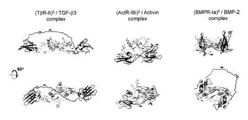
Figure 4. Depicts diagrams relating to the feasibility of specific embodiments
of trap
constructs with natural linkers from three-dimensional structural models.
Shown are
molecular mechanics energy-minimized natural linkers for embodiments of (TbR-
I1)2,
(ActR-11b)2 and (BMPR-Ia)2 homo-bivalent single-chain traps in complex with
the TGF-
(33, Activin and BMP-2 growth factors, respectively. Each growth factor
covalent dimer is
rendered in gray. Each single-chain trap is rendered in black, and consists of
two folded
binding domains and in intervening unstructured linker. Each dot indicates the
point of
fusion in the linker region between two receptor ectodomains to generate the
single-
chain trap. Arrowheads indicate polypeptide chain direction in the trap's
linker. Two 90 -
rotated views are provided for each complex.
Figure 5A. Depicts molecular dynamics (MD) model for an embodiment of the
(T13R-11)2
homo-bivalent single-chain trap bound to the TGF-133 growth factor (right
images). An
3
CA 02681177 2015-06-18
initial model with energy-minimized linker and with ligand-binding domains in
crystallographic positions bound onto the growth factor is also shown for
reference (left
images, see also Figs. 3 and 4). The single-chain trap is rendered in black
and the
growth factor covalent dimer is rendered in gray. Ten time-averaged structures
(each
over 1 ns) covering 10 ns timeframe of MD simulation are overlaid. Two 90 -
rotated
views are provided
Figure 5B. is a graphical representation of per residue root-mean-square (RMS)
fluctuations of an embodiment of the (Tf3R11)2 / TGF-I33 complex, time-
averaged over
the last 10 ns of MD simulation.
Figure 5C. is a graphical representation of solvated Interaction Energy (SIE)
between
an embodiment of a single-chain (TORI1)2 trap and the TGF-I33 ligand over the
last 10
ns of MD simulation of their complex, with an average value of -25.4 kcal/mol.
Figure 6. Depicts a schematic of embodiments of prototype (Tf3R11)2 and
modified N-His
(T13R11)2 traps.
Figure 7A. Depicts surface plasmon resonance (SPR)-based biosensor (BiacoreTM)
sensograms showing an embodiment of a prototype (Tf3R11)2 (in diluted
conditioned
media from different % transfections) binding to surface-immobilized TGF-I33
ligand.
Figure 7B. Depicts surface plasmon resonance sensograms comparing binding of
embodiments of bivalent prototype (Tf3R11)2, bivalent TpRII-Fc and monovalent
TpRII to
270 RUs surface-immobilized TGF-f33 ligand.
Figure 8. Is a photographic depiction of a gel showing high level production
and
purification yield of an embodiment of N-His (TpRII)2 protein from 500 ml
culture of
transfected 293 cells.
Figure 9A. Is a graphical depiction of inhibition of TGF-13 signaling in MO Lu
luciferase
reporter cells by an embodiment of prototype (Tf3R11)2 compared to TpRII-Fc.
Figure 9B, Is a graphical depiction of SPR-based determination of trap binding
of TGF-
f3 in solution by an embodiment of prototype (Tf3R11)2 and Tf3R1I-Fc compared
to
monomeric TpRII-ED.
Figure 9C. Is a graphical depiction of inhibition of TGFf31-induced 4T1 cell
invasion in
vitro by an embodiment of prototype (Tf3R11)2 and Tf3R11-Fc traps.
Figure 10A. Is a Biacore TM sensogram showing direct binding of embodiments of
N-His
(TpR11)2 and monomeric N-His TI3R11 to different isoforms of TGF-13.Figure
10B. Is a
4
CA 02681177 2015-06-18
graphical depiction of a BiacoreTM comparison of performance of embodiments of
100
nM N-His (TORI1)2 and TpRII-Fc to bind to 500 RUs each of TGF-f31 or TGF-p3
Figure 10C. Is a graphical depiction of SPR-based determination of IC50 for
trap
binding to TGF-131 (5 nM) in solution. The graph shows efficient binding of
TGF-p1 by
an embodiment of a N-His (TORI1)2 trap and TpRII-Fc trap versus reduced
binding by
monomeric Tr3R11 (293 cell-produced or E.coli-produced).
Figure 10D. Is a graphical depiction showing efficient inhibition of TGF-p
signaling in
Mv1Lu luciferase reporter cells by an embodiment of N-His (T13RI1)2 and TpRII-
Fc
compared to poor inhibition by monomeric Tf3R11 (293 cell-produced and E-Coli-
produced).
Figure 11. (A) is a photographic depiction and (B) is a graphical depiction of
results
showing that an embodiment of N-His (TpRI1)2 exhibits long-term stability and
activity in
10% serum at 37 C
Figure 12. Provides graphical depictions showing efficient neutralization of
TGF-131 (A)
and binding of TGF-I31 in solution (B) by an embodiment of a (TpR11)2 trap
(ligand
binding agent) having a 60 amino acid linker.
Figure 13. Is a graphical depiction showing efficient inhibition of Myostatin
signaling in
A204 cells by an embodiment of an (ActRI1B)2 trap (ligand binding agent)
compared to
the less potent inhibition of ActRIIB-Fc and monomeric ActRI1B.
.. Figure 14. Is a graphical depiction of results showing that an embodiment
of a bivalent
(BMPR1a)2 trap (ligand binding agent) is more potent than monovalent BMPR1a
trap
for neutralization of BMP2.
Figure 15A. Provides schematic diagrams exemplifying embodiments of in-line
fusions
of receptor ectodomains leading to embodiments of heterovalent single-chain
traps of
TGF-p-superfamily growth factors.
Figure 15B parts 1 and 2. Depict embodiments of amino-acid sequences
exemplifying
embodiments of heterovalent single-chain traps (ligand binding agents) of TGF-
p-
superfamily growth factors, and corresponding to the domain organization
diagrams
depicted in Figure15A. Natural or linker sequences are underlined; artificial
linker is in
underlined italics; Tf3R-1-ED structured domain is shown in bold-italics; T13R-
11-ED
structured domain is shown in bold; regular type denotes the unstructured
region of
TpR-11-ED that becomes structured in the ternary complex TPR-I/ Tf3R-II/ TGF-p
(Groppe et al, 2008).
5
CA 02681177 2015-06-18
Summary of Invention
The invention relates to ligand binding agents capable of permitting
modulation of cellular
response to members of the TGF-I3 superfamily by binding one or more members
of the
TGF-6 superfamily and preventing interaction with cellular receptors, and
methods of
designing and using such agents. The ligand binding agents taught herein are
preferably
single chain multivalent ligand binding agents. However, it would be possible
to link such
single-chain constructs to other uni- or multivalent molecules and/or to
combine two or
more such single chain traps uing multimerization domains known in the art
(e.g. coiled-
coil domains, Fc domains, pentabodies) to form a multimeric trap if so desired
and any
such trap having a multivalent single chain portion falls within the scope of
the present
invention.
In an embodiment of the invention there is provided methods and processes to
engineer
multivalent receptor ectodomains using a single-chain approach.
The ligand binding agents of the invention are preferably multivalent ligand
traps, having
at least two binding domains (bd) which recognize different sites on (or the
same site of
different portions of) the same member of the TGF-6 superfamily. The binding
domains
may be modified, for example to facilitate purification, so long as such
modifications do not
reduce binding affinity to unacceptable levels.
The binding domains (bd) of the ligand traps are preferably joined by a
flexible polypeptide
linker region. This linker should preferably include an unstructured amino
acid sequence
which in some embodiments is be either the same as or derived from
conservative
modifications to the sequence of a natural unstructured region in the
extracellular portion
of the receptor for the ligand of interest or another receptor in the TGF-6
superfamily. In
other instances, such linkers may be entirely artificial in composition and
origin but will
contain amino acids selected to provide an unstructured flexible linker with a
low likelihood
of encountering electrostatic or steric hindrance complications when brought
into close
proximity to the ligand of interest. =
In some instances, the linker will include regions to facilitate purification
(e.g. His tags) or
to facilitate the addition of cargo or accessory molecules. When such
additions affect the
unstructured nature of the linker or introduce potential electrostatic or
steric concerns,
appropriate increases to the linker length will be made to ensure that the two
binding
6
CA 02681177 2015-06-18
domains are able to bind their respective sites on the ligand. In light of the
methods and
teachings herein, such determinations could be made routinely by one skilled
in the art.
In an embodiment of the invention there are provided ligand traps having the
general
.. Structure I:
(<bd1>-linkerl )k-H<bd1>-1inker2-<bd2>-11nker3f-In -(<bd3>)m-(1inker4-
<bd4>)db,
where:
- n and h are independently greater than or equal to 1;
d, f, m and k are independently equal to or greater than zero;
- bd1, bd2, bd3 and bd4 are polypeptide binding domains having an affinity for
the same
member of the TGF-6 superfamily, with bd1, bd2, bd3, and bd4 being
independently the
same or different from each other; and,
-linker1, 1inker2, 11nker3 and linker4 are unstructured polypeptide sequences;
wherein the number of amino acids in each linker is determined independently
and is
greater than or equal to X/2.5; where,
X equals the shortest linear distance between:
(a) the C-terminus of an isolated form of the binding domain that is located
at the N-
terminus of the linker and that is specifically bound to its ligand; and,
(b) the N-terminus of an isolated form of the binding domain that is located
at the C-
terminus of the linker and that is specifically bound to its ligand.
As used herein "an isolated form" of a binding domain is a form of that
binding domain
acting as a monovalent monomer.
Subject to the constraints described herein, linkers 1, 2, 3, and 4 may be the
same or
different. In certain embodiments the linker is between 25 and 60 amino acids
in length
Also provided are nucleic acid sequences encoding such ligand traps.
Depending on the values seleCted for d, f, h, k, m, and n, the ligand trap
structure may
comprise a large number of repeating units in various combinations or may be a
relatively simple structure such as Structure II <bd1>-linker-<bd2>.
In certain embodiments of the invention, the member of the TGF-6 superfamily
to which
the binding domains (bd) have affinity is selected from the group consisting
of: TGF-61,
7
CA 02681177 2015-06-18
TGF-132, TGF-63, activin f3A, activin 6B, activin pc, activin pE, bone
morphogenic
protein (BMP) 2, BMP 3, BMP4, BMP 5, BMP 6, BMP 7, BMP 8, BMP 9, BMP 10, BMP
11, BMP 12, BMP 13, BMP 14, BMP 15, growth differentiation factor (GDF) 1, GDF
3,
GDF 8, GDF 9, GDF 15, Nodal, lnhibin a, anti-Mullerian Hormone, Lefty 1, Lefty
2,
arteman, Persephin and Neurturin.
In an embodiment of the invention there is provided a binding agent wherein
one or
more of bd1, bd2, bd3, and bd4 is selected from one of SEQ ID NO 43-48.
.. In an embodiment of the invention the binding agent comprises one or more
of SEQ ID
No 31 and 75 to 81.
In an embodiment of the invention the binding agent comprises one or more of
SEQ ID
NO 31-42 or 49-74 as a linker sequence.
The invention also provides a method of designing a multivalent binding agent
useful in
modulating responsiveness of a cell to a member of the TGF-6 superfamily, said
method comprising:
a) identifying a member of the TGF-p superfamily of interest;
.. b) obtaining two polypeptide binding domains having affinity for different
sites on the
member of the TGF-6 superfamily member;
c) obtaining an unstructured polypeptide linker of at least a number of amino
acids
equal to (X/2.5) where
X equals the shortest linear distance between:
(i) the C-terminus of an isolated form of the binding domain that is located
at the
N-terminus of the linker and that is specifically bound to its ligand; and,
(ii) the N-terminus of an isolated form of the binding domain that is located
at the
C-terminus of the linker and that is specifically bound to its ligand; and,
d) modelling the linker between the binding domains and carrying out molecular
.. dynamics simulations to substantially minimize molecular mechanics energy
and reduce
steric and electrostatic incompatibility between the linker and the member of
the TGF-6
superfamily.
8
CA 02681177 2015-06-18
The design method can optionally be expanded to further include a step e) of
producing
a fusion protein comprising the two polypeptide binding domains joined by the
unstructured polypeptide linker.
The ligand binding agents disclosed herein are also useful in purification of
ligand, for
example, by immobilization on an inert matrix on a solid support, on, for
example, to
nanoparticles to concentrate levels of ligand in a sample.
The invention also provides novel polypeptide sequences useful in a Variety of
applications. These sequences include SEQ ID NOs 53 to 74. Also provided are
nucleic acid sequences encoding these polypeptide sequences.
Also provided is a method of modulating the response of a cell to TGF-p in its
environment, said method comprising exposing the cell to a multivalent ligand
trap
comprising a ligand binding agent (ligand trap) disclosed herein.
In an embodiment of the invention there is provided a binding agent having the
general
structure V:
R3 R4 R5 R6 R7 Rg Rg
R1-(<bd1>-linker1)k-R<bd1>-linker2-<bd2>-linker31-ln -(<bd3>)m-(linker4-
<bd4>)dh-R2
Wherein R1, R2, R3, R4, R5r R6, R7, R8, R9, may be the same or different, may
not be
present and when present, may independently be one or more of a fusion protein
for
targeting, a single domain antibody, a radiotherapy agent, an imaging agent, a
fluourescent dye, a fluorescent protein tag, a cytotoxic agent for
chemotherapy a nano
particle-based carrier, a polymer-conjugated to drug, nanocarrier or imaging
agent, a
stabilizing agent, a drug a nanocarrier and a dendrimer and a support for use
in
purification or concentration of ligand; and wherein bd1, bd2, bd3, bd4,
linker1, linker2,
linker3, 1inker4, k, f, n, m, d, and h are defined as in Structure I. In light
of the
disclosure herein, one skilled in the art can select suitable R-groups for
diagnostic
therapeutic or other applications.
9
CA 02681177 2015-06-18
In an embodiment of the invention there is provided an isolated polypeptide
having at
least 80%, 85%, 90%, 95%, 98%, 99% and 100% sequence identity to a natural
unstructured region in the extracellular portion of a receptor for a member of
the TGF-3
superfamily and being substantially free of structured regions capable of
specific binding
to a member of the TGF-3 superfamily. In some instances, this isolated
polypeptide has
at least 80%, 85%, 90%, 95 %, 98%, 99 % sequence identity to one or more of
SEQ ID
NO 31-42 and SEQ ID NOs 49-74.
In an embodiment of the invention there is provided a polypeptide comprising a
region
having at least 80%, 85%, 90%, 95 %, 98%, 99 % sequence identity to one or
more of
SEQ ID NOs 53-74 and SEQ ID NOs 31-42 and 49-74. In some instances this
polypeptide has a region with at least 90 %, 95 %, 98%, 99 % sequence identity
to one
or more of SEQ ID NOs 53-74.
In an embodiment of the invention there is provided a polypeptide having
between 43 %
and 99 % sequence identity to a naturally unstructured region in the
ectodomain of a
receptor for a member of the TGF-3 superfamily.
In an embodiment of the invention there is provided a nucleic acid sequence
encoding a
polypeptide disclosed herein.
In an embodiment of the invention there is provided a method of modulating the
response of a cell to a TGF-3 superfamily member in its environment, said
method
comprising exposing the cell to a ligand binding agent disclosed herein.
In an embodiment of the invention there is provided a data storage medium
comprising
instructions for determining the minimum linker length when designing a ligand
binding
agent.
In an embodiment of the invention there is provided a data storage medium
comprising
a means for identifying acceptable minimal linker length when designing a
ligand
binding agent.
CA 02681177 2015-06-18
Linker length will be considered acceptable when it permits binding of binding
domains
located on each of the N- and C-termini of the linker to bind their natural
binding sites on
their natural ligand such that, with both binding domains so bound, the ligand
is bound
with a higher affinity than it would be bound by binding of only one of the
binding
domains.
Brief Description of the Invention
In an embodiment of the invention there is provided a single-chain non-
naturally
occurring polypeptide useful as a ligand binding agent. The ligand binding
agent
comprises structured ligand-binding domains (denoted bd) derived from or based
on the
extracellular portion of a natural receptor or receptors, joined by one or
more
polypeptide linkers. The ligand binding agent provides a multivalent binding
agent and
does not require fusion to any conventional dimerizing or multimerizing
moieties such as
coiled-coil domains of Fc domains in order to be multivalent.
In an embodiment of the invention, there is provided a multivalent binding
agent with
affinity for a member of the TGF-I3 superfamily, said agent comprising the
general
structure I:
(<bd1>dinker1)k-[{<bd1>-linker2-<bd2>-linker3fin -(<bd3>)m-(linker4-<bd4>)db,
where:
- n and h are independently greater than or equal to 1;
- d, f, m and k are independently equal to or greater than zero;
- bdi, bd2, bd3 and bd4 are polypeptide binding domains having an affinity for
the same
member of the TGF-(3 superfamily, with bd1, bd2, bd3, and bd4 being
independently the
same or different from each other; and,
linker2, linker3 and linker4 are unstructured polypeptide sequences;
wherein the number of amino acids in each linker is determined independently
and is
greater than or equal to X/2.5; where,
X equals the shortest linear distance between:
(a) the C-terminus of an isolated form of the binding domain that is located
at the N-
terminus of the linker and that is specifically bound to its ligand; and,
11
CA 02681177 2015-06-18
(b) the N-terminus of an isolated form of the binding domain that is located
at the C-
terminus of the linker and that is specifically bound to its ligand.
As used herein "an isolated form" of a binding domain is a form of that
binding domain
acting as a monovalent monomer.
The length of the linker is considered to be the number of amino acids
between:
(a) the C-terminal main chain carbon atom of the binding domain located at the
linker's
N-terminal end; and
(b) the N-terminal main-chain nitrogen atom of binding domain located at the
linker's C-
terminal end.
Non-limiting examples of useful linkers are found in the amino acid sequences
in SEQ
ID NOs 53 to 74 which should be read conventionally with the N-terminus on the
left and
.. the C-terminus on the right, and in corresponding reverse sequences having
the same
amino acids but wherein the C terminus is on the left and the N- terminus is
on the right
as the sequences are written in full. In some embodiments, such reverse
sequences
will preferably be produced using D-amino acids. Where immunogencity is of
concern,
it will generally be desired to screen such reverse sequences for
immunogenicity at an
early stage. (For examples of reverse sequences, see SEQ ID NOs 31-42 and 49-
74).
All amino acids sequences in this document are written N-terminus to C-
terminus unless
otherwise noted. All sequences disclosed herein except SEQ ID NOs 31-42 and 49-
74
are disclosed as using L-amino acids and the use of a D-amino acid is
considered a
variant affecting the percent sequence identity to the sequences as stated.
In an embodiment of the invention, the ligand binding agent has the general
Structure II:
<bd1>-linker2-<bd2>.
In an embodiment of the invention, the ligand binding agent has the general
Structure III
<bd1>-(linker2-<bd2>)n.
In an embodiment of the invention, the polypeptide has the general Structure
IV:
([13d1]-[linker1MbdThrilinker2]-([bd2]-[1inker3Hbd3Dg,
when f and g are greater than or equal to one.
12
CA 02681177 2015-06-18
In an embodiment where bd2 and bd3 are the same, and f and g are the same
number,
this can result in a substantially mirror symmetric structure around linker 2,
subject to
differences in the linkers. In instances where bd3 is different from bd2 and
and/or
where f and g are different numbers, different structures will be produced. It
is within
the capacity of one of ordinary skill in the art to select suitable binding
domains, linkers,
and repeat frequencies in light of the disclosure herein.
In some instances, the binding domain region of the single-chain polypeptide
will be
selected for its ability to bind a growth-factor ligand having a covalently-
stabilized
dimeric quaternary structure, and may be selected from a list of growth
factors from
within the TGF-p family, e.g., transforming growth factor beta (TGF-D, bone
morphogenetic protein (BMP), activin, myostatin, and including their naturally
occurring
isoforms. =
In some instances, the polypeptide is designed to bind simultaneously to
equivalent but
spatially distinct sites on a multimeric ligand. As used herein "multimeric"
includes
dimeric, trimeric, and greater numbers of units, and "multivalent" includes
bivalent,
trivalent, and greater numbers of binding domains.
In some instances, the linker is independently selected to have varying
degrees of
sequence identity to naturally occurring unstructured amino acid sequences
found in the
native receptor sequence in the regions flanking the ligand binding domain,
for example
70%, 80%, 90%, 95%, 98 %, 99% or 100% sequence identity, whereas for entirely
artificial linkers (e.g. poly-Gly or poly-Ser linkers, sequence identity will
be even lower.
Examples of linker sequences of varying degree of identity to the natural
receptor
sequence are given in Fig. 2C. and SEQ ID NOs 31-42 and 49-74 (Table II).
In some instances, the number of amino acid residues in the linker of either
natural or
artificial origin is selected to be equal to or greater than the minimum
required distance
for simultaneous (bridged) binding to two binding sites on the growth factor
to be bound
by the relevant binding domains. An example of an embodiment of such a
determination is given in the section "Feasibility assessment procedure for
designed
single-chain bivalent traps". Examples of natural and artificial linker
sequences of
varying length are given in Fig. 2D and SEQ ID NOs 31-42 and 49-74. In some
instances, linker length is between 18-80 a.a., 25-60 a.a., 35-45 a.a.
13
CA 02681177 2015-06-18
In some instances, the overall molecular mass of bivalent agents disclosed
herein
before glycosylation is between about 29 kDa and 37 kDa, and the overall mass
following typical glycosylation is between about 40 kDa and 60 kDa. Thus,
there is
provided herein, multivalent ligand traps having a pre-glycosylation size of
between
about 12 kDa and 19 kDa per binding domain.
The ligand traps disclosed herein will generally have a lower molecular mass
than
comparable multimeric ligand traps constructed using known multimerization
domains.
Example of Selected Ligand Trap Sizes
Agent Predicted for protein Actual (with
glycosylation)
based on SDS-PAGE
(Tf3R11)2 34kDa 50-60kDa
(TpRI1b)2 37kDa 50-60kDa
(ActRIIB)2 30kDa 50-60kDa
(BMPR1a)2 29kDa 40-50kDa
37Kd+40kDa 77kDa
TpRII-Fc 60Kd+60kDa = 120kDa
=
Polypeptides of the invention can be useful as therapeutic agents that
neutralize the
action of disease-associated covalently-stabilized dimeric ligands such as
growth
factors. They may also have commercial potential for use as diagnostic agents
to detect
the presence of disease-associated covalently-stabilized dimeric ligands such
as growth
factors in imaging and non-imaging diagnostic applications. They can also be
useful in
the purification and/or concentration or segregation of ligand in vitro.
Detailed Description of Invention
Although the invention is described with reference to specific examples, it
will be
understood that it is not so limited.
Experiment #1: Design strategy of single-chain bivalent traps for TGF-13-
family
ligands
14
CA 02681177 2015-06-18
1. Single-chain recombinant traps were designed against growth factors that
belong to
the transforming growth factor TGF-P superfamily of cysteine-knot cytokines
according
to SCOP (Andreeva et al., 2008, Nucl. Acid Res. 36: D419) and Pfam (Finn et
al., 2006,
Nucl Acid Res. 34: D247) structural classifications. More specifically, these
growth
factors including, for example, TGF-Ps, activins and BMPs, share the same 3D
architecture and form covalent disulfide-linked homodimers. The method
disclosed
herein is applicable to all members of the TGF-Psuperfamily, including TGF-
131, -p2, -
133; activin pA, pB, pc, pE; bone morphogenetic proteins (BMP) 2-15; growth
differentiation factors (GDF) 1, 3, 8 (myostatin), 9 and 15; Nodal; lnhibin a;
anti-
Mullerian hormone (AMH); Lefty 1 and 2; Arteman, Persephin and Neurturin.
2. Single-chain recombinant traps against TGF-p superfamily growth-factors
were
designed from the extracellular portion of their cognate natural receptors.
The
extracellular segment of all these TGF-13 superfamily receptors contain a
single
structured domain that belongs to the snake-toxin family according to SCOP
(Andreeva
et al., 2008, Nucl. Acid Res. 36; D419) and Pfam (Finn et al., 2006, Nucl Acid
Res. 34:
D247) structural classifications. The complete extracellular portion of these
receptors
typically includes unstructured segments flanking their folded ligand-binding
domain.
These unstructured extracellular portions were apparent from the
experimentally
determined 3D structures available from the PDB database (Berman et al., 2000,
Nucl.
Acid Res. 28: 235), e.g., crystal structures for type II TGF-p receptor
ectodomain (Hart
et al., 2002 Nat. Struct. Biol. 9: 203; Boesen et al., 2002, Structure 10:
913; Groppe et
al., 2008, Mol. Cell 29: 157), type 1 TGF-p receptor ectodomain (Groppe et
al., 2008,
Mol. Cell 29:157), type Ila activin receptor ectodomain (Allendorph et al.,
2006, Proc.
Natl. Acad. Sci. USA 103: 7643), type 1lb activin receptor ectodomain
(Thompson et al.,
2003, EMBO J. 22: 1555; Greenwald et al., 2004, Mol. Cell 15: 485), type I BMP
receptor ectodomain (Kirsch et al., 2000, Nat. Struct. Biol. 7: 492), or the
NMR structure
of the type ll TGF-(3 receptor ectodornain (Deep et al., 2003, Biochemistry
42: 10126)].
In the absence of experimental data, as for example in the case the
extracellular region
of the 1lb splicing variant of the TGF-13 type II receptor, unstructured
extracellular
segments were defined by: (i) sequence portions falling outside of the folded
ligand-
binding domain boundaries located by comparative analysis against structurally
characterized homologs, and (ii) predictions based on knowledge-based
algorithms,
e.g., DISOPRED (Ward et al., 2004, J. Mol. Biol. 337: 635). Amino acid
sequences
CA 02681177 2015-06-18
corresponding to the unstructured (i.e., flexible) and structured (i.e.,
folded, ligand-
binding domain) regions from the ectodomains of several receptors of TGF-13-
superfamily growth factors, are given in Figs. 1A and 16, respectively.
3. Homo-bivalent single-chain recombinant traps hereby designed against TGF-3-
superfamily growth factors disclosed herein were designed with regard to the
experimentally determined binding mode between TGF-p-family ligands and the
extracellular portion of their cognate natural receptors. The ligand-receptor
binding
mode was provided at atomic level by the high-resolution 3D structures
available for
several members of the TGF-p-superfamily ligands in complex with their cognate
receptor ectodomains. Examples of experimental molecular structures for TGF-p-
superfamily-growth-factor/receptor ectodomain complexes include TGF-p3 bound
to
Tf3R-11-ED (Hart et al., 2002 Nat. Struct. Biol. 9: 203), activin bound to
ActR-1Ib-ED
(Thompson et al., 2003, EMBO J. 22: 1555; Greenwald et al., 2004, Mol. Cell
15: 485),
BMP-2 bound to type la BMP receptor ectodomain BMPR-la-ED (Kirsch et al.,
2000,
Nat. Struct. Biol. 7: 492) and ActR-1Ia-ED (Allendorph et al., 2006, Proc.
Natl. Acad. Sci.
USA 103: 7643), BMP-7 bound to ActR-11a-ED (Greenwald et al., 2003, Mol. Cell
11:
605). These structures provided the relative spatial orientation between two
separate
receptor ectodomain chains (molecules) binding simultaneously onto one
covalently
homodimerized ligand molecule, i.e., 2:1 receptorligand stoichiometry. Higher-
order
.. ligand-receptor assemblies between a particular TGF-p-superfamily growth
factor and
ectodomains from different receptor types have also been determined, for
example the
ternary complexes between TGF-f33, Tf3R-11-ED and Tf3R-1-ED (Groppe et al.,
2008,
Mol. Cell 29:157) or between BMP-2, ActR-1Ia-ED and BMPR-la-ED (Allendorph et
al.,
2006, Proc. Natl. Acad. Sci. USA 103: 7643). These structures provide the
relative
spatial orientation between four separate receptor ectodomain chains
(molecules)
binding simultaneously onto one covalently homodimerized ligand molecule,
i.e., 2:2:1
high-affinity-receptor: low-affinity-receptor: ligand stoichiometry. Such
structures were
used as guides to design hetero-bivalent, hetero-trivalent and hetero-
tetravalent single-
chain traps of TGF-p-superfamily growth factors and are useful in designing
single-
.. chain traps for other suitable ligands of interest involving the TGF-f3
superfamily.
4. Homo-bivalent single-chain traps of TGF-B-family ligands were therefore
designed as
unnatural fusion proteins consisting of the sequence (excluding the signal
peptide) of
the natural extracellular portion of the receptor repeated twice. Fig. 2A
presents
schematically homo-bivalent single-chain traps with natural linkers for three
TGF-B-
16
CA 02681177 2015-06-18
family ligands, where structured and unstructured regions are based on
experimental
data for single-domain extracellular portions, as presented in Figs. 'IA and
1B.This
resulted in constructs with two structured domains for binding to select TGF-
13-
superfamily ligand(s), spaced by an unstructured flexible linker formed by
fusing the
unstructured C-terminus of the first domain to the unstructured N-terminus of
the
second domain. The natural linker was also progressively substituted by
artificial
sequences as well as varied in length (Figs. 2B-D). From thermodynamic and
kinetic
considerations, it was expected that divalent receptor ectodomains would
provide
increased ligand-binding affinities and slower ligand-dissociation rates
relative to single-
domain receptor ectodomains.
Experiment #2: Feasibility assessment procedure for designed single-chain
bivalent traps
To the extent to which the structures of various TGF-p-superfamily growth
factors are
conserved, the structures of their cognate receptor ectodomains are conserved,
and the
2:1 receptor-ligand binding stoichiometry is conserved, the concept of fusing
two natural
receptor ectodomain sequences to produce single-chain homo-bivalent traps with
improved in vitro ligand binding affinity and cellular ligand neutralizing
activity relative to
respective monovalent receptor ectodomains, is applicable to the entire family
of TGF-p
family. The feasibility of these ligand traps can be theoretically assessed
routinely by
following the stepwise procedure
outlined below. Although the procedure is presented for homo-bivalent single-
chain
traps, it also applies to other designs covered here, e.g., hetero-bivalent
and hetero-
tetravalent single-chain traps.
1. The linear distance is measured between the C-terminal main-chain carbon
atom of
one domain and the N-terminal main-chain nitrogen atom of the other domain
when
bound to the covalently-dimerized ligand. Alternate structures of the complex
reflecting
internal geometrical flexibility in the homodimerization mode of the disulfide-
stabilized
ligand when bound to the receptor ectodomains, as reported in several cases
(Greenwald et al., 2004,
17
CA 02681177 2009-09-17
WO 2008/113185
PCT/CA2008/000547
Mol. Cell 15: 485), can be included in the design process. A computer hardware
equipped with commercial/public software appropriate for manipulating
molecular
structures on an available graphics device can be routinely employed to this
end.
2. The linear distance (in A units, 1 A = 10-10 m) is divided by a factor of
2.5 to calculate
the minimum number of amino acid residues that the flexible linker should
posses
(Table 1) in order to allow simultaneous binding of the folded domains to
their binding
sites on the homodimeric ligand. The 2.5 factor is based on the Ca-Cpextent of
fully
extended linkers, which peaks at 3.0 A (George and Heringa, 2002, Protein Eng.
15:
871), minus an average tolerance of 0.5 A per amino acid residue to allow for
deviations
of the linker path from linearity.
(Table 1. Linker characteristics for select examples of single-chain traps of
TGF-13-
family growth factors. Minimum number of residues required for linkage
represents the
structure-based linear distance for linkage (A) divided by a factor of 2.5.)
3. The number of amino acid residues in the unstructured linker portion of the
bivalent
single-chain trap should be at least equal to the estimated minimum number of
linker
residues required. Receptor isoforms that differ in the length of the
extracellular
unstructured segments, such as the TGF-13 receptor isoforms II and Ilb (Fig.
2B), can
.. be included in the design process. The natural sequence-based linker can
also be
shortened up to the estimated minimum number of amino acid residues without
significantly impairing the ligand binding affinity and neutralizing activity
of the trap. A
preferable location for shortening the unstructured linker is from the point
of fusion (see
Fig. 3) in either or both directions relative to the amino acid sequence.
Example of
shortened natural linkers that can be utilized in single-chain trap design are
given in Fig.
2D. As listed in Table 1, the required minimal length of the linker varies
between various
single-chain traps of TGF-13-superfamily growth factors. An upper limit for
the length of
the unstructured linker is not defined. Hence, ligand binding agent (trap)
constructs with
linkers comprising unstructured sequence segments repeated in whole or in part
are
envisioned to comply with bivalent design and preserve the desired
characteristics of
the trap. The natural linker can be progressively substituted by artificial
sequences,
which may or may not result in different linker lengths. Examples of linkers
longer than
the natural linker designed by repeating of natural sequence or by introducing
of
artificial sequence are given in Fig. 2D.
18
CA 02681177 2009-09-17
WO 2008/113185
PCT/CA2008/000547
4. Finally, atomic-level theoretical analysis is to be carried out, where the
linker is
modeled between the structured domains and the molecular structure of the trap-
ligand
complex is refined by minimizing the molecular mechanics energy and by
carrying out
molecular dynamics simulations (Cornell et al., 1995, J. Am. Chem. Soc. 117:
5179).
This may, in some cases, highlight regions of steric and/or electrostatic
incompatibility
between the trap's linker and the growth-factor, and suggest that the length
and/or
composition of the linker may be incompatible with the bivalent design, even
lithe linker
complies with the minimum number of amino acids requirement as per step (3.)
above.
If the linker can be accommodated without affecting the simultaneous binding
of the
structured domains to their binding sites on the ligand, then the trap
construct is
deemed feasible for the proposed application. Computer hardware equipped with
commercial/public software appropriate for manipulating molecular structures
on an
available graphics device, and for performing energy calculation and
simulation based
on molecular mechanics force fields, e.g., the AMBER force field (Cornell et
al., 1995, J.
Am. Chem. Soc. 117: 5179), can be routinely employed by one skilled in the art
in order
to carry out this structural modeling analysis. A detailed molecular modeling
analysis of
the (T13R-11)2 homo-bivalent single-chain trap is provided as an example in
the following
section and includes molecular dynamics simulation. Examples of molecular
mechanics
energy-refined models of three single-chain homo-bivalent traps: (T3R-I1)2,
(ActR-1Ib)2
and (BMPR-Ia)2, bound to their respective growth factors are shown in Figs. 3
and 4.
These atomic-level models represent starting points for further computer-based
optimization of linker composition and length.
This process is explained in greater detail in the example below:
Modeling Example A, Experiment #2
i. In one example, the atomic-level solution structure of the single-chain
homo-bivalent
trap (TOR-I1)2 was simulated in complex with the growth factor TGF-133.
The starting point for molecular design of the (Tf3R-11)2 trap was the 2.15 A-
resolution crystal structure of the disulfide-linked dimeric human TGF-133
complexed
with two TGF-13 type II receptor ectodomains (Hart et al., 2002 Nat. Struct.
Biol. 9: 203),
deposited in the Protein Data Bank (Berman et al., 2000, Nucl. Acid Res. 28:
235) under
19
CA 02681177 2009-09-17
WO 2008/113185
PCT/CA2008/000547
the code 1KTZ. Because this structure displays the growth factor in a non-
canonical
conformation probably due to the low pH used in the crystallization conditions
(Hart et
al., 2002 Nat. Struct. Biol. 9: 203; Groppe et al., 2008, Mol. Cell 29:157),
the binary
complex structure was first reconstructed with the ligand in canonical
conformation as
reported previously (Hinck et al., 1996, Biochemistry 35: 8517; Mittl et al.,
1996, Protein
Sci. 5: 1261), which was also recently confirmed by the ternary structure of
TGF-13
ligand-receptor assembly (Groppe et al., 2008, Mol. Cell 29:157). An initial
3D molecular
model of the (1-13R-11)2 trap incorporating an inter-domain natural linker of
35 amino-acid
residues (as per sequence listed in Fig. 2B) was constructed from standard
geometries
followed by conjugate-gradient energy minimization of the molecular mechanics
force
field energy using an AMBER all-atom force field (Cornell et al., 1995, J. Am.
Chem.
Soc. 117: 5179) and the AMBER 9 suite of programs (Case et al., 2005, J
Comput.
Chem. 26: 1668). During energy minimizations, only the linker regions of the
traps were
allowed to move, while the coordinates of the growth factors and of the folded
domains
of the traps were fixed. The resulting 3D molecular model of the homo-bivalent
single-
chain trap (Ti3R11)2 bound to TGF-(33 is depicted in Fig. 4.
This initial model of the complex was used as input for molecular dynamics
(MD)
simulation carried out together with AMBER FF03 force field (Duan et al.,
2003, J.
Comput. Chem. 24: 1999; Lee & Duan, 2004, Proteins 55: 620) within the AMBER 9
suite of programs (Case et al., 2005, J. Comput. Chem. 26: 1668). The
molecular
system consisting of 245 amino-acid residues from the single-chain trap (from
the full-
length (Tr3R-11)2 trap with 272 amino-acid residues, 21 unstructured residues
from the N-
terminus and 6 flexible residues from the C-terminus were not included in the
MD
simulation), 224 amino-acid residues of the TGF-133 dimer and 14 Na+
counterions
(added to maintain electroneutrality) was solvated in rectangular water box
using the
Xleap program in the AMBER 9 software. The distance between the wall of the
box and
the closest atom of the solute was 12.0 A, and the closest distance between
the solute
and solvent atoms was 0.8 A. The entire system was energy-minimized by
applying
harmonic restraints with force constants of 10 kcal/mol/A2 to all solute
atoms, followed
by heating from 100K to 300K over 25 ps in the canonical ensemble (NVT), and
by
equilibrating to adjust the solvent density under 1 atm pressure over 25 ps in
the
isothermal-isobaric ensemble (NPT) simulation. The harmonic restraints were
then
gradually reduced to zero with four rounds of 25 ps NPT simulations. After
additional 25
PS simulation, a 15 ns production run was obtained with snapshots collected
every 1 ps.
CA 02681177 2009-09-17
WO 2008/113185
PCT/CA2008/000547
For all simulations, 2 fs time-step and 9 A non-bonded cutoff were used. The
Particle
Mesh Ewald method (Darden et al., 1993, J. Chem. Phys. 98: 10089) was used to
treat
long-range electrostatics, and bond lengths involving bonds to hydrogen atoms
were
constrained by SHAKE (Ryckaert et al. 1977, J. Compt. Phys. 23: 327). No other
constraints were imposed during the MD simulation.
As seen from Fig. 5A, the single-chain trap (T6R-I1)2 bound to TGF-63 attains
a
stable MD solution structure that preserves the simultaneous binding of the
two ligand-
binding domains onto the dimeric growth factor as observed in the crystal
structure of
unlinked receptor ectodomains (Hart et al., 2002 Nat. Struct. Biol. 9: 203,
Groppe et al.,
2008, Mol. Cell 29:157). This substantiates the feasibility of the designed
single-chain
TGF-6 trap in terms of the length of the linker. The MD analysis also reveals
that in the
complex, the linker region of the single-chain trap becomes relatively rigid,
with only 6
residues experiencing greater mobility (expressed as per-residue and time-
averaged
root-mean-square fluctuations) than the rest of the trap's amino acid residues
(Fig. 5B).
In addition, the single-chain trap established favorable interaction with the
growth factor,
as evaluated by the solvated interaction energy function for scoring protein-
ligand
binding affinity (SIE) (Naim et al., 2007, J. Chem. Inf. Model. 47: 122). A
highly
favorable SIE value of -25.4 kcal/mol was calculated as an average over the
last 10 ns
of MD simulation (Fig. 5C). This further indicates the feasibility of the
employed natural
linker in terms of amino acid composition, that is, there were no significant
unfavorable
steric and electrostatic contacts predicted between the trap's linker and the
growth
factor.
ii. In one example, structure-based design leads to a divalent molecule
consisting of two
human T6RII ectodomains that are fused in tandem into a single polypeptide
chain
(schematically shown in Fig. 6). In this construct, an intervening linker
sequence is
formed from the unstructured natural C-terminal sequence of one ectodomain
(black, 10
residues) and the unstructured natural N-terminal sequence of the other
ectodomain
(white, 25 residues). This linker bridges between the two structured TGF-(3-
binding
domains. This TGF-13 trap is hereby named prototype (-113R11)2. The construct
also
contains an N-terminal myc tag and C-terminal 6xHis tag for ease of detection
and
protein purification. In the prototype (T3RI1)2 the native IPP sequence is
replaced by
GGR within the linker due to a Notl restriction site inserted during
construction of the
21
CA 02681177 2009-09-17
WO 2008/113185
PCT/CA2008/000547
(TI3R11)2 gene. Also shown is another construct with a 35 amino acid residues
linker with
native IPP restored, and having a N-terminal His tag. This construct is termed
"modified
N-His" (Tf3R11)2 and features a native linker sequence. Predicted molecular
models of
(T13R11)2 bound to TGF-I3 are given
Figs. 3-5.
Experiment #3: Small scale production of prototype (T13RII)2 and demonstration
of
TGF-fl-bindinq activity
Fig. 6 shows a schematic of prototype (Tf3R11)2. The prototype (113R11)2 gene
was cloned
into mammalian expression vector pTT and increasing amounts were transiently
transfected into HEK293 cells. The conditioned media from these transfected
cells were
collected after 5 days and tested via SPR Biacore analysis for the binding of
secreted
(TI3R11)2 to a TGF-133 surface (Fig. 7A). The sensogram shows increasing
levels of
binding that correlates with cells transfected with increasing levels of
(113R11)2 plasmid
(ranging from 1% to 95% transfected cells), indicating a dosage effect and
specific
binding. The binding characteristics of (Tf3R11)2 (produced from 95%-
transfected cells)
was compared with dimerized Tf3R11-Fc and monomeric Tf3RII (Fig. 7B). The
sensogram of prototype
(1-13R11)2 was similar to the T13R11-Fc interaction (slow off rate), and both
were distinct
from monomeric T13R11 interaction (fast off rate), indicating that (T13R11)2
interacts with
the TGF-433 surface in a high-affinity, bivalent manner.
Experiment #4: Production and purification of prototype and modified N-His
(1-13R//)2
Scaleup production of prototype (T13R11)2 in 293 cells resulted in variable
yields of
protein (1-3 mgs per 1 liter culture) upon purification via cobalt column,
perhaps due to
a less accessible His tag at the C-terminus. A modified version was
constructed having
a N-terminal His tag, termed N-His (T13R11)2, as shown in Fig. 6. N-His
(T13R11)2-
transfected HEK293 cells were grown in 500 ml culture. The media was
collected,
concentrated 5-fold by 10 kDa Centricon filtration and then passed through a
10 ml
Fractogel Cobalt column. Fig. 8 shows a SDS-PAGE analysis of N-His (Tl3R11)2
at the
22
CA 02681177 2009-09-17
WO 2008/113185
PCT/CA2008/000547
various stages of purification. The N-His (TPRI1)2 in the eluted fractions
(lane 6) is
relatively pure and migrates as a smear (likely due to glycosylation) in the
50-60 kDa
range. The total yield from 500 ml culture was 7-8 mgs, indicating that the N-
His
(T3RII)2 protein is amenable to large-scale production.
Experiment #5: Demonstration that (Tf3R11)2 is a potent TGF-13 trap
The ability of purified prototype (TI3R11)2 to neutralize TGF-I3 was tested on
Mv1Lu cells
having a TGF-p-responsive luciferase reporter gene and compared with TPRII-Fc
from
two sources, commercial R&D and collaborator H. Lin (Fig. 9A). The resulting
inhibition
curves indicated the average IC50 for prototype (TI3R11)2 is 0.58 nM (S.D.
0.64) which is
in the same range as for TpRII-Fc Lin (0.45 nM) and slightly higher than TPRII-
Fc R&D
(0.1 nM). Purified prototype (TORI1)2 was also compared with dimeric TPRII-Fc
and
monomeric TPRII-ED for their ability to competitively bind TGF-p in solution
via Biacore
analysis (Fig. 9B). Increasing amounts of each binder was added separately to
a
constant amount of TGF-131 or 433 (5 nM) followed by coinjection of this
mixture over a
TGF-p-specific antibody surface. The level of unbound TGF-I3 at equilibrium is
assessed by the maximum/plateau level of the surface binding curve (Fig. 9B)
Prototype (TOR11)2 and TpRII-Fc have similarly low 1050s in the range of 5-8
nM, as
would be expected for intra-molecular, divalent binding of TGF-p. In contrast,
the IC50
for monovalent Tf3R1I-ED is 10-20 fold higher. One might predict, for full
avidity, that the
IC50 for dimeric (TI3R11)2 could be at least 100-fold greater than for
monomeric TPRII-
ED. In order to augment avidity, variable linker lengths may be sampled for
(T13RI1)2
(see Figs. 2C and 2D). These results (Figs. 9A and 9B) indicate that (T3RI1)2
is an
excellent trapping/neutralizing reagent for TGF-13 and hence is a good
candidate
therapeutic and/or diagnostic agent for diseases in which TGF-13 is causative
and
overexpressed/overactive (e.g. breast tumors). To this end we examined the
ability of
prototype (T131:211)2 to prevent TGF-13-induced invasion of 4T1 breast cancer
cells in vitro
(Fig. 9C). Similar to TPRII-Fc, prototype (T13R11)2 reduced 4T1 cell invasion
to
approximately 20% of the non-trap treated (+ TGF-13) control.
Experiment #6: Assessment of binding characteristics and efficacy of N-His
(T6R11)2
23
CA 02681177 2009-09-17
WO 2008/113185
PCT/CA2008/000547
The two different SPR Biacore assays that were utilized in Figure 7 and Figure
9B were
used again to characterize the N-His (T13RII)2 ¨ TGF-p ligand interaction.
First, the
direct binding assay was utilized where the TGF-f3 trap was injected over
various
immobilized TGF-P isoform surfaces. While this assay can verify binding to
different
TGF-p isoform surfaces, it cannot verify that a 1:1 trap: TGF-I3 homodimer
interaction is
occurring in solution due to the nature of using an immobilized TGF-f3
surface. In order
to show trap binding enhancement to soluble TGF-13 ligand, indirect binding
assays
were carried out in which a constant TGF-13 concentration was preincubated
with
various trap or TPRII monomer concentrations and then injected over a 1D11
antibody
surface (anti-TGF-131 to 3). In this manner, the 1D11 surface measures the
amount of
free (or unbound) TGF-P. A lower IC50 indicates binding enhancement due solely
to
avidity. In the direct binding assay, the binding of bivalent N-His
(T13RI1)2t0 immobilized
TGF-I31 and 3 was compared to that of the monomeric N-His TpRII construct
(Fig. 10A).
N-His (T13R11)2 bound to all TGF-P isoforms (1-3), showing a fast on rate and
significantly slower off rate of binding to TGF-P1 and 03 compared with
monomeric N-
His TpRII, as is expected for a bivalent binding interaction. In addition, N-
His (Tf3R11)2
showed binding to TGF-32 whereas monomeric TPRII binding to this isoform was
undetectable. We also compared binding of N-His (T3RII)2 and TORII-Fe to TGF-
01 and
33 (Fig. 10B). Both traps showed similar binding kinetics with characteristic
fast on
rates and slow off rates. In order to assess trap binding to ligand in
solution, the indirect
binding assay to determine 1050s as carried out using 5nM TGF-P1 . I050 curves
for
bivalent N-His (TpR11)2 , TpRII-Fc and monovalent TpRII (produced either in
293 cells or
E.coli) were generated (Fig. 10C). N-His (TpRI1)2 and TPRII-Fc both showed
efficient
binding, having 1050s of 1.1 and 1.6 nM, respectively. The 1050s of N-His
Tf3R11 (293
cells) and TpRIIED (E.coli) were approximately 8 and 70 fold higher
(respectively) than
that of N-His (Ti3R11)2. Similar differences between bivalent and monovalent
traps were
observed in neutralization assays using MO Lu luciferase reporter cells (Fig.
10D). The
1050s for N-His (TpR11)2 and TPR11-Fc in this assay were in the sub nM range
whereas
monomeric N-His TORII (293 cells) showed only partial neutralization in the 10-
100 nM
range, and monomeric TpRII (E.coli) was unable to neutralize TGF-P. The
results also
show that, compared to prototype (TPRII)2, the modified N-His (T0R11)2 was
most
efficient in neutralizing TGF-I3 (compare Figs 9A and 100).
24
CA 02681177 2009-09-17
WO 2008/113185
PCT/CA2008/000547
Experiment #7: N-His (TOR11)2 exhibits long-term stability and activity
The susceptibility of the N-His (T6R11)2 to proteolytic degradation was
assessed by
incubating N-His (T6RII)2 in the presence of 10% fetal bovine serum at 37 C
for a
period of 7 days (Fig. 11). The western blot on the left shows that N-His
(T6R11)2 protein
remains intact throughout the 7 day period. In addition, the neutralization
curves on the
right demonstrate that N-His (T6R11)2 retains its activity. These results show
that N-His
(T6RII)2 is not adversely sensitive to proteolysis and therefore is a good
candidate
therapeutic and/or imaging agent for animal studies.
Experiment #8: The N-His (TI3R11b)2, which has a long linker (60 amino acids,
see
Figs. 1A and 2A) is more potent than N-His (Ti3R11)2
The IC50 for N-His (T6R11b)2 for neutralization of TGF61 was 0.04 nM (Fig.
12A), which
is 4-fold more potent than N-His (T6RII)2 (IC50 = 0.16 nM, Fig. 10D).
Similarly, when
tested by SPR analysis (Biacore) for binding TGF-61 in solution, N-His
(T6R11b)2 was
more potent that N-His (T6R11)2 (Fig.12B). These results illustrate that
modification of
linker length is at least one parameter whereby trap efficiency can be
improved.
Experiment #9: (ActRIlb)2: another example of a single-chain receptor trap
within
the TGF-I3 family
In order to show that the single-chain bivalent receptor strategy taught
herein can be
applied to other ligands of the TGF-6 family, (ActRI1b)2 (shown schematically
in Fig. 2A)
was constructed from the human ActRIlb receptor using this strategy. ActRIlb
is the
high affinity receptor for both myostatin and activin B. (ActRI1b)2 and
monomeric ActRIlb
were produced in 293 cells and their ability to neutralize myostatin was
tested using
human rhabdosarcoma A204 cells. These cells have the ActRIlb receptor and were
transfected with (CAGA)12-luciferase reporter gene (responsive to activin and
myostatin) (Fig. 13). (ActRI1b)2 exceeded the neutralization potency of
monomeric
ActRIlb (IC50 of 0.1 and 0.38 nM, respectively), thus demonstrating the better
binding
efficiency of this bivalent trap. In addition, (ActRI1b)2 was 10-fold more
potent than
dimeric ActRIlb-Fc. These results therefore indicate that the single-chain
receptor
CA 02681177 2009-09-17
WO 2008/113185
PCT/CA2008/000547
strategy taught herein can be used as a platform technology to develop
effective
trapping reagents of other ligands within the TGF-I3 family.
Experiment #10: (BMPR1a)2: another example of a single-chain receptor trap
.. within the TGF-13 family
Another example of a TGF-I3 family member trap is (BMPR1a)2, shown
schematically in
Fig. 2A. The (BMPR1a)2 trap was compared with monomeric BMPR1 a for
neutralization
of BMP2 (Fig. 14). The bivalent (BMPR1a)2 trap was clearly able to neutralize
BMP2
.. whereas monomeric BMPR1a showed poor neutralization.
The multivalent polypeptide ligand binding agents described herein allow for
high affinity
and specificity by single-chain multivalency. This single-chain attribute is
fundamentally
different from existing multi-chain agents such as Fc-based fusions (covalent
dimer),
E/K-coiled-coil-based fusions (non-covalent dimer), or described cytokines and
ligand
traps that include fused multimerizing moieties. The present design can
facilitate tissue
penetration, thereby increasing access to sites of interest. The present
design can also
provide a shorter half life in systemic circulation, which can be desirable
for certain
applications such as imaging and other diagnostic applications, as well as
where
ongoing abundant systemic distribution of the antagonist is not desirable. In
addition,
the present design permits linkage of other cargo molecules (for example
imaging
agents like fluorescent molecules), toxins, etc.
Linkers can be designed to facilitate purification of the linker and/or ligand
binding
agent. The exact purification scheme chosen will determine what modifications
are
needed, for example, additions of purification "tags" such as His tags is
contemplated.
The general Structure I
(<bd1>-linker1)k-[{<bd1>-(linker2-<bd2>)-Iinker3r}n ¨(<bd3>)m-(1inker4-
<bd4>)din
can be modified to add one or more cargo and/or accessory molecules (referred
to
collectively herein by R1, R2, R3, R4, etc.).
For example, to provide Structure V:
26
CA 02681177 2009-09-17
WO 2008/113185
PCT/CA2008/000547
Rg R4 R5 R6 R7 R8 Rg
R1-(<bd1>-finker1)k-[{<bd1>-(linker2-<bd2>)-linker3f-}n ¨(<bd3>),,,-(linker4-
<bd4>)d]n-R2
Where bd1, bd2, bd3, bd4, linker1, linker2, linker3, linker4, k, f, n, m, d,
and h are
defined as in Structure!.
Without limiting the generality of R substituients available, R1, R2, R3, R4,
R5, R6, R7, R8,
Rg, may be the same or different, may not be present and when present, may
independently be one or more of:
A fusion protein for targeting such as an antibody fragment (e.g. single chain
Fv)
and/or a single domain antibody (sdAb).
A radiotherapy and/or imaging agent such as a radionuceotide (e.g. 1231,
1111n, 18F,
64c, 68y, 1241, 1311, , 90¨
Y 177LU, 67cu, 213Bi, 211At), =,a fluorescent dye (e.g. Alexa Fluor ,
Cy dye) and/or a fluorescent protein tag (e.g. GFP, DsRed).
A cytotoxic agent for chemotherapy such as doxorubicin, calicheannicin, a
maytansinoid derivatives (e.g.DM1, DM4), a toxin (eg. truncated Pseudomonas
endotoxin A, diphteria toxin).
A nano particle-based carrier such as polyethylene glycol (PEG), a polymer-
conjugated to drug, nanocarrier or imaging agent (e.g. of a polymer N-(2-
hydorxylpropyl)methacrylamide (HPMA), glutamic acid, PEG, dextran).
A drug (e.g.doxorubicin, camptothecin, paclitaxel, palatinate).
A nanocarrier such as a nanoshell or liposome.
An imaging agent such as Supermagnetic Iron Oxide (SPIO)
A dendrimer
27
CA 02681177 2009-09-17
WO 2008/113185
PCT/CA2008/000547
A solid support for use in ligand purification, concentration or sequestration
(e.g.
nanoparticles, inert resins, suitable silica supports).
In general, it will not be preferable to have cargo or accessory molecules in
all possible
positions, as this may cause steric or electrostatic complications. However,
the effects
of adding a cargo or accessory molecule to any given position or positions on
the
structure can be determined routinely in light of the disclosure herein by
modeling the
linker between the binding domains and carrying out molecular dynamics
simulations to
substantially minimize molecular mechanics energy and reduce steric and
electrostatic
incompatibility between the linker and the member of the TGF-f3 superfamily as
taught
herein.
It will frequently be preferable to add the cargo or accessory molecule to the
linker
portion of the agent, rather to the binding domain, to reduce the likelihood
of
interference in binding function. However, addition to the binding domain is
possible
and could be desirable in some instances and the effect of such an addition
can be
determined routinely in advance by modeling the binding agent and the linker
with the
proposed addition as described herein.
In certain embodiments of conjugation to cargo molecules and accessory
molecules,
the following structures will be produced:
R-[bd]-(linker-[bd])n
[bd]-(R-linkerqbdpn
R-[bd]-(linker-[bd]-R)n
R-[bd]-(R-linkerqbdPn
[bd]-(R-linker-[bd]-R)n
R-[bd]-(R-linker-[bd]-R),
28
CA 02681177 2009-09-17
WO 2008/113185
PCT/CA2008/000547
Conjugation methodologies are somewhat diverse but typically can be performed
using
commercial kits that enable conjugation via common reactive groups such as
primary
amines, succinimidyl (NHS) esters and sulfhydral-reactive groups. Some
examples are;
Alexa Fluor 488 protein labeling kit (Molecular Probes, lnvitrogen detection
technologies) and PEGylation kits (Pierce Biotechnology Inc.).
Many embodiments of the binding agents taught herein will have a lower
molecular
mass, as compared with competing multivalent receptor-based neutralizing
agents.
In an embodiment of the invention there is provided ligand binding agents
wherein the
intervening linker sequence, between the ligand-binding domains, is composed
of native
amino acids, the sequence of which is based on the receptor ectodomains (e.g.
the
various linkers shown in Fig. 2B and the "repeat" and "delete" linkers shown
in Fig. 2D)
or conservative substitutions of natural or unnatural amino acids into such
regions or
reversal of such natural or modified sequences. It will frequently be
considered
preferable to use unstructured regions from these receptor ectodomains as the
template
for linker design. Once linkers have been designed, it will generally be
preferred to test
their effectiveness using the procedures described herein or other
substantially
functionally equivalent procedures. Routine testing for immunogenicity may be
desired
for in vivo use.
In some instances, it will be desirable to subject the polypeptide-based
linking design of
the ligand binding agents disclosed herein to optimization of characteristics
desired for a
particular application. For example, the linker may be modified in length and
composition based on atomic-level simulations and knowledge-based design in
order to
improve binding affinity, specificity, immunogenicity and stability. This is
applicable to a
wide range of molecular systems exhibiting homomeric, heteromeric, dimeric and
multimeric ligand-receptor structural characteristics
Additional different binding domains can be incorporated to generate
multivalent traps
with even higher binding potency.
In an embodiment of the invention, a non-naturally occurring single-chain
hetero-
bivalent polypeptide is produced by the inline fusion of two or more different
structured
29
CA 02681177 2009-09-17
WO 2008/113185
PCT/CA2008/000547
ligand-binding domains (denoted <bd1>, <bd2>, <bd3> and <bd4>) from the
extracellular portion of distinct natural receptors, and which is not fused to
any
dimerizing or multimerizing moieties. In some instances, this polypeptide will
have the
general structure <bd1>-11nker2-<bd2>. In some instances, the binding domains
will be
selected from the ectodomains of the TOR-11 and TORI receptors, and fused to
produce
hetero-bivalent single-chain traps active against TGF-13 isoforms. In other
instances, the
binding domains will be selected from the ectodomains of the ActR-Ila and BMPR-
la
receptors and fused to generate single-chain hetero-bivalent traps active
against activin,
myostatin and BMP isoforms. In other embodiments, the binding domains are
selected
from other receptors tomembers of the TGF-13 superfamily.
In another embodiment of the invention a non-naturally occurring single-chain
hetero-
trivalent polypeptide is produced by the inline fusion of two or more
different structured
ligand-binding domains (denoted bd1 and bd2) from the extracellular portion of
distinct
natural receptors, and which is not fused to any dimerizing or multimerizing
moieties. In
some instances, this polypeptide will have the general structure [bd1]-1inker1-
[bd2]-
linker2-[bd2]. In other instances, this polypeptide will have the general
structure [bd1]-
1inker1-[bd1]-1inker2-[bd2]. In some instances, [bd1] and [bd2] will be
selected from the
ectodomains of the TOR-II and TOR, receptors, and fused to produce hetero-
bivalent
-- single-chain traps active against TGF-I3 isoforms. In other instances, bd1
and bd2 will
be selected from the ectodomains of the ActR-Ila and BMPR-Ia receptors and
fused to
generate single-chain hetero-bivalent traps active against activin, myostatin
and BMP
isoforms.
-- In another embodiment of the invention a non-naturally occurring single-
chain hetero-
tetravalent polypeptide is produced by the inline fusion of two or more
identical or
different structured ligand-binding domains from the extracellular portion of
natural
receptors repeated twice or more times in various orders. In an embodiment to
the
invention this hetero-tetravalent polypeptide is not fused to any dimerizing
or
multimerizing moieties. In one embodiment, this polypeptide will have the
general
structure [bd1]-linkert-[bd2]-1inker2-[bd1Flinkert-[bd2]. In other instances,
this
polypeptide will have the general structure [bd1]-linkerl-[bd1]-linker2-[bd2]-
1inker3-[bd2].
In one embodiment, this polypeptide will have the general structure [bd1]-
linker11bd21-
linker21bd21-1inker3-[bd1]. In some instances, [bd1] and [bd2} will be
selected from the
CA 02681177 2009-09-17
WO 2008/113185
PCT/CA2008/000547
ectodomains of the T6R-II and T6R-I receptors, and fused to produce single-
chain
hetero-tetravalent traps active against TGF-6 isoforms. In other instances,
[bd1] and
[bd21 will be selected from the ectodomains of the ActR-Ila and BMPR-la
receptors and
fused to generate single-chain hetero-tetravalent traps active against
activin, myostatin
and BMP isoforms.
Specific non-limiting examples of embodiments of heteromeric single-chain
traps
against TGF-6 are represented schematically as well as with full sequence
details in
Figs. 15A and 15B.
A nucleotide sequence encoding a single-chain protein produced according to
the
teachings herein can be cloned and inserted into any suitable vector and
therefore is
very amenable to production (i.e. there is no requirement for two vectors, or
one vector
with two promoters, to express two receptor ectodomains).
The linker region provides a segment that is distinct from the structured
ligand binding
domains and thus can be used for conjugation to accessory molecules (for
example,
molecules useful in increasing stability such as PEGylation moieties) or cargo
molecules
such as contrast agents (for imaging) without having to chemically modify the
binding
domains.
In an embodiment of the invention in which the ligand-binding domains and the
linker
contain primarily natural sequences they would not ordinarily be expected to
be severely
immunogenic or toxic in a typical patient.
Smaller size (for example, 50-60 kDa for (Tf3R11)2 compared to 100-120 kDa for
T6R1I-
Fc or 150 kDa for monoclonal antibodies) will generally be expected to
increase access
to target tissues.
Large scale production is an attainable goal. One 500 ml scale-up of N-His
(T6RII)2 in
293 cells yielded 7 mg of purified protein.
In some instances, it may be desirable to permit a computer or other machine
capable
of calculation to determine linker length according to the disclosure herein.
Thus, in an
embodiment of the invention there is provided a data storage medium comprising
31
CA 02681177 2009-09-17
WO 2008/113185
PCT/CA2008/000547
instructions for determining the minimum linker length. In an embodiment of
the
invention there is provided a data storage medium comprising a means for
identifying
acceptable minimal linker length.
Linker length will be considered acceptable when it permits binding of binding
domains
located on each of the N- and C-termini of the linker to bind their natural
binding sites on
their natural ligand such that, with both binding domains so bound, the ligand
is bound
with a higher affinity than it would be bound by binding of only one of the
binding
domains.
Methods
Construction and cloning of TGFB family traps
1) Prototype (TBRII)2
Step 1: The mammalian expression vector pTT2-RIIE (De Crescenzo el al., 2003,
J.
Mol. Biol. 328: 1173), which contains a myc-tagged ectodomain of the human
type II
TGF-6 receptor (T6RII) was cut with Notl and BamHI to eliminate E-coil/His
regions.
Step 2: A second ectodomain of T6RII was PCR amplified from plasmid
huTG93RII/pCDNA3 as template and using primers R2ECD3'Bamrev2 and R2ECD
5'Not to incorporate a 3' 6-His tag+Bam HI site, and a 5' Not I restriction
site,
respectively.
R2ECD3'Bamrev2: SEQ ID NO 1
GACAGGATCCTAGTGATGATGGTGGTGATGGTCAGGATTGCTGGTGTTATATTC
R2ECD 5'Not: SEQ ID NO 2
CACGGCGGCCGCCACGTTCAGAAGTCGGTTAATAAC
This PCR product was ligated to pCDNA3 cut with Notl and BamHI. The insert was
verified by sequencing, re-excised by Not I /Bam HI digestion and then cloned
into the
vector from step 1, resulting in the assembly of two T6RII ectodomains in
tandem. The
32
CA 02681177 2009-09-17
WO 2008/113185
PCT/CA2008/000547
sequence of this construct was verified by sequencing and the amino acid
sequence of
the prototype (TpR11)2 protein is shown below and is presented schematically
in Fig. 6.
Prototype (TI3R11)2: SEQ ID NO 3
*-A7.:-* ,1.4` " = -
1IPPEQKLISEEDLLHVQKSVNNDMIVTDNNGAVKF
PQLCKFCDVRFSTCDNQKSCMSNCSITSICEKPQEVCVAVWRKNDENITLETVCHDPK
LPYHDFILEDAASPKCIMKEKKKPGETFFMCSCSSDECNDNII FSEEYNTSNPDGGRHV
QKSVNNDMIVIDNNGAVKFPQLCKFCDVRFSTCDNQKSCMSNCSITSICEKPQEVCVA
VWRKNDENITLETVCHDPKLPYHDFILEDAASPKCIMKEKKKPGETFFMCSCSSDECND
NIIFSEEYNTSNPDHHHHHH
* The gray sequence within brackets denotes the huTpRII signal peptide which
is
cleaved upon processing in 293 cells. The boxed sequences (EQK...LL and
HH...HH),
are Myc and His tags, respectively.
2) Modified N-His (TORI1)2 and N-His TORII monomer
Step 1: Reversion of non-native amino acids GGR to native amino acids IPP in
the
linkers of prototype (T13RI1)2
The Not1 site within the linker coding sequence of prototype (TOR11)2 created
a GGR
sequence (underlined in the amino acid sequences shown above). This was
reverted
back to the native IPP sequence by PCR-based mutagenesis. Internal primers
2XR2mutfor and 2Xmutrev span the region to be mutated and two flanking primers
pTT2 5' and pTT2 3' contain the flanking regions.
2XR2mutfor : SEQ ID NO 4
CCTGACATCCCACCGCAGGTTCAGAAG
2Xmutrev: SEQ ID NO 5
GAACGTGCGGTGGGATGTCAGGATTGC
pTT2 5' SEQ ID NO 6
ATACACTTGAGTGACAATGACA
pTT2 3'SEQ ID NO 7
AAAATTCCAACACACTACTTTGCAATCT
33
CA 02681177 2009-09-17
WO 2008/113185
PCT/CA2008/000547
The template used was pTT2-prototype(Ti3R11)2 . Primers pTT2 5' and 2XR2mutrev
were paired to create PCR fragment 1. Primers 2XR2mutfor and pTT2 3' were
paired to
create PCR fragment 2. The PCR fragments 1 and 2 were heated at 95 C and
allowed
to anneal together. The flanking primers were then used to amplify the
assembled
fragment 1+2. The amplified fragment 1+2 was then cut with HindlIl and BamH1
and
inserted into pTT vector also cut with HindlIl and BamHI. The resultant
plasmid was
designated pTT2-native(TI3R11)2 .
Step 2: Elimination of C-terminal His and N-terminal Myc tags and fusion with
N-
terminal His tag/thrombin cleavage site.
Two primers were designed, incorporating the desired sequence change
(restriction
sites, thrombin cleavage site, and eliminating the mycMyc tag, and the His
tag).
BamHI-Thr-IPP-R2ECD_for: SEQ ID NO 8
GGATCCTTCAACCCGCGTATTCCGCCGCACGTTCAGAAGTCGGTT
BstBI stop R2ECD rev: SEQ ID NO 9
GCGTTCGAACTAGICAGGATTGCTGGIGTTATATTC
These primers were used to generate two fragments by PCR using pTT2-
native(T3R11)2
as a template: fragment 1XECD (monomer) and 2XECD (dimer). Both fragments were
digested with BstBI and BamHI and cloned separately into plasmid vector
pTTVH8G
(unpublished, derived from pTT vector; Durocher et al., 2002, Nucl. Acid. Res.
30: No.
2 e9) which has the human VEGF signal sequence/10 N-terminal amino acids of
VEGF
and 8Xhis tag. The protein sequences of the resulting constructs are as
follows:
N-His (T3RI1)2: SEQ ID NO 10
fMNFLLSWVHVVSLALLLYI-HH46WSQA] APMAEGGGQNHHHH
HHHHGGSFNPRIPP HVOKSVNNDMIVTDNNGAVKFPOLCKF
CDVRFSTCDNOKSCMSNCSITSICEKPOEVCVAVWRKNDE
NITLETVCHDPKLPYHDFILEDAASPKCIMKEKKKPGETFFM
CSCSSDECNDNIIFSEEYNTSNPD_IPP HVQKSVNNDMIVTD
NNGAVKFPOLCKFCDVRFSTCDNQKSCMSNCSITSICEKPQ
34
CA 02681177 2009-09-17
WO 2008/113185
PCT/CA2008/000547
EVCVAVWRKNDENITLETVCHDPKLPYHDFILEDAASPKCIM
KEKKKPGETFFMCSCSSDECNDNIIFSEEYNTSNPD
* The gray sequence within brackets denotes the VEGF signal peptide. The boxed
sequences; (HH...HH and IPP), are the His tag and reverted IPP sequence,
respectively.
N-His TORII monomer: SEQ ID NO 11
[ = ' IAPMAEGGGQNHHHH
HHHH1GGSFNPRIPP HVQKSVNNDMIVTDNNGAVKFPQLCKF
CDVRFSTCDNQKSCMSNCSITSICEKPQEVCVAVWRKNDE
NITLETVCHDPKLPYHDFILEDAASPKCIMKEKKKPGETFFM
CSCSSDECNDNIIFSEEYNTSNPD
3) N-His (TI3R11b)2
Step 1: Assembly of (T13R11b)2 gene.
a. Plasmid pTT2-native(Tr3R11)2 was cut with Notl and BamHI to eliminate the
second
Tf3RIIECD.
b. A PCR fragment was generated using plasmid pRC/CMV-huTORIlb (containing the
human TPRIlb gene) as a template with the following primers:
R2ECD-3'Bamrev-2: SEQ ID NO 12
GACAGGATCCTAGTGATGATGGTGGTGATGGTCAGGATTGCTGGTGTTATATTC
and
.. R2bECD 5'Not for: SEQ ID NO 13
CACGGCGGCCGCCACGTTCAGAAGTCGGATGTGG
The resulting fragment comprised the TWIlb with a 3' 6-His tag , Barn HI site
and stop
codon, and Not I at the 5' end. This fragment was cut with Not I and Barn HI
and then
cloned into the the vector from step a.
Step 2:
CA 02681177 2009-09-17
WO 2008/113185
PCT/CA2008/000547
Using the plasmid from step lb as template a PCR fragment was generated (that
eliminates the N-terminal Myc tag and C-terminal His tags) with the following
primers:
BamHI-Thr-IPP-R2-ECD-for: SEQ ID NO 14
GGATCCTTCAACCCGCGTATTCCGCCGCACGTTCAGAAGTCGGTT
BstBI stop R2ECD rev: SEQ ID NO 15
GCGTTCGAACTAGTCAGGATTGCTGGTGTTATATTC
The resulting PCR fragment was digested with the appropriate enzymes and
subcloned
into pTTVH8G. The protein sequence of the this trap is as follows:
N-His (T131211b)2: SEQ ID NO 16
A APMAEGGGQNHHHHHHHH1GGSF
NPRIPPHVQKSVNNDMIVTDNNGAVKFPQLCKFCDVRFSTCDNQKSCMSNCSITSICE
KP
QEVCVAVWRKNDENITLETVCHDPKLPYHDFILEDAASPKCIMKEKKKPGETFFMCSCS
DECNDNIIFSEEYNTSNPDIPPHVQKSDVEMEAQKDEIICPSCNRTAHPLRHINNDMIVT
DNNGAVKFPQLCKFCDVRFSTCDNQKSCMSNCSITSICEKPQEVCVAVWRKNDENITL
ET
VCHDPKLPYHDFILEDAASPKCIMKEKKKPGETFFMCSCSSDECNDNIIFSEEYNTSNP
* The gray sequence within brackets denotes the VEGF signal peptide. The boxed
sequence is the His tag.
30 4) (ActRIIB)2 and ActRIIB monomer
For construction of (ActRIIB)2, three primer pairs (1+2, 3+4, 5+6) were used
to generate
3 PCR fragments (A, B and C) using a plasmid containing the human ActRIIB
sequence
as a template.
36
CA 02681177 2009-09-17
WO 2008/113185
PCT/CA2008/000547
Primer1: SEQ ID NO 17
cgcagatctgcggccgcATGACGGCGCCCTGGGTGGCCCTCGCCCTCCTCTGGGGATCG
CTGTGCGCAGGATCAGGATCAGAACAGAAGCTGATCaGCG
Primer2: SEQ ID NO 18
CCGtGATCAGCTTCTGTTCTGATCCTGATCCTGCGCACAGCGATCCCCAGAGGAGG
GCGAGGGCCACCCAGGGCGCCGTCATgcggccgcagatctggc
Primer3: SEQ ID NO 19
GGCaGATCTCCGAGGAAGATTTACTAGGGCGTGGGGAGGCTGAGACACGGGAGT
G CATC
Primer4: SEQ ID NO 20
ccgactagtGGGGGCTGTCGGGGGTGGCTC
Primer5: SEQ ID NO 21
ccgactagtGGGCGTGGGGAGGCTGAGAC
Primer6: SEQ ID NO 22
cgctggatccCTAATGGTGATGATGGTGATGGGTGGGGGCTGTCGGGGGTGGC
PCR Fragment A containins 5' Bgl II and Notl sites , the ATG start codon , the
signal
peptide and the 5' half of the Myc tag and Bc1 I site. PCR fragment B contains
the first
ECD, with a BglIl site and the 3' half of the Myc tag and Spel site. Fragment
C contains
the second ECD, Spel site at the 5' end, and a BamHI site, stop codon, the
6Xhis tag
at the 3' end. These fragments were subcloned into pGemT vector (Promega),
digested
with the appropriate enzymes, and ligated together. The resulting A+B+C
fragment,
which encodes the (ActRIIB)2 single chain dimer, was cut using Notl and BamH1
and
inserted into pTT expression vector. The ActRIIB monomer was assembled in a
similar
manner using primer pairs 1+2 and 3+6. The resulting constructs have the
following
protein sequences:
(ActRIIB)2: SEQ ID NO 23
fMTAPWVALALLWGSLCAGJSGS,EQKLISEEDLLGRGEAETRECIYYNANWELERTNQ
SG
LERCEGEQDKRLHCYASWRNSSGTIELVKKGCWLDDFNCYDRQECVATEENPQVYF
CCCE
37
CA 02681177 2009-09-17
WO 2008/113185
PCT/CA2008/000547
GNFCNERFTHLPEAGGPEVTYEPPPTAPTSGRGEAETRECIYYNANWELERTNQSGL
ERC
EGEQDKRLHCYASWRNSSGTIELVKKGCWLDDFNCYDRQECVATEENPQVYFCCCE
GNFC
NERFTHLPEAGGPEVTYEPPPTAPTHHHHHH
ActRIIB monomer: SEQ ID NO 24
-i SOS EQKLISEEDLLGRGEAETRECIYYNANWELERTNQ
SOLE
RCEGEQDKRLHCYASWRNSSGTIELVKKGCWLDDFNCYDRQECVATEENPQVYFCC
CEGN
FCNERFTHLPEAGGPEVTYEPPPTAPTHHHHHH
*The gray sequence within brackets denotes the human ActRIIB signal peptide.
The
boxed sequences; (EQK...LL and HH...HH), are Myc and His tags, respectively.
5) (BMPR1a)2 and BMPRla monomer
For construction of (BMPR1a)2, 2 primer pairs (1+2, 5+6) were used to generate
two
PCR fragments (A, B) using a plasmid containing the human BMPR1a (ALK3)
sequence as a template.
Primerlfor: SEQ ID NO 25 GCG MG CTT ATG CCT CAG CTA TAC ATT TAC ATC
Primer4rev: SEQ ID NO 26 CGGC CTC CGG ATG CTG CCA TCA MA MC GG
Primer5for: SEQ ID NO 27 CCGCG CGC CGG CAG AAT CTG GAT AGT ATG CTT
Primer6rev: SEQ ID NO 28 CGAC AGG ATC CIA GTG ATG ATG GTG GTG ATG
TCG AAT GCT GCC ATC AAA AAA CGG
PCR fragment A contains a 5'Hind III site, start codon, signal peptide and
first
BMPR1aECD. PCR fragment B contains the second BMPR1aECD, 6Xhis tag, stop
codon and BamHI site.
These fragments were subcloned into pGemT vector (Promega), digested with the
appropriate enzymes, and ligated together. The resulting A+B fragment, which
encodes
38
CA 02681177 2009-09-17
WO 2008/113185
PCT/CA2008/000547
the (BMPR1a)2 single-chain dimer, was cut with HindlIl and BamH1 and inserted
into
pTT2 expression vector. The BMPR1a monomer was assembled using primer pairs
1+6. The resulting constructs have the following protein sequences:
(BMPR1a)2: SEQ ID NO 29
11J1gA46104,114141iliWki gQNLDSMLHGTGMKSDSDQKKSENGVT
LAPEDTLPFLKCYCSGHCPDDAINNICITNGHCFAIIEEDDQGETTLASGCMKYEGSDF
CKDSPKAQLRRTIECCRTNLCNQYLQPTLPPVVIGPFFDGSIRQNLDSMLHGTGMKSD
SD
QKKSENGVTLAPEDTLPFLKCYCSGHCPDDAINNICITNGHCFAIIEEDDQGETTLASG
MKYEGSDFQCKDSPKAQLRRTIECCRTNLCNQYLQPTLPPVVIGPFFDGSIRIHHHHHH
BMPR1a monomer: SEQ ID NO 30
= = ' ,IQNLDSMLHGTGMKSDSDQKKSENGVTLAPEDTLPFL
CYCSGHCPDDAINNTCITNGHCFAIIEEDDQGETTLASGCMKYEGSDFQCKDSPKAQL
RR
TIECCRTNLCNQYLQPTLPPVVIGPFFDGSIRIHHHHHH
* The gray sequence within brackets denotes the human BMPR1a signal peptide.
The
boxed sequence is the His tag.
Expression and purification of ligand binding agents
Modifed human embryonic kidney cells (293-EBNA1 clone 6E) stably expressing
EBNA1 were transfected using 25kDa linear polyethylenimine (PEI) (Poysciences,
Warrington, PA) as described below (and Durocher et al., 2002, Nucl. Acid Res.
30:
e9)). The cells growing as suspension cultures in Freestyle medium
(Invitrogen) were
transfected at 1X10⁶ cells/ml with variable amounts of pTT vector plasmid DNA
(for
small scale cultures), or a fixed amount of plasmid DNA (for large scale
culture), and 2
ug/ml PEI.
1. Small-scale transient transfections:
39
CA 02681177 2009-09-17
WO 2008/113185
PCT/CA2008/000547
Five hundred microliters of the suspension culture was distributed per well in
a 12-well
plate. DNA was diluted in Freestyle medium (in a volume equivalent to one-
tenth of the
culture to be transfected), PEI was added, and the mixture immediately
vortexed and
incubated for 10 min at room temperature prior to its addition to the cells.
Following 3 h
incubation with DNA¨PEI complexes, culture medium was completed to 1 ml. The
culture was harvested 5 days after transfection and the media was clarified by
centrifugation at 3500 g for 10 min and sterile filtered. Aliquots of
conditioned media
were analyzed for TGF-13 binding activity via SPR analyses (see below and Fig.
7A)
2. Large-scale cultures and protein purification:
Large scale cultures were processed as per (Pham et al., 2005: Biotechnol.
Bioeng. 90:
332). Bioreactors of 1L (Biostat Q, B. Braun, Germany) were equipped with 450
pitched
blade impellers and stirring speed was maintained at 100 rpm. Surface aeration
was
applied with a gas mixture of nitrogen, carbon dioxide and oxygen at a gas-
flow rate of
100 standard cubic cm/min). The dissolved oxygen tension was controlled at 40%
air
saturation. The temperature was maintained at 37 C and the pH was maintained
at 7.15
with CO2 at the beginning of the run and with NaHCO3 (7.5% w/v) during the
cell growth
phase. A feed with 0.5% (w/v) TN1 peptone (OrganoTechnie) was done 24 hours
post-
transfection. The culture medium was harvested 120 hours post transfection and
trap
protein was purified by immobilized metal affinity chromatography on Fractogel-
Cobalt
column as previously described (Cass et al., 2005, Protein Expr. Purl. 40: 77)
except
that wash and elution steps contained 25 mM and 300 mM imidazole respectively.
A 10
ml column packed with 5 cm Talon Metal Affinity Resin (BD Biosciences,
Mississauga,
Ont.) and was equilibrated with 10 column bed volumes (CVs) of Talon Wash
Buffer
(TWB: 50 mM sodium phosphate, 300 mM NaCI, pH 7). The conditioned medium was
passed through a 0.22 pm filter, and then loaded by gravity. The column was
washed
with 10 CVs of TWB and (1-131R11)2 was eluted in 1 ml fractions using 300 mM
imidazole
in TWB. Eluted trap protein was then desalted in PBS using a HiPrep 26/10
desalting
column (GE-Healthcare) as recommended by the manufacturer. Protein
concentration
was determined by Bradford using BSA as a standard. The progress of the
various
stages of purifcation for N-His (TOR11)2 is seen in Fig. 8.
CA 02681177 2009-09-17
WO 2008/113185
PCT/CA2008/000547
Surface Plasmon Resonance (SPR) experiments
Analyses of conditioned media of transfected 293-EBNA1 clone 6E cells for TGFp-
binding activity
Conditioned media from cells, transfected with increasing amounts of pTT2-
prototype
(T3RI1)2 plasmid (to generate increasing percentages of transfected cells
ranging form
1-95%), were collected 5 days post-transfection and sterile filtered
(0.22)/m). The
samples were diluted to 1:100 or 1:20 using HBS buffer (10 mM HEPES, 150 mM
NaCI,
3.4 mM EDTA, and 0.02% Tween 20) prior to surface plasmon resonance (SPR)
analysis of the (T13RI1)2 interaction with TGF-03. SPR data was generated
using a
Biacore 3000 instrument (G.E. Healthcare Inc.) at 25 C using HBS as running
buffer.
Ligand was prepared by covalently immobilizing 2000 resonance units (RUs) of
TGF-133,
along with a mock blank control surface, onto a Biacore CM-5 sensor chip using
= 15 standard amine coupling methods. Samples were injected
simultaneously over the TGF-
I33-immobilized and blank surfaces for 240 s followed by a 240 s dissociation
time at a
flow rate of 10 pl/min. Specific (Ti3R11)2¨TGF133 interaction sensograms were
generated by subtracting the sensogram generated from the blank surface from
the one
generated from the TGF-133-immobilized surface. The sensograms were aligned to
the
injection start points using BiaEval software version 4.1 (Biacore Inc.), as
shown in Fig.
7A.
Association and dissociation of TORII, prototype (TORI1)2 and TORII-Fc with
TGFO
Sensograms comparing (TDR11)2 with Ti3R1I-Fc and E.coli-produced monomeric
TORII
ectodomain were generated. The ligand surface and injection conditions were
the same
as described above except injection times were for 120 s. Solutions containing
200 nM
purified Ti3R11 and 25 nM Ti3R1I-Fc in HBS buffer were used for analysis.
(T13R11)2-
conditioned media (from 95% transfected cells) was diluted 1:20 in HBS buffer.
The
SPR sensograms generated from these sample injections were aligned to their
injection
start point and normalized to a maximum RU response of 100 using Biacore
BiaEval
software version 4.1 (Biacore Inc.), as shown in Fig. 7B.
41
CA 02681177 2009-09-17
WO 2008/113185
PCT/CA2008/000547
Comparison of the TGFO-binding efficacies of purified (TRH)2, TORII-Fe and
TORII
monomer
Solutions containing a TORII variant ((Tf3R11)2, Tf3R11-Fc or E.coli-produced
Ti3R11) and 5
nM of TGF-0 (01 or 03) were pre-incubated and then injected over covalently
immobilized 1D11 anti-TGF-13 antibody (R&D Systems) under mass-transport
limiting
conditions to measure free TGF-13. SPR data was generated using a Biacore 3000
instrument (G.E. Healthcare Inc.) using HBS as running buffer. A high density
1D11
surface (approximately 10,000 RUs) and matching blank control surface were
created
on a Biacore CM-5 sensorchip using standard amine coupling methods. A twenty-
fold
stock concentration of TGF-0 (100 nM in 10mM acetic acid) was used. This gave
the
final 1-fold assay concentration of TGF-0 (5 nM) when 10 pl was added to 190
pl HBS
containing a T13R11 variant at a 1.05 times final concentration. Blank
injection samples
were made from 10 p110 mM acetic acid mixed with 190 uL HBS. TGF-0 was added
to
the Tf3R11 variant solution using the TRANSFER command, mixed, and incubated
for
120 s at 4 C prior to injection over the 1D11 surface. Using the KINJECT
command,
samples were simultaneously injected for 5 min over the 1D11 and control
surfaces with
a 30 s dissociation time at a flow rate of 5 plimin at 25 C. The 1D11 surface
was
regenerated for the next cycle by injecting 10 mM HCI for 15 s at 20 pl/min
using the
.. INJECT command. All sensogram analysis was carried out using Biacore
BiaEvaluation
software v4.1 (G.E. Healthcare Inc.). T13R11 variant sensograms were aligned
to the
injection start point, and double-referenced using the control surface and
blank injection
sensograms. The plateau levels (which measure the amount of free TGF13) were
taken
from the average value of the stabilized dissociation phase of each double-
referenced
sensogram. Examples are shown in Figs. 9B, 10C and 12B.
Comparison of the antagonistic/inhibitor potencies of various binding agents
by
luciferase reporter assays
1. Luciferase assay for a TGF-O binding agent in mink lung epithelial (Mv1Lu)
cells.
Mink lung epithelial cells, stably transfected with the TGF-13-responsive PA 1-
1 promoter
fused to the firefly luciferase reporter gene (Abe et al., 1994, Anal.
Biochem. 216: 276),
42
CA 02681177 2009-09-17
WO 2008/113185
PCT/CA2008/000547
were used. These cells were plated in 96-well tissue culture plates (2X10⁴
cells/well) in
Dulbecco's modified Eagle's medium containing 5% fetal bovine serum and were
allowed to attach for at least 6 h at 37 C. Cells were then washed with
phosphate
buffered saline (PBS), and the medium was replaced by Dulbecco's modified
Eagle's
medium containing 1.0% fetal bovine serum and 0.1% bovine serum albumin (DMEM-
1,
0.1% BSA). Various concentrations of purified (Ti3R11)2 or (TPRI1b)2 trap or
Tf3R1I-Fc
(either from R&D Systems or collaborator Dr. Herbert Lin) were mixed with 20
pM TGF-
(31 in DMEM-1, 0.1% BSA) and added to the cells. After 16 hr. incubation at 37
C, the
medium was removed, and the cells were washed once with PBS. Cells were then
lysed
with 25 pl reporter lysis buffer (Promega Corp.) and assayed for luciferase
activity using
the Promega luciferase assay kit according to the manufacturer's instructions.
Luminescence was measured in a MRX (Dynex Inc.) or Lumioskan RS (Global
Medical
Instrumentation, Inc.) microplate reader. The activity is expressed as the
percentage of
the maximum TGF-131 activity (i.e. in the absence of any antagonist) or
relative
luciferase units (RLU) (see examples shown in Figs. 9A, 10D and 12A).
2. Luciferase assay for ActRIIB binding agents in A204 cells.
A204 cells (rhabdomyosarcoma, ATCC) were plated in 48-well culture plates
(5X10⁴
cells/well) in McCoy's 5A Media (ATCC) supplemented with 10% Fetal bovine
serum.
After 24 hrs. the cells were transfected with (CAGA)12MLP-Luc (luciferase
reporter
responsive to Activin and myostatin; Dennler et al., 1998, EMBO J. 17: 3091)
and pRL-
CMV (constitutive renilla reporter for normalization of transfections, Promega
Corp.)
using Lipofectamine 2000 transfection reagent, according to the manufacturer's
.. specifications (Gibco-BRL). After 24 hours the cells were washed once with
DMEM-1,
0.1% BSA and then treated with 4 nM human myostatin (GDF8, R&D Systems)
without
or with increasing concentrations of ActRI lb trap or ActRIlb-Fc (R&D Systems)
for 6 hrs,
37 C. The cells were then washed once with PBS and lysed with 50 pi 1X Passive
lysis
buffer. The lysates were measured by a dual firefly/renilla luciferase
reporter kit,
according to the manufacturer's (Promega Corp). The activity is expressed as
firefly
RLU normalized to renilla (see example shown in Fig. 13).
3. Luciferase assay for BMPR1a binding agents in C2C12BRA cells.
43
CA 02681177 2009-09-17
WO 2008/113185
PCT/CA2008/000547
C2C12BRA cells (mouse myoblast cells stably transfected with a BMP-luciferase
reporter; Zilderberg et al., 2007, BMC Cell Biology 8: 41) were plated onto 96-
well
culture plates (5X10³ cells/well) in DMEM supplemented with 10% fetal bovine
serum.
After 24 hrs. the cells were washed once with DMEM-1, 0.1% BSA and then
treated with
1 nM human BMP2 with or without increasing amounts of BMPR1a trap or BMPR1a-Fc
(R&D Systems) for 24
hrs. at 37 C. The cells were then washed once with PBS and lysed with 50 ill
1X
Reporter lysis buffer. The lysates were measured by a firefly luciferase
reporter kit,
according to the manufacturer's (Promega Corp). The activity is expressed as
firefly
RLU (see example Fig. 14).
Neutralization/Inhibition of TGF-I3-induced 4T1 cell invasion by TGFf3 binding
agents
411 cells (mouse mammary carcinoma, ATCC) were seeded onto BD BioCoat Matrigel
invasion chambers (BD Biosciences) at 1X10⁵ cells/chamber in DMEM containing
no
serum, without or with 100 pM TGF-P, and with 400 nM prototype (TpRI1)2 trap
or TpRII-
Fc. The cells were allowed to invade through the matrigel into the bottom
chamber for
18 hrs at 37 C. The cells on the upper side of matrigel membrane were removed
by
scraping and invaded cells were stained/fixed with 0.2% crystal violet, 100%
ethanol.
The number of invaded cells was quantified for 4 fields of equal size via
light
microscopy. The example shown in Fig. 9C shows the average % invasion
(relative to +
TGF-p control) from 3 experiments.
Western blot to determine N-His (TI3R11)2 protein stability in serum
Equal amounts N-His (T3RI1)2 protein were incubated for 1-7 days at 37 C in
DMEM+10% fetal bovine serum. Equal aliquots were electophoresed in a 8% SDS-
reducing gel followed by western blotting and probing with anti-TpRII antibody
(R&D
Systems). The result is shown in Fig. 11A.
44
_
0
t..)
o
o
ce
,-,
Reference
Linear Minimum
(...)
Receptor Residues
Single- Targeted structures
distance residues ce
ectodomain in "natural"
u,
(A) for required chain trap
ligand(s) (PDB
used linker
entries)
linkage for linkage
2G00,
(ActR-11a)2 BMP-7 ActR-1Ia-ED 28
70 28
1 LX5
Activin 1 S4Y,
--t' (Act R- I Ib)2
Myostatin ActR-1Ib-ED
1 NYU 25
45,50 18
n
\ 2G00,
0
(BMPR-1a)2 BMP-2 BMPR-la-ED 41
60 24 I.)
1 ES7
0,
CO
H
1 KTZ,
H
-1
(T8 TGF-81 R-I1)2 TOR-II-ED 1 PLO,
35 80 .. 32
TG F-83
I.)
1 M9Z
0
0
l0
I
1 KTZ,
0
TGF131
'.01 (TI3R-11b)2
TG F-03 TpR-11b-ED 1 PLO, 60
80 32 H
1 M9Z
-1
1-d
Table 1. Linker characteristics for select examples of single-chain traps of
TGF-p-family growth n
1-i
factors. Minimum number of residues required for linkage represents the
structure-based linear n
i.'.)
distance for linkage (A) divided by a factor of 2.5.
=
=
=
=
u,
4,.
-4
CA 02681177 2015-06-18
Table II:
In addition to linkers disclosed elsewhere herein, the following polypeptide
sequences
may be useful as linkers or components thereof. These polypeptides may be
useful
when produced using either L- or 0-amino acids. However, with respect to SEQ
ID NOs
31-42 and 49-74, use of 0-amino acids will frequently be preferred.
COOH -IPPHVQKSVNNDMIVTDNNGAVKFP- NH2SEQ ID NO 31
COOH -SEEYNTSNPD NH2 SEQ ID NO 32
COOH IPPHVQKSDVEMEAQKDEIICPSCNRTAHPLRHINNDMIVTDNNGAVKFP-NH2
SEQID 33
COOH -SEEYNTSNPD NH2 SEQ ID NO 34
COOH -AALLPGAT NH2 SEQ ID NO 35
COOH -PTTVKSSPGLGPVE NH2 SEQ ID NO 36
COOH -AILGRSE NH2 SEQ ID NO 37
COON -EMEVTQPTSNPVTPKPPYYNI NH2 SEQ ID NO 38
COOH -SGRGEAET NH2 SEQ ID NO 39
COOH -EAGGPEVTYEPPPTAPT NH2 SEQ ID NO 40
COOH -QNLDSMLHGTGMKSDSDQKKSENGVTLAPED NH2 SEQ ID NO 41
COOH -PVVIGPFFDGSIR NH2 SEQ ID NO 42
COOH -SEEYNTSNPDIPPHVQKSVNNDMIVTDNNGAVKFP NH2 SEQ ID NO 49
COOH -SEEYNTSNPDIPPHVQKSDVEMEAQKDEIICPSCNRTAHPLRHINNDMIVT
DNNGAVKFP NH2 SEQ ID NO 50
COOH -EAGGPEVTYEPPPTAPTSGRGEAET NH2 SEQ ID NO 51
COOH -PVVIGPFFDGSIRQNLDSMLHGTGMKSDSDQKKSENGVTLAPED NH2
SEQ ID NO 52
COON -PVVIGPFFDGSIRGNLDSMLHGTGMKSDSDQKKSENGVTLAPED NH2 SEQ
ID NO 53
COOH -SEEYNTSNPDGPPHVQKSVNNDMIVTDNNGAVKFP NH2 SEQ ID NO 54
COOH -EAGGPEVTGEPPPTAPTSGRGEAET NH2 SEQ ID NO 55
COON -SEEYNTSNPDGGRHVQKSDVEMEAQKDEIICPSCNRTAHPLRHINND
MIVTDNNGAVKFP NH2 SEQ ID NO 56
COOH -SEEYNTSNPDGGPHVQKSVNNDMIVTDNNGAVKFP NH2 SEQ ID NO 57
COOH -SEEYNTSNPDGGRHVQKSVNNDMIVTDNNGAVKFPNH2 -SEQ ID NO 58
46
CA 02681177 2015-06-18
COOH-SEEYNTSNPSGGGSGGGSGGGMEAQKDEIICPSCNRTAHPLRHINND
MIVTDNNGAVKFP NH2 SEQ ID NO 59
COON -SEEYNTSNPSGGGSGGKSVNNDMIVTDNNGAVKFP NH2 SEQ ID NO 60
COOH -SEEYNTSNPSGGGSGGGSGGGDMIVTDNNGAVKFP NH2 SEQ ID NO 61
-SEEYNTSNPDIPPHVQKSGGGSGGGSGGGSGGGSGGGSGGGSGGNNDMI
VTDNNGAVKFP NH2 SEQ ID NO 62
COOH -SEEYNTSNPDGGGSGGGSGGGSGGGSGGGSGGGSGGGSGGGSGGNND
MIVTDNNGAVKFP NH2 SEQ ID NO 63
COOH -SEEYNTSNPDIPPHVQKSVNNDMIVIDNNGAVKFP NH2 SEQ ID NO 64
COON -SEEYNTSNPDIPPHVQKSVNNDMIPPHVQKSVNNDMIVTDNNGAVKFP NH2
SEQ ID NO 65
COOH -SEEYNTSNPPHVQKSVNNDMIVTDNNGAVKFP NH2 SEQ ID NO 66
COOH -SEEYNTSNPDGGGGGGGGIPPHVQKSVNNDMIVTDNNGAVKFP NH2 SEQ
ID NO 67
COON -SEEYNTSNPDGGGSGGGSGGGSIPPHVQKSVNNDMIVTDNNGAVKFP NH2
SEQ ID NO 68
COON -SEEYNTSNPDIPPHVQKSDVEMEAQKDEIICPSCNRTAHPLRHINNDM
IVTDNNGAVKFP NH2 SEQ ID NO 69
COON -SEEYNTSNPDIPPHVQKSDVEMEAQKDERTAHPLRHINNDMIVTDNNGAVK
FP NH2 SEQ ID NO 70
COON -EAGGPEVTYEPPPTAPTSGRGEAET NH2 SEQ ID NO 71
C0011 -EAGGPEVTYEPPPTAPTGGGGGGGGGGSGRGEAET NH2 SEQ ID NO 72
COON -PVVIGPFFDGSIRQNLDSMLHGTGMKSDSDQKKSENGVTLAPED NH2
SEQ ID NO 73
COON -PVVIGPDGSIRQNLDSHGTGMKSDSDQKKSENGVTLAPED NH2 SEQ ID NO
74
Also contemplated are nucleic acid sequences encoding such linkers.
47