Note: Descriptions are shown in the official language in which they were submitted.
CA 02681269 2013-03-27
- 1 -
Laser system for refractive eye surgery
The invention relates to a laser system for refractive eye surgery, and to a
refractive
eye treatment method.
In ophthalmology, "refractive surgery" by means of lasers is understood to be
the
interaction of laser radiation with parts of the eye, in order to alter the
refractive
properties of the eye, and consequently its imaging properties, for the
purpose of
eliminating, or at least alleviating, imaging defects.
A particularly important example of refractive surgery is the correction of
the defec-
tive vision of an eye by means of the LASIK technique. In the case of LASIK
accord-
ing to the prior art, the cornea is first cut open laterally by means of, for
example, a
microkeratome, and the thus resulting small cover (also termed flap) is folded
to the
side. In the thus exposed stroma of the cornea, laser radiation is used to
effect a so-
termed ablation, i.e. the removal of tissue in accordance with a calculated
ablation
profile. The small cover is subsequently folded back, and a relatively
painless and
rapid healing process ensues. Following this procedure, the cornea has
different
imaging properties, and the defective vision has been remedied or reduced.
Usually, in the case of the prior art, the lateral incision into the cornea,
described
above, is effected by means of a so-termed microkeratome, i.e. an oscillating
me-
chanical cutting edge. More recently, use has also been made of so-termed
femto-
second microkeratomes, in which femtosecond laser pulses are focussed in the
tissue
of the cornea, in order to produce therein, by means of closely adjacent focal
points
of the radiation, a so-termed laser-induced perforation, or so-termed laser-
induced
photo-disruptions in the corneal tissue, which are performed in the corneal
tissue in
such a way that a cut is ultimately produced, as in the case of a mechanical
mi-
crokeratome.
CA 02681269 2009-09-18
- 2 -
Depending on the nature of the treatment (e.g. incision or ablation) and/or
the
tissue type, laser radiation of differing wavelengths and/or pulse durations
is
used in laser-optical eye surgery. For the application of cuts (incision) in
the
cornea (for instance, for the preparation of a flap), it is usual to use laser
radia-
. - 5 tion in the range from approximately 340 to 350 nm or in the near-
infrared
(NIR) wavelength range, for example between 1000 and 1100 nm, with a pulse
duration in the femtosecond range. Such a system is also referred to as a fem-
tosecond microkeratome. By contrast, for photoablation of stroma tissue, laser
radiation in the ultraviolet wavelength range, for example 193 nm, is
generally
used, wherein the pulse duration used can also be longer, as far as into the
nanosecond range.
In general, in the case of cutting of the flap by means of the femtosecond
laser,
there occurs an energy transmission of approximately 40% through the cornea.
This energy transmitted through the cornea can result, in the eye, in a strong
radiation impact that is manifested in the patient as a side-effect, for
example as
the so-termed transient light syndrome (TLS), over several months.
If a visible wavelength is used in the case of cutting of the flap by means of
a
femtosecond laser, for example a titanium-sapphire laser having a wavelength
of
710 nm to 810 nm, or a frequency-doubled infrared system having a wavelength
of approximately 517 nm, there occurs during the operation process a visual
stressing of the patient that is unacceptable.
UV femtosecond laser systems having a frequency tripling of infrared radiation
emit a wavelength of approximately 345 nm in that, for example, the third har-
monic is produced. In the case of this wavelength, an extremely efficient con-
version of the laser-beam energy is produced during the photo-disruption
process. Nevertheless, approximately 5 % of the energy enters further into the
eye and is absorbed in the lens. Further, there is produced a blue-light
fluores-
cence with a fluorescence maximum at 440
CA 02681269 2014-02-05
- 3 -
nm, which corresponds to the peak of the so-termed blue-light hazard effect
(blue-
light hazard) and results, most importantly, in damage to the retina.
WO 89/06519 A2 discloses the use of a wavelength in the range from 1400 to
1800 nm in the case of intrastromal keratomileusis for the purpose of
modifying the
curvature of the cornea.
US 6,258,082 B1 discloses a diode laser having a wavelength of 980 nm, 1.5 pm
and
1.9 pm, as well as a diode-pumped Er:YAG laser having a wavelength of approxi-
mately 2.94 pm. The radiation emitted by this laser is applied in the case of
photore-
fractive keratectomy, phototherapeutic keratectomy, intrastromal
photokeratectomy,
LASIK and LASE.
US 5,656,186 is concerned with ablation and a laser-induced perforation of the
cor-
n nea by means of laser pulses of the range from 150 fsec to 7 nsec. A
wavelength of
770 nm is used for this purpose.
The publication "Generation of 20-fs pulses by a prismless Cr4+:YAG laser"
Ripin et
alteri, Optics Letter, Vol. 27, No. 1, 1 January 2002 discloses a Cr4+:YAG
laser in
20 which pulses shorter than 20 fsec were measured in the case of a
prismless laser.
The pulses had their maximum at 1450 nm, and in a range from 1310 nm to
1500 nm the intensity was greater than half of the maximum. By means of a loga-
rithmic scale it was possible to observe a spectrum from 1140 nm to 1700 nm,
which
was the limit of the spectral instrument used.
It is an object of the invention to create an eye treatment device.
The object is achieved by an eye treatment device according to the present
inven-
tion. The light emitted by the radiation source has such a wavelength range
that it
brings about a reaction in a treatment region of an eye and is absorbed, at
least
partially, in at least one of the regions located behind same in the direction
of the
retina. This has the advantage that the light radiation passing through the
treatment
region is absorbed, and damage to the structures located behind the treatment
re-
gion can be prevented.
CA 02681269 2009-09-18
- 3a
The light emitted by the radiation source can have a wavelength range in which
the
treatment region is partially transmissive.
The treatment region can be the cornea. The light radiation passing through
the
cornea can be absorbed, for example, in the aqueous humour. Consequently, dam-
age to structures located behind the aqueous humour, for example the iris, the
lens,
the vitreous body and the retina, can be prevented.
The reaction brought about in the treatment region by the light can be an
ablation of
tissue. The ablation of tissue enables the cornea to be reshaped, in order to
correct
any defective vision. The reaction brought about in the treatment region by
the light
can also be a laser-induced optical perforation of tissue, which is also
referred to as
photo-disruption. The laser-induced perforations, or the photo-disruptions,
can be
used to produce a cut in the cornea.
CA 02681269 2009-09-18
- 4 -
The radiation source can be a laser source. A femtosecond laser source can be
used to produce the laser-induced perforations.
The wavelength range of the light emitted by the radiation source is approxi-
mately 1600 nm to approximately 1700 nm, preferably approximately 1625 nm
to approximately 1675 nm, most preferably approximately 1640 nm to approxi-
mately 1660 nm. In these wavelength ranges the cornea is transmissive of light
and the light passing through the cornea is absorbed in the aqueous humour, as
a result of which damage to the structures located behind the aqueous humour,
for example the iris, the lens, the vitreous body or the retina, can be
prevented.
In particular, the systems Co:MgF2 and Cr:YAG are possibilities as femtosecond
laser systems for the wavelength range 1600 ¨ 1700 nm.
A method for treating the eye with light has such a wavelength range that it
brings about a reaction in a treatment region and is absorbed, at least
partially,
in at least one of the regions located behind same in the direction of the
retina.
This has the advantage that the light radiation emerging from the treatment
region is absorbed, and damage to the structures located behind the treatment
region can be prevented.
The treatment region can be substantially transmissive in the wavelength
range.
The treatment region can be the cornea. As mentioned previously, the light
radiation passing through the cornea can be absorbed, for example, in the
aqueous humour. Consequently, damage to structures located behind the
aqueous humour, for example the iris, the lens, the vitreous body and the ret-
ina, can be prevented.
The reaction brought about in the treatment region by the light can be an abla-
tion of tissue. The reaction brought about in the treatment region by the
light
can also be a laser-induced optical perforation of tissue. The radiation
source
can be a laser source.
The wavelength range in the case of the method is approximately 1600 nm to
approximately 1700 nm, preferably approximately 1625 nm to approximately
1675 nm, most preferably approximately 1640 nm to approximately 1660 nm.
As mentioned previously, in these wavelength ranges the cornea is transmissive
of light and the light passing through the cornea is absorbed in the aqueous
CA 02681269 2009-09-18
- 5 -
humour, as a result of which damage to the structures located behind the aque-
ous humour, for example the iris, the lens, the vitreous body and the retina,
can
be prevented.
The invention is now described in greater detail with reference to the
appended
figures.
Figure 1 shows an eye model according to Gullstrand-Le Grand,
Figure 2 is a diagram representing the transmission of the cornea, and
Figure 3 is a diagram representing the transmission of the aqueous
humour.
Radiation sources suitable for ablation and incision are known to the
specialist in
the field of ophthalmology, in particular refractive surgery. These radiation
sources include laser light sources. As mentioned at the beginning, pulse dura-
tions in the femtosecond range are used for incision, and longer pulse
durations
are used for ablation. So-termed frequency multipliers can be used to adapt
the
wavelength of the laser to the application. Such laser systems are known to
the
specialist in the art, and need not be described in greater detail.
Figure 1 shows an eye model by Gullstrand-Le Grand. The cornea 1 has a front
surface 2 and a rear surface 3. Located behind the rear surface 3 of the
cornea
1 is the aqueous humour 4. Located behind the aqueous humour 4 is the lens
5, having a front surface 6 and a rear surface 7. Adjoining behind the lens is
the
vitreous body 8. Located behind the vitreous body 8 is the retina 9. The light
goes into the eye through the cornea 1 and is imaged onto the retina 9.
As mentioned at the beginning, in the case of refractive surgery a laser-
induced
optical perforation is used to produce a cut in the cornea 1, by means of a
fem-
tosecond laser. It is understood that the laser radiation is not fully
absorbed
through the laser-induced perforation in the cornea 1. In the case of eye
treat-
ment devices of the prior art, the portion of the laser radiation not absorbed
in
the cornea 1 passes through the aqueous humour 4, the lens 5 and the vitreous
body 8, and strikes the retina 9. Depending on the wavelength used, the
patient
may have side-effects, for example the previously mentioned transient light
syndrome or damage to the retina 9 resulting from the blue-light hazard
effect.
CA 02681269 2013-03-27
- 6 -
Likewise, in the case of ablation of tissue in the cornea 1 for the purpose of
correct-
ing defective vision, in the case of eye treatment devices of the prior art
the portion
of the radiation not absorbed in the cornea 1 can pass through the aqueous
humour
4, the lens 5 and the vitreous body 8, and strike the retina 9. In this case,
likewise,
the previously described side-effects occur, depending on the wavelength used.
By
contrast, the radiation of the excinner laser, having a wavelength of 193 nm,
is ab-
sorbed fully in the cornea.
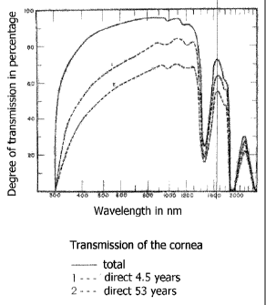
Figure 2 is a diagram representing the degree of transmission of the cornea.
Figure
lo 3 is a diagram representing the degree of transmission of the aqueous
humour. In
Figure 2, the unbroken line shows the total transmission of the cornea 1,
which
transmission was ascertained from six eyes. The curve denoted by 1 in Figure 2
shows the direct transmission in the case of an eye of a child of 41/2 years
of age,
and the curve denoted by 2 shows the direct transmission in the case of an eye
of a
person of 53 years of age.
In the case of both incision and ablation, the cornea is the aforementioned
treatment
region. A treatment is to be possible, not only on the surface of the cornea
1, but
also in deeper regions of the cornea 1. Consequently, the wavelength range
selected
for the treatment is to be selected in such a way that the cornea 1 is
partially trans-
missive in this wavelength range. Suitable for this purpose, according to
Figure 3,
are, on the one hand, the wavelength range from 300 nm to 1300 nm and the wave-
length range from 1600 nm to 1700 nm.
It was realized that the previously described side-effects can be prevented if
the
radiation not absorbed in the treatment region, i.e. the cornea, is absorbed
in a re-
gion located behind same. In this case, the radiation not absorbed in the
cornea
cannot reach, for example, the lens and/or the retina 9.
It is proposed, for the purpose of treating the cornea 1, a wavelength range
lies from
approximately 1600 nm to 1700 nm. If the radiation not absorbed in the cornea
1 to
use in the aqueous humour approximately 1700 nm, since in this range the
aqueous
humour 4 has a comparatively low transmission. Consequently, the absorption of
the
radiation not absorbed in the cornea 1 occurs in a region that absorbs as
close as
possible to the cornea 1, it cannot pass through the
CA 02681269 2009-09-18
- 7 -
iris, the lens 5 and the vitreous body 8 and strike the retina 9, or it can do
so
only having been weakened by the absorption in the aqueous humour 4.
=