Note: Descriptions are shown in the official language in which they were submitted.
CA 02681531 2014-07-03
1
Imniunoglobulin Heavy Chains Comprising Heterologous T-Cell Epitopes
The present invention relates to nucleic acids and to their use as vaccines,
the nucleic
acids encoding T cell epitopes against which an immune response is to be
raised. Such
vaccines may be used in the treatment of tumours.
In the field of cancer vaccines and chronic viral infections, it is now
becoming clear that
factors other than frequency, such as functional avidity of tumour specific T
cells and route
of priming, are major determinants in maximising vaccine efficacy. A number of
groups
have shown that high avidity CD8+ T cells demonstrate superior anti-tumour
activity
(Alexander-Miller, Immunologic Research, 2005;31: 13-24, Hodge et al, J
Immunol
2005;174: 5994-6004, Valmori et al, J Immunol 2002;168: 4231-40, Zeh eta!, J
Immunol
1999;162: 989-94). It has been suggested that high avidity T cells play a
vital role in
tumour regression in patients. This is exemplified in a study where high
avidity antigen-
specific tumour infiltrating lymphocytes (TIL) were detected in a patient with
dramatic
tumour regression (Khong & Rosenberg, J Immunol 2002;168: 951-6). Evidence is
also
emerging demonstrating that adoptive transfer of in vitro stimulated
autologous tumour-
specific T cells is successful, possibly as in vitro stimulation enables
selection of the high
avidity T cells (Vignard et al., J Immunol 2005;175: 4797-805, Dudley etal., J
lmmunother
2001;24: 363-73, Morgan eta!, J Immunol 2003;171: 3287-95, Rosenberg & Dudley,
Proceedings of the National Academy of Sciences of the United States of
America
2004;101 Suppl 2: 14639-45).
Hitherto, a number of groups have attempted to raise a cellular immune
response against
a pre-determined epitope using an antibody as a carrier for that epitope. For
example,
WO 96/19584 (Bona et al.) discloses chimeric antibodies in which T cell
epitopes are
inserted into the complementarity determining regions (CDRs) of an antibody,
and alleges
that such chimeric antibodies are suitable for raising a cytotoxic T cell
(CTL) response.
However, this document teaches that the DNA must encode a functional
CA 02681531 2009-09-22
WO 2008/116937
PCT/EP2008/053761
2
protein. Thus in the abstract, it is stated that "the functional capabilities
of the
epitope and the parent immunoglobulin are retained." Also, on page 21, it is
stated "that the insertion of the desired epitope should be at a region of the
nucleic acid encoding the parent immunoglobulin molecule that is not
essential for expression or function of the parent immunoglobulin molecule."
Furthermore, all the examples in WO 96/19584 show that intact
immunoglobulin is produced following insertion of the T cell epitope.
US Patent No. 7,067,110 discloses a method for eliciting an immune
response against an antigen using a fusion protein of antibody which lacks an
immunoglobulin variable region domain fused to the antigen by a polypeptide
bond. The fusion protein retains the ability to bind to Fc.
EP0759944 discloses a method of incorporating T cell epitopes within an
antibody molecule that is secreted as an intact immunoglobulin protein and
which can target CTL epitopes to tumours to make them better targets for
CTLs.
WO 00/64488 discloses that a CTL response can be raised by nucleic acid
encoding a chimeric antibody having heterologous T cell epitopes inserted in
the CDRs but not the variable region thereof, provided that the nucleic acid
is
directed for expression in B cells. As B cells cannot stimulate naïve T cell
responses, the vaccine described in WO 00/64488 would only be useful in
boosting pre-existing T cell responses.
WO 02/092126 discloses that a CTL response can be raised by a polypeptide
comprising a heterologous T cell epitope and the part of human Fc which
binds to the high affinity CD64 receptor. However, the present inventors have
now shown that disruption of the antibody sequence by inserting a T cell
epitope, for example within an inappropriate CDR or even within the variable
region of an antibody, prevents association of heavy and light chain and no
functional antibody is secreted. DNA encoding these mis-folded antibodies
CA 02681531 2016-08-16
CA 2681531
3
unexpectedly generates strong T cell responses. Furthermore, this is not
mediated via
CD64 as human IgG2 ¨ which does not bind to mouse or human 0064 ¨ works just
as
efficiently as human IgG1.
In one aspect of the present invention, there is provided a nucleic acid which
comprises a
non-specific promoter and at least one sequence that encodes a polypeptide
that has at
least one heterologous T cell epitope therein but does not have any regulatory
T cell
epitopes.
This polypeptide is preferably a homologous carrier, e.g. when used to raise a
T cell
response in humans it may be a human protein, or a foreign protein or
human/foreign
chimeric protein that has had all T regulatory epitopes identified and
removed.
Various aspects of the disclosure related to a nucleic acid which comprises
(a) a promoter
and (b) at least one sequence that encodes a recombinant heavy chain, wherein
the heavy
chain has at least one heterologous T cell epitope therein such that the heavy
chain does
not fold correctly when the nucleic acid is expressed, stimulates a T cell
response against
the at least one heterologous T cell epitope, the T cell response not being
mediated via
0064, and cannot associate with a light chain to form an intact antibody or
associates with
a light chain to form decreased amounts of intact antibody as compared to a
normal control
antibody.
Various embodiments of the claimed invention relate to a nucleic acid which
comprises (a)
a promoter that causes expression of the nucleic acid in dendritic cells
and/or keratinocytes
and (b) at least one sequence that encodes a recombinant heavy chain, wherein
the heavy
chain has at least one heterologous T cell epitope therein such that the heavy
chain does
not fold correctly when the nucleic acid is expressed, stimulates a T cell
response against
the at least one heterologous T cell epitope, the T cell response not being
mediated via
0064, and cannot associate with a light chain to form an intact antibody or
associates with
a light chain to form decreased amounts of intact antibody as compared to a
normal control
antibody.
CA 02681531 2015-08-07
CA 2681531
3a
Various embodiments of the invention provide a vaccine comprising a nucleic
acid as
described above and an adjuvant.
Various embodiments of the invention provide a pharmaceutical composition
comprising a
nucleic acid as described above and a pharmaceutically acceptable carrier,
excipient or
diluent.
Various embodiments of the invention provide a use of a nucleic acid as
described above in
the manufacture of a medicament for stimulating an immune response against at
least one
of the at least one T cell epitopes.
Various embodiments of the invention provide a use of a nucleic acid as
described above
for stimulating an immune response against at least one of the at least one T
cell epitopes.
The polypeptide is preferably one chain of a heterodimer, the heterologous T
cell epitope
causing disruption of the heterodimer chain such that it cannot bind or
associate with the
other chain of the heterodimer. Many molecules are herodimeric, with one chain
being
dependent upon the other for folding and then secretion. If the secondary
structure is
disrupted due to insertion of a heterologous T cell epitope, folding and
secretion is
inhibited. In certain embodiments, one chain is secreted and includes a
heterologous CTL
epitope, and the other chain includes a heterologous helper epitope but, due
to disruption
of the secondary folding, is not secreted. Thus, the nucleic acid may encode
both chains
of the heterodimer, wherein one chain includes a heterologous cytotoxic T cell
(CTL)
epitope and is secreted when expressed, and the other chain includes a
heterologous
helper epitope and is not secreted when expressed. Alternatively, the
respective
heterodimer chains may be encoded on separate nucleic acid molecules.
The heterodimer may be an immunoglobulin molecule. The heavy chain of the
immunoglobulin molecule may include a heterologous cytotoxic T cell (CTL)
epitope and
be secreted when expressed, and the light chain of the
CA 02681531 2009-09-22
WO 2008/116937 PCT/EP2008/053761
4
immunoglobulin molecule may include a heterologous helper epitope and not
be secreted when expressed.
The nucleic acid of the first aspect of the present invention encodes an
polypeptide that does not include any regulatory T cell (T reg) epitopes.
These polypeptides act as inert carriers for the T cell epitope(s) and may be
a
molecule, or part of a molecule, that can be used by the immune system to
stimulate immune responses, as these molecules by definition do not express
competing T reg epitopes. Suitable molecules include HLA molecules, T cell
receptors, TOL receptors, TOL ligands, cytokines, cytokine receptors,
chemokines, chemokine receptors. It is preferred that the molecule is an
antibody or part thereof.
Without wishing to be bound by theory, the present invention is based, at
least
in part, on the concept that a T cell response can be generated against a
specific T cell epitope (such as a CTL epitope), by administration of a
nucleic
acid encoding a polypeptide including the T cell epitope but no regulatory T
cell epitopes. It is believed that nucleic acid is either taken up by antigen
presenting cells (APCs), migrates to lymph nodes and is directly presented, or
is expressed to produce a polypeptide which is secreted and which is then
taken up by other APCs. The former nucleic acid is suitable for stimulating
helper T cell epitopes and the latter is suitable for stimulating CTL
responses.
The polypeptide that is encoded by the nucleic acid ideally does not have any
natural T cell epitopes. Suitable polypeptides in this regard are immune
molecules, such as antibodies. Antibody heavy and light chains which cannot
associate so that the light chain remains in the APCs and so that the heavy
chain is secreted are suitable for the practice of the present invention,
although the present invention is not limited to the use of antibodies as
carriers for the T cell epitopes.
A suppressor T cell population was identified approximately 40 years ago, but
progress was hampered by the lack of specific techniques to identify the cells
CA 02681531 2009-09-22
WO 2008/116937 PCT/EP2008/053761
and because of scientific scepticism regarding the existence of suppression.
However, Sakaguchi et al resurrected interest in suppressor cells in 1995 by
demonstrating that the transfer of lymphocytes depleted of CD4+CD25+T cells
into athymic mice caused the development of various autoimmune diseases in
5 the recipient mice and that reconstitution with CD4+CD25+T cells
prevented
autoimmune reactions in these mice (Sakaguchi et a/J.Immunol
1995;155:1151-1164). Subsequently, numerous studies in mice and humans
have shown that diverse T cell populations with regulatory activity play an
important role in the suppression of immune responses (both innate and
adaptive) to self (controlling self tolerance) (Sakaguchi et al J Immunol
1995;155:1151-1164) as well as foreign antigens (Shevach, Immunity 2006;
25: 195-201, Coleman et al, J. Cell Mol. Med. 2007; 11: 1291-1325). Treg-
cell depletion in mouse models of cancer has shown to improve endogenous
immune-mediated tumour rejection (Shimizu, et al, J. lmmunol. 1999; 163:
5211-5218, Onizuka et al, Cancer Research 1999; 59: 3128-3133) and
antigen-specific anti-tumour immunity (Tanaka, et al, J. lmmunother.
2002;25:207-217). In addition, Treg-cell depletion augments tumour
immunotherapy including vaccination (Tanaka, et al, J. lmmunother.
2002;25:207-217, Dannull et al, J. Clin. Invest. 2005;115:3623-3633) and
CTLA-4 blockade (Sutmuller et al, J. Exp. Med. 2001;194:823-832).
Furthermore, numbers of Treg-cells are increased in the peripheral blood
(Woo et al, Cancer Research 2001;61:4766-4772, Curiel et al, Nature
Medicine 2004;10:942-949, Wolf et al, Clin. Cancer Research 2003;9:606-
612, Sasada et al, Cancer 2003;98:1089-1099) and populate the tumour
microenvironment and draining lymph nodes (Curiel et al, Nature Medicine
2004;10:942-949, Sasada et al, Cancer 2003;98:1089-1099, Liyanage et al, J.
Immunology 2002;169:2756-2761, Matsuura et al, Cancer 2006;106:1227-
1236, Yang et al, Blood 2006;107:3639-3646, Alvaro et al, Clin. Cancer
Research 2005;11:1467-1473) of patients with different cancers. In patients
with gastric carcinoma (Sasada et al, Cancer 2003;98:1089-1099, lchihara et
al, Clinical Cancer Research 2003;9:4404-4408) and ovarian cancer (Curiel et
al, Nature Medicine 2004;10:942-949), poor prognosis and decreased survival
CA 02681531 2009-09-22
WO 2008/116937 PCT/EP2008/053761
6
rates were associated with higher Treg-cell frequencies. Treg-cells have also
been shown to suppress/inhibit the proliferation, cytokine-production (IFNy,
IL-
2) and cytolytic activity of tumour-specific CD8+ (Liyanage et al, J.
Immunology 2002;169:2756-2761, Piccirillo eta!, J. Immunology
2001;167:1137-1140, Mempel eta!, Immunity 206;25:129-141, Annacker et
al, J. Immunology 2001;166:3008-3018, Woo eta!, J. Immunology
2002;168:4272-4276) and CD4+ (Liyanage et al, J. Immunology
2002;169:2756-2761, lchihara et al, Clinical Cancer Research 2003;9:4404-
4408, Nishikawa et al, Blood 2005;106:1008-1011) T cells. In addition, Treg-
cells can suppress the functions of dendritic cells (Romagnani eta!, Eur. J.
Immunol. 2005;35:2452-2458), NK cells (Ralainirina eta!, J. Leukoc. Biol.
2007;81:144-153) and B cells (Lim eta!, J. Immunology 2005;175:4180-
4183). Taken together, these studies suggest an important role of Treg-cells
in tumour immunopathology and indicate a close correlation between Treg-
cell frequencies and tumour growth.
Treg-cells are divided into natural CD4+CD25+T cells and diverse populations
of induced/adaptive Treg-cells (Shevach, Immunity 2006; 25: 195-201,
Bluestone eta!, Nat. Immunol. 2005;6:345-352) (Table 1). About 5%-10% of
CD4+ T cells in mice and humans are natural Treg-cells (Sakaguchi eta!, Nat.
Immunology 2005;6:345-352). Natural Treg-cells develop in the thymus by
strong TCR interaction with self peptide (Picca et al, Current Opinion in
Immunology 2005;17:131-136, Jordan eta!, Nature Immunology
2001;2(4):301-306, Picca et al, Immunological Reviews 2006;212:74-85),
while induced Treg-cells develop from non-regulatory T cells in the periphery.
This extrathymic conversion requires special immunological conditions such
as continuous exposure to low dose antigen, exposure to a systemic
peripheral antigen or exposure to TGFr3 (Shevach, Immunity 2006; 25: 195-
201, Akbar et al, Nat. Rev. lmmunol. 2007;7:231-237). Treg-cells may
mediate their suppression by one or a combination of the following
mechanisms: i) cell-cell contact dependent mechanism, ii) through the
secretion of immunosuppressive cytokines like IL-10 or TGFr3 or iii) direct
CA 02681531 2009-09-22
WO 2008/116937 PCT/EP2008/053761
7
killing of the target cells perforin-granzyme pathway (von Boehmer, Nature
Immunology 2005;6(4):338-344).
To date, very little is known about the antigen-specificity of human Treg-
cells.
Wang et al reported the identification of LACE-1-specific CD4+CD25+GITR+
functional Treg-cell clones in cancer patients (Wang et al, Immunity
2004;20:107-118). Vence et a/ demonstrated the presence of tumour antigen-
specific CD4+ Treg-cells in the peripheral blood of metastatic melanoma
patients (Vence et al, PNAS 2007;104(52):20884-20889). These Treg-cells
recognised a broad range of tumour antigens, including TRP1, NY-ES0-1,
gp100 and survivin. In addition, Vence et al were the first to demonstrate the
presence of NY-ES0-1-specific Treg-cell epitopes within the NY-ES0-1
molecule. Furthermore, vaccination of melanoma patients with dendritic cells
either loaded with synthetic peptides or tumour lysates was shown to induce
increased frequencies of Treg-cells, concomitant with the expansion of
tumour-specific CD8+ T cells (Chakraborty et al, Hum. Immunology
2004;65:794-802). This suggests the possibility that the vaccine contained
unidentified Treg-cell epitopes as well as CD8+ T cell epitopes, which lead to
the expansion of Treg-cells in vivo by ligand-specific activation through the
Treg-cell T cell receptor (TCR). It is widely accepted that Treg-cells require
antigen-specific activation through TCR recognition/engagement but mediate
antigen-nonspecific bystander suppression (Thorton & Shevach, J.
Immunology 2000;164:183190). Furthermore, Li et al suggested the
existence of dominant Treg epitopes within the Hepatitis C Virus core protein
that stimulated HCV-specific Treg-cells in infected patients (Li et al,
lmmunol.
Cell Biol. 2007;85(3)1 97-204). Collectively, these studies in addition to the
recent finding that immunization of HHD transgenic mice with the anti-
endothelial DNA construct C200Fc, failed to stimulate a significant Tie-21-196-
specific anti-tumour immune response and the increased frequency of Tie-21_
196-specific IFNy secreting cells from splenocytes of HHD mice after the
depletion of CD4+CD25+Treg cells (by administration of 400pg PC61
monoclonal antibody) prior to C200Fc DNA immunization (Middleton, PhD
CA 02681531 2009-09-22
WO 2008/116937 PCT/EP2008/053761
8
Thesis. University of Nottingham, November 2007) indicates that the Tie-21_
196 within the DNA vaccine contains unidentified Treg-cell epitopes as well as
the CD8+ epitope. This would explain the failure of the vaccine to break
tolerance to the self antigen Tie-2 and to elicit anti-tumour immunity in HHD
mice due to abundant antigen-specific expanded Treg-cells suppressing the
cell-mediated anti-tumour immune response. There is therefore an advantage
to express T effector epitopes with inert immune carriers which fail to
express
T reg epitopes to direct the immune response to the effector epitope and
prevent stimulation of the dominant T reg response.
Advantageously, the nucleic acid of the present invention includes a
sequence encoding a sequence, such as a leader sequence, that allows the
expressed polynucleotide to be secreted. This allows the polynucleotide to be
transferred to antigen presenting cells (APCs). The sequence could be a
leader sequence that is naturally expressed with the polynucleotide or could
be a heterologous leader sequence, such as an immunoglobulin leader
sequence, which is added. The latter is especially suitable where the
polynucleotide encodes a membrane-bound molecule.
According to another aspect of the present invention, there is provided a
nucleic acid which comprises a non-specific promoter and at least one
sequence that encodes a recombinant heavy chain of an immunoglobulin
molecule, wherein the heavy chain has at least one heterologous T cell
epitope therein such that the heavy chain cannot take its native conformation
when the nucleic acid is expressed.
The nucleic acid of the this aspect of the present invention encodes a
recombinant heavy chain of an immunoglobulin molecule. The structure of
such a heavy chain is known to those of skill in the art, and generally
includes
variable and constant regions. The heavy chain may be from an antibody.
The antibody may be monoclonal or polyclonal and may be IgA, IgD, IgE, IgG
or IgM, although IgG is preferred. The IgG antibody may be any IgG
CA 02681531 2009-09-22
WO 2008/116937
PCT/EP2008/053761
9
subclass, such as human IgG1, IgG2, IgG3 or IgG4, or mouse IgG1, IgG2a,
IgG2b or IgG3. The IgG antibody may be a human IgG1 antibody having the
IgG2 Fc binding domain, or a human IgG2 antibody having the IgG1 Fc
binding domain. The heavy chain may have the constant region of a human
antibody, and the variable or hypervariable (CDR) region of a mouse
monoclonal antibody into which heterologous T cell epitopes have been
inserted. The variable region other than the hypervariable region may also be
derived from the variable region of a human antibody. When applied to
antibodies (i.e. comprising a heavy chain and a light chain), the antibody is
said to be humanised. Methods for making humanised antibodies are known
in the art. Methods are described, for example, in Winter, U.S. Patent No.
5,225,539. The variable region of the heavy chain outside of the mouse
hypervariable region may also be derived from a mouse monoclonal antibody.
In such case, the entire variable region is derived from murine monoclonal
antibody and, when applied to antibodies, the antibody is said to be
chimerised. Methods for making chimerised antibodies are known in the art.
Such methods include, for example, those described in U.S. patents by Boss
(Celltech) and by Cabilly (Genentech). See also U.S. Patent Nos. 4,816,397
and 4,816,567, respectively.
In certain embodiments, the nucleic acid of the present invention further
comprises at least one sequence that encodes a light chain of an
immunoglobulin molecule. Alternatively, a separate nucleic acid encoding a
light chain of an immunoglobulin molecule may be provided. The light chain
may have at least one heterologous T cell epitope therein. The T cell epitope
may be such that the light chain cannot take its native conformation when the
nucleic acid is expressed. The light chain may have any of the features
described herein in respect of the heavy chain. Accordingly, the invention
also provides a nucleic acid encoding a recombinant light chain of an
immunoglobulin molecule, wherein the light chain has at least one
heterologous T cell epitope therein such that the light chain cannot take its
native conformation when the nucleic acid is expressed. The nucleic acid
CA 02681531 2009-09-22
WO 2008/116937
PCT/EP2008/053761
may include a non-specifc promoter. Such nucleic acid(s) encode an
immunoglobulin molecule, such as an antibody.
Thus, according to a further aspect of the present invention, there is
provided
5 a nucleic acid which comprises a non-specific promoter and at least one
sequence that encodes a recombinant immunoglobulin molecule, wherein the
immunoglobulin molecule has at least one heterologous T cell epitope therein
such that the immunoglobulin molecule cannot take its native conformation
when the nucleic acid is expressed. Preferably, the recombinant
10 immunoglobulin molecule, and heavy and light chains described above do
not
have any regulatory T cell epitopes.
The invention also provides:
= a vaccine comprising a nucleic acid of the invention and an adjuvant;
= a pharmaceutical composition comprising a nucleic acid of the
invention and a pharmaceutically acceptable carrier, excipient or
diluent;
= a nucleic acid of the invention for use in medicine;
= the use of such a nucleic acid of the invention in the manufacture of a
medicament for stimulating an immune response against at least one
of the T cell epitope(s)
= a nucleic acid of the invention for stimulating an immune response
against at least one of the T cell epitope(s); and
= a method for stimulating an immune response against a T cell epitope,
comprising administering to a subject in need of such immune
response a therapeutically effective amount of a nucleic acid of the
invention.
Surprisingly, the present inventors have found that antibodies, such as
monoclonal antibodies, which may be human or non-human, that have pre-
determined T cell epitopes cloned within their variable regions, so as to
disrupt the primary antibody structure, inhibit folding and/or limit secretion
to
CA 02681531 2009-09-22
WO 2008/116937 PCT/EP2008/053761
11
either just heavy chain or very low amounts of intact antibody, stimulate
strong helper and antigen-specific T cell responses. The inventors have also
found that this effect can be achieved using nucleic acid encoding the heavy
chain of such an antibody. It is believed that the T cell epitope is processed
but not destroyed by the immunoproteosome. In certain embodiments, the
invention provides a DNA vaccine presenting pre-defined T cell epitopes
within denatured immunoglobulin which enhances the frequency and the
avidity of the T cell response. The polypeptides encoded by the nucleic acids
of the invention may be referred to herein as "Immunobodies".
The finding that an immune response against a T cell epitope can be
stimulated by a nucleic acid encoding at least the heavy chain of an
immunoglobulin molecule into which the T cell epitope has been inserted such
that the an immunoglobulin molecule cannot take its native conformation runs
contrary to the expectations in the art, where it is taught that the antibody
must be expressed in a functional form. For example, as discussed above,
WO 96/19584 teaches that, where a nucleic acid encodes an antibody in
which T cell epitopes are inserted into the CDRs of the antibody, the nucleic
acid must encode a functional antibody. Similarly, EP0759944 describes a
method of incorporating T cell epitopes within an antibody molecule that is
secreted as an intact immunoglobulin protein. Although US patent no.
7,067,110 discloses that an immune response can be raised against an
antigen by a fusion protein of antibody and the antigen, the antibody is
disclosed as lacking an immunoglobulin variable region. In addition, this
fusion protein will have regulatory T cell epitopes in the antigen. Thus,
although the protein may stimulate an antibody response, it will not stimulate
high avidity T cells responses due to regulatory T cell epitopes s in the
antigen.
As discussed above, WO 00/64488 discloses a nucleic acid encoding a
chimeric antibody having heterologous T cell epitopes inserted in the CDRs
but not the variable region thereof, which nucleic acid is directed for
CA 02681531 2009-09-22
WO 2008/116937 PCT/EP2008/053761
12
expression in B cells. The nucleic acid of the present invention is not
directed
for expression in B cells, and thus will not target B cells specifically
either in
vitro or in vivo. The nucleic acid of the present invention can be taken up by
any antigen presenting cells, including dendritic cells, and can therefore
prime
naïve CTL and helper T cell responses, whereas the vaccine described in WO
00/64488 would only be useful in boosting pre-existing T cell responses.
Analysis of the functional avidity of responses induced by nucleic acids in
accordance with the invention demonstrated that a high avidity response can
be generated when compared to immunisation with synthetic peptide. This
also correlated with enhanced ability to recognise and kill tumour cells in
vitro
and in vivo. This observation is comparable to that documented in other
studies where better anti-tumour activity is shown by high avidity TRP2
specific CTL (Zeh eta!, J Immunol 1999;162: 989-94, Harada eta!,
Immunology 2001104: 67-74).
The nucleic acids of the present invention have a non-specific promoter, i.e.
a
promoter that will promote expression of the nucleic acid but which has no
specificity for cells in which expression is promoted. The promoter preferably
causes expression of the nucleic acid in dendritic cells and/or keratinocytes.
Examples of suitable promoters include the CMV promoter, the SV40
promoter, and other non-specific promoters known to those of skill in the art.
Alternatively, the nucleic acid of the present invention may have one or more
promoters that cause specific expression in dendritic cells (e.g. Cd1lb
promoter) and in keratinocytes (e.g. MHCII promoter, Chin etal., 2001 J.
Immunol. 167, 5549-5557).
The nucleic acid of certain aspects of the invention encodes an
immunoglobulin molecule, preferably an antibody that includes all of the major
features of an antibody, that is to say heavy and light chains which include
variable and constant regions. The antibody may be monoclonal or polyclonal
and may be IgA, IgD, IgE, IgG or IgM, although IgG is preferred. The IgG
CA 02681531 2009-09-22
WO 2008/116937
PCT/EP2008/053761
13
antibody may be any IgG subclass, such as human IgG1, IgG2, IgG3 or IgG4,
or mouse IgG1, IgG2a, IgG2b or IgG3. The IgG antibody may be a human
IgG1 antibody having the IgG2 Fc binding domain. The antibody may have
the constant region of a human antibody, and the variable or hypervariable
region of a mouse monoclonal antibody into which heterologous T cell
epitopes have been inserted. The variable region other than the
hypervariable region may also be derived from the variable region of a human
antibody. Such an antibody is said to be humanised. Methods for making
humanised antibodies are known in the art. Methods are described, for
example, in Winter, U.S. Patent No. 5,225,539. The variable region of the
antibody outside of the mouse hypervariable region may also be derived from
a mouse monoclonal antibody. In such case, the entire variable region is
derived from murine monoclonal antibody and the antibody is said to be
chimerised. Methods for making chimerised antibodies are known in the art.
Such methods include, for example, those described in U.S. patents by Boss
(Celltech) and by Cabilly (Genentech). See also U.S. Patent Nos. 4,816,397
and 4,816,567, respectively.
The nucleic acid of certain aspects of the invention is such that the heavy
chain, light chain or immunoglobulin molecule expressed therefrom has at
least one heterologous T cell epitope therein so that the heavy chain, light
chain or the immunoglobulin molecule cannot take its native conformation.
The T cell epitope may disrupt the expressed protein so that the heavy chain
or the immunoglobulin molecule can no longer bind to its antigen, so that the
heavy and light chains (where present) can no longer associate, or so that the
heavy chain or immunoglobulin molecule cannot be secreted properly, for
example. The disruption may be in the tertiary structure of the
immunoglobulin molecule which may prevent formation of the disulphide
bonds.
As discussed in more detail below, where the immunoglobulin molecule is an
antibody, the T cell epitope(s) may be inserted into or substituted for the
CA 02681531 2009-09-22
WO 2008/116937 PCT/EP2008/053761
14
CDR1 and CDR2 regions of the antibody. CDR1 and CDR2 form part of the
antibody 13 sheet conformation and are partially submerged within the folded
molecule. Any change to their length, amino acid composition or charge will
disrupt this structure and prevent heavy and light chain folding and
association. CDRH3 is exposed on the surface of the immunoglobulin
molecule and is therefore more permissive of alterations. In the present
invention, it is preferred if CDR1 and/ or CDR2 are substituted with T cell
epitope(s). Indeed, in certain embodiments, loss of framework regions at the
CDRH junctions completely disrupts antibody folding yet insertion of epitopes
in these regions gives good T cell responses. Incorporation of any epitope
within the CDRH1 (5 amino acids in length) or CDRH2 (17 amino acids in
length) causes sufficient disruption to allows secretion of heavy chain but
only
very low amounts of intact antibody, even if the light chain has its native
sequence. This shows that the secondary structure is important for heavy
and light chain pairing. Incorporation of any epitope within CDRL1 of the
light
chain results in low level secretion of light chain, even if there is only a
single
epitope incorporated into the CDRH3 of the heavy chain.
"Heterologous T cell epitope" is intended to mean a T cell epitope which is
heterologous to the antibody. For example, a heterologous T cell epitope may
be one which was not previously present in the antibody. The heterologous T
cell epitope may be inserted as a whole, although it may be made up from an
inserted amino acid sequence, together with flanking amino acids of the
second portion. This is to ensure that the inserted epitope has a similar
processing profile in the heterologous nucleic as from the original antigen.
One or more CTL/helper epitopes can be inserted within the same variable
region.
The T cell epitope(s) can be inserted anywhere in the heavy chain or light
chain. It is preferred if the or each epitope is inserted in the variable
region of
the heavy chain and/or light chain, although nucleic acids encoding heavy
chains or antibodies having T cell epitopes inserted in just the constant
CA 02681531 2009-09-22
WO 2008/116937
PCT/EP2008/053761
region, or in the constant region and the variable region of a heavy chain
and/or light chain are included within the invention. In the nucleic acids of
the
present invention, the sequence(s) encoding the T cell epitopes may be
inserted into (i.e. added to) the sequence encoding the heavy chain and/or
5 light chain, or may be substituted into the sequence encoding the heavy
chain
and/or light chain.
In the variable region, the T cell epitope(s) may be inserted in, or
substituted
for, any one or more the CDRs of the heavy and/or light chain, i.e. L1, L2,
L3,
10 H1, H2, or H3. Of these, L1, H1 and H2 are currently preferred. In
certain
embodiments, the T cell epitopes are inserted in, or substituted for, CDRL1
and/or H1 and/or H2. Preferably the incorporated T cell epitopes are not of
similar size and charge to the amino acids of the original CDR of the antibody
so that the antibody does not take its native conformation, e.g. does not fold
15 and is not secreted correctly. Alternatively or additionally, they may
be
inserted in, or substituted for, the framework region surrounding the CDRs.
The inserted T cell epitopes are preferably cytotoxic T cell (CTL or CD8)
epitopes. Alternatively or additionally, helper T cell (CD4) epitopes may be
inserted. T cell epitopes can be predicted using known T cell algorithms or
synthesised as peptides and screened using standard T cell assays. The T
cell epitopes may have an amino acid length in the range of from 5 to 50, 7 to
40, 8 to 30 or 9 to 20 amino acids, such as 9, 10, 11, 12, 13, 14, 15, 16 ,
17,
18, 19, 20, 21, 22, 23, 24, 25, 26, 27, 28, 29 or 30 amino acids. The epitopes
may inserted using complementary oligonucleotides that encode the antigenic
epitopes, which are annealed and cloned into specific sites of the antibody
framework where CDR's (or other region) have been replaced with unique
restriction enzyme sites. The ability of the recombinant antibody to stimulate
helper and cytotoxic T cell responses can be screened as exemplified herein.
Various combinations are possible within the present invention. In certain
embodiments, one or a plurality of CD8 epitopes is/are inserted in, and/or
CA 02681531 2009-09-22
WO 2008/116937 PCT/EP2008/053761
16
substituted for, the CDR H1 and/or H2, or in the non-CDR variable region, of
the heavy chain or the antibody. Additionally or alternatively, one or a
plurality
of CD4 epitopes may be inserted in, and/or substituted for, the CDR L1, or in
the non-CDR variable region, of the light chain or the antibody. Where there
is a plurality of T cell epitopes, the T cell epitopes may be the same or
different. Those of skill in the art will appreciate that numerous
combinations
are possible, including:
= a CD8 epitope in CDR H1, and a CD4 epitope in CDR L1;
= a CD8 epitope in CDR H2, and a CD4 epitope in CDR L1;
= a CD8 epitope in CDR H1 and CDR H2, and a CD4 epitope in CDR L1;
= 2 CD8 epitopes in CDR H1, and a CD4 epitope in CDR L1;
= 2 CD8 epitopes in CDR H2, and a CD4 epitope in CDR L1; etc
Nucleic acids of the present invention can incorporate multiple T cell
epitopes
from a single target antigen that can bind to the majority of both class I and
class II MHC molecules. This may create a vaccine that can be used in
widespread population vaccination. Alternatively nucleic acids useful in the
invention can incorporate multiple T cell epitopes from multiple target
antigens
that can bind to the most common class I and class ll phenotypes. This may
create a vaccine that may prevent selection of antigen loss variants. Target
antigens may be from a single pathogen or tumour type or may be selected to
give an immune response against a variety of pathogens or cancers. Nucleic
acids useful in the present invention targeting specific common HLA
phenotypes may incorporate numerous T cell epitopes from a wide variety of
cancers and/or pathogens, providing a single vaccine to prevent disease.
Any T cell epitope can be inserted, provided that it stimulates helper and/or
cytotoxic T cell responses. T cell epitopes from pathogens such as HIV,
Hepatitis C and other infections that require CTLs to clear latent infections
may be used, although it is preferred if the epitope is a "self-epitope", i.e.
associated with a condition/disorder associated with cell proliferation such
as
CA 02681531 2009-09-22
WO 2008/116937 PCT/EP2008/053761
17
cancer. Preferably, the T cell epitope is such that the heavy chain or
antibody
cannot fold correctly and be secreted. It is therefore preferred if the
inserted
epitopes are of not of similar size and amino acid composition to the original
variable region. The nucleic acid may have a plurality of different T cell
epitopes so as to generate a wide variety of T cell responses. The nucleic
acid may incorporate multiple epitopes from a single antigen, thereby
ensuring that the majority of individuals with different HLA types respond to
the single vaccine. Alternatively, multiple T cell epitopes from multiple
antigens targeting a restricted spectrum of HLA types could be used. The
nucleic acid molecules of the invention may include a variety of antigens from
a single pathogen or cancer type or they could include disparate antigens
targeting a wide range of solid tumours or pathogens. The nucleic acid
molecules of the invention may even be designed to target different cell
populations within a tumour, such as tumour epithelial and endothelial
antigens.
Surprisingly the inventors have found that, when T cell epitopes were inserted
into structurally confined CDRs or non-CDR regions of the heavy chain, they
gave superior CTL responses. This appears to be due to secretion of large
amounts of heavy chain, which can only weakly associate with light chain due
to the insertion of bulky epitopes into their variable regions. This is
contrary to
dogma, which states "that only proteins synthesised endogenously by antigen
presenting cells are presented on MHC class I molecules and recognised by
CTLs" ¨ WO 96/19584. Uptake of exogenous antigen and presentation on
MHC class I is a process known as cross presentation and usually requires
uptake via specific receptors. This could be the CD64 receptor for human
Fcy1 antibodies. However, it would be predicted that large amounts of intact
antibody or antigen-antibody complexes would be better at targeting this
receptor. In contrast, the results presented herein clearly show very low
levels of intact antibody and large amounts of free heavy chain, which should
not bind to CD64, give superior CTL responses. Indeed, it is shown herein
that CTL responses can be stimulated when CTL epitopes are inserted in
CA 02681531 2009-09-22
WO 2008/116937 PCT/EP2008/053761
18
antibodies which cannot bind to CD64, such as Ig02 antibodies or IgG1
molecules with their CD64 binding region replaced with the non-CD64 binding
region from Ig02.
The nucleic acid encoding the heavy chain preferably includes a leader
sequence to allow it to be secreted. The present inventors have found that, if
the leader sequence of the heavy chain is removed to prevent secretion and
allow more endogenous protein to be produced, this reduces the CTL
response. This is completely contrary to expectations. Whilst not wishing to
be bound by theory, the inventors believe that this implies that the nucleic
acid
is expressed in non-antigen presenting cells, which secrete high levels of
heavy chain and low amounts of native protein which can then be taken up by
antigen presenting cells. Alternatively, the nucleic acid may directly
transfect
antigen presenting cells which migrate to the draining lymph node where they
secrete low amounts of native protein and large amounts of heavy chain that
is taken up by the same or adjacent antigen presenting cells and presented
on MHC class I to naïve CTLs. Therefore, for a nucleic acid vaccine to
stimulate efficient CTL responses, it must preferably encode CTL epitopes
within a protein that is secreted at very low levels and/or at the same time
secretes large amounts of denatured protein. However, a CTL response
cannot mature to a high affinity memory response in the absence of helper
responses. Therefore, it is preferred if T helper epitopes are inserted into
the
heavy chain or the immunoglobulin molecule, preferably into the variable
region of antibody light chains. Again, surprisingly and in contrast to the
dogma which states "only proteins taken up exogenously by the target cells
are presented by MHC class II molecules and recognised by helper T cells",
light chain was only secreted in very low amounts. Removal of the leader
sequence to prevent secretion of the light chain had no effect on the helper
responses. Accordingly, the nucleic acid of the present invention may or may
not have a leader sequence for the light chain of the antibody. These results
imply that the nucleic acid is taken up by the antigen presenting cells which
present the T helper epitopes in the context of MHC class II from
CA 02681531 2009-09-22
WO 2008/116937
PCT/EP2008/053761
19
endogenously-synthesised protein, possibly by autophagy. For helper T cells
to assist CTL responses, both the T cell epitopes they recognise must be
presented on the same antigen presenting cells in a process known as linked
T cell help. This implies that the antigen presenting cell synthesising the
light
chain, encoded by the nucleic acid, must either also synthesise, secrete and
cross present the CTL epitopes themselves or take up heavy chain from an
adjacent APC.
The present invention also provides isolated dendritic cells which present the
heterologous helper T cell epitopes on MHC class II from endogenously-
produced light chain and heterologous CTL epitopes from cross-presented
heavy chain. Such dendritic cells may be used in the therapies described
herein.
Nucleic acids of the present invention can make existing T cell epitopes more
immunogenic by encoding a denatured antibody which leads to an increase in
both the frequency and avidity of T cell responses.
The nucleic acid of the invention may be DNA, cDNA, or RNA such as mRNA,
obtained by cloning or produced wholly or partly by chemical synthesis. For
therapeutic use, the nucleic acid is preferably in a form capable of being
expressed in the subject to be treated.
The nucleic acid of the present invention may be recombinant or provided as
an isolate, in isolated and/or purified form. It may be free or substantially
free
of nucleic acid flanking the gene in the human genome, except possibly one
or more regulatory sequence(s) for expression. Where nucleic acid according
to the invention includes RNA, reference to the sequences shown herein
should be construed as reference to the RNA equivalent, with U substituted
for T.
CA 02681531 2009-09-22
WO 2008/116937 PCT/EP2008/053761
Nucleic acids of the present invention can be readily prepared by the skilled
person, for example using the information and references contained herein
and techniques known in the art (for example, see Sambrook, Fritsch and
Maniatis, "Molecular Cloning", A Laboratory Manual, Cold Spring Harbor
5 Laboratory Press, 1989, and Ausubel eta!, Short Protocols in Molecular
Biology, John Wiley and Sons, 1992), given the nucleic acid sequences and
clones available. These techniques include (i) the use of the polymerase
chain reaction (PCR) to amplify samples of such nucleic acid, e.g. from
genomic sources, (ii) chemical synthesis, or (iii) preparing cDNA sequences.
10 DNA encoding the polypeptide may be generated and used in any suitable
way known to those of skill in the art, including by taking encoding DNA,
identifying suitable restriction enzyme recognition sites either side of the
portion to be expressed, and cutting out said portion from the DNA. The
portion may then be operably linked to a suitable promoter in a standard
15 commercially available expression system. Another recombinant approach
is
to amplify the relevant portion of the DNA with suitable PCR primers.
Modifications to the sequences can be made, e.g. using site directed
mutagenesis, to lead to the expression of modified peptide or to take account
of codon preferences in the host cells used to express the nucleic acid.
In order to obtain expression of the nucleic acid sequences, the sequences
can be incorporated into a vector having one or more control sequences
operably linked to the nucleic acid to control its expression. The vectors may
include other sequences such as promoters or enhancers to drive the
expression of the inserted nucleic acid, nucleic acid sequences so that the
polypeptide is produced as a fusion and/or nucleic acid encoding secretion
signals so that the polypeptide produced in the host cell is secreted from the
cell. If desired, polypeptide can then be obtained by transforming the vectors
into host cells in which the vector is functional, culturing the host cells so
that
the polypeptide is produced and recovering the polypeptide from the host cells
or the surrounding medium. Prokaryotic and eukaryotic cells are used for this
purpose in the art, including strains of E. coli, yeast, and eukaryotic cells
such
CA 02681531 2009-09-22
WO 2008/116937
PCT/EP2008/053761
21
as insect cells, and animal cells, for example, COS, CHO cells, Bowes
Melanoma and other suitable human cells. Where the present invention
relates to nucleic acid(s) encoding the heavy and light chains of an antibody,
the respective nucleic acids may be present in the same expression vector,
driven by the same or different promoters, or in separate expression vectors.
The nucleic acids of the present invention may be used to stimulate an
immune response against at least one of the T cell epitope(s) in a patient
such as a mammal, including human. Helper and/or cytotoxic T cell
responses may be stimulated. The T cell response against a particular
epitope obtained by the present invention may have a higher avidity than that
obtained by immunisation with the same epitope as a simple peptide, or by
immunisation with the same epitope encoded within an antigen either as a
peptide or a nucleic acid. The nucleic acids of the invention may be
administered as a combination therapy, i.e. a nucleic acid encoding the light
chain and nucleic acid encoding the heavy chain. The nucleic acid may be
administered intravenously, intradermally, intramuscularly, orally or by other
routes. Intradermal or intramuscular administration is preferred because
these tissues contain dendritic cells
As used herein, the term "treatment" includes any regime that can benefit a
human or non-human animal. The treatment may be of an inherited or
acquired disease. Preferably, the treatment is of a condition/disorder
associated with cell proliferation such as cancer or of infectious disease.
Examples of types of cancer that can be treated with the nucleic acid include
any solid tumour, colorectal cancer, lung, breast, gastric, ovarian, uterine,
liver, kidney, pancreatic, melanoma, bladder, head and neck, brain,
oesophageal, pancreatic, and bone tumours, as well as soft tissue cancers,
and leukaemias. Examples of infectious diseases that can be treated with the
nucleic acid include infection with HIV, Hepatitis C, or any chronic infection
that requires T cell immunity for clearance.
CA 02681531 2009-09-22
WO 2008/116937 PCT/EP2008/053761
22
The nucleic acid may be employed in combination with a pharmaceutically
acceptable carrier or carriers. Such carriers may include, but are not limited
to, saline, buffered saline, dextrose, liposomes, water, glycerol, ethanol and
combinations thereof.
Adjuvants may be employed to facilitate stimulation of the host's immune
response, and may include, aluminium hydroxide, lysolecithin, pluronic,
polyols, polyanions, peptides, proteins and oil emulsions.
The nucleic acids useful in the invention can be formulated in pharmaceutical
compositions. These compositions may comprise, in addition to one of the
above substances, a pharmaceutically acceptable excipient, carrier, buffer,
stabiliser or other materials well known to those skilled in the art. Such
materials should be non-toxic and should not interfere with the efficacy of
the
active ingredient. The precise nature of the carrier or other material may
depend on the route of administration, e.g. intradermal, oral, intravenous,
cutaneous or subcutaneous, nasal, intramuscular, intraperitoneal routes. The
formulation is preferably nucleic acid as a stable dry powder precipitated
onto
the surface of microscopic gold particles and suitable for injection via a
gene
gun. The formulation may be suitable for intradermal or intramuscular
administration using electroporation.
The compositions comprising, or for the delivery of, nucleic acids are
preferably administered to an individual in a "therapeutically effective
amount",
this being sufficient to show benefit to the individual. The actual amount
administered, and rate and time-course of administration, will depend on the
nature and severity of what is being treated. Prescription of treatment, e.g.
decisions on dosage etc, is within the responsibility of general practitioners
and other medical doctors, and typically takes account of the disorder to be
treated, the condition of the individual patient, the site of delivery, the
method
of administration and other factors known to practitioners. The nucleic acids
of the invention are particularly relevant to the treatment of existing cancer
CA 02681531 2009-09-22
WO 2008/116937 PCT/EP2008/053761
23
and in the prevention of the recurrence of cancer after initial treatment or
surgery. Examples of the techniques and protocols mentioned above can be
found in Remington's Pharmaceutical Sciences, 16th edition, Oslo, A. (ed),
1980.
Preferably, the nucleic acid of the invention stimulate helper and/or
cytotoxic T
cells that can significantly inhibit the growth of tumour cells when
administered
to a human in an effective amount. The optimal dose can be determined by
physicians based on a number of parameters including, for example, age,
sex, weight, severity of the condition being treated, the active ingredient
being
administered and the route of administration. For example, a dose of 1-
100014 of DNA is sufficient to stimulate both helper and cytotoxic T cell
responses.
The nucleic acids of the invention may be administered along with additional
pharmaceutically acceptable ingredients. Such ingredients include, for
example, immune system stimulators.
A composition may be administered alone or in combination with other
treatments, either simultaneously or sequentially dependent upon the
condition to be treated. Other cancer treatments include other monoclonal
antibodies, other chemotherapeutic agents, other radiotherapy techniques or
other immunotherapy known in the art. One particular application of the
compositions of the invention are as an adjunct to surgery, i.e. to help to
reduce the risk of cancer reoccurring after a tumour is removed.
Injections (id) may be the primary route for therapeutic administration of the
nucleic acid of this invention.
The nucleic acids may be administered in a localised manner to a tumour site
or other desired site or may be delivered in a manner in which it targets
tumour or other cells.
CA 02681531 2009-09-22
WO 2008/116937
PCT/EP2008/053761
24
The dose of nucleic acid will be dependent upon the properties of the agent
employed, e.g. its binding activity and in vivo plasma half-life, the
concentration of the polypeptide in the formulation, the administration route,
the site and rate of dosage, the clinical tolerance of the patient involved,
the
pathological condition afflicting the patient and the like, as is well within
the
skill of the physician. For example, doses of 10014 of nucleic acid per
patient
per administration are preferred, although dosages may range from about
1014 to 1 mg per dose. Different dosages are utilised during a series of
sequential inoculations; the practitioner may administer an initial
inoculation
and then boost with relatively smaller doses of nucleic acid.
In certain other embodiments, the present invention relates to a method of
engineering T cell epitopes from target antigens into the variable regions of
antibodies, and the use of such engineered antibodies as vaccines to
stimulate both helper and cytotoxic T cell responses.
A further aspect of the present invention provides a host cell containing a
nucleic acid as disclosed herein. The nucleic acid of the invention may be
integrated into the genome (e.g. chromosome) of the host cell. Integration
may be promoted by inclusion of sequences that promote recombination with
the genome in accordance with standard techniques. The nucleic acid may
be on an extra-chromosomal vector within the cell, or otherwise identifiably
heterologous or foreign to the cell.
A still further aspect provides a method, which comprises introducing the
nucleic acid of the invention into a host cell. The introduction, which may
(particularly for in vitro introduction) be generally referred to without
limitation
as "transformation", may employ any available technique. For eukaryotic
cells, suitable techniques may include calcium phosphate transfection, DEAE-
Dextran, electroporation, liposome-mediated transfection and transduction
using retrovirus or other virus, e.g. vaccinia or, for insect cells,
baculovirus.
CA 02681531 2009-09-22
WO 2008/116937 PCT/EP2008/053761
For bacterial cells, suitable techniques may include calcium chloride
transformation, electroporation and transfection using bacteriophage. As an
alternative, direct injection of the nucleic acid could be employed.
5 Marker genes such as antibiotic resistance or sensitivity genes may be
used
in identifying clones containing nucleic acid of interest, as is well known in
the
art.
The introduction may be followed by causing or allowing expression from the
10 nucleic acid, e.g. by culturing host cells (which may include cells
actually
transformed although more likely the cells will be descendants of the
transformed cells) under conditions for expression of the gene, so that the
encoded polypeptide (or peptide) is produced. If the polypeptide is expressed
coupled to an appropriate signal leader peptide it may be secreted from the
15 cell into the culture medium. Following production by expression, a
polypeptide or peptide may be isolated and/or purified from the host cell
and/or culture medium, as the case may be, and subsequently used as
desired, e.g. in the formulation of a composition which may include one or
more additional components, such as a pharmaceutical composition which
20 includes one or more pharmaceutically acceptable excipients, vehicles or
carriers (e.g. see below).
The present inventon also provides a method for identifying T cell epitopes in
a candidate antigen, comprising:
25 depleting T regulatory cells in a non-human animal;
immunising the non-human animal with a candidate antigen; and
screening to see whether a T cell response is raised against either
peptides to predicted epitopes in the candidate antigen or all the possible
overlapping peptides within the candidate antigen.
The method may be carried out in a non-human animal, such as a mouse or a
rat. T regulatory cells can be depleted in the non-human animal using anti-
CA 02681531 2009-09-22
WO 2008/116937 PCT/EP2008/053761
26
CD25 antibodies, which optionally may be conjugated with toxins such as
Ontak, or by chemotherapy such as cyclophosphamide which preferentially
kills T regulatory cells. Once T regulatory cells have been depleted, the non-
human animal may be immunised with DNA encoding the candidate antigen,
or by the candidate antigen itself. It is preferred that the candidate antigen
is
provided as an antigen-Fc fusion protein. In the screening step, the peptide
against which any T cell response stimulated in the non-human animal is
identified. This can be done in vitro using a technique such as ELISPOT. If a
T cell response is elicited to a candidate epitope, this epitope can be used
to
immunise a non-human animal. If this peptide elicits a T cell response, the
avidity and frequency can be enhanced by encoding the epitope within a
nucleic acid in accordance with the present invention. This method can allow
the identification of T cell epitopes that are processed by the
immunoproteosome.
Preferred features of each aspect of the invention are as for each of the
other
aspects mutatis mutandis. The prior art documents mentioned herein are
incorporated to the fullest extent permitted by law.
The invention will now be described further in the following non-limiting
examples.
Reference is made to the following drawings:
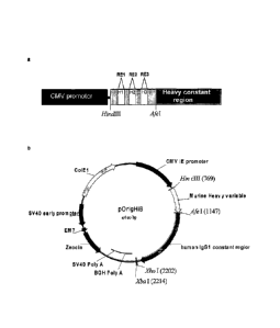
Figure 1: Map depicting features of the heavy chain vector pOrigHIB
The wild type de-immunised heavy variable region of antibody SC100 was
cloned using HindIII/Afel inframe with the human IgG1 Fc constant region.
The Fc region comprises the CH1, CH2, CH3 domains and the hinge region.
High-level expression in mammalian cells is driven from the human
cytomegalovirus immediate early promoter. BGH polyadenylation signals
downstream of the Orig HIB human IgG1 chain to ensure mRNA stability and
effective termination. EM7 is a bacterial promoter that controls expression of
the zeocin resistance gene allowing antibiotic selection in E.coli while the
SV40 early promoter upstream of the resistance gene allows selection in
CA 02681531 2009-09-22
WO 2008/116937 PCT/EP2008/053761
27
mammalian cells. SV40 polyadenylation signals downstream of the
resistance gene in order to direct proper processing of the 3'end of the zeor
mRNA. The vector also contains within its backbone the C0lE1 origin of
replication for propagation in bacteria. Complimentary determining DNA
sequences were effectively removed and exchanged for restriction sites RE1,
RE2 and RE3 (Fspl, Mscl and Sill respectively) singly and in combination.
Figure 2: Map depicting features of the heavy chain vector pOrigLIB
The wild type de-immunised light variable region of antibody SC100 was
cloned using BamHI/BsiWI inframe with the human kappa constant region.
High-level expression in mammalian cells is driven from the human
cytomegalovirus immediate early promoter. BGH polyadenylation signals
downstream of the Orig LIB chain to ensure mRNA stability and effective
termination. The vector also includes the C0lE1 origin of replication and the
antibiotic resistance gene for ampicillin allowing propagation and selection
in
bacteria. Complimentary determining regions were effectively removed and
exchanged for restriction sites RE4, RE5 and RE6 (EcoRV, Ssp I and Hpa I
respectively) singly and in combination.
Figure 3: Sequence of the wild type lmmunobody chimeric heavy chain.
Nucleotide and on translation amino acid sequence are illustrated for the full
length chimeric igG1 heavy chain. Locations of CDR's are within boxes
defined by the kabat numbering scheme. The stop codon is depicted by a red
astrix. The HindIII/Afe I restriction sites are highlighted utilised in
transfer of
the variable heavy region.
Figure 4: Sequence of the wild type lmmunobody chimeric kappa chain
Nucleotide and on translation amino acid sequence are illustrated for the full
length chimeric kappa chain. Locations of CDR's are within boxes defined by
the kabat numbering scheme. The stop codon is depicted by an asterisk.
The BamHI/BsiWI restriction sites utilised in transfer of the variable light
region are highlighted.
CA 02681531 2009-09-22
WO 2008/116937 PCT/EP2008/053761
28
Figure 5: Overlapping extension PCR
CDR's were removed and replaced with unique restriction sites by
overlapping PCR. The forward primers H1, H2. H3, L1, L2 and L3 (Table 2)
were designed to replace CD R1, 2 and 3 within the heavy and light chain
variable region respectively. Each primer contained, centrally located, the
chosen unique enzyme recognition sequence devoid of the CDR sequence to
be removed (green section) and flanked by 10-20bp of wild type sequence.
The forward primers were used in a first round of PCR in conjunction with a
general reverse primer, huHeClonR or huLiClonR (Table 2), that anneals to
the human heavy and light constant domains within the wild type constructs
pOrigHIB and pOrigLIB respectively. The fragment generated does not
contain wild type CDR sequence (red section), but is effectively exchanged for
the restriction site. In order to amplify the entire variable heavy and light
region, a second round of PCR is required using the PCR product generated
from the first round as a reverse primer with the general CMV forward primer
that anneals to the CMV promoter within the single plasmids. Second round
PCR products were subcloned into pCR2.1 (Invitrogen) and, after sequence
confirmation, the heavy/light (VH and VL) variable regions containing H1, H2,
H3, L1, L2 and L3 versions singly, in combination and together were inserted
back into the single constructs pOrigHIB and pOrigLIB, exchanging the wild
type regions using HindIII/Afel and BamHI/BsiWI respectively.
Figure 6: Sequence of the ImmunoBody heavy chain variable region
Nucleotide and amino acid sequence of the heavy variable region where
CDR's have been replaced with their corresponding enzyme site H1, H2 and
H3, singly in combination and together. The unique restriction enzyme sites
are highlighted. CDR1, 2 and 3 were replaced with Fspl, Mscl and Srfl
respectively
CA 02681531 2009-09-22
WO 2008/116937 PCT/EP2008/053761
29
Figure 7: Sequence of the ImmunoBody kappa chain variable region
Nucleotide and amino acid sequence of the heavy variable region where
CDR's have been replaced with their corresponding enzyme site L1, L2 and
L3, singly in combination and together. The unique restriction enzyme sites
are highlighted. CDR1, 2 and 3 were replaced with EcoRV, Sspl and Hpal
respectively.
Figure 8: Map depicting features of the double expression vector pDCOrig
Once all epitopes have been incorporated into the variable heavy and variable
light sites within the single vectors, they are transferred into the double
expression vector utilising as highlighted HindIII/Afel and BamHI/BsiWI in
frame with their respective human constant regions. The Fc region of the
heavy chain comprises of the CH1, CH2, CH3 domains and the hinge region.
High-level expression of both the heavy and light chains in mammalian cells is
driven from the human cytomegalovirus immediate early promoter. BGH
polyadenylation signals downstream of both chains to ensure mRNA stability
and effective termination. EM7 is a bacterial promoter that controls
expression of the zeocin resistance gene allowing antibiotic selection in
E.coli
while the SV40 early promoter upstream of the resistance gene allows
selection in mammalian cells. SV40 polyadenylation signals downstream of
the resistance gene in order to direct proper processing of the 3'end of the
zeor mRNA. The vector also contains within its backbone the ColE1 origin of
replication for propagation in bacteria.
Figure 9: Sequence of the immunobody IB15 heavy chain containing a stop
codon preventing synthesis of the FC region
Nucleotide and amino acid sequence of the chimeric heavy chain, pDCOrig
IB15 CH1 stop. A stop codon was inserted by site directed mutagenesis after
the CH1 domain of the human igG1 Fc constant region as depicted by a
asterisk. Nucleotides and amino acids in bold represent the CH1 domain.
Amino acids within boxes represent the 0P100210M epitope in H1
(TIMDQVPFSV) and the TRP2 epitope in H2 (SVYDFFVWL). The HindIII/Afe
CA 02681531 2009-09-22
WO 2008/116937 PCT/EP2008/053761
I restriction sites are highlighted utilised in transfer of the variable heavy
region from the single construct.
Figure 10: Nucleotide and amino acid sequence of the DCIB15 heavy variable
5 region without a leader.
The leader was removed by PCR using the forward primer pOrig heavy no
leader with the reverse primer huHeClonR (Table 2) that binds to the human
IgG1 CH1 domain effectively re amplifying the heavy variable (VH) region.
After sequence confirmation, the VH region minus leader was cloned back into
10 the double expression construct DCIB15 using HindIII/Afel inframe with
the
human IgG1 constant region. Amino acids within boxes represent the
GP100210M epitope in H1 (TIMDQVPFSV) and the TRP2 epitope in H2
(SVYDFFVWL). The HindIII/Afe I restriction sites utilised in transfer of the
variable heavy region are highlighted.
Figure 11: Nucleotide and amino acid sequence of the DCIB15 kappa variable
region without a leader
The leader was removed by PCR using the forward primer pOrig light no
leader with the reverse primer huLiClonR (Table 2) re amplifying the light
variable (VL) region. After sequence confirmation, the VL region minus leader
was cloned back into the double expression construct DCIB15 using
BamHI/BsiWI in frame with the human kappa constant region. Amino acids
within boxes represent the HepB CD4 epitope in L1 (TPPAYRPPNAPIL). The
BamHI/BsiWII restriction sites are highlighted utilised in transfer of the
variable light region.
Figure 12: Sequence of human Ig02 constant region
Nucleotide and amino acid sequence of the heavy human Ig02 constant
region amplified. The Afel and Sapl restriction sites are highlighted utilised
in
transfer and replacement of the huigG1 constant region in the double
expression vector DCIB15.
CA 02681531 2009-09-22
WO 2008/116937 PCT/EP2008/053761
31
Figure 13: Sequence of human igG3 constant region
Nucleotide and amino acid sequence of the heavy human igG2 constant
region amplified. The Afel and Sapl restriction sites are highlighted utilised
in
transfer and replacement of the huigG1 constant region in the double
expression vector DCIB15
Figure 14: Human isotypes of the immunobody double expression vector
A Map of the double expression vector pDCOrigIB15 huig02.
B Map of the double expression vector pDCOrigIB15huigG3.
The Hind1111Afe I and BamH11BsiWI restriction sites utilised in transfer of
the
variable heavy and light region are highlighted.
Figure 15: Sequence of DCIB66 heavy chain containing the 02 motif
Nucleotide and amino acid sequence of the chimeric heavy chain. The amino
acids E233 L234 L235 within a critical binding motif for interaction with the
high affinity FcyR1(CD64) were substituted with P233 V234 A235 from human
igG2 highlighted in bold within a box. Other amino acids within boxes
represent the GP100210M epitope in H1 (TIMDQVPFSV) and the TRP2
epitope in H2 (SVYDFFVWL).
The AgellAhdl sites highlighted were used in transfer of the section
containing
the substitutions into pDCOrigIB15 huigG1. The Hind1111Afe I restriction sites
utilised in transfer of the variable heavy region are depicted in bold.
Figure 16: Sequence of DCIB67 heavy chain containing the 01 binding motif
Nucleotide and amino acid sequence of the chimeric heavy chain. The amino
acids P233 V234 A235 within the human Ig02 constant region were
substituted with the critical binding motif for interaction with the high
affinity
Fc7R1 (CD64) E233 L234 L235 0236 from human IgG1 highlighted in bold
within a box. Other amino acids within boxes represent the GP100210M
epitope in H1 (TIMDQVPFSV) and the TRP2 epitope in H2 (SVYDFFVWL).
CA 02681531 2009-09-22
WO 2008/116937 PCT/EP2008/053761
32
The AgellAhdl sites highlighted were used in transfer of the section
containing
the substitutions into pDCOrigIB15 huigG2. The Hind1111Afe I restriction sites
utilised in transfer of the variable heavy region are depicted in bold.
Figure 17: Murine IgG2a lmmunobody expression vectors
Maps of (A) Single chain pMoOrigHIB vector, (B) Double expression vector
DCIB53 containing the GP100210M epitope in H1 (TIMDQVPFSV), the TRP2
epitope in H2 (SVYDFFVWL) and the HepB CD4 epitope in L1
(TPPAYRPPNAPIL) and (C) Double expression vector DCIB63 containing the
HLA-DR7 restricted gp100 CD4 epitope ( GTGRAMLGTHTMEVTVYH) in H1,
the TRP2 epitope (SVYDFFVWL) in H2 and the HLA-DR4 restricted gp100
CD4 epitope in H3 (WNRQLYPEWTEAQRLD). Restriction sites utilised are
depicted.
Figure 18: Schematic diagram to depict construction of the regulatory
compliant plasmid pVAXDCIB54
The heavy single chain vector pVaxIB54 HIB (A) was linearised using Nrul.
The light chain expression cassette from pOrigLIB (B) was excised using Nrul
and Hpal and cloned into the linearised plasmid to generate the double
expression vector pVaxDCIB54 (C). The Hind1111Afe I and BamH11BsiWI
restriction sites utilised in transfer of the variable heavy and light region
are
highlighted.
Figure 19: Sequence of DCIB15
Nucleotide and amino acid sequence of the heavy and light variable regions
cloned in frame with the human IgG1 Fc and kappa constant regions within
the expression vector pDCOrig. Amino acids within boxes represent the
GP100210M epitope in H1 (TIMDQVPFSV), the TRP2 epitope in H2
(SVYDFFVWL) and the HepB CD4 epitope in L1 (TPPAYRPPNAPIL). The
Hind1111Afe I and BamH11BsiWI restriction sites utilised in transfer of the
variable heavy and light region from the single construct are highlighted.
CA 02681531 2009-09-22
WO 2008/116937
PCT/EP2008/053761
33
Figure 20: ImmunoBody constructs produce low levels of intact protein.
A, quantification of the level of ImmunoBody heavy chain by sandwich Elisa
from the supernatant of CHO-S cells transfected with ImmunoBody containing
gp100/H1, TRP2/H2 and HepB CD4/L1 (DCIB15). Supernatant was used
neat and diluted 1 in 3, 1 in 10 and 1 in 30 in media and compared to a
human IgG positive control.
B, Analysis of purified ImmunoBody containing gp100/H1, TRP2/H2 and
HepB help/L1 (DCIB15) by sandwich Elisa compare to a positive control.
C and D, Determination of expression of heavy chain and intact ImmunoBody
from supernatant of CHO-S transfectants by sandwich Elisa. Plates were
coated with an anti-human Fc specific antibody. To detect heavy chain an
anti-human IgG Fc specific HRP antibody was used and to detect intact
ImmunoBody an anti-human kappa chain specific HRP antibody was used.
E, Determination of heavy chain, light chain and intact ImmunoBody from
supernatant of CHO-S transfectants (DCIB15, DCIB31, DCIB32, DCIB36,
DCIB48, DCIB49, DCIB52, DCIB54) by sandwich Elisa. Plates were coated
with an anti-human Fc specific antibody or anti-human kappa chain antibody.
To detect heavy chain an anti-human IgG Fc specific HRP antibody was used
in combination with the anti-human Fc specific coating antibody. To detect
intact ImmunoBody an anti-human kappa chain specific HRP antibody was
used in combination with anti-human Fc specific coating antibody. To detect
light chain anti-human kappa chain specific HRP antibody was used in
combination with the anti-human kappa chain specific antibody.
Figure 21: Sequence of DCIB24
Nucleotide and amino acid sequence of the heavy and light variable regions
cloned in frame with the human IgG1 Fc and kappa constant regions within
the expression vector pDCOrig. Amino acids within boxes represent the
ovalbumin epitope in H2 (SI INFEKL) and the HepB CD4 epitope in L1
(TPPAYRPPNAPIL). The Hind1111Afe I and BamH11BsiWI restriction sites
utilised in transfer of the variable heavy and light region from the single
construct are highlighted.
CA 02681531 2009-09-22
WO 2008/116937 PCT/EP2008/053761
34
Figure 22 Sequence of DCIB25
Nucleotide and amino acid sequence of the heavy and light variable regions
cloned in frame with the human IgG1 Fc and kappa constant regions within
the expression vector pDCOrig. Amino acids within boxes represent the
GP100210M epitope in H1 (TIMDQVPFSV), the TRP2 epitope in H2
(SVYDFFVWL) and the HepB CD4 epitope in L3 (TPPAYRPPNAPIL). The
Hind1111Afe I and BamH11BsiWI restriction sites utilised in transfer of the
variable heavy and light region from the single construct are highlighted.
Figure 23: Sequence of DCIB31
Nucleotide and amino acid sequence of the heavy and light variable regions
cloned inframe with the human IgG1 Fc and kappa constant regions within the
expression vector pDCOrig. Amino acids within boxes represent the TRP2
epitope (SVYDFFVWL)
in H3. The Hind1111Afe I and BamH11BsiWI restriction sites utilised in
transfer of
the variable heavy and light region from the single construct are highlighted.
Figure 24: Sequence of DCIB32
Nucleotide and amino acid sequence of the heavy and light variable regions
cloned inframe with the human IgG1 Fc and kappa constant regions within the
expression vector pDCOrig. Amino acids within boxes represent the TRP2
epitope (SVYDFFVWL) in H3 and the HepB CD4 epitope in L3
(TPPAYRPPNAPIL). The Hind1111Afe I and BamH11BsiWI restriction sites
utilised in transfer of the variable heavy and light region from the single
construct are highlighted.
Figure 25: Sequence of DCIB36
Nucleotide and amino acid sequence of the heavy and light variable regions
cloned inframe with the human IgG1 Fc and kappa constant regions within the
expression vector pDCOrig. Amino acids within boxes represent the TRP2
epitope (SVYDFFVWL) in L3. The Hind1111Afe I and BamH11BsiWI restriction
CA 02681531 2009-09-22
WO 2008/116937 PCT/EP2008/053761
sites utilised in transfer of the variable heavy and light region from the
single
construct are highlighted.
Figure 26: Sequence of DCIB48
5 Nucleotide and amino acid sequence of the heavy and light variable
regions
cloned inframe with the human IgG1 Fc and kappa constant regions within the
expression vector pDCOrig. Amino acids within boxes represent the TRP2
epitope (SVYDFFVWL) in H2 and the HLA-DR4 restricted gp100 CD4 epitope
in H3 (WNRQLYPEWTEAQRLD). The Hind1111Afe I and BamH11BsiWI
10 restriction sites utilised in transfer of the variable heavy and light
region from
the single construct are highlighted.
Figure 27: Sequence of DCIB49
Nucleotide and amino acid sequence of the heavy and light variable regions
15 cloned inframe with the human IgG1 Fc and kappa constant regions within
the
expression vector pDCOrig. Amino acids within boxes represent the HepB
CD4 epitope (TPPAYRPPNAPIL) in H3. The Hind1111Afe I and BamH11BsiWI
restriction sites utilised in transfer of the variable heavy and light region
from
the single construct are highlighted.
Figure 28: Sequence of DCIB52
Nucleotide and amino acid sequence of the heavy and light variable regions
cloned inframe with the human IgG1 Fc and kappa constant regions within the
expression vector pDCOrig. Amino acids within boxes represent the TRP2
epitope (SVYDFFVWL) in H2 and the HepB CD4 epitope (TPPAYRPPNAPIL)
in H3. The Hind1111Afe I and BamH11BsiWI restriction sites utilised in
transfer
of the variable heavy and light region from the single construct are
highlighted.
Figure 29: Sequence of DCIB54
Nucleotide and amino acid sequence of the heavy and light variable regions
cloned inframe with the human IgG1 Fc and kappa constant regions within the
CA 02681531 2009-09-22
WO 2008/116937 PCT/EP2008/053761
36
expression vector pDCOrig. Amino acids within boxes represent the HLA-
DR7 restricted gp100 CD4 epitope ( GTGRAMLGTHTMEVTVYH) in H1, the
TRP2 epitope (SVYDFFVWL) in H2 and the HLA-DR4 restricted gp100 CD4
epitope in H3 (WNRQLYPEWTEAQRLD). The Hind1111Afe I and BamH11BsiWI
restriction sites utilised in transfer of the variable heavy and light region
from
the single construct are highlighted.
Figure 30: Sequence of DCIB18
Nucleotide and amino acid sequence of the heavy and light variable regions
cloned inframe with the human IgG1 Fc and kappa constant regions within the
expression vector pDCOrig. Amino acids within boxes represent the TRP2
epitope in H2 (SVYDFFVWL) and the HepB CD4 epitope in L1
(TPPAYRPPNAPIL). The Hind1111Afe I and BamH11BsiWI restriction sites
utilised in transfer of the variable heavy and light region from the single
construct are highlighted.
Figure 31: CTL epitopes incorporated into ImmunoBody framework are
processed and presented to elicit an immune response in vivo.
A, C57BI/6 mice were immunised on days 0, 7, and 14 with an ImmunoBody
construct containing the TRP2 epitope in CDR H2 and HepB CD4 epitope in
CDR L1 (DCIB18). On day 19 splenocytes were analysed by IFNy elispot
assay against TRP2 peptide, HepB helper peptide and a media control.
Responses are measured as spots/million splenocytes.
B, Splenocytes from immunised mice were assayed for avidity to the TRP2
epitope by measuring responses to increasing peptide concentration in IFNy
elispot assay. Responses are measured as spots/million splenocytes and
avidity is assigned as the concentration which gives 50% maximal effector
function.
C, splenocytes from immunised mice were depleted of CD8 T cells and
analysed against TRP2 peptide, HepB helper peptide and a media control for
the presence epitope specific responses in IFNy elispot assay. Responses
are measured as spots/million splenocytes.
CA 02681531 2009-09-22
WO 2008/116937 PCT/EP2008/053761
37
D, cytotoxicity of splenocytes from immunised mice in a 4 hour 51Cr-release
assay against the B16F10, B16F10 IFNa and B16F10 siKb melanoma cell
lines after 6 days in vitro TRP2 peptide stimulation.
E, C57BI/6 or HLA-DR4 transgenic mice were immunised on days 0, 7, and
14 with ImmunoBody DNA (DCIB15, DCIB31, DCIB32, DCIB36, DCIB48,
DCIB52 and DCIB54). On day 19 splenocytes were analysed by IFNy elispot
assay against TRP2 peptide and a media control. Responses are measured
as spots/million splenocytes.
F, Splenocytes from immunised mice were assayed for avidity to the TRP2
epitope by measuring responses to increasing peptide concentration in IFNy
elispot assay. Responses are measured as spots/million splenocytes and
avidity is assigned as the concentration which gives 50% maximal effector
function.
G, C57BI/6 or HLA-DR4 transgenic mice were immunised on days 0, 7, and
14 with ImmunoBody DNA (DCIB15, DCIB48, DCIB49, DCIB52 and DCIB54).
On day 19 splenocytes were analysed by IFNy elispot assay against HepB
helper peptide (DCIB15, DCIB49 and DCIB52) or gp100DR4 helper peptide
(DCIB48 and DCIB54) and a media control. Responses are measured as
spots/million splenocytes.
Figure 32: ImmunoBody DNA immunisation is better than peptide immunisation
or immunisation with whole antigen.
A, ImmunoBody DNA immunisation (DCIB18) was compared to s.c.
immunisation with peptide epitope in Incomplete Freund adjuvant or
immunisation with a DNA expressing the TRP2 antigen. C57BI/6 mice were
immunised on days 0, 7, and 14 and on day 19 splenocytes were analysed by
IFNy elispot assay against TRP2 peptide (=), HepB helper peptide ) and a
media control (0). Responses are measured as spots/million splenocytes.
B, Splenocytes from ImmunoBody DNA (0) and peptide (*) immunised mice
were assayed for avidity to the TRP2 epitope by measuring responses to
increasing peptide concentration in IFNy elispot assay. Responses are
CA 02681531 2009-09-22
WO 2008/116937 PCT/EP2008/053761
38
measured as spots/million splenocytes and avidity is assigned as the
concentration which gives 50% maximal effector function.
C, cytotoxicity of splenocytes from immunised mice in a 4 hour 51Cr-release
assay against the B16F10 (=), B16F10 IFNa ) and B16F10 siKb (0)
melanoma cell lines after 6 days in vitro TRP2 peptide stimulation.
D, ImmunoBody DNA immunisation (DCIB18) was compared to immunisation
with TRP2 peptide pulsed DCs. C57BI/6 mice were immunised on days 0, 7,
and 14 and on day 19 splenocytes were analysed by IFNy elispot assay against
titrating quantities of TRP2 peptide. Responses are measured as spots/million
splenocytes and avidity is assigned as the concentration which gives 50%
maximal effector function.
E, ImmunoBody DNA immunisation (DCIB18) was compared to immunisation
with TRP2 peptide pulsed DCs. C57BI/6 mice were immunised on days 0, 7,
and 14 and on day 19 splenocytes were stimulated in vitro with TRP2 peptide
pulsed LPS blasts. Six days post stimulation CTL lines were assessed by
chromium release assay for ability to lyse B16F10 or B16F10 siKb melanoma
lines. Responses are measured as % cytotoxicity.
F, ImmunoBody DNA immunisation (DCIB24) was compared to immunisation
with SIINFEKL peptide. C57BI/6 mice were immunised on days 0, 7, and 14
and on day 19 splenocytes were analysed by IFNy elispot assay against
SIINFEKL peptide and a control peptide. Responses are measured as
spots/million splenocytes.
G, ImmunoBody DNA immunisation (DCIB15) was compared to immunisation
with gp100 210M peptide. HHDII mice were immunised on days 0, 7, and 14
and on day 19 splenocytes were analysed by IFNy elispot assay against
titrating quantities of gp100 210M peptide and a control. Responses are
measured as spots/million splenocytes.
H, ImmunoBody DNA immunisation (DCIB24) was compared to immunisation
with SIINFEKL peptide. C57BI/6 mice were immunised on days 0, 7, and 14
and on day 19 splenocytes were analysed by IFNy elispot assay against
titrating quantities of SIINFEKL peptide. Responses are measured as
CA 02681531 2009-09-22
WO 2008/116937 PCT/EP2008/053761
39
spots/million splenocytes and avidity is assigned as the concentration which
gives 50% maximal effector function.
I, ImmunoBody DNA immunisation (DCIB15) was compared to immunisation
with gp100 210M peptide. HHDII mice were immunised on days 0, 7, and 14
and on day 19 splenocytes were analysed by IFNy elispot assay against
titrating quantities of gp100 210M peptide. Responses are measured as
spots/million splenocytes and avidity is assigned as the concentration which
gives 50% maximal effector function.
Figure 33: Sequence of DCIB21
Nucleotide and amino acid sequence of the heavy and light variable regions
cloned in frame with the human IgG1 Fc and kappa constant regions within
the expression vector pDCOrig. Amino acids within boxes represent the
HepB S Ag epitope in H2 (IPQSLDSWWTSL) and the I-Ad restricted Flu HA
CD4 epitope in L1 (FERFEIFPKE). The Hind1111Afe I and BamH11BsiWI
restriction sites utilised in transfer of the variable heavy and light region
from
the single construct are highlighted.
Figure 34: Multiple epitopes can be processed from CDR H2 site.
A, C57BI/6 mice were immunised on days 0, 7 and 14 with ImmunoBody
construct containing SIINFEKL epitope in CDR H2 and HepB CD4 epitope in
CDR L1 (DCIB24). On day 19, splenocytes were analysed in IFNy elispot
assay against SIINFEKL peptide, an irrelevant peptide, HepB CD4 peptide and
media control. Responses are measured as spots/million splenocytes.
B, Balb/c mice were immunised on days 0, 7 and 14 with ImmunoBody
construct containing HepB CD8 epitope in CDR H2 and Flu HA CD4 epitope in
CDR L1 (DCIB21). On day, 19 splenocytes were analysed in IFNy elispot
assay against HepB CD8 peptide, an irrelevant peptide, Flu HA CD4 peptide
and media control. Responses are measured as spots/million splenocytes.
CA 02681531 2014-07-03
Figure 35: Sequence of DCIB17
Nucleotide and amino acid sequence of the heavy and light variable regions
cloned in frame
with the human IgG1 Fc and kappa constant regions within the expression vector
pDCOrig.
Amino acids within boxes represent the GP100210M epitope in H1 (TIMDQVPFSV)
and the
5 HepB CD4 epitope in L1 (TPPAYRPPNAPIL). The HindIIIIAfe I and BamHI/BsANI
restriction
sites utilised in transfer of the variable heavy and light region from the
single construct are
highlighted.
Figure 36: Sequence of DCIB26
10 Nucleotide and amino acid sequence of the heavy and light variable
regions cloned in frame with
the human IgG1 Fc and kappa constant regions within the expression vector
pDCOrig. Amino
acids within boxes represent the Tie--2 Z84 epitope in H1 (FLPATLTMV) and the
HepB CD4
epitope in L1 (TPPAYRPPNAPIL). The HindIII/Afe I and BamF11/13s1W1 restriction
sites utilised
in transfer of the variable heavy and light region from the single construct
are highlighted.
Figure 37: Multiple CTL epitopes can be processed from the variable region.
A, HHDII mice were immunised on days 0, 7 and 14 with ImmunoBody TM construct
containing
gp100 IMDQVPFSV epitope in CDR H1 with removal of part of the framework and
HepB CD4
epitope in CDR L1 (DCIB17). On day 19, splenocytes were analysed in IFNy
elispot assay
against gp100 IMDQVPFSV peptide, HepB CD4 peptide and media control. Responses
are
measured as spots/million splenocytes.
B, HHDII mice were immunised on days 0, 7 and 14 with ImmunoBody construct
containing Tie2
epitope in CDR H1 with removal of part of the framework and HepB CD4 epitope
in CDR L1
(DCIB26). On day 19, splenocytes were analysed in IFNy elispot assay against
Tie2 peptide,
HepB CD4 peptide and media control. Responses are measured as spots/million
splenocytes.
CA 02681531 2009-09-22
WO 2008/116937 PCT/EP2008/053761
41
Figure 38: Multiple CTL responses can be generated from different epitopes
within the same ImmunoBody construct.
HLA-A2 restricted gp100 epitope IMDQVPFSV was engineered into the CDR
H1 site alongside the TRP2 epitope SVYDFFVWL in CDR H2 and the HepB
CD4 epitope was present in the CDR L1 site (DCIB15).
A, HHDII mice were immunised on days 0, 7, and 14 with ImmunoBody DNA.
On day 19 splenocytes were analysed by IFNy elispot assay against gp100
peptide, TRP2 peptide, HepB helper peptide and a media control. Responses
are measured as spots/million splenocytes.
B, Splenocytes from immunised mice were assayed for avidity to the gp100
modified IMDQVPFSV (*) epitope, gp100 wt ITDQVPFSV epitope (A) and
TRP2 epitope (=) by measuring responses to increasing peptide concentration
in IFNy elispot assay. Responses are measured as spots/million splenocytes
and avidity is assigned as the concentration which gives 50% maximal effector
function.
C, cytotoxicity of splenocytes from immunised mice in a 4 hour 51Cr-release
assay against T2 cells pulsed with gp100 IMDQVPFSV peptide, TRP2 peptide
or control and the B16F10 and B16F10 HHD melanoma cell lines.
D, HHDII mice were immunised on days 0, 7, and 14 with ImmunoBody DNA
containing either i) gp100 epitope in CDR H1, TRP2 epitope in CDR H2 and
HepB CD4 epitope in CDR L1 (DCIB15) or ii) TRP2 epitope in CDR H2 and
HepB CD4 epitope in CDR L1 (DCIB18). On day 19, splenocytes were
analysed by IFNy elispot assay against gp100 peptide (=), TRP2 peptide (a),
HepB helper peptide ) and a media control (0). Responses are measured as
spots/million splenocytes.
E, C57BI/6 mice were immunised i.m. with 10pg DNA solution combined with
electroporation. Immunisations were performed three times at weekly intervals
in the tibialis muscle. Mice were immunised with DCIB24 or DCIB18 alone,
both combined in the same site or with both at the same time but in separate
sites. On day 19 splenocytes were analysed for the presence of TRP2,
SIINFEKL peptide specific immune responses. Responses are measured as
spots/million splenocytes.
CA 02681531 2009-09-22
WO 2008/116937
PCT/EP2008/053761
42
Figure 39: Sequence of DCIB37
Nucleotide and amino acid sequence of the heavy and light variable regions
cloned in frame with the human IgG1 Fc and kappa constant regions within
the expression vector pDCOrig. Amino acids within boxes represent the
GP100 F7L epitope in H1 (TITDQVPLSV) and the HepB CD4 epitope in L1
(TPPAYRPPNAPIL). The Hind1111Afe I and BamH11BsiWI restriction sites
utilised in transfer of the variable heavy and light region from the single
construct are highlighted.
Figure 40: Sequence of DCIB40
Nucleotide and amino acid sequence of the heavy and light variable regions
cloned in frame with the human IgG1 Fc and kappa constant regions within
the expression vector pDCOrig. Amino acids within boxes represent the
GP100 F7I epitope in H1 (TITDQVPISV) and the HepB CD4 epitope in L1
(TPPAYRPPNAPIL). The Hind1111Afe I and BamH11BsiWI restriction sites
utilised in transfer of the variable heavy and light region from the single
construct are highlighted.
Figure 41: Sequence of DCIB41
Nucleotide and amino acid sequence of the heavy and light variable regions
cloned in frame with the human IgG1 Fc and kappa constant regions within
the expression vector pDCOrig. Amino acids within boxes represent the
GP100 wild type epitope in H1 (TITDQVPFSV) and the HepB CD4 epitope in
L1 (TPPAYRPPNAPIL). The Hind1111Afe I and BamH11BsiWI restriction sites
utilised in transfer of the variable heavy and light region from the single
construct are highlighted.
Figure 42: Sequence of DCIB42
Nucleotide and amino acid sequence of the heavy and light variable regions
cloned in frame with the human IgG1 Fc and kappa constant regions within
the expression vector pDCOrig. Amino acids within boxes represent the
CA 02681531 2009-09-22
WO 2008/116937 PCT/EP2008/053761
43
GP100 F7Y epitope in H1 (TITDQVPYSV) and the HepB CD4 epitope in L1
(TPPAYRPPNAPIL). The Hind1111Afe I and BamH11BsiWI restriction sites
utilised in transfer of the variable heavy and light region from the single
construct are highlighted.
Figure 43: Sequence of DCIB43
Nucleotide and amino acid sequence of the heavy and light variable regions
cloned in frame with the human IgG1 Fc and kappa constant regions within
the expression vector pDCOrig. Amino acids within boxes represent the
GP100 V5L epitope in H1 (TITDQLPFSV) and the HepB CD4 epitope in L1
(TPPAYRPPNAPIL). The Hind1111Afe I and BamH11BsiWI restriction sites
utilised in transfer of the variable heavy and light region from the single
construct are highlighted.
Figure 44: Modification at non-anchor residues can enhance epitope
immunogenicity.
HHDII mice were immunised at days 0, 7 and 14 with ImmunoBody constructs
containing modified gp100 epitopes in the CDR H1 region (DCIB37, DCIB40,
DCIB41, DCIB42 and DCIB43). On day 19, splenocytes were analysed by IFNy
elispot assay against gp100 wild type epitope peptide and a media control.
Responses are measured as spots/million splenocytes.
Figure 45: Sequence of DCIB35
Nucleotide and amino acid sequence of the heavy and light variable regions
cloned in frame with the human IgG1 Fc and kappa constant regions within
the expression vector pDCOrig. Amino acids within boxes represent the
GP100210M epitope in H1 (TIMDQVPFSV), the TRP2 epitope in H2
(SVYDFFVWL) and the HLA-DR4 restricted gp100 CD4 epitope in L1
(WNRQLYPEWTEAQRLD). The Hind1111Afe I and BamH11BsiWI restriction
sites utilised in transfer of the variable heavy and light region from the
single
construct are highlighted.
CA 02681531 2009-09-22
WO 2008/116937 PCT/EP2008/053761
44
Figure 46: Multiple CD4 helper responses can be processed and presented to
elicit an immune response in vivo.
A, HHDII or C57BI/6 mice were immunised at days 0, 7 and 14 with
ImmunoBody constructs containing the I-Ab restricted HepB CD4 epitope in the
CDR L1 region (DCIB15).
B, Balb/c mice were immunised at days 0, 7 and 14 with ImmunoBody
constructs containing the I-Ad restricted Flu HA CD4 epitope in the CDR L1
region (DCIB21).
C, HLA-DR4 transgenic mice were immunised at days 0, 7 and 14 with
ImmunoBody constructs containing the HLA-DR4 restricted gp100 CD4 epitope
in the CDR L1 (DCIB35). On day 19, splenocytes were analysed by IFNy elispot
assay against corresponding peptide, an irrelevant peptide and a media
control.
Responses are measured as spots/million splenocytes.
D, HLA-DR4 transgenic mice were immunised at days 0, 7 and 14 with
ImmunoBody constructs containing the HLA-DR4 restricted gp100 CD4 epitope
in the CDR L1 (DCIB35), in the CDR H3 (DCIB54) and in the CDR L3
(DCIB50). On day 19, splenocytes were analysed by IFNy elispot assay
against corresponding peptide, an irrelevant peptide and a media control.
Responses are measured as spots/million splenocytes.
Figure 47: Sequence of DCIB50
Nucleotide and amino acid sequence of the heavy and light variable regions
cloned inframe with the human IgG1 Fc and kappa constant regions within the
expression vector pDCOrig. Amino acids within boxes represent the
GP100210M epitope (TIMDQVPFSV) in H1, the TRP2 epitope
(SVYDFFVWL) in H2 and the HLA-DR4 restricted gp100 CD4 epitope
(WNRQLYPEWTEAQRLD) in L3. The HindIII/Afe I and BamHI/BsiWI
restriction sites utilised in transfer of the variable heavy and light region
from
the single construct are highlighted.
CA 02681531 2009-09-22
WO 2008/116937 PCT/EP2008/053761
Figure 48: CD8 T cell responses are partially dependent upon secreted heavy
chain but helper responses do not require secreted light chain.
A, HHDI I mice were immunised at day 0, 7 and 14 with ImmunoBody DNA
constructs containing i) gp100 epitope in CDR H1, TRP2 epitope in CDR H2
5 and HepB CD4 epitope in CDR L1 (DCIB15), ii) gp100 epitope in CDR H1,
TRP2 epitope in CDR H2 and HepB CD4 epitope in CDR L1 without the leader
sequence on the heavy chain, iii) gp100 epitope in CDR H1, TRP2 epitope in
CDR H2 and HepB CD4 epitope in CDR L1 without the leader sequence on the
light chain. On day 19, splenocytes were analysed by IFNy elispot assay
10 against gp100 (=) and HepB CD4 ) peptides and a media control (0).
Responses are measured as spots/million splenocytes.
B, Determination of heavy chain, light chain and intact ImmunoBody from
supernatant of CHO-S transfectants by sandwich Elisa. Plates were coated
with an anti-human Fc specific antibody or anti-human kappa chain antibody.
15 To detect heavy chain an anti-human IgG Fc specific HRP antibody was
used
in combination with the anti-human Fc specific coating antibody. To detect
intact ImmunoBody an anti-human kappa chain specific HRP antibody was
used in combination with anti-human Fc specific coating antibody. To detect
light chain anti-human kappa chain specific HRP antibody was used in
20 combination with the anti-human kappa chain specific antibody.
C, C57BI/6 mice were immunised at day 0, 7 and 14 with ImmunoBody DNA
constructs containing i) gp100 epitope in CDR H1, TRP2 epitope in CDR H2
and HepB CD4 epitope in CDR L1 (DCIB15), ii) gp100 epitope in CDR H1,
TRP2 epitope in CDR H2 and HepB CD4 epitope in CDR L1 without the leader
25 sequence on the heavy chain. On day 19, splenocytes were analysed by
IFNy
elispot assay against TRP2 peptide. Responses are measured as spots/million
splenocytes.
D, C57BI/6 mice were immunised at day 0, 7 and 14 with ImmunoBody DNA
constructs containing i) gp100 epitope in CDR H1, TRP2 epitope in CDR H2
30 and HepB CD4 epitope in CDR L1 (DCIB15), ii) gp100 epitope in CDR H1,
TRP2 epitope in CDR H2 and HepB CD4 epitope in CDR L1 without the leader
sequence on the heavy chain. On day 19, splenocytes were analysed by IFNy
CA 02681531 2009-09-22
WO 2008/116937 PCT/EP2008/053761
46
elispot assay against HepB helper peptide. Responses are measured as
spots/million splenocytes.
Figure 49: ImmunoBody Fc region is beneficial for establishing an efficient
immune response.
A, C57BI/6 mice were immunised at day 0, 7 and 14 with ImmunoBody DNA
constructs containing i) gp100 epitope in CDR H1, TRP2 epitope in CDR H2
and HepB CD4 epitope in CDR L1 (DCIB15), ii) gp100 epitope in CDR H1,
TRP2 epitope in CDR H2 and HepB CD4 epitope in CDR L1 lacking the Fc
region. On day 19, splenocytes were analysed by IFNy elispot assay against
TRP2 (1) peptide, a media control (0), the B16F10 melanoma line (=) and the
B16F10 siKb negative control cell line ( ). Responses are measured as
spots/million splenocytes.
B, C57BI/6 mice were immunised at day 0, 7 and 14 with ImmunoBody DNA
constructs containing i) gp100 epitope in CDR H1, TRP2 epitope in CDR H2
and HepB CD4 epitope in CDR L1 (DCIB15), ii) gp100 epitope in CDR H1,
TRP2 epitope in CDR H2 and HepB CD4 epitope in CDR L1 lacking the Fc
region. On day 19, splenocytes were analysed by IFNy elispot assay against
TRP2 peptide. Responses are measured as spots/million splenocytes.
C, The same mice were analysed for responses specific for the HepB helper
peptide. Responses are measured as spots/million splenocytes.
D, Splenocytes from mice immunised with DCIB15 or DCIB15 lacking the Fc
region (DCIB15 FcStop) were assayed for avidity to the TRP2 epitope by
measuring responses to increasing peptide concentration in IFNy elispot assay.
Responses are measured as spots/million splenocytes and avidity is assigned
as the concentration which gives 50% maximal effector function.
E, C57BI/6 mice were immunised at day 0, 7 and 14 with ImmunoBody DNA
constructs containing i) gp100 epitope in CDR H1, TRP2 epitope in CDR H2
and HepB CD4 epitope in CDR L1 Human IgG1 (DCIB15), ii) The same
construct with Human Ig02 constant region (DCIB33), iii) The same construct
with Human Ig03 constant region (DCIB65), iv) The same construct with the
Human IgG1 binding motif replaced with the binding motif from Human Ig02
CA 02681531 2009-09-22
WO 2008/116937 PCT/EP2008/053761
47
(DCIB66) and v) DCIB33 with the binding motif replaced by the motif from
Human IgG1 (DCIB67). On day 19, splenocytes were analysed by IFNy elispot
assay against TRP2 peptide (=), a media control (0) and the HepB helper
peptide ). Responses are measured as spots/million splenocytes.
F, Determination of heavy chain, light chain and intact ImmunoBody from
supernatant of CHO-S transfectants (DCIB15, DCIB33, DCIB65, DCIB66 and
DCIB67) by sandwich Elisa. Plates were coated with an anti-human Fc
specific antibody or anti-human kappa chain antibody. To detect heavy chain
an anti-human IgG Fc specific HRP antibody was used in combination with
the anti-human Fc specific coating antibody. To detect intact ImmunoBody an
anti-human kappa chain specific HRP antibody was used in combination with
anti-human Fc specific coating antibody. To detect light chain anti-human
kappa chain specific HRP antibody was used in combination with the anti-
human kappa chain specific antibody.
G, Determination of heavy chain ImmunoBody from supernatant of CHO-S
transfected with DCIB53 by sandwich Elisa. Plates were coated with an anti-
mouse Fc specific antibody. To detect heavy chain an anti-mouse IgG2a
specific HRP antibody was used.
Figure 50: ImmunoBody immunisation enhances immune responses and
overcomes regulation observed from whole antigen.
A, HLA-A2 transgenic mice (HHDII) were immunised at day 0, 7 and 14 with
ImmunoBody DNA constructs DCIB15 or whole gp100 antigen in pcDNA3
vector. On day 19, splenocytes were analysed by IFNy elispot assay against
gp100 peptide or control. Responses are measured as spots/million
splenocytes.
B, C57BI/6 mice were depleted of CD25 positive cells by injection of anti-CD25
antibody (PC61) 400pg i.p. Both CD25 depleted mice and undepleted animals
were subsequently immunised at day 4, 11 and 18 with ImmunoBody DNA
constructs DCIB15 or whole TRP2 antigen in pOrig vector. On day 23,
splenocytes were analysed by IFNy elispot assay against TRP2 peptide or
control. Responses are measured as spots/million splenocytes.
CA 02681531 2009-09-22
WO 2008/116937 PCT/EP2008/053761
48
C and D, HHDII mice were either untreated (c) or treated with 400pg
PC61mAb i.p., (d). 4 days later, all mice were immunized with the Tie2
C200HFc DNA construct. DNA immunizations were repeated at 7 day
intervals for a total of 3 immunisations. 6 days after the final immunisation,
splenocytes were harvested and restimulated in an ex-vivo IFNy ELISPOT
assay with 1pg/m1 of each of the predicted CTL epitopes from Tie-2. Bars
indicate the mean of triplicate values for each individual mouse, normalized
to
background controls, with error bars representing the standard deviation from
the mean.
E and F, HHDII mice were either untreated (e) (n = 3) or treated (f) (n = 2)
with 400pg PC61 antibody i.p. After 4 days, all mice were immunised with
100pg Z12 peptide and 100pg Z48 peptide, mixed 1:1 in IFA (s.c.). Repeat
peptide immunisations were administered 7 days after the first peptide
immunisation. Splenocytes were harvested 14 days after the final
immunisation and restimulated with 1pg/m1 Z12 peptide (black bars) or media
alone (open bars) in an IFNy ELIspot assay. Bars indicate the mean of
triplicate values with error bars representing the standard deviation from the
mean.
G, HHDII mice were immunised with 100pg Z12 peptide mixed 1:1 in IFA (s.c.).
Repeat peptide immunisations were administered at days 7 and 14 days after
the first peptide immunisation. Splenocytes were harvested 7 days after the
final immunisation and analysed for the presence of epitope specific responses
to increasing peptide concentration in IFNy elispot assay. Responses are
measured from individual mice as spots/million splenocytes and avidity is
assigned as the concentration which gives 50% maximal effector function.
H, HHDII mice were immunised with ImmunoBody DNA construc DCIB71 via
gene gun at days 0, 7 and 14. Splenocytes were harvested 7 days after the
final immunisation and analysed for the presence of epitope specific responses
to increasing peptide concentration in IFNy elispot assay. Responses are
measured from individual mice as spots/million splenocytes and avidity is
assigned as the concentration which gives 50% maximal effector function.
CA 02681531 2009-09-22
WO 2008/116937 PCT/EP2008/053761
49
Figure 51: Sequence of DCIB71
Nucleotide and amino acid sequence of the heavy and light variable regions
cloned inframe with the human IgG1 Fc and kappa constant regions within the
expression vector pDCOrig. Amino acids within boxes represent the Tie-2 Z12
epitope (ILINSLPLV) in H1 and the HepB CD4 epitope (TPPAYRPPNAPIL) in
Li .The Hind1111Afe I and BamH11BsiWI restriction sites utilised in transfer
of
the variable heavy and light region from the single construct are highlighted.
Figure 52: Sequence of DCIB72
Nucleotide and amino acid sequence of the heavy and light variable regions
cloned inframe with the human IgG1 Fc and kappa constant regions within the
expression vector pDCOrig. Amino acids within boxes represent the Tie-2 Z12
epitope (ILINSLPLV) in H2 and the HepB CD4 epitope (TPPAYRPPNAPIL) in
L1. The Hind1111Afe I and BamH11BsiWI restriction sites utilised in transfer
of
the variable heavy and light region from the single construct are highlighted.
Figure 53: The role of xenogenic Fc in providing T cell help and the
requirement
for antigen specific T cell help.
A, C57BI/6 or HHDII mice were immunised at day 0, 7 and 14 with Heavy chain
ImmunoBody DNA constructs containing gp100 epitope in CDR H1 or TRP2
epitope in CDR H2 (IB17 and IB18 respectively). On day 19, splenocytes were
analysed by IFNy elispot assay against gp100 peptide or TRP2 peptide and
control. Responses are measured as spots/million splenocytes.
B, Splenocytes from mice immunised with ImmunoBody heavy chain containing
TRP2 epitope in CDR H2 were assayed for avidity to the TRP2 epitope by
measuring responses to increasing peptide concentration in IFNy elispot assay.
Responses are measured as spots/million splenocytes and avidity is assigned
as the concentration which gives 50% maximal effector function.
C, C57BI/6 mice were immunised at day 0, 7 and 14 with ImmunoBody DNA
constructs containing gp100 epitope in CDR H1, TRP2 epitope in CDR H2 and
HepB CD4 epitope in CDR L1 Human IgG1 (DCIB15) or gp100 epitope in CDR
H1, TRP2 epitope in CDR H2 and HepB CD4 epitope in CDR L1 with murine
CA 02681531 2009-09-22
WO 2008/116937 PCT/EP2008/053761
IgG2a constant region (DCIB53). On day 19, splenocytes were analysed by
IFNy elispot assay against TRP2 peptide, HepB helper peptide and control.
Responses are measured as spots/million splenocytes.
D, Splenocytes from mice immunised with DCIB15 or DCIB53 were assayed for
5 avidity to the TRP2 epitope by measuring responses to increasing peptide
concentration in IFNy elispot assay. Responses are measured as spots/million
splenocytes and avidity is assigned as the concentration which gives 50%
maximal effector function.
E, HLA-DR4 transgenic mice were immunised at day 0, 7 and 14 with
10 ImmunoBody DNA constructs containing gp100DR4 epitope in CDR H1, TRP2
epitope in CDR H2 and gp100DR7 epitope in CDR H3 Human IgG1 (DCIB54)
or gp100DR4 epitope in CDR H1, TRP2 epitope in CDR H2 and gp100DR7
epitope in CDR H3 with murine IgG2a constant region (DCIB64). On day 19,
splenocytes were analysed by IFNy elispot assay against TRP2 peptide,
15 gp100DR4 helper peptide and control. Responses are measured as
spots/million splenocytes.
F, Splenocytes from mice immunised with DCIB54 or DCIB64 were assayed for
avidity to the TRP2 epitope by measuring responses to increasing peptide
concentration in IFNy elispot assay. Responses are measured as spots/million
20 splenocytes and avidity is assigned as the concentration which gives 50%
maximal effector function.
Figure 54: Sequence of DCIB53
Nucleotide and amino acid sequence of the murine heavy and light full length
25 chains within the expression vector pDCOrig moigG2a. Amino acids within
boxes represent theGP100210M epitope in H1 (TIMDQVPFSV), the TRP2
epitope in H2 (SVYDFFVWL) and the HepB CD4 epitope in L1
(TPPAYRPPNAPIL) in L1. The Hind1111Afe I and BamHI1Hpal restriction sites
utilised in transfer of the variable heavy and light region from the single
30 construct are highlighted.
CA 02681531 2009-09-22
WO 2008/116937 PCT/EP2008/053761
51
Figure 55: Sequence of DCIB64
Nucleotide and amino acid sequence of the murine heavy and light full length
chains within the expression vector pDCOrig moigG2a. The stop codon is
depicted by an asterisk. Amino acids within boxes represent the HLA-DR7
restricted gp100 CD4 epitope (GTGRAMLGTHTMEVTVYH) in H1, the TRP2
epitope (SVYDFFVWL) in H2 and the HLA-DR4 restricted gp100 CD4 epitope
in H3 (WNRQLYPEWTEAQRLD). The Hind1111Afe I and BamHI1Hpal
restriction sites utilised in transfer of the variable heavy and light region
from
the single construct are highlighted.
Figure 56: lmmunoproteasome processing is important in the generation of
responses from epitopes within ImmunoBody constructs.
HHDII mice were immunised at day 0, 7 and 14 with ImmunoBody constructs
containing the gp100209-217epitope in CDR H1 (DCIB41) or the modified version
gp100210M in CDR H1 (DCIB15). On day 19, splenocytes were analysed by
IFNy elispot assay against gp100209-217peptide or gp100210M peptide and
control. Responses are measured as spots/million splenocytes.
Figure 57: Different immunisation methods are efficient at eliciting immune
responses from ImmunoBody vaccine.
A, C57BI/6 mice were immunised with ImmunoBody DNA (DCIB15) via gene
gun, i.m. +/- electroporation or i.d. +/- electroporation at days 0, 7 and 14.
On
day 19, splenocytes were analysed by IFNy elispot assay against TRP2
peptide, HepB helper peptide and control. Responses are measured as
spots/million splenocytes.
B, Splenocytes from mice immunised by different routes were assayed for
avidity to the TRP2 epitope by measuring responses to increasing peptide
concentration in IFNy elispot assay. Responses are measured as spots/million
splenocytes and avidity is assigned as the concentration which gives 50%
maximal effector function.
CA 02681531 2009-09-22
WO 2008/116937 PCT/EP2008/053761
52
Figure 58: ImmunoBody immunisation induces vitiligo-like depigmentation and
protects against tumour challenge.
A, C57BI/6 mice immunised with ImmunoBody DNA containing the TRP2
epitope in CDR H2 and the HepB CD4 epitope in CDR L1 (DCIB18)
demonstrate depigmentation in hair growth at the site of immunisation.
B, Immunised C57BI/6 mice were challenged between 3rd and 4th
immunisations with 2x10⁴ B16F10 IFNa cells i.v. Tumour burden in the lungs
was assessed at 49 days post tumour challenge. Tumour burden is expressed
as a mean tumour area as a percentage of total lung area. Immunised mice
were challenged 7 days post final immunisation with 2x10⁴ B16F10 IFNa cells
s.c. Tumour size was measured at 3-4 day intervals and mice euthanized once
tumour growth exceeded limit.
C, Tumour size assessed at day 46 post tumour injection.
D, survival.
Figure 59: ImmunoBody immunisation significantly delays tumour growth.
A, C57BI6 mice were injected with 2x10⁴ B16F10 cells s.c. Four days post
tumour injection mice were immunised with DCIB52 ImmunoBody DNA.
Repeat immunisation were performed at days 11 and 18 post tumour injection.
Tumour burden was analysed at 3-4 day intervals and mice euthanized once
tumour growth exceeded maximum permitted limit. Tumour volume over time
was plotted.
B, C57BI6 mice were injected with 2x10⁴ B16F10 IFNa cells s.c. Fourteen days
post tumour injection mice were immunised with DCIB52 ImmunoBody DNA.
Repeat immunisations were performed at days 21 and 28 post tumour injection.
Tumour burden was analysed at 3-4 day intervals and mice euthanized once
tumour growth exceeded maximum permitted limit. Tumour volume is shown at
day 47 post tumour implant.
C, C57BI6 mice were injected with 2x10⁴ B16F10 cells s.c and anti-CD25
antibody i.p. where appropriate. Four days post tumour injection mice were
immunised with DCIB52 ImmunoBody DNA or control ImmunoBody DNA.
Repeat immunisations were performed at days 11 and 18 post tumour injection
CA 02681531 2009-09-22
WO 2008/116937 PCT/EP2008/053761
53
Immunisation at day 11 was combined with the injection of anti-CTLA-4
antibody i.p. where appropriate. Tumour burden was analysed at 3-4 day
intervals and mice euthanized once tumour growth exceeded maximum
permitted limit. Tumour volume over time was plotted.
Figure 60: Sequence of DCIB68
Nucleotide and amino acid sequence of the heavy and light variable regions
cloned inframe with the human IgG1 Fc and kappa constant regions within the
expression vector pDCOrig. Amino acids within boxes represent the HLA-
DR7 restricted gp100 CD4 epitope (GTGRAMLGTHTMEVTVYH) in H1 and
L3, the TRP2 epitope (SVYDFFVWL) in H2 and the HLA-DR4 restricted
gp100 CD4 epitope in H3 and L1 (WNRQLYPEWTEAQRLD). The Hind1111Afe
I and BamH11BsiWI restriction sites utilised in transfer of the variable heavy
and light region from the single construct are highlighted.
Figure 61: Immune responses can be generated from ImmunoBody constructs
expressed from different vector backbones.
C57BI/6 mice were immunised at day 0, 7 and 14 with ImmunoBody DNA
constructs containing gp100DR4 epitope in CDR H1, TRP2 epitope in CDR H2
and gp100DR7 epitope in CDR H3 Human IgG1 (DCIB54, B1-3) an equivalent
construct in the pVax vector (VaxDCIB54, C1-3). On day 19, splenocytes were
analysed by IFNy elispot assay against TRP2 peptide and control. Responses
are measured as spots/million splenocytes.
EXAMPLES
Methods
Generation of DNA vectors
The deimmunised murine heavy and light variable regions of SC100 clone
VHd VKb (W001/88138) within the vectors pSVgptHuigG1 and pSVhygHuCk
(Biovation Ltd) were amplified by PCR. VH and VL region PCR products were
CA 02681531 2009-09-22
WO 2008/116937 PCT/EP2008/053761
54
cloned in frame with the human IgG1 and kappa constant regions using
HindIII/Afel and BamHI/BsiWI sites to produce the single chain constructs
pOrigHIB and pOrigLIB (see Figures 1 and 2). The sequence of the full-
length chimeric heavy and kappa chain was confirmed by the dideoxy chain
termination method (Sanger et al, Proceedings of the National Academy of
Sciences of the United States of America 1977;74: 5463-7). DNA and
translated protein sequences for the chimeric heavy and light chain are shown
in Figures 3 and 4 respectively. Locations of the complementarily determining
regions (CDR's) are depicted.
With exception of the heavy CDR2 region that retains six amino acids, the
CDRs of the heavy and light chains were completely removed and exchanged
for unique restriction enzyme sites. This was achieved by careful examination
of the regions either side of the sequence for a removal that will permit a
restriction enzyme site to be generated. These unique restriction sites are
used to open up the DNA such that an oligonucleotide encoding an antigenic
epitope can be inserted. Most framework sequence that is lost on generation
of the restriction site is replaced by including in the epitope primers to
ensure
that, on translation, amino acids are retained and that the sequence remains
in frame. Table 1 lists chosen enzyme sites and epitope oligonucleotide
sequences for all CDRs.
CDR regions were removed and replaced with unique restriction sites by
Overlap Extension PCR as shown in Figure 5. For the heavy variable region,
the oligonucleotides H1, H2 and H3 (see Table 2) were designed to replace
each of the three CDR's. Each specific primer contains 10-20bp of sequence
either side of the enzyme site to be incorporated. Used in conjunction with
the general reverse primer huHeClonR (see Table 2) that binds to the human
IgG1 constant region first round PCR's were set up consisting of 1 I of the
template plasmid pOrigHIB, 2 IdNTPS (2.5mM), 51.11 10 x taq polymerase
buffer, 1 I of forward and reverse primer (25pmols), 5units of taq polymerase
(New England Biolabs) made up to a final volume of 50 I with sterile distilled
CA 02681531 2014-07-03
water. Reactions were subjected to an initial denaturation of 5 minutes at 95
C followed by 35
cycles of 30s at 95 C, 1 minute at 55 C (annealing) and 1 minutes at 72 C
(extension). The
final cycle contained a 10 minute extension using a Techne TM PHC-1
programmable cyclic
reactor. Similarly, for the light variable region, the oligonucleotides L1,
L2, and L3 were
5 designed to replace each of the three CDR's (see Table 2). First round
PCR's were set up as
described above but with the reverse primer huLiClonR (see Table 2) that binds
to the
constant region of the human kappa chain and the template pOrigLIB.
Table 1. List of CDR replacement enzymes and epitope oligonucleotide sequences
CDR RE site Epitope Oligo
H1 Fsp 1 5'NNNNNNTGGGTTCG3'
3'NNNNNNACCCAAGC5'
H2 Msc / 5'TNNNNNNCGATTCA3'
3'ANNNNNNGCTAAGT5'
H3 Si-fl 5'GANNNNNNTG3'
3'CTNNNNNNAC5'
Li Eco RV 5'CTCTTGCNNNNNNTGGT3'
3'GAGAACGNNNNNNACCA5'
L2 Ssp I 5'CTACNNNNNNAG3'
3'GATGNNNNNNTC5'
L3 Hpa 1 5'TATTACTGCNNNNNNTTCGGTGGAGG3'
'ATAATGACGNNNNNNAAGCCACCTCC5'
N represents epitope DNA sequence
The remaining letters represent framework nucleotides that need to be
incorporated
1 pl of the resulting PCR products was then used in a subsequent PCR as a
reverse primer in
conjunction with the CMV forward primer set up as outlined
CA 02681531 2009-09-22
WO 2008/116937 PCT/EP2008/053761
56
above. The 450bp amplified DNA fragment was cloned directly into the TA
TOPO vector pCR2.1 (lnvitrogen) and clones sequenced to confirm
amplification of the VH and VL region devoid of the CDRs and replacement of
restriction site.
CDR's within the variable heavy and light have been replaced with their
corresponding enzyme site H1, H2, H3, L1, L2 and L3 singly, in combination
and altogether (Figure 6 and 7). The different versions were then inserted
into pOrig HIB and pOrigLIB using HindIII/Afel and BamHI/BsiWI with direct
replacement of the parental wild type deimmunised SC100 VH and VL regions.
This allows generation of molecules containing single or multiple epitopes
(from the same or different antigens).
Table 2 ¨ Primers
Oligonucleotide Sequence
H1 Fspl
5'-CCT GAG AAT GTC CTG CTG CGC AGG GTG CGG GGA AG-
H2 Mscl
5'-CAT TGG TAG TGG TGG CCA TTT CCA GAG AC-3'
H3 Sill
5'-CCG TGT ATT ACT GTG CCC GGG CCA AGG AAG CAC GGT C-3'
L1 EcoRV
5'-GGA GCC AGG GTG GAT ATC TGG AGA AAG GAG GC-3'
L2 Sspl
5'-CCA GAG GTG CTA ATA TTC AGT GGC AGT GGA TG-3'
L3 Hpa I
5'-GGT GAG GAT ACC GGA GTT AAC CAA GGT GGA AAT C-3'
huHeClonR 5'- CGC CTG AGT TGG AGG ACA CC-3'
huLiClonR 5-GAG GGA CAC AAG AGA GGC-3'
CMV Forward 5'-GGC GTG GAT AGG GGT TTG AC-3'
OrigstophuHeCH
1 For 5'-CCA AGG TGG ACA AGA AAG TTT GAG CCA AAT GTT GTG ACA
OrigstophuHeCH 5'-GAG TTT TGT CAC AAG ATT TGG GTG AAA GTT TGT TGT CCA
1 Rev GGT TGG-3'
pOrig light no 5'-AGG ATC CAC CAT GGA TGT GTT GAT GAG CC-3'
leader For
CA 02681531 2009-09-22
WO 2008/116937 PCT/EP2008/053761
57
Oligonucleotide Sequence
pOrig heavy no 5'-AAA GCT TAT GCA GGT GCA GCT GGT G-3'
leader For
huigG3rev2 5'-ATC GAT ATC ATT TAG CCG GAG ACA GG-3'
_
IgG3hufor2 5'-ACT GTG TGG AGG GCT TGG ACC AAG-3'
IgG2 for 5'-AGT CAC GGT TTG GAG GGG TTG CAC-3'
IgG2 rev 5'-AGT GGA TAT CAT TTA CCC GGA GAG AGG-3'
HIBF 5'-AAC AGT GTG AGG GCT GAG GA-3'
huigG1PVA REV 5'-A GAG
TGA GGG TGG CCC GGG GAG TGG AGG TGG TGG-3'
HuigG2ELLGRev 5'-A GAG
TGA GGG TGG TGG TAA GAG TTG TGG TGG TGG-3'
SV4OpremFOR 5'-A GCT AGG ATC AGG AGG TGT TGA CAA TTA ATC ATC-3'
SV4OpremREV 5'-AAC GAT TGG GAA GGG CAA GGT TTG ATA G-3'
migG2aC1Afe1 F2 5'-TTT ACA GGG GTA AAA CAA GAG CCC CAT GGG TG-3'
migG2aXbaRA 5'-TCT
AGA TGA TTT ACC GGG AGT GGG GGA GAA GCT C-3'
MoLC1BsiF1 5'-TTT GGT AGG GAT GCT GCA GGA ACT GTA TGG-3'
MoLCXhoR1 5'-TTT GTG GAG TGA ACA GTG ATT GGT GTT GAA GC-3'
MolgG2BamHI 5'-CC TTG ACC TGG AAC TCT GGT TGG GTG TGG AGT GGT G-3'
For
MoigG2BamHI 5'-C ACC ACT GGA GAG GGA ACC AGA GTT GGA GGT CAA GG-3'
Rev
MoigG2Xhol For 5'-GC AGG TGA GTG ACT GTA ACT TGG AGG ACC TGG CCC AGG-3'
MoigG2Xhol Rev 5'-GCT GGG GGA GGT GCT GGA AGT TAG AGT CAC TGA GCT GC-3'
wtkappavarL1for 5'-C TCT TGG AGA TCT AGT GAG AGG GTG GTA CAT AGT AAT GGA
AAC ACC TAT TTA GAA TGG T-3'
wtkappavarL1rev 5'-A GGA TTG TAA ATA GGT GTT TGG ATT ACT ATG TAG GAG GCT
GTG ACT AGA TCT GCA AGA G-3'
Murine TRP2
5'-TTT GTA AGG TTA TGG GGG TTG TGG GAT GGG GGG TTG-3'
Forward
Murine TRP2 5'-TTT GTG ATA TCT GAG GCT TGG TGG GTG TAT GTG TTG C-3'
Reverse
CA 02681531 2009-09-22
WO 2008/116937
PCT/EP2008/053761
58
Oligonucleotide Sequence
GP100 Forward 5'-
TTT CTG ATA TCA TGG GTG TCC AGA GAA GGA GCT TC-3'
Gp100 Reverse 5'-TTT CTC TCG AGT GAG ACC TGC TGT CCA CTG AGG AGC-3'
Insertion of antigenic epitopes into CDR sites of single chain vectors
A number of CD8 CTL and CD4 helper epitopes are listed in Table 3,
although any epitope can easily be inserted into any of the sites within the
single chain vectors. For example, insertion of the TRP2 epitope into the H2
site of the pOrigHIB vector was achieved as follows.
Complementary oligonucleotides were designed to encode nucleotide
sequence that on translation expresses the epitope. DNA sequence that
encodes the epitope was flanked by the corresponding CDR nucleotides to
ensure that, on translation, amino acids were retained and that the sequence
remained in frame (see Table 1). Primers were sent for synthesis (MWG) and
5' end phosphorylated.
S V YDF F V W L
5'-Phosphorylated-T AGT GTT TAT GAT TTT TTT GTG TGG CTC CGA TTC A-3'
3'- A TCA CAA ATA CTA AAA AAA CAC ACC GAG GOT AAG T-Phosphorylated-5'
Complementary oligonucleotides were resuspended to a final concentration of
1mg/m1 in sterile double distilled water and annealed together by setting up a
reaction with 10 I of each primer made up to a final volume of 50 I with TE
buffer. The reaction was cycled for 95 C- 5mins (0.1 C/sec), 72 C - 20mins
0.1 C/sec, 55 C- 20mins then held at 4 C
For insertion into the H2 site, the vector pOrigHIB H2 and/ or pOrigHIB Hi H2
was linearised by setting up a Mscl restriction digest (dependent on CDR to
be utilised for insertion of epitope) and incubated overnight at 37 C. The
digest was electrophoresed on a 1.5% agarose gel and the cut vector purified
CA 02681531 2009-09-22
WO 2008/116937 PCT/EP2008/053761
59
by gel extraction. To prevent self ligation of the linearised vector,
phosphate
groups from the 5' ends of the vector were removed by treatment and
overnight incubation at 37 QC with calf intestinal alkaline phosphatase (CIAP)
units, 10 I 10 x NEB buffer 3 made up to a final volume of 100 I with sterile
5 distilled water. Dephosphorylated vector was purified and ligations set
up with
neat, 1/100 and 1/200 dilutions of the annealed oligonucleotides to clone
directly into the H2 site using standard techniques. Epitope insertions were
confirmed by sequencing within the single vectors using the universal primer
CMV forward.
Table 3 CTL and helper epitopes
PROTEIN CO- SEQUENCE HLA
ORDINATES
RFSTRICTION
TRP2 180-188 SVYDFFVWL
A2, Kb
adtcattatdattlltttcadtddctc
GP100 209-217 ITDQVPFSV A2
accattactqaccaddtqcctttctccdtd
GP100 (210M) 209-217(M) IMDQVPFSV A2
accattatqqaccaddtqcctttctccdtd
GP100 (F7L) 209-217 ITDQVPLSV A2
accattactdaccaddtdcciltdtccdtd
WNROLYPEWTEAQRLD
GP100 44-59
DR0401
twaacaddcadctdtatccadadtddacadaadcccadadacttgac
HEPB S AG 28-39 I PQSLDSWWTSL Kd
(CTL)
ataccdcadadtctadactaitcaddacttctctc
HepB TPPAYRPPNAPIL
128-140
I-Ab (helper)
nucleoprotein actcctccadcttatadaccaccaaatmccctatccta
MAGE3 271-279 FLWGPRALV A2
ttcctdtcmcitccaadcmcctaitt
Tie2 (Z83) 124-132 FLPATLTMT A2
ttcctaccadctactttaactatdact
Tie2 (Z84) 124-132 FLPATLTMV A2
ttcctaccadctactttaactatqcitt
Tie2 (Z9) 431-439 GMVEKPFNI A2
qqqatcaddaaaadcccttcaacatt
Tie2 (mZ9) 431-439 GMVEKPFNV A2
ciddatcaddaaaadcccttcaaccitt
CA 02681531 2009-09-22
WO 2008/116937 PCT/EP2008/053761
PROTEIN CO- SEQUENCE HLA
ORDINATES
RESTRICTION
FLU HA 111-120 FERFEIFPKE I-Ad
(helper)
tttqaaacattqaciatattccccaaqqaa
ovalbumin 258-265 SIINFEKL Kb
aqtataatcaactttqaaaaactq
Triosephosphate GELIGTLNAAKVPAD
23-37 DR0101
isomerase (wt) qqqqacictcatccicicattctqaacquiciccaacacicamcciac
Triosephosphate GELIGILNAAKVPAD
23-37 DR0101
Isomerase (m1) coxiadctcatoxicactctdaaccianccaacadcanccdac
VEGFR2 773-781 VIAMFFWLL A2
cadattpccatdttcttctddctactt
mVEGFR2 773-781 VLAMFFWLL A2
qtqcttqccatqcittcttctqcictactt
Transfer into the double expression vector pDCOrig
Once all epitopes have been incorporated into the VH and VL sites within the
single vectors, they are transferred into the double expression vector pDCOrig
5 using HindIII/Afel and BamHI/BsiWI in frame with their respective human
constant regions. To generate the ImmunoBody TM double expression vector
pDCOrig, pOrigHIB was linearised using the blunt ended restriction
endonuclease Nrul located adjacent to the CMV promoter. pOrigLIB was
digested with the blunt ended Nrul and Hpal endonucleases to excise the
10 entire light chain expression cassette consisting of the CMV promoter,
deimmunised human kappa chain and the BGH polyA signal. After gel
electrophoresis, isolation and gel extraction of the linerised vector pOrigHIB
and the light chain expression cassette the vector was dephosphorylated and
light chain expression cassette ligated to form the construct pDCOrig (Figure
15 8). Orientation of the light chain cassette within pDCOrig was confirmed
by
restriction analysis.
pDCOrig contains both the heavy and light chain gene coding sequences
combined within the same construct, eliminating intronic sequences and the
20 two vector system. Expression is driven by the high level CMV Immediate
Early promoters and other DNA control elements, such as Bovine Growth
Hormone polyadenylation signal. The selection marker Zeocin has also been
CA 02681531 2009-09-22
WO 2008/116937 PCT/EP2008/053761
61
included to maximise expression and efficiency of production. Careful design
of this vector has retained the unique restriction enzyme sites at the
junctions
of the variable and constant regions and provides a quick and easy method to
create different combinations of the variable regions (epitope insertions, see
Figure 8). Table 4 lists some of the pDcOrig IB constructs generated.
Table 4. pDCOrig constructs
H1 H2 H3 Li L3
Gp100 210M TRP2 HepB nucleoprotein
DCIB15
TIMDQVPFSV SVYDFFVWL TPPAYRPPNAPIL
Gp100 210M HepB nucleoprotein
DCIB17
TIMDQVPFSV TPPAYRPPNAPIL
TRP2 HepB nucleoprotein
DCIB18
SVYDFFVWL TPPAYRPPNAPIL
HepB S Ag Flu HA
DCIB21
IPQSLDSWWTSL FERFEIFPKE
OVALBUMIN HepB nucleoprotein
DCIB24
SI INFEKL TPPAYRPPNAPIL
Gp100 210M TRP2
HepB nucleoprotein
DCIB25
TIMDQVPFSV SVYDFFVWL
TPPAYRPPNAPIL
Tie-2 Z84 HepB nucleoprotein
DCIB26
FLPATLTMV TPPAYRPPNAPIL
Gp100 F7L TRP2 HepB nucleoprotein
DCIB30
TITDQVPLSV SVYDFFVWL TPPAYRPPNAPIL
TRP2
DCIB31
SVYDFFVWL
TRP2 HepB nucleoprotein
DCIB32
SVYDFFVWL
TPPAYRPPNAPIL
DCIB33 Gp100 210M TRP2 HepB nucleoprotein
huigG2 TIMDQVPFSV SVYDFFVWL TPPAYRPPNAPIL
Gp100 210M TRP2 Gp100
DCIB35
TIMDQVPFSV SVYDFFVWL
WNRQLYPEWTEAQRLD
TRP2
DCIB36
SVYDFFVWL
Gp100 F7L HepB nucleoprotein
DCIB37
TITDQVPLSV TPPAYRPPNAPIL
Gp100 F7I HepB nucleoprotein
DCIB40
TITDQVPISV TPPAYRPPNAPIL
CA 02681531 2009-09-22
WO 2008/116937 PCT/EP2008/053761
62
H1 H2 H3 Li L3
Gp100 wt HepB nucleoprotein
DCIB41
TITDQVPFSV TPPAYRPPNAPIL
Gp100 F7Y HepB nucleoprotein
DCIB42
TITDQVPYSV TPPAYRPPNAPIL
Gp100 V5L HepB nucleoprotein
DCIB43
TITDQLPFSV TPPAYRPPNAPIL
TRP2 Gp100
DCIB48
SVYDFFVWL WNRQLYPEWTEAQRLD
HepB nucleoprotein
DCIB49
TPPAYRPPNAPIL
Gp100 210M TRP2 Gp100
DCIB50 TIMDQVPFSV SVYDFFVWL
WNRQLYPEW
TEAQRLD
TRP2 HepB nucleoprotein
DCIB52
SVYDFFVWL TPPAYRPPNAPIL
DCIB53 Gp100 210M TRP2 HepB nucleoprotein
TIMDQVPFSV SVYDFFVWL TPPAYRPPNAPIL
MoigG2a
Gp100 TRP2 Gp100
DCIB54 GTGRAMLGTHTM SVYDFFVWL WNRQLYPEWTEAQRLD
EVTVYH
DCIB64 Gp100 TRP2 Gp100
GTGRAMLGTHTM SVYDFFVWL WNRQLYPEWTEAQRLD
MoigG2a EVTVYH
DCIB65 Gp100 210M TRP2 HepB nucleoprotein
huigG3 TIMDQVPFSV SVYDFFVWL TPPAYRPPNAPIL
DCIB66 Gp100 210M TRP2
huigG1 + TIMDQVPFSV SVYDFFVWL HepB
nucleoprotein
TPPAYRPPNAPIL
G2 motif
DCIB67 Gp100 210M TRP2
huigG2 + TIMDQVPFSV SVYDFFVWL HepB
nucleoprotein
TPPAYRPPNAPIL
G1 motif
Gp100 TRP2 Gp100 Gp100 Gp100
DCIB68 GTGRAMLGTHTM SVYDFFVWL WNRQLYPEWTEAQRLD WNRQLYPEWTEAQRLD GTGRAMLGTHTM
EVTVYH EVTVYH
DCIB69 Gp100 TRP2 Gp100 Gp100 Gp100
GTGRAMLGTHTM SVYDFFVWL WNRQLYPEWTEAQRLD WNRQLYPEWTEAQRLD GTGRAMLGTHTM
MoigG2a EVTVYH
EVTVYH
Tie-2 Z12 HepB nucleoprotein
DCIB71
ILINSLPLV TPPAYRPPNAPIL
Tie-2 Z12 HepB nucleoprotein
DCIB72
ILINSLPLV TPPAYRPPNAPIL
CA 02681531 2014-07-03
63
Generation of pDcOrig 11315 CH1 stop
A stop codon was incorporated after the CH1 domain of the human IgG1 constant
region within the construct pDCOrig 1E315 using the QuikTM change site
directed
mutagenesis kit (Stratagene) and the complementary oligonucleotides
origstophuHeCH1 Forward and OrigstophuHeCH1 reverse primers (see Table 2) as
instructed by the manufacturer. Incorporation of the stop codon was confirmed
by
DNA sequencing (Figure 9)
Removal of leader sequences from pDCOrig 815
In order to remove the leader sequence from the heavy and light chain of the
vector
pDCOrig IB15, PCR's were set up using the template pDCOrig IB15 with the
forward
primers pOrig light no leader and pOrig heavy no leader in conjunction with
the
reverse primers huHeClonR and hiLiClonR respectively (Table 2). Amplified
fragments were TA TOPO ligated into the vector pCR2.1 (Invitrogen) and clones
confirmed by sequencing. Both the 11315 VH and VL regions devoid of leader
were
cloned back into pDCOrig 11315 using HindIII/Afel and BamHI/BsiWI sites
respectively.
DNA sequence and translation for the VH and VL regions are shown in Figures 10
and
11 respectively.
Construction of human IgG2 and IgG3 isotypes of the ImmunobodyTM double
expression vector pDCOrig
The human IgG3 constant region was amplified by PCR using huigg3 forward and
reverse primers (Table 2) incorporating a Afel and EcoRV respectively with the
template pOTB7huigG3 (Image clone 4566267 MGC 45809). Similarly the human
IgG2 constant region was amplified using igG2For and igG2Rev primers (Table 2)
with
the template pT0B7 huigG2 (Image clone 6281452 MGC 71314).
Both fragments were TOPO ligated into pCR2.1 and sequence confirmed (Figures
12
and 13). The huigG1 constant region within the construct
CA 02681531 2009-09-22
WO 2008/116937
PCT/EP2008/053761
64
pDCOrigIB15 was effectively replaced with both huigG2 and huigG3 cloned
inframe with the heavy variable using Afel and Sapl sites to generate
pDCOrigIB15 huigG2 and pDCOrigIB15huigG3 (Figure14). Both the vectors
retain the same unique restriction sites at the variable/ constant region
junction. This permits easy exchange of variable regions between all human
isotype single and double chain lmmunobody vectors.
Mutation of human IgG1 Fcy and human Ig02 Receptor binding domain
To substitute the amino acids E233 L234 L235 of the huigG1 binding motif
within the CH2 domain with P233 V234 A235 of huigG2, a short section was
reamplified by PCR incorporating the mutation. The reverse primer
huigG1PVA Rev containing the substitutions and the constitutive restriction
site Ahdl was utilised with the forward primer HIBF (Table 2) and the template
pDCOrig IB15. The resulting fragment was ligated into the vector pCR2.1
(Invitrogen). After sequence confirmation, the wild type sequence was
effectively replaced with the section containing the mutations by inserting
into
the single cutter Agel/Ahdl sites of the plasmid pDCOrig IB15 huigG1 (Figure
15).
The amino acids P233 V234 A235 within the huigG2 constant domain of the
construct pDCOrig IB15 huigG2 was also substituted with the huigG1 binding
motif ELLG. As before, the reverse primer huigG2ELLGRev (Table 2)
containing the substitutions and the constitutive restriction site Ahdl was
utilised with the forward primer HIBF and the template pDCOrig IB15 human
igG2. The fragment was TA TOPO ligated into the vector pCR2.1. After
sequence confirmation, the wild type sequence again was replaced with the
section containing the huigG1 binding motif using Agel/Ahdl sites of the
plasmid pDCOrig IB15 huigG2 (Figure 16).
Generation of pDCOrig murine IgG2a plasmids DCIB53 and DCIB63
To construct a murine igG2a version of the double expression vector
pDCOrig, cDNA was synthesised from total RNA isolated from the hybridoma
CA 02681531 2014-07-03
cell line 337. For amplification of the murine igG2a constant region, the
forward primer
migG2aC1AfeF2 containing the restriction site Afel was used in conjunction
with the
reverse primer migG2aXbaRA harbouring a Xbal site after the stop codon. PCR
fragment was TOPO ligated into the vector pCR2.1. After sequence confirmation,
the
5 murine igG2a constant region was excised and cloned inframe with the
murine heavy
variable region into the Afe1/Xbal sites of the vector pOrigHIB effectively
replacing
human igG1. A BamHI and Xhol site was removed without altering, on
translation,
amino acid sequence from the murine igG2a constant region, sequentially by
site
directed mutagenesis using Quik change site directed mutagenesis kit
(StratageneTM)
10 and the complimentary primers M01gG2BamHIFOR and REV, MoigG2XholFOR and
REV respectively. This generated the single chain ImmunoBodyTM vector
pMoOrigHIB
(Figure 17A). A section of pMoOrigHIB containing the MoigG2a constant region
was
transferred from the single construct into the double expression vector
pDCOrig IB15
inframe with the murine heavy variable region using Afel and the single cutter
Awl!
15 located in the SV40 promoter to generate the intermediate vector
pDCOrigIB15MoigG2a hukappa still containing a human kappa region.
For amplification of the murine kappa region, the cDNA was used as a template
with the
primers MoLC1BsiF1 containing a BsiWI site and MoLCXhol incorporating a Xhol
site
20 after the stop codon. The amplified fragment was TOPO cloned into the
vector pCR2.1
as before. The murine kappa region was excised and ligated into the
lmmunoBodyTM
vector pOrigLIB L1 and pOrigLIB hepB help/L1 replacing the human kappa
constant
using BsiWI/Xhol generating the intermediate vector pM0LIBL1Bsi and pMoLIB
HepB
help/L1Bsi. The lmmunobodyTM system involves transfer of variable regions
using a
25 unique restriction site at the junction of the variable and constant
regions while the
junction between the murine heavy variable and moigG2a constant can
accommodate
an Afel site (present within all the human immunobody vectors) and not alter
amino acid
sequence on translation, the region between the murine variable and kappa is
problematic. On analysis of
CA 02681531 2009-09-22
WO 2008/116937 PCT/EP2008/053761
66
sequence at this junction no unique restriction site could be incorporated
that
would not alter amino acid sequence. The BsiWI site at the junction was
removed to revert to wild type sequence. This was achieved by amplifying the
entire murine full length chain by overlapping PCR. A first PCR was set up
using the forward primer MoKappaSDMfor containing wild type sequence at
the junction and flanking region effectively removing BsiWI, the BGH reverse
primer and the intermediate light chain vectors pM0LIBL1Bsi and pMoLIB
hepB help/L1Bsi as template respectively. Around a 430bp amplified
fragment from the first round of PCR was used as a reverse primer with the
forward primer ImmunoLikozFor containing a BamHI site. The amplified full
length murine kappa chains were TOPO ligated into pCR2.1 and sequence
confirmed. The full length murine kappa chain containing hepB help in the L1
site in pCR2.1 was excised and cloned into the BamHI/Xhol sites of the
intermediate double expression vector pDCOrigIB15MoigG2a hukappa
replacing the human kappa chain to generate the murine double expression
vector pDCOrigIB GP100210m/H1 TRP2/H2 HepB help/L1 molgG2a (DCIB
53, Figure17 B and 54).
Similarly, the full length murine kappa chain containing an L1 site was
excised
and cloned into the BamHI/Xhol sites of the intermediate double expression
vector pDCOrigIB15MoigG2a hukappa replacing the human kappa chain to
generate the intermediate murine double expression vector
pDCOrigIB15molgG2a with an empty L1 site. To generate the construct with
a wild type light variable region, the complimentary 5' phosphorylated primers
wtkappavarL1for and rev (Table2) were annealed and inserted into the L1 site
after linearization with EcoRV as described above. Finally the heavy variable
region from DCIB 54 containing GP100DR7/H1 TRP2/H2 and GP100DR4/H3
was transferred using HindIII/Afel to generate pDCOrig GP100DR7/H1
TRP2/H2 GP100DR4/H3 moigG2a wild type kappa (DCIB68 Figure17C and
60).
CA 02681531 2009-09-22
WO 2008/116937 PCT/EP2008/053761
67
Removal of the eukaryotic SV40 promoter from the lmmunobody double
expression vector pDCOrig for regulatory DNA vaccine requirements
The EM7 bacterial promoter and zeocin gene was amplified using the forward
primer SV40PremFOR incorporating a Nhel site and SV4OremREV reverse
primer (Table 2) with the template pOrigHIB. The resultant 511 bp PCR
fragment was pCR2.1 TOPO ligated and confirmed by sequencing. The EM7
promoter and a section of the zeocin gene was excised using Nhel and Fsel
from pCR2.1 and cloned directly into pOrigHIB H1 effectively removing the
SV40 promoter. The Nhel site resides before the SV40 promoter while the
Fsel recognition sequence is a single cutter within the zeocin gene of the
vector. After sequence confirmation a larger section was transferred from the
single vector into the pDCOrig IB68 vector encoding the tail end of huigG1,
BGH polyA, EM7 and part of the zeocin gene digesting with Sapl and Fsel
effectively removing the SV40 promoter from the double expression vector.
Alteration of the pDCOrig backbone for the FDA regulatory compliant one of
pVax1 (lnvitrogen)
The lmmunobody full length human igG1 heavy chain was excised from the
construct DCIB54 using HindlIl and Xbal and inserted into these sites within
the MCS of the vector pVax1 (Figure18 A). In order to generate the pVax
version of the double chain expression vector, pVaxIB54HIB was linearised
using the blunt ended restriction endonuclease Nrul located adjacent to the
CMV promoter. pOrigLIB (Figure18 B) was digested with the blunt ended
Nrul and Hpal endonucleases to excise the entire light chain expression
cassette consisting of the CMV promoter, lmmunobody human kappa chain
and the BGH polyA signal. After gel electrophoresis, isolation and gel
extraction of the linerised vector pVaxIB54HIB and the light chain expression
cassette the vector was dephosphorylated and light chain expression cassette
ligated to form the construct pVaxDCIB54 (Figure18 C). Orientation of the
light chain cassette within pVaxDCIB54 was confirmed by restriction analysis.
pVaxDCIB54 retains the same unique restriction sites at the variable/ constant
region junction permitting easy exchange of variable regions between all
CA 02681531 2014-07-03
68
human isotype single and double chain Immunobody TM vectors. For example to
generate pVaxDCIB68 (Figure 60) the murine light variable region containing
Gp100DR4/L1 and Gp100DR7/L3 was excised from DCIB68 using BamHI/BsiWI and
cloned into pVaxDCIB54 effectively replacing the light wild type variable
region.
Generation of pOrig murine TRP2 and pCDNA3 GP100
To construct pOrig murine TRP2, cDNA synthesised from 5pg of total RNA
isolated
from the cell line B16F10 was used as a template for the amplification of full
length
murine tyrosinase related protein 2 (TRP2) using the primers murine TRP2
forward
and reverse (Table 2) with incorporation of a HindlIl or EcoRV site
respectively. Full
length TRP2 was ligated into the Hind111/Ec0RV multiple cloning site of the
vector
pOrigH1B. Full length murine GP100 was also amplified from the cDNA using the
designed murine GP100 forward and reverse primers containing EcoRV and Xhol
sites respectively (Table 1). The PCR product was cloned into the EcoRV/Xhol
sites
of the mammalian expression vector pCDNA3 (Invitrogen). Both plasmids were
identified by restriction analysis and confirmed by DNA sequencing.
Sandwich Elisas
Falcon 96-well flexible plates were coated, overnight at 4 C, with 50u1 of
anti-human
IgG, Fc specific antibody (Sigma 12136) or anti-human kappa light chain
antibody (Dako
A0191) at 10pg/m1 in PBS. Plates were washed three times with 200pl/well PBS-
Tween
20TM (0.05%), using a Skan Washer 400 (Molecular Devices), and wells blocked
with
1% fish skin gelatin (Sigma) in PBS (1% FSG/PBS). Plates were incubated 1 hr
at room
temperature and washed with 1% FSG/PBS. Tissue culture supernatant containing
expressed lmmunoBodyTM or purified lmmunoBodyTM protein (50p1) was added to
the
wells, in triplicate, and plates were incubated for lhr at room temperature.
Plates were
washed with 1% FSG/PBS and bound lmmunoBodyTM was detected by adding 50pl/well
of peroxidase-conjugated anti-human IgG, Fc specific antibody (Sigma A0170) or
anti-
human kappa light chain antibody (Sigma
CA 02681531 2009-09-22
WO 2008/116937 PCT/EP2008/053761
69
A7164), diluted 1/2000 in 1% FSG/PBS, and incubated 1hr at room
temperature. Plates were washed with 1 /0 FSG/PBS and developed by
adding TMB substrate(R & D Systems) at 50p1/well. Absorbance was
measured at 650nm in a VERSA max microplate reader (Molecular Devices).
Mice and immunisations
Animal work was carried out under a Home Office approved project licence.
Male and female C57BI/6 (Harlan) or HLA-A2 transgenic (HHDII) (Pasteur
Institute, Paris) were used between 6 and 12 weeks of age. Synthetic peptides
(manufactured by John Keyte, Department of Biomedical Sciences,
Nottingham University, UK) were emulsified with incomplete Freunds adjuvant
and injected via a sub-cutaneous route. Each mouse received 10pg
peptide/immunisation. DNA was coated onto 1.0pm gold particles (BioRad,
Hemel Hempstead, UK) using the manufacturer's instructions and
administered intradermally by the Helios Gene Gun (BioRad). Each mouse
received lpg DNA/immunisation. Naked DNA solution was also administered
i.d. or i.m (10pg/immunisation) combined immediately post injection with a
short electric pulse. Mice were immunised at 0, 1 and 2 weeks and spleens
removed at week 3. Depletion of T cell subsets in vivo was performed by
injection of 400pg anti-CD25 antibody (PC61) i.p. four days prior to
immunisation or 200pg anti-CTLA-4 antibody i.p. concurrent with secondary
immunisation.
Restimulations in vitro
Five days post final immunisation, splenocytes (5x10⁶/m1) were cocultured at
37 C with syngeneic, irradiated (20Gy), peptide pulsed lipopolysaccharide
(LPS) blasts (0.5 to 1x10⁶ cells/nil) in 2m1 RPMI-1644 with 10% FBS, 2mM
glutamine, 20mM HEPES buffer, 100 units/ml penicillin, 100pg/m1-1
streptomycin and 10-6M 2-mercaptoethanol in 24 well plates. LPS blasts were
obtained by activating splenocytes (1.5x10⁶ cells/nil) with 25pg/mILPS (Sigma)
and 7pg/mIdextran sulphate (Pharmacia, Milton Keynes, UK) for 3 days at
37 C. Before use, 2x10⁷ LPS blasts were cultured with 100pg/mIsynthetic
CA 02681531 2014-07-03
peptide for lhr. Cultures were assayed for cytotoxic activity on day 6 in a
51Cr-release assay.
"Cr-release assay
Target cells were labelled for lhr with 1.85MBq sodium ("Cr) chromate
(Amersham, Essex,
5 UK) with or without 100pg/m1 peptide. Post incubation they were washed 3
times in RPM! and
incubated for a further 1hr with 100pg/m1 peptide. 5x10³ targets/well of 96-
well V-bottomed
plates were set up and coincubated with different densities of effector cells
in a final volume of
200p1. After 4hrs at 37 C, 50p1 of supernatants were removed from each well
and transferred to
a Lumaplate (Packard, Rigaweg, the Netherlands). Plates were read on a
Topcount Microplate
10 Scintillation Counter (Packard). Percentage specific lysis was
calculated using the following
formula:
specific lysis = 100x[(experimental release-spontaneous release)/(maximum
release-
spontaneous release)]
Ex vivo Elispot assay
Elispot assays were performed using murine 1FNy capture and detection reagents
according to
the manufacturer's instructions (Mabtech, Sweden). In brief, anti-IFNy
antibodies were coated
onto wells of 96-well Immobilin-P plate and replicate wells were seeded with
5x10⁵ splenocytes.
Synthetic peptides (at a variety of concentrations) or 5x10⁴ target melanoma
cells were added
to these wells and incubated for 40hrs at 37 C. After incubation, captured
IFNy was detected
with by a biotinylated anti-IFNy antibody and development with a strepatavidin
alkaline
phosphatase and chromogenic substrate. Spots were analysed and counted using
an
automated plate reader (CTL). Functional avidity was calculated as the
concentration
mediating 50% maximal effector function using a graph of effector function
versus peptide
concentration. Depletion of CD8 T cells from splenocyte populations was
performed using CD8
Dynabeads TM (Dynal) according to manufacturer's instructions and then added
to ex vivo
elispot assay.
CA 02681531 2009-09-22
WO 2008/116937 PCT/EP2008/053761
71
Tumour studies
C57BI/6 mice were randomised into treatment groups and immunised at
weekly intervals for five weeks. Between the third and fourth immunisation
they were challenged by i.v. injection into the tail vein with 1x10⁴ B16F10
IFNa
melanoma cells. When injected i.v., B16F10 cells migrate to the lungs to form
metastases. Mice were monitored for signs of tumour growth and distress. At
day 49 post tumour challenge, mice were euthanised and lungs analysed for
the presence of metastases. Spleens were analysed for the presence of
epitope and tumour specific immune responses in ex vivo elispot assay.
HHDII mice were immunised at weekly intervals for three weeks and 7 days
post-final immunisation were challenged s.c. in the right flank with 2x10⁴
B16F10 HHD melanoma cells. Tumour growth was monitored at 3-4 day
intervals and size of the tumour was measured using a calliper.
Example 1 ¨ ImmunoBody constructs produce low levels of intact
antibody
Stable CHO-S cell transfectants were made with an ImmunoBody construct
containing the gp100 epitope IMDQVPFSV and the TRP2 epitope
SVYDFFVWL in CDR H1 and CDR H2 respectively with the HepB CD4 epitope
TPPAYRPPNAPIL in CDR L1 (DCIB15; Figure 19).
The supernatant from these transfectants was analysed for expression of
ImmunoBody protein by sandwich elisa. Plates were coated with anti-human
IgG Fc specific antibody and supernatant added. Bound ImmunoBody was
detected using an anti-human Fc specific HRP antibody to detect heavy chain.
Heavy chain was detected in the supernatant at a concentration of
approximately lpg/m1 compared to the control (Figure 20a). ImmunoBody was
purified from the supernatant using a protein A affinity column and analysed
for
presence of ImmunoBody. Purification of ImmunoBody yielded far lower
CA 02681531 2009-09-22
WO 2008/116937 PCT/EP2008/053761
72
quantities of protein than previously expected compared to the control (Figure
20b). Since such low yields of intact protein could be purified, ImmunoBody
constructs were analysed for the expression of both heavy chain and intact
antibody in the supernatant of transfected cells by sandwich ELISA.
Constructs with the HepB CD4 epitope in CDR L1 and the SIINFEKL epitope in
CDR H2 (DCIB24; Figure 21) or the gp100 epitope IMDQVPFSV and the TRP2
epitope SVYDFFVWL in CDR H1 and CDR H2 respectively with the HepB CD4
epitope TPPAYRPPNAPIL in CDR L3 (DCIB25; Figure 22) were also tested.
Plates were coated with anti-human IgG Fc specific antibody and supernatant
added. Bound ImmunoBody was detected using an anti-human Fc specific
HRP antibody to detect heavy chain or an anti-human kappa chain specific
HRP antibody to detect intact ImmunoBody. ImmunoBody transfectants show
high level of heavy chain secretion but very low levels of intact ImmunoBody
(Figure 20c and d).
This data indicates that the incorporation of CD8 and CD4 T cell epitopes into
the heavy and light chain variable regions has disrupted the overall structure
of
the ImmunoBody preventing formation of intact antibody.
Additional data on analysis of supernatant from transfected CHO-S cells
demonstrates that only constructs with CTL epitopes incorporated into the
CDRH3 or CDRL3 are secreted as intact antibody (Figure 20e). In contrast,
incorporation of any epitope within the CDRH1 or CDRH2 allowed secretion of
heavy but low amounts of intact antibody even if there was nothing
incorporated within the light chain and it was secreted. Incorporation of any
epitope within CDRL1 any of the light chain resulted in low level secretion of
light chain even if there was only an epitopes incorporated into the CDRH3 of
the heavy chain.
Example 2 ¨ CTL epitopes incorporated into ImmunoBody framework are
processed and presented to elicit an immune response in vivo
CA 02681531 2009-09-22
WO 2008/116937 PCT/EP2008/053761
73
The previously-published CTL epitope from TRP2, aa280-288 (Bloom et al,
The Journal of Experimental Medicine 1997;185: 453-9), was engineered into
the CDR H2 region of the ImmunoBody construct alongside a Hepatitis B
universal CD4 epitope in CDR L1 (DCIB18; Figure 30). C57BI/6 mice were
immunised three times at weekly intervals intradermally with ImmunoBody
DNA via the gene gun. Splenocytes were subsequently analysed by IFNy
elispot for TRP2 specific responses. Mice immunised with ImmunoBody DNA
demonstrated considerable TRP2 peptide specific responses compared to
control but lower level responses specific for the HepB CD4 peptide (Figure
31a). The avidity of the TRP2 specific responses were also studied by peptide
titration in IFNy elispot. Over the fifteen mice tested within five different
experiments, the avidity of the responses ranges from 10-9 M to 10-11 M
peptide. A representative example is shown in Figure 31b.
In order to confirm that this TRP2 specific response was mediated by CD8 T
cells, the C57BI/6 mice were immunised three times with ImmunoBody DNA at
weekly intervals. Six days after the last immunisation splenocytes were
isolated and analysed in vitro for specific responses by IFNy elispot. To
determine if the TRP-2 specific response was mediated by CD8 T cells, CD8 T
cells were depleted prior to analysis in elispot assay. Depletion of CD8 T
cells
led to abolition of the TRP2-specific response; however CD8 depletion did not
affect the HepB CD4 peptide response, suggesting it is most likely mediated by
CD4 T cells (Figure 31c).
To determine if the responses generated by ImmunoBody DNA immunisation
are capable of killing target cells in vitro, splenocytes were stimulated with
TRP2 peptide pulsed LPS blasts in vitro for 6 days and analysed in a
chromium release assay against B16F10 melanoma cells. Splenocytes from
ImmunoBody DNA immunised mice demonstrated superior lysis of both
B16F10 cells, which have low levels of surface MHC class I, and of B16F10
IFNa cells, which have high surface MHC class I expression compared to that
of B16F10 line that expresses no H-2Kb molecules (B16F10 siKb). The
CA 02681531 2009-09-22
WO 2008/116937 PCT/EP2008/053761
74
abolition of killing against the B16F10 siKb cell line demonstrates that
killing is
CD8 dependent and restricted through H-2Kb (Figure 31d).
These results show that TRP2 (SVYDFFVWL) CD8 epitope incorporated into
the CDR H2 region of the ImmunoBody framework is processed and presented
to elicit high frequency responses mediated via MHC class I. The HepB CD4
epitope is also processed and presented in the context of MHC class II to
elicit
good CD4 mediated responses from DNA immunisation.
TRP2 epitope specific responses were also analysed from other TRP2 epitope
containing constructs using identical methodology. Incorporation of the TRP2
epitope into CDRs within the heavy chain resulted in high frequency peptide
specific responses (Figure 31e). In contrast incorporation of CTL epitopes
within the light chain resulted in a significant reduction in CTL frequency
(DCIB36). Analysis of the avidity of the TRP2 epitope specific responses
reveals that they are of high avidity when generated from epitopes within the
heavy chain but this is considerably lower upon expression of epitopes from
the light chain (Figure 31f). High frequency high avidity helper responses
where observed for all constructs (Figure 31g). Suggesting that secretion of
heavy chain was an advantage for stimulating CTL responses but not for
helper responses.
Example 3 ¨ ImmunoBody DNA immunisation is better than peptide
immunisation or immunisation with whole antigen
To analyse the efficiency of ImmunoBody DNA immunisation, it was compared
to s.c. immunisation with peptide epitope in Incomplete Freund's adjuvant or
immunisation with a DNA expressing the TRP2 antigen.
C57BI/6 mice received three weekly immunisations with DNA or peptide
comprising of the TRP2 epitope linked to the universal helper epitope in IFA.
TRP2 and helper peptide specific responses generated in ImmunoBody
CA 02681531 2009-09-22
WO 2008/116937 PCT/EP2008/053761
immunised mice were far superior in magnitude to those elicited by peptide
immunisation or immunisation with the whole TRP2 antigen (Figure 32a).
Further analysis of the avidity of these peptide specific responses revealed
that
responses generated by mice immunised with ImmunoBody DNA have greater
5 than a log higher avidity than those from peptide immunised individuals
(Figure
32b). The responses generated in C57BI/6 mice were subsequently analysed
for cytotoxic ability in vitro against the B16F10 cell line and, as a negative
control, the B16F10 siKb cell line. Figure 32c shows that ImmunoBody DNA
immunised mice are capable of anti-tumour activity in vitro that is H-2Kb
10 restricted and both peptide immunised mice and whole antigen immunised
mice are unsuccessful at killing the same melanoma cell lines.
ImmunoBody immunisation was also compared to immunisation with DC +
peptide. C57BI/6 mice received three weekly immunisations with DNA or DC +
15 peptide. TRP2 peptide specific responses were of comparable frequency
but
ImmunoBody immunised mice generated higher avidity responses compared
to those immunised with DC + peptide (Figure 32d). This is also demonstrated
when these responses were analysed for ability to kill B16F10 melanoma cells
in vitro (Figure 32e). The responses generated by ImmunoBody immunisation
20 showed higher killing of B16F10 melanoma at lower effector to target
ratio than
responses from DC + peptide immunised mice. They also showed higher
specific lysis of the B1 6F20 siKb melanoma line which has knocked down
levels of H-2Kb.
25 ImmunoBody constructs containing the H-2Kb restricted Ovalbumin epitope,
SIINFEKL, and the anchor modified HLA-A2 restricted gp100 epitope,
IMDQVPFSV (210M) were compared with the corresponding epitope peptide
immunisation in C57BI/6 or HHDII mice respectively. Mice received three
weekly immunisations with DNA or peptide in IFA. Analysis of the responses
30 after the final immunisation reveals that ImmunoBody DNA immunised mice
generate higher frequency peptide specific responses compared to peptide
immunised mice (Figure 32f and g). These responses were also analysed for
CA 02681531 2009-09-22
WO 2008/116937 PCT/EP2008/053761
76
avidity by peptide titration. ImmunoBody immunisation elicits significantly
higher avidity responses than peptide immunisation (Figure 32h and i).
The magnitude of TRP2 specific response generated by the ImmunoBody
DNA vaccine is far superior to that generated by either synthetic peptide or
whole TRP2 antigen. However, evidence from clinical trials suggests that the
presence of a high frequency of tumour specific CD8 T cells does not
necessarily lead to tumour regression and generally in vaccine trials the
objective clinical response rate is very low (Rosenberg et al, J Immunol
2005;175: 6169-76; Rosenberg eta!, Nature Medicine 2004;10: 909-15). It is
now becoming clear that factors other than frequency such as functional
avidity of tumour specific T cells and route of priming are major determinants
in maximising vaccine efficacy. A number of groups have shown that high
avidity CD8 T cells demonstrate superior anti-tumour activity (Alexander-
Miller, Immunologic research, 2005;31: 13-24; Hodge eta!, J Immunol
2005;174: 5994-6004; Valmori eta,', J Immunol 2002;168: 4231-40; Zeh eta!,
J Immunol 1999;162: 989-94; Alexander-Miller et al, Proceedings of the
National Academy of Sciences of the United States of America 1996; 93:
4102-7). In our study, analysis of the functional avidity of ImmunoBody
induced TRP2 specific responses demonstrated that a high avidity response
can be generated when compared to immunisation with synthetic peptide.
This high avidity response also correlated with the enhanced ability to
recognise and kill tumour cells in vitro. The signal from the APC or route of
priming of the response is also crucial for the induction of high avidity
immune
responses (Oh eta!, J Immunol 2003;170: 2523-30).
Example 4 ¨ Multiple epitopes can be processed from CDR H2 site
To demonstrate that multiple epitopes can be processed and presented from
CDR H2 to elicit an immune response, the H-2Kb restricted epitope SI INFEKL
(DCIB24; Figure 21) from ovalbumin and the H-2Kd restricted Hepatitis B
epitope IPQSLDSWWTSL (DCIB21; Figure 33) were engineered into the H2
CA 02681531 2009-09-22
WO 2008/116937 PCT/EP2008/053761
77
site in the heavy variable region. These ImmunoBody constructs also
contained a I-Ab restricted (TPPAYRPPNAPIL) epitope Hepatitis B CD4
epitope or I-Ad restricted Influenza haemagluttinin (FERFEIFPKE) epitope in
the CDR L1 site in the light variable region.
C57BI/6 or Balb/c mice were immunised three times at weekly intervals
intradermally with ImmunoBody DNA via the gene gun. Splenocytes were
subsequently analysed by IFNy elispot for the presence of epitope specific CD8
and CD4 responses.
C57BI/6 immunised mice demonstrated high frequency SIINFEKL specific
responses but lower responses specific for the helper epitope (Figure 34a).
Balb/c mice also created high frequency Hepatitis B epitope specific CD8
responses with similar level responses to the helper epitope (Figure 34b).
This data suggests that processing and presentation of CD8 epitopes from the
CDR H2 site is not restricted by specific epitope sequence or length.
Example 5 ¨ Multiple CTL epitopes can be processed from the variable
region
To demonstrate that epitopes can be processed and presented from the
variable region and not solely the CDR regions, epitopes were incorporated
into the CDR H1 site with the removal of part of the framework region.
Example epitopes are the modified HLA-A2 restricted epitopes IMDQVPFSV
(DCIB17; Figure 35) from gp100 and FLPATLTMV from Tie-2 (DCIB26; Figure
36). ImmunoBody constructs also contained the Hepatitis B CD4 epitope in
the CDR L1 site.
HLA-A2 transgenic mice (HHDII) mice were immunised three times at weekly
intervals intradermally with ImmunoBody DNA via the gene gun. Splenocytes
CA 02681531 2009-09-22
WO 2008/116937 PCT/EP2008/053761
78
were subsequently analysed by IFNy elispot for the presence of epitope
specific CD8 and CD4 responses.
HHDII mice elicited high frequency gp100 210M epitope specific responses
with reasonable responses to the HepB CD4 epitope (Figure 37a). Responses
in HHDII mice immunised with the Tie2 epitope containing construct were not
of as high frequency but considerable responses were generated specific for
both the Tie2 epitope and the HepB CD4 epitope (Figure 37b).
Data in this example indicates that epitopes inserted within the variable
region
can be processed and presented to elicit an immune response in vivo. It is
also apparent that this is not restricted to one epitope sequence.
Example 6 ¨ Multiple CTL responses can be generated from different
epitopes within the same ImmunoBody construct
The previously-mentioned HLA-A2 restricted gp100 epitope IMDQVPFSV was
engineered into the CDR H1 site alongside the TRP2 epitope SVYDFFVWL
which is also restricted through HLA-A2 in the CDR H2 site of the same
construct. The HepB CD4 epitope was present in the CDR L1 site (DCIB15;
Figure 19).
HHDII mice were immunised three times at weekly intervals intradermally with
ImmunoBody DNA via the gene gun. Splenocytes were subsequently
analysed by IFNy elispot for the presence of epitope specific CD8 and CD4
responses.
Figure 38a shows that responses are generated specific for both the gp100
and TRP2 epitopes, although the frequency of the TRP2 specific responses
are lower. Responses to the HepB CD4 peptide are also generated. The
avidity of the TRP2 specific responses were also studied by peptide titration
in
IFNy elispot. The avidity of the responses ranges from 10-1 M to 10-11 M
CA 02681531 2009-09-22
WO 2008/116937 PCT/EP2008/053761
79
peptide for the gp100 epitope and 10-9 M to 10-1 M peptide for the TRP2
epitope. Representative examples are shown in Figure 38b. To determine if
the responses are capable of killing target cells in vitro, splenocytes were
stimulated with TRP2 and gp100 peptide pulsed LPS blasts in vitro for 6 days
and analysed in a chromium release assay against peptide labelled T2 cells
and B16F10 HHD melanoma cells. Specific killing of B16F10 HHD melanoma
line compared to the control B16F10 melanoma line. Responses also
demonstrated specific lysis of peptide labelled T2 cells compared to control
(Figure 38c).
Combining two CD8 epitopes in a single ImmunoBody construct appears to
result in a degree of immunodominance between epitopes. The
immunodominant epitope is the epitope with the highest affinity for MHC class
I. When mice are immunised with the construct containing both gp100 and
TRP2 CD8 epitopes are compared to those immunised with a construct
containing only the TRP2 CD8 epitope, the frequency of the TRP2 response
decreases (Figure 38d).
This data demonstrates that epitope specific immune responses can be
generated from the same DNA construct specific for two different CD8
epitopes. These are also capable of anti-tumour activity in vitro. However,
there is a degree of immunodominance that governs the frequency of the
response to the subdominant epitope.
A similar study was performed with separate ImmunoBody constructs
containing the TRP2 epitope in CDRH2 (DCIB18) or the SIINFEKL epitope in
CDRH2 (DCIB24). Mice were immunised with either DCIB18 or DCIB24 alone,
DCIB18 and DCIB24 combined in the same site or DCIB18 and DCIB24 at the
same time but in separate sites. Immunisations were performed three times at
weekly intervals and DNA was injected i.m in the tibialis muscle combined with
electroporation. Analysis of the immune responses generated shows that high
frequency peptide specific responses can be elicited when mice were
CA 02681531 2009-09-22
WO 2008/116937 PCT/EP2008/053761
immunised with DCIB18 or DCIB24 alone (Figure 38e). Immunising mice with
these constructs in the same site results in significant loss of the TRP2
peptide
specific response. This suggests that the SIINFEKL epitope is dominant over
the TRP2 epitope. The TRP2 specific response can be recovered if mice are
5 immunised with constructs in separate sites (p=0.0026). This data
suggests
that immunodominance does influence immune responses generated by IB
immunisation but this can be resolved by immunisation in spatially separate
sites.
10 Example 7 ¨ Non anchor residue modifications can enhance T cell
recognition
The previous example shows that the modified gp100 epitope IMDQVPFSV is
immunodominant and has a high affinity for HLA-A2 (predicted using the
15 SYFPEITHI algorithm and demonstrated in T2 stabilisation assay - Table
5).
Since the wild type gp100 epitope ITDQVPFSV is not immunogenic,
modifications were made at non anchor residues that would have a similar
HLA-A2 binding affinity to the wild type epitope but also enhance the
immunogenicity. These modified epitopes were engineered into the CDR H1
20 site of the ImmunoBody construct and tested alongside the wild type
epitope
(DCIB37, DCIB40, DCIB41, DCIB42, DCIB43; Figures 39-43).
HHDII mice were immunised three times at weekly intervals intradermally with
ImmunoBody heavy chain DNA alone via the gene gun. Splenocytes were
25 subsequently analysed by IFNy elispot for the presence of epitope
specific CD8
responses. Two modifications (F7L and F7I; DCIB37; Figure 39, DCIB40;
Figure 40) to the wild type gp100 epitope which retain affinity for HLA-A2
(Table 5) demonstrated superior ability to induce epitope specific immune
responses compared to the wild type epitope (Figure 44a).
CA 02681531 2009-09-22
WO 2008/116937 PCT/EP2008/053761
81
Table 5
Antigen Epitope T2 stabilisation SYFPEITHI
assay (m.f.i) score
Gp100 (210M) IMDQVPFSV 23.1 22
Gp100 (wt) ITDQVPFSV 18.5 18
Gp100 (F7L) ITDQVPLSV 18 19
Gp100 (F7I) ITDQVPISV Nd 18
TRP2 SVYDFFVWL 19 21
Control 7.29 -
Example 8 ¨ Multiple CD4 helper responses can be processed and
presented to elicit an immune response in vivo
To examine if CD4 helper epitopes could be processed and presented to elicit
an immune response in vivo, different epitopes were engineered independently
into the CDR L1 site of the ImmunoBody construct. These included the I-Ad
restricted epitope FERFEIFPKE (DCIB21; Figure 33) from Influenza
haemagluttinin, the I-Ab restricted epitope TPPAYRPPNAPIL from HBcAg
(DCIB15; Figure 19) and the HLA-DR4 restricted epitope
WNRQLYPEWTEAQRLD from gp100 (DCIB35; Figure 45).
Balb/c, C57BI/6 or HHDII and DR4 transgenic mice were immunised three
times at weekly intervals intradermally with ImmunoBody DNA via the gene
gun. Splenocytes were subsequently analysed by IFNy elispot for the
presence of epitope specific CD4 responses. Figures 46a, b and c
demonstrate that all three CD4 helper epitopes can be processed and
presented from the CDR L1 site to elict an epitope specific immune response
in vivo.
CA 02681531 2009-09-22
WO 2008/116937 PCT/EP2008/053761
82
The gp100 HLA-DR4 restricted epitope was also tested for processing and
presentation from different CDRs. Constructs incorporating the epitope into
CDRL1 (DCIB35; Figure 45), CDRH3 (DCIB54; Figure 29) or CDRL3 (DCIB50;
Figure 47) were used to immunise HLA-DR4 transgenic mice three times at
weekly intervals. Figure 46d shows that helper epitope can be efficiently
processed from different CDRs to elicit high frequency helper responses.
Example 9 ¨ CTL responses are partially dependent upon secreted heavy
chain but helper responses do not require secreted light chain
Classically CD4 T cell epitopes are processed from proteins that are acquired
exogenously and CD8 T cell epitopes from endogenously produced proteins.
There is evidence now for the cross presentation of epitopes from exogenously
acquired antigen to elicit a CD8 T cell mediated response. This route of
priming has also been proposed to be more efficient in the development of
CD8 T cell-mediated immune responses. Recently there have been similar
findings for CD4-mediated responses. Mounting evidence suggests that CD4
T cell epitopes derived from intracellular proteins can be processed and
presented in the context of MHC class II.
In order to determine if secreted ImmunoBody is required for the induction of
CD8 and CD4 T cell responses, ImmunoBody constructs containing the HLA-
A2 restricted gp100 epitope IMDQVPFSV in the CDR H1 site and the I-Ab
restricted HepB helper epitope TPPAYRPPNAPIL in the CDR L1 site were
made without leader sequences on the heavy chain or light chain (Figures 10
and 11).
HHDII mice were immunised three times at weekly intervals intradermally with
ImmunoBody DNA via the gene gun. Splenocytes were subsequently
analysed by IFNy elispot for the presence of epitope specific CD8 and CD4 T
cell responses. When the responses were analysed for gp100 specific CD8
response, it was observed that removal of the leader sequence from the heavy
CA 02681531 2009-09-22
WO 2008/116937 PCT/EP2008/053761
83
chain of the ImmunoBody construct resulted in a decrease in epitope specific
responses however the CD4 responses was not affected (Figure 48a).
Removal of the leader sequence from the heavy chain affected secretion of
heavy chain by transfected CHO-S cells (Figure 48b). Removal of the leader
sequence from the light chain, thus preventing light chain secretion, did not
appear to affect the epitope specific CD8 or CD4 responses (Figure 48c). CD8
responses were significantly reduced in the absence of a leader sequence on
the heavy chain but CD4 responses remained unaffected (Figure 48c and d).
This data implies that the secretion of heavy chain is important for the
efficient
induction of a CD8 T cell response, suggesting that CD8 epitopes are
undergoing cross presentation. Secondly, it implies that CD4 epitopes are
derived from intracellular ImmunoBody to elicit an immune response.
Example 10 ¨ Reduced CTL responses without Fc due to lack of protein
secretion
This experiment examines whether the presence of the Fc region is beneficial
for establishing an efficient immune response. The Fc region has been
removed from the ImmunoBody construct, containing the H-2Kb restricted
TRP2 epitope SVYDFFVWL in CDR H2 and the I-Ab restricted HepB CD4
epitope TPPAYRPPNAPIL in CDR L1 (DCIB15), by incorporating a stop codon
before the Fc to prevent transcription and translation (Figure 9).
C57BI/6 mice were immunised three times at weekly intervals intradermally
with ImmunoBody DNA via the gene gun. Splenocytes were subsequently
analysed by IFNy elispot for the presence of epitope specific CD8 and CD4 T
cell responses.
Mice immunised with the ImmunoBody construct lacking the Fc region
generated a low level TRP2 peptide specific response that was capable of very
low level recognition of the tumour cell line B16F10 compared to a construct
CA 02681531 2009-09-22
WO 2008/116937 PCT/EP2008/053761
84
with the Fc region (Figure 49a). Analysis of both the TRP2 and HepB helper
peptide specific responses from a number of experiments demonstrates that
constructs lacking the Fc region generate significantly lower TRP2 peptide
specific responses (Figure 49b). However the HepB helper responses is
unaffected by removal of the Fc region (Figure 49c). This is consistent with
our
previous results showing that help works best in the light chain where it is
not
secreted and is therefore working by direct presentation. In contrast CTL
responses are stimulated by both direct and indirect presentation and the
latter
may benefit from Fc targeting. Alternatively the Fc stop construct results in
lower secretion of the truncated heavy chain which may explain the reduced
response. An ImmunoBody encoding TRP-2 was therefore engineered with an
Ig02 (DCIB33) and an Ig03 constant region (DCIB65) the former should not
bind to CD64 but can bind to CD32 and may also bind to Fc receptor IV in
mice. Human Ig03 can bind to both CD32 and CD64. Both ImmunoBodies
stimulated strong CTL responses (Figure 49e). This suggests that Fc targeting
is not a strong component of the indirect presentation. To further verify this
issue, the Fc targeting domain of IgG1 was replaced with the equivalent Ig02
domain and vica versa (DCIB66, 67, Figures 15 and 16). Both constructs
stimulated strong CTL responses (Figure 49e). This may be due to the
lmmunoBodyTM vaccines only secreting heavy chain which may not associate
and allow Fc binding (Figure 49f and g).
Example 11- ImmunoBody immunisation enhances immune responses
and overcomes regulation observed from whole antigen. It also allows
identification of new heterologous T cell epitopes.
This may lead to the second benefit of immunising with a human antibody
encoding T cell epitope which is that, in contrast to most self antigens, it
is an
inert carrier that does not express regulatory epitopes. An ImmunoBodyTm
expressing either a gp100 epitope or a TRP-2 epitope stimulated a high
frequency, high avidity T cell response (frequency 1/103 avidity 10-13M)
whereas immunisation with the whole gp100 of TRP-2 antigen stimulated T
CA 02681531 2009-09-22
WO 2008/116937 PCT/EP2008/053761
cells with low frequency and avidity (frequency iii avidity 10-7M). CD25
depletion partially restored the response to the antigen but ImmunoBody was
still 100 fold superior (Figure 50a and b).
5 Similarly immunisation with DNA encoding the first 200 amino acids of Tie-
2
linked to-Fc, failed to stimulate an immune response to the top 10 predicted
epitopes. The sequence of the first 196 amino acids of Tie-2 was entered into
the EpiJen and NetCTL online prediction algorithms. Both of these methods
take into account proteasomal cleavage and TAP transport in addition to
10 predicting HLA-A*0201 binding affinity. The MHCpred and Syfpeithi
algorithms were also used as examples of the older prediction algorithms that
only take into account predicted MHC binding affinity. The whole Tie-2
molecule could contain additional CTL epitopes that may exert an
immunodominant effect over those present in the first 196 amino acids. The
15 whole sequence of Tie-2 was therefore also entered into the same
algorithms
in order to obtain the ranks of each predicted epitope from the whole
molecule. Peptides that were not homologous in mouse and man were
discounted. Six of the remaining peptides that were consistently predicted to
represent good CTL epitopes by several different prediction algorithms were
20 selected. The relative scores obtained with the different algorithms for
each
of these peptides, along with the results for Z83 (a previously identified
epitope), are summarised in Table 6.
Additional data on analysis of supernatant from transfected CHO-S cells
25 demonstrates that only constructs with CTL epitopes incorporated into
the
CDRH3 or CDRL3 are secreted as intact antibody (Figure 20e). In contrast,
incorporation of any epitope within the CDRH1 or CDRH2 allowed secretion of
heavy but low amounts of intact antibody even if there was nothing
incorporated within the light chain and it was secreted. Incorporation of any
30 epitope within CDRL1 any of the light chain resulted in low level
secretion of
light chain even if there was only an epitopes incorporated into the CDRH3
of the heavy chain.
CA 02681531 2009-09-22
WO 2008/116937 PCT/EP2008/053761
86
In order to determine whether a T cell repertoire exists in HLA-A*0201
transgenic mice that recognizes any of the predicted CTL epitopes from Tie-2,
animals were immunised with the native Tie2 C200hFc DNA construct
(Ramage eta!, Int. J. Cancer 2004;110:245-250) and splenocytes were
screened for peptide specific IFNy responses in an ELISPOT assay. A
separate group of mice were immunized with C200hFc following treatment
with PC61 mAb, as before, 4 days prior to DNA immunisation.
Mice that were immunised with the native C200HFc DNA construct did not
mount an IFNy response that recognised Z83, regardless of whether the
animals were depleted of CD25+ regulatory T cells prior to immunisation or
not. There were no significant IFNy responses to any of the new peptides
tested from animals that were not depleted of regulatory T cells prior to
immunisation, with the exception of Z284 which appeared to stimulate a
response in one animal (M3) with a mean of 69 SFC/million splenocytes
(Figure 50c and d). From the animals that were depleted of regulatory T cells
prior to DNA immunisation, 2/3 animals (M1 and M3) demonstrated an IFNy
response to restimulation with Z282 peptide, with mean values of 320 and 94
SFC/million splenocytes respectively. M1 also demonstrated a partial
response to restimulation with Z285, with a mean of 85 SFC/million
splenocytes.
The apparently conflicting results from the in vivo screen of the predicted
CD8+ epitopes from Tie-2 could be the result of immunodominance, as the
IFNy responses from mice that were immunized with the native C200HFc
construct in the absence of CD25+ cells appeared to be skewed towards one
predominant peptide. In order to further investigate the T cell repertoire
that is
available to respond to the Z282 epitope, in the absence of competition from
other potential CD8+ epitopes, a group of HHD mice were immunized with the
Z282 peptide in IFA in the presence or absence of CD25+ regulatory T cells.
CA 02681531 2009-09-22
WO 2008/116937 PCT/EP2008/053761
87
All of the mice immunised with Z282 mounted peptide-specific IFNy
responses, even when immunised in the presence of CD25+ regulatory T
cells. Mouse 3 of the non-depleted animals mounted the highest response,
with a mean value of 215 SFC/million cells. The highest response from the
depleted animals was observed from mouse 2 with a mean value of 137
SFC/million cells (Figure 50e and f).
Responses induced by peptide immunisation remain of low frequency. To
examine if higher frequency responses can be generated if the epitope is
removed from any regulatory influence generated by the whole antigen, the
z282 (also known as z12) epitope was engineered into the H1 site of an
ImmunoBody construct alongside Hep B CD4 in L1 (DCIB71, Figure 51) HLA-
A2 transgenic mice were then immunised with z12 peptide or ImmunoBody
DNA (via gene gun) three times at weekly intervals and then analysed for the
presence of epitope specific immune responses. All mice immunised with z12
peptide exhibit low frequency and avidity epitope specific responses (Figure
50g). However when the z12 epitope is engineered into the ImmunoBody
construct higher frequency and avidity responses are induced in all mice
(Figure 50h).
To summarize, if CD25 cells were depleted prior to immunisation an immune
response was stimulated to 3/10 of the Tie2 epitopes. Similarly if one of
these
epitopes was presented as a peptide, weak immune responses could be
generated. However if this epitope was presented within an lmmunoBodyTM
construct high frequency and high avidity T cell responses were generated.
These results suggest that there are T-reg epitopes within the first 200 amino
acids of Tie-2 which inhibit CTL responses. If these T-regs or their epitopes
are removed it is possible to uncover a response to self antigens which can
be further enhanced by presentation within an ImmunoBody.
CA 02681531 2009-09-22
WO 2008/116937 PCT/EP2008/053761
88
EpiJen 4 NetCTL 5 Syfpeithi 6
MHCPred 7
Name' Start 2 Peptide 3 Score
Score
(IC50 Rank Score Rank Score Rank
Rank
(1C5OnM)
nM)
9 18
96
Z83 124 FLPATLTMT --- --- 0.73 19 2978
(27) (55)
(633)
1
2
Z282 27 ILINSLPLV 0.05 1 1.39* 1 29 16
(2)
(6)
2 10 5
9
Z283 146 VLIKEEDAV 0.23 0.7 24 89
(5) (31) (11)
(34)
3 3 9
11
Z284 64 LMNOHODPL 0.98 0.88* 21 113
(7) (11) (32)
(51)
4 2 4
21
Z285 8 VLCGVSLLL 1.19 0.94* 24 242
(10) (9) (10)
(125)
8 15
65
Z286 34 LVSDAETSL --- --- 0.74 19 887
(26) (52)
(349)
4 7
57
Z287 26 LILINSLPL --- --- 0.88* 23 607
(12) (16)
(271)
Z18 (flu) G I L G F V F T L 0.19 (1) 1.29 (2) 30
419 (87)
Table 6. Predicted HLA-A*0201 restricted CTL epitopes from Tie-2. 1 Name of
peptide. 2
Amino acid residue start position within Tie-2 molecule. 3 Peptide sequence. 4
Prediction
using the EpiJen web server. The score is given in units of IC50nM, with lower
scores
representing higher affinity peptides. 5 Prediction using the NetCTL 1.2 web
server. Score
represents the weighted sum of three individual prediction methods, with a
relative weighting
on MHC binding of 1. * indicates a score above the threshold value of 0.75
identified as the
cut off point for CTL epitopes from the dataset obtained for known epitopes. 6
Prediction
using the SYFPEITHI programme. Maximal score for HLA-A*0201 binding peptides
is 36. 7
Prediction using the MHCPred additive method to predict peptide affinity for
MHC and TAP.
The score is again given in units of IC50nM, with lower scores representing
higher affinity
peptides. Suggested IC50 values are between 0.01 to 5000 nM. For all
prediction methods,
the rank values indicate the order in which epitopes are predicted from the
196 amino acid
fragment, with values in brackets representing the rank predictions from the
whole Tie-2
molecule. Values obtained for the known Z18 CTL epitope derived from the
matrix protein of
Influenza A virus are included for comparison.
CA 02681531 2009-09-22
WO 2008/116937 PCT/EP2008/053761
89
Example 12¨ The role of xenogenic Fc in providing T cell help and the
requirement for antigen specific T cell help
Stimulation of high avidity T cell responses usually requires T cell help
during
the priming. It was originally conceived that this would be provided by the
Hep B foreign helper epitope encoded within the light chain. Indeed strong
helper responses were generated to this epitope. However as the heavy
chain was secreted and the light chain was not although the hep B epitope
could have provided help for direct presentation when both chains would be
produced by the same APC it is unlikely that it could be providing help for
the
indirectly presented heavy chain as this is unlikely to be taken up by the
same
antigen presenting cell. Mice were therefore immunised with a DNA vector
only encoding heavy chain. High frequency, high avidity CTL responses were
still generated (Figure 53 a and b). This implies that either help is not
required or that the human Fc which is xenogenic in mice is providing linked
foreign help. A mouse IgG2a construct was therefore assessed for secretion
of Heavy and light chains (Figure 49g) and screened for generation of
immune responses (DCIB53 figure 54). Although it still gave high frequency
high avidity T cell responses these were not as strong as the equivalent
human construct suggesting that the xenogenic Fc was providing linked help
(Figure 53c and d). An HLA-DR4 gp100 epitope was then incorporated into
the mouse IgG2a construct (DCIB64, Figure 55) to provide both linked help
for CTL generation but also antigen specific T cell help to stimulate
inflammation within the tumour environment. These constructs stimulate high
frequency and high avidity CTL and helper responses (Figure 53e and f). A
hIgG1 construct expressing the same epitope can be used in human patients.
Example 13 ¨ Immunoproteasome processing is important in the
generation of responses from epitopes within ImmunoBody constructs
It has been suggested that the immunoproteasome has the ability to alter the
array of epitopes generated from self antigens as it possess a different
pattern
CA 02681531 2009-09-22
WO 2008/116937 PCT/EP2008/053761
of cleavage. In some cases, new epitopes are generated upon upregulation
of the immunoproteasome and in others epitopes are destroyed. There is
evidence that the immunoproteasome is unable to generate several epitopes
derived from melanoma antigens namely MelanA/MART-1, gp100 209-217 and
5 Tyrosinase 369-377 (Chapiro et a/ 2006. J Immunol; 176: 1053-61). Chapiro
and colleagues have suggested that the ability to process and present the
gp100 epitope is related to the upregulation of the immunoproteasome.
Mature DCs are believed to be responsible for the priming of immune
responses and are known to constitutively express the immunoproteasome
10 (Macagno etal. 2001. Eur J Immunol; 31:3271-80). The gp100 209-217
epitope was therefore engineered into the CDRH1 site of an ImmunoBody
construct and tested for its ability to induce peptide specific immune
responses in HLA-A2 transgenic mice. No peptide specific responses were
observed from this construct (Figure 56). However when the epitope was
15 modified to possess a methionine at position 210 (210M) instead of
threonine
this prevents its cleavage by the immunoproteasome and epitope specific
responses were observed (Figure 56).
A HLA-A2 restricted peptide derived from VEGFR2 (aa 773-781 VIAMFFWLL)
20 and two modified hTERT peptides (aa 572-580 YLFFYRKSV and aa 988-997
YLQVNSLQTV) were also tested for generation of responses from
ImmunoBody constructs. These epitopes were initially discovered by in silico
epitope prediction and peptide immunisation therefore negating the
requirement for proteasomal processing. However they are presented upon
25 the surface of host endothelial/tumour cells which suggests they are
processed from whole antigen via the constitutive proteasome. None of these
epitopes generated responses when engineered into the ImmunoBody
construct suggesting that processing via the immunoproteasome may be
required for efficient generation of immune responses.
CA 02681531 2009-09-22
WO 2008/116937
PCT/EP2008/053761
91
Example 14 ¨ Different immunisation methods are efficient at eliciting
immune responses from ImmunoBody vaccine
ImmunoBody vaccine has been shown to be effective at eliciting high
frequency and avidity CD8 and CD4 responses when administered via gene
gun. ImmunoBody vaccine was subsequently tested for generation of T cell
responses using other methods of immunisation.
C57BI/6 mice were immunised with ImmunoBody DNA containing the TRP2
epitope in CDRH2 via either the i.d. or i.m. route. Immunisations were
combined with and without electroporation and performed three times at
weekly intervals.
Mice immunised with gene gun show high frequency TRP2 peptide specific
responses. These are comparable in mice immunised either via i.m. or i.d.
route combined with electroporation. Immunisation via i.m. or i.d. route in
absence of electroporation generated lower frequency TRP2 peptide specific
responses (Figure 57a). All TRP2 peptide specific responses are of high
avidity as measured by peptide titration (Figure 57b).
Example 15 ¨ ImmunoBody immunisation induces vitiligo-like
depigmentation and protects against tumour challenge
Since mice immunised with ImmunoBody DNA generate immune responses
capable of cytotoxic activity against the highly metastatic and poorly
immunogenic tumour cell line B16F10, the vaccine was tested for protective
efficacy in vivo.
Mice were immunised with IB DNA (DCIB18; Figure 30) via gene gun into
shaved skin of the abdomen at five weekly intervals. Part way through the
schedule of immunisations, mice were injected i.v with 1x10⁴ B16F10 cells
expressing IFNa which forms metastatic tumours in the lung. When the hair
CA 02681531 2009-09-22
WO 2008/116937 PCT/EP2008/053761
92
was permitted to grow back after last immunisation, mice immunised with
ImmunoBody DNA were observed to have growth of white hair at the site of
immunisation (Figure 58a). Seven weeks post tumour cell injection, mice were
sacrificed and the number of internal and external lung metastases analysed.
ImmunoBody DNA immunised mice exhibited a significant reduction in the
number of lung metastases compared to untreated control mice (Figure 58b).
Mice were also immunised with IB DNA (DCIB18) via gene gun at three weekly
intervals. Seven days post final immunisation mice were challenged with 2x10⁴
B16F10 cells expressing IFNa subcutaneously. Mice were monitored for
tumour growth and survival. Mice were euthanized once tumours reached the
maximum limit according to Home Office regulations. ImmunoBody DNA
immunised mice exhibited significantly slower subcutaneous tumour growth
and prolonged survival (Figure 58c & d).
The TRP2 specific response is CD8 mediated as depletion of the CD8+ cells
abrogates the response. CD8 T cells have been identified as a major player
in anti-tumour immunity and our results show that ImmunoBody DNA
immunisation elicits in vivo anti-tumour immunity in a mouse model. All
immunised mice with no signs of disease exhibited vitiligo-like depigmentation
of fur at the site of immunisation. Previously vitiligo is often associated
with
tumour protection in mice and has been highly correlated with successful IL-2
immunotherapy in patients with metastatic melanoma (Overwijk et al,
Proceedings of the National Academy of Sciences of the United States of
America 1999; 96: 2982-7; Lane et al, Cancer Research 2004;64: 1509-14;
Steitz eta!, Cancer Immunol lmmunother 2006; 55: 246-53; Rosenberg &
White, J lmmunother Emphasis Tumor Immunol 1996;19: 81-4).
CA 02681531 2009-09-22
WO 2008/116937 PCT/EP2008/053761
93
Example 16¨ ImmunoBody immunisation significantly delays tumour
growth.
ImmunoBody immunisation has previously shown to significantly protect
against tumour challenge. The vaccine was subsequently tested for efficacy
in a therapeutic setting.
C57BI/6 mice were injected s.c. with 2x10⁴ B16F10 tumour cells. Four days
post injection mice were immunised with ImmunoBody DNA containing TRP2
epitope in CDRH2 or control ImmunoBody DNA. Repeat immunisations were
performed at days 11 and 18 post tumour injection. Tumour growth was
monitored at 3-4 day intervals. ImmunoBody immunised mice demonstrate a
significant delay in growth of the aggressive B16F10 melanoma compared to
control immunised mice (Figure 59a).
A similar study was also performed using the less aggressive B16F10
IFNalpha tumour line. C57BI/6 mice were injected with 2x10⁴ tumour cells s.c.
and immunised at day 14 with ImmunoBody DNA or control DNA. Repeat
immunisation were performed at days 21 and 28 post tumour injection.
ImmunoBody immunised mice exhibited significantly lower tumour growth
than control immunised mice at day 47 post tumour injection (Figure 59b).
Previous data has suggested that depletion of T regulatory cells enhances
generation of immune responses therefore an anti-tumour study was
performed. In this study mice were injected with 2x10⁴ B16F10 tumour cells
s.c. and immunised at day 4, 11 and 18 with ImmunoBody DNA or control
DNA. On day 0 mice were depleted of T regulatory cells via injection of anti-
CD25 antibody (PC61). Concurrent with the second immunisation mice were
also injected with anti-CTLA-4 antibody as blockade of CTLA-4 has also
shown to be beneficial in the inhibition of regulatory T cells. Tumour growth
was monitored and although ImmunoBody immunisation significantly delays
the tumour growth (p=0.0188) this was further enhanced by treatment with
anti-CD25 and anti-CTLA-4 antibodies (p=0.001) (Figure 59c). The treatment
CA 02681531 2009-09-22
WO 2008/116937 PCT/EP2008/053761
94
with anti-CD25 antibody did not appear to significantly delay the tumour
growth observed in ImmunoBody immunised mice.
Example 17 ¨ Immune responses can be generated from ImmunoBody
constructs expressed from different vector backbones.
Responses were analysed when ImmunoBody constructs were expressed from
different vector backbones. ImmunoBody construct containing gp100DR7
epitope in CDRH1, TRP2 epitope in CDRH2 and gp100DR4 epitope in CDRH3
with wildtype light chain was engineered into the double expression vectors
DCOrig and DCVax (DCIB54, Figures 18 and 29). HLA-DR4 transgenic mice
were immunised via gene gun three times at weekly intervals and responses
analysed ex vivo by IFNy elispot assay.
Similar experiments were performed using an ImmunoBody construct
containing gp100DR7 epitope in CDRH1 and CDRL3, TRP2 epitope in
CDRH2, gp100DR4 epitope in CDRH3 and CDRL1 (DCIB68, Figure 60). This
construct was engineered into both the DCOrig, DCOrig devoid of the SV40
promoter and DCVax vector backbones.
Mice immunised with the ImmunoBody construct in the Orig vector (B1-3)
demonstrate similar frequency epitope responses compared to the
lmmunobody cionstruct in the pVax vector (C1-3) (Figure 61).
In summary, ImmunoBody technology has superior ability to elicit high
frequency and avidity CD8 and CD4 immune responses from a non-
immunogenic antibody framework that can efficiently prevent tumour growth in
vivo. It has the ability to target up to six different antigens simultaneously
and
has the capability to avert the problem of regulatory T cells that often
occurs
when whole antigen immunogens are used. This technology presents a novel
approach to vaccination and demonstrates the potential for the ImmunoBody
CA 02681531 2016-08-16
system to be used as a multivalent vaccine for many other cancer types and
micro-organism related diseases.
SEQUENCE LISTING IN ELECTRONIC FORM
This description contains a sequence listing in electronic form in ASCII
text format (file no. 80514-67_ca_seglist_v1_22Sep2009.txt).
A copy of the sequence listing in electronic form is available from the
Canadian Intellectual Property Office.