Note: Descriptions are shown in the official language in which they were submitted.
CA 02681752 2009-09-23
WO 2008/121301 PCT/US2008/004006
CRYSTALLINE ANTI-HUMAN IL-12 ANTIBODIES
Cross Reference to Related Applications
This application claims priority to U.S. Provisional Application Serial No.
60/920,608, filed on March 29, 2007.
Field of the Invention
The present invention relates to a batch crystallization method for
crystallizing an
antibody, which allows the production of the antibody on an industrial scale;
crystals of
antibodies, in particular as obtained according to the disclosed method; and
composi-
tions containing the crystals as well as methods of use of the crystals and
composi-
tions.
Back_ground of the Invention
a) Antibody crystals
With over 100 monoclonal antibodies (mAbs) currently being evaluated in
clinical
study phases 2 or 3, the mAb market is considered one of the most promising
bio-
pharmaceutical markets. Since these drugs are delivered in single doses often
ex-
ceeding 100 mg, there is an urgent need to find suitable formulation
strategies that
satisfy stability, safety, and patient compliance. However, highly
concentrated liquid
mAb formulations show increased viscosity, hindering syringeability through
patient
friendly thin needles. Furthermore, the tendency for mAb molecules to
aggregate at
such high concentrations exponentially increases when compared to moderately
con-
centrated solutions. This is unacceptable, with regard to safety and stability
require-
ments.
Thus, the delivery of high mAb doses is reserved for large volumes, which gen-
erally have to be delivered via infusion. This way of dosing is cost intensive
and sig-
nificantly reduces the patient's compliance.
Therefore, pharmaceutically applicable low volume mAb crystal suspensions for
subcutaneous injection would be highly desirable. Theoretically, degradation
pathways
1
CA 02681752 2009-09-23
WO 2008/121301 PCT/US2008/004006
influencing the mAb integrity should be significantly decelerated due to the
rigidity of a
crystal lattice, where motions in the protein structure are hindered.
Moreover, an in-
crease in viscosity would be significantly reduced when comparing highly
concentrated
crystal suspensions with liquid formulations. With respect to sustained
release, it might
be possible to generate or alter protein crystals such that they dissolve
slowly when
brought into a patient's body. This would be a very elegant way to deliver a
sustained
release formulation, as the extensive use of excipients and processes harming
the
mAb structure would be prevented.
Despite the great potential in using protein crystals as a drug substance, few
attempts have been made to systematically evaluate this strategy.
A well-known exemption is insulin, which was successfully crystallized decades
ago. Today, the use of crystal suspensions of insulin is well described,
offering stable
and long acting formulations being well established on the market. The
discrepancy
between the development of insulin crystals and crystallization of all other
proteins
might be related to the fact that ordered insulin aggregates are natively
formed in the
pancreas. Thus, insulin crystals are easily obtained when insulin is brought
in contact
with an excess of zinc ions. Most other proteins tend to form unordered
precipitates
rather than crystals, and therefore, finding crystallization conditions for a
protein is a
time consuming, non-trivial task.
Despite a great interest in harvesting protein crystals for x-ray diffraction
analy-
sis, finding suitable crystallization conditions still is an empirical
science, as in principle
any protein behaves differently. To date, no general rule has been found that
might
reliably predict a successful crystallization condition for a protein of
choice. Thus, ob-
taining crystals of a given protein always is referred to as the "bottle neck"
of whatever
intended application is planned later on.
Antibodies are especially hard to crystallize, due to the flexibility of the
molecule.
Nevertheless, examples of immunoglobulin crystals have been known for a long
time.
The first example of immunoglobulin crystals were described 150 years ago by
an Eng-
lish physician, Henry Bence Jones; he isolated crystals of an abnormal Ig
light chain
dimer from the urine of a myeloma patient ( Jones, H. B. (1848) Philosophical
Transac-
tions of the Royal Society, London 138: 55-62). Such abnormal Igs have been
known
ever since as Bence Jones proteins. In 1938, the spontaneous crystallization
of a dis-
tinct abnormal Ig from the serum of a myeloma patient was described (von
Bonsdorf,
B. et al. (1938) Folia Haematologia 59: 184-208), apparently an Ig heavy chain
oli-
gomer (MW 200 kDa).
2
CA 02681752 2009-09-23
WO 2008/121301 PCT/US2008/004006
Crystalline human immunoglobulins of normal structure (two heavy chains linked
to two light chains) were described over the next thirty years, again mostly
isolated
from myeloma patients ( Putnam, F. W. (1955) Science 122: 275-7). Davies and
co-
workers were the first to characterize the structure of an intact human
myeloma anti-
body, named "Dob", using x-ray crystallography ( Terry, W. D. et al. (1968)
Nature
220(164): 239-41), and they determined its three-dimensional structure in 1971
( Sar-
ma, V. R. et al. (1971) J. Biol. Chem. 246(11): 3753-9). Their pioneering work
was
followed by that of others, yielding the crystal structures of the IgG "Kol"
(Huber, R. et
al. (1976) Nature 264(5585): 415-20), the IgG "Mcg"( Rajan, S. S. et al.
(1983) Mol.
Immunol. 20(7): 787-99), and a canine lymphoma IgG2a ( Harris, L. J. et al.
(1992 Na-
ture 360(6402): 369-72).
Crystals of immunoglobulins retain their distinctive immunological activities
upon
re-dissolution. Nisonoff et al. reported in 1968 on a rabbit anti-p-
azobenzoate antibody,
"X4", that was easily crystallized (Nisonoff, A. et al. (1968) Cold Spring
Harbor Sympo-
sia on Quantitative Biology 32: 89-93). Antibody X4 was extensively
characterized
before crystallization as well as after re-dissolution of the crystals. [1251]-
p-
iodobenzoate was found to bind specifically and potently to re-dissolved X4;
the re-
dissolved crystals also exhibited multiple specific Ouchterlony
immunodiffusion reac-
tions typical of the unpurified rabbit serum (Nisonoff et al., 1968). Connell
and co-
workers described a human myeloma gamma-immunoglobulin-1 kappa (IgG-K), called
"Tem", that crystallized spontaneously from serum at cold temperatures (
Connell, G.
E. et al. (1973) Canad. J. Biochem. 51(8): 1137-41). Tem crystals were found
to be
well-formed and possessed rhombohedral symmetry. Tem-containing serum was ex-
tensively characterized by agarose immunodiffusion techniques. Electrophoresis
and
immunodiffusion of a re-dissolved solution of the Tem crystals showed them to
be
identical with the material obtained from the serum by cryoprecipitation, and
with the
isolated myeloma protein (Connell et al., 1973).
Mills and co-workers reported in 1983 an unusual crystallocryoglobulinemia re-
sulting from human monoclonal antibodies to albumin (Mills, L. E. et al.
(1983) Annals
of Internal Med 99(5): 601-4). Here, very similar cuboidal crystals were
isolated from
two patients. Redissolution of the crystals followed by electrophoresis and
immu-
noelectrophoresis indicated that the crystals were composed of two protein
compo-
nents, a monoclonal IgG-lambda and human serum albumin in a 1:2 ratio
(Jentoft, J. E.
et al. (1982) Biochem. 21(2): 289-294). The components were separated on
prepara-
tive scale by dissolution of the original crystals followed by column
chromatography.
3
CA 02681752 2009-09-23
WO 2008/121301 PCT/US2008/004006
Although neither separated component crystallized on its own, upon
recombination the
original bipartite complex reformed and then crystallized. Further study of
the distinc-
tive sedimentation characteristics and immunological reactivity of the
redissolved,
separated IgG and its Fab fragment with human serum albumin indicated that
reasso-
ciation of the two redissolved, separated components was immunologic in
nature, i.e.,
that the crystalline antibody once redissolved still possessed its native,
highly specific
(for human serum albumin) binding characteristics (Mills et al. 1983).
Recently, Margolin and co-workers reported on the potential therapeutic uses
of
crystalline antibodies ( Yang, M.X. et al. (2003) Proc. Natl. Acad. Sci.
100(12): 6934-
6939). They found that the therapeutic monoclonal antibody trastuzumab
(Herceptin )
could be crystallized (Shenoy, B. et al. (2002) PCT Int. Appl. WO/2002/072636,
(Altus
Biologics Inc., USA). 173 pp.). Crystalline trastuzumab suspensions were
therapeuti-
cally efficacious in a mouse tumor model, thus demonstrating retention of
biological
activity by crystalline trastuzumab (Yang et al., 2003).
b) Crystallization techniques
The crystallization of diverse proteins cannot be carried out successfully
using
defined methods or algorithms. Certainly, there have been great technical
advances in
the last 20-30 years, as noted by the world-renowned expert in protein
crystallization,
A. McPherson. McPherson provides extensive details on tactics, strategies,
reagents,
and devices for the crystallization of macromolecules. ( McPherson, A. (1999)
Crystal-
lization of Biological Macromolecules. Cold Spring Harbor, New York, Cold
Spring
Harbor Laboratory Press, p. 159). He does not, however, provide a method to
ensure
that any given macromolecule can indeed be crystallized by a skilled person
with rea-
sonable expectation of success. McPherson states for example: "Whatever the
proce-
dure, no effort must be spared in refining and optimizing the parameters of
the system,
both solvent and solute, to encourage and promote specific bonding
interactions be-
tween molecules and to stabilize them once they have formed. This latter
aspect of
the problem generally depends on the specific chemical and physical properties
of the
particular protein or nucleic acid being crystallized.".
It is widely accepted by those skilled in the art of protein crystallization
that no
algorithm exists to take a new protein of interest, apply definite process
steps, and
thereby obtain the desired crystals.
Several screening systems are commercially available (for example Hampton 1
and 2, Wizzard I and II) which allow, on a microliter scale, to screen for
potentially suit-
4
CA 02681752 2009-09-23
WO 2008/121301 PCT/US2008/004006
able crystallization conditions for a specific protein. However, positive
results obtained
in such a screening system do not necessarily allow successful crystallization
in a lar-
ger, industrially applicable batch scale. Conversion of microliter-size
crystallization tri-
als into industrial dimensions is described to be a challenging task (see Jen
, A.,
Merkle, H. P. (2001) Pharm. Res. 18, 11, 1483).
Baldock et al. reported on a comparison of microbatch and vapor diffusion for
initial screening of crystallization conditions (Baldock, P. et al. (1996) J.
Crystal Growth
168(1-4):170-174. Six commercially available proteins were screened using a
set of
crystallization solutions. The screens were performed using the most common
vapor
diffusion method and three variants of a microbatch crystallization method,
including a
novel evaporation technique. Out of 58 crystallization conditions identified,
43 (74%)
were identified by microbatch, while 41 (71 %) were identified by vapor
diffusion.
Twenty-six conditions were found by both methods, and 17 (29%) would have been
missed if microbatch had not been used at all. This shows that the vapor
diffusion
technique, which is most commonly used in initial crystallization screens does
not
guarantee positive results.
c) Anti-human IL-12 Antibody Crystals
Human IL-12 plays a critical role in the pathology associated with several dis-
eases involving immune and inflammatory responses, for example multiple
sclerosis,
Crohn's disease and psoriasis. There is, therefore, a great need for suitable
methods
of treating such human IL-12 related disorders. One promising therapeutic
approach
comprises the administration of pharmaceutically effective doses of anti-human
IL-12
antibodies.
Due to the role of human IL-12 in a variety of human disorders, therapeutic
strategies have been designed to inhibit or counteract IL-12 activity. In
particular, anti-
bodies that bind to, and neutralize, IL-12 have been sought as a means to
inhibit IL-12
activity. Some of the earliest antibodies were murine monoclonal antibodies
(mAbs),
secreted by hybridomas prepared from lymphocytes of mice immunized with IL-12
(see, e.g., WO 97/15327). These murine IL-12 antibodies are, however, limited
for
their use in vivo due to problems associated with administration of mouse
antibodies to
humans, such as short serum half life, an inability to trigger certain human
effector
functions and elicitation of an unwanted immune response against the mouse
antibody
in a human (the "human anti-mouse antibody" (HAMA) reaction).
5
CA 02681752 2009-09-23
WO 2008/121301 PCT/US2008/004006
In general, attempts to overcome the problems associated with the use of fully-
murine antibodies in humans, have involved genetically engineering the
antibodies to
be more "human-like." For example, chimeric antibodies, in which the variable
regions
of the antibody chains are murine-derived and the constant regions of the
antibody
chains are human-derived, have been prepared. However, because these chimeric
and humanized antibodies still retain some murine sequences, they still may
elicit an
unwanted immune reaction, the human anti-chimeric antibody (HACA) reaction,
espe-
cially when administered for prolonged periods.
US Patent No. 6,914,128 discloses human antibodies, preferably recombinant
human antibodies, that specifically bind to human interleukin-12 (hIL-12).
Preferred
antibodies disclosed therein, have high affinity for hIL-12 and neutralize hIL-
12 activity
in vitro and in vivo. The antibodies, or antibody portions, are useful for
detecting hIL-
12 and for inhibiting hIL-12 activity, e.g., in a human subject suffering from
a disorder in
which hIL-12 activity is detrimental. Nucleic acids, vectors and host cells
for express-
ing the recombinant human antibodies of the invention, and methods of
synthesizing
the recombinant human antibodies, are also enclosed. Crystalline forms of the
anti-
hIL-12 antibodies or methods for preparing the same are not specifically
described in
the '128 patent.
The problem to be solved according to the present invention is, therefore, to
de-
velop suitable crystallization conditions, in particular batch crystallization
conditions, for
anti-IL-12 antibodies, and to establish crystallization process conditions
applicable to
volumes relevant for industrial antibody crystal production. At the same time,
a crystal-
lization process is established that does not make use of toxic agents, which
might
negatively affect the pharmaceutical applicability of such antibodies.
Summary of the Invention
The above-mentioned problem was, surprisingly, solved by the finding that it
is
possible to obtain crystals of a whole anti-human IL-12 antibody in batch
crystallization
volumes above the microliter scale by applying physiologically acceptable
polyalkylene
polyols as the crystallization-inducing agent.
In a first aspect, the invention provides a batch crystallization method for
crystal-
lizing an anti-human IL-12 antibody, comprising the steps of:
(a) providing an aqueous solution of the IL-12 antibody in admixture with at
least one
crystallization agent of the polyalkylene polyol type, as defined in more
detail be-
low, for example polyalkylene glycol; for example by mixing an aqueous
solution
6
CA 02681752 2009-09-23
WO 2008/121301 PCT/US2008/004006
of the antibody, wherein the antibody preferably is present in dissolved form,
with
an aqueous crystallization solution comprising at least one polyalkylene
glycol as
crystallization agent in dissolved form, or alternatively by adding the
crystalliza-
tion agent in solid form;
(b) and incubating the aqueous crystallization mixture until crystals of the
antibody
are formed.
According to a further embodiment, the method of the present invention may
also be performed such that the crystallization mixture obtained in step a)
may be sup-
plemented with a suitable amount of pre-existing anti-human IL-12 antibody
crystals as
seed crystals in order to initiate or boost the crystallization.
The crystallization method of the invention generally is performed at a pH of
the
aqueous crystallization mixture in the range of about pH 4 to about 6.5, in
particular
about 4.5 to about 6.0, about 5.0 to about 5.8 or about 5.3 to about 5.7, such
as, for
example, 5.4, 5.5 or 5.6.
Moreover, the aqueous crystallization mixture may contain at least one buffer.
The buffer may comprise an acetate component as a main component, especially
an
alkali metal salt thereof, for example a sodium or a potassium salt, such as
sodium
acetate. The salt is adjusted by addition of an acid, in particular acetic
acid, to the re-
quired pH. In a preferred embodiment of the crystallization method, the buffer
concen-
tration (total acetate) in the aqueous crystallization mixture is about 0 to
about 0.5 M,
or about 0.02 to about 0.5 M, as for example about 0.05 to about 0.3 M, or
about 0.07
to about 0.2 M, or about 0.09 to about 0.12 M.
A "crystallization agent of the polyalkylene polyol type" is defined in more
detail
below:
A skilled reader will realize that the term has to be understood broadly and
com-
prises polyalkylene polyols as well as derivatives thereof.
A "polyalkylene polyol" as used according to the invention is a straight or
branched chain, in particular straight chain, poly-C2-C6-alkylene polyol.
TheThe poly-
ether is formed from at least one type of a polyfunctional aliphatic alcohol
carrying 2 to
6, 2 to 4 and in particular 2 or 3, preferably vicinal, hydroxyl groups and
having 2 to 6,
in particular 2, 3 or 4 carbon atoms, preferably forming a linear carbon
backbone.
Non-limiting examples are ethylene-1,2-diol (glycol), propylene-1,2-diol,
propylene-1,3-
diol, and n-butylene-1,3-diol and n-butylene-1,4-diol. A particularly
preferred diol is
glycol.
7
CA 02681752 2009-09-23
WO 2008/121301 PCT/US2008/004006
The polyalkylene polyols of the invention may be composed of one single type
of
polyol or mixtures of at least to different polyols, which may be polymerized
at random
or may be present as block copolymers.
Furthermore, the term "polyalkylene polyol" also comprises derivatives of the
same. Non-limiting examples are alkyl esters and ethers, in particular
monoalkyl ethers
and dialkyl ethers. "Alkyl" is in particular defined as straight or branched-
chain C,-C6-
alkyl residue, in particular, methyl, ethyl, n- or i-propyl, n-, i-, sec.-
oder tert.-butyl, n- or
i-pentyl; and n-hexyl.
The polyalkylene polyols, in particular the polyalkylene glycols, as used
accord-
ing to the invention are further characterized by a wide range of molecular
weights.
The molecular weight range, stated as number- or weight average molecular
weight,
typically is in the range of 400 to 10,000, as for example 1,000 to 8,000, or
2,000 to
6,000 3,000 to 6,000 or 3,200 to 6,000, as for example 3,350 to 6,000, 3,350
to 5000,
or 3,800 to 4,200, in particular about 4,000.
Particular polyalkylene polyols are polyethylene glycols (PEGs) and polypropyl-
ene glycols (PPGs) and corresponding random or block copolymers. Specific exam-
ples of suitable polyols are PEG 2,000, PEG 3,000, PEG 3,350, PEG 4,000, PEG
5,000 and PEG 6,000.
In particular, the polyalkylene polyol concentration, in particular the
polyethylene
glycol concentration, in the crystallization mixture is in the range of about
5 to about 30
% (w/v), as for example about 7 to about 15 % (w/v) or about 9 to about 16
%(w/v) or
about 10 to about 14 % (w/v) or about 11 to about 13 % (w/v). Preferably,
polyethyl-
ene glycol with an average molecular weight of about 4,000 is used in a
concentration
in the crystallization mixture of about 11 to about 13 % (w/v).
In a preferred embodiment of the invention, antibody protein solution and
crystal-
lization solution are combined in a ratio of about 1:1. Thus, molarities of
the buffering
agents / crystallization agents in the original crystallization solution are
about double as
high as in the crystallization mixture.
Typically, the crystallization method is performed in a batch volume in the
range
of about 1 ml to about 20,000 I, or 1 ml to about 15,000 I, or 1 ml to about
12,000 I, or
about 1 ml to about 10,000 I, or 1 ml to about 6,000 I, or 1 ml to about 3,000
I, or 1 ml
to about 1,000 I, or 1 ml to about 100 I, as for example about 50 ml to about
8,000 ml,
or about 100 ml to about 5,000 ml, or about 1,000 ml to about 3,000 ml; or
about 1 1 to
about 1,000 I; or about 10 I to about 500 I.
8
CA 02681752 2009-09-23
WO 2008/121301 PCT/US2008/004006
In addition, the crystallization method of the invention may be performed so
that
at least one of the following additional crystallization conditions is
achieved:
a) incubation is performed for between about 1 hour to about 250 days, or 1 to
250
days or 13 to 250 days, for example about 1 to about 30 days, or about 2 to 10
days;
b) incubation is performed at a temperature between about 0 C and about 50 C,
for example about 4 C and about 37 C or about 15 C and about 25 C;
c) the antibody concentration (i.e., protein concentration) in the
crystallization mix-
ture is in the range of about 0.5 to 280 mg/mI or about 1 to 200 mg/mI or 1 to
100
mg/mI, for example 1.5 to 20 mg/mI, in particular in the range of about 2 to
15
mg/ml, or 5 to 10 mg/mI. The protein concentration may be determined accord-
ing to standard procedures for protein determination.
In a preferred embodiment, the crystallization method, for example with
polyeth-
ylene glycol as the crystallization agent, is performed such that the
incubation is per-
formed for between about 13 to 60 days at a temperature of about 20 C and at
an
antibody concentration of about 5 to 10 mg/mI.
According to a particularly preferred method, crystallization is performed
under
the following conditions of the crystallization mixture:
Polyalkylene glycol: PEG 4000 10 to 15 % (w/v)
buffer: sodium acetate, 0 to 0.3 M, (total acetate)
pH: 5.3 to 5.8
anti-hIL-12 concentration: 3 to 10 mg/mI
Temperature: 18 to 24 C
Batch volume: 1 to 100 I
Agitation: None
Duration: about 1 to 60 days
The crystallization mixtures as outlined above are usually obtained by adding
a
crystallization agent in solution or as solid to the protein solution. Both
solutions may
be, but do not have to be buffered. Crystallization agent concentration and
buffer mo-
9
CA 02681752 2009-09-23
WO 2008/121301 PCT/US2008/004006
larity in the original crystallization solution is usually higher than in the
crystallization
mixture as it is "diluted" with the protein solution.
In a further embodiment, the crystallization method of the invention may
further
comprise the step of drying the obtained crystals. Suitable drying methods
comprise
evaporative drying, spray drying, lyophilization, vacuum drying, fluid bed
drying, spray
freeze drying, near critical drying, supercritical drying, and nitrogen gas
drying.
In a further embodiment, the crystallization method of the invention may
further
comprise the step of exchanging the crystallization mother liquor with a
different liquid
or buffered buffer, e.g., a liquid or buffer containing a polyalkylene polyol
different from
the one used for crystallization with a molar mass in the range of about 300
to 8,000
Daltons or mixtures of such polyols, for example by centrifugation,
diafiltration, ultrafil-
tration or other commonly used buffer exchange techniques. The different
liquid or
buffer may also be designated as an "artificial mother liquor" which differs
from the
"natural" crystallization mother liquor of the crystals and prevents a
dissolution of the
crystals formed.
The present invention also relates to a crystal of an anti-hIL-12 antibody,
obtain-
able by a crystallization method as defined above and in general to crystals
of an anti-
hIL-12 antibody.
The crystals of the invention may be of different shape. The shape generally
is
designated as "sword-like". In particular, the term also comprises
"platelets", "needles"
or "needle-clusters" (sea urchin-like). For example, the crystals of the
invention may
be characterized by a needle-like morphology with a maximum length (I )of
about 2 -
500 pm or about 100 - 300 pm and a length/diameter (1/d) ratio of about 1 to
100. The
height of such needle-like crystals is roughly in the dimension of the
diameter.
Platelets of the invention may have the following dimensions: A maximum length
(I) of about 2 - 500 pm or about 100 - 300 pm and a length/diameter (I/d)
ratio of about
1 to 100. The height of such platelets is considerably smaller than the
diameter.
Needle-clusters of the invention may have the following dimensions. A maximum
length I of about 2 - 200 pm or about 10 - 100 pm and a length/diameter (1/d)
ratio of
about 1 to 3.
The crystal may be obtained from a polyclonal antibody or, preferably, a mono-
clonal antibody.
CA 02681752 2009-09-23
WO 2008/121301 PCT/US2008/004006
In particular, the antibody is selected from the group consisting of non-
chimeric
or chimeric antibodies, humanized antibodies, non-glycosylated antibodies,
human
antibodies and mouse antibodies. In particular the antibody to be crystallized
is a non-
chimeric, human antibody optionally further processed for improving the
antigen-
binding and/or efficacy.
Preferably, the crystals are obtained from an IgG antibody such as, for
example,
an IgG1, IgG2, IgG3 or IgG4 antibody. In particular, the antibody is a whole
anti-human
IL-12 antibody of the group IgG1.
In a preferred embodiment, the crystals are prepared from an isolated human
antibody, that dissociates from hIL-12 with a Kd of 1 x10"10 M or less and a
koff rate
constant of 1 x 10⁻³ s"' or less, both determined by surface plasmon
resonance.
In particular, the crystals may be prepared from an isolated human antibody
with
a light chain variable region (LCVR) comprising the amino acid sequence of SEQ
ID
NO: 2 and a heavy chain variable region (HCVR) comprising the amino acid
sequence
of SEQ ID NO: 1.
Preferred human antibodies are, for example described in US Patent No.
6,914,128.
Most preferred are crystals prepared from the antibody ABT-874.
In a further embodiment, the invention relates to a solid, liquid or semi-
solid pharma-
ceutical composition comprising: (a) crystals of an anti-hlL-12 antibody as
defined
above, and (b) at least one pharmaceutically acceptable excipient stably
maintaining
the antibody crystals.
Another aspect of this invention relates to a solid, liquid or semi-solid
pharmaceu-
tical composition comprising: (a) crystals of an anti-hlL-12 antibody as
defined herein,
and (b) at least one pharmaceutically acceptable excipient encapsulating or
embed-
ding the antibody crystals. The composition may further comprise (c) at least
one
pharmaceutically acceptable excipient stably maintaining the antibody
crystals. More-
over, encapsulation and embedding may be implemented in conjunction.
In particular, the compositions of the invention may have an antibody crystal
concentration higher than about 1 mg/mI, in particular about 200 mg/mI or
more, for
example about 200 to about 600 mg/mI, or about 300 to about 500 mg/mI.
The excipients may comprise at least one polymeric, optionally biodegradable
carrier or at least one oil or lipid carrier.
11
CA 02681752 2009-09-23
WO 2008/121301 PCT/US2008/004006
The polymeric carrier may be one or more polymer selected from the group con-
sisting of: poly (acrylic acid), poly (cyanoacrylates), poly (amino acids),
poly (anhy-
drides), poly (depsipeptide), poly (esters), poly (lactic acid), poly (lactic-
co-glycolic
acid) or PLGA, poly (9-hydroxybutryate), poly (caprolactone), poly
(dioxanone); poly
(ethylene glycol), poly (hydroxypropyl) methacrylamide, poly (organo)
phosphazene,
poly (ortho esters), poly (vinyl alcohol), poly (vinylpyrrolidone), maleic
anhydride alkyl
vinyl ether copolymers, pluronic polyols, albumin, alginate, cellulose and
cellulose de-
rivatives, collagen, fibrin, gelatin, hyaluronic acid, oligosaccharides,
glycaminoglycans,
sulfated polysaccharides, blends and copolymers thereof.
The oil (or oily liquid) may be one or more oil (or oily liquid) selected from
the
group consisting of oleaginous almond oil, corn oil, cottonseed oil, ethyl
oleate, isopro-
pyl myristate, isopropyl palmitate, mineral oil, light mineral oil,
octyldodecanol, olive oil,
peanut oil, persic oil, sesame oil, soybean oil, squalane, liquid
triglycerides, liquid
waxes, and higher alcohols.
The lipid carrier may be one or more lipid selected from the group consisting
of
fatty acids and salts of fatty acids, fatty alcohols, fatty amines, mono-, di-
, and triglyc-
erides of fatty acids, phospholipids, glycolipids, sterols and waxes and
related similar
substances. Waxes are further classified in natural and synthetic products.
Natural
materials include waxes obtained from vegetable, animal or minerals sources
such as
beeswax, carnauba or montanwax. Chlorinated naphthalenes and ethylenic
polymers
are examples for synthetic wax products.
In a preferred embodiment, the composition is an injectable composition
compris-
ing anti-hlL-12 antibody crystals as defined above and having an antibody
crystal con-
centration in the range of about 10 to about 400 mg/mI or about 50 to about
300 mg/mI.
In a further aspect the invention relates to a crystal slurry comprising anti-
hlL-12
antibody crystals as defined above having an antibody crystal concentration
higher
than about 100 mg/mI, for example about 150 to about 600 mg/mI, or about 200
to
about 400 mg/mI.
The present invention also relates to a method for treating a mammal
comprising
the step of administering to the mammal an effective amount of whole anti-hlL-
12 anti-
body crystals as defined above or an effective amount of the composition as
defined
above. Preferably, the composition is administered by parenteral route, oral
route, or
by injection.
12
CA 02681752 2009-09-23
WO 2008/121301 PCT/US2008/004006
Furthermore, the present invention relates to a method of treating a hIL-12-
related disorder in a subject that comprises administering a therapeutically
effective
amount of antibody crystals as defined above.
In particular, the hIL-12-related disorder is selected from:
rheumatoid arthritis, osteoarthritis, juvenile chronic arthritis, Lyme
arthritis, psoriatic
arthritis, reactive arthritis, spondyloarthropathy, systemic lupus
erythematosus, Crohn's
disease, ulcerative colitis, inflammatory bowel disease, insulin dependent
diabetes
mellitus, thyroiditis, asthma, allergic diseases, psoriasis, dermatitis
scleroderma, atopic
dermatitis, graft versus host disease, organ transplant rejection, acute or
chronic im-
mune disease associated with organ transplantation, sarcoidosis,
atherosclerosis, dis-
seminated intravascular coagulation, Kawasaki's disease, Grave's disease,
nephrotic
syndrome, chronic fatigue syndrome, Wegener's granulomatosis, Henoch-
Schoenlein
purpurea, microscopic vasculitis of the kidneys, chronic active hepatitis,
uveitis, septic
shock, toxic shock syndrome, sepsis syndrome, cachexia, infectious diseases,
para-
sitic diseases, acquired immunodeficiency syndrome, acute transverse myelitis,
Hunt-
ington's chorea, Parkinson's disease, Alzheimer's disease, stroke, primary
biliary cir-
rhosis, hemolytic anemia, malignancies, heart failure, myocardial infarction,
Addison's
disease, sporadic, polyglandular deficiency type I and polyglandular
deficiency type II,
Schmidt's syndrome, adult (acute) respiratory distress syndrome, alopecia,
alopecia
areata, seronegative arthopathy, arthropathy, Reiter's disease, psoriatic
arthropathy,
ulcerative colitic arthropathy, enteropathic synovitis, chlamydia, yersinia
and salmo-
nella associated arthropathy, spondyloarthopathy, atheromatous dis-
ease/arteriosclerosis, atopic allergy, autoimmune bullous disease, pemphigus
vulgaris,
pemphigus foliaceus, pemphigoid, linear IgA disease, autoimmune haemolytic
anae-
mia, Coombs positive haemolytic anaemia, acquired pernicious anaemia, juvenile
per-
nicious anaemia, myalgic encephalitis/Royal Free Disease, chronic
mucocutaneous
candidiasis, giant cell arteritis, primary sclerosing hepatitis, cryptogenic
autoimmune
hepatitis, Acquired Immunodeficiency Disease Syndrome, Acquired
Immunodeficiency
Related Diseases, Hepatitis C, common varied immunodeficiency (common variable
hypogammaglobulinaemia), dilated cardiomyopathy, female infertility, ovarian
failure,
premature ovarian failure, fibrotic lung disease, cryptogenic fibrosing
alveolitis, post-
inflammatory interstitial lung disease, interstitial pneumonitis, connective
tissue disease
associated interstitial lung disease, mixed connective tissue disease
associated lung
disease, systemic sclerosis associated interstitial lung disease, rheumatoid
arthritis
associated interstitial lung disease, systemic lupus erythematosus associated
lung
13
CA 02681752 2009-09-23
WO 2008/121301 PCT/US2008/004006
disease, dermatomyositis/polymyositis associated lung disease, Sjodgren's
disease
associated lung disease, ankylosing spondylitis associated lung disease,
vasculitic
diffuse lung disease, haemosiderosis associated lung disease, drug-induced
interstitial
lung disease, radiation fibrosis, bronchiolitis obliterans, chronic
eosinophilic pneumo-
nia, lymphocytic infiltrative lung disease, postinfectious interstitial lung
disease, gouty
arthritis, autoimmune hepatitis, type-1 autoimmune hepatitis (classical
autoimmune or
lupoid hepatitis), type-2 autoimmune hepatitis (anti-LKM antibody hepatitis),
autoim-
mune mediated hypoglycemia, type B insulin resistance with acanthosis
nigricans, hy-
poparathyroidism, acute immune disease associated with organ transplantation,
chronic immune disease associated with organ transplantation, osteoarthrosis,
primary
sclerosing cholangitis, idiopathic leucopenia, autoimmune neutropenia, renal
disease
NOS, glomerulonephritides, microscopic vasulitis of the kidneys, lyme disease,
discoid
lupus erythematosus, male infertility idiopathic or NOS, sperm autoimmunity,
multiple
sclerosis (all subtypes), insulin-dependent diabetes mellitus, sympathetic
ophthalmia,
pulmonary hypertension secondary to connective tissue disease, Goodpasture's
syn-
drome, pulmonary manifestation of polyarteritis nodosa, acute rheumatic fever,
rheu-
matoid spondylitis, Still's disease, systemic sclerosis, Takayasu's
disease/arteritis,
autoimmune thrombocytopenia, idiopathic thrombocytopenia, autoimmune thyroid
dis-
ease, hyperthyroidism, goitrous autoimmune hypothyroidism (Hashimoto's
disease),
atrophic autoimmune hypothyroidism, primary myxoedema, phacogenic uveitis, pri-
mary vasculitis and vitiligo. The human antibodies, and antibody portions of
the inven-
tion can be used to treat autoimmune diseases, in particular those associated
with in-
flammation, including, rheumatoid spondylitis, allergy, autoimmune diabetes,
autoim-
mune uveitis.
Moreover, the present invention relates to the use of whole anti-hlL-12
antibody
crystals as defined above for preparing a pharmaceutical composition for
treating a
hIL-12-related disease as defined above.
Finally, the present invention provides anti-hlL-12 antibody crystals as
defined
above for use in medicine.
Brief Description of the Drawin9s
The foregoing and other objects, features and advantages of the present inven-
tion, as well as the invention itself, will be more fully understood from the
following de-
scription of preferred embodiments when read together with the accompanying
draw-
ings, in which:
14
CA 02681752 2009-09-23
WO 2008/121301 PCT/US2008/004006
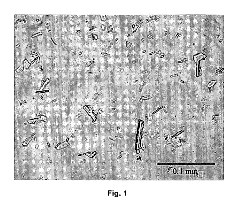
Figure 1 shows a light micrograph of ABT-874 crystals in crystallization.
Figures 2 to 5 show SEMs of ABT-874 crystals at different magnification;
Figure 2:
1,250x; Figure 3: 10,000x; Figure 4: 3,227x; Figure 5: 15,000x.
Figure 6 shows the results of Capillary Isoelectric Focusing (clEF)
Experiments with
ABT-874; A) ABT-874 crystal buffer and pl markers of pl 8.4, 8.5, 10.1 and
10.4; B)
ABT-874 crystals; same pl marker and characteristic ABT-874 signal at p1=9,29;
C)
Reference Standard; same pl marker and characteristic ABT-874 signal at
p1=9,29.
Figure 7 shows light microscopic pictures of crystals (needle-clusters)
obtained ac-
cording to Example 28 (crystallization with agitation).
Figure 8 shows light microscopic pictures of crystals (needles) obtained
according to
Example 32 (crystallization without agitation).
Figure 9 shows light microscopic pictures of crystals (needles) obtained
according to
Example 33 (crystallization without agitation).
Figure 10 shows light microscopic pictures of crystals (needles) obtained
according to
Example 34b (crystallization without agitation).
Figure 11 shows second derivative IR spectra of ABT-874 samples. Figure 1 1A
shows
spectra of crystal suspension recorded with an BioATR cell. Figure 11 B shows
spectra
of redissolved crystals recorded with an AquaSpec cell. Solid lines represent
samples
from crystalline ABT-874, dashed lines reprsent liquid standards. An offset
between
sample and standard was inserted for better illustration.
Figure 12 shows second derivative IR spectra of ABT-874 samples, 50 mg/mL
crystalline protein in 22% PEG 4,000 buffer in 0.1 M sodium acetate buffer, pH
5.5,
stored for 3 months at 25 C. Figure 11A shows spectra of crystal suspension
recorded
with an BioATR cell. Figure 116 shows spectra of redissolved crystals recorded
with
an AquaSpec cell. An offset between sample and standard was inserted for
better
illustration.
Figure 13: 40 mL batch crystallization of ABT-874 with and without seeding
(e.g.,
using 3.25% crystallized protein as seeding material in relation to ABT-874
mass from
the batch). R 2 are 0.9711 for non seeded, and 0.9763 for the seeded batch,
respectively.
Detailed Description of the Invention
A. Definitions
CA 02681752 2009-09-23
WO 2008/121301 PCT/US2008/004006
A "batch method of crystallization" comprises the step of adding the
crystalliza-
tion solution comprising the crystallization agent, preferably in dissolved
form, to the
solution of the antibody to be crystallized.
A "micro scale crystallization method", which may for example be based upon
vapor diffusion, comprises the steps of mixing a small volume of antibody
solution in
the microliter range with a reservoir buffer containing a crystallization
agent; placing a
droplet of the mixture in a sealed container adjacent to an aliquot of the
reservoir
buffer; allowing exchange of solvent between the droplet and the reservoir by
vapor
diffusion, during which the solvent content in the droplet changes and
crystallization
may be observed if suitable crystallization conditions are reached.
A "crystallization agent", e.g., a polyethylene glycol, favors crystal
formation of
the antibody to be crystallized.
A "crystallization solution" contains a crystallization agent in dissolved
form.
Preferably the solution is an aqueous system, i.e., the liquid constituents
thereof pre-
dominantly consist of water. For example, 80 to 100 wt.-% or 95 to 100 wt.-%
or 98 to
100 wt.-% may be water.
Antibody "crystals" are one form of the solid state of matter of the protein,
which
is distinct from a second solid form, i.e., the amorphous state, which exists
essentially
as an unorganized, heterogeneous solid. Crystals have a regular three-
dimensional
structure, typically referred to as a lattice. An antibody crystal comprises a
regular
three-dimensional array of antibody molecules (see Giege, R. and Ducruix, A.
Barrett,
Crystallization of Nucleic Acids and Proteins, a Practical Approach, 2nd ed.,
pp. 1-16,
Oxford University Press, New York (1999)).
A "whole" or "intact" anti-hlL-12 antibody as crystallized according to this
inven-
tion, is a functional antibody that is able to recognize and bind to its
antigen human IL-
12 in vitro and/or in vivo. The antibody may initiate subsequent immune system
reac-
tions of a patient associated with antibody-binding to its antigen, in
particular Direct
Cytotoxicity, Complement-Dependent Cytotoxicity (CDC), and Antibody-Dependent
Cytotoxicity (ADCC). The antibody molecule has a structure composed of two
identical
heavy chains (MW each about 50 kDa) covalently bound to each other, and two
identi-
cal light chains (MW each about 25 kDa), each covalently bound to one of the
heavy
chains. The four chains are arranged in a classic "Y" motif. Each heavy chain
is com-
prised of a heavy chain variable region (abbreviated herein as HCVR or VH) and
a
heavy chain constant region. The heavy chain constant region is comprised of
three
16
CA 02681752 2009-09-23
WO 2008/121301 PCT/US2008/004006
domains, CH1, CH2 and CH3. Each light chain is comprised of a light chain
variable
region (abbreviated herein as LCVR or VL) and a light chain constant region.
The light
chain constant region is comprised of one domain, CL. The VH and VL regions
can be
further subdivided into regions of hypervariability, termed complementarity
determining
regions (CDR), interspersed with regions that are more conserved, termed
framework
regions (FR). Each VH and VL is composed of three CDRs and four FRs, arranged
from amino-terminus to carboxy-terminus in the following order: FR1, CDR1,
FR2,
CDR2, FR3, CDR3, FR4. The complete antibody molecule has two antigen binding
sites, i.e., is "bivalent". The two antigen binding sites are specific for one
hIL-12 anti-
gen, i.e., the antibody is "mono-specific".
"Monoclonal antibodies" are antibodies that are derived from a single clone of
B
lymphocytes (B cells), and recognize the same antigenic determinant. Whole
mono-
clonal antibodies are those that have the above-mentioned classic molecular
structure
that includes two complete heavy chains and two complete light chains.
Monoclonal
antibodies are routinely produced by fusing the antibody-producing B cell with
an im-
mortal myeloma cell to generate B cell hybridomas, which continually produce
mono-
clonal antibodies in cell culture. Other production methods are available, for
example,
expression of monoclonal antibodies in bacterial, yeast, insect, or mammalian
cell cul-
ture using phage-display technology; in vivo production in genetically
modified animals,
such as cows, goats, pigs, rabbits, chickens, or in transgenic mice which have
been
modified to contain and express the entire human B cell genome; or production
in ge-
netically modified plants, such as tobacco and corn. Anti-hlL-12 antibodies
from all
such sources may be crystallized according to this invention.
The monoclonal antibodies to be crystallized according to the invention
include
"chimeric" anti-hlL-12 antibodies in which a portion of the heavy and/or light
chain is
identical with or homologous to corresponding sequences in antibodies derived
from a
particular species or belonging to a particular antibody class or subclass,
while the
remainder of the chain(s) is identical with or homologous to corresponding
sequences
in antibodies derived from another species or belonging to another antibody
class or
subclass. For example, a mouse/human chimera contains the variable antigen-
binding
portions of a murine antibody and the constant portions derived from a human
anti-
body.
"Humanized" forms of non-human (e.g., murine) anti-hlL-12 antibodies are also
encompassed by the invention. Humanized antibodies are chimeric antibodies
that
contain minimal sequence derived from a non-human immunoglobulin. For the most
17
CA 02681752 2009-09-23
WO 2008/121301 PCT/US2008/004006
part, humanized antibodies are human immunoglobulins in which residues from
one or
more complementarity determining regions (CDRs) or hypervariable loops (HVLs)
of
the human immunoglobulin are replaced by residues from a CDR or HVL of a non-
human species, such as mouse, rat, rabbit or nonhuman primate, having the
desired
functionality. Framework region (FR) residues of the human immunoglobulin may
re-
placed by corresponding non-human residues to improve antigen binding
affinity. Fur-
thermore, humanized antibodies may comprise residues that are found neither in
the
corresponding human or non-human antibody portions. These modifications may be
necessary to further improve antibody efficacy.
A "human antibody" or "fully human antibody" is one, which has an amino acid
sequence which corresponds to that of an antibody produced by a human or which
is
recombinantly produced. The term "human antibody", as used herein, is intended
to
include antibodies having variable and constant regions derived from human
germline
immunoglobulin sequences. The human antibodies of the invention may include
amino
acid residues not encoded by human germline immunoglobulin sequences (e.g.,
muta-
tions introduced by random or site-specific mutagenesis in vitro or by somatic
mutation
in vivo), for example in the CDRs and in particular CDR3. However, the term
"human
antibody", as used herein, is not intended to include antibodies in which CDR
se-
quences derived from the germline of another mammalian species, such as a
mouse,
have been grafted onto human framework sequences.
The term "recombinant human antibody", as used herein, is intended to include
all human antibodies that are prepared, expressed, created or isolated by
recombinant
means, such as antibodies expressed using a recombinant expression vector
trans-
fected into a host cell, antibodies isolated from a recombinant, combinatorial
human
antibody library, antibodies isolated from an animal (e.g., a mouse) that is
transgenic
for human immunoglobulin genes (see, e.g., Taylor, L.D. et al. (1992) Nucl.
Acids Res.
20:6287-6295) or antibodies prepared, expressed, created or isolated by any
other
means that involves splicing of human immunoglobulin gene sequences to other
DNA
sequences. Such recombinant human antibodies have variable and constant
regions
derived from human germline immunoglobulin sequences. In certain embodiments,
however, such recombinant human antibodies are subjected to in vitro
mutagenesis
(or, when an animal transgenic for human Ig sequences is used, in vivo somatic
mutagenesis) and thus the amino acid sequences of the VH and VL regions of the
re-
combinant antibodies are sequences that, while derived from and related to
human
18
CA 02681752 2009-09-23
WO 2008/121301 PCT/US2008/004006
germline VH and VL sequences, may not naturally exist within the human
antibody
germline repertoire in vivo.
A "neutralizing antibody", as used herein (or an "antibody that neutralized
hIL-12
activity"), is intended to refer to an antibody whose binding to hIL-12
results in inhibi-
tion of the biological activity of hIL-12. This inhibition of the biological
activity of hIL-12
can be assessed in vitro or in vivo by measuring one or more indicators of hIL-
12 bio-
logical activity, such as hIL-12-induced cell proliferation and hIL-12 binding
to hIL-12
receptors or hIL-12 induced decrease of white blood cells in vivo.
These indicators of hIL-12 biological activity can be assessed by one or more
of
several standard in vitro or in vivo assays known in the art. Preferably, the
ability of an
antibody to neutralize hIL-12 activity is assessed by inhibition of hIL-12-
induced cell
proliferation in phytohemagglutinin blasts and murine 2D6 cells.
An "affinity matured" anti-hlL-12 antibody is one with one or more alterations
in
one or more hypervariable regions, which result in an improvement in the
affinity of the
antibody for antigen, compared to a parent antibody. Affinity matured
antibodies will
have nanomolar or even picomolar affinities values for the target antigen.
Affinity ma-
tured antibodies are produced by procedures known in the art. Marks et al.
(1992)
Bio/Technology 10:779-783 describes affinity maturation by VH and VL domain
shuf-
fling. Random mutagenesis of CDR and/or framework residues is described by
Barbas
et al. (1994) Proc. Nat. Acad. Sci. USA 91:3809-3813 (1994); Scier et al.
(1995) Gene
169:147-155; Yelton et al. (1995) J. Immunol. 155:1994-2004; Jackson et al.
(1995) J.
Immunol. 154(7):3310-9; and Hawkins et al. (1992) J. Mol Biol. 226:889-896.
An "isolated antibody", as used herein, is intended to refer to an antibody
that is
substantially free of other antibodies having different antigenic
specificities (e.g., an
isolated antibody that specifically binds hIL-12 is substantially free of
antibodies that
specifically bind antigens other than hIL-12). An isolated antibody that
specifically
binds hIL-12 may, however, have cross-reactivity to other antigens, such as
hIL-12
molecules from other species. Moreover, an isolated antibody may be
substantially
free of other cellular material and/or chemicals.
The phrase "human interleukin 12" (abbreviated herein as hIL-12, or IL-12), as
used herein, includes a human cytokine that is secreted primarily by
macrophages and
dendritic cells. The term includes a heterodimeric protein comprising a 35 kD
subunit
(p35) and a 40 kD subunit (p40) which are both linked together with a
disulfide bridge.
The heterodimeric protein is referred to as a "p70 subunit". The structure of
human IL-
19
CA 02681752 2009-09-23
WO 2008/121301 PCT/US2008/004006
12 is described further in, for example, Kobayashi, et al. (1989) J. Exp Med.
170:827-
845; Seder, et al. (1993) Proc. Natl. Acad. Sci. 90:10188-10192; Ling, et al.
(1995) J.
Exp Med. 154:116-127; Podlaski, et al. (1992) Arch. Biochem. Biophys. 294:230-
237.
The term human IL-12 is intended to include recombinant human IL-12 (rh IL-
12),
which can be prepared by standard recombinant expression methods.
The term "koff', as used herein, is intended to refer to the off rate constant
for
dissociation of an antibody from the antibody/antigen complex.
The term "Kd", as used herein, is intended to refer to the dissociation
constant of
a particular antibody-antigen interaction.
A "functional equivalent" of a specific "parent" anti-hlL-12 antibody as
crystallized
according to the invention is one that shows the same antigen-specificity, but
differs
however with respect to the molecular composition of the "parent" antibody on
the
amino acid level or glycosylation level. The differences may be merely such
that the
crystallization conditions do not deviate from the parameter ranges as
disclosed
herein.
"Encapsulation" of antibody crystals refers to a formulation where the incorpo-
rated crystals are individually coated by at least one layer of a coating
material. In a
preferred embodiment, such coated crystals may have a sustained dissolution
rate.
"Embedding" of antibody crystals refers to a formulation where the crystals,
which might be encapsulated or not, are incorporated into a solid, liquid or
semi-solid
carrier in a disperse manner. Such embedded crystallized antibody molecules
may be
released or dissolved in a controlled, sustained manner from the carrier.
B. Method of crystallization
The crystallization method of the invention is in principle applicable to any
anti-
hIL-12 antibody. The antibody may be a polyclonal antibody or, preferably, a
mono-
clonal antibody. The antibody may be chimeric antibodies, humanized
antibodies, hu-
man antibodies or non-human, as for example mouse antibodies, each in
glycosylated
or non-glycosylated form. In particular the method is applicable to ABT-874
and func-
tional equivalents thereof.
Preferably the anti-hlL-12 antibody is an IgG antibody, in particular an anti
hu-
man IL-12 antibody of the group IgG1.
Unless otherwise stated the crystallization method of the invention makes use
of
technical equipment, chemicals and methodologies well known in the art.
However, as
CA 02681752 2009-09-23
WO 2008/121301 PCT/US2008/004006
explained above, the present invention is based on the surprising finding that
the se-
lection of specific crystallization conditions, in particular, the selection
of specific crys-
tallization agents, optionally further combined with specific pH conditions
and/or con-
centration ranges of the corresponding agents (buffer, antibody,
crystallization agent),
allows for the first time to prepare reproducibly and in a large scale stable
crystals of
antibodies, in particular non-chimeric, human antibodies, directed against hIL-
12,
which can be further processed to form an active ingredient of a superior,
highly ad-
vantageous pharmaceutical composition.
The starting material for performing the crystallization method normally com-
prises a concentrated solution of the antibody to be crystallized. The protein
concentra-
tion may, for example, be in the range of about 5 to about 300 mg/mI,
preferably about
5 to about 200 mg/mI, preferably about 5 to about 75 mg/mI. The solution may
contain
additives stabilizing the dissolved antibody, and it may be advisable to
remove the ad-
ditives in advance. This can be achieved by performing a buffer exchange step.
Preferably the starting material for performing the crystallization contains
the an-
tibody in an aqueous solution, having a pH adjusted in the range of about 3.2
to about
8.2, or about 4.0 to about 8.0, in particular about 4.5 to about 6.5,
preferably about 5.0
to about 5.5. The pH may be adjusted by means of a suitable buffer applied in
a final
concentration of about 1 to about 500 mM, in particular about 1 to about 100
mM or 1
to about 10 mM. The solution may contain additives, as for example in a
proportion of
about 0.01 to about 15, or about 0.1 to about 5, or about 0.1 to about 2 wt.-%
based on
the total weight of the solution, such as salts, sugars, sugar alcohols and
surfactants,
in order to further stabilize the solution. The excipients are preferably be
selected from
physiologically acceptable compounds, routinely applied in pharmaceutical
prepara-
tions. As non-limiting examples, excipients include salts, such as NaCI;
surfactants,
such as polysorbate 80 (Tween 80), polysorbate 20 (Tween 20); sugars, such as
sucrose, trehalose; sugar alcohols, such as mannitol, sorbitol; and buffer
agents, such
as phosphate-based buffer systems, sodium and potassium hydrogen phosphate
buff-
ers as defined above, acetate buffer, phosphate buffer, citrate buffer, TRIS
buffer,
maleate buffer or succinate buffer, histidine buffer; amino acids, such as
histidine, ar-
ginine and glycine.
The buffer exchange may be performed by means of routine methods, for exam-
ple dialysis, diafiltration or ultrafiltration.
The initial protein concentration of the aqueous solution used as starting
material
should be in the range of about 0.5 to about 200 or about 1 to about 50 mg/mI.
21
CA 02681752 2009-09-23
WO 2008/121301 PCT/US2008/004006
Depending on the intended final batch size (which may be in the range of 1 ml
to
20,000 litres) an initial volume of the aqueous antibody solution is placed in
an appro-
priate container (as for example a vessel, bottle or tank) made of inert
material, as for
example glass, polymer or metal. The initial volume of the aqueous solution
may cor-
respond to about 30 to 80%, normally about 50% of the final batch size.
If necessary the solution after having been filled into the container will be
brought
to standardized conditions. In particular, the temperature will be adjusted in
the range
of about 4 C and about 37 C.
Then the crystallization solution, containing the crystallization agent in an
appro-
priate concentration, optionally pre-conditioned in the same way as the
antibody solu-
tion, is added to the antibody solution.
The addition of the crystallization solution is performed continuously or
discon-
tinuously optionally under gentle agitation in order to facilitate mixing of
the two liquids.
Preferably the addition is performed under conditions where the protein
solution is pro-
vided under agitation and the crystallization solution (or agents in its solid
from) is / are
added in a controlled manner.
The formation of the antibody crystals is initiated by applying a polyalkylene
polyol as defined above, in particular a polyalkylene glycol, and preferably a
polyethyl-
ene glycol (PEG), or a mixture of at least two different polyalkylene glycols
as defined
above as the crystallization agent. The crystallization solution contains the
agent in a
concentration, which is sufficient to afford a final concentration of the
polyalkylene
polyol in the crystallization mixture in the range of about 5 to 30 % (w/v).
Preferably, the crystallization solution additionally contains an acidic
buffer, e.g.,
different from that of the antibody solution, in a concentration suitable to
allow the ad-
justment of the pH of the crystallization mixture in the range of about 4 to
6.
After having finished the addition of the crystallization solution, the
obtained mix-
ture may be further incubated for about 1 hour to about 250 days in order to
obtain a
maximum yield of antibody crystals. If appropriate, the mixture may, for
example, be
agitated, gently stirred, rolled or otherwise moved.
Finally, the crystals obtained may be separated by known methods, for example
filtration or centrifugation, as for example by centrifugation at about 200 -
20,000 rpm,
preferably 500 - 2,000 rpm, at room temperature or 4 C. The remaining mother
liquor
may be discarded or further processed.
22
CA 02681752 2009-09-23
WO 2008/121301 PCT/US2008/004006
If necessary, the isolated crystals may be washed and subsequently dried, or
the
mother liquor can be exchanged by a different solvent system suitable for
storage and
/or final use of the antibodies suspended therein.
Antibody crystals formed according to the present invention may vary in their
shape. as already explained above For therapeutic administration, the size of
the crys-
tals will vary depending on the route of administration, for example, for
subcutaneous
administration the size of the crystals may be larger than for intravenous
administra-
tion.
The shape of the crystals may be altered by adding specific additional
additives
to the crystallization mixture, as has been previously described for both
protein crystals
and crystals of low molecular weight organic and inorganic molecules.
If necessary, it may be verified that the crystals are in fact crystals of the
anti-
body. Crystals of an antibody can be analyzed microscopically for
birefringence. In
general, crystals, unless of cubic internal symmetry, will rotate the plane of
polarization
of polarized light. In yet another method, crystals can be isolated, washed,
resolubi-
lized and analyzed by SDS-PAGE and, optionally, stained with an anti-Fc
receptor an-
tibody. Optionally, the resolubilized antibody can also be tested for binding
to its hIL-12
utilizing standard assays.
Crystals as obtained according to the invention may also be crosslinked to one
another. Such crosslinking may enhance stability of the crystals. Methods for
crosslink-
ing crystals described, for example, in U.S. Patent No. 5,849,296. Crystals
can be
crosslinked using a bifunctional reagent such as glutaraldehyde. Once
crosslinked,
crystals can be lyophilized and stored for use, for example, in diagnostic or
therapeutic
applications.
In some cases, it may be desirable to dry the crystal. Crystals may be dried
by
means of inert gases, like nitrogen gas, vacuum oven drying, lyophilization,
evapora-
tion, tray drying, fluid bed drying, spray drying, vacuum drying or roller
drying. Suitable
methods are well known.
Crystals formed according to the invention can be maintained in the original
crystallization solution, or they can be washed and combined with other
substances,
like inert carriers or ingredients to form compositions or formulations
comprising crys-
tals of the invention. Such compositions or formulations can be used, for
example, in
therapeutic and diagnostic applications.
23
CA 02681752 2009-09-23
WO 2008/121301 PCT/US2008/004006
A preferred embodiment is to combine a suitable carrier or ingredient with
crys-
tals of the invention in that way that crystals of the formulation are
embedded or en-
capsulated by an excipient. Suitable carriers may be taken from the non
limiting group
of: poly (acrylic acid), poly (cyanoacrylates), poly (amino acids), poly
(anhydrides), poly
(depsipeptide), poly (esters), poly (lactic acid), poly (lactic-co-glycolic
acid) or PLGA,
poly (f3-hydroxybutryate), poly (caprolactone), poly (dioxanone); poly
(ethylene glycol),
poly (hydroxypropyl) methacrylamide, poly (organo) phosphazene, poly (ortho
esters),
poly (vinyl alcohol), poly (vinylpyrrolidone), maleic anhydride alkyl vinyl
ether copoly-
mers, pluronic polyols, albumin, alginate, cellulose and cellulose
derivatives, collagen,
fibrin, gelatin, hyaluronic acid, oligosaccharides, glycaminoglycans, sulfated
polysac-
charides, blends and copolymers thereof, SAIB, fatty acids and salts of fatty
acids, fatty
alcohols, fatty amines, mono-, di-, and triglycerides of fatty acids,
phospholipids, glycol-
ipids, sterols and waxes and related similar substances. Waxes are further
classified in
natural and synthetic products. Natural materials include waxes obtained from
vegeta-
ble, animal or minerals sources such as beeswax, carnauba or montanwax.
Chlorin-
ated naphthalenes and ethylenic polymers are examples for synthetic wax
products.
C. Compositions
Another aspect of the invention relates to compositions/formulations
comprising
anti-hlL-12 antibody crystals in combination with at least one
carrier/excipient.
The formulations may be solid, semisolid or liquid.
Formulations of the invention are prepared, in a form suitable for storage
and/or
for use, by mixing the antibody having the necessary degree of purity with a
physio-
logically acceptable additive, like carrier, excipient and/or stabilizer (see
for example
Remington's Pharmaceutical Sciences, 16th Edn., Osol, A. Ed. (1980)), in the
form of
suspensions, lyophilized or dried in another way. Optionally further active
ingredients,
as for example different antibodies, biomolecules, chemically or enzymatically
synthe-
sized low-molecular weight molecules may be incorporated as well.
Acceptable additives are non-toxic to recipients at the dosages and concentra-
tions employed. Nonlimiting examples thereof include:
- Acidifying agents, like acetic acid, citric acid, fumaric acid, hydrochloric
acid, malic
acid, nitric acid, phosphoric acid, diluted phosphoric acid, sulfuric acid,
tartaric acid.
- Aerosol propellants, like butane, dichlorodifluoromethane,
dichlorotetrafluoroethane,
isobutane, propane, trichloromonofluoromethane.
24
CA 02681752 2009-09-23
WO 2008/121301 PCT/US2008/004006
- Air displacements, like carbon dioxide, nitrogen;
- Alcohol denaturants, like methyl isobutyl ketone, sucrose octacetate;
- Alkalizing agents, like ammonia solution, ammonium carbonate,
diethanolamine,
diisopropanolamine, potassium hydroxide, sodium bicarbonate, sodium borate,
sodium
carbonate, sodium hydroxide, trolamine;
- Antifoaming agents, like dimethicone, simethicone.
- Antimicrobial preservatives, like benzalkonium chloride, benzalkonium
chloride solu-
tion, benzelthonium chloride, benzoic acid, benzyl alcohol, butylparaben,
cetylpyridin-
ium chloride, chlorobutanol, chlorocresol, cresol, dehydroacetic acid,
ethylparaben,
methylparaben, methylparaben sodium, phenol, phenylethyl alcohol,
phenylmercuric
acetate, phenylmercuric nitrate, potassium benzoate, potassium sorbate,
propylpara-
ben, propylparaben sodium, sodium benzoate, sodium dehydroacetate, sodium
propi-
onate, sorbic acid, thimerosal, thymol.
- Antioxidants, like ascorbic acid, ascorbyl palmitate, butylated
hydroxyanisole, buty-
lated hydroxytoluene, hypophosphorous acid, monothioglycerol, propyl gallate,
sodium
formaldehyde sulfoxylate, sodium metabisulfite, sodium thiosulfate, sulfur
dioxide, to-
copherol, tocopherols excipient;
- Buffering agents, like acetic acid, ammonium carbonate, ammonium phosphate,
boric
acid, citric acid, lactic acid, phosphoric acid, potassium citrate, potassium
metaphos-
phate, potassium phosphate monobasic, sodium acetate, sodium citrate, sodium
lac-
tate solution, dibasic sodium phosphate, monobasic sodium phosphate,
histidine.
- Chelating agents, like edetate disodium, ethylenediaminetetraacetic acid and
salts,
edetic acid;
- Coating agents, like sodium carboxymethylcellulose, cellulose acetate,
cellulose ace-
tate phthalate, ethylcellulose, gelatin, pharmaceutical glaze, hydroxypropyl
cellulose,
hydroxypropyl methylcellulose, hydroxypropyl methylcellulose phthalate,
methacrylic
acid copolymer, methylcellulose, polyethylene glycol, polyvinyl acetate
phthalate, shel-
lac, sucrose, titanium dioxide, carnauba wax, microcystalline wax, zein, poly
amino
acids, other polymers like PLGA etc., and SAIB.
- Coloring agents, like ferric oxide.
- Complexing agents, like ethylenediaminetetraacetic acid and salts (EDTA),
edetic
acid, gentisic acid ethanolamide, oxyquinoline sulfate.
CA 02681752 2009-09-23
WO 2008/121301 PCT/US2008/004006
- Desiccants, like calcium chloride, calcium sulfate, silicon dioxide.
- Emulsifying and/or solubilizing agents, like acacia, cholesterol,
diethanolamine
(adjunct), glyceryl monostearate, lanolin alcohols, lecithin, mono-and di-
glycerides,
monoethanolamine (adjunct), oleic acid (adjunct), oleyl alcohol (stabilizer),
poloxamer,
polyoxyethylene 50 stearate, polyoxyl 35 caster oil, polyoxyl 40 hydrogenated
castor
oil, polyoxyl 10 oleyl ether, polyoxyl 20 cetostearyl ether, polyoxyl 40
stearate,
polysorbate 20, polysorbate 40, polysorbate 60, polysorbate 80, propylene
glycol
diacetate, propylene glycol monostearate, sodium lauryl sulfate, sodium
stearate,
sorbitan monolaurate, soritan monooleate, sorbitan monopaimitate, sorbitan
monostearate, stearic acid, trolamine, emulsifying wax.
- Filtering aids, like powdered cellulose, purified siliceous earth.
- Flavors and perfumes, like anethole, benzaidehyde, ethyl vanillin, menthol,
methyl
salicylate, monosodium glutamate, orange flower oil, peppermint, peppermint
oil, pep-
permint spirit, rose oil, stronger rose water, thymol, tolu balsam tincture,
vanilla, vanilla
tincture, vanillin.
- Glidant and/or anticaking agents, like calcium silicate, magnesium silicate,
colloidal
silicon dioxide, talc.
- Humectants, like glycerin, hexylene glycol, propylene glycol, sorbitol;
- Ointment bases, like lanolin, anhydrous lanolin, hydrophilic ointment, white
ointment,
yellow ointment, polyethylene glycol ointment, petrolatum, hydrophilic
petrolatum, white
petrolatum, rose water ointment, squalane.
- Plasticizers, like castor oil, lanolin, mineral oil, petrolatum, benzyl
benyl formate,
chlorobutanol, diethyl pthalate, sorbitol, diacetylated monoglycerides,
diethyl phthalate,
glycerin, glycerol, mono-and di-acetylated monoglycerides, polyethylene
glycol, pro-
pylene glycol, triacetin, triethyl citrate, ethanol.
- Polypeptides, like low molecular weight (less than about 10 residues);
Proteins, such as serum albumin, gelatin, or immunoglobulins;
- Polymer membranes, like cellulose acetate membranes.
- Solvents, like acetone, alcohol, diluted alcohol, amylene hydrate, benzyl
benzoate,
butyl alcohol, carbon tetrachloride, chloroform, corn oil, cottonseed oil,
ethyl acetate,
glycerin, hexylene glycol, isopropyl alcohol, methyl alcohol, methylene
chloride, methyl
isobutyl ketone, mineral oil, peanut oil, polyethylene glycol, propylene
carbonate, pro-
26
CA 02681752 2009-09-23
WO 2008/121301 PCT/US2008/004006
pylene glycol, sesame oil, water for injection, sterile water for injection,
sterile water for
irrigation, purified water, liquid triglycerides, liquid waxes, higher
alcohols.
- Sorbents, like powdered cellulose, charcoal, purified siliceous earth,
Carbon dioxide
sorbents, barium hydroxide lime, soda lime.
- Stiffening agents, like hydrogenated castor oil, cetostearyl alcohol, cetyl
alcohol, cetyl
esters wax, hard fat, paraffin, polyethylene excipient, stearyl alcohol,
emulsifying wax,
white wax, yellow wax.
- Suppository bases, like cocoa butter, hard fat, polyethylene glycol;
- Suspending and/or viscosity-increasing agents, like acacia, agar, alginic
acid, alumi-
num monostearate, bentonite, purified bentonite, magma bentonite, carbomer
934p,
carboxymethylcellulose calcium, carboxymethylcellulose sodium,
carboxymethycellu-
lose sodium 12, carrageenan, microcrystalline and carboxymethylcellulose
sodium
cellulose, dextrin, gelatin, guar gum, hydroxyethyl cellulose, hydroxypropyl
cellulose,
hydroxypropyl methylcellulose, magnesium aluminum silicate, methylcellulose,
pectin,
polyethylene oxide, polyvinyl alcohol, povidone, propylene glycol alginate,
silicon diox-
ide, colloidal silicon dioxide, sodium alginate, tragacanth, xanthan gum;
- Sweetening agents, like aspartame, dextrates, dextrose, excipient dextrose,
fructose,
mannitol, saccharin, calcium saccharin, sodium saccharin, sorbitol, solution
sorbitol,
sucrose, compressible sugar, confectioner's sugar, syrup;
- Tablet binders, like acacia, alginic acid, sodium carboxymethylcellulose,
microcrystal-
line cellulose, dextrin, ethylcellulose, gelatin, liquid glucose, guar gum,
hydroxypropyl
methylcellulose, methycellulose, polyethylene oxide, povidone, pregelatinized
starch,
syrup.
- Tablet and/or capsule diluents, like calcium carbonate, dibasic calcium
phosphate,
tribasic calcium phosphate, calcium sulfate, microcrystalline cellulose,
powdered cellu-
lose, dextrates, dextrin, dextrose excipient, fructose, kaolin, lactose,
mannitol, sorbitol,
starch, pregelatinized starch, sucrose, compressible sugar, confectioner's
sugar;
- Tablet disintegrants, like alginic acid, microcrystalline cellulose,
croscarmellose so-
dium, corspovidone, polacrilin potassium, sodium starch glycolate, starch,
pregelati-
nized starch.
- Tablet and/or capsule lubricants, like calcium stearate, glyceryl behenate,
magnesium
stearate, light mineral oil, polyethylene glycol, sodium stearyl fumarate,
stearic acid,
purified stearic acid, talc, hydrogenated vegetable oil, zinc stearate;
- Tonicity agent, like dextrose, glycerin, mannitol, potassium chloride,
sodium chloride
27
CA 02681752 2009-09-23
WO 2008/121301 PCT/US2008/004006
Vehicle: flavored and/or sweetened aromatic elixir, compound benzaldehyde
elixir, iso-
alcoholic elixir, peppermint water, sorbitol solution, syrup, tolu balsam
syrup.
- Vehicles, like oleaginous almond oil, corn oil, cottonseed oil, ethyl
oleate, isopropyl
myristate, isopropyl palmitate, mineral oil, light mineral oil, myristyl
alcohol, octyldode-
canol, olive oil, peanut oil, persic oil, sesame oil, soybean oil, squalane;
solid carrier
sugar spheres; sterile bacteriostatic water for injection, bacteriostatic
sodium chloride
injection, liquid triglycerides, liquid waxes, higher alcohols
- Water repelling agents, like cyclomethicone, dimethicone, simethicone;
- Wetting and/or solubilizing agents, like benzalkonium chloride, benzethonium
chlo-
ride, cetylpyridinium chloride, docusate sodium, nonoxynol 9, nonoxynol 10,
octoxynol
9, poloxamer, polyoxyl 35 castor oil, polyoxyl 40, hydrogenated castor oil,
polyoxyl 50
stearate, polyoxyl 10 oleyl ether, polyoxyl 20, cetostearyl ether, polyoxyl 40
stearate,
polysorbate 20, polysorbate 40, polysorbate 60, polysorbate 80, sodium lauryl
sulfate,
sorbitan monolaureate, sorbitan monooleate, sorbitan monopalmitate, sorbitan
monostearate, and tyloxapol.
The crystals may be combined with a polymeric carrier to provide for stability
and/or sustained release. Such polymers include biocompatible and
biodegradable
polymers. A polymeric carrier may be a single polymer type or it may be
composed of
a mixture of polymer types. Nonlimiting examples of polymeric carriers have
already
been stated above.
Examples of preferred ingredients or excipients include:
- salts of amino acids such as glycine, arginine, aspartic acid, glutamic
acid, lysine,
asparagine, glutamine, proline, histidine;
- monosaccharides, such as glucose, fructose, galactose, mannose, arabinose,
xylose,
ribose;
- disaccharides, such as lactose, trehalose, maltose, sucrose;
- polysaccharides, such as maltodextrins, dextrans, starch, glycogen;
- alditols, such as mannitol, xylitol, lactitol, sorbitol;
- glucuronic acid, galacturonic acid;
- cyclodextrins, such as methyl cyclodextrin, hydroxypropyl- (3-cyclodextrin)
28
CA 02681752 2009-09-23
WO 2008/121301 PCT/US2008/004006
- inorganic salts, such as sodium chloride, potassium chloride, magnesium
chloride,
phosphates of sodium and potassium, boric acid ammonium carbonate and ammonium
phosphate;
- organic salts, such as acetates, citrate, ascorbate, lactate;
- emulsifying or solubilizing agents like acacia, diethanolamine, glyceryl
monostearate,
lecithin, monoethanolamine, oleic acid, oleyl alcohol, poloxamer,
polysorbates, sodium
lauryl sulfate, stearic acid, sorbitan monolaurate, sorbitan monostearate, and
other
sorbitan derivatives, polyoxyl derivatives, wax, polyoxyethylene derivatives,
sorbitan
derivatives; and
- viscosity increasing reagents like, agar, alginic acid and its salts, guar
gum, pectin,
polyvinyl alcohol, polyethylene oxide, cellulose and its derivatives propylene
carbonate,
polyethylene glycol, hexylene glycol and tyloxapol.
Formulations described herein also comprise an effective amount of crystalline
antibody. In particular, the formulations of the invention may include a
"therapeutically
effective amount" or a "prophylactically effective amount" of antibody
crystals of the
invention. A "therapeutically effective amount" refers to an amount effective,
at dos-
ages and for periods of time necessary, to achieve the desired therapeutic
result. A
"therapeutically effective amount" of the antibody crystals may vary according
to fac-
tors such as the disease state, age, sex, and weight of the individual, and
the ability of
the antibody to elicit a desired response in the individual. A therapeutically
effective
amount is also one in which any toxic or detrimental effects of the antibody
are out-
weighed by the therapeutically beneficial effects. A "prophylactically
effective amount"
refers to an amount effective, at dosages and for periods of time necessary,
to achieve
the desired prophylactic result. Typically, since a prophylactic dose is used
in subjects
prior to or at an earlier stage of disease, the prophylactically effective
amount will be
less than the therapeutically effective amount.
Suitable dosages can readily be determined using standard methodology. The
antibody is suitably administered to the patient at one time or over a series
of treat-
ments. Depending on the above mentioned factors, about 1 pg/kg to about 50
mg/kg,
as for example 0.1-20 mg/kg of antibody is an initial candidate dosage for
administra-
tion to the patient, whether, for example, by one or more separate
administrations, or
by continuous infusion. A typical daily or weekly dosage might range from
about 1
pg/kg to about 20 mg/kg or more, depending on the condition, the treatment is
re-
peated until a desired suppression of disease symptoms occurs. However, other
dos-
29
CA 02681752 2009-09-23
WO 2008/121301 PCT/US2008/004006
age regimens may be useful. In some cases, formulations comprise a
concentration of
antibody of at least about 1 g/L or greater when resolubilized. In other
embodiments,
the antibody concentration is at least about 1 g/L to about 100 g/L when
resolubilized.
Crystals of an antibody, or formulations comprising such crystals, may be
admin-
istered alone or as part of a pharmaceutical preparation. They may be
administered by
parenteral, oral or topical routes. For example, they may be administered by
oral, pul-
monary, nasal, aural, anal, dermal, ocular, intravenous, intramuscular,
intraarterial,
intraperitoneal, mucosal, sublingual, subcutaneous, transdermal, topical or
intracranial
routes, or into the buccal cavity. Specific examples of administration
techniques com-
prise pulmonary inhalation, intralesional application, needle injection, dry
powder inha-
lation, skin electroporation, aerosol delivery, and needle-free injection
technologies,
including needle-free subcutaneous administration.
The present invention will now be explained in more detail by means of the fol-
lowing, non-limiting, illustrative examples. Guided by the general part of the
description
and on the basis of his general knowledge a skilled reader will be enabled to
provide
further embodiments to the invention without undue experimentation.
ExempliFication
A. Materials
a) Protein
Frozen monoclonal antibody (mAb) ABT-874 was obtained from Abbott Laborato-
ries. All experiments were performed from a product lot, where the original
mAb con-
centration was 64 mg/mL.
b) Fine chemicals
Sodium acetate was obtained from Grussing GmbH, Filsum. Polyethylene glycols
of different polymerization grades were obtained from Clariant GmbH, Sulzbach.
Fur-
thermore, commercial crystallization screens and reagents (Hampton Research,
Nextal
Biotechnologies) were used for certain microscale experiments. All other
chemicals
were from Sigma-Aldrich, Steinheim, or Merck, Darmstadt.
B. General methods
a) Thawing of ABT-874 drug substance
ABT-874 was thawed at 25 C in agitated water baths.
b) Buffer Exchange - Method A
CA 02681752 2009-09-23
WO 2008/121301 PCT/US2008/004006
An aliquot of ABT-874 solution was pipetted into a 30 KDa MWCO Vivaspin 20
concentrator (Vivascience). The protein sample was diluted with the new buffer
in a
ratio of 1:10, and by centrifugation at 5,000 x g at 4 C (Sigma 4 K 15 lab
centrifuge)
the sample volume was brought back to the original sample volume. The dilution
/ cen-
trifugation steps were repeated once, resulting in a dilution of 1:100 of the
original
sample buffer. After adjustment of protein concentration, the solution was
sterile fil-
tered through a 0.2 pm syringe driven filter unit.
b) Buffer Exchange - Method B
An aliquot of ABT-874 solution was placed into a SLIDE-A-LYZER dialysis cas-
sette (Pierce Biotechnology Inc.). The dialysis cassette was placed into a
beaker con-
taining the buffer of choice, and the buffer exchange was performed at 4 C
overnight
with stirring. After adjustment of protein concentration, the solution was
sterile filtered
through a 0.2 pm syringe driven filter unit.
c) OD280 - protein concentration measurements
A ThermoSpectronics UV1 device was used to assess protein concentration at a
wavelength of 280 nm, applying an extinction coefficient of 1.42 cm2 mg-'. For
this pur-
pose, aliquots of crystallization slurries were centrifuged at 14,000 rpm, and
residual
protein concentration was determined in the supernatant.
d) pH Measurements
pH measurements were conducted by using a Mettler Toledo MP220 pH meter.
Inlab 413 electrodes and Inlab 423 microelectrodes were utilized.
e) Crystallization Methods
el) Microscale Crystallization - Sitting Drop Vapor Diffusion Hydra II
Initial crystallization screens were performed using a Hydra II
crystallization robot
and Greiner 96 well plates (three drop wells, Hampton Research). After setting
up the
plates, the wells were sealed with Clearseal film (Hampton Research).
e2) Microscale Crystallization - Hanging Drop Vapor Diffusion
Hanging drop vapor diffusion experiments were conducted using VDX plates
(with sealant, Hampton Research) and OptiClear plastic cover slides (squares,
Hamp-
ton Research) or siliconized glass cover slides (circle, Hampton Research),
respec-
tively. After preparation of reservoir solutions, one drop of reservoir
solution was ad-
mixed with one drop of the protein solution on a cover slide, and the well was
sealed
31
CA 02681752 2009-09-23
WO 2008/121301 PCT/US2008/004006
with the inverted cover slide in such a way that the drop was hanging above
the reser-
voir.
e3) Batch Crystallization - Method A (24 Well Plate)
Batch crystallization was performed by admixing the protein solution with an
equal amount of crystallization buffer (500 NI) in a well. The well was
subsequently
sealed with adhesive tape to prevent water evaporation.
e4) Batch Crystallization - Method B(Eppendorff Reaction Tube)
Batch crystallization was performed by admixing the protein solution with an
equal amount of crystallization buffer in a 1.5 mL or a 2 mL Eppendorff
reaction tube.
e5) Batch Crystallization - Method C (Falcon Tubes, No Agitation)
Batch crystallization was performed by admixing the protein solution with an
equal amount of crystallization buffer in a 15 mL or 50 mL Falcon tube.
e6) Batch Crystallization - Method D (Falcon Tubes, Agitation)
Batch crystallization was performed by admixing the protein solution with an
equal amount of crystallization buffer in a 15 mL or 50 mL Falcon tube. Right
after clos-
ing, the tube was put on a laboratory shaker (GFL 3013 or GFL 3015) or was
alterna-
tively agitated by tumbling. By application of these methods, introduction of
stirrers into
the sample was avoided.
f) SDS-PAGE
Samples were prepared by adjusting protein concentration to 8 pg / 20 pL. The
samples were diluted with an SDS / Tris / glycerine buffer containing
bromophenol
blue. Qualitative SDS PAGE analysis was performed using Invitrogen NuPage 10%
Bis-Tris Gels, NuPage MES SDS Running Buffer and Mark12 Wide Range Protein
Standards. 20 pL of sample was pipetted into a gel pocket. After running the
gel and
fixation with acetic acid / methanol reagent, staining was performed using the
Novex
Colloidal Blue Stain Kit. Gels were dried using Invitrogen Gel-Dry drying
solution.
g) Light Microscopy
Crystals were observed using a Zeiss Axiovert 25 or a Nikon Labophot micro-
scope. The latter was equipped with a polarization filter set and a JVC TK
C1380 color
video camera.
h) SE-HPLC
32
CA 02681752 2009-09-23
WO 2008/121301 PCT/US2008/004006
Aggregation levels of ABT-874 samples were assessed by SE-HPLC. A Dionex
P680 pump, ASI-100 autosampler and UVD170U detector device were used. Aggre-
gated species were separated from the monomer by an Amersham Bioscience Super-
dex 200 10/300 GL gel filtration column, applying a validated Abbott standard
protocol
(A-796874.0 - ABT 874, J 695).
C. Vapor Diffusion Crystallization Experiments
Concentration values given in the following examples are initial values
referring
to the antibody solution and the reservoir solution before mixing of the two
solutions.
All pH values, if not described otherwise, refer to the pH of an acetate
buffer
stock before it was combined with other substances, like the crystallization
agent.
All buffer molarities, if not described otherwise, refer to sodium acetate
concen-
trations in a stock solution before pH adjustment, typically performed using
acetic acid
glacial.
Example 1- PEG 4,000 / Sodium Acetate Grid Screen In Hanging Drop Vapor Dif-
fusion Mode
A hanging drop vapor diffusion crystallization method was performed on ABT-
874. ABT-874 was buffered into a buffer containing about 0.1 M sodium acetate
at a
pH of about 5.2. The protein concentration was adjusted to 10 mg/mL.
A greased VDX plate and square OptiClear plastic cover slides were used. 500
pL of a particular reservoir solution was prepared by admixing acetate buffer,
50% w/v
PEG 4,000 solution and Milli Q water (fully desalted and optionally pre-
distilled) in each
well. In this example, the acetate buffer molarity was kept constant at about
0.1 M, and
PEG 4,000 was varied from about 6% w/v to about 28% w/v in 2% steps. The pH
was
about 5.2 throughout. Each condition was assessed in duplicate. About 1 pL of
the
protein solution was admixed with about 1 pL of a particular reservoir
solution on a
square OptiClear plastic cover slide, and the well was sealed with the
inverted slide,
generating a hanging drop experiment. The plates were stored at ambient
temperature.
Microscopy of the drops was performed multiple times during the following
thirty days.
The conditions were classified into clear drops, drops containing random
precipitation,
drops containing crystals and drops containing mixtures of precipitated
species and
crystals.
RESULTS: From the 24 wells assessed, no crystals were observed.
33
CA 02681752 2009-09-23
WO 2008/121301 PCT/US2008/004006
Example 2 - PEG 4,000 / Sodium Acetate Grid Screen In Hanging Drop Vapor Dif-
fusion Mode, Different Protein Concentration
A hanging drop vapor diffusion crystallization method was performed on ABT-
874 at different protein concentration. ABT-874 was buffered into a buffer
containing
about 0.1 M sodium acetate at a pH of about 5.2. The protein concentration was
ad-
justed to 50 mg/mL.
A greased VDX plate and square OptiClear plastic cover slides were used. 500
pL of a particular reservoir solution was prepared by admixing acetate buffer,
50% w/v
PEG 4,000 solution and Milli Q water in each well. In this example, the
acetate buffer
molarity was kept constant at about 0.1 M, and PEG 4,000 was varied from about
6%
w/v to about 28% w/v in 2% steps. The pH was about 5.2 throughout. Each
condition
was assessed in duplicate. About 1 pL of the protein solution was admixed with
about
1 pL of a particular reservoir solution on a square OptiClear plastic cover
slide, and the
well was sealed with the inverted slide, generating a hanging drop experiment.
The
plates were stored at ambient temperature. Microscopy of the drops was
performed
multiple times during the following thirty days. The conditions were
classified into clear
drops, drops containing random precipitation, drops containing crystals and
drops con-
taining mixtures of precipitated species and crystals.
RESULTS: From the 24 wells assessed, crystals were observed at a PEG 4,000
concentration of about 16%. The crystals showed needle or needle cluster like
mor-
phology.
Example 3 - PEG 400 / Sodium Acetate Grid Screen In Hanging Drop Vapor Diffu-
sion Mode
A hanging drop vapor diffusion crystallization method was performed on ABT-
874 using PEG 400. ABT-874 was buffered into a buffer containing about 0.1 M
so-
dium acetate at a pH of about 5.2. The protein concentration was adjusted to
10
mg/mL.
A greased VDX plate and square OptiClear plastic cover slides were used. 500
pL of a particular reservoir solution was prepared by admixing acetate buffer,
50% w/v
PEG use PEG 400 solution and Milli Q water in each well. In this example, the
acetate
buffer molarity was kept constant at about 0.1 M, and PEG 400 was varied from
about
30% w/v to about 40% w/v in 2% steps. The pH was about 5.2 throughout. Each
condi-
tion was assessed in duplicate. About 1 pL of the protein solution was admixed
with
about 1 pL of a particular reservoir solution on a square OptiClear plastic
cover slide,
34
CA 02681752 2009-09-23
WO 2008/121301 PCT/US2008/004006
and the well was sealed with the inverted slide, generating a hanging drop
experiment.
The plates were stored at ambient temperature. Microscopy of the drops was per-
formed multiple times during the following thirty days. The conditions were
classified
into clear drops, drops containing random precipitation, drops containing
crystals and
drops containing mixtures of precipitated species and crystals.
RESULTS: From the 12 wells assessed, no crystals were observed.
Example 4 - PEG 400 / Sodium Acetate Grid Screen In Hanging Drop Vapor Diffu-
sion Mode, Different Protein Concentration
A hanging drop vapor diffusion crystallization method was performed on ABT-
874 at different protein concentration. ABT-874 was buffered into a buffer
containing
about 0.1 M sodium acetate at a pH of about 5.2. The protein concentration was
ad-
justed to 50 mg/mL.
A greased VDX plate and square OptiClear plastic cover slides were used. 500
pL of a particular reservoir solution was prepared by admixing acetate buffer,
50% w/v
PEG use PEG 400 solution and Milli Q water in each well. In this example, the
acetate
buffer molarity was kept constant at about 0.1 M, and PEG 400 was varied from
about
30% w/v to about 40% w/v in 2% steps. The pH was about 5.2 throughout. Each
condi-
tion was assessed in duplicate. About 1 pL of the protein solution was admixed
with
about 1 NL of a particular reservoir solution on a square OptiClear plastic
cover slide,
and the well was sealed with the inverted slide, generating a hanging drop
experiment.
The plates were stored at ambient temperature. Microscopy of the drops was per-
formed multiple times during the following thirty days. The conditions were
classified
into clear drops, drops containing random precipitation, drops containing
crystals and
drops containing mixtures of precipitated species and crystals.
RESULTS: From the 12 wells assessed, no crystals were observed.
Example 5 - PEG 400 / Sodium Acetate Grid Screen In Hanging Drop Vapor Diffu-
sion Mode, Different Protein Concentration And Set Up
A hanging drop vapor diffusion crystallization method was performed on ABT-
874 using different protein concentration and a different set up. ABT-874 was
buffered
into a buffer containing about 0.1 M sodium acetate at a pH of about 5.2. The
protein
concentration was adjusted to 50 mg/mL.
A greased VDX plate and square OptiClear plastic cover slides were used. 500
pL of a particular reservoir solution was prepared by admixing acetate buffer,
50% w/v
CA 02681752 2009-09-23
WO 2008/121301 PCT/US2008/004006
PEG 400 solution and Milli Q water in each well. In this example, the acetate
buffer
molarity was kept constant at about 0.1 M, and PEG 400 was varied from about
30%
w/v to about 40% w/v in 2% steps. The pH was about 5.7 or 6.7, respectively.
Each
condition was assessed in duplicate. About 1 pL of the protein solution was
admixed
with about 1 pL of a particular reservoir solution on a square OptiClear
plastic cover
slide, and the well was sealed with the inverted slide, generating a hanging
drop ex-
periment. The plates were stored at ambient temperature. Microscopy of the
drops was
performed multiple times during the following twenty-one days. The conditions
were
classified into clear drops, drops containing random precipitation, drops
containing
crystals and drops containing mixtures of precipitated species and crystals.
RESULTS: From the 24 wells assessed, no crystals were observed.
Example 6 - PEG 10,000 / Sodium Acetate Grid Screen In Hanging Drop Vapor
Diffusion Mode
A hanging drop vapor diffusion crystallization method was performed on ABT-
874 using PEG 10,000. ABT-874 was buffered into a buffer containing about 0.1
M
sodium acetate at a pH of about 5.2. The protein concentration was adjusted to
10
mg/mL.
A greased VDX plate and square OptiClear plastic cover slides were used. 500
NL of a particular reservoir solution was prepared by admixing acetate buffer,
50% w/v
PEG 10,000 solution and Milli Q water in each well. In this example, the
acetate buffer
molarity was kept constant at about 0.1 M, and PEG 10,000 was varied from
about 4%
w/v to about 14% w/v in 2% steps. The pH was about 5.2 throughout. Each
condition
was assessed in duplicate. About 1 pL of the protein solution was admixed with
about
1 pL of a particular reservoir solution on a square OptiClear plastic cover
slide, and the
well was sealed with the inverted slide, generating a hanging drop experiment.
The
plates were stored at ambient temperature. Microscopy of the drops was
performed
multiple times during the following thirty days. The conditions were
classified into clear
drops, drops containing random precipitation, drops containing crystals and
drops con-
taining mixtures of precipitated species and crystals.
RESULTS: From the 12 wells assessed, no crystals were observed.
Example 7 - PEG 10,000 / Sodium Acetate Grid Screen In Hanging Drop Vapor
Diffusion Mode, Different Protein Concentration
A hanging drop vapor diffusion crystallization method was performed on ABT-
874 using PEG 10,000 and at different protein concentration. ABT-874 was
buffered
36
CA 02681752 2009-09-23
WO 2008/121301 PCT/US2008/004006
into a buffer containing about 0.1 M sodium acetate at a pH of about 5.2. The
protein
concentration was adjusted to 50 mg/mL.
A greased VDX plate and square OptiClear plastic cover slides were used. 500
pL of a particular reservoir solution was prepared by admixing acetate buffer,
50% w/v
PEG 10,000 solution and Milli Q water in each well. In this example, the
acetate buffer
molarity was kept constant at about 0.1 M, and PEG 10,000 was varied from
about 4%
w/v to about 14% w/v in 2% steps. The pH was about 5.2 throughout. Each
condition
was assessed in duplicate. About 1 pL of the protein solution was admixed with
about
1 pL of a particular reservoir solution on a square OptiClear plastic cover
slide, and the
well was sealed with the inverted slide, generating a hanging drop experiment.
The
plates were stored at ambient temperature. Microscopy of the drops was
performed
multiple times during the following thirty days. The conditions were
classified into clear
drops, drops containing random precipitation, drops containing crystals and
drops con-
taining mixtures of precipitated species and crystals.
RESULTS: From the 12 wells assessed, no crystals were observed.
Example 8 - PEG 4,000 / Sodium Acetate Grid Screen In Hanging Drop Vapor Dif-
fusion Mode, Different Set Up
A hanging drop vapor diffusion crystallization method was performed on ABT-
874 using PEG 4,000 and a different set up. ABT-874 was buffered into a buffer
con-
taining about 0.1 M sodium acetate at a pH of about 5.2. The protein
concentration was
adjusted to 10 mg/mL.
A greased VDX plate and square OptiClear plastic cover slides were used. 500
pL of a particular reservoir solution was prepared by admixing acetate buffer,
50% w/v
PEG 4,000 solution and Milli Q water in each well. In this example, the
acetate buffer
molarity was kept constant at about 0:1 M, and PEG 4,000 was varied from about
22%
w/v to about 28% w/v in 2% steps. The pH was about 4.2, 4.7, 5.2, 5.7, 6.2 and
6.7,
respectively. Each condition was assessed in duplicate. About 1 pL of the
protein solu-
tion was admixed with about 1 pL of a particular reservoir solution on a
square Opti-
Clear plastic cover slide, and the well was sealed with the inverted slide,
generating a
hanging drop experiment. The plates were stored at ambient temperature.
Microscopy
of the drops was performed multiple times during the following thirty days.
The condi-
tions were classified into clear drops, drops containing random precipitation,
drops
containing crystals and drops containing mixtures of precipitated species and
crystals.
RESULTS: From the 48 wells assessed, no crystals were observed.
37
CA 02681752 2009-09-23
WO 2008/121301 PCT/US2008/004006
Example 9 - PEG 4,000 / Sodium Acetate Grid Screen In Hanging Drop Vapor Dif-
fusion Mode, Different Set Up
A hanging drop vapor diffusion crystallization method was performed on ABT-
874 using PEG 4,000 and another set up. ABT-874 was buffered into a buffer
contain-
ing about 0.1 M sodium acetate at a pH of about 5.2. The protein concentration
was
adjusted to 10 mg/mL.
A greased VDX plate and square OptiClear plastic cover slides were used. 500
pL of a particular reservoir solution was prepared by admixing acetate buffer,
50% w/v
PEG 4,000 solution and Milli Q water in each well. In this example, the
acetate buffer
molarity was kept constant at about 0.1 M, and PEG 4,000 was varied from about
8%
w/v to about 14% w/v in 2% steps. The pH was about 5.7, 6.2 and 6.7,
respectively.
Each condition was assessed in duplicate. About 1 pL of the protein solution
was ad-
mixed with about 1 pL of a particular reservoir solution on a square OptiClear
plastic
cover slide, and the well was sealed with the inverted slide, generating a
hanging drop
experiment. The plates were stored at ambient temperature. Microscopy of the
drops
was performed multiple times during the following thirty days. The conditions
were
classified into clear drops, drops containing random precipitation, drops
containing
crystals and drops containing mixtures of precipitated species and crystals.
RESULTS: From the 24 wells assessed, crystals were observed at a PEG 4,000
concentration of about 10 to 14% at all pH included in this example. The
crystals
showed needle or needle cluster like morphology.
Example 10 - PEG 400 Combined With 4,000 / Sodium Acetate Grid Screen In
Hanging Drop Vapor Diffusion Mode
A hanging drop vapor diffusion crystallization method was performed on ABT-
874 using PEG 400 with 4,000/Sodium Acetate. ABT-874 was buffered into a
buffer
containing about 0.1 M sodium acetate at a pH of about 5.2. The protein
concentration
was adjusted to 10 mg/mL.
A greased VDX plate and square OptiClear plastic cover slides were used. 500
pL of a particular reservoir solution was prepared by admixing acetate buffer,
50% w/v
PEG 4,000 solution and Milli Q water in each well. In this example, the
acetate buffer
molarity was kept constant at about 0.1 M, and PEG 4,000 was varied from about
8%
w/v to about 12% w/v in 2% steps. Simultaneously, PEG 400 was brought into the
PEG
4,000 / acetate solutions at concentrations of about 26% w/v, 28% w/v, 30% w/v
and
32% w/v, respectively. The pH was about 5.2 throughout. Each condition was as-
38
CA 02681752 2009-09-23
WO 2008/121301 PCT/US2008/004006
sessed in duplicate. About 1 pL of the protein solution was admixed with about
1 pL of
a particular reservoir solution on a square OptiClear plastic cover slide, and
the well
was sealed with the inverted slide, generating a hanging drop experiment. The
plates
were stored at ambient temperature. Microscopy of the drops was performed
multiple
times during the following thirty days. The conditions were classified into
clear drops,
drops containing random precipitation, drops containing crystals and drops
containing
mixtures of precipitated species and crystals.
RESULTS: From the 24 wells assessed, no crystals were observed.
Example 11 - PEG 400 Combined With 4,000 / Sodium Acetate Grid Screen In
Hanging Drop Vapor Diffusion Mode, Different Protein Concentration
A hanging drop vapor diffusion crystallization method was performed on ABT-
874 using PEG 400 with 4,000/Sodium Acetate with different protein
concentrations.
ABT-874 was buffered into a buffer containing about 0.1 M sodium acetate at a
pH of
about 5.2. The protein concentration was adjusted to 50 mg/mL.
A greased VDX plate and square OptiClear plastic cover slides were used. 500
pL of a particular reservoir solution was prepared by admixing acetate buffer,
50% w/v
PEG 4,000 solution and Milli Q water in each well. In this example, the
acetate buffer
molarity was kept constant at about 0.1 M, and PEG 4,000 was varied from about
4%
w/v to about 8% w/v in 2% steps. Simultaneously, PEG 400 was brought into the
PEG
4,000 / acetate solutions and concentrations of about 30% w/v, 32% w/v, 34%
w/v and
36% w/v, respectively. The pH was about 5.2 throughout. Each condition was as-
sessed in duplicate. About 1 pL of the protein solution was admixed with about
1 pL of
a particular reservoir solution on a square OptiClear plastic cover slide, and
the well
was sealed with the inverted slide, generating a hanging drop experiment. The
plates
were stored at ambient temperature. Microscopy of the drops was performed
multiple
times during the following thirty days. The conditions were classified into
clear drops,
drops containing random precipitation, drops containing crystals and drops
containing
mixtures of precipitated species and crystals.
RESULTS: From the 24 wells assessed, no crystals were observed.
Example 12 - PEG 4,000 / sodium acetate grid screen in hanging drop vapor dif-
fusion mode, different protein buffer
A hanging drop vapor diffusion crystallization method was performed on ABT-
874 using PEG 4,000 with different protein buffers. ABT-874 was buffered into
a buffer
39
CA 02681752 2009-09-23
WO 2008/121301 PCT/US2008/004006
containing about 0.1 M sodium acetate at a pH of about 5.5. The protein
concentration
was adjusted to 10 mg/mL.
A greased VDX plate and square OptiClear plastic cover slides were used. 500
pL of a particular reservoir solution was prepared by admixing acetate buffer,
50% w/v
PEG 4,000 solution and Milli Q water in each well. In this example, the
acetate buffer
molarity was kept constant at about 0.1 M, and PEG 4,000 was varied from about
4%
w/v to about 26% w/v in 2% steps. The pH was 5.5 throughout. Each condition
was
assessed in duplicate. About 1 pL of the protein solution was admixed with
about 1 pL
of a particular reservoir solution on a square OptiClear plastic cover slide,
and the well
was sealed with the inverted slide, generating a hanging drop experiment. The
plates
were stored at ambient temperature. Microscopy of the drops was performed
multiple
times during the following five days. The conditions were classified into
clear drops,
drops containing random precipitation, drops containing crystals and drops
containing
mixtures of precipitated species and crystals.
RESULTS: From the 24 wells assessed, crystals were observed at a PEG 4,000
concentration of about 12% w/v, 18% w/v, 20% w/v, 22% w/v and 24% w/v, respec-
tively. The crystals showed needle or needle cluster like morphology.
Example 13 - PEG 4,000 / Sodium Acetate Grid Screen In Hanging Drop Vapor
Diffusion Mode, Different Protein Concentration
A hanging drop vapor diffusion crystallization method was performed on ABT-
874 using PEG 4,000 with different protein concentrations. ABT-874 was
buffered into
a buffer containing about 0.1 M sodium acetate at a pH of about 5.5. The
protein con-
centration was adjusted to 5 mg/mL.
A greased VDX plate and square OptiClear plastic cover slides were used. 500
pL of a particular reservoir solution was prepared by admixing acetate buffer,
50% w/v
PEG 4,000 solution and Milli Q water in each well. In this example, the
acetate buffer
molarity was kept constant at about 0.1 M, and PEG 4,000 was varied from about
4%
w/v to about 26% w/v in 2% steps. The pH was 5.5 throughout. Each condition
was
assessed in duplicate. About 1 pL of the protein solution was admixed with
about 1 pL
of a particular reservoir solution on a square OptiClear plastic cover slide,
and the well
was sealed with the inverted slide, generating a hanging drop experiment. The
plates
were stored at ambient temperature. Microscopy of the drops was performed
multiple
times during the following five days. The conditions were classified into
clear drops,
CA 02681752 2009-09-23
WO 2008/121301 PCT/US2008/004006
drops containing random precipitation, drops containing crystals and drops
containing
mixtures of precipitated species and crystals.
RESULTS: From the 24 wells assessed, crystals were observed at a PEG 4,000
concentration of about 10% w/v and 14% w/v, respectively. The crystals showed
nee-
dle or needle cluster like morphology.
Example 14 - PEG 4,000 / Sodium Acetate Grid Screen In Hanging Drop Vapor
Diffusion Mode, Different Protein Buffer
A hanging drop vapor diffusion crystallization method was performed on ABT-
874 using PEG 4,000/Sodium Acetate with different protein buffer. ABT-874 was
buff-
ered into a buffer containing about 0.1 M sodium acetate at a pH of about 5.5.
The pro-
tein concentration was adjusted to 20 mg/mL.
A greased VDX plate and square OptiClear plastic cover slides were used. 500
pL of a particular reservoir solution was prepared by admixing acetate buffer,
50% w/v
PEG 4,000 solution and Milli Q water in each well. In this example, the
acetate buffer
molarity was kept constant at about 0.1 M, and PEG 4,000 was varied from about
4%
w/v to about 26% w/v in 2% steps. The pH was 5.5 throughout. Each condition
was
assessed in duplicate. About 1 pL of the protein solution was admixed with
about 1 pL
of a particular reservoir solution on a square OptiClear plastic cover slide,
and the well
was sealed with the inverted slide, generating a hanging drop experiment. The
plates
were stored at ambient temperature. Microscopy of the drops was performed
multiple
times during the following five days. The conditions were classified into
clear drops,
drops containing random precipitation, drops containing crystals and drops
containing
mixtures of precipitated species and crystals.
RESULTS: From the 24 wells assessed, crystals were observed at a PEG 4,000
concentration of about 10% w/v, 14% w/v, 16% w/v, 20% w/v and 22% w/v, respec-
tively. The crystals showed needle or needle cluster like morphology.
Example 15 - Broad Screening Of Conditions In Vapor Diffusion Mode
A broad screening hanging drop vapor diffusion crystallization method was per-
formed on ABT-874. ABT-874 was buffered into a 20mM HEPES / 150mM sodium
chloride buffer at pH 7.4. The protein concentration was adjusted to 10 mg/mL.
In an-
other case, protein concentration was adjusted to 5 mg/mL. In another case,
protein
concentration was adjusted to 20 mg/mL.
41
CA 02681752 2009-09-23
WO 2008/121301 PCT/US2008/004006
Using the Hydra II crystallization roboter, 96 well Greiner plates were set up
at
ambient temperature, using several commercially available crystallization
screens. The
protein solution and the crystallization agent were admixed in a ratio of
about 1:1, pref-
erably 1:1.
The following screens were used. Hampton Crystal Screen 1& 2,Hampton In-
dex Screen, Hampton SaItRX Screen (all from Hampton Research), Nextal The Clas-
sics, The Classics Lite, The PEGs, The Anions, The pH clear and The Ammonium
sul-
phate (all from Nextal Biotechnologies).
After addition of the protein to the crystallization agent (three drops per
condition,
containing the three different protein concentrations as described above), the
plates
were sealed with Clearseal film. Any plate was set up in quadruplicate and
stored at
ambient temperature, 4 C, 27 C and 37 C, respectively. Microscopy of the drops
was
performed after six days. The conditions were classified into clear drops,
drops con-
taining random precipitation, drops containing crystals and drops containing
mixtures
of precipitated species and crystals.
RESULTS: From the 10,368 conditions tested, 4 rendered crystals. The condi-
tions comprised following protein concentrations and crystallization agents as
declared
by the manufacturers:
- ambient temperature, ABT-874 at about 20 mg/mL
0.2M ammonium sulphate, 30% w/v PEG 8,000
(Hampton Crystal Screen, C6)
- 4 C, ABT-874 at about 5 mg/mL
0.1 M HEPES pH 7.5, 5% w/v PEG 8,000
(Nextal The Classics Lite, F4)
- 4 C, ABT-874 at about 10 mg/mL
0.1 M HEPES pH 7.5, 5% w/v PEG 6,000, 2.5% v/v MPD
(Nextal The Classics Lite, H9)
- 4 C, ABT-874 at about 20 mg/mL
0.1 M HEPES, 5% w/v PEG 6,000, pH 7.00
(Nextal pH clear, C4)
The crystals showed needle like or needle cluster like morphologies.
42
CA 02681752 2009-09-23
WO 2008/121301 PCT/US2008/004006
Example 16 - PEG 4,000 / Sodium Acetate Grid Screen In Hanging Drop Vapor
Diffusion Mode, Different Set Up
A hanging drop vapor diffusion crystallization method was performed on ABT-
874 using PEG 400 with 4,000/Sodium Acetate with a different set up. ABT-874
was
buffered into a 20mM HEPES / 150mM sodium chloride buffer at pH 7.4. The
protein
concentration was adjusted to 10 mg/mL. In another case, protein concentration
was
adjusted to 5 mg/mL.
A greased VDX plate and circle siliconized glass cover slides were used. 500
pL
of a particular reservoir solution was prepared by admixing acetate buffer,
50% w/v
PEG 4,000 solution and Milli Q water in each well. In this example, the
acetate buffer
molarity was kept constant at about 0.1 M, and PEG 4,000 concentration was
used at
about 12% w/v, 18% w/v, 24% w/v and 30% w/v, respectively. The pH was varied
from
about 3.6 to about 5.6 in 0.2 steps, generating 48 different conditions. Any
condition
was set up with the two protein concentrations as described above. About 1 NL
of the
protein solution was admixed with about 1 pL of a particular reservoir
solution on a
circle siliconized glass cover slide, and the well was sealed with the
inverted slide,
generating a hanging drop experiment. The plates were stored at ambient
temperature.
Microscopy of the drops was performed after six days. The conditions were
classified
into clear drops, drops containing random precipitation, drops containing
crystals and
drops containing mixtures of precipitated species and crystals.
RESULTS: From the 96 conditions tested, crystals in the shape of needle clus-
ters were observed with the 5 mg/mL ABT-874 and about 24% PEG 4,000 at pH
about
5.6.
Example 17 - PEG 4,000 / Sodium Citrate Grid Screen In Hanging Drop Vapor
Diffusion Mode, Different Set Up
A hanging drop vapor diffusion crystallization method was performed on ABT-
874 using PEG 4,000/Sodium Citrate with a different set up. ABT-874 was
buffered
into a 20mM HEPES / 150mM sodium chloride buffer at pH 7.4. The protein
concentra-
tion was adjusted to 10 mg/mL. In another case, protein concentration was
adjusted to
5 mg/mL.
A greased VDX plate and circle siliconized glass cover slides were used. 500
pL
of a particular reservoir solution was prepared by admixing citrate buffer,
50% w/v PEG
4,000 solution and Milli Q water in each well. In this example, the citrate
buffer molarity
was kept constant at about 0.1 M, and the PEG 4,000 concentration was used at
about
43
CA 02681752 2009-09-23
WO 2008/121301 PCT/US2008/004006
12% w/v, 18% w/v, 24% w/v or 30% w/v. The pH was varied from about 4.2 to
around
6.4 in 0.2 steps, generating 48 different conditions. Any condition was set up
with the
two protein concentrations as described above. Around 1 pL of the protein
solution was
admixed with around 1 pL of a particular reservoir solution on a circle
siliconized glass
cover slide, and the well was sealed with the inverted slide, generating a
hanging drop
experiment. The plates were stored at ambient temperature. Microscopy of the
drops
was performed after six days. The conditions were classified into clear drops,
drops
containing random precipitation, drops containing crystals and drops
containing mix-
tures of precipitated species and crystals.
RESULTS: From the 96 conditions tested, no crystals were observed.
D. Batch crystallization experiments
A batch crystallization method was performed on ABT-874. Concentration val-
ues given in the following examples are initial values referring to the
antibody solution
and the crystallization solution before mixing of the two solutions.
All pH values, if not described otherwise, refer to the pH of an acetate
buffer
stock before it was combined with other substances, like the crystallization
agent.
All buffer molarities, if not described otherwise, refer to sodium acetate
concen-
trations in a stock solution before pH adjustment, typically performed using
acetic acid
glacial.
Example 18 - PEG 4,000 / Sodium Acetate Condition At 1 MI Batch Volume
A crystallization method was performed on ABT-874 using PEG 4,000/Sodium
Acetate at 1 MI batch volume. ABT-874 was buffered into a buffer containing
about
0.1 M sodium acetate at a pH of around 5.2. The protein concentration was
adjusted to
10 mg/mL.
Batch crystallization was performed by admixing about 500 pL of the protein so-
lution with an equal volume of the crystallization buffer in a 1.5 mL
Eppendorff reaction
tube. 500 pL of a particular reservoir solution was prepared by admixing
acetate buffer,
50% w/v PEG 4,000 solution and Milli Q water. In this example, the acetate
buffer mo-
larity was 0.1 M, and the acetate buffer pH was around 6.7. PEG 4,000 was used
at a
concentration of around 14% w/v. The reaction tube was stored at ambient
tempera-
ture. Microscopy of a 1 pL aliquot was performed after 16 days.
RESULTS: No crystals were observed after 16 days.
44
CA 02681752 2009-09-23
WO 2008/121301 PCT/US2008/004006
Example 19 - PEG 4,000 / Sodium Acetate Grid Screen In 300 NL Volume Batch
Mode
A crystallization method was performed on ABT-874 using PEG 4,000/Sodium
Acetate in a 300pL volume batch mode. ABT-874 was buffered into a buffer
contain-
ing about 0.1 M sodium acetate at a pH of around 5.5. The protein
concentration was
adjusted to 10 mg/mL.
Batch crystallization was performed by admixing about 150 pL of the protein so-
lution with an equal volume of the crystallization buffer in a well. The well
plate was
subsequently sealed with adhesive tape to prevent water evaporation. 150 pL of
a
particular reservoir solution was prepared by admixing acetate buffer, 50% w/v
PEG
4,000 solution and Milli Q water in each well. In this example, the acetate
buffer molar-
ity was kept constant at around 0.1M, and the acetate buffer pH was around 5.5
throughout. PEG 4,000 was varied from around 12% w/v to around 34% w/v in 2%
steps. Any condition was assessed in triplicate. The plate was stored at
ambient tem-
perature. Microscopy of the drops was performed during the following two days.
RESULTS: From the 36 wells examined, crystals were observed in experiments,
that were set up with between 22% w/v and 26% w/v PEG 4,000.
Example 20 -PEG 4,000 / Sodium Acetate Condition At I MI Batch Volume, Dif-
ferent PEG 4,000 Concentrations
A crystallization method was performed on ABT-874 using PEG 4,000/Sodium
Acetate in a 1 MI batch volume using different PEG 4,000 concentrations. ABT-
874
was buffered into a buffer containing around 0.1 M sodium acetate at a pH of
around
5.5. The protein concentration was adjusted to 10 mg/mL.
Batch crystallization was performed by admixing around 500 pL of the protein
solution with an equal volume of the crystallization buffer in a 1.5 mL
Eppendorff reac-
tion tube. 500 pL of a particular reservoir solution was prepared by admixing
acetate
buffer, 50% w/v PEG 4,000 solution and Milli Q water. In this example, the
acetate
buffer molarity was 0.1 M, and the acetate buffer pH was around 5.5. PEG 4,000
was
used at a concentration of about 22% w/v. The experiment was set up in
quadruplicate.
The reaction tubes were stored at ambient temperature. Microscopy of 1 pL
aliquots
were performed multiple times during the following 78 days. Furthermore, the
crystal
yield was determined by OD 280. An aliquot of the suspension was centrifuged
at
14,000 rpm, and the protein concentration in the supernatant was assessed.
CA 02681752 2009-09-23
WO 2008/121301 PCT/US2008/004006
RESULTS: Sword-like crystals appeared after seven days. No precipitated spe-
cies were observed during the following months of storage. The crystal yield
as deter-
mined by OD280 from residual protein concentration in the supernatant was
between
50 and 70% after sixty days.
Example 21 - PEG 4,000 / Sodium Acetate Condition At 1 MI Batch Volume, Dif-
ferent PEG 4,000 Concentrations
A crystallization method was performed on ABT-874 using PEG 4,000/Sodium
Acetate in a 1 MI batch volume using different PEG 4,000 concentrations. ABT-
874
was buffered into a buffer containing about 0.1 M sodium acetate at a pH of
about 5.5.
The protein concentration was adjusted to 10 mg/mL.
Batch crystallization was performed by admixing about 500 pL of the protein so-
lution with an equal volume of the crystallization buffer in a 1.5 mL
Eppendorff reaction
tube. 500 pL of a particular reservoir solution was prepared by admixing
acetate buffer,
50% w/v PEG 4,000 solution and Milli Q water. In this example, the acetate
buffer mo-
larity was 0.1 M, and the acetate buffer pH was about 5.5. PEG 4,000 was used
at a
concentration of about 26% w/v. The reaction tube was stored at ambient
temperature.
Microscopy of a 1 pL aliquot was performed multiple times during the following
months.
RESULTS: After one day, precipitated species were observed. Sword-like crys-
tals were observed after five days besides the precipitate.
Example 22 - PEG 4,000 / Sodium Acetate Condition At 1 MI Batch Volume, Dif-
ferent PEG 4,000 Concentrations
A crystallization method was performed on ABT-874 using PEG 4,000/Sodium
Acetate in a 1 MI batch volume using different PEG 4,000 concentrations. ABT-
874
was buffered into a buffer containing about 0.1 M sodium acetate at a pH of
about 5.5.
The protein concentration was adjusted to 10 mg/mL.
Batch crystallization was performed by admixing about 500 pL of the protein so-
lution with an equal volume of the crystallization buffer in a 1.5 mL
Eppendorff reaction
tube. 500 NL of a particular reservoir solution was prepared by admixing
acetate buffer,
50% w/v PEG 4,000 solution and Milli Q water. In this example, the acetate
buffer mo-
larity was 0.1 M, and the acetate buffer pH was about 5.5. PEG 4,000 was used
at a
concentration of about 24% w/v. The reaction tube was stored at ambient
temperature.
Microscopy of a 1 NL aliquot was performed multiple times during the following
months.
Furthermore, the crystal yield was determined by OD 280. An aliquot of the
suspension
46
CA 02681752 2009-09-23
WO 2008/121301 PCT/US2008/004006
was centrifuged at 14,000 rpm, and the protein concentration in the
supernatant was
assessed.
RESULTS: Needle cluster like crystals appeared after one day. After five days,
needle like crystals and platelets were observed besides the needle cluster
like crys-
tals. The crystal yield as determined by OD280 from residual protein
concentration in
the supernatant was between 60 and 70% after thirteen days.
Example 23 - PEG 4,000 / Sodium Acetate Grid Screen In 1 MI Volume Batch
Mode, Different Protein Concentration
A crystallization method was performed on ABT-874 using PEG 4,000/Sodium
Acetate in a 1 MI batch volume using different protein concentrations. ABT-874
was
buffered into a buffer containing about 0.1 M sodium acetate at a pH of about
5.5. The
protein concentration was adjusted to 5 mg/mL.
Batch crystallization was performed by admixing about 500 pL of the protein so-
lution with an equal volume of the crystallization buffer in a well. The well
plate was
subsequently sealed with adhesive tape to prevent water evaporation.
500 pL of a particular reservoir solution was prepared by admixing acetate
buffer,
50% w/v PEG 4,000 solution and Milli Q water in each well. In this example,
the ace-
tate buffer molarity was kept constant at about 0.1 M, and the acetate buffer
pH was
about 5.5 throughout. PEG 4,000 was varied from about 12% w/v to about 34% w/v
in
2% steps. Any condition was assessed in duplicate. The plate was stored at
ambient
temperature. Microscopy of the drops was performed during the following month.
RESULTS: From the 24 wells examined, sword-like crystals were observed in
experiments that were set up with about 24% w/v and 26% w/v PEG 4,000.
Example 24 - PEG 4,000 / Sodium Acetate Grid Screen In 1 MI Volume Batch
Mode, Different Set Up
A crystallization method was performed on ABT-874 using PEG 4,000/Sodium
Acetate in a 1 MI batch volume using different set up. ABT-874 was buffered
into a
buffer containing about 0.1 M sodium acetate at a pH of about 5.5. The protein
concen-
tration was adjusted to 10 mg/mL.
Batch crystallization was performed by admixing about 500 pL of the protein so-
lution with an equal volume of the crystallization buffer in a well. The well
plate was
subsequently sealed with adhesive tape to prevent water evaporation. 500 pL of
a
particular reservoir solution was prepared by admixing acetate buffer, 50% w/v
PEG
47
CA 02681752 2009-09-23
WO 2008/121301 PCT/US2008/004006
4,000 solution and Milli Q water in each well. In this example, the acetate
buffer molar-
ity was kept constant at about 0.1 M, and the acetate buffer pH was about 4.1,
4.6 and
5.1, respectively. PEG 4,000 was varied from about 20% w/v to about 28% w/v in
2%
steps. The plate was stored at ambient temperature. Microscopy of the drops
was per-
formed during the following four days.
RESULTS: From the 18 wells examined, sword-like crystals were observed in
experiments that were set up with 28% w/v PEG 4,000 and sodium acetate buffer
at
pH 5.1.
Example 25 - PEG 4,000 / Sodium Acetate Condition At 2 MI Batch Volume, Dif-
ferent Temperature
A crystallization method was performed on ABT-874 using PEG 4,000/Sodium
Acetate in a 2MI batch volume using different temperatures. ABT-874 was
buffered
into a buffer containing about 0.1 M sodium acetate at a pH of about 5.5. The
protein
concentration was adjusted to 10 mg/mL.
Batch crystallization was performed by admixing about 1 mL of the protein solu-
tion with an equal volume of the crystallization buffer in a 2 mL Eppendorff
reaction
tube. 1 mL of a particular reservoir solution was prepared by admixing acetate
buffer,
50% w/v PEG 4,000 solution and Milli Q water. In this example, the acetate
buffer mo-
larity was 0.1 M, and the acetate buffer pH was about 5.5. PEG 4,000 was used
at a
concentration of about 22% w/v. The reaction tube was stored at 4-8 C.
Microscopy of
a 1 pL aliquot was performed multiple times during the following month.
RESULTS: Precipitated species were observed after storage overnight.
Example 26 - PEG 4,000 / Sodium Acetate Crystallization Condition At 10 MI
Batch Volume, Agitation
A crystallization method was performed on ABT-874 using PEG 4,000/Sodium
Acetate in a 10MI batch volume using agitation. ABT-874 was buffered into a
buffer
containing about 0.1 M sodium acetate at a pH of about 5.5. The protein
concentration
was adjusted to 10 mg/mL.
Batch crystallization was performed by admixing about 5 mL of the protein solu-
tion with an equal volume of the crystallization buffer in a 50 mL Falcon
tube. 5 mL of
the crystallization buffer was prepared by admixing acetate buffer, 50% w/v
PEG 4,000
solution and Milli Q water in the tube. In this example, the acetate buffer
molarity was
about 0.1 M, and the acetate buffer pH was about 5.5. PEG 4,000 was used at a
con-
48
CA 02681752 2009-09-23
WO 2008/121301 PCT/US2008/004006
centration of about 24% w/v. The tube was stored at ambient temperature,
agitating
the batch on a laboratory shaker. Microscopy of a 1 pL aliquot of the solution
was per-
formed multiple times during the following weeks.
RESULTS: Sword-like crystals appeared after six days, but were almost com-
pletely adsorbed to the container surface. It could not be concluded from
microscopy
that the batch was free of precipitated species. The crystallization liquor
was almost
clear.
Example 27 - PEG 4,000 / Sodium Acetate Crystallization Condition At 10 Mi
Batch Volume, No Agitation
A crystallization method was performed on ABT-874 using PEG 4,000/Sodium
Acetate in a 10MI batch volume with no agitation. ABT-874 was buffered into a
buffer
containing about 0.1 M sodium acetate at a pH of about 5.5. The protein
concentration
was adjusted to 10 mg/mL.
Batch crystallization was performed by admixing about 5 mL of the protein solu-
tion with an equal volume of the crystallization buffer in a 50 mL Falcon
tube. 5 mL of
the crystallization buffer was prepared by admixing acetate buffer, 50% w/v
PEG 4,000
solution and Milli Q water in the tube. In this example, the acetate buffer
molarity was
about 0.1 M, and the acetate buffer pH was about 5.5. PEG 4,000 was used at a
con-
centration of about 24% w/v. The tube was stored at ambient temperature.
Microscopy
of a 1 pL aliquot of the solution was performed multiple times during the
following
weeks. Furthermore, the crystal yield was determined by OD 280. An aliquot of
the
suspension was centrifuged at 14,000 rpm, and the protein concentration in the
super-
natant was assessed.
RESULTS: Needle cluster like crystals appeared after one day. After four days,
needle like crystals were observed besides the needle cluster like crystals.
The crystal
yield as determined by OD280 from residual protein concentration in the
supernatant
was between 30 and 40% after seven days.
Example 28 - PEG 4,000 / Sodium Acetate Crystallization Condition At 10 MI
Batch Volume, Agitation, Different Container Material
A crystallization method was performed on ABT-874 using PEG 4,000/Sodium
Acetate in a 10MI batch volume using agitation and different container
materials. ABT-
874 was buffered into a buffer containing about 0.1 M sodium acetate at a pH
of about
5.5. The protein concentration was adjusted to 10 mg/mL.
49
CA 02681752 2009-09-23
WO 2008/121301 PCT/US2008/004006
Batch crystallization was performed by admixing about 5 mL of the protein solu-
tion with an equal volume of the crystallization buffer in a 50 mL glass class
I vial. 5
mL of the crystallization buffer was prepared by admixing acetate buffer, 50%
w/v PEG
4,000 solution and Milli Q water in the vial. In this example, the acetate
buffer molarity
was about 0.1 M, and the acetate buffer pH was about 5.5. PEG 4,000 was used
at a
concentration of about 24% w/v. The vial was stored at ambient temperature,
agitating
the batch on a laboratory shaker. Microscopy of a 1 pL aliquot of the solution
was per-
formed multiple times during the following weeks. Furthermore, the crystal
yield was
determined by OD 280. An aliquot of the suspension was centrifuged at 14,000
rpm,
and the protein concentration in the supernatant was assessed.
RESULTS: Sword-like crystals were observed after eighteen days. The crystal
yield as determined by OD280 from residual protein concentration in the
supernatant
was between 40 and 50% after eighteen days. A light microscopic picture of the
nee-
dle-clusters (width of the picture corresponding to a length of 450 pm) is
shown in Fig-
ure 7.
Example 29 - PEG 4,000 / Sodium Acetate Crystallization Condition At 10 MI
Batch Volume, Agitation, Different Container Material And Influence Of Polysor-
bate 80
A crystallization method was performed on ABT-874 using PEG 4,000/Sodium
Acetate in a 10MI batch volume using agitation, different container materials
and influ-
ence of polysorbate 80. ABT-874 was buffered into a buffer containing about
0.1 M
sodium acetate at a pH of about 5.5. The protein concentration was adjusted to
10
mg/m L.
Batch crystallization was performed by admixing about 5 mL of the protein solu-
tion with an equal volume of the crystallization buffer in a 50 mL glass class
I vial. 5
mL of the crystallization buffer was prepared by admixing acetate buffer, 50%
w/v PEG
4,000 solution and Milli Q water in the vial. In this example, the acetate
buffer molarity
was about 0.1 M, and the acetate buffer pH was about 5.5. PEG 4,000 was used
at a
concentration of about 24% w/v. Furthermore, polysorbate 80 in a concentration
of
0.1% was added to the buffer. The vial was stored at ambient temperature,
agitating
the batch on a laboratory shaker. Microscopy of a 1 pL aliquot of the solution
was per-
formed multiple times during the following weeks. Furthermore, the crystal
yield was
determined by OD 280. An aliquot of the suspension was centrifuged at 14,000
rpm,
and the protein concentration in the supernatant was assessed.
CA 02681752 2009-09-23
WO 2008/121301 PCT/US2008/004006
RESULTS: Sword-like crystals were observed after eighteen days. No difference
was observed between the crystal shape of this example and Example 28 (no
addition
of polysorbate 80). The crystal yield as determined by OD280 from residual
protein
concentration in the supernatant was between 25 and 35% after eighteen days.
Example 30 - Different PEG 4,000 / Sodium Acetate Crystallization Conditions
At
MI Batch Volume And Comparison Of Agitated And Non Agitated Batches
A crystallization method was performed on ABT-874 using PEG 4,000/Sodium
Acetate in a 10MI batch volume using a comparison of agitated and non-agitated
batches. ABT-874 was buffered into a buffer containing about 0.1 M sodium
acetate at
10 a pH of about 5.5. The protein concentration was adjusted to 10 mg/mL.
Batch crystallization was performed by admixing about 5 mL of the protein solu-
tion with an equal volume of the crystallization buffer in a 50 mL glass class
I vial. 5
mL of the crystallization buffer was prepared by admixing acetate buffer, 50%
w/v PEG
4,000 solution and Milli Q water in the vial. In this example, the acetate
buffer molarity
was about 0.1 M, and the acetate buffer pH was about 5.5. PEG 4,000 was used
at a
concentration of about 22% w/v and 24% w/v. The vials were stored at ambient
tem-
perature, either without agitation or agitating the batch by tumbling .
Microscopy of a 1
pL aliquot of the solution was performed multiple times during the following
weeks.
Furthermore, the crystal yield of one batch was determined by OD 280. An
aliquot of
the suspension was centrifuged at 14,000 rpm, and the protein concentration in
the
supernatant was assessed.
RESULTS: In both agitated batches, precipitated species were observed after 26
days. The non-agitated batch with the buffer of about 22% w/v PEG 4,000
contained
sword-like crystals after 26 days, but the crystal yield was deemed low as the
suspen-
sion was almost clear macroscopically. The non-agitated batch with the buffer
of about
24% w/v PEG 4,000 contained sword-like crystals after 26 days. The yield as
deter-
mined from the supernatant after 70 days was between 65 and 75%.
Example 31 - Influence of Seeding
The influence of seeding on ABT-874 crystal yield was examined. The non-
agitated batch with the crystallization buffer containing about 22% w/v PEG
4,000 from
Example 30 showed very low crystal yield after 26 days. Therefore, the batch
was in-
cubated with about 100 pL of the non-agitated batch with the crystallization
buffer con-
taining about 24% w/v PEG 4,000 from the same example.
51
CA 02681752 2009-09-23
WO 2008/121301 PCT/US2008/004006
RESULTS: No obvious yield extension resulted from the incubation with seed
crystals.
Example 32 - PEG 4,000 / Sodium Acetate Crystallization Conditions At 10 MI
Batch Volume, Different Protein Concentration, Comparison Of Agitated And
Non Agitated Batches
A crystallization method was performed on ABT-874 using PEG 4,000/Sodium
Acetate in a 10MI batch volume using different protein concentrations and a
compari-
son of agitated and non-agitated batches. ABT-874 was buffered into a buffer
contain-
ing about 0.1 M sodium acetate at a pH of about 5.5. The protein concentration
was
adjusted to 5 mg/mL.
Batch crystallization was performed by admixing about 5 mL of the protein solu-
tion with an equal volume of the crystallization buffer in a 15 mL glass class
I vial. 5
mL of the crystallization buffer was prepared by admixing acetate buffer, 50%
w/v PEG
4,000 solution and Milli Q water in the vial. In this example, the acetate
buffer molarity
was about 0.1 M, and the acetate buffer pH was about 5.5. PEG 4,000 was used
at a
concentration of about 22% w/v, 24% w/v and 26% w/v. The vials were stored at
ambi-
ent temperature, either without agitation or with agitating the batch on a
laboratory
shaker. Microscopy of a 1 pL aliquot of the solution was performed multiple
times dur-
ing the following weeks. Furthermore, the crystal yield of one batch was
determined by
OD 280. An aliquot of the suspension was centrifuged at 14,000 rpm, and the
protein
concentration in the supernatant was assessed.
RESULTS: The batches containing the buffer with about 22% w/v and about 24%
w/v
PEG 4,000 were clear after 65 days. While the agitated batch containing the
crystalli-
zation buffer with about 26% w/v PEG 4,000 contained precipitated species
after 4
days, the non-agitated batch of the same crystallization buffer contained
sword-like
crystals after 4 days. The crystal yield of this particular batch as
determined from the
supernatant after 26 days was between 40 and 50%. A light microscopic picture
of the
crystals (width of the picture corresponding to a length of 225 pm) obtained
without
agitation is shown in Figure 8.
Example 33 - PEG 4,000 / Sodium Acetate Crystallization Condition At 10 MI
Batch Volume, Different Set Up
A crystallization method was performed on ABT-874 using PEG 4,000/Sodium
Acetate in a 10MI batch volume using a different set up. ABT-874 was buffered
into a
52
CA 02681752 2009-09-23
WO 2008/121301 PCT/US2008/004006
buffer containing about 0.1 M sodium acetate at a pH of about 5.5. The protein
concen-
tration was adjusted to 10 mg/mL.
Batch crystallization was performed by admixing about 5 mL of the protein solu-
tion with an equal volume of the crystallization buffer in a 15 mL Falcon
tube. 5 mL of
the crystallization buffer was prepared by admixing acetate buffer, 50% w/v
PEG 4,000
solution and Milli Q water in the tube. In this example, the acetate buffer
molarity was
about 0.1 M, and the acetate buffer pH was about 5.5. PEG 4,000 was used at a
con-
centration of about 22% w/v. The tube was stored at ambient temperature.
Microscopy
of a 1 pL aliquot of the solution was performed multiple times during the
following
weeks. Furthermore, the crystal yield of the batch was determined by OD 280.
An ali-
quot of the suspension was centrifuged at 14,000 rpm, and the protein
concentration in
the supernatant was assessed.
RESULTS: Sword-like crystals were observed after 11 days. The crystal yield of
this batch as determined from the supernatant after 26 days was between 40 and
50%.
A light microscopic picture of the crystals (width of the picture
corresponding to a
length of 450 pm) obtained without agitation after 26 days is shown in Figure
9.
Example 34a - PEG 4,000 / sodium acetate crystallization condition at 50 mL
batch volume
A crystallization method was performed on ABT-874 using PEG 4,000/Sodium
Acetate in a 50 mL batch volume. ABT-874 was buffered into a buffer containing
about
0.1 M sodium acetate at a pH of about 5.5. The protein concentration was
adjusted to
10 mg/mL.
Batch crystallization was performed by admixing about 25 mL of the protein
solu-
tion with an equal volume of the crystallization buffer in a 50 mL Falcon
tube. 25 mL of
the crystallization buffer was prepared by admixing acetate buffer, 50% w/v
PEG 4,000
solution and Milli Q water in the tube. In this example, the acetate buffer
molarity was
about 0.1 M, and the acetate buffer pH was about 5.5. PEG 4,000 was used at a
con-
centration of about 22% w/v. The tube was stored at ambient temperature.
Microscopy
of a 1 pL aliquot of the solution was performed multiple times during the
following
weeks. Furthermore, the crystal yield of the batch was determined by OD 280.
An ali-
quot of the suspension was centrifuged at 14,000 rpm, and the protein
concentration in
the supernatant was assessed.
RESULTS: Sword-like crystals were observed after 3 days. The crystal yield of
this batch as determined from the supernatant after 16 days was between 50 and
60%.
53
CA 02681752 2009-09-23
WO 2008/121301 PCT/US2008/004006
Example 34b - PEG 4,000 / Sodium Acetate Crystallization Condition At 700 MI
Batch Volume
A crystallization method was performed on ABT-874 using PEG 4,000/Sodium
Acetate in a 700mL batch volume. ABT-874 was buffered into a buffer containing
about 0.1 M sodium acetate at a pH of about 5.5. The protein concentration was
ad-
justed to 10 mg/mL.
Batch crystallization was performed by admixing about 350 mL of the protein
solution with an equal volume of the crystallization buffer in a 1 L poly
propylene bottle.
350 mL of the crystallization buffer was prepared by admixing acetate buffer,
PEG
4,000 and Milli Q water. In this example, the acetate buffer molarity was
about 0.1 M,
and the acetate buffer pH was about 5.5. PEG 4,000 was used at a concentration
of
about 22% w/v. The bottle was stored at ambient temperature. Microscopy of a 1
NL
aliquot of the solution was performed after 40 days. Furthermore, the crystal
yield of
the batch was determined by OD 280. An aliquot of the suspension was
centrifuged at
14,000 rpm, and the protein concentration in the supernatant was assessed.
RESULTS: Sword-like crystals were observed after 40 days. The crystal yield of
this batch as determined from the supernatant after 40 days was between 50 and
60%.
A light microscopic picture of the crystals (width of the picture
corresponding to a
length of 450 pm) obtained after 40 days without agitation is shown in Figure
10.
54
CA 02681752 2009-09-23
WO 2008/121301 PCT/US2008/004006
The experimental conditions of the above batch experiments are summarized in
the following Table 1:
Table 1 - Batch Experiments
U C C U
E O cD
O N C _ o~ E N 0 N 0) >
Q > fU ~ U~ .0 (0 C_ C E p
(u E o ~ v~ 5 oo ti - cu c E
X (a - Z' 7 C fd < C.) } a d LL ~' 'v c U
w m E Um_.w
18 1 14%' PEG4000, 0,1M NaAc - 6,7 5 amb 16d
25 2 22% PEG4000, 0,1 M NaAc precip. 5,5 5,6 5 4-8 C 1 d
19 0,3 22-26 % PEG4000, 0,1 M NaAc + (n.d.) 5,5 5 amb 2d
20 1 22 % PEG4000, 0,1 M NaAc +(50-70) 5,5 5,6 5 amb 7d
21 1 26 % PEG4000, 0,1 M NaAc + (n.d.) 5,5 5,6 5 amb 5d
22 1 24% PEG4000, 0,1M NaAc + 5,5 5,6 5 amb 1d
(60-70) (13 d)
23 1 24-26% PEG4000, 0,1M NaAc + (n.d.) 5,5 5,6 2,5 amb 2d
24 1 28 % PEG4000, 0,1M NaAc + (n.d.) 5,1 5,2- 5 amb 4d
5,3
26 10 24% PEG4000, 0,1 M NaAc + + (n.d.) 5,5 5,6 5 amb 6d
27 10 24% PEG4000, 0,1M NaAc +(30-40) 5,5 5,6 5 amb 1d
28 10 24% PEG4000, 0,1M NaAc + +(40-50) 5,5 5,6 5 amb 18d
29 10 24% PEG4000, 0,1M NaAc + +(25-35) 5,5 5,6 5 amb 18d
0,1% polysorbate 80
30 10 22% PEG4000, 0,1 M NaAc + (n.d.) 5,5 5,6 5 amb 26d
22% PEG4000, 0,1 M NaAc + precip.
24% PEG4000, 0,1 M NaAc + 26d
65-75 (70d)
24% PEG4000, 0,1 M NaAc + precip
32 10 22% PEG4000, 0,1M NaAc none 5,5 5,6 2,5 amb 64d
24% PEG4000, 0,1 M NaAc none 64d
26% PEG4000, 0,1 M NaAc +(40-50) 4d
26% PEG4000, 0,1 M NaAc + precip. 4d
33 10 22% PEG4000, 0,1M NaAc + 5,5 5,6 5 amb 11d
(40-50) (26d)
34a 50 22% PEG4000, 0,1M NaAc + 5,5 5,6 5 amb 3d
(50-60) (16d)
34b 700 22% PEG4000, 0,1M NaAc + 5,5 5,6 5 amb 40
(50-60) (40d)
' % (w/v)
CA 02681752 2009-09-23
WO 2008/121301 PCT/US2008/004006
E. Methods For Crystal Processing And Analysis
Example 35 - Washing of crystals
After formation of the crystals, a washing step without redissolving the
crystals
may be favorable. After the crystallization process was finished, the crystal
slurry was
transferred into a centrifugation tube and centrifuged at 500 to 1000 x g for
twenty
minutes. The centrifugation was performed at 4 C or ambient temperature. After
cen-
trifugation, the supernatant was decanted, and the crystal pellet were easily
resus-
pended in a buffer containing about 24% w/v PEG 4,000 in about 0.1 M sodium
acetate
at a pH about 5.5. No measurable solubility of ABT-874 crystals in such a
washing
buffer occurred, as analyzed by OD280. The centrifugation / resuspension steps
were
subsequently repeated for one to three times, and after this washing
procedure, the
pellet was resuspended and stored in such a buffer.
Example 36 - Analysis of crystals by SDS PAGE
To confirm the protein character of the crystals, the crystals were washed
with a
washing buffer as described in example 32. After assuring by OD280 that no
more
dissolved protein was in the liquor, the crystals were centrifuged, the
supernatant was
decanted, and the crystals were subsequently dissolved in distilled water.
OD280
measurement of this solution revealed that protein was now present, as the
absorb-
ance of the sample was now significantly higher as in the residual washing
buffer. SDS
PAGE analysis of this solution of redissolved crystals, when compared to an
original
ABT-874 sample, showed the same pattern.
Example 37 - Analysis of crystals by SE-HPLC
To assess the content of aggregated species of the ABT-874 crystals, an
aliquot
of washed crystals was centrifuged and redissolved in the SE-HPLC running
buffer (92
mM di sodium hydrogen phosphate / 211mM di sodium sulfate pH 7.0). Right after
the
end of the crystallization process, in this example 16 days at ambient
temperature, the
aggregate content typically increased slightly from about 0.9% to about 1.6 -
1.7%. It is
not yet clear whether such aggregates are contained in the crystals or at
their surface
and were not properly removed by the washing process.
F. Miscellaneous Examples
Concentration values given in the following examples are initial values
referring
to the antibody solution and the crystallization solution before mixing of the
two solu-
tions.
56
CA 02681752 2009-09-23
WO 2008/121301 PCT/US2008/004006
All pH values, if not described otherwise, refer to the pH of an acetate
buffer
stock before it was combined with other substances, like the crystallization
agent.
All buffer molarities, if not described otherwise, refer to sodium acetate
concen-
trations in a stock solution before pH adjustment, typically performed using
acetic acid
glacial.
Example 38 - Solid Crystallization Agent
ABT-874 was buffered into a buffer containing about 0.1 M sodium acetate at a
pH of about 5.5. The protein concentration was adjusted to 10 mg/mL.
Batch crystallization was performed by admixing about 500 pL of the protein so-
lution with about 380 pL acetate buffer (0.1 M, pH 5.5) in a 2 mL Eppendorf
reaction
tube. Subsequently, solid polyethylene glycol was added to a final
concentration of
12% m/v (120mg/mL). The tube was subsequently closed and agitated until
complete
dissolution of the crystallization agent. The tube was stored at ambient
temperature
without agitation. Microscopy of aliquots of the crystallization mixture was
performed
multiple times during the following weeks.
RESULTS: Sword-like crystals were observed after seven days.
Example 39 - Different buffer preparation protocol and preparation of crystals
In this example, the acetate buffers were prepared as described in the
following:
60 g of acetic acid glacial were diluted with about 840 mL of purified water.
The pH
was adjusted with sodium hydroxide solution and the volume adjusted to 1,000
mL. In
this case, the total acetate amount was fixed at 1 M (100 mM in the protein
solution, the
crystallization buffer and the crystallization mixture).
Crystallization is performed as according to Example 34a; sword-like crystals
are
observed after three days.
Example 40 - Preparation of Encapsulated Crystals
Crystals as obtained in Example 34 are positively charged as determined via
zeta potential measurement using a Malvern Instruments Zetasizer nano. The
crystals
are washed and suspended in a buffer containing excipients which conserve
crystallin-
ity, and which has a pH that keeps the crystals charged. Subsequently, an
appropriate
encapsulating agent is added to the crystal suspension. In this context, an
appropriate
encapsulating agent is a (polymeric) substance with low toxicity,
biodegradability and
counter ionic character. Due to this counter ionic character, the substance is
attracted
to the crystals and allows coating. By this technique, the dissolution of
crystals in me-
57
CA 02681752 2009-09-23
WO 2008/121301 PCT/US2008/004006
dia, which do not contain any other excipient maintaining crystallinity is
preferably sus-
tained.
Example 41 - Preparation of encapsulated / embedded crystals
Crystals are obtained as described in Example 34. The crystals are washed and
suspended in a buffer containing excipients which conserve crystallinity.
The crystals can then be embedded by drying the crystals and combining these
dried crystals with a carrier, e.g. by compression, melt dispersion, etc.
- encapsulated / embedded by combining a crystal suspension with a carrier so-
lution which is not miscible with water. The carrier precipitates after
removal of
the solvent of the carrier. Subsequently, the material is dried.
- encapsulated / embedded by combining a crystal suspension with a water mis-
cible carrier solution. The carrier precipitates as its solubility limit is
exceeded in
the mixture.
- embedded by combining dried crystals or a crystal suspension with a water
miscible carrier solution.
- embedded by combining dried crystals with a carrier solution which is not
water
miscible.
Example 42 - Investigation of Precipitated ABT-874
a) Precipitation
Acetate buffer was prepared by dissolving 1 mole of sodium acetate in water
and
adjusting pH to 5.5 with acetic acid (100%). The stock solution was diluted
1:10 with
water for buffer exchange. The PEG 4000 solution was prepared by dissolving 20
g
PEG 4000 in 5mL 1M sodium acetate buffer pH 5.5 and water. After dissolution,
the
volume was adjusted to 50mL with water. 5 mL of 10 mg/mL ABT874 (in 0.1 M
sodium
acetate buffer pH 5.5) (original buffer exchanged by diafiltration) were
admixed with 5
mL 40% PEG 4000 in 0.1 M sodium acetate buffer pH 5.5.
The precipitate batch was kept at room temperature overnight without
agitation.
Non-birefringent particles in the magnitude of approx. 1-10 pm formed.
b) Washing of precipitate
2mL of the precipitate slurry were put into a centrifuge and centrifuged at
500 x g
for 20 min. The supernatant was discarded, and the pellet was resuspended in 2
mL of
a 40% PEG 4000 in 0.1 M sodium acetate buffer pH 5.5 (prepared in accordance
to the
58
CA 02681752 2009-09-23
WO 2008/121301 PCT/US2008/004006
procedure above). Protein concentration of the final suspension was determined
by
OD280 to be 3.9 mg/mL.
G. Crystal characterization
In the following section, experiments that were performed to determine whether
crystalline monoclonal antibody ABT-874 retains the bioactivity characteristic
of never-
crystallized ABT-874 upon redissolution of the crystalline material are
summarized.
G1. Bioactivity test by determination of the IFN-y production of NK-92 cells
a) General method
The biological activity of redissolved ABT-874 crystals was measured by a cell-
based assay that monitors the IFN-y production of NK-92 cells in response to
stimula-
tion by IL-12. Prior to analysis the samples were diluted first to 30 pg/mL in
cell culture
medium (a-MEM medium with 20% FCS and 200 mM L-glutamine). Subsequently
samples were further diluted in 11 steps from 3 pg/mL to 0.1 ng/mL. The IL-12
solution
was diluted to 10 ng/mL in cell culture medium and added to the ABT-874
samples.
The mixtures were then incubated at 37 C and 5% C02 for 1 hour.
A suspension of NK-92 cells (2.0 x 10⁶ cells/mL) was pipetted into a 96-well
mi-
croplate, the ABT-874/IL-12 mixtures were added to the cells and the
microplates were
then incubated at 37 C and 5% C02 for about 20 hours. After incubation the mi-
croplates were centrifuged at 1,000 rpm and 5 C for 10 min and 50 NI of the
super-
natant of each well were used to measure the amount of IFN-y produced by the
cells
by an ELISA (ELISA Kit Human Interferon-y, Pierce, Cat. No. EHIFNG).
The biotinylated anti IFN-y antibody solution was pipetted into the 96-well
pre-
coated microplate and the cell culture supernatants were added (4 rows for
each of
both samples). After incubation of the microplate for 2 hours at room
temperature it
was washed. After this the Streptavidin-HRP solution was added and the
microplate
was incubated for another 30 min and then washed. After the TMB substrate was
added, the microplate was incubated at room temperature for about 20 min in
the dark
and the reaction was then stopped by adding the stop solution.
Finally the absorption was measured within the next 5 min in a microplate
reader
at 450 nm (correction wavelength 550 nm) and the results were plotted versus
the
ABT-874 concentration. The IC50 values were then assessed using a 4-parameter
nonlinear curve fit and the relative biological activity of the sample was
calculated by
59
CA 02681752 2009-09-23
WO 2008/121301 PCT/US2008/004006
dividing the IC50 value of the reference standard by the IC50 value of the
sample and
multiplication by 100%.
b) Relative activity for ABT-874 Crystals
The test was performed as a comparison of the biological activity of the
sample
to that of a reference standard. The amounts of IFN-y produced by the cells
were
measured by a commercially available ELISA kit and were reported as absorption
units
at a wavelength of 450 nm. These values, plotted versus the concentration of
ABT-874
and assessed by a 4-parameter nonlinear regression, revealed the IC50 values
for the
inhibition of the IL-12 effect by ABT-874. Since both samples were run in four
repeats
on one microplate this results in four IC50 values for ABT-874 reference
standard and
the sample respectively. Subsequently, the mean of the IC50 values of the
reference
standard was calculated and the relative activity of each repeat of the sample
was as-
sessed by dividing the mean IC50 value of the reference standard by the
relevant IC50
value of the sample and multiplication by 100%.
The test of the sample (crystal suspension 2.9 mg/mL) revealed a relative bio-
logical activity of 98%. Thus, the sample can be considered as fully
biologically active.
G2. Microscopic Characterization
In the following, data on microscopic characterization of crystals of ABT-874
will
be presented.
a) Optical analysis of mAb crystal batch samples
After homogenization, aliquots of 1 to 10 pL sample volume were pipetted onto
an object holder plate and were covered with a glass cover slide. The crystal
prepara-
tions were assessed using a Zeiss Axiovert 25 inverted light microscope
equipped with
E-PI 10x oculars and 10x, 20x and 40x objectives, respectively. Pictures were
taken
using a digital camera (Sony Cybershot DSC S75).
b) Scanning Electron Microscope (SEM) Characterization of ABT-874 Crystals
To image protein crystals with an electron microscope they must be dry,
electri-
cally conductive and stable enough to tolerate high vacuum and the energy of
an elec-
tron beam. This protocol separates the crystals from their buffer by
filtration, stabilizes
the crystals by chemically fixing them with a glutaraidehyde based fixative,
dehydrates
them through a graded series of ethanol, dries them by the critical point
method and
plasma coats them with gold to make them electrically conductive.
CA 02681752 2009-09-23
WO 2008/121301 PCT/US2008/004006
bl) Materials
- 0.2 M Sorensen's Phosphate Buffer (SPB) - 0.15 M disodium phosphate, 0.05
M monobasic potassium phosphate, pH 7.3
- Karnovsky's fixative - 2.5 % glutaraldehyde, 1.5% paraformaldehyde, 0.1 M
SPB
- 50%, 75%, 95% and 100% ethanol
- ABT-874 crystal sample in crystallization buffer (from Example 34, stored in
washing buffer from Example 35)
- ABT-874 crystallization buffer (washing buffer from example 35)
- Millipore stainless steel filter assembly for attaching 13 mm filter
membranes to
syringes
- 0.4 m polycarbonate filter membranes (Nucleopore, Cat# 110407)
b2) Equipment
- Critical Point Dryer (CPD) - Baltec Model CPD030, Asset LC978501
- Scanning electron microscope (SEM) - Philips XL30 field emission scanning
electron microscope
- Sputter Coater - Denton Desk II sputter coater, Asset LC827847
b3) Procedure
Steps 3-12 are performed by flushing solution through the filter assembly and
hold-
ing the syringe on the filter assembly for designated hold time.
1. Load syringe filter holder with polycarbonate filter;
2. Mix 0.1 ml of crystal sample with 0.4 ml of crystal buffer in 1.0 ml
syringe;
3. Dispense diluted crystal solution through filter assembly;
4. Dispense 1 mi of crystal buffer and hold for 2 min;
5. Dispense 1 ml of 50% fix, 50% crystal buffer and hold for 2 min;
6. Dispense 1 ml of 100% fixative and hold for 2 min;
7. Dispense 1 ml of SPB and hold for 2 min;
8. Dispense 1 ml of SPB and hold for 2 min, again;
9. Dispense 1 ml of 50% ethanol and hold for 2 min;
61
CA 02681752 2009-09-23
WO 2008/121301 PCT/US2008/004006
10. Dispense 1 ml of 75% ethanol and hold for 2 min;
11. Dispense 1 ml of 95% ethanol and hold for 2 min;
12. Dispense 1 ml of 100% ethanol and hold for 2 min, repeat step 3 times;
13. Transfer filter membrane with attached crystals to CPD filled w/ 100%
ethanol;
14. Process filter through CPD as follows:
a. Five exchanges of liquid C02 at 10 C, mixing for 5 minutes per ex-
change;
b. Heat to 40 C, 80 bar pressure; and
c. Slowly bleed back to atmosphere over 20 minutes;
15. Mount filter membrane on SEM support;
16. Sputter coat w/ gold for 60 seconds;
17. Examine with SEM;
c) Results
In the attached Figures 1 to 5 representative pictures of ABT-874 crystals are
presented.
Figure 1 shows a light micrograph of ABT-874 crystals in crystallization
buffer
(from Example 34, stored in washing buffer from example 35) obtained according
to
Example 34. The crystal habit is similar to habit of fixed dried crystals
shown in Fig-
ures 2 to 5. The crystals exhibited birefringence.
Figures 2 to 5 show SEMs at different magnification of ABT-874 crystals
obtained
according to Example 34.
G3. Birefringence
Crystals as generated from all batch experiments exhibited birefringence.
G4. Syringeability.
An ABT-874 crystal suspension of 150 mg/mL protein incorporated in crystals
and formulated in a washing buffer from example 35 is syringeable through a
27G
needle
H. Capillary Isoelectric Focusing (clEF) Experiments with ABT-874
a) Equipment
62
CA 02681752 2009-09-23
WO 2008/121301 PCT/US2008/004006
The iCE280 analyzer (Convergent Bioscience) was used for the analysis. System
ID
1054 (IS # 2785).
b) Material
The capillary used was of 50 mm length, 100 pm ID column, coated (Convergent,
Catalogue # 101700. The Electrolytes used were - Anolyte (80 mM H3PO4) and Ca-
tholyte (100 mM NaOH). (Convergent, Catalogue # 101800). Carrier ampholyte is
4%
Pharmalyte (8-10.5), (GE Healthcare, Catalogue # 17-0455-01. Additive was
methyl
cellulose (0.35%), (Convergent, Catalogue # 101876). Internal pl markers were
from
BioRad (8.4, 8.5, 10.1 and 10.4 - BioRad, Catalogue number 148-2100, Lot# 482-
511)
pI marker mix.
Volume (pL)
PI marker 8.4 2.5
PI marker 8.5 2.5
PI marker 10.1 2.5
PI marker 10.4 2.5
Water 40
Total 50
c) Methods
Focusing time was 2 minute at 1500V and 20 minutes at 3000V. Sample prepa-
ration procedure - Mab crystals, Mab precipitate and the reference standard
were all
diluted to about 1 mg/mI in Milli-Q water. Sample preparation procedure (with
urea).
Volume (pL)
Milli-Q water 92
1% Methyl cellulose 70
Carrier - Pharmalyte 8
Sample (1 mg/mL) 30
PI marker mix (Table 1) 16
Total 216
The samples were mixed in 1.5 mL micro-centrifuge tubes as shown in the ta-
ble above. Urea was then added (20 mg) to give a final concentration of about
1.6 M.
The centrifuge tubes were then vortexed, centrifuged for 10 minutes and then
carefully
transferred into vials for analysis.
63
CA 02681752 2009-09-23
WO 2008/121301 PCT/US2008/004006
d) Results
The following samples were analyzed:
ABT-874 crystal buffer (washing buffer from example 35)
ABT-874 crystals (obtained according to Example 33, in washing buffer from
example
35)
Reference Standard (ABT-874 liquid sample)
The results are shown in the attached Figures 6A to C.
Example 43: Retention Of Native Secondary Structure Upon Crystallization / Re-
dissolution Of Crystals
IR spectra were recorded with a Confocheck system on a Bruker Optics Tensor
27 according to manufacturers instructions. Liquid samples were analyzed using
a
MicroBiolytics AquaSpec cell. Measurements of protein suspensions were
performed
with a Harrick BioATRII cellT"". Each sample was assessed by performing at
least two
measurements of 120 to 500 scans at 25 C. Blank buffer spectra were subtracted
from the protein spectra, respectively. Protein second derivative spectra were
gener-
ated by Fourier transformation and vector normalised from 1580-1720 cm"' for
relative
comparison.
Redissolution of crystals was performed as follows. Crystal suspensions were
centrifuged, the supernatant discarded, and the crystal pellet was dissolved
in 0.1 M
sodium acetate buffer pH 5.5 to 10 mg/mL protein concentration.
Figure 11 depicts FT-IR second derivative spectra of crystalline ABT-874 sus-
pensions, which were crystallized following the process as described in
Example 34b,
washed following the procedure introduced in Example 35, and redissolved. The
spec-
tra demonstrate that no significant alterations of the secondary structure
were ob-
served, either in the crystalline solid state or after redissolution.
Example 44: Stability Data (SE HPLC, FT-IR, morphology)
ABT-874 was crystallized using the crystallization procedure described in Exam-
ple 34b. The crystals were washed as described in Example 35, with a
dispersion
buffer containing 22% PEG 4,000 and 0.1 M sodium acetate and the pH was
adjusted
to 5.5 with acetic acid glacial. Subsequently, the crystals were concentrated
to 5
mg/mL and 50 mg/mL protein by centrifugation, respectively, and stored at 2-8
C.
64
CA 02681752 2009-09-23
WO 2008/121301 PCT/US2008/004006
Stability data of 5 mg/mI and 50 mg/mL crystalline ABT-874 over 3 months stor-
age at 2-8 C indicated retention of above 90% monomer.
(a) SE-HPLC
Table 2 Stability Data of 5 mg/mL crystalline ABT-874 after redissolution
Time point Aggregates (%) Monomer (%) Fragments (%)
TO 3.9 95.8 0.3
1 months 5.7 94.0 0.3
3 months 8.7 91.0 0.3
Table 3 Stability Data of 50 mg/mL crystalline ABT-874 after redissolution
Time point Aggregates (%) Monomer (%) Fragments (%)
TO 3.8 96.0 0.2
1 months 4.6 95.0 0.4
3 months 6.3 93.4 0.3
A Dionex HPLC system (P680 pump, ASI 100 autosampler, UVD170U) was used
to measure stability of the ABT-874 antibody. ABT-874 samples were separated
on a
GE Superdex 200 column, applying a flow rate of 0.75 mUmin. Detection was
carried
out at a wavelength of 214 nm. The running buffer consisted of 0.2 M di sodium
sul-
phate in 0.09 M sodium phosphate buffer, pH 7Ø
(b) FT-IR
IR spectra were recorded with a Confocheck system on a Bruker Optics Tensor
27. Liquid samples were analyzed using a MicroBiolytics AquaSpec cell. Measure-
ments of protein suspensions were performed with a Harrick BioATRII ceIITM
Each
sample was assessed by performing at least two measurements of 120 to 500
scans at
C. Blank buffer spectra were subtracted from the protein spectra,
respectively.
CA 02681752 2009-09-23
WO 2008/121301 PCT/US2008/004006
Protein second derivative spectra were generated by Fourier transformation and
vector
normalised from 1580-1720 cm"' for relative comparison.
Redissolution of crystals was performed as follows. Crystal suspensions were
centrifuged, the supernatant discarded, and the pellet was dissolved in 0.1 M
sodium
acetate buffer pH 5.5 to 10 mg/mL protein concentration.
Figure 2 depicts FT-IR second derivative spectra of crystalline ABT-874 suspen-
sions (50 mg/mL shelf stability samples, prepared as described above and
stored for 3
months at 25 C) and after redissolution of such pre-treated crystals. The
spectra
demonstrate that no significant alterations of the secondary structure were
observed
upon storage at 25 C for three months, either in the crystalline solid state
or after re-
dissolution.
(c) Morphology
After 3 months storage at 2-8 C, no significant morphological change was ob-
served in light microscopy analysis of the crystals. Aliquots of 1 to 10 pL
sample vol-
ume were pipetted onto an object holder plate, diluted with formulation buffer
(22%
PEG) and covered with a glass cover slide. The preparations were assessed
using a
Zeiss Axiovert 25 inverted light microscope equipped with E-PI lOx oculars and
lOx,
20x and 40x objectives, respectively.
Example 45 - Yield extension of the Crystallization Process
The endpoint of a crystallization process can be defined as the time point
when
OD2S0 measurements of aliquots of the supernatant of the crystallization
slurry are con-
stant, e.g., for three subsequent days. A yield extension is possible by
adding a cer-
tain amount of additional PEG 4,000 (50% w/v solution in around 0.1 M sodium
acetate
buffer at a pH of around 5.5) to the supernatant of the crystallization
slurry. Crystals
that are similar to the first crop form during the following days. Applying
this proce-
dure, the overall yield is easily driven beyond 90%, without the introduction
of precipita-
tion.
For example, the PEG 4,000 concentration is raised from around 11 % w/v to
around 22% w/v, around 20% w/v, around 18% w/v, around 16% w/v, or around 14%
w/v, in aliquots of the supernatant of Example 34b. After storage for several
days at
ambient temperature (e.g., between about 20 and about 25 C), precipitated
species
are observed at certain PEG 4,000 concentrations, e.g., around 22% w/v, around
20%
w/v or around 18% w/v PEG 4,000. Crystals without concomitant precipitation
are
found at lower PEG 4,000 concentrations, e.g., at around 16% w/v and around
14%
66
CA 02681752 2009-09-23
WO 2008/121301 PCT/US2008/004006
w/v PEG 4,000. By adding PEG 4,000 to an overall concentration of, e.g.,
around 14%
w/v to the residual supernatant of the crystallization slurry, the overall
crystal yield is
driven from around 60% to around 70% to over 90% in a few days.
Example 46: Yield extension Applying a Continuous Process
In this example, additional precipitant and / or protein is "titrated" to a
crystalliza-
tion batch (optionally containing a certain amount of crystallization agent)
at a prede-
fined rate. Continuous crystallization over time is induced, finally resulting
in over 90%
crystal yield.
Example 47 - Seeding of ABT-874 Crystallization Batches
Spontaneous nucleation is statistic in nature. Seeds, which might consist of
the
same protein (homogeneous seeding) or another substance (heterogeneous
seeding)
than the one being crystallized, provide a template on which further molecules
can
assemble. Thus, seeding may thereby accelerate crystallization.
An ABT-874 crystallization batch was prepared as described in Example 34b.
After mixing the protein solution with the crystallization buffer, the mixture
was seeded
by homogeneous seeding with ABT-874 crystals. For example, an aliquot of a
crystal
suspension prepared as described in Example 34b, exhibiting around 50 to 60%
crys-
tal yield, was added, e.g., in a 1 / 20 ratio (v/v) to the crystallization
batch. Applying
this strategy, total crystal yields and process durations were further
optimized towards
higher yields in shorter process times.
Briefly, an ABT-874 crystallization mixture (5 mg/mL protein and 11 % PEG
4,000
in 0.1 M acetate buffer pH 5.5) was prepared and divided into two 40 mL
aliquots. The
first batch was stored at RT without further procedures and the second batch
was
seeded by adding 2 mL of a crystallization mixture of the same composition
that al-
ready exhibited 65% of crystal yield (6.5 mg seeds, calculated on the base of
crystal-
lized protein, in comparison to 200 mg ABT-874 in the batch). The plots
depicted in
Figure 13 illustrate that by applying this seeding approach, the overall yield
was ex-
tended by around 15% within 80 days, whereas the parallel curve progression
sug-
gested that process times to reach maximum yield were not significantly
reduced. Fig-
ure 13 suggests that although the non-seeded batch reached a plateau of yield
after
around 80 days, the theoretically possible yield might be as high as for the
seeded
batch, meaning that seeding reduced the duration of the crystallization
process rather
than extending the yield.
67
CA 02681752 2009-09-23
WO 2008/121301 PCT/US2008/004006
Incorporation by Reference
The contents of all cited references (including literature references,
patents,
patent applications, and websites) that maybe cited throughout this
application are
hereby expressly incorporated by reference in their entirety, as are the
references cited
therein. The practice of the present invention will employ, unless otherwise
indicated,
conventional techniques of crystallization and formulation, which are well
known in the
art.
Equivalents
The invention may be embodied in other specific forms without departing from
the spirit or essential characteristics thereof. The foregoing embodiments are
therefore
to be considered in all respects illustrative rather than limiting of the
invention de-
scribed herein. Scope of the invention is thus indicated by the appended
claims rather
than by the foregoing description, and all changes that come within the
meaning and
range of equivalency of the claims are therefore intended to be embraced
herein.
68