Note: Descriptions are shown in the official language in which they were submitted.
WO 2007/126982 PCT/US2007/007803
CA 02681933 2009-09-24
METHODS AND KIT FOR ENDOMETRIOSIS SCREENING
CROSS-REFERENCE TO RELATED APPLICATIONS
[0001] This application claims the benefit of priority to provisional
application no. 60/788,058 filed on April 3, 2006 and provisional application
no.
60/875,527 filed on December 19, 2006.
FIELD OF THE INVENTION
[0002] The present invention is directed to improved and reliable systems,
methods and kits for detecting one or more physiological factors expressed in
a bodily
sample, such as saliva, as part of an endometriosis screening evaluation. In
preferred
embodiments of the invention, the bodily sample is obtained in a non-invasive
manner, and the systems, methods, and kits allow for early and reliable
identification
of endometriosis.
BACKGROUND OF THE INVENTION
[00031 Endometriosis has been classified as an immune deficiency disease
("Pathogenesis of Endometriosis: Natural Immunity Dysfunction or Autoimmune
Disease," Trends Mol Med., 9(5):223-8, May 2003, G. Matarase, G. De Placido,
Y.
Nikas, C. Alviggi) that affects about 7 percent of the pre-menopausal women
worldwide in their reproductive years (www.mindbranch.com/products).
Endometriosis is characterized by ectopic lesions of endometrial tissue in
various
organs of the body outside the uterus. Harvard Medical School Family Health
Guide,
p. 1071, 1999, A. Komaroff. Ectopic lesions of endometrial tissue are often
found on
the ovaries, fallopian tubes, ligaments that support the uterus, areas around
the vagina
and uterus, areas within the pelvic cavity, and combinations of these areas.
Other
1
CA 02681933 2009-09-24
WO 2007/126982 PCT/US2007/007803
sites of ectopic lesions may include the vagina, cervix, vulva, bladder, and
bowel, as
well as other areas. The ectopic lesions form benign tumors on organs when
there is
an immune deficiency in the patient. (www.coobgyn.com/pt/re/coobg abstract)
[0004] The ectopic endometrial lesions characteristic of endometriosis are
similar to endometrial tissue which lines the uterus. Unlike endometrial
tissue lining
the uterus, however, ectopic endometrial lesions are unable to discharge from
the
body during menstruation. Internal bleeding results from the ectopic
endometrial
lesions, leading to the development of inflammation and scar tissue. The
ectopic
endometrial lesions have also been reported to generate blood vessels by a
process
known as angiogenesis. The ectopic endometrial lesions can also develop nerve
tissue, which enhances sensitivity to inflammation.
[0005] Several theories exist with regard to the etiology and pathogenesis of
endometriosis and the growth of the ectopic lesions. It is generally accepted
that
endometrial cells and fragments desquamate during the menstrual period and are
transported through the fallopian tubes. The endometrial cells and fragments
are
implanted, proliferate, and develop outside the uterus, such as in the
peritoneal cavity.
Studies have suggested that alterations in the immune response of a woman
predispose her to the ectopic implants of endometrial cells. New
considerations for
the pathogenesis of endometriosis, Int. J. Gynaecol Obstet, 2002 Feb;
76(2):117-26,
Gazavani, R, Templeton, A.
[0006] There are many factors involved in the pathogenesis of endometriosis
that vary considerably within the population of females having. endometriosis.
Endometriosis, The Comulete Reference for Taking Charge of Your Health
Contemporary Books, p.175, 2003, M. Ballweg. These different factors manifest
in
different manners, resulting in a likewise high variation of symptoms within
the
2
WO 2007/126982 PCT/US2007/007803
CA 02681933 2009-09-24
relevant female population. For example, one of the symptoms of endometriosis
is
infertility. While endometriosis is one of the leading causes of infertility
in women, it
is estimated that about 30 to 40 percent - less than half -- of women with
endometriosis suffer from fertility problems.
(www.healthywomen.org/healthtopics/endometriosis/qlL2/24/L1/3//) Similarly,
while the above-discussed increased sensitivity brought about by inflammation,
scar
tissue, and nerve tissue growth manifests as discomfort or severe pain, not
all women
afflicted with endometriosis experience the severe pain, and oftentimes severe
pain
can be attributed to another cause.
twww.healthywomen.org/healthtopics/endometriosis/q/L2/24/.L1//) Other symptoms
of endometriosis may include inflammation, chronic pain, diarrhea, intestinal
pain,
painful intercourse, abdominal tenderness, crainping, back ache, menstrual
cramps,
excessive menstrual bleeding, and pelvic pain. However, linking these symptoms
to
an endometriosis diagnosis is extremely difficult. These symptoms are not
universally experienced throughout the population of females having
endometriosis.
Further, these symptoms can be brought about by other illnesses.
10007] The wide variety of symptoms exhibited by females having
endometriosis, combined with the other possible explanations and diagnoses for
the
symptoms, imparts a large degree of uncertainty to endometriosis diagnoses.
Using
conventional models, it may take many years and/or many repeated tests before
a
practitioner can confidently verify whether or not a woman has endometriosis.
Endometriosis, The Complete Reference for Taking Charge of Your Health,
Contemporary Books, p. 354-357, 2003, M. Ballweg.
j0008] Accordingly; there is a need in the art for an endometriosis screening
system, kit, and method- that are= able to overcome the difficulties and
uncertainties
3
WO 2007/126982 PCT/US2007/007803
CA 02681933 2009-09-24
inherent to conventional symptom diagnoses. The system, kit, and method
preferably
are non-invasive in application to promote regular testing and to remove the
fear
associated with invasive methods, thereby encouraging repeated and periodic
testing
and early stage intervention.
SUMMARY OF THE INVENTION
[0009] In accordance with the principles of the invention as embodied and
described herein, a first aspect of the invention provides a method of
screening for
endometriosis in a female subject, comprising collecting at least one sample
(specimen) from the female subject, performing a first assay on the sample to
evaluate
for a first physiological factor, the presence or absence of which correlates
to
endometriosis, performing a second assay on the sample to evaluate for a
second
physiological factor, the presence or absence of which correlates to
endometriosis, the
first and second physiological factors differing from one another, and
evaluating
whether the female subject has endometriosis based on the presence or absence
of the
physiological factors.
[0010] Many (but not necessary all) of the aspects and embodiments of the
present invention, including the first aspect described above and assay
methbds and
assay kits described below, operate under the premise that there are multiple
different
physiological factors associated with the pathogenesis of endometriosis, and
that these
factors are not all shared universally with females afflicted with
endometriosis. As
will be explained in greater detail below, and without wishing to be bound by
any
theory, some of these factors are sensitive to estrogen metabolism imbalances,
while
other factors are due to immune responses with or without inflammation
responses.
By concurrently testing for a plurality of these physiological factors, the
reliability
and accuracy of the diagnosis can be increased because female subjects lacking
4
WO 2007/126982 PCT/US2007/007803
CA 02681933 2009-09-24
certain physiological factors will not be overlooked by a test that focuses on
only one
physiological factor. Beneficially, the likelihood of early detection is
greatly
improved, and the diagnosed subject can receive timely treatment for the
particular
form of endometriosis from which she may be suffering before it progresses too
far.
[0011] Aspects of the present invention also relate to individual
endometriosis
screening methods, assay methods, and assay kits that may be implemented as
part of
the method of the first aspect described above, or which may be implemented in
other
manners, e.g., as an individual screening system, assay method, or kit.
100121 A second aspect of the invention provides a method of screening for
endometriosis in a female subject, comprising subjecting a bodily sample to a
denaturing procedure, and evaluating a property of the bodily sample after the
denaturing procedure as part of a endometriosis screening procedure.
[00131 In an embodiment of this second aspect of the invention, the
denaturing procedure comprises subjecting the bodily fluid to at least one
freezing and
thawing cycle. In accordance with another embodiment of this second aspect of
the
invention, the measured property is pH, and the evaluating comprises
determining the
difference in the property measured before and after the denaturing procedure.
In
another embodiment of this second aspect, the property comprises crystalline
formations of the dehydrated bodily fluid. Optionally, as part of the
evaluation the
crystalline formations are compared to reference microphotographs.
[0014] A third inventive aspect provides a method of screening for presence
of endometriosis in a female subject, comprising providing a mixture
comprising a
bodily sample and a flavonoid pigment (such as quercetin or an anthocyanin),
measuring first and second optical density values of the mixture at first and
second
wavelengths, respectively, calculating a mathematical relationship value
between the
WO 2007/126982 PCT/US2007/007803
CA 02681933 2009-09-24
first and second optical density values in order to evaluate the "slope" or
rate of
change in absorbency value between any two wavelengths, and comparing this
mathematical relationship or slope value to a reference scale as part of an
endometriosis screening procedure.
[0015] A fourth aspect of the invention provides a method of screening for
endometriosis in a female subject, comprising combining a bodily fluid sample
mixed
with a flavonoid pigment, preferably yet optionally quercetin, treating the
sample with
an indicator or reagent, preferably diluted tincture of iodine, and evaluating
a color
response as part of an endometriosis screening procedure.
[0016] A fifth aspect of the invention provides a method for the detection of
endometriosis in a female subject, comprising subjecting a bodily sample to a
filtering
procedure, measuring a property of the bodily sample prior and subsequent to
the
filtering procedure, and evaluating the measured property as part of an
endometriosis
screening procedure. In an embodiment of this fifth aspect of the invention,
the
bodily sample is combined with a color response system, such as a pigment, and
the
first and second measured properties are color observations which are compared
to
one another in order to determine whether filtering caused a color change to
the
treated bodily fluid sample.
[001-7] A sixth aspect of the invention provides a method of screening for
endometriosis in a female subject, comprising combining a bodily fluid sample
with
quercetin, subjecting the bodily fluid sample to a chromatography procedure,
and
evaluating results of the chromatography procedure as part of an endometriosis
screening procedure.
[0018] A seventh aspect of the invention provides an assay method
comprising performing a first assay on a first non-invasive bodily sample;
evaluating
6
WO 2007/126982 PCT/US2007/007803
CA 02681933 2009-09-24
the first assay for a first physiological factor, the presence or absence of
which in the
first non-invasive bodily sample correlates to endometriosis; performing a
second
assay on a second non-invasive bodily sample; and evaluating the second assay
for a
second physiological factor, the presence or absence of which in the second
non-
invasive bodily sample correlates to endometriosis, the first and second
physiological
factors differing from one another.
100191 An assay method according to an eighth aspect of the invention
comprises performing a denaturing assay on a non-invasive bodily sample, and
evaluating a property of the bodily sample after the denaturing assay. In a
preferred
embodiment of this aspect, the denaturing assay comprises at least one
freezing and
thawing cycle, and first and second pH measurements of the non-invasive bodily
sample are respectively taken before and after the freezing and thawing cycle.
In
another preferred embodiment of this aspect, the denaturing assay comprises
dehydrating the bodily sample, and crystalline formations of the dehydrated
bodily
sample are evaluated. According to another preferred embodiment of this eighth
aspect, the assay method comprises combining the bodily sample with a
flavonoid
pigment, -measuring first and second optical density values of the combination
of the
bodily sample and the flavonoid pigment at first and second wavelengths,
respectively, prior to the denaturing assay, measuring first and second
optical density
values of the combination of the bodily sample and the flavonoid pigment at
first and
second wavelengths, respectively, subsequent to the denaturing assay, and
comparing
the optical density values prior and subsequent to the denaturing assay.
[0020] A ninth aspect of the invention resides in an assay method, comprising
measuring first and second optical density values of a mixture comprising a
bodily
7
WO 2007/126982 PCT/US2007/007803
CA 02681933 2009-09-24
sample and a flavonoid pigment at first and second wavelengths, respectively,
and
calculating a mathematical relationship value between the optical density
values.
[0021] An assay method according to a tenth aspect of the invention
comprises combining a non-invasive bodily sample with a quercetin pigment,
treating the sample and the quercetin pigment with diluted tincture of iodine
and detecting for a color response, and evaluating the color response.
[0022] An eleventh aspect of the invention provides an assay method
comprising combining a non-invasive bodily fluid sample with quercetin,
subjecting
the bodily fluid sample to a chromatography procedure, and evaluating results
of the
chromatography procedure.
[0023] A twelfth aspect of the invention provides a kit comprising a platform
for receiving bodily samples and carrying out a first assay on a first non-
invasive
bodily sample to evaluate for a first physiological factor correlating to
endometriosis
and a second assay on a second non-invasive bodily sample to evaluate for a
second
physiological factor correlating to endometriosis, the first and second
physiological
factors differing from one another.
[0024] Other aspects of the invention reside in kits for carrying out the
methods and assay methods of the first to twelfth aspects of the invention.
BRIEF DESCRIPTION OF THE DRAWINGS
[0025] The accompanying drawings are incorporated in and constitute a part
of the specification. The drawings, together with the general description
given above
and the detailed description of the preferred embodiments and methods given
below,
serve to explain the principles of the invention. In such drawings:
[0026] Fig. I is a perspective view of a kit for the screening of
endometriosis
according to an embodiment of the invention;
8
WO 2007/126982 PCT/US2007/007803
CA 02681933 2009-09-24
[00271 Fig. 2 shows chemical structures of flavonoids and transformations
experienced by anthocyanins over different pH ranges;
[0028] Figs. 3 and 4 are graphs plotting ratios of absorption (560 nm/538 run)
of an anthocyanin pigment mixed with saliva samples from women with and
without
endometriosis over parts of their menstrual cycles;
[0029] Fig. 5 shows plots comparing pigment stability versus cycle day in
saliva samples mixed with different concentrations of estradiol, the saliva
samples
taken from women with and without endometriosis;
[0030] Fig. 6 are reference photographs taken at 200X magnification of saliva
crystals observed in samples from women with endometriosis in the follicular
phase;
[0031] Fig. 7 are reference photographs taken at 200X magnification of saliva
crystals observed in samples from women without endometriosis in the
follicular
phase;
[00321 Fig. 8 are reference photographs taken at 200X magnification of saliva
crystals observed in samples from women with endometriosis in the fertile
phase;
[0033] Fig. 9 are reference photographs taken at 200X magnification of saliva
crystals observed in samples from women without endometriosis in the fertile
phase;
[0034] Fig. 10 are reference photographs taken at 200X magnification of
saliva crystals observed in samples from women with endometriosis in the
luteal
phase;
[0035] Fig. 11 are reference photographs taken at 200X magnification of
saliva crystals observed in samples from women without endometriosis in the
luteal
phase;
[0036] Fig. 12 is a graph illustrating the relationship between saliva crystal
formation and menstrual cycle for women with and without endometriosis;
9
WO 2007/126982 PCT/US2007/007803
CA 02681933 2009-09-24
100371 Fig. 13a-13d are graphs comparing absorbency between 400 nm and
600 nm for several women with and without endometriosis; and
[00381 Figs. 14A and 14B are comparative spectra measurements for a saliva
sample taken from a woman not having endometriosis and a woman with
endometriosis, respectively.
DETAILED DESCRIPTION OF THE PREFERRED
EMBODIMENTS AND PREFERRED METHODS OF THE INVENTION
[0039] Reference will now be made in detail to the presently preferred
embodiments and methods of the invention as illustrated in the accompanying
drawings, in which like reference characters designate like or corresponding
parts
throughout the drawings. It should be noted, however, that the invention in
its
broader aspects is not limited to the specific details, representative devices
and
methods, and illustrative examples shown and described in this section in
connection
with the preferred embodiments and methods. The invention according to its
various
aspects is particularly pointed out and distinctly claimed in the attached
claims read in
view of this specification, and appropriate equivalents.
[0040] The methods and systems described herein provide for the screening of
endometriosis by making use of a bodily sample which possesses physiological
factors that allow for the detection and screening of endometriosis. In
particularly
preferred embodiments of the invention, multiple testing procedures or methods
are
carried out in vitro on the bodily sample or plurality of samples to detect
for the
presence or absence of multiple physiological factors. Subjects of the
population
having endometriosis randomly differ from one another in the particular
factors they
possess. Measuring for multiple different factors increases the likelihood
that at least
WO 2007/126982 PCT/US2007/007803
CA 02681933 2009-09-24
one of the physiological factors possessed by an individual having
endometriosis will
'be detected.
[00411 The selected bodily sample preferably yet optionally is considered a
non-invasive medium that is attainable from the subject without requiring
penetration
of the skin, such as with a needle or scalpel as part of a surgical procedure.
Preferably
saliva is selected as the non-invasive bodily sample. Saliva is known to
contain some
of the same physiological factors found in serum, in concentrations that are
linearly
proportional to the concentrations measured in serum. ("Human Saliva as a
Diagnostic Specimen", Journal ofNutrition, 131:1621S-1625S, 2001, L. Hofinan)
Further, saliva includes factors that respond to inflammation resulting from
ectopic
lesions. While saliva is preferred for use with the kits and systems of the
invention
and in practice of the methods of the invention, other bodily fluids and
matter, such as
epithelial cells, blood, sweat, or fluids from epithelial cells, etc.,
possessing factors
that allow for like testing procedures to be carried out may be selected.
[0042] As will be discussed in further detail below, embodiments of the
invention comprise endometriosis detection kits, systems, and methods for the
detection of one or more physiological factors which are detectable in a
bodily sample
and correlate to endometriosis.
[0043] As noted above, inflammation is a symptom common in many but not
all women afflicted with endometriosis. Endometrial inflammation is
accompanied
by an increase in concentration of certain factors, which presumably cause the
inflammation and/or arise in response to the inflammation. In either case,
saliva is an
example of a bodily fluid rich in factors correlating to endometrial
inflammation.
Cytokines such as interleukin-6 have been recognized as potential markers for
endometriosis. ("Prediction ofEndometriosis With Serum and Peritoneal Fluid
I1
WO 2007/126982 CA 02681933 2009-09-24 PCT/US2007/007803
Markers," Human Reproduction," Human Reproduction, 17(2):426-31 ISSN: 0268-
1161, 2002 Feb., M.A. Bedaiwy, T. Falcone; R.K. Sharma, J.M. Goldberg, M.
Attaran, Nelson) Cytokines have been implicated in the implantation and the
growth
of endometrial cells outside the uterus, as well as the development of
inflammation.
The concentration of cytokines increases in women with endometrial
inflammation.
(ScienTotal Environment, 65:85-94 1987, Hojo) (See also "Interleukin-6 (IL-6),
an
inflammatory cytokine is expressed abnormally in women with endometriosis" J.
of
Clinical Endocrinology & Metabolism, Vol. 90, No. 1, 529-537, 2005, Koshiba,
et al.)
[0044] Peroxidases are another physiological factor found in increased levels
in women with endometrial inflammation. Selenium glutathione peroxidase is
also
related to inflammatory responses and is present in human saliva and other
human
body fluids. Other peroxidases and lysomes also increase in both serum and
saliva of
women with endometriosis.
[00451 Certain flavonoids are particularly useful for enabling the detection
of
factors correlating to inflammation. A discussion of the relationship between
the
flavonoid quercetin and inflammation is found in "Luteolin Inhibits an
Endotoxin-
Stimulated Phosphorylation Cascade and Proinflammatory Cytokine Production in
Macrophages," Pharmacology: Therapeutics, Vol. 296, Issue 1, 181-187, January
2001, A. Xagorari, A. Papapetropoulos, A. Mauromatis, M. Economou, T. Fotsis
and
C. Roussos. As will be described in greater detail below, quercetin can be
used as a
marker for certain of these peroxidases.
[0046] Physiological factors associated with immune responses and found in
saliva also may serve as the subject of an endometriosis detection procedure.
Certain
prosthetic enzymes that are known to be different in women with endometriosis
than
women without endometriosis represent examples of such factors. For example,
some
12
WO 2007/126982 PCT/US2007/007803
CA 02681933 2009-09-24
women with endometriosis have carbonic anhydrases characterized by defective
ligands. ("Salivary Carbonic Anhydrase Isoenzyme," The Journal of Physiology,
520,
Part 2, pp. 315-320, 1999, Kivela et al.) Carbonic anhydrase II and IV have
been
documented to be different in peritoneal fluid and in serum obtained from
women
with endometriosis. Carbonic anhydrase enzymes also play a role in managing
buffer
capacity in saliva. (Archives of Oral Biology, 48(8): 547-51 (2003)) Women
with
endometriosis, who*have immune deficiencies in certain carbonic anhydrase
types,
reportedly possess saliva with different kinds of ligands on the carbonic
anhydrase
when compared to carbonic anhydrases in the saliva of women without
endometriosis.
(J Physiol., 299:29-44, February 1980) Further, lower secretion levels of
saliva by
women with endometriosis can also affect how buffer capacity is controlled in
saliva.
Based on experimental evidence for effects of carbonic anhydrase inhibitors on
phosphate buffer levels in sheep, it can be speculated that decreases of
carbonic
anhydrase levels in saliva of women with endometriosis might also lead to a
proportionally greater percent of phosphate based buffer in the saliva. (The
Effect of
Carbonic Anhydrase Inhibitors on the Anionic Composition of Sheep's Parotid
Saliva.
With an Appendix on Uncatalysed Carbon Dioxide-Water Kinetics by P. T.
McTigue.
J R Blair-West, R T Fernley, J F Nelson, E M Wintour, and R D Wright) In
accordance with certain embodiments of the invention, these differences, both
in the
composition and types of carbonic anhydrase present in saliva, are the subject
of an
initial screening procedure for identifying some immune deficiencies that
correlate to
endometriosis. These deficiencies/differences in the carbonic anhydrases are
observed by comparing response patterns for electrophoresic SDS gel studies in
saliva
samples from women with and without endometriosis. It can be noted that the
band
for carbonic anhydrase (s) is weaker in women with endometriosis.
13
WO 2007/126982 CA 02681933 2009-09-24 PCT/US2007/007803
[0047] Another factor that affects (or is affected by) the pathology of
endometriosis is the metabolism of estradiol, which is regulated both by the
menstrual
cycle, and by tissues that produce estradiol and stimulate the growth of
endometrial
lesions. Approximately 90 percent of the estradiol in saliva is in a free
unbound state,
making saliva a good medium for measuring changes in estradiol activity.
("Evolution of Salivary Estradiol Levels During the Spontaneous Menstrual
Cycle.
Correlation Between Saliva and Plasma," J. Gynecol Obstel. Biol. Reprod.
(Paris),
18(1):47-52, 1989, J. Berthonneau,'F. Begon, J.Y. Bounaud, JP. Chansigaud, L.
Cedard) Saliva is also a good medium to compare different estradiol
metabolites such
as catechol estradiol, estrone or estriol, which appear to be different
between women
with and without endometriosis. A discussion of the differences observed in
estrone
to estriol ratios in women with endometriosis is found in "Estrogen Production
and
Metabolism in Endometriosis," Endometriosis Annals of the New York Academy of
Sciences, 955:75-85 (2002), 2002 New York Academy of Sciences, S. Bulun, S.
Yang, Z. Fang, B. Gurates, M. Tamura, and S. Sebastian, Departments of
Obstetrics
and Gynecology and Molecular Genetics, University of Illinois at Chicago,
Chicago,
Illinois 60612, USA.
[0048] Without necessarily wishing to be bound by any theory, it is believed
that estradiol forms metabolites 2-hydroxy and 4-hydroxy catechols, which have
been
implicated in endometriosis pathogenesis and the growth of ectopic endometrial
lesions. The presence of these metabolites can stimulate cell proliferation.
The
interaction of certain metabolites of estradiol with the cells of the
misplaced ectopic
lesions compounds the disease and leads to added stress and side effects that
cause
inflammatory responses that circulate in the blood and serum to other parts of
the
body, including saliva, thus causing additional injury to the immune system.
("The
14
WO 2007/126982 CA 02681933 2009-09-24 PCT/US2007/007803
Pains of Endometriosis," Science, 308 (5728): 1587-1589, 2005, K. Berkley, A.
Rapkin, R. Papka)
[0049] The 2-hydroxy and 4-hydroxy catechols are believed to transform into
2-methoxyestradiol, which is found in increased levels in the luteal phase of
the
menstrual cycle. Presumably, this transfornnation occurs in order to protect
the body
from any toxic effects of estradiol itself. A discussion of how these
different forms of
estradiol metabolites interact with each other is set forth in "Cardiovascular
Pharmacology of Estradiol Metabolites," Journal of Pharmacology and
Experimental
Therapeutics, 308:403-409, 2004, R. Dubey, S. Tofovic, E. Jackson.
10050] A distinction between women having endometriosis and women not
having endometriosis relates to their ability to convert catechol forms of
estradiol into
2-methoxyestradiol. Women with active endometriosis have defects in their
ability to
control free estradiol metabolite formation. For example, women with active
endometriosis are deficient in 17-beta hydroxysteroid dehydrogenase-2 (17 beta-
HSD-2), thus limiting the metabolic pathway to convert estradiol to
methyoxyestradiol. As a consequence, women with active endometriosis have
excess
4-catechol estradiol, especially in the luteal phase compared to women without
active
endometriosis.
[0051] Without necessarily wishing to be bound by any theory, it is believed
that the excess catechol levels in women having endometriosis leads to the
chelation
of metallic ions that are otherwise responsible for the proper function of
certain
prosthetic enzymes, such as carbonic anhydrase. Hence, it is believed that it
is the
excess catechol estradiol levels may be at least partially responsible for the
carbonic
anhydrase deficiencies and defects discussed above.
WO 2007/126982 CA 02681933 2009-09-24 PCT/US2007/007803
[0052] The existence of excess catechol levels can be detected using flavonoid
pigments that change in response to different catechol estradiol
concentrations. The
degree to which color responses occur, whether discerned via the eye or
quantitatively
measured, can be used as a screening means or marker in evaluating for the
presence
or absence of endometriosis. For example, according to an embodiment of the
invention discussed in greater detail below, spectral absorbency values
measured for
flavonoids such as malvidin-3,5-diglucoside mixed with saliva can be
correlated with
different phases in the menstrual cycle that reflect differences in how saliva
responds
to increased estradiol concentrations. Without wishing to be bound by theory,
it is
believed that endometrial inflammation causes the production of glycosidases
in
saliva, and glycosidases react with malvidin-3,5-diglucoside to form malvidin-
3-
glucoside in women with endometriosis to generate clear or faded colored
products.
The chelation of metallic ions (caused by catechol estradiol forms discussed
above)
could destabilize certain prosthetic enzymes in saliva, causing the color of
the
anthocyanins-(a species of flavonoids) to fade.
100531 To investigate this relationship between flavonoid pigments and
changes in estradiol metabolism, experiments were carried out to determine the
relationship between flavonoid pigment color stability and free estradiol
levels in
saliva. The data is illustrated in and explained in reference to Fig. 5. The
left set of
data is a plot based on saliva mixed with 0 pg/ml of estradiol, the middle set
of data
points is a plot based on saliva mixed with 2 pg/ml of estradiol, and the
right set of
data points is a plot based on saliva mixed with 4 pg/ml of estradiol. Samples
were
taken over a 30 day period for each set of data. The ordinate represents
percent
change in flavonoid pigment stability between color absorbance measurements
taken
at t1= 0 and t2 = 30 minutes.
16
WO 2007/126982 PCT/US2007/007803
CA 02681933 2009-09-24
[0054] As shown in Fig. 5, pigment mixed with saliva from women with
endometriosis did not show significant variation in its stability between the
follicular
and luteal phase of the menstrual cycle, regardless of the concentration of
estradiol
added. That is, the amount of estradiol added to the saliva-pigment mixture
did not
significantly affect differences in the stability of the pigment in the
follicular and
luteal phases. Stability tended to range between 70 and 80 percent. On the
other
hand, women without endometriosis had a greater variation between the
follicular and
luteal phase of the cycle in flavonoid pigment stability because the body
fluid
contains enzymes that respond differently to estradiol in different phases of
the
menstrual cycle. It is believed that women with endometriosis have
deficiencies in
these enzymes_
[0055] Further, pigment stability, i.e., as measured as the difference in
absorbance betvveen time 0 and time 30 minutes, is consistently lower for
women with
endometriosis. As shown in Fig. 5, it has been observed that the pigment can
exceed
100% stability due to gains in intensity over time, e.g., between the 0 and 30
minute
measurements. For example, the pigment stability of a woman not having
endometriosis at two days before ovulation with no additional incubation of
estradiol
was 110% (i.e., the absorbency value at t=0 was 0.522, and at t=30 was 0.578.)
This
pattern of increase in color absorption was observed in the saliva from women
who do
not have endometriosis before ovulation if no additional estradiol was
incubated in the
saliva. This pattern of increased color absorption was also found when the
saliva was
incubated in different concentrations of estradiol three days after ovulation.
[0056] Additionally, some women with endometriosis have other factors that
affect fertility and prevent an embryo from implantation in the uterus. These
factors
are also due to an immune problem that differs from the above-described immune
17
WO 2007/126982 PCT/US2007/007803
CA 02681933 2009-09-24
problems resulting in misplaced ectopic tumors, but still are associated with
the
presence of endometriosis. (www.nichd.nih.gov/new/releases/infertility.cfin)
[0057] Endometriosis screening procedures that may be performed on saliva
and other bodily fluids and matter, collectively referred to as bodily
samples, are
described in detail below. Each screening procedure is in theory designed for
the
evaluation of one or more physiological factors, and more particularly factor
imbalances, present in the bodily samples of women with endometriosis. As
described above, the factor imbalances are believed to be responsible for or
caused by
certain forms of estradiol and imbalances in estradiol metabolite levels,
inflammation,
immune deficiencies that correlate with the presence of endometriosis, and
other
symptoms of endometriosis.
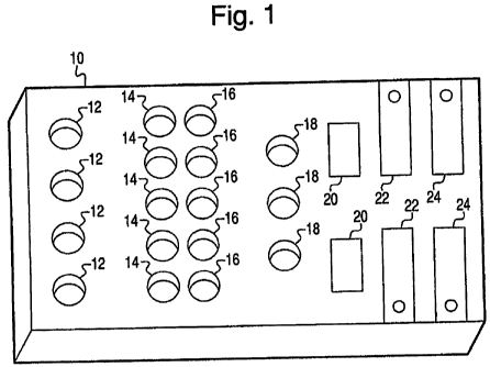
[00581 Turning now to the drawings, Fig. I generally depicts a platform that
can hold an array of multiple samples for permitting multiple tests (assays)
to be
carried out on the platform with respect to a bodily sample or multiple bodily
samples
of a subject. The assays may provide quantitative, semi-quantitative, and/or
qualitative analysis. The use of multiple assays permits testing for multiple
factors
responsive to, responsible for, or otherwise correlating to or indicative of
endometriosis. As a result, the test results taken in their totality provide
an accurate
and a more complete assessment as to whether or not the tested patient is
afflicted
with endometriosis significantly enough to warrant therapy or monitoring for
further
investigation. For example, where a first assay serves to detect for a first
physiological factor not possessed by an individual, the individual may still
be
reliably diagnosed with endometriosis by the second assay which detects for a
different second physiological factor. Furthermore, the use of different types
of
assays which measure different factor imbalances allows for a broader
perspective as
18
WO 2007/126982 PCT/US2007/007803
CA 02681933 2009-09-24
to the etiology of the disease in a particular individual. By multi-testing
for more than
one factor, it is possible to discern whether or not the responses are
specific to
endometriosis or some other condition that may have similar symptoms to
endometriosis, but which is not endometriosis. Repeated testing over an
extended
time period permits determination of whether or not the response is getting
progressively stronger or weaker, thereby indicating whether or not a certain
therapy
or treatment is effective. Consequently, the patient may be advised as to
whether or
not she has endometriosis or whether or not a treatment therapy is effective.
[0059] Fig. 1 shows a platform 10 preferably made of a transparent material,
such as glass or plastic (e.g., acrylic, vinyl, acetate, or carbonate
compound) inert with
respect to aqueous solutions having a pH between 4 and 9. It is also preferred
that
platform 10 undergoes no degradation over a wide temperature range, such as
from
about -20 C to about 80 C, for instance at least about 20 C to about 60 C.
[0060] Platform 10 as illustrated is a preferred implementation for carrying
out multiple testing procedures, each directed to the detection or measurement
of a
respective physiological factor. It should be understood that it is within the
scope of
the invention to modify platform 10 to permit for one, two, three, or more
samples to
be collected and retained for each of the testing procedures/assays. It is
also within
the scope of the invention to modify platform 10 to hold multiple bodily
samples for a
single testing procedure, or a single bodily sample for a single testing
procedure.
Further, other arrangements than shown may be selected. For example, the
assays
may be arranged concentrically with respect to one another.
[0061] Platform 10 includes a first set of wells 12, which is described below
as used in conjunction with pH testing, although it should be understood that
wells 12
may serve other purposes and testing procedures. Wells 12, and for that matter
the
19
WO 2007/126982 PCT/US2007/007803
CA 02681933 2009-09-24
other wells 14, 16, 18 included on platform 10, are not particularly limited
to a
specific size or shape. Wells preferably are capable of holding disposable and
removable cuvettes, tubes, or ampules with volumes ranging from 1 microliter (
l) to
1000 microliters ( l) of solution, e.g., from 25 to 100 microliters ( l) of
solution
suitable for many testing procedures. Wells 12, 14, 16, 18 may be formed in
removable slides, e.g., glass slides, to permit relocation of the slide to a
microscope
for observation. The opening area of wells 12, 14, 16, 18 at the upper surface
of
platform 10 is preferably about 3 mm to about 10 mm in diameter, to provide an
opening area of about 2.25 mm2 to about 25 mm2, e.g., about 1 cm2. It should
be
understood that wells 12, 14, 16, 18 may possess different diameters from one
another
and vary in capacity. Further, wells 12, 14, 16, and/or 18 may have non-
circular
openings, e.g., oval, square, etc. Platform 10 may possess a greater or lesser
number
of wells 12, 14, 16, 18 for each screening procedure.
100621 Testing procedures will be described in greater detail below. Several
of the procedures involve the use of pigments, including flavonoids such as
anthocyanins. Chemical structures for flavonoids, including the flavonol
quercetin,
are shown in Fig. 2. The transformations experienced by anthocyanins are also
shown
in Fig. 2. The Markush groups of the flavonoids, including the anthocyanin
structures, may be defined as follows: RQ is selected from the group
consisting of
hydrogen, hydroxy, and keto group; R1 is selected from the group consisting of
hydrogen, hydroxyl, and Ci-C4 alkoxy; R2 is selected from the group consisting
of
hydrogen, hydroxyl, and C}-C4 alkoxy; R3 is selected from the group consisting
of
hydrogen, hydroxyl, or glycoside selected from the group consisting of
glucosides,
rutinosides, arabinosides, sophorosides, p-coumaroyl rutinosides, and
rhamnosides;
WO 2007/126982 PCT/US2007/007803
CA 02681933 2009-09-24
Ra is a keto group in flavonoids including quercetin; R5 is selected from the
group
consisting of hydrogen, hydroxyl, and a glycoside selected from the group
consisting
of glucosides; and R7is selected from the group consisting of hydrogen,
hydroxyl, a
keto group and Cl-C4 alkoxy.
[00631 The color exhibited by the different pigment forms is pH sensitive.
Form I favors a pink color and is dominant below pH 4Ø Form YI favors a
purple
color and is dominant between pH 5.5-7Ø Form III favors a blue to purple
color and
is dominant between pH 6.8-7.2. Forms IV and V are deep blue and favored above
pH 7.2. Forms II and III are known as anhydrous base forms, whereas Forms N
and
V are referred to as ionized anhydrobase forms, which can be stabilized
through
chemical interaction with substances such as chelating agents, such as
divalent
metallic ions, co-pigmenting with reducing sugars or phenolic compounds, or by
association with certain charged molecules that have glycosides. The degree to
which
stabilization occurs is dependent upon ionic strength, pH, and concentration
of the
pigment and stabilizing agent. As described above, women with endometriosis
have
catechol estradiol imbalances which can lead to chelation of the catechol
estradiols
with some metallic ions, thus causing the anhydrobase Forms IV and V to be
less
stabilized.
[0064] The concentration of pigment for embodiments described herein
involving the use of anthocyanins preferably yet optionally falls within the
range of
8x10'6 molar to 1xl0"3 molar. When the pigment is provided in an aqueous
solution,
such as saliva, the pigment is preferably yet optionally present at levels
between
8x10"5 molar and 1.0x10⁻⁴molar at pH levels between 5.0 and 9, more preferably
between 5.8 and 8Ø The pigment may be combined with methanol to provide a
1xi 0'3 molar concentration, then optionally diluted with water or saliva to
the
21
WO 2007/126982 PCT/US2007/007803
CA 02681933 2009-09-24
concentration levels mentioned above, for example. If methanol preparation is
diluted
in the saliva to form an aqueous solution, the methanol acts as a surfactant.
It should
be understood that agents other than methanol may be selected for making the
solution, and that dilution in an aqueous solution is also optional. Further,
it is within
the scope of the invention to employ the anthocyanins without the addition of
methanol or other agents.
[0065] The structure of the flavonoid quercetin is set forth below and
reproduced in Fig. 2:
OH
OH
HO
1 I
OH
OH 0
[0066] X. Denaturing Effect - pH Testing
[00671 A first embodiment of the invention provides an endometriosis
screening method comprising subjecting a bodily sample to a denaturing
procedure,
and measuring a property such as pH of the bodily sample after, and optionally
before, the denaturing procedure. Without wishing to be bound by any theory,
it is
believed that the denaturing procedure relies on the sensitivity, such as
temperature
sensitivity, of certain physiological factors that experience imbalances in
women with
endometriosis.
22
WO 2007/126982 PCT/US2007/007803
CA 02681933 2009-09-24
[00681 According to an implementation of this embodiment, the denaturing
procedure is designed to cause certain heat sensitive enzymes that appear to
be
relevant for maintaining buffer capacity in saliva to denature, while having
no
substantial effect on the phosphate buffer levels in the same saliva sample.
As
mentioned above, certain carbonic anhydrase enzymes control buffer capacity in
saliva and are found to be different in saliva from woman with endometriosis
compared to the saliva of a woman without endometriosis. The denaturing
procedure
has been observed to have a greater effect on the saliva pH levels of a
healthy woman
than on the saliva pH levels of a woman with endometriosis. Without wishing to
be
bound by any theory, it is believed the denaturing may be caused by
differences in
ionic strength that result because of the differences in buffer controlling
mechanisms
in saliva with and without endometriosis, dependent upon interactions with
certain
heat sensitive enzymes. Whereas the denaturing procedure neutralizes the
buffering
effects of certain enzymes or buffer controlling proteins in healthy women,
thus
altering the saliva sample pH when it is exposed to air, the buffer control
mechanism
in samples from women with endometriosis is left substantially unaffected by
the
denaturing procedure. Consequently, the denaturing procedure will produce less
change in the pH of the saliva that is exposed to air in women with
endometriosis.
[0069] A freezing and thawing cycle is an example of a denaturing procedure
that will cause certain buffer maintaining enzymes such as carbonic anhydrases
to
denature, as well as some other calcium sensitive components to precipitate
out of the
saliva solution. Using wells 12, endometriosis is screened by assessing how
well or
poorly a bodily fluid sample maintains buffer capacity when subjected to one
or more
freezing and thawing cycles. Saliva samples are collected over a period of
consecutive days and each sample is aliquotted into a separate one of wells
12. In
23
WO 2007/126982 PCT/US2007/007803
CA 02681933 2009-09-24
Fig. 1, four wells 12 are shown for receiving samples taken over four
consecutive
days. It should be understood that platform 10 may contain a lesser or greater
number
of wells 12. Further, samples may be taken at different intervals, e.g., the
daily
sampling may be replaced by sampling every 48 hours. Preferably, a first pH
sample
is taken prior to the first freezing and thawing cycle. Each of the samples is
subjected
to a first freezing and thawing cycle, and a second pH value is recorded. The
sample
is optionally subjected to one or more additional freezing and thawing cycles,
and a
third and optionally subsequent pH value(s) is/are recorded. The difference
between
pH values is calculated and recorded. As shown in Table 1 below, the first and
second pH values used were taken subsequent to a first thaw and subsequent to
a
fourth thaw, respectively. In the experiments summarized in Table 2 below, the
first
and second pH values were taken subsequent to a first thaw and a second thaw,
respectively. These results are compared with a patient without endometriosis
(a
man). It should be understood that the values may be taken prior and
subsequent to
subsequent thaws. Additionally, more than one freezing and thawing cycle may
be
conducted between measurement of the pH values
[0070] Example 1
[0071] A preliminary study of thawed saliva samples from ten women with
histories of endometriosis found an increased frequency of low changes in pH
of
thawed saliva samples. This is documented in the following Table I that tracks
pH
changes in thawed saliva from a woman with a history of endometriosis. pH
measurements were taken with a pH meter. The procedure for collecting and
processing the saliva samples is set forth below.
[0072] 1. First morning whole saliva is collected passively by putting an
absorbent pad under the tongue. The saliva is allowed to collect into the pad.
24
WO 2007/126982 CA 02681933 2009-09-24 PCT/US2007/007803
1007312. The absorbent pad is put into a container having a plunger which
compresses the pad to expel the saliva.
[0074] 3. The extracted saliva is centrifuged at 7500x for 30 minutes.
[0075] 4. After centrifugation the pH is measured and recorded.
[0076] S. The processed saliva sample is then frozen at --20 C.
[0077] 6. After the sample has been frozen for at least 24 hours, the sample
is
thawed to room temperature in an ice bucket for about an hour.
[0078] 7. When the sample has thawed a second measurement of pH is made
and recorded.
[0079] S. An evaluation is made of the difference between the two pH
measurements.
[0080] It has been observed that samples obtained from women having
endometriosis have a high percent of difference values that are less than 0.2,
in some
instances approximately 0 or negative, as noted in the Table 1 below.
WO 2007/126982 PCT/US2007/007803
CA 02681933 2009-09-24
Table 1: pH changes in saliva from women with endometriosis
Cycle day pH 1 st thaw pH 4th thaw Change in
r pH
-10 7.35 7.59 0.24
-9 7.35 7.60 0.25
-8 7.24 7.56 0.32
-7 7.30 7.31 0.01
-6 7.19 7.10 -0.09
-5 7.21 7.19 -0.02
-4 7.05 7.04 -0.01
-3 7.02 7.08 0.06
-2 7.44 7.25 -0.19
-1 7.21 7.22 0.01
LH spike 6.82 7.15 0.33
+1 6.86 6.82 -0.04
+2 6.59 6.71 0.12
+3 6.82 6.95 0.13
10081] The above test procedures were repeated on saliva samples taken from
a woman known to not have endometriosis and from a man. After repeated thawing
cycles, considerably higher pH values were observed. The results are reported
in
Table 2 below.
26
WO 2007/126982 PCT/US2007/007803
CA 02681933 2009-09-24
Table 2: pH changes in saliva samples from people with no endometriosis
Sample 1 st thaw 2nd thaw Difference in
day H
Woman without endometriosis
1 7.22 7.68 0.44
2 7.25 7.36 0.11
3 6.90 7.65 0.75
4 7.25 7.84 0.59
7.29 7.84 0.55
6 7.46 8.01 0.55
Man
1 6.96 7.22 0.26
2 7.06 7.23 0.17
3 6.73 7.08 0.35
4 7.10 7.43 0.33
5 6.62 7.01 0.39
6 6.82 7.22 0.40
[00821 As exhibited by Table I above, women who have endometriosis show
a high frequency of pH changes of 0.2 or less between repeated freezing and
thawing
of saliva samples, whereas as shown in Table 2 saliva samples from subjects
without
endometriosis exhibited pH changes upon freezing and thawing that tended to
register
at 0.3 and higher, even when subjected to only a single freezing and thawing
cycle.
[0083] These tests are preferably conducted on samples collected daily over a
period of at least one week, preferably at least two weeks to minimize
deviations that
could be caused by outside influences, such as time of day. It should be
understood
that while a pH meter was used for measuring pH values in the above example,
other
quantitative approaches or semi-quantitative or qualitative measurement
approaches
may also be implemented. For example, conductivity meters may be selected.
Other
commercial pH indicators that use color changes can also be used, although the
27
WO 2007/126982 PCT/US2007/007803
CA 02681933 2009-09-24
indicator preferably is capable of measuring pH changes within a 0.2 deviation
between pH values ranging from 5.8 to 8.
[0084] 2. Denaturing .Effect: Crystallography
[0085] According to another embodiment of the invention, crystallography of
a saliva sample is examined as part of an endometriosis screening procedure.
Depending upon the cycle phase and whether the subject has endometriosis,
crystal
formations can be observed in saliva samples subjected to a denaturing
procedure,
such as a freezing and thawing cycle, or dehydration under ambient conditions.
Crystals form when metallic ions such as sodium, calcium or other bivalent
metallic
ions are in solution with available soluble sugars. This condition can result
when
glycosidase enzymes break glycosidic bonds in glycoproteins thus allowing for
the
presence of soluble sugars in the saliva. Inflammation can cause an increase
in
glycosidases in saliva. "Enzymatic protective systems of saliva in
inflammation of the
periodontium ", Patol Fiziol Eksp Ter., 1991 Jan-Feb;(1):32-4, Vavilova TP,
Petrovich
IuA, Malyshkina LT.
[00$6) Without wishing to be bound by any theory, it is believed that women
with endometriosis appear to have factors in saliva that allow these crystal
formations
to occur at any time during the menstrual cycle, possibly because there are
higher
levels of glycosidases, due to inflammatory factors resulting from
endometriosis.
Glycosidases in the peritoneal fluid from infertile women with and without
endometriosis, Clin Biochem., 1998, 31(3):181-6 (ISSN: 0009- 9120) Brandelli
A;
Passos EP. In contrast, women who do not have endometriosis only show the
characteristic ferning patterns of crystals just before ovulation when
increased
estradiol levels lead to increased activity of glycosidases, and about a week
later on
the day of implantation, presumably because of a surge in estradiol production
about 7
28
WO 2007/126982 PCT/US2007/007803
CA 02681933 2009-09-24
days after ovulation. Temporal Surge of Glycotransferase Activities in the
Genital
Tract of the Hamster during the Estrous Cycle, Biology of Reproduction 54,
1032-
1037 1996, Daulat, Tulsiani, Catherine Chayko, Marie-Claire Orgebin-Crist
Yoshihiko Araki, www.biolreprod.org/cg'i/reprint/54/S/1032.pdf.
[00871 According to an implementation of this embodiment of the invention,
wells 20 are provided for conducting the crystallinity assay to detect for the
presence
or absence of endometriosis. A 25 microliter sample of saliva having been
subjected
to a freezing and thawing cycle is deposited in one or more of wells 20 and
allowed to
dry and crystallize at ambient (e.g., room) temperature. These wells are
designed to
be viewed under a microscope at 200x magnification. Using a microscope, it is
possible to visually see distinctive crystal shapes and patterns that are
produced,
depending upon whether or not the female subject has endometriosis and the
cycle
phase from which the sample was obtained.
10088] It has been observed that women having endometriosis exhibit crystal
formations in their dried saliva in the luteal phase, and often in or
throughout the other
4
phases of the menstrual cycle. Generally, women with endometriosis have saliva
characterized by crystals that extend outward radially from a center or hub of
the
crystal in multiple directions so that the distal ends of the crystals form a
generally
rounded or polygonal outline around the center. These crystal formations are
also
referred to herein as axial formations due to the branching out of crystals
from a
central axis.
[0089] On the other hand, women not having endometriosis exhibit crystal
formations in their dried saliva only immediately before the fertile stage,
and for one
day which occurs at the time of implantation, about one week after ovulation.
Generally, the crystal formations for women not having endometriosis often are
less
29
WO 2007/126982 CA 02681933 2009-09-24 PCT/US2007/007803
dense and more linear than crystal formations of women having endometriosis.
These
linear crystal formations possess an elongate spine with branch pendent
crystals
extending outward generally perpendicular to the spine to provide a generally
fem-
like or skeletal appearance. Referring to Figs. 14A and 14B, the elongated or
fern-
like crystal formations of women not having endometriosis are documented to
contain
proportionally higher levels of sodium salts than women having endometriosis.
Figs.
14A and 14B are comparative spectra measurements for a saliva sample taken
from a
woman not having endometriosis and a woman having endometriosis, respectively.
Spectral analysis was conducted using electron microscope and a light element
detector system (SE1VI/EDS). The woman without endometriosis had a relatively
high
sodium (Na) peak, as seen in Fig. 14A, compared to the relatively low sodium
(Na)
peak of Fig. 14B for the woman having endometriosis.
[0090] Women having endometriosis also tend to have a high frequency of the
crystal formation that can be axially oriented as compared with a low
frequency of
any type of crystal formation observed in women who do not have,endometriosis.
This visual distinction is best viewed under a microscope at 200X
magnification.
Figs. 6, 8, and 10 provide reference photographs of saliva crystals observed
in
samples from women with endometriosis in the follicular, fertile, and luteal
phases,
respectively. Crystal formations were observed in each of the phases, with the
crystals generally showing the axial formation. Figs. 7, 9, and 11 provide
reference
photographs of saliva crystals observed in samples from women without
endometriosis in the follicular, fertile, and luteal phases, respectively. In
the follicular
and luteal phases shown in Figs. 7 and 11, respectively, little or no crystal
formation
was observed. Fig. 9 depicts the fertile phase in which less dense crystals
characterized by a spine and branches was observed.
WO 2007/126982 PCT/US2007/007803
CA 02681933 2009-09-24
100911 Procedures for collecting the saliva samples for this embodiment were
as follows.
[0092] 1. One milliliter samples of whole saliva are collected in the morning
and stored in a plastic ampoule. The time and date are noted on each sample.
It is
possible to let the sample sit for a few hours, or prepare the saliva sample
immediately
for crystal observation by drying a small amount on a glass slide. It is also
possible
(yet optional) to freeze the sample and then thaw it as described below.
[0093] 2. The freezing temperature and thawing rate are variables affecting
the outcome of the crystallization process. See Art of Science, Vo. 11, No. 2,
HyClone Laboratories Inc., "Freezing and Thawing Serum and Other Biological
Materials", p.l -4 (Spring 1992). To account for these variables, freshly
collected
saliva was centrifuged at 7500xG for 30 minutes. The centrifugation allows
different
size particles to settle in more uniform order, thus permitting for more
standardized
procedures in the pipetting process. The supernatant was frozen to 20 C.
Thawing
was conducted within 1 hour at room temperature and ambient conditions.
[0094] 3. A 0.25 microliter sample of thawed saliva is pipetted from the top
part of the saliva sample onto we1120 of a slide, and allowed to dry under
ambient
conditions for at least one hour.
[0095] 4. The dried saliva is viewed in a microscope at 200X power.
[0096] 5. Optionally a digital camera attached to a computer can be attached
to the microscope and a digital photograph on the computer records the view on
the
slide.
[009716. The digital photos of the samples are viewed on a computer using a
commercial software program.
31
WO 2007/126982 PCT/US2007/007803
CA 02681933 2009-09-24
[009817. The samples are compared to a digital reference library that has
reference photos stored to serve as comparisons for determining what type of
crystal
formation is prevalent in the sample. If the crystal formation shows axial
formation, it
is highly probable that the sample originated from a woman suffering from
endometriosis. If the crystal formation is linear with parallel line
formations, then the
sample originated from a woman not suffering from severe endometriosis. If
there is
no crystal formation observed, it is also highly probable that the sample
originated
from a woman not suffering from endometriosis.
[0099] 8. A record is kept of the type of crystal observed, what cycle day it
originated from, and the frequency that the crystal formation is observed
within a
given phase of the menstrual cycle. The dried saliva samples may keep the
crystalline
patterns intact for years and can be stored for future reference.
[0100] 9. If a predetermined number (e.g., three or more) of dried samples
have crystal formations that match (or do not match) the reference
photographs, an
= assessment can be made as to whether or not the saliva originates from a
woman with
endometriosis.
.~;
[0101] It should be understood that the freezing and cooling cycle described
in
the above procedure is only one example of a denaturing procedure imparted on
the
sample.
(0102] Example 2
[0103] The photo analysis process was performed on 10 women, each of
whom provided 30 samples from different days of the menstrual cycle. Five (5)
of the
women had endometriosis, and 5 of the women did not have endometriosis. The
photos were catalogued as to cycle day which had been calculated according to
results
for ovulation detection performed using urine-based commercial ovulation
tests. A
32
WO 2007/126982 CA 02681933 2009-09-24 PCT/US2007/007803
measurement was made for the frequency (% slides with crystal formation) for
any
given cycle day in the study.
Table 3
% saliva samples
from women with % saliva samples from
endometriosis who women without
had some form of endometriosis who had
crystal formation some form of crystal
CYCLE DAY based on - observed in the formation observed in the
urinary LH levels sample sam le
-5 20% 10%
-4 10% 0%
-3 0% 10%
-2 60% 10%
-1 60% 20%
0
1 day before ovulation 80% 80%
1 50% 0%
2 60% 20%
3 60% 0%
4 80% 0%
100% 0%
6 60% 0%
7 60% 0%
8 80% 0%
9 80% 20%
60% 0%
[01041 The slides were transferred to a microscope, and the frequency of any
visible crystal forms on glass slides prepared according to procedures
described above
was counted. The data of Table 3 is set forth in graphical form in Fig. 12 to
illustrate
the frequency of any crystal form observed in each of the two groups of women:
women with and without endometriosis. As seen in Fig. 12, women with
endometriosis exhibited crystal formations throughout each phase of the
menstrual
cycle, whereas women with no endometriosis exhibited greatly reduced and less
dense
33
WO 2007/126982 CA 02681933 2009-09-24 PCT/tTS2007/007803
crystal formations for significant portions of the menstrual cycle, in
particular the
leuteal phase.
1010513. Optical Density Comparison
[0106] Another embodiment of an endometriosis screening method of the
present invention features a comparison of two optical density peak values
between
400 nm and 700 nm of a bodily fluid mixed with a flavonoid pigment as part of
an
endometriosis screening procedure.
[0107] Referring to Fig. 1, any of wells 12, 14, 16, and/or 18 may be selected
for carrying out this embodiment. Samples of saliva or another bodily fluid
are
obtained from a woman, preferably periodically over at least a five-day span,
and the
samples are pipetted into the wells or disposable cuvettes or tubes that fit
into wells
12, 14, 16 and 18. The samples are mixed with a flavonoid pigment, which is
preferably evenly pre-impregnated into the well, cuvette, or tube.
[01081 Impregnation of the pigment into the well or disposable cuvette or tube
is cazxied out as follows: -
[0109] 1. A defined amount of pigment that is weighed to reach a 1x10"3
molar concentration for 1 ml sample is mixed with I ml of methanol. .
[0110] 2. The pigment mixture is vortexed until all the pigment is dissolved.
[0111] 3. A defined volume of this solution is pipetted into the well or
disposable tube or cuvette.
[0112] 4. The solution is allowed to evaporate so that the pigment is evenly
distributed on to the surface of the well or disposable cuvette or tube.
[011315. The well, tube or cuvette having the evaporated pigment distributed
onto it can now be stored for several months under cool dry conditions until
ready for
use.
34
WO 2007/126982 PCT/US2007/007803
CA 02681933 2009-09-24
[01141 When samples are ready for evaluation, the samples are pipetted in
defined amounts into the wells, disposable tubes or cuvettes so that the
resulting
concentration of the dried pigment is reconstituted to be at 1 x 10`4molar
concentration.
[0115] The samples are then measured with a spectrophotometer for
absorbency values read between 400nm and 700nm. A comparison is made between
two different wavelengths on the same sample in order to determine the rate of
change
in the absorbency value between different wavelengths. The chart below
compares
ratio values of 600nm/400nm for saliva samples mixed with the anthocyanin
malvidin
3,5-diglucoside and quercetin.
WO 2007/126982 PCT/US2007/007803
CA 02681933 2009-09-24
Table 4
Cycle Day Endometriosis - No Endometriosis - Endometriosis -
Malvidin 3,5- Malvidin 3,5- Quercetin
di lucoside diglucoside
-10 1
-9 1.25
-8 5.14
-7
-6 0.91
-5 1.21 5.19
-4 2.18
-4 2.53 1.5
-3 2.1 2.8 0.22
-2
-1 2.05 0.33
0 1.2
+1 3.3
+2
+3 5.85
+4
+5 3.72 5.15
+6
+7
+8 1.21
+9 1.33
+10 1.22 5.15
+11
+12 5.21
+13 3.33 1 77771
[0116] Fig. 13 provides further examples of absorbency patterns between 400
nxn and 600 nm for women with endometriosis and women without endometriosis.
The specimens from women without endometriosis increased at a greater rate, or
had
a greater slope, between 400 mu and 600 nm than the specimens from women with
endometriosis over the same range.
[0117] Without wishing to be bound by any theory, it is believed that the
ratio
values represent the degree to which certain anhydrobase forms of anthocyanins
are
36
WO 2007/126982 PCT/US2007/007803
CA 02681933 2009-09-24
present in the sample. Referring back to Fig. 2, the ratio values represent
the stability
of certain anhydrobase forms of anthocyanins, and in particular Forms II and
III in
relationship to the ionized anhydrobase Forms IV and V, when data is taken
within
the range of 400 to 700 nm. The ratios, when considered in conjunction with
the
cycle day the bodily fluid was taken, permit an assessment to be made as to
whether
or not a woman has endometriosis.
[011S] For example, it has been observed that women with endometriosis
provide uniform 560nm/538nm ratio values for saliva samples mixed with 1x10"3
molar concentration of the anthocyanin, malvidin 3,5-diglucoside.
Specifically, the
560 nrn1538 nm ratio values are generally about 1 throughout the cycle, more
broadly
between about I and about 1.2 for the women with endometriosis. On the other
hand,
saliva samples from women who do not have endometriosis can be mixed with the
same anthocyanin pigment in identical concentrations to produce 560 nm/538 nm
ratio values ranging from 0.8 to 1.7, with the values mostly ranging between
1.2 and
1.5, depending on the cycle day, ionic strength, and the pH of the saliva
sample.
Referring to the examples shown in Figs. 3 and 4, the ratio ranges from 1.2 to
1.5 in
the non-fertile phase of the menstrual cycle (after ovulation has occurred).
The ratio
shifts below 1, i.e., 0.8 to 1.0 in the ovulatory phase of the cycle shown in
Fig. 3.
[0119] Without wishing to be bound by theory, a more detailed explanation of
the chemistry follows. Anhydrobase anthocyanin Forms II and III are in
equilibrium
with one another in a pH range of 4<pH<7. The anhydrobase Form III has a
maximum absorption at about 556 nm and the anhydrobase Form II has a maximum
absorption at about 534 nm. =As illustrated in Fig. 4, when anthocyanin
pigment is
combined with saliva of a woman suffering from endometriosis, equilibrium is
favored towards equal distribution between anhydrobase Form II and anhydrobase
37
WO 2007/126982 PCT/US2007/007803
CA 02681933 2009-09-24
Form III, which produces a ratio close to 1, more generally between 1 and 1.2.
This is
pH independent within the range from pH 5.8 to 7.8. It is believed that the
higher
absorbency ratio values, e.g., greater than 1.3, of women without
endometriosis is the
result of greater stabilization of anhydrobase III relative to anhydrobase H.
[0120] Without wishing to be bound by theory, it is believed that the
difference in the ratio values observed between women with endometriosis and
women who do not have endometriosis may be due to some differences in ionic
strength, which may affected by certain immune factors present in saliva that
are
sensitive to changes in ionic strength and pH regulation. Differences in the
composition of enzymes between saliva samples from women with and without
endometriosis may cause structural changes in the flavonoid pigments that can
be
used as chemical markers to distinguish between women with endometriosis from
women without endometriosis.
[0121] The following experiments evaluated the effect of heat on the above-
described biological mechanism which controls the ratio of the anhydrobase
forms.
[0122] The saliva samples are mixed with anthocyanin pigment malvidin 3,5-
diglucoside. Each sample is placed in 1 ml tube. Optical density measurements
are
taken at a wavelength between 500nm and 600nm. Measurements are made at 538nm
and 560 nm. The measurements are taken both before and after the samples are
subjected to a cycle of heating at over 100 C for 20 minutes and cooling to
ambient
temperature. Measurements are reported below in Table 5.
Table 5
538 nm 560 nm ratio value
woman with
endometriosis
- not heated 0.18 0.185 1.0
38
WO 2007/126982 PCT/US2007/007803
CA 02681933 2009-09-24
- heated 0.175 0.16 0.9
buffer
- not heated 0.13 0.19 1.46
- heated 0.16 0.23 1.44
woman with no
endometriosis
- not heated 0.14 0.17 1.21
- heated 0.10 0.10 1.00
[0123] The results of the unheated and heated saliva samples of Table 5 show
that heating has a minimal effect on the absorption patterns for the buffer,
with a
difference of 0.02 in the ratio value. In contrast, the saliva of the woman
not having
endometriosis showed a 10 fold decrease in absorbency ratio of 0.21 as the
result of
the heating. The saliva sample from the woman with endometriosis showed a
decrease in ratio value of 0.01, similar to the decrease observed in the
heated buffer.
There appear to be heat sensitive factors present in the unheated sample of
saliva from
the woman without endometriosis that affect stabilization of the anhydrobase
form III.
[0124] While data comparison is described above as involving a calculation of
the ratio of absorption readings at the wavelengths of 560 nm and 538 nm, it
should
be understood that the absorption data may be subject to other forms of data
manipulation and mathematical functions (e.g., subtraction) for comparison.
Additionally, the above procedures and calculations of ratio values can also
be
performed at other wavelengths between 400 and 700 nm, and with other types of
anthocyanins, or flavonoids, realizing that different types of flavonoids
would have
different ratio values because of the differences in the k max value. The
mechanism
and basis for determining these values would, however, remain the same.
Reference
values for different wavelengths and different wavelength ratios can be
obtained by
39
WO 2007/126982 CA 02681933 2009-09-24 PCT/US2007/007803
measuring or otherwise obtaining data from women known to have endometriosis
and
known not to have endometriosis.
[0125] Example 3
[01261 Procedure for measurements was as follows:
[0127] 1. The saliva sample is thawed in an ice bucket.
1012812. A record is made of the pH of the sample.
1012913. Depending on the volume of the well, between 90 microliters to 450
microliters of saliva can be pipetted into a well or disposable cuvette or
tube that fits
into a well. The well or disposable tube or cuvette has a defined amount of
malvidin
3,5-diglucoside at 10% concentration to yield a 1x10'4 molar concentration.
2.76 mg
of crystal form malvidin 3,5-diglucoside may be dissolved in 1 ml of methanol
and
mixed with 3 ml ethylene glycol monoethyl ether ( EGME). The solution is then
aliquotted into each well, cuvette or tube at the appropriate volume ratio,
while
allowing the solvent to evaporate. Once the malvidin 3,5-diglucoside is
contacted to
the cuvette or tube and kept in cool dry conditions at room temperature, it
can remain
intact and ready for use for long periods of time.
[013014. The sample mixed with the pigment is measured in a
spectrophotometer or plate reader at the wavelengths 560nm and 538nm.
[0131] 5. A calculation is made as to the ratio for absorbance for
560nm/538nm. If the ratio value > 1.25 for samples taken in the luteal phase,
the
sample is preliminarily determined to be from a woman who does not have active
endometriosis, preferably subject to additional sample testing with consistent
results.
If the ratio value is less than 1.2, the sample may be preliminarily
determined to be
from a woman who has active endometriosis: If a predetermined percentage,
e.g., 3/5
(60% or more), of the samples taken during consecutive days of the luteal
phase
WO 2007/126982 PCT/US2007/007803
CA 02681933 2009-09-24
provide ratio values for absorbency as measured at 560nm/538nm of less than
1.2,=
then the samples reflect the presence of endometriosis. On the other hand, if
0/5
consecutive days of the luteal phase give ratios between 1.0 and 1.2, then a
conclusion
is drawn-that the samples originate from a woman who does not have
endometriosis.
[0132] Table 6 below represents the ratio 560 nm/538 nm for women having
(right column) and not having (left column) endometriosis. Saliva samples were
mixed with lx10'4molar concentration of malvidin-3,5-diglucoside. Each row in
Table 6 corresponds to a cycle day:
41
WO 2007/126982 PCT/US2007/007803
CA 02681933 2009-09-24
Table 6
No Endometriosis Endometriosis
Cycle Day Trial #1 Trial #2 C cle Day
Da -14 Day -14
Da -13 Da -13
Day-12 Day-12
Da -11 Da -11
Day -10 Da -10 1.22 1.1
Da -9 Day -9 1.13
Day -8 1.3 1.5 Day -8
Day -7 1.5 Day -7 1.2
Day -6 Day -6
Day -5 Day -5 1.06
Da -4 1.35 1.53 Day -4 1.09 1.02
Day -3 1.3 Da -3 1.13
Day -2 1.2 Da -2
Day -1 1.1 Da -1
Day 0 0.8 1.06 Day 0
Day +1 1.4 1.2 Day +1
Day +2 1.15 1.28 Day +2 1.1
Day +3 1.4 1.25 Day +3
Da +4 1.3 Day +4
Day +5 1.4 1.72 Day +5 1.09
Day +6 1.5 Day +6
Da +7 1.3 1.5 Day +7 1.0
Day +8 1.4 1.32 Day +8 1.08
Day +9 1.5 Day +9 1.03 1.08
Day +10 1.3 1.52 Day +10 1.07
Day +11 1.5 1.47 Day +11 0.95
Day +12 1.7 Day +12 1.05 1
[0133] Testing was performed on 10 women known not to have
endometriosis, and 5 women known to have endometriosis. Of the former group
reported in Table 6, 27 out of 30 samples (i.e., 90%) taken produced results
with ratio
values for 560nm/538nm of 1.2 or greater. This data is consistent with other
data
measuring the same ratio values for woman not having endometriosis. It should
be
noted that ratio values for women who do not have endometriosis will have
values
42
WO 2007/126982 PCT/US2007/007803
CA 02681933 2009-09-24
near 1 about one day before ovulation, during ovulation and immediately after
ovulation. Without wishing to be bound by any theory, it is believed this is
because
the composition of saliva changes during the ovulation period due to the
effects of 17
beta estradiol on certain enzymes such as glycosidases and some prosthetic
enzymes.
As a result, stabilization of the anhydrobase is compromised and the ratio
value is
diminished during ovulation. Therefore, it is best to measure differences
between
women with and without endometriosis after ovulation in the luteal phase,
because
during the luteal phase, saliva from women who do not have endometriosis is
able to
stabilize the anhydrobase form; whereas saliva from women who have
endometriosis
still has factors present that inhibit anhydrobase stabilization. Of the 20
out of 30
samples measured in the luteal phase, 19 of the 20 samples produced a ratio of
560nm/538nm of greater than 1.2. Of the latter group of women having
endometriosis, 10 out of 10 samples reported in Table 6 taken during the
luteal phase
(i.e., 100%) were characterized by a ratio of less than 1.2, as expected.
[01341 Fig. 4 is a graph plotting the ratio of absorption at 560 nm to 538 nm
for woman with endometriosis and a woman without endometriosis through the
luteal
phase of the menstrual cycle. The selected pigment was malvidin 3,5-
diglucoside,
which was mixed with the saliva samples to give a 1xlOmolar concentration. The
woman who did not have endometriosis exhibited ratios of absorption
(560nm/538nm) repeatedly ranging between about 1.3 and about 1.6. A relative
stable blue color was observed. In contrast, the woman with endometriosis
repeatedly
exhibited ratios of absorption (560nm/538nm) in a range of 1 and not exceeding
1.2,
and although a blue color response temporarily resulted, the response was not
stable
and the blue faded over a matter of 20 minutes. Without wishing to be bound by
any
43
WO 2007/126982 PCT/US2007/007803
CA 02681933 2009-09-24
theory, it is believed that the fading of color was caused by the lack of
stabilizing
metal ions.
[013514. Indicator-Activated Color Changes
[0136] The following embodiment of the invention provides an endometriosis
screening procedure based on the ability of certain pigment-indicator
combinations,
such as quercetin with diluted iodine, to alter the color of the pigment mixed
with the
bodily fluid sample, preferably saliva.
[0137] Quercetin can be used as a marker for certain types of peroxidases,
which are inflammatory substances found at elevated levels in women having
endometrial inflammation. One example is interleukin L-6 production which is
known to be inhibited by certain flavonoids such as luteolin and quercetin.
("Luteolin
Inhibits an Endotoxin-Stimulated Phosphorylation Cascade and Proinflammatory
Cytokine Production in Macrophages", Pharmacology: Therapeutics, Vol. 296,
Issue
1, 181-187, January 2001, A. Xagorari, A. Papapetropoulos, A. Mauromatis, M.
Economou, T. Fotsis and C. Roussos) Strips 22 provide colorimetric system
assays
for endometriosis detection that use flavonoids to detect inflammatory
responses
resulting from endometriosis. Each strip 22 comprises a matrix such as ashless
cellulose, cellulose acetate, or nylon mesh embedded with a pigment, such as
quercetin or an anthocyanin. The saliva sample and the flavonoid pigment are
combined on strip 22. It is preferred to embed the pigment into strip 22
before the
saliva is added. The pigment may be embedded onto the cellulose as follows.
[0138] Quercetin is dissolved in a methanol solution to provide, for example,
a 1x10'3 molar concentration. Twenty microliters are pipetted onto the
cellulose
absorbent surface, and the methanol is allowed to evaporate until dry. Once
strip 22
is prepared, a body fluid sample is applied. By way of example, the sample
size may
44
WO 2007/126982 PCT/US2007/007803
CA 02681933 2009-09-24
be 25 microliters. After about 5 seconds, an approximately equal amount of
diluted
tincture of iodine (e.g., 1:10 concentration) is added.
[0139] Cellulose or nitrous cellulose treated with quercetin yields a yellow
color that is stable under ambient conditions. The addition of the saliva
sample does
not significantly alter the yellow color, regardless of whether or not the
woman
supplying the sample has endometriosis. However, the color response produced
by
addition of the diluted iodine tincture differs between a woman having
endometriosis
and a woman not having endometriosis. In the case of a sample collected from a
woman not having endometriosis, the diluted iodine tincture will cause strip
22 to turn
immediately a stable blue. On the other hand, for a woman having
endometriosis,
strip 22 will not yield a blue color. Instead, within approximately 5 seconds
the color
response will begin to fade to yellow and continue to maintain this yellow
color over
a period of time such as 30 minutes. Time and intensity may vary from sample
to
sample. However, any degree of fading at all can be associated with some form
of
inflammation response relating to endometriosis.
[0140) Preferably, multiple color response tests are conducted and evaluated
before making a conclusion. For example, a series (e.g., five or more) of
color
responses yielding yellow or faded yellow color responses from multiple saliva
samples collected from a woman over consecutive days may be interpreted as
evidence that the female subject may have endometriosis. A series (e.g.,=five
or more)
of samples, collected over consecutive days, yielding a deep blue color in
response to
the addition of diluted tincture of iodine may be interpreted as'evidence that
the
subject does not have endometriosis.
(0141] Without wishing to be bound by any theory, it is believed that the
instability of the sample from the woman with endometriosis is due to elevated
WO 2007/126982 PCTIUS2007/007803
CA 02681933 2009-09-24
peroxidase levels that relate to increased cytokines present in saliva as a
result of
inflammatory responses to the presence of endometriosis.
(014215. Chromatography
[0143] Additionally, it is possible to measure for presence of endometriosis
by
using chromatography techniques to evaluate how a flavonoid interacts with the
saliva
sample. The chromatography can also be performed on a solid matrix system such
as
ash-free-cellulose in strip 22. As described above, 25 microliters of 1x10"3
molar
solution of quercetin is pipetted at a defined location on strip 22. A marker
(e.g.,
pencil mark) may be applied to record the location of the quercetin mark. Then
25
microliters of fresh whole saliva is pipetted just below the quercetin mark. A
solution
of butanol, acetic acid and water in ratios 4:1:5 respectively is allowed to
filter
through the paper. The distance that the front travels versus the distance
that the
quercetin travels is measured. The Rf value is defined as the ratio of the
front
distance divided by the distance that the pigment travels.
[0144] The Rf value corresponds to a measurement that can predict whether or
not a woman is suffering for inflammation related to endometriosis. Women with
endometriosis having inflammation show Rf values closer to the standard buffer
value
ranging between 0.5 and 1Ø Women who do not have endometriosis and do not
have
inflammation from endornetriosis show values much lower than the standard
control
of around 0.2, usually in a range of 0.2 to 0.4.
Table 7
Standard Rf for Rf for quercetin Rf value for Rf value for
quercetin mixed with saliva quercetin mixed quercetin mixed
Control sample: from woman with with saliva from with saliva from
buffer at same pH as no endometriosis woman with woman with
bodily fluid endometriosis endometriosis after
before hysterectomy hysterectomy
0.92 0.22 0.72 0.18
, - .
46
WO 2007/126982 PCT/US2007/007803
CA 02681933 2009-09-24
0.92 0.26 0.72 0.10
0.8 0.28 0.75 0.38
1 0.25 0.40 0.50
1 0.27 1.0 0.37
1 0.24 0.50 0.40
1 0.25 0.8 0.25
1 0.23 1.0 Na
[01451 As can be noted in the data in Table 7, the woman who has
endometriosis shows significantly higher Rf values for quercetin on the
chromatography assay than the woman who did not have endometriosis. After a
hysterectomy, the Rf values declined for this woman. It can be noted that the
Rf
values after the hysterectomy were in some cases lower than the Rf values
obtained
from the woman with no endometriosis.
[0146] The variability in the Rf values for the woman with endometriosis may
.
correlate with certain factors that vary from day to day such as inflammation
responses to the way the immune system responds to certain environmental
factors
that affect women predisposed to endometriosis. It has been noted that women
who
have hysterectomies to control for endometriosis can still generate other
physiological
responses and imbalances in their immune system that are not controlled by
just the
removal of reproductive organs. The data in Table 7 suggests that hysterectomy
had
an effect that diminished the Rf values for quercetin, but that the woman is
still
predisposed, although to a lesser degree, to whatever responses quercetin may
generate in saliva from a women with endometriosis.
[0147] 6. Filtration
[0148] Strips 24 placed on platform 10 provide for additional colorimetric
system assays that involve an absorbent surface with a filtering mechanism.
According to a preferred embodiment, the absorbent surface can comprise a
47
WO 2007/126982 PCT/US2007/007803
CA 02681933 2009-09-24
Sephadex (cross-linked dextran gel) preparation having antigens that respond
to
factors found in the saliva of women without endometriosis, but not found in
saliva of
= women having endometriosis (or vice versa). A saliva sample is contacted
with and
allowed to pass through strip 24. The saliva sample is brought into contact
with a
color response system both before and after passing through the Sephadex
filter strip
24.
[0149] A comparison is made between the color response systems to
determine whether any color changes occurred as the result of the sample
passing
through strip 24, and an interpretation is made as to whether or not the
saliva
originated from a woman with endometriosis.
[0150] The absorbent surface of strips 24 can also be designed to remove
certain factors that might interfere with the assay. For example it might be
necessary
or desired to remove certain molecules such as those greater than 100,000
Daltons in
order to assay for only those components in the saliva that have sizes smaller
than
100,000 Daltons. By imposing a molecular sieve into strip 24, one can adapt
the
assay to meet these requirements.
[0151] By way of example, certain divalent metallic components that are
sensitive to the absence or presence of endometriosis can be removed by
filtering.
This is especially true for certain calcium-dependent factors. By putting EDTA
(a
calcium chelation agent) inside the strip one can remove or denature any
calcium
dependent factors that affect the assay. An indicator that produces different
color
= responses dependirig upon whether the calcium-dependent factors is present
is
selected and used to treat the sample before and after filtration. The color
responses
both before contact with the EDTA and after contact with the EDTA are
compared.
48
WO 2007/126982 PCT/US2007/007803
CA 02681933 2009-09-24
[0152] Platform 10 may possess a greater or lesser number of strips 22, 24
than shown in Fig. 1.
[0153] Collection of Samples
[0I54] The procedure for collecting saliva from an individual is not
particularly limited. The saliva sample may be placed in a container extracted
in
measured or random amounts. For example, a person may spit into a tube and
then
use a pipette to extract a defined amount of saliva such as 100 microliters,
and
optionally deliver the sample to the desired well. It is also be possible to
use a device
that absorbs saliva directly inside the mouth, such as a suction device or a
device that
uses osmotic pressure to transport saliva into a pouch that has a fixed
volume.
Another technique is to absorb saliva onto a sponge or vessel that has
multiple
capillary tubes that hold fixed amounts of saliva. Saliva can also be absorbed
using
an absorbent material, which is then inserted into a tube with a plunger to
push the
saliva out of the absorbent.
[0155] The timing of the saliva collection can be particularly important, both
in terms of its relationship to the menstrual cycle (as discussed above) and
in terms of
the time of day, e.g., moming or evening. ("Circadian Rhythms in Human
Salivary
Flow Rate and Composition," The Journal of Physiol-- 220, 529-545, 1974,
Dawes,
C.; "The effects of flow rate and duration of stimulation on the
concentrations of
protein and the main electrolytes in human submandibular saliva," Archives of
Oral
BioloQV. 19, 887-895) Saliva changes in its composition according to time of
day. It
has been observed and documented that collection of unstimulated saliva taken
from
under the tongue provides the most consistent results. Additionally, it is
preferred to
collect the samples in the morning before eating any food. Alternatively, at
least a
period of 20 minutes from food intake should elapse before samples are taken.
It is
49
WO 2007/126982 PCT/US2007/007803
CA 02681933 2009-09-24
additionally preferred not to stimulate the saliva through any means. A
passive
collection system works well for the assays defined in this invention.
[0156) It should be understood that the saliva multi-based assay system has
multiple presentations. It can be used in different formats than illustrated
in the
attached drawings. For example, platform 10 can also have a circular shape or
be a
tube whereby the indicators are located inside the tube. It is also possible
to omit one
or more of the illustrated assays, and/or include additional assays for
screening of
endometriosis_ Further, each of the assays can be isolated on its own platform
without
other assays.
[0157] A series of samples compared in multiple assays on one platform
offers several benefits. By assessing different factors simultaneously from a
saliva
sample and comparing these factors to additional samples taken periodically,
e.g.,
daily, it is possible to generate more accurate and more defniitive
assessments as to
whether or not a woman has endometriosis. Such a system with multiple assays
not
only generates more information about the condition of an individual, but also
reduces
the time aind cost in the process of evaluating for the presence of
endometriosis.
Furthermore, the use of different types of assays allows for a broader
perspective as to
the etiology of the disease in a particular individual. For example,
distinctions can be
made as to whether or not the individual is suffering from immune deficiencies
and
inflammatory responses, or whether the endometriosis is related to defects in
estradiol
metabolism or not. Additionally, results of these tests can assist a
practitioner in
determining whether or not a certain therapy is effective in treating a
particular
individual who may be suffering from endometriosis.
[0158] The non-invasive based assay system with multiple assays according to
preferred embodiments of the invention offers great improvements over current
WO 2007/126982 PCT/US2007/007803
CA 02681933 2009-09-24
technologies that evaluate women with endometriosis. The system can provide a
test
that allows for home-based collection of samples taken over a period of days
or weeks
of a menstrual cycle. While some tests may be evaluated in home use, others
may be
delivered to the doctor or laboratory for testing. Importantly, the woman is
not
required to visit the doctor on a daily basis to provide bodily fluid sample;
rather,
these samples may be taken at home, so that the test subject need not visit
the doctor
or laboratory until after all samples have been collected. The system may be
designed
solely of assays that allow for home-based evaluation so as to allow a woman
to make
a preliminary evaluation as to whether or not she might be subject to
endometriosis in
that particular cycle and should therefore see a specialist who can advise her
on
possible treatments.. It is also possible to perform these measurements in a
doctor's
office where the woman can have immediate evaluation as to whether on not the
pain
she is having or her infertility problem is due to endometriosis or due to
some other
cause. The system and methods of various embodiments of the present invention
are
also useful for use during routine physical examinations, and thereby promote
early
stage intervention.
[0159] The procedures and systems described herein are based on the premise
that many factors are present in the pathogenesis of endometriosis. Some of
these
factors are sensitive to estrogen metabolism imbalances which can be tracked
by
measuring ratio of ionized anhydrobase forms of certain anthocyanins. Other
factors
involve differences in immune factors such as ligands and glycosides present
on
certain temperature-sensitive enzymes that in turn are sensitive to the
presence of
certain metallic ions. When catechol estrogens interfere with the functioning
of these
enzymes, other cellular processes are affected and a cascade of immune
responses and
inflammatory responses occur, resulting in other markers to indicate
endometriosis.
51
WO 2007/126982 PCT/US2007/007803
CA 02681933 2009-09-24
The procedures and systems herein distinguish between these different factors
to
screen for the presence of endometriosis at high accuracy.
[0160) The flavonoids and anthocyanin pigments used in several embodiments
of the invention may be obtained from various commercial sources, such as, for
example, Sigma Aldrich and Polyphenols in Norway. Other sources may also be
used.
[0161) The foregoing detailed description of the preferred embodiments of the
invention has been provided for the purposes of illustration and description,
and is not
intended to be exhaustive or to limit the invention to the precise embodiments
disclosed. The embodiments were chosen and described in order to best explain
the
principles of the invention and its practical application, thereby enabling
others skilled
in the art to understand the invention for various embodiments and with
various
modifications as are suited to the particular use contemplated. It is intended
that the
scope of the invention cover various modifications and equivalents included
within
the spirit and scope of the appended claims.
52