Note: Descriptions are shown in the official language in which they were submitted.
CA 02681986 2009-10-08
TROCAR ASSEMBLY
BACKGROUND
Technical Field
[0002] The present disclosure relates to a trocar assembly for use in
minimally invasive
surgical procedures including endoscopic, laparoscopic and arthroscopic type
procedures.
Discussion of Related Art
[0003] Minimally invasive procedures are continually increasing in number
and
variation. Forming a relatively small diameter temporary pathway to the
surgical site is a key
feature of most minimally invasive surgical procedures. The most common method
of providing
such a pathway is by inserting a trocar assembly through the skin. In many
procedures, the
trocar assembly is inserted into an insufflated body cavity of a patient. In
such procedures, the
trocar assemblies with seal mechanisms are utilized to provide the necessary
pathway to the
surgical site while minimizing leakage of insufflation gases.
[0004] Trocar assemblies typically include an obturator which is removably
inserted
through a cannula. The obturator may include a safety shield which protects
against
1
CA 02681986 2016-05-24
unintentional puncturing by the sharpened tip of the obturator. The safety
shield includes a
mechanism which controls the relative movement and locking of the safety
shield. One example
of a safety shield mechanism is disclosed in commonly assigned U.S. Patent No.
6,319,266 to
Stellon et al.
SUMMARY
[0005] Accordingly,
the present disclosure is directed to a surgical system for penetrating
tissue. The surgical system for penetrating tissue includes a cannula assembly
and an obturator
assembly at least partially positionable within the cannula assembly. The
cannula assembly
includes a cannula housing having a cannula sleeve depending from the cannula
housing, an
object seal disposed relative to the cannula housing and being adapted to
establish a substantial
seal about an object inserted therethrough and a cover mounted to the cannula
housing and
having a cover aperture therethrough. The cover has a trailing end face
defining a predetermined
geometrical configuration. At least a portion of the trailing end face is
obliquely arranged
relative to the longitudinal axis and terminates in, and leads toward, the
cover aperture to
facilitate guiding of the surgical object through the cover aperture. The
obturator assembly
includes an obturator housing having a housing base defining a leading end
face. The leading
end face defines a predetermined geometrical configuration corresponding to
the predetermined
geometrical configuration of the trailing end face of the cover to mate
therewith upon assembly
of the obturator assembly with the cannula assembly. An obturator member
extends from the
obturator housing and has a leading penetrating member adapted to penetrate
tissue. The
obturator assembly includes an obturator sleeve dimensioned to at least
partially accommodate
2
CA 02681986 2009-10-08
the obturator member. The obturator sleeve is adapted for longitudinal
movement from an
advanced position to a retracted position relative to the obturator member.
[00061 In disclosed embodiments, the obturator assembly includes an
obturator sleeve,
the obturator sleeve dimensioned to at least partially accommodate the
obturator member.
[00071 In disclosed embodiments, the obturator sleeve is adapted for
longitudinal
movement from an advanced position to a retracted position relative to the
obturator member.
[00081 In disclosed embodiments, the surgical system includes a zero
closure disposed in
mechanical cooperation with the cannula housing, the zero closure valve
configured to open to
permit passage of a surgical object and thereafter close in the absence of the
surgical object.
[00091 The present disclosure also relates to an object seal for use with
a cannula
assembly. The object seal is configured to maintain a substantially fluid-
tight seal with respect
to an object inserted therethrough, and comprises a rigid insert and an
elastomeric seal. The rigid
insert includes a first horizontal surface, a first vertical annular wall
disposed inwardly of the
first horizontal surface, and a second vertical annular wall disposed inwardly
of the first vertical
annular wall. The first vertical annular wall having a first diameter, the
second vertical annular
wall having a second diameter, the first diameter is larger than the second
diameter. The
elastomeric seal is disposed in mechanical cooperation with the rigid insert
and including a
horizontal surface disposed within the second vertical annular wall. Te
elastomeric seal includes
an aperture disposed therein for accommodating insertion of a surgical
instrument therethrough.
The aperture defines a third diameter which is smaller than the second
diameter.
[00101 In disclosed embodiments, the elastomeric seal is over-molded onto
the rigid
insert.
[00111 In disclosed embodiments, the rigid insert is made of plastic.
3
CA 02681986 2009-10-08
[0012] In disclosed embodiments, the rigid insert comprises a distally-
depending lip, the
lip being configured to engage a portion of a cannula housing.
100131 The present disclosure also relates to a surgical system for
penetrating tissue,
which comprises a cannula assembly and an obturator assembly. The cannula
assembly includes
a cannula housing, a cannula sleeve and an object seal. The object seal
includes a rigid insert
and an elastomeric seal. The rigid insert includes a first horizontal surface,
a first vertical wall
disposed inwardly of the first horizontal surface, and a second vertical wall
disposed inwardly of
the first vertical wall. The first vertical wall has a first diameter, and the
second vertical wall has
a second diameter. The first diameter is larger than the second diameter. The
elastomeric seal is
disposed in mechanical cooperation with the rigid insert and includes a
horizontal surface
disposed within the second vertical annular wall, and an aperture for
accommodating insertion of
a surgical instrument therethrough. The aperture defines a third diameter
which is smaller than
the second diameter. The obturator assembly is at least partially positionable
within the cannula
assembly. The obturator assembly includes an obturator housing and an
obturator member
extending from the obturator housing and having a leading penetrating member
adapted to
penetrate tissue.
[0014] In disclosed embodiments, the obturator assembly includes an
obturator sleeve,
the obturator sleeve dimensioned to at least partially accommodate the
obturator member.
[0015] In disclosed embodiments, the obturator sleeve is adapted for
longitudinal
movement from an advanced position to a retracted position relative to the
obturator member.
[0016] In disclosed embodiments, the elastomeric seal is over-molded onto
the rigid
insert.
[0017] In disclosed embodiments, the rigid insert is made of plastic.
4
CA 02681986 2009-10-08
[0018] In disclosed embodiments, the rigid insert comprises a distally-
depending lip, the
lip being configured to engage a portion of a cannula housing.
[0019] In disclosed embodiments, the surgical system includes a zero
closure disposed in
mechanical cooperation with the cannula housing, the zero closure valve
configured to open to
permit passage of a surgical object and thereafter close in the absence of the
surgical object.
BRIEF DESCRIPTION OF THE DRAWINGS
[0020] Various embodiments of the present disclosure are described
hereinbelow with
references to the drawings, wherein:
[0021] Figures 1-21 illustrate various components of the trocar assembly
in accordance
with embodiments of the present disclosure;
[0022] Figures 22-23 illustrate various components of another embodiment
of a trocar
assembly of the present disclosure;
[0023] Figures 24-31 illustrate various components of another embodiment
of a trocar
assembly of the present disclosure; and
[0024] Figures 32-39 illustrate various components of another embodiment
of a trocar
assembly of the present disclosure.
DETAILED DESCRIPTION
[0025] Referring now in detail to the drawing figures, in which, like
references numerals
identify similar or identical elements, there is illustrated, in Figure 1, a
trocar assembly
constructed in accordance with various embodiments of the present disclosure
and designated
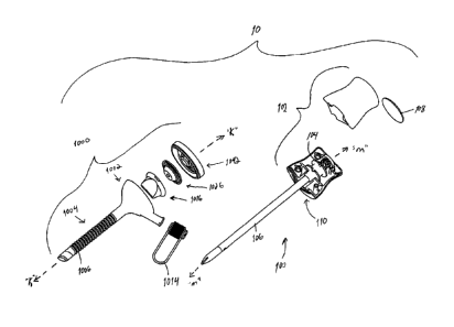
generally by reference numeral 10. Trocar assembly 10 is particularly adapted
for use in
CA 02681986 2009-10-08
minimally invasive surgical procedures such as endoscopic or laparoscopic
procedures.
Generally, trocar assembly 10 includes two principal subassemblies, namely,
obturator assembly
100 and cannula assembly 1000. Trocar assembly 10 may have various dimensions
or diameters.
In one embodiment, trocar assembly 10 provides a 5 mm portal to an underlying
tissue or target
site.
[0026] Cannula assembly 1000 may be suitable for use in any endoscopic
procedures
including, e.g., laparoscopic and arthroscopic. In disclosed embodiments,
cannula assembly
1000 includes cannula housing 1002 and cannula sleeve 1004 extending from the
cannula
housing 1002. Either or both cannula housing 1002 and cannula sleeve 1004 may
be transparent
in part or in whole and may be fabricated from biocompatible metal or
polymeric material.
Cannula 1004 may include a plurality of spaced locking ribs or projections
1006 extending about
the periphery of the sleeve (Figures 1-2). Ribs 1006 may be generally annular
in configuration
and may be spaced along longitudinal axis "k". Ribs 1006 may further define a
tapered leading
surface 1008 and a trailing locking surface 1010 traversing the longitudinal
axis "k". Tapered
leading surface 1008 facilitates insertion of a cannula sleeve 1004 within the
tissue. Trailing
locking surfaces 1010 are dimensioned to engage the tissue substantially
preventing or
minimizing retropulsion of cannula sleeve 1004 relative to the tissue. Cannula
housing 1002
may include port extension 1012 depending from base of the housing 1002. Port
extension 1012
is in fluid communication with the internal passageway of cannula sleeve 1004.
Port extension
1012 may have luer connector 1014 releasably connected thereto, or permanently
affixed to
cannula housing 1002. Luer connector 1014 may be adapted for connection to a
source of
insuffiation gases or another fluid source such as, e.g., an irrigant fluid
used in an arthroscopic
procedure.
6
CA 02681986 2016-05-24
[0027] Cannula housing 1002 further includes zero closure valve 1016.
(Figures 1 and 3-
6). Zero closure valve 1016 is adapted to open to permit passage of the
surgical object and
thereafter close in the absence of the object. Zero closure valve 1016
includes outer flange 1018
and inner valve surfaces 1020 depending radially inwardly from the outer
flange 1018. Inner
seal surfaces 1020 may extend in both a radial and longitudinal direction and
terminate at slit
1022. A pair of opposed rails 1024 are disposed on the leading end face of
zero closure valve
1016. Rails 1024 provide additional rigidity or support to inner valve
surfaces 1020 to facilitate
closing of the valve 1016 and/or minimize damage to valve 1016 during
insertion of a relatively
sharp object. Other zero closure valves such as duck bill valves are also
envisioned.
[0028] With reference to Figures 1 and 7-10, cannula housing further
includes object seal
1026. Object seal 1026 may be substantially similar to the seal disclosed in
commonly assigned
U.S. patent Application Publication Serial No. 2006/0253077, filed
April 19, 2006. Object seal 1026 includes annular seal
mount 1028 and resilient seal 1030 connected to the mount 1028. Seal mount
1028 may be
formed of a relatively rigid material such as a suitable polymeric material or
alternatively may be
fabricated from a resilient material. Seal mount 1028 incorporates a plurality
of apertures 1032
extending through the wall of the seal mount 1028. Resilient seal 1030 defines
aperture 1034
and is arranged to form a substantial seal about an instrument inserted
therethrough. In an
embodiment, resilient seal 1030 is adapted to form a seal about an instrument
having a diameter
ranging from about 3 mm to about 7 mm, for example, about 5 mm. In this
regard, aperture
1034 of seal 1030 defines a diameter ranging from about 2 mm to about 3 mm.
Seal 1030 may
be formed of any suitable elastomeric material. In disclosed embodiments, seal
1030 is
integrally formed with seal mount 1028 such that the elastomeric material
communicates through
7
CA 02681986 2016-05-24
apertures 1032 to form the integrally coupled unit depicted in the drawing
sheets. Seal mount
1028 and seal 1030 may be co-molded as is known in the art. In embodiments,
seal 1030 is
molded with seal mount 1028 to provide annular entry seal portion 1030,
anchoring segments or
spokes 1038 extending through apertures 1032 of seal mount 1028 and planar
inner seal portion
1040. Annular entry seal portion 1030 defines a general frusto-conical
configuration. Inner seal
portion 1040 defines aperture 1034.
[0029] Seal 1030 may include the fabric seal disclosed in commonly-assigned
U.S.
Patent No. 6,702,787 to Racenet. The seal disclosed in the
Racenet '787 patent may be a septum seal having a first
layer of resilient material and at least one fabric layer juxtaposed relative
to the first layer. The
fabric layer may include a SPANDEX material containing 20% LYCRA from
Milliken. Other
arrangements for seal 1030 are also envisioned. Seal 1030 may be flat,
hemispherical or have
any other shape as desired.
[0030] With reference now to Figures 1 and 11-12, cannula assembly 1000
further
includes cover 1042 which is mounted to cannula housing 1002 to enclose both
zero closure
valve 1016 and object seal 1026. Cover 1042 may be secured to housing 1002
with the use of
adhesives, cements, or via mechanical coupling means such as a segment
coupling or snap fit.
Cover 1042 includes outer segment 1044 and inner segment 1046 depending
radially inwardly
from the outer segment 1044. Cover 1042 may be oblong or elliptical in plan
view. The trailing
end face of cover 1042 defines a substantial recessed portion or mounting
recess 1048 of
predetermined geometrical configuration. In embodiments, mounting recess 1048
is generally
diamond shaped, key shaped or the particular configuration depicted in the
views in Figure 11.
Inner segment 1046 is inclusive of mounting recess 1048 and defines a
substantially planar
8
CA 02681986 2009-10-08
surface obliquely arranged with respect to the longitudinal axis and tapering
toward cover
aperture 1050. Tapered planar surface 1050 facilitates guiding of a surgical
object towards and
through cover aperture 1050. Seal cover 1042 further includes peripheral rib
1052 which is
received within or over the inner boundary of cannula housing 1002 to
facilitate securement to
the cannula housing 1002. Ribs 1052 may be dimensioned to frictionally engage
the inner
boundary of cannula housing 1002.
[00311
With reference to Figures 1 and 14-21, obturator assembly 100 of trocar
assembly
will be discussed. Obturator assembly 100 includes obturator housing 102,
elongated
obturator rod 104 extending distally from the housing 102 and outer sleeve 106
coaxially
mounted about the obturator rod 104. In general, outer sleeve 106 is adapted
to reciprocate or
move in a longitudinal direction between an unarmed and armed condition of
obturator rod 104.
Obturator rod 104 defines obturator axis "m" and will be discussed in greater
detail hereinbelow.
Obturator housing 102 includes housing cover or dome 108 mounted thereto.
Obturator housing
102 may be two half components connected to each other along respective
peripheries thereof.
Obturator housing 102 includes leading end 110 which projects outwardly from
the obturator
housing 102. Leading end 110 is correspondingly dimensioned to be received
within mounting
recess 1048 of seal cover 1042 in the assembled condition of obturator 100 and
cannula housing
1000. For example, leading end 110 may be substantially similar in
configuration, e.g., generally
key shaped or diamond shaped arrangement, to the configuration of mounting
recess 1048 of seal
cover 1042. Leading end 110 of obturator housing 102 is generally tapered to
facilitate insertion
within mounting recess 1048 of seal cover 1042. Thus, in the assembled
condition, leading end
110 or face of obturator housing 102 fits or mates with mounting recess 1048
of seal cover 1042.
In one embodiment, leading end 110 of obturator housing 102 and mounting
recess 1048 of seal
9
CA 02681986 2016-05-24
cover 1042 establish a mechanical interference or transition fit whereby
obturator housing 102
may be readily mounted and dismounted relative to cannula housing 1002. A
frictional fit for a
more secured condition is also envisioned.
[00321 With reference to the Figures 1 and 14-17, obturator rod 104 of
obturator
assembly 100 will be discussed. Obturator rod 104 includes obturator collar
112 at its proximal
end, and penetrating head 114 at its distal end. Obturator collar 112 has a
plurality of axial ribs
116 extending therefrom along the outer surface of rod 104. Rod 104 may have
an outer
diameter at its proximal or trailing end which is greater than the outer
diameter of the obturator
rod. Obturator collar 112 is received within recess defined between walls
117a, 117b, 117c of
obturator housing 102 to longitudinally fix the obturator rod 104 relative to
obturator housing
102 (Figure 13). Thus, the aforedescribed mounting arrangement of obturator
rod 104 and
obturator housing 102 secures the obturator rod 104 from moving in an axial
direction relative to
obturator housing 102.
[0033] Penetrating head 114 may be substantially similar in design to the
penetrating
member disclosed in commonly assigned U.S. Patent Application Publication
Serial No 2009/0093833, filed August 20, 2008. Penetrating
head 114 includes cylindrical element 118 and dissecting elements 120
extending contiguously
from the cylindrical element 118. Cylindrical element 118 defines an arcuate
or rounded leading
surface 120 which is atraumatic to tissue and extends a predetermined distance
beyond planar
dissecting element 120. This consequent narrow profile provided by cylindrical
element 118
permits initial insertion within tissue and facilitates, e.g., dissection or
advancement, within the
tissue without an incising action. Cylindrical element 118 may extend through
planar dissecting
element 120 to obturator rod 104. Planar dissecting element 120 defines a
triangular
CA 02681986 2009-10-08
arrangement having oblique side surfaces 122 leading to parallel end surfaces
124. Side surfaces
122 may be arcuate or rounded as shown to be atraumatic to tissue. In the
alternative, side
surfaces 122 may be sharpened. End surfaces 124 may be blunt or sharp.
[00341 Obturator rod 102 and penetrating head 114 may be integrally,
i.e., monolithically
formed, as a single unit. In one method, obturator member 104 and head 114 may
be formed of a
suitable polymeric material through known injection molding techniques. In the
alternative,
penetrating head 114 and obturator rod 104 may be separate components and
connected through
a slot and groove arrangement.
[0035] As depicted in Figures 1 and 18-21, outer sleeve 106 of obturator
assembly 100
will be discussed. Outer sleeve 106 is adapted for longitudinal movement
relative to obturator
rod 104. Outer sleeve 106 includes first and second collars 126, 128 at its
proximal end. Collars
126, 128 are longitudinally spaced. Collars 126, 128 reside within collar
mounting walls 117b,
130 of obturator housing 102 (Figure 13) and move within the walls 117b, 130
during traversing
movement of outer sleeve 106. Obturator sleeve 106 includes obturator nose 132
at its distal
end. Obturator nose 132 may be substantially similar in configuration to the
nose 132 described
in the aforementioned '629 application. Nose 132 moves relative to penetrating
head 114 during
longitudinal movement of outer sleeve 106. In the initial or unarmed
condition, nose 132 is
positioned relative to penetrating head 114 whereby cylindrical element 118 of
the penetrating
head 114 at least partially extends beyond the nose 132. In addition, side
surfaces 122 of planar
dissecting element also may extend beyond nose 132, i.e., protrude outwardly
from central slot
134. Nose 132 may be generally conical in configuration. Alternatively, nose
132 may also
have a slight inward contour along opposed peripheral portions. Various other
configurations are
also envisioned.
11
CA 02681986 2009-10-08
[0036] Obturator sleeve 106 may be spring biased in the distal direction
by coil spring
134. Coil spring 134 is mounted about obturator rod 102 and engages first
collar 126 of
obturator sleeve 106.
[0037] During use, as obturator sleeve 106 is advanced within tissue,
obturator nose 132
engages the tissue causing retraction of the obturator nose 132 and the
obturator sleeve 106
against the bias of coil spring 134 to thereby further expose penetrating head
114. Once
obturator nose 132 passes through the tissue, obturator sleeve 106 and
obturator nose 132 return
to its initial position.
[0038] Figures 22-23 illustrate another embodiment of the present
disclosure. In this
embodiment, trocar assembly 10' establishes a 3 mm portal for accessing the
underlying tissue
site. Most of the components are similar to the aforedescribed embodiment with
the exception of
a reduction in size. In addition, in accordance with this embodiment,
obturator sleeve 106' and
obturator rod (from the embodiment illustrated in FIG. 1) are integrally or
monolithically formed
(FIGS. 22-23). Thus, obturator sleeve 106' will not move in a longitudinal
direction relative to
the obturator rod. In other regards, this embodiment is substantially similar
to the previous
embodiment.
[0039] Figures 24-39 illustrate another embodiment of the present
disclosure. With
particular reference to Figures 24-31, a trocar assembly 2010 (e.g., a 3 mm
low profile version)
is shown. Trocar assembly 2010 includes an obturator housing 2012 disposed in
mechanical
cooperation with an elongated obturator member 2014, and defines a
longitudinal axis "A-A."
The elongated obturator member 2014 extends distally from the obturator
housing 2012. The
trocar assembly 2010 also includes a cannula assembly 3100 which receives the
elongated
obturator member 2014.
12
CA 02681986 2009-10-08
[00401 With reference to Figures 25 and 26, the obturator member 2014
includes an
obturator rod 2018 mechanically engagable with the obturator housing 2012 and
a penetrating
head 2020 adjacent the distal end of the obturator rod 2018. The penetrating
head 2020 includes,
from distal to proximal, a cylindrical element 2022 and a dissecting element
contiguously
extending from the cylindrical element 2022. The cylindrical element 2022
defines a rounded
leading surface which is atraumatic to tissue. The cylindrical element 2022
permits initial
insertion within an opening in the tissue and facilitates the advancement of
the penetrating head
2020 within the tissue. The dissecting element incorporates upper and lower
tapered surfaces
2030, 2032 and rounded side surfaces which define a pair of outwardly disposed
dissecting fins
2024. The tapered surfaces 2030, 2032 and dissecting fins 2024 are also
atraumatic to tissue.
The tapered surfaces 2030, 2032 and dissecting fins 2024 further enlarge the
opening within the
tissue as the penetrating head 2020 is advanced.
[0041] The cannula assembly 3100 of the trocar assembly 2010 includes an
elongated
portion 3102, defining a longitudinal axis "B-B," and a cover 3110 (Figures 27-
29). The cover
3110 encloses an object seal 3130 (Figures 30-31) and a zero-closure seal 3150
(Figure 24). The
object seal 3130 is disposed proximally of the zero-closure seal 3150. The
cover 3110 includes
an outer lip 3116 and an aperture 3120 having a diameter of Dl. A horizontal
shelf 3124
interconnects the outer lip 3116 with the aperture 3120. The outer lip 3116
includes vertical,
inner sidewalls 3118. Additionally, the aperture 3120 is defined between
vertical, inner
sidewalls 3122.
[0042] The object seal 3130 includes an elastomeric septum seal 3130b
which is over-
molded onto a rigid plastic insert 3130a. Rigid plastic insert 3130a includes
a horizontal surface
3132, a first vertical, annular wall 3134 and a second vertical, annular wall
3136. An inner
13
CA 02681986 2009-10-08
vertical surface 3134a of annular wall 3134 defines diameter D2. An inner
vertical surface
3136a of annular wall 3136 defines diameter D3. Additionally, the elastomeric
septum seal
3130b of the object seal 3130 defines a horizontal surface 3138 disposed
within annular wall
3136. The elastomeric septum seal 3130b includes an aperture 3139 having a
diameter D4. The
diameter D1 of the cover's aperture 3120 is less than the diameter D3 of the
annular wall 3136.
Thus, upon insertion, the obturator member 2014 is only able to contact the
horizontal surface
3138 and the walls defining the aperture 3139 of the object seal 3130.
(0043] The object seal 3130 also includes a lip 3140 depending downwardly
from
horizontal surface 3132. The lip 3140 engages a corresponding detent (not
shown) on the
housing 3102, such that the object seal 3130 cannot move circumferentially
(i.e., about
longitudinal axis "B-B") or radially (i.e., transversely with respect to
longitudinal axis "B-B").
Additionally, when the cannula assembly 3100 is assembled, object seal 3130 is
clamped to a
portion of the housing 3102, thus preventing axial (i.e., along longitudinal
axis "B-B")
movement of the object seal 3130 and further preventing circumferential and
radial movement of
the object seal 3130.
100441 In use, the obturator member 2014 of the trocar assembly 2010 is
introduced
within the cannula assembly 3100, through the aperture 3139 of the object seal
3130 and through
the zero-closure seal 3150. The assembled unit is positioned against the
targeted tissue, e.g., the
abdominal lining. When the obturator member 2014 passes through the aperture
3139 (either
when longitudinal axis "A-A" is substantially aligned with longitudinal axis
"B-B" or when
longitudinal axis "A-A" is non-aligned (i.e. spaced from and/or angled) with
longitudinal axis
"B-B"), the only portion of the object seal 3130 that is capable of
circumferential, axial or radial
movement is the horizontal surface 3138 adjacent aperture 3139 and disposed
within vertical
14
CA 02681986 2009-10-08
surface 3136a of annular wall 3136. The other portions of the object seal 3130
(including the
rigid plastic insert 3130a and the portions of the elastomeric septum seal
3130b disposed
outwardly of rigid plastic insert 3130a) are not capable of moving axially or
radially with respect
to the aperture 3139.
[0045] The penetrating head 2020 is manipulated relative to the tissue
whereby the
cylindrical element 2022, the tapered surfaces 2030, 2032, the dissecting fins
2024 and the
central section 2034 engage tissue and dissect or separate the tissue to gain
access to an
underlying cavity. The obturator member 2014 may then be removed from the
cannula assembly
3100. Instruments may be introduced within the cannula assembly 3100 to
perform a surgical
procedure.
[0046] With particular reference to Figures 32-39, a trocar assembly 4010
(e.g., a 5 mm
version) is shown. Trocar assembly 4010 includes an elongated obturator member
4214 (Figures
35-37) and a protective shield 4216 (Figures 38-39) coaxially mounted about
the obturator
member 4214. Trocar assembly 4010 is similar to trocar assembly 2010,
discussed above,
however the object seal 4330 (Figures 33-34) of trocar assembly 4010 is
different from the
object seal 3130 of trocar assembly 2010 and will be described herein.
[0047] The object seal 4330 includes an elastomeric septum seal 4330b
which is over-
molded onto a rigid plastic insert 4330a. Rigid plastic insert 4330a includes
a horizontal surface
4332, and a vertical, annular wall 4334. An inner vertical surface 4334a of
annular wall 4334
defines diameter D5. The elastomeric septum seal 4330b of the object seal 4330
defines a
horizontal surface 4338 disposed within annular wall 4334. The elastomeric
septum seal 4330b
includes an aperture 4339 having a diameter D6. The diameter D1 of the cover's
aperture 3120
is less than the diameter D5 of the annular wall 4334. Thus, upon insertion,
the obturator
CA 02681986 2009-10-08
member 2014 is only able to contact the horizontal surface 4338 and the walls
defining the
aperture 4339 of the object seal 4330.
100481 The object seal 4330 also includes a lip 4340 depending downwardly
from its
horizontal surface 4332. The lip 4340 engages a corresponding detent (not
shown) on the
housing, such that the object seal 4330 cannot move circumferentially (i.e.,
about longitudinal
axis "B-B") or radially (i.e., transversely with respect to longitudinal axis
"B-B"). Additionally,
when the cannula assembly 3100 is assembled, object seal 4330 is clamped to a
portion of the
housing 3102, thus preventing axial (i.e., along longitudinal axis "B-B")
movement of the object
seal 4330 and further preventing circumferential and radial movement of the
object seal 4330.
100491 The obturator member 4214 includes an obturator rod 4218 and a
penetrating
head 4220 at the end of the obturator rod 4218. The penetrating head 4220
includes, from distal
to proximal, a cylindrical element 4222 and a dissecting element contiguously
extending from
the cylindrical element 4222. The cylindrical element 4222 defines a rounded
leading surface
which is atraumatic to tissue. The cylindrical element 4222 permits initial
insertion within an
opening in the tissue and facilitates the advancement of the penetrating head
4220 within the
tissue without any cutting or incising of the tissue. The dissecting element
incorporates upper
and lower planar surfaces and rounded side surfaces which interconnect the
planar surfaces, to
thereby define a pair of outwardly disposed dissecting fins 4224. The
dissecting fins 4224 also
are atraumatic to tissue. The dissecting fins 4224 further enlarge the opening
within the tissue as
the penetrating head 4220 is advanced.
100501 The protective shield 4216 is adapted for reciprocal longitudinal
movement
relative to the obturator member 4214 between an advanced position and a
retracted position.
The protective shield 4216 includes a sleeve having a shield head 4226 which
is mounted about
16
CA 02681986 2009-10-08
the penetrating head 4220 of the obturator member 4214. The protective shield
4216 is normally
biased toward the advanced position by a coil spring mounted within the
obturator housing 4212
and engageable with the sleeve. In the initial or advanced position of the
protective shield 4216,
the penetrating head 4220 is partially exposed from the shield head 4226. In
the retracted
position of the protective shield 4216, the cylindrical element 4222 and the
dissecting fins 4224
are further exposed from the shield head 4226.
[0051] In use, trocar assembly 4010 is introduced through the cannula
assembly 3100
and the assembled unit is positioned against the targeted tissue, e.g., the
abdominal lining. That
is, when the obturator member 4214 passes through the aperture 4339 (either
when longitudinal
axis "A-A" is substantially aligned with longitudinal axis "B-B" or when
longitudinal axis "A-
A" is non-aligned (i.e. spaced from and/or angled) with longitudinal axis "B-
B"), the only
portion of the object seal 4330 that is capable of circumferential, axial or
radial movement is the
horizontal surface 4338 adjacent aperture 4339. The other portions of the
object seal 4330
(including rigid plastic insert 4330a and the portions of the elastomeric
septum seal 4330b
disposed outwardly of rigid plastic insert 4330a) are not capable of moving
axially or radially
with respect to the aperture 4339.
[0052] Once adjacent the targeted tissue, the penetrating head 4220 is
manipulated to
engage tissue and initiate the dissecting action on the tissue. The
penetrating head 4220 is
advanced causing the shield head 4226 to contact the tissue and be driven
proximally toward the
retracted position. In this position, the dissecting fins 4224 are further
exposed to further dissect
the tissue. After access to the underlying cavity has been achieved, the
protective shield 4216
and the shield head 4226 are returned to the advanced position by the biasing
force of the coil
spring. The obturator member 4214 may then be removed from the cannula
assembly 3100.
17
CA 02681986 2016-05-24
Instruments may be introduced within the cannula assembly 3100 to perform a
surgical
procedure.
[0053] The materials utilized in the components of the presently disclosed
trocar
assembly generally include materials such as, for example, ABS, polycarbonate,
stainless steel,
titanium and any other suitable biocompatible metals and/or polymeric
materials. A preferred
ABS material is CYCOLAC which is available from General Electric. A preferred
polycarbonate material is also available from General Electric under the
trademark LEXAN. An
alternative polycarbonate material which may be utilized is CALIBRE
polycarbonate available
from Dow Chemical Company. The polycarbonate materials may be partially glass
filled for
added strength.
[0054] Although the illustrative embodiments of the present disclosure have
been
described herein with reference to the accompanying drawings, it is to be
understood that the
disclosure is not limited to those precise embodiments, and that various other
changes and
modifications may be effected therein by one skilled in the art without
departing from the scope
of the disclosure.
18