Note: Descriptions are shown in the official language in which they were submitted.
CA 02682055 2009-09-24
WO 2008/118992 PCT/US2008/058324
HIGH RESOLUTION ELECTROPHYSIOLOGY CATHETER
FIELD OF THE INVENTION
The invention relates to systems and methods for providing therapy to a
patient, and more particularly to systems and methods for mapping and ablating
tissue within the heart of the patient.
BACKGROUND OF THE INVENTION
Physicians make use of catheters today in medical procedures to gain access
into interior regions of the body to ablate targeted tissue regions. It is
important for
the physician to be able to precisely locate the catheter and control its
emission of
energy within the body during these tissue ablation procedures. For example,
in
electrophysiological therapy, ablation is used to treat cardiac rhythm
disturbances in
order to restore the normal function of the heart.
Normal sinus rhythm of the heart begins with the sinoatrial node (or "SA
node") generating a depolarization wave front that propagates uniformly across
the
myocardial tissue of the right and left atria to the atrioventricular node (or
"AV node").
This propagation causes the atria to contract in an organized manner to
transport
blood from the atria to the ventricles. The AV node regulates the propagation
delay
to the atrioventricular bundle (or "HIS" bundle), after which the
depolarization wave
front propagates uniformly across the myocardial tissue of the right and left
ventricles, causing the ventricles to contract in an organized manner to
transport
blood out of the heart. This conduction system results in the described,
organized
sequence of myocardial contraction leading to a normal heartbeat.
Sometimes, aberrant conductive pathways develop in heart tissue, which
disrupt the normal path of depolarization events. For example, anatomical
obstacles
1
CA 02682055 2009-09-24
WO 2008/118992 PCT/US2008/058324
in the atria or ventricles can disrupt the normal propagation of electrical
impulses.
These anatomical obstacles (called "conduction blocks") can cause the
depolarization wave front to degenerate into several circular wavelets that
circulate
about the obstacles. These wavelets, called "reentry circuits," disrupt the
normal
activation of the atria or ventricles. As a further example, localized regions
of
ischemic myocardial tissue may propagate depolarization events slower than
normal
myocardial tissue. The ischemic region, also called a "slow conduction zone,"
creates errant, circular propagation patterns, called "circus motion." The
circus
motion also disrupts the normal depolarization patterns, thereby disrupting
the
normal contraction of heart tissue. The aberrant conductive pathways create
abnormal, irregular, and sometimes life-threatening heart rhythms, called
arrhythmias. An arrhythmia can take place in the atria, for example, as in
atrial
tachycardia (AT), atrial fibrillation (AFIB), or atrial flutter (AF). The
arrhythmia can
also take place in the ventricle, for example, as in ventricular tachycardia
(VT).
In treating these arrhythmias, it is essential that the location of the
sources of
the aberrant pathways (called substrates) be located. Once located, the tissue
in the
substrates can be destroyed, or ablated, by heat, chemicals, or other means of
creating a lesion in the tissue, or otherwise can be electrically isolated
from the
normal heart circuit. Electrophysiology therapy involves locating the aberrant
pathways via a mapping procedure, and forming lesions by soft tissue
coagulation
on the endocardium (the lesions being 1 to 15 cm in length and of varying
shape)
using an ablation catheter to effectively eliminate the aberrant pathways. In
certain
advanced electrophysiology procedures, as part of the treatment for certain
categories of atrial fibrillation, it may be desirable to create a curvilinear
lesion
around or within the ostia of the pulmonary veins (PVs), and a linear lesion
2
CA 02682055 2009-09-24
WO 2008/118992 PCT/US2008/058324
connecting one or more of the PVs to the mitral valve annulus. Preferably,
such
curvilinear lesion is formed as far out from the PVs as possible to ensure
that the
conduction blocks associated with the PVs are indeed electrically isolated
from the
active heart tissue.
Referring to Fig. 1, a prior art electrophysiological catheter 2 includes a
flexible catheter body 4, a tip electrode 6 mounted to the distal end of the
catheter
body 4, and a plurality of ring electrodes 8 (distal ring electrode 8(1),
medial ring
electrode 8(2), and proximal ring electrode 8(3)) mounted to the distal end of
the
catheter body 4 proximal to the tip electrode 6. In this embodiment, the tip
electrode
6 serves as both a tissue ablation electrode and a tissue mapping electrode,
and the
ring electrodes 8 serve as dedicated mapping electrodes. In a typical mapping
procedure, the tip electrode 6, and if possible the ring electrodes 8, are
placed into
contact with the endocardial tissue of the heart chamber stricken with the
arrhythmia
to obtain multiple electrocardiograms (ECGs) or monophasic action potentials
(MAPs) by measuring electrical signals at the electrodes 6, 8. For example,
three
bipolar ECG recordings may be obtained by measuring the voltage potentials
between various pairs of the electrodes (e.g., between the tip electrode 6 and
the
distal ring electrode 8(1), between the distal ring electrode 8(1) and the
medial ring
electrode 8(2), or between the medial ring electrode 8(2) and the proximal
ring
electrode 8(3)).
Based on the ECG or MAP recordings, the physician can determine the
relative location of the catheter in the heart and/or the location of any
aberrant
pathways. In one technique, the morphologies of the ECG or MAP recordings,
themselves, can be analyzed by a physician to determine the relative location
of the
catheter in the heart. In another technique, the electrode recordings are
processed
3
CA 02682055 2009-09-24
WO 2008/118992 PCT/US2008/058324
to generate isochronal electrophysiology maps, which may be combined with
three-
dimensional anatomical maps, such as those generated in three-dimensional
medical systems (e.g., the Realtime Position Management (RPM) tracking system,
developed commercially by Boston Scientific Corporation and described in U.S.
Pat.
Nos. 6,216,027 and 6,950,689, and the CARTO EP Medical system, developed
commercially by Biosense Webster and described in U.S. Pat. No. 5,391,199).
Primarily due to the relatively large size of tip electrodes, current catheter
designs, such as the type illustrated in Fig. 1, may detect far field
electrical activity
(i.e., the ambient electrical activity away from the recording electrode(s)),
which can
negatively affect the detection of local electrical activity. That is, due to
the relatively
large size of the tip electrode and the distance from the next ring electrode,
the
resulting electrical recordings are signal averaged and blurred, and thus not
well-
defined. This far-field phenomenon becomes more exaggerated, thereby
decreasing
the mapping resolution, as the length of distal tip electrode increases.
Thus, the electrical activity measured by such catheters does not always
provide a physician with enough resolution to accurately identify an ablation
site and
or provide the physician with an accurate portrayal of the real position of
the tip
electrode, thereby causing the physician to perform multiple ablations in
several
areas, or worse yet, to perform ablations in locations other than those that
the
physician intends.
In addition, many significant aspects of highly localized electrical activity
may
be lost in the far-field measurement. For example, the high frequency
potentials that
are encountered around pulmonary veins or fractioned ECGs associated with
atrial
fibrillation triggers may be lost. Also, it may be difficult to determine the
nature of the
tissue with which the tip electrode is in contact, or whether the tip
electrode is in
4
CA 02682055 2009-09-24
WO 2008/118992 PCT/US2008/058324
contact with tissue at all, since the far-field measurements recorded by the
tip
electrode may indicate electrical activity within the myocardial tissue even
though the
tip electrode is not actually in contact with the endocardial tissue.
For example, it may be very important to ascertain whether the tip electrode
is
in contact with endocardial tissue or venous tissue during an ablation
procedure.
This becomes especially significant when ablating in and around the ostia of
the
pulmonary veins, since ablation within the pulmonary veins, themselves,
instead of
the myocardial tissue, may cause stenosis of the pulmonary veins. However, the
far
field measurements taken by the tip electrode may indicate that the tip
electrode is in
contact with endocardial tissue, when in fact, the tip electrode is in contact
with
venous tissue. As another example, it may be desirable to ascertain lesion
formation
by measuring the electrical activity of the tissue in contact with the tip
electrode (i.e.,
the lack of electrical activity indicates ablated tissue, whereas the presence
of
electrical activity indicates live tissue). However, due to the far-field
measurements,
electrical activity may be measured from nearby live tissue, even though the
tip
electrode is actually in contact with ablated tissue.
Accordingly, there remains a need for an electrophysiology catheter that is
capable of measuring electrical activity of tissue at a higher resolution.
SUMMARY OF THE INVENTION
In accordance with first embodiment of the invention, a medical probe
comprises an elongated member (e.g., a flexible elongated member), and a
metallic
electrode mounted to the distal end of the elongated member. In one
embodiment,
the metallic electrode is cylindrically shaped and comprises a rigid body. The
medical probe further comprises a plurality of microelectrodes (e.g., at least
four
microelectrodes) embedded within, and electrically insulated from, the
metallic
5
CA 02682055 2009-09-24
WO 2008/118992 PCT/US2008/058324
electrode, and at least one wire connected to the metallic electrode and the
microelectrodes. Each microelectrode may have a suitably small size, e.g.,
less than
2mm. The exterior surfaces of the microelectrodes may conform to an exterior
surface of the metallic electrode to form an electrode assembly with a
substantially
continuous exterior surface.
In one embodiment, the metallic electrode has a cylindrical wall, a bore
surrounded by the cylindrical wall, and a plurality of holes extending through
the
cylindrical wall in communication with the bore. In this case, the
microelectrodes are
respectively disposed within the holes. The distal end of the elongated member
may
be disposed within the bore of the metallic electrode, and the medical probe
may
further comprise an electrically insulative potting material disposed within
the bore.
In this embodiment, the medical probe may further comprise a plurality of
electrically
insulative bands respectively disposed within the holes, in which case, the
microelectrodes are respectively disposed within the electrically insulative
bands.
In accordance with a a second embodiment of the invention, a medical probe
comprises an elongated member (e.g., a flexible elongated member), and a cap
electrode mounted to the distal tip of the elongated member. In one
embodiment,
the cap electrode has a length equal to or greater than 4mm and is composed of
a
metallic material. The medical probe further comprises a plurality of
microelectrodes
(e.g., at least four microelectrodes) disposed on, and electrically insulated
from, the
cap electrode, and at least one wire connected to the cap electrode and the
microelectrodes. The cap electrode and microelectrodes may be integrated
together
in the same manner as the metallic electrode and microelectrodes described
above.
In one embodiment, the medical probe further comprises at least one ring
electrode
6
CA 02682055 2009-09-24
WO 2008/118992 PCT/US2008/058324
mounted around the elongated member proximal to the cap electrode, in which
case,
the wire(s) is connected to the ring electrode(s).
In accordance with a third embodiment of the invention, a medical probe
comprises an elongated member (e.g., a flexible elongated member), and a rigid
electrode mounted to the distal end of the elongated member. In one
embodiment,
the metallic electrode is cylindrically shaped and is composed of a metallic
material.
The medical probe further comprises a plurality of microelectrodes (e.g., at
least four
microelectrodes) disposed on, and electrically insulated from, the cap
electrode, and
at least one wire connected to the cap electrode and the microelectrodes. The
rigid
electrode and microelectrodes may be integrated together in the same manner as
the metallic electrode and microelectrodes described above.
In accordance with a fourth embodiment of the invention, a medical system
comprises any of the medical probes described above, a radio frequency (RF)
ablation source coupled to the wire(s), and a mapping processor coupled to the
wire(s).
In accordance with a fifth embodiment of the invention, a medical method
comprises using any of the medical probes described above into a patient for
purpose of better understanding the invention. The method further comprises
placing the metallic electrode, cap electrode, or rigid electrode into contact
with
tissue (e.g., cardiac tissue) within the patient, sensing the tissue via at
least one of
the microelectrodes, and conveying ablation energy from the metallic
electrode, cap
electrode, or rigid electrode to ablate the tissue. In one method, the medical
probe is
intravenously introduced into the patient, in which case, the.cardiac tissue
may be
endocardial tissue.
7
CA 02682055 2009-09-24
WO 2008/118992 PCT/US2008/058324
In accordance with a sixth embodiment of the invention, a method of
manufacturing a medical probe comprises providing a cylindrically-shaped
electrode
having a wall and a bore surrounded by the wall. The method further comprises
forming a plurality of holes through the wall into the bore (e.g., by drilling
the holes),
mounting a plurality of microelectrodes (e.g., at least four microelectrodes)
respectively into the holes, mounting the distal end of an elongated member
(e.g., a
flexible elongated member) into the bore, connecting at least one wire to the
electrode and microelectrodes, and disposing the wire(s) through the elongated
member.
In one method, the electrode has a hemi-spherical distal tip, in which case,
the distal tip of the elongated member is mounted into the bore. One method
further
comprises mounting a plurality of electrically insulative bands respectively
into the
holes, in which case, the microelectrodes are respectively mounted within the
electrically insulative bands. In one method, each of the microelectrodes has
a
diameter equal to or less than 4mm. Another method further comprises
introducing
an electrically insulative potting material within the bore prior to mounting
the distal
end of the elongated member within the bore. Still another method further
comprises
grinding an exterior surface of the electrode and the exterior surfaces of the
microelectrodes to form an electrode assembly with a substantially continuous
exterior surface.
The use of microelectrodes in the manner described above eliminates
detection of the far field electrical activity, thereby increasing the
resolution and
fidelity of the mapping performed by the medical probe, allowing a user to
more
precisely measure complex localized electrical activity, and more accurately
8
CA 02682055 2009-09-24
WO 2008/118992 PCT/US2008/058324
detecting tissue contact and tissue characterization, including lesion
formation
assessment.
BRIEF DESCRIPTION OF THE DRAWINGS
The drawings illustrate the design and utility of embodiments of the
invention,
in which similar elements are referred to by common reference numerals, and in
which:
Fig. 1 is a partially cutaway plan view of a prior art electrophysiology
catheter;
Fig. 2 is a plan view of one embodiment of an electrophysiology system
constructed in accordance with the invention;
Fig. 3 is a partially cutaway plan view of an electrophysiology catheter used
in
the system of Fig. 2, particularly showing a first arrangement of
microelectrodes;
Fig. 4 is a cross-sectional view of the electrophysiology catheter of Fig. 3,
taken along the line 4-4;
Fig. 5 is a cross-sectional view of one microelectrode incorporated into the
electrophysiology catheter of Fig. 3;
Fig. 6 is a partially cutaway plan view of the electrophysiology catheter of
Fig.
3, particularly showing a second arrangement of microelectrodes;
Fig. 7 is a partially cutaway plan view of the electrophysiology catheter of
Fig.
3, particularly showing a third arrangement of microelectrodes;
Fig. 8 is a partially cutaway plan view of the electrophysiology catheter of
Fig.
3, particularly showing a fourth arrangement of microelectrodes;
Fig. 9 is a partially cutaway plan view of the electrophysiology catheter of
Fig.
3, particularly showing a fifth arrangement of microelectrodes;
Fig. 10 is a distal view of the electrophysiology catheter of Fig. 3;
9
CA 02682055 2009-09-24
WO 2008/118992 PCT/US2008/058324
Fig. 11 is a cross-sectional view of another microelectrode incorporated into
the electrophysiology catheters of Figs. 5 and 6;
Figs. 12A-12C are plan views of a method of using the electrophysiology
system of Fig. 2 to map and create lesions within the left atrium of a heart;
Fig. 13 is a diagram illustrating electrocardiograms generated by the
electrophysiology system of Fig. 2, particularly when the distal end of the
electrophysiology catheter is slowly placed into firm contact with endocardial
tissue;
Fig. 14 is a diagram illustrating electrocardiograms generated by the
electrophysiology system of Fig. 2, particularly when the distal end of the
electrophysiology catheter is removed from a superior vena cava into contact
with
endocardial tissue;
Fig. 15 is a diagram illustrating electrocardiograms generated by the
electrophysiology system of Fig. 2, particularly when RF ablation energy is
delivered
from the electrophysiology catheter into endocardial tissue; and
Fig. 16 is a diagram illustrating electrocardiograms generated by the
electrophysiology system of Fig. 2, particularly when the distal end of the
electrophysiology catheter is placed into contact with the left ventricle of a
heart
adjacent the atrioventricular node.
DETAILED DESCRIPTION OF THE ILLUSTRATED EMBODIMENTS
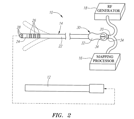
Referring to Fig. 2, an exemplary electrophysiology system 10 constructed in
accordance with the invention is shown. The system 10 may be used within body
lumens, chambers or cavities of a patient for therapeutic and diagnostic
purposes in
those instances where access to interior bodily regions is obtained through,
for
example, the vascular system or alimentary canal and without complex invasive
surgical procedures. For example, the system 10 has application in the
diagnosis
CA 02682055 2009-09-24
WO 2008/118992 PCT/US2008/058324
and treatment of arrhythmia conditions within the heart. The system 10 also
has
application in the treatment of ailments of the gastrointestinal tract,
prostrate, brain,
gall bladder, uterus, and other regions of the body. As an example, the system
10
will be described hereinafter for use in the heart for mapping and ablating
arrhythmia
substrates.
The system 10 generally comprises a conventional guide sheath 12, and an
electrophysiology catheter 14 that can be guided through a lumen (not shown)
in the
guide sheath 12. As will be described in further detail below, the
electrophysiology
catheter 14 is configured to be introduced through the vasculature of the
patient, and
into one of the chambers of the heart, where it can be used to map and ablate
myocardial tissue. The system 10 also comprises a mapping processor 16 and a
source of ablation energy, and in particular, a radio frequency (RF) generator
18,
coupled to the electrophysiology catheter 14 via a cable assembly 20. Although
the
mapping processor 16 and RF generator 18 are shown as discrete components,
they
can alternatively be incorporated into a single integrated device.
The mapping processor 16 is configured to detect, process, and record
electrical signals within the heart via the electrophysiology catheter 14.
Based on
these electrical signals, a physician can identify the specific target tissue
sites within
the heart, and ensure that the arrhythmia causing substrates have been
electrically
isolated by the ablative treatment. Based on the detected electrical signals,
the
mapping processor 16 outputs electrocardiograms (ECGs) to a display (not
shown),
which can be analyzed by the user to determine the existence and/or location
of
arrhythmia substrates within the heart and/or determine the location of the
electrophysiology catheter 14 within the heart. In an optional embodiment, the
mapping processor 16 can generate and output an isochronal map of the detected
11
CA 02682055 2009-09-24
WO 2008/118992 PCT/US2008/058324
electrical activity to the display for analysis by the user. Such mapping
techniques
are well known in the art, and thus for purposes of brevity, will not be
described in
further detail.
The RF generator 18 is configured to deliver ablation energy to the
electrophysiology catheter 14 in a controlled manner in order to ablate the
target
tissue sites identified by the mapping processor 16. Ablation of tissue within
the
heart is well known in the art, and thus for purposes of brevity, the RF
generator 18
will not be described in further detail. Further details regarding RF
generators are
provided in U.S. Patent No. 5,383,874.
The electrophysiology catheter 14 may be advanced though the guide sheath
12 to the target location. The sheath 12, which should be lubricious to reduce
friction
during movement of the electrophysiology catheter 14, may be advanced over a
guidewire in conventional fashion. Alternatively, a steerable sheath may be
provided. With respect to materials, the proximal portion of the sheath 12 is
preferably a Pebax material and stainless steel braid composite, and the
distal
portion is a more flexible material, such as unbraided Pebax , for steering
purposes.
The sheath 12 should also be stiffer than the electrophysiology catheter 14. A
sheath introducer (not shown), such as those used in combination with basket
catheters, may be used when introducing the electrophysiology catheter 14 into
the
sheath 12. The guide sheath 12 preferably includes a radio-opaque compound,
such as barium, so that the guide sheath 12 can be observed using fluoroscopic
or
ultrasound imaging, or the like. Alternatively, a radio-opaque marker (not
shown)
can be placed at the distal end of the guide sheath 12.
The electrophysiology catheter 14 comprises an integrated flexible catheter
body 22, a plurality of distally mounted electrodes, and in particular, a
tissue ablation
12
CA 02682055 2009-09-24
WO 2008/118992 PCT/US2008/058324
electrode 24, a plurality of mapping ring electrodes 26, a plurality of
mapping
microelectrodes 28, and a proximally mounted handle assembly 30. In
alternative
embodiments, the flexible catheter 14 may be replaced with a rigid surgical
probe if
percutaneous introduction or introduction through a surgical opening within a
patient
is desired.
The handle assembly 30 comprises a handle 32 composed of a durable and
rigid material, such as medical grade plastic, and ergonomically molded to
allow a
physician to more easily manipulate the electrophysiology catheter 14. The
handle
assembly 30 comprises an external connector 34, such as an external multiple
pin
connector, received in a port on the handle assembly 30 with which the cable
assembly 20 mates, so that the mapping processor 16 and RF generator 18 can be
functionally coupled to the electrophysiology catheter 14. The handle assembly
30
may also include a printed circuit (PC) board (not shown) coupled to the
external
connector 34 and contained within the handle 32. The handle assembly 30
further
including a steering mechanism 34, which can be manipulated to bidirectionally
deflect the distal end of the electrophysiology catheter 14 (shown in phantom)
via
steering wires (not shown). Further details regarding the use of steering
mechanisms are described in U.S. Patent Nos. 5,254,088 and 6,579,278.
The catheter body 22 is preferably about 5 French to 9 French in diameter,
and between 80cm to 150cm in length. The catheter body 22 preferably has a
cross-sectional geometry that is circular. However, other cross-sectional
shapes,
such as elliptical, rectangular, triangular, and various customized shapes,
may be
used as well. The catheter body 22 is preferably preformed of an inert,
resilient
plastic material that retains its shape and does not soften significantly at
body
temperature; for example, Pebax , polyethylene, or Hytrel (polyester).
13
CA 02682055 2009-09-24
WO 2008/118992 PCT/US2008/058324
Alternatively, the catheter body 22 may be made of a variety of materials,
including,
but not limited to, metals and polymers. The catheter body is preferably
flexible so
that it is capable of winding through a tortuous path that leads to a target
site, i.e., an
area within the heart. Alternatively, the catheter body 22 may be semi-rigid,
i.e., by
being made of a stiff material, or by being reinforced with a coating or coil,
to limit the
amount of flexing.
In the illustrated embodiment, the tissue ablation electrode 24 takes the form
of a cap electrode mounted to the distal tip of the catheter body 22. In
particular,
and with further reference to Fig. 3, the ablation electrode 24 has a
cylindrically-
shaped proximal region 36 and a hemispherical distal region 38. As shown
further in
Fig. 4, the proximal region 36 of the ablation electrode 24 has a wall 40 and
a bore
42 surrounded by the wall 40. The ablation electrode 24 may have any suitable
length; for example, in the range between 4mm and 10mm. In the illustrated
embodiment, the length of the ablation electrode 24 is 8mm. Preferably, the
ablation
electrode 24 is composed of a solid, electrically conductive material, such as
platinum, gold, or stainless steel. The wall 40 of the ablation electrode 24
has a
suitable thickness, such that the ablation electrode 24 forms a rigid body.
For the
purposes of this specification, an electrode is rigid if it does not deform
when
pressed into firm contact with solid tissue (e.g., cardiac tissue). The
ablation
electrode 24 is electrically coupled to the RF generator 18 (shown in Fig. 2),
so that
ablation energy can be conveyed from the RF generator 18 to the ablation
electrode
24 to form lesions in myocardial tissue. To this end, an RF wire 44 (shown in
Fig. 3)
is electrically connected to the ablation electrode 24 using suitable means,
such as
soldering or welding. The wire 44 is passed in a conventional fashion through
a
lumen (not shown) extending through the associated catheter body 22, where it
is
14
CA 02682055 2009-09-24
WO 2008/118992 PCT/US2008/058324
electrically coupled either directly to the external connector 34 or
indirectly to the
external connector 34 via the PC board located in the handle assembly 30,
which, in
turn, is electrically coupled to the RF generator 18 via the cable assembly
20.
The mapping ring electrodes 26 include a distal mapping ring electrode 26(1),
a medial mapping ring electrode 26(2), and a proximal mapping ring electrode
26(3).
The mapping ring electrodes 26, as well as the tissue ablation electrode 24,
are
capable of being configured as bipolar mapping electrodes. In particular, the
ablation electrode 24 and distal mapping ring electrode 26(1) can be combined
as a
first bipolar mapping electrode pair, the distal mapping ring electrode 26(1)
and the
medial mapping ring electrode 26(2) may be combined as a second bipolar
mapping
electrode pair, and the medial mapping ring electrode 26(2) and the proximal
mapping ring electrode 26(3) may be combined as a third bipolar mapping
electrode
pair.
In the illustrated embodiment, the mapping ring electrodes 26 are composed
of a solid, electrically conducting material, like platinum, gold, or
stainless steel,
attached about the catheter body 22. Alternatively, the mapping ring
electrodes 26
can be formed by coating the exterior surface of the catheter body 22 with an
electrically conducting material, like platinum or gold. The coating can be
applied
using sputtering, ion beam deposition, or equivalent techniques. The mapping
ring
electrodes 26 can have suitable lengths, such as between 0.5mm and 5mm. The
mapping ring electrodes 26 are electrically coupled to the mapping processor
16
(shown in Fig. 2), so that electrical events in myocardial tissue can be
sensed for the
creation of electrograms or monophasic action potentials (MAPs), or
alternatively,
isochronal electrical activity maps. To this end, signal wires 46 (shown in
Fig. 3) are
respectively connected to the mapping ring electrodes 26 using suitable means,
CA 02682055 2009-09-24
WO 2008/118992 PCT/US2008/058324
such as soldering or welding. The signal wires 46 are passed in a conventional
fashion through a lumen (not shown) extending through the associated catheter
body
22, where they are electrically coupled either directly to the external
connector 34 or
indirectly to the external connector 34 via the PC board located in the handle
assembly 30, which, in turn, is electrically coupled to the mapping processor
16 via
the cable assembly 20.
Like the mapping ring electrodes 26, the mapping microelectrodes 28 are
electrically coupled to the mapping processor 16 (shown in Fig. 2), so that
electrical
events in myocardial tissue can be sensed for the creation of electrograms or
MAPs,
or alternatively, isochronal electrical activity maps. To this end, signal
wires 48
(shown in Fig. 3) are respectively connected to the mapping microelectrodes 28
using suitable means, such as soldering or welding. The signal wires 48 are
passed
in a conventional fashion through a lumen (not shown) extending through the
associated catheter body 22, where they are electrically coupled either
directly to the
external connector 34 or indirectly to the external connector 34 via the PC
board
located in the handle assembly 30, which, in turn, is electrically coupled to
the
mapping processor 16 via the cable assembly 20.
Significantly, the microelectrodes 28 are disposed on the tissue ablation
electrode 24, and in particular, are embedded within the wall 40 of the tissue
ablation
electrode 24. This allows the localized intracardial electrical activity to be
measured
in real time at the point of energy delivery from the ablation electrode 24.
In addition,
due to their relatively small size and spacing, the microelectrodes 28 do not
sense
far field electrical potentials that would normally be associated with bipolar
measurements taken between the tissue ablation electrode 24 and the mapping
ring
electrodes 26.
16
CA 02682055 2009-09-24
WO 2008/118992 PCT/US2008/058324
Instead, the microelectrodes 28 measure the highly localized electrical
activity
at the point of contact between the ablation electrode 24 and the endocardial
tissue.
Thus, the arrangement of the microelectrodes 28 substantially enhances the
mapping resolution of the electrophysiology catheter 14. The high resolution
inherent in the microelectrode arrangement will allow a user to more precisely
measure complex localized electrical activity, resulting in a powerful tool
for
diagnosing ECG activity; for example, the high frequency potentials that are
encountered around pulmonary veins or the fractioned ECGs associated with
atrial
fibrillation triggers.
Moreover, the microelectrode arrangement lends itself well to creating MAPs,
which may play an important role in diagnosing AFIB triggers. In particular, a
focal
substrate may be mapped by the microelectrodes 28, and without moving the
ablation electrode 24, the mapped focal substrate may be ablated. The
microelectrode arrangement also allows for the generation of high density
electrical
activity maps, such as electrical activity isochronal maps, which may be
combined
with anatomical maps, to create electro-anatomical maps. In addition, due to
the
elimination or minimization of the detected far field electrical activity,
detection of
tissue contact and tissue characterization, including lesion formation
assessment, is
made more accurate.
The microelectrodes 28 may be disposed on the ablation electrode 24 in any
one of a variety of different patterns. In the embodiment illustrated in Fig.
3, four
microelectrodes 28 (only three shown) are circumferentially disposed about the
cylindrical-shaped region 36 of the ablation electrode 24 at ninety degree
intervals,
so that they face radially outward in four different directions. In another
embodiment
illustrated in Fig. 6, four microelectrodes 28 are arranged into two
longitudinally
17
CA 02682055 2009-09-24
WO 2008/118992 PCT/US2008/058324
disposed pairs (only pair shown) circumferentially disposed about the
cylindrical-
shaped proximal region 36 of the ablation electrode 24 at a one hundred degree
interval, so that the electrode pairs face radially outward in two opposite
directions.
Other embodiments illustrated in Figs. 7 and 8, are respectively similar to
the
embodiments illustrated in Figs. 5 and 6, with the exception that a fifth
microelectrode 28 is disposed on the hemispherical distal region 38 of the
ablation
electrode 24, so that it faces distally outward. In yet another embodiment, as
shown
in Fig. 9, ten microelectrodes 28 are arranged into two longitudinally
disposed trios
(only one shown) and two longitudinally disposed pairs circumferentially
disposed
about the cylindrical-shaped proximal region 36 of the ablation electrode 24
at ninety
degree intervals, so that the electrode trios and pairs face radially outward
in four
different directions. Notwithstanding the different microelectrode patterns,
as a
general rule, it is preferable that the microelectrodes 28 be located as
distal on the
ablation electrode 24 as possible. In this manner, the microelectrodes 28 will
be
placed into contact with tissue when the distal end of the electrophysiology
catheter
14 is oriented perpendicularly to the tissue.
In the illustrated embodiments, each of the microelectrodes 28 has a circular
profile for ease of manufacture, although in alternative embodiments, the
microelectrodes 28 may have other profiles, such as elliptical, oval, or
rectangular.
The microelectrodes 28 have relatively small diameters and are spaced a
relatively
small distance from each other in order to maximize the mapping resolution of
the
microelectrodes 28, as will be described in further detail below. Ultimately,
the size
and spacing of the microelectrodes 28 will depend upon the size of the
ablation
electrode 24, as well as the number and particular pattern of the
microelectrodes 28.
Preferably, the diameter of each microelectrode 28 is equal to or less than
half the
18
CA 02682055 2009-09-24
WO 2008/118992 PCT/US2008/058324
length of the ablation electrode 24, and more preferably equal to or less than
one-
quarter the length of the ablation electrode 24. For example, if the length of
the
ablation electrode 24 is 8mm, the diameter of each microelectrode 28 may be
equal
to or less than 4mm, and preferably equal to or less than 2mm. The spacing of
the
microelectrodes 28 (as measured from center to center) may be equal to or less
than
twice the diameter, and preferably equal to or less than one and half times
the
diameter of each microelectrode 28.
Each microelectrode 28 is composed of an electrically conductive material,
such as platinum, gold, or stainless steel, but preferably is composed of a
silver/silver chloride to maximize the coupling between the microelectrode 28
and
blood, thereby optimizing signal fidelity. As shown in Fig. 5, each
microelectrode 28
is substantially solid, having a small bore 50 formed in one end of the
microelectrode
28 along its axis, thereby providing a convenient means for connecting a
signal wire
48 to the microelectrode 28 via suitable means, such as soldering or welding.
Each microelectrode 28 also has a tissue-contacting surface 52 opposite the
bore 42 that preferably conforms with the tissue-contacting surface of the
ablation
electrode 24. Thus, because the tissue-contacting surface of the ablation
electrode
24 is curved, the tissue-contacting surface 52 of each microelectrode 28 is
likewise
curved, with the radii of curvature for the respective surface being the same,
thereby
forming an electrode assembly with a substantially continuous surface (i.e., a
surface
with very little discontinuities or sharp edges). In this manner, RF energy
will not be
concentrated within localized regions of the ablation electrode 24 to create
"hot
spots" that would undesirably char tissue, which may otherwise occur at
discontinuities. To ensure that the electrode assembly has a continuous
external
19
CA 02682055 2009-09-24
WO 2008/118992 PCT/US2008/058324
surface, the exterior surfaces of the ablation electrode 24 and
microelectrodes 28
can be ground to a fine finish (e.g., #16 grit).
Referring to Fig. 4, the ablation electrode 24 comprises a plurality of holes
54
laterally extending through the wall 40 in communication with the bore 42, and
the
microelectrodes 28 are respectively disposed in the holes 54. The holes 54 may
be
formed by drilling through the wall 40 of the ablation electrode 24.
Significantly, the
microelectrodes 28 are electrically insulated from the ablation electrode 24,
and thus,
from each other, so that they can provide independent mapping channels. The
microelectrodes 28 are also thermally insulated from the ablation electrode 24
to
prevent saturation of the mapping channels that would otherwise cause
interference
from the heat generated during a radio frequency (RF) ablation procedure.
To this end, the ablation electrode 24 comprises a plurality of insulative
bands
56 (best shown in Fig. 5) composed of the suitable electrically and thermally
insulative material, such as a high temperature thermoset plastic with high
dielectric
properties, e.g., polyimide or plastics from the phenolic group, such as
Bakelite@ or
Ultem@ plastics. The insulative bands 56 are respectively mounted within the
holes
54, and the microelectrodes 28 are mounted in the insulative bands 56. In this
manner, the insulative bands 56 are interposed between the wall 40 of the
ablation
electrode 24 and the microelectrodes 28 to provide the desirable electrical
and
thermal insulation. The insulative bands 56 and microelectrodes 28 may be
respectively mounted within the holes 54 using a suitable bonding material,
such as,
epoxy. An electrically and thermally insulative potting material 58 (such as a
multicomponent (resin and hardener component) thermosetting or ultra-violet
(UV)-
curable resin, for example, silicone, urethane or epoxy) can also be
introduced into
the bore 42 of the ablation electrode 24 to ensure electrical insulation
between the
CA 02682055 2009-09-24
WO 2008/118992 PCT/US2008/058324
microelectrodes 28 and ablation electrode 24, to further secure the
microelectrodes
28 to the ablation electrode 24, and to prevent cross-talk between the
otherwise
electrically insulated microelectrodes 28.
The electrophysiology catheter 14 further comprises a temperature sensor 60,
such as a thermocouple or thermistor, which may be located on, under, abutting
the
longitudinal end edges of, or in the ablation electrode 24. In the illustrated
embodiment, the temperature sensor 60 is mounted within a bore 42 formed at
the
distal tip of, and along the longitudinal axis of, the ablation electrode 24,
as illustrated
in Fig. 10, or, if a microelectrode 28 is incorporated into the distal tip of
the ablation
electrode 24, as illustrated in Figs. 7 and 8, within a bore 42 formed within,
and along
the longitudinal axis of, a microelectrode 28, as illustrated in Fig. 11. For
temperature control purposes, signals from the temperature sensors are
transmitted
to the RF generator 18 via signal wires 62, so that RF energy to the ablation
electrode 24 may be controlled based on sensed temperature. To this end, the
signal wires 62 are passed in a conventional fashion through a lumen (not
shown)
extending through the associated catheter body 22, where they are electrically
coupled either directly to the external connector 34 or indirectly to the
external
connector 34 via the PC board located in the handle assembly 30, which, in
turn, is
electrically coupled to the RF generator 18 via the cable assembly 20.
Having described the structure of the medical system 10, its operation in
creating a lesion within the left atrium LA of the heart H to ablate or
electrically
isolate arrhythmia causing substrates will now be described with reference to
Figs.
12A-12C. It should be noted that other regions within the heart H can also be
treated using the medical system 10. It should also be noted that the views of
the
heart H and other interior regions of the body described herein are not
intended to be
21
CA 02682055 2009-09-24
WO 2008/118992 PCT/US2008/058324
anatomically accurate in every detail. The figures show anatomic details in
diagrammatic form as necessary to show the features of the embodiment
described
herein.
First, the guide sheath 12 is introduced into the left atrium LA of the heart
H,
so that the distal end of the sheath 12 is adjacent a selected target site
(Fig. 12A).
Introduction of the guide sheath 12 within the left atrium LA can be
accomplished
using a conventional vascular introducer retrograde through the aortic and
mitral
valves, or can use a transeptal approach from the right atrium, as illustrated
in Fig.
12A. A guide catheter or guide wire (not shown) may be used in association
with the
guide sheath 12 to aid in directing the guide sheath 12 through the
appropriate artery
toward the heart H.
Once the distal end of the guide sheath 12 is properly placed, the
electrophysiology catheter 14 is introduced through the guide sheath 12 until
its
distal end is deployed from the guide sheath 12 (Fig. 12B). The steering
mechanism
34 located on the handle assembly 30 (shown in Fig. 2) may be manipulated to
place
the ablation electrode 24 into firm contact with the endocardial tissue at a
perpendicular angle to the wall of the heart H.
Once the ablation electrode 24 is firmly and stably in contact with the
endocardial tissue, the mapping processor 16 (shown in Fig. 2) is operated in
order
to obtain and record ECG or MAP signals from the myocardial tissue via bipolar
pairs
of the microelectrodes 28 (shown in Fig. 2). These ECG or MAP signal
measurements can be repeated at different locations within the left atrium LA
to
ascertain one or more target sites to be ablated. The user can analyze the
ECGs or
MAPs in a standard manner, or if electrical activity isochronal maps (whether
or not
combined with anatomical maps), can analyze these, to ascertain these target
sites.
22
CA 02682055 2009-09-24
WO 2008/118992 PCT/US2008/058324
Significantly, the use of the microelectrodes 28 substantially increases the
resolution
and enhances the fidelity of the ECG or MAP measurements. Alternatively, the
mapping processor 16 can be operated to obtain and record ECG or MAP signals
from the myocardial tissue via bipolar pairs of the ablation electrode 24 and
mapping
ring electrodes 26 if far field electrical potentials are desired; that is
generalized
mapping, in addition to highly localized mapping is desired.
Once a target site has been identified via analysis of the ECG or MAP signals
or isochronal electrical activity maps, the ablation electrode 24 is placed
into firm
contact with the target site, and the RF generator 18 (shown in Fig. 1) is
then
operated in order to convey RF energy to the ablation electrode 24 (either in
the
monopolar or bipolar mode), thereby creating a lesion L (Fig. 12C). Firm
contact
between the ablation electrode 24 and the endocardial tissue of the heart H
can be
confirmed by analyzing the ECG or MAP signals measured by the microelectrodes
28, with the amplitude of the ECG or MAP signals increasing as contact between
the
ablation electrode 24 and the endocardial tissue increases.
In the case where ablation is performed in or around the ostia PV of blood
vessels, such as pulmonary veins or the superior vena cava, the contact with
the
endocardial tissue, as opposed to venous tissue, can be confirmed via analysis
of
the highly localized ECG or MAP signals measured by the microelectrodes 28.
Ablation of the target site can be confirmed, again, by analyzing the highly
localized
ECG or MAP signals measured by the microelectrodes 28 during and after the
ablation procedure, with the amplitude of the ECG or MAP signals gradually
decreasing to zero as the tissue is successfully ablating. Significantly,
since the
microelectrodes 28 are incorporated into the ablation electrode 24, target
site
23
CA 02682055 2009-09-24
WO 2008/118992 PCT/US2008/058324
identification, electrode-tissue contact and characterization, tissue
ablation, and
lesion confirmation can all be performed without moving the ablation electrode
24.
To test the ability of the electrophysiology catheter 14 to record highly
localized ECGs, a prototype was built to determine if the localized electrode-
tissue
contact is assessable with the localized ECG recordings, determine if the
localized
ECG recordings can be used as a lesion assessment tool, determine if the
localized
ECG recordings are stable during RF ablation energy delivery, and assess if
the
microelectrodes 28 undesirably create tissue char during RF ablation energy
delivery. The ablation electrode 24 of the prototype 8mm long, and the four
0.070"
diameter microelectrodes 28 were embedded around the ablation electrode 24 in
a
manner similar to that illustrated in Fig. 3.
Tests of the prototype of the electrophysiology catheter 14 comparing the
ECG measurements taken by the mapping microelectrodes 28 to ECG
measurements taken by the mapping ring electrodes 26 were conducted in the
right
atrium of a dog. While recording ECGs with the microelectrodes 28 and ring
electrodes 26, the distal end of the electrophysiology catheter 14 was (1)
placed
gradually into firm contact with the endocardial tissue via manipulation of
the steering
mechanism 34 (corresponding ECG tracings shown in Fig. 13); (2) placed into
the
superior vena cava and then slowly pulled into the right atrium (corresponding
ECG
tracings shown in Fig. 14); (3) operated to conduct an RF ablation in the
right atrium
(corresponding ECG tracings shown in Fig. 15); and (4) placed into contact
with the
right ventricle near the atrial-ventricular (AV) node (corresponding ECG
tracings
shown in Fig. 16). In each case, four bipolar ECG recordings were made by the
four
microelectrodes 28 (me1-me2, me2-me3, me3-me4, me4-mel), and three bipolar
ECG recordings were made by the ablation electrode 24 and three ring
electrodes
24
CA 02682055 2009-09-24
WO 2008/118992 PCT/US2008/058324
26 (ablation electrode-distal ring electrode (AE-DRE), distal ring electrode-
medial
ring electrode (DRE-MRE), and medial ring electrode-proximal ring electrode
(MRE-
PRE)).
As shown in Figs. 13-16, the microelectrodes 28 clearly separate the localized
electrical activity at the ablation electrode 24 from the far field electrical
activity that
is normally associated with the ring electrode 26 measurements. That is, the
higher
resolution microelectrodes 28 generate very distinctly sharp, high amplitude,
ECG
tracings, compared to the typically slurred ECG tracings generated by the
lower
resolution ablation electrode 24 and ring electrodes 26.
As shown in Fig. 13, the amplitudes of the complexes of the ECG tracings
recorded by the microelectrodes 28 increases as the contact between the
ablation
electrode and the tissue increases. In particular, the amplitudes of the ECG
complexes recorded by the microelectrodes 28 become distinctly exaggerated
when
firm contact between the ablation electrode 24 and the tissue is achieved, in
contrast
to the ECG tracings recorded by the ablation/ring electrodes 24, 26, which
have
complexes of very low amplitudes during such firm contact that are virtually
indistinguishable from the complexes when no contact between the ablation
electrode and tissue occurs. As a result, the incorporation of microelectrodes
within
an ablation electrode proves to be a very useful tool for assessing electrode-
tissue
contact.
As shown in Fig. 14, the amplitudes of the complexes of the ECG tracings
recorded by the microelectrodes 28 are essentially zero when the ablation
electrode
24 is located within the superior vena cava, and then distinctly increase when
the
ablation electrode 24 is outside of the superior vena cava (SVC) in contact
with the
endocardial tissue. In contrast, the amplitudes of the complexes of the ECG
tracings
CA 02682055 2009-09-24
WO 2008/118992 PCT/US2008/058324
recorded by the ablation/ring electrodes 24, 26 are non-zero even when the
ablation
electrode 24 is located within the superior vena cava and do not substantially
increase when the ablation electrode 24 is located outside of the superior
vena cava
in contact with the endocardial tissue. As discussed above, distinguishing
between
the endocardial tissue and venous tissue important when ablating in or around
the
ostia of pulmonary veins. Thus, the incorporation of microelectrodes within an
ablation electrode proves to be a very useful tool for ensuring that an
ablation
procedure is not performed within a pulmonary vein.
As shown in Fig. 15, the amplitudes of the complexes of the ECG tracings
recorded by the microelectrodes 28 significantly decrease about 5-10 seconds
after
initiation of RF energy delivery during an ablation procedure. Significantly,
due to
the proximity of the microelectrodes 28 to the ablation electrode 24, the
changes to
the complexes of the ECG tracings are very discernible during the ablation
procedure. This is significant in that the distinct reduction of the
amplitudes of the
ECG tracings during ablation is a reliable indicator that the ablation
electrode 24 is in
firm contact with the tissue and that a lesion is forming. In contrast, the
amplitudes
of the complexes of the ECG tracings recorded by the ablation/ ring electrodes
24,
26 do not significantly change during the ablation procedure. Thus, the
incorporation
of microelectrodes within an ablation electrode proves to be a very useful
tool for
ensuring that the ablation procedure is efficiently creating a lesion within
the
myocardial tissue.
As shown in Fig. 16, the morphologies of the complexes of the ECG tracings
are significantly different when recorded by the microelectrodes 28 and
opposed to
the ablation electrode/ring electrodes 24, 26, when the ablation electrode 24
is
located in the ventricle adjacent the atrial-ventricular node. In particular,
the ECG
26
CA 02682055 2009-09-24
WO 2008/118992 PCT/US2008/058324
complexes recorded by the microelectrodes 28 reflect ventricular electrical
activity,
indicating that the ablation electrode 24 is located in the ventricle, whereas
the ECG
complexes recorded by the ablation /ring electrodes 24, 26 reflect both atrial
and
ventricular electrical activity, indicating that the ablation electrode 24 is
located at the
atrial-ventricular node, when in fact, it is not. Thus, the incorporation of
microelectrodes within an ablation electrode proves to be a very useful tool
for
determining whether the ablation electrode is located in a region of the heart
that can
be distinguished from other regions of the heart based on the nature of
electrical
activity expected to be at the region.
27