Note: Descriptions are shown in the official language in which they were submitted.
CA 02682293 2012-01-31
ENDOSCOPIC SUCTION DEVICE FOR MUCOSECTOMY
CROSS-REFERENCE TO RELATED APPLICATIONS
[0001] This application claims the benefit of U.S. Provisional Application
serial
no. 60/920,829, filed on March 29, 2007, entitled "ENDOSCOPIC SUCTION DEVICE
FOR
MUCOSECTOMY."
BACKGROUND OF THE INVENTION
[0002] The present invention relates to endoscopic suction devices and
apparatus for medical procedures involving endoscopic procedures.
[0003] The treatment of tissue encompasses a variety of techniques such as
electrocauterization, heat therapy, resection (removal of tissue), and
sclerotherapy (the
injection of medicine into target tissue). These treatment techniques usually
involve the
passing of medical instruments through the operating channel of the endoscope.
The
endoscope permits minimally invasive access, as well as visualization and
suction aids.
[0004] Another technique that frequently utilizes the operating channel of
the
endoscope is ligation, which involves applying a band or ligature around a
vessel or
portion of tissue, thereby cutting off blood or fluid flow and causing the
tissue to
necrose and separate from adjacent healthy tissue. Ligation is widely used to
treat a
number of medical tissue conditions, including, but not limited to,
hemorrhoids, polyps,
ballooning varices, and other types of lesions, including those that are
cancerous.
Typically, ligators are also used with a suction or vacuum means to draw the
tissue into
the distal tip, whereby the band is deployed over the base of the diseased
tissue to cut
off blood flow. The ligating device is typically activated by
1
CA 02682293 2009-09-29
WO 2008/121696
PCT/US2008/058399
retracting a line (string, wire, or cable) that is attached to the ligator at
the distal end
of an endoscope and is threaded through the operating channel of the endoscope
to
the proximal end of the instrument. The ligator can be activated by
mechanically
pulling the activating line by means of a hand-operated reel or trigger, or a
motor
drive mechanism. Various other ligating devices use cooperating inner and
outer
members that slide the individual bands by pushing or pulling them from the
tip of
the inner or outer member, the bands being preloaded onto the inner or outer
member prior to deployment.
[0005] To
prevent having to withdraw the instrument from the patient, reload,
and reintroduce it for treating additional tissue or vessels, devices have
been
developed capable of sequentially delivering multiple bands that are
preloaded, thus
shortening the procedure time and improving patient comfort. Multiple band
ligating
devices include designs that individually tether or otherwise secure the bands
to the
dispenser and then release them sequentially as needed, often by use of one or
more strings extending to the proximal end.
[0006] For
example, during mucosectomy, the excision of a mucosa, the
clinician faces challenges in removing mucosa merely due to the design of the
distal
end of a typical endoscope. More specifically, the suction port formed on the
distal
end of the endoscope may at times cause challenges, e.g., visual obstruction
when
a polypectomy snare is used to position around a lesion for removal thereof.
[0007] Thus, it
is desirable to provide an endoscopic suction device that is
compatible with an endoscope and that provides reduced visual obstruction
during
mucosectomy.
2
CA 02682293 2009-09-29
WO 2008/121696
PCT/US2008/058399
BRIEF SUMMARY OF THE INVENTION
[0008] The
present invention generally provides an endoscopic suction device
that is compatible with an endoscope for endoscopic mucosa! resection (EMR).
Embodiments of the present invention provide a device to allow a more
simplified
way of mucosectomy, especially when a polypectomy snare is used therewith. A
device allows for a relatively easier way of removing a lesion during
mucosectomy.
[0009] In one
embodiment, the device comprises a connecting base having
an open end attachable to the distal end of the endoscope. The device further
comprises a distal tip extending from the connecting base and having a closed
distal
end. The distal tip comprises a suction chamber formed therein and in fluid
communication with the open end. The suction chamber has a suction opening
formed laterally therethrough for suctioning lesions during mucosectomy.
[0010] In
another embodiment, the present invention provides an endoscope
apparatus. The apparatus comprises the endoscopic suction device and an
endoscopic assembly for endoscopy. The apparatus comprises an insertion tube
having a plurality of channels through which endoscopic parts may be disposed.
The apparatus further comprises a control system in mechanical and fluid
communication with the insertion tube. The control system is configured to
control at
least one of the endoscopic parts.
[0011] In
another example, the present invention provides a method of
mucosectomy of mucosal tissue of a patient. The method comprises disposing a
polypectomy snare distally through a working channel of the endoscope having a
distal end and advancing the snare through the distal end to the endoscopic
suction
device attached to the distal end. The method further comprises opening the
snare
3
CA 02682293 2013-12-19
adjacent the suction opening to receive the mucosal tissue and suctioning the
mucosal
tissue through the suction opening. The method further comprises receiving the
mucosal
tissue with the snare.
[0011a] Provided herein is an endoscopic suction device of a distal end of
an
endoscope for mucosectomy, the device comprising: a connecting base having an
open end
removably attachable to the distal end of the endoscope by cooperating
threads, the
connecting base having a larger diameter than and mountable over the distal
end of the
endoscope; and a distal tip extending from the connecting base and having a
closed distal
end, the distal tip comprising a suction chamber formed therein and in fluid
communication
with the open end, the suction chamber having a suction opening formed
laterally
therethrough for suctioning lesions during mucosectomy, the distal tip having
a cylindrical
shape with a rounded tip, wherein the entire suction opening is located on a
planar or flat
surface of distal tip the to increase contact with mucosal tissue and create a
seal for
enhanced suctioning.
[0011 b] Also provided herein is an endoscope apparatus having a suction
apparatus,
the apparatus comprising:an endoscopic assembly for endoscopy, the assembly
comprising:
an insertion tube, the insertion tube having a plurality of channels through
which
endoscopic parts are disposed; and a control system in mechanical and fluid
communication with the insertion tube, the control system being configured to
control at
least one of the endoscopic parts; and an endoscopic suction device of a
distal end of the
endoscope, the endoscopic suction device comprising: a connecting base having
an open
end removably attachable to the distal end of the insertion tube by
cooperating threads, the
connecting base having a larger diameter than and mountable over the distal
end of the
insertion tube; and a distal tip extending from the connecting base and having
a closed
distal end, the distal tip comprising a suction chamber formed therein and in
fluid
communication with the open end, the suction chamber having a suction opening
formed
laterally therethrough for suctioning lesions during mucosectomy, the distal
tip having a
cylindrical shape with a rounded tip, wherein the entire suction opening is
located on a
planar or flat surface of the distal tip to increase contact with mucosal
tissue and create a
seal for enhanced suctioning.
[0011c] Further provided herein is an endoscopic suction device of a
distal end of an
endoscope for mucosectomy, the device comprising: a connecting base having an
open end
4
CA 02682293 2013-12-19
attachable to the distal end of the endoscope; and a distal tip extending from
the connecting
base and having a closed distal end, the distal tip comprising a suction
chamber formed
therein and in fluid communication with the open end, the suction chamber
having a suction
opening formed laterally therethrough for suctioning lesion during
mucosectomy, wherein
the entire suction opening is located on a planar or flat surface of the
distal tip to increase
contact with mucosal tissue and create a seal for enhanced suctioning; wherein
the distal tip
has a cylindrical shape with a rounded tip; and wherein the open end is
removably
attachable to the distal end of the endoscope by cooperating threads, the
connecting base
having a larger diameter than and mountable over the distal end of the
endoscope.
[0012]
Further objects, features, and advantages of the present invention will
become apparent from consideration of the following description and the
appended claims
when taken in connection with the accompanying drawings.
BRIEF DESCRIPTION OF THE DRAWINGS
[0013]
Figure 1 is side view of an endoscopic suction device in accordance with one
embodiment of the present invention;
[0014]
Figure 2 is a perspective view of a flexible endoscopic apparatus comprising
the endoscopic suction device in accordance with one embodiment of the present
invention;
[0015]
Figure 3 is an elevated view of a distal tip of the endoscope and the
endoscopic suction device in accordance with one embodiment of the present
invention;
[0016] Figure 4 is a cross-sectional view of the endoscopic apparatus taken
along line 4-4 of
Figure 3;
[0017]
Figure 5 is an environmental view of the assembly applying suction on a
lesion during mucosectomy; and
[0018]
Figure 6 is an environmental view of an exposed muscularis propria layer
after snare excision.
DETAILED DESCRIPTION OF THE INVENTION
[0019]
Embodiments of the present invention provide an endoscopic suction device
for an endoscope during mucosectomy. The device is a distal cap that is
4a
CA 02682293 2009-09-29
WO 2008/121696
PCT/US2008/058399
placed directly over the distal end of an endoscope. The distal cap includes a
side
or lateral opening to suction mucosa into its chamber so that the mucosa can
be
resected using a snare. In one embodiment, the device or distal cap comprises
a
connecting base attachable to the endoscope and a distal tip extending from
the
base. The tip has a lateral suction opening formed through the side in fluid
communication with a suction chamber to receive a lesion during mucosectomy.
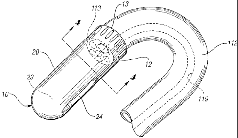
[0020] Figure 1
illustrates an endoscopic suction device or distal cap 10
comprising a connecting base 12 having an open end 13 attachable to the
insertion
tube of an endoscope. The open end 13 may be attached to the insertion tube by
any suitable means, e.g., threaded connection, press fit, or bonded
attachment. As
shown, the endoscopic suction device 10 further comprises a distal tip 20
integrally
extending distally from the connecting base 12. In this embodiment, the distal
tip 20
comprises a suction chamber 23 formed therein and in fluid communication with
the
open end 13. The suction chamber is able to hold a lesion to be removed during
mucosectomy. Preferably, the suction chamber has a lateral suction opening 24
formed through the side of the device 10 for suctioning a lesion to be held in
the
suction chamber 23 during mucosectomy. The lateral suction opening 24 is in
fluid
communication with the open end 13 so that a vacuum or suction source may be
used. Preferably, the distal cap 10 may have any suitable length, e.g.,
between
about 1.5 and 4 centimeters. Furthermore, although the drawings depict the
distal
cap 10 having an elongate or bullet shaped tip, the distal cap may take on any
other
suitable shape.
[0021] As
shown, the suction opening 24 is formed laterally through the distal
tip 20. This allows the suction opening 24 to be more easily disposed over the
CA 02682293 2009-09-29
WO 2008/121696
PCT/US2008/058399
lesion, thereby being removed more conveniently as will be described in
greater
detail below. Preferably, the suction opening 24 may have an oval or a
generally
circular shape; however, the suction opening 24 may take on any other shape
without falling beyond the scope or spirit of the present invention. Moreover,
the
area adjacent the suction opening 24 may be planar or flat to increase contact
with
the mucosal tissue and create a seal for enhanced suctioning. With a
transparent
distal tip 20 along with the laterally formed suction opening 24, the
physician is able
to more clearly maneuver or manipulate the endoscopic parts to perform the
mucosectomy procedure. In one embodiment, the suction opening 24 is configured
to fit over mucosa for a mucosectomy treatment. Alternatively, the suction
opening
24 may be relatively smaller in area to fit over the lesion, but with a
suction source
that effectively suctions the lesion in the chamber for mucosectomy.
[0022] The
endoscopic suction device 10 may be made of any suitable
material, preferably transparent material. In one embodiment, at least a
portion of
the endoscopic device could be made of metal, metal alloy, or an opaque
material.
However, it is advantageous for the device 10 to be made of transparent
material.
For example, the endoscopic suction device 10 may comprise one of super
elastic
material, polycarbonate plastic, nitinol, cobalt-chromium-nickel-molybdenum-
iron
alloy, or cobalt-chrome alloy, polytetrafluoroethylene (PTFE), polyethylene,
polypropylene, perfluoroelastomer, fluoroelastomer, nitrile, neoprene,
polyurethane,
silicone, polytetrafluroethylene, styrene-butadiene, rubber, or
polyisobutylene.
[0023] In this
embodiment, the endoscopic suction device is preferably
configured to be able to receive a snare disposed through the open end and
situated
adjacent the lateral suction opening for receiving mucosa or a lesion during a
6
CA 02682293 2012-01-31
mucosectomy procedure. Thus, in use, a lesion is suctioned through the opening
24 and
received within a loop of the snare for resection or removal during
mucosectomy.
[0024] Figure 2 illustrates a flexible endoscopic apparatus or instrument
110
comprising the endoscopic suction device 10 in accordance with one embodiment
of the
present invention. The apparatus 110 has a length that permits access to the
deeper
regions of a hollow body organ. In certain embodiments, the flexible apparatus
110 can
be sized for insertion into the alimentary tract. In accordance with one
embodiment, the
apparatus 110 includes a conventional endoscope with an operating control
section 111
and a flexible section 112 that terminates at a distal insertion end 113. The
operating
control section 111 includes a viewing end 114 remote from the insertion end
113,
through which a ligating procedure can be directly observed.
[0025] It is to be understood that any other suitable endoscopic
apparatus may
be used with the ligator assembly described above. For example, various
endoscopic
ligating apparatus may be used including but not limited to U.S. Patent No.
6,007,551
entitled "Endoscopic Ligating Apparatus" filed on September 6, 1996 and U.S.
Patent No.
5,624,453 entitled "Endoscopic Ligating Instrument" filed on October 30, 1995.
[0026] Referring to Figures 2 and 4, the endoscopic instrument 110 may
include
a plurality of channels extending from the operating control section 111 and
through
the flexible section 112 to the insertion end 113. For example, the instrument
110 can
include an illumination channel 116 through which a fiberoptic cable is
inserted for the
transmission of light from a light source. A viewing channel
7
CA 02682293 2009-09-29
WO 2008/121696
PCT/US2008/058399
117 can also be provided with a fiberoptic cable for viewing purposes, while a
third
channel 118 can be provided for application of suction at the surgical site.
The
endoscopic instrument 110 can also include a working channel 119 through which
a
plurality of tools and instruments can be extended such as a polypectomy snare
for
resection of a lesion, an irrigation channel 120 to allow delivery of fluid to
the ligation
site, and an air channel 121 that can be used to deliver pressurized air, such
as for
cleaning the lens at the insertion end of the viewing channel 117.
[0027] In one
embodiment, the endoscopic instrument 110 also includes an
auxiliary port portion 123 having a proximal opening 124. The working channel
119
extends into the auxiliary port 123 by way of a working channel extension
119a.
Each of the channels preferably opens at the distal or insertion end 113 of
the
flexible section 112 of the endoscopic instrument 110.
[0028] The
endoscope forming part of the instrument 110 of Figure 3 can be
of many different types. For example, the endoscope can be of the type
commercially provided by Olympus, Pentax, or Fujinon. While most of the
working
components of these endoscopes are similar, each may have a different
configuration for the proximal opening 124 and the auxiliary port 123. Each of
these
specifically identified endoscopes, and other commercially available
endoscopes,
utilize different sealing members (not shown) at the proximal opening 124 of
the
auxiliary port 123. It is understood that the various aspects of the present
invention
accommodate the secure attachment to various configurations and dimensions of
a
variety of endoscopes.
[0029]
Referring now to Figure 4, details of the endoscopic suction device 10
can be seen. In this embodiment, the device 10 is disposed at the insertion
end 113
8
CA 02682293 2009-09-29
WO 2008/121696
PCT/US2008/058399
of the flexible section 112 of the endoscope. The material of the device 10
should
be sufficiently strong or rigid to receive lesions to be suctioned therein and
resected
by a polypectomy snare. The device 10 is preferably removably mountable to the
insertion end 113 of the flexible endoscope section 112.
[0030] In use,
the endoscopic suction device is preferably provided separately
from the endoscopic instrument. At an appropriate time in the use of the
flexible
endoscopic apparatus, the device 10 can be mounted about the cylindrical
surface
of the flexible endoscope section by any suitable means such as by cooperating
threads.
[0031] Figures
5 and 6 illustrate a method of mucosectomy in accordance
with one example of the present invention. In this example, the technique of
mucosectomy uses a braided snare. In another example, a monofilament stiff-
wire
polypectomy snare may be used. As shown in Figure 5, the polypectomy snare is
disposed distally through a working channel of the endoscope. The snare is
then
moved through the distal end of the endoscope and is opened for positioning
relative
to a lesion to be removed during mucosectomy. Upon suctioning proximally
through
the distal tip, the polypectomy snare is opened and placed against the mucosal
surface about the lesion. The snare is then relatively slowly moved towards a
closed
position, thereby reducing blood flow through the lesion. In another example,
the
snare may also be introduced outside of the distal tip prior to suctioning
through the
distal tip.
[0032] Figure 5
illustrates the polypectomy snare 140 in a relatively closed
position around the neck of the lesion 142. In this example, once tightened
around
the lesion, the endoscopic suction device 10 is positioned about the lesion
for
9
CA 02682293 2009-09-29
WO 2008/121696
PCT/US2008/058399
suctioning. More specifically, the lesion 142 is disposed through the suction
opening
24 into the suction chamber 23 of the distal tip 20 of the endoscopic suction
device
110. The device 10 maintains a suitable vacuum to maintain the lesion within
the
suction chamber 23 of the device 10. The snare 140 is then further tightened
around the lesion 142 and lifted away from its vessel wall. Pure coagulation
current
as known may be applied to transect the lesion 142. Lifting the lesion 142
further
into the chamber 23 as current is applied helps further prevent transmural
extension
of the burn.
[0033] Figures
5 and 6 illustrate a muscularis propria layer 143. In this
example, the muscularis propria layer 143 is preferably exposed after the
snare
excision. This indicates that a complete mucosectomy has been performed. The
lesion 142 is received in the suction chamber 23 of the device 10 as a vacuum
is
maintained therethrough. The device 10 then may be retracted through the
suction
channel and out of the system for disposal or retainment of the lesion.
[0034] While
the present invention has been described in terms of preferred
embodiments, it will be understood, of course, that the invention is not
limited thereto
since modifications may be made to those skilled in the art, particularly in
light of the
foregoing teachings.