Note: Descriptions are shown in the official language in which they were submitted.
CA 02682297 2013-07-24
Description
Measuring arrangement and method for three-dimensional measuring of an object
The invention relates to a measuring arrangement for three-dimensional
measuring of at
least one part on an object, in particular a semitransparent object,
comprising one light
source with a continuous spectrum, one device to generate a multi-focal
illumination
pattern, an objective lens with high chromatic aberration to image foci of the
illumination
pattern onto the object, a detector unit such as a CCD chip to determine the
wave length
spectra of the foci imaged confocally onto the object via the objective lens,
as well as one
spectrum dispersing device that is arranged between the confocally imaged foci
and the
detector unit.
The invention further relates to a method for measuring the shape of at least
one section of
an object, in particular a semitransparent object such as at least a section
of a tooth,
utilizing one light source to generate light with a continuous spectrum, one
device to
generate a multi-focal illumination pattern, one objective lens with high
chromatic
aberration to image foci of the illumination pattern onto the object, one
detector unit to
determine the wave length spectra of the foci imaged confocally onto the
object via the
objective lens, whereby from the respective wavelength spectrum the spectral
peak position
of each focus is determined, from which subsequently one computes the extent
of the object
along the direction of the imaging ray (Z coordinate).
In many technical fields it is necessary to determine, i.e. measure, the three-
dimensional
structure of objects. One example is the determination of the shape of a
tooth, which is
necessary to produce a dental prosthesis. For dental reconstructions that are
still produced
by conventional methods using a plaster cast, one endeavours to leave behind
the
corresponding classical method and to determine the geometry of the dental
shape using a
non-contacting method.
1
CA 02682297 2013-07-24
A large number of methods for the acquisition of the three-dimensional
structure of bodies
are known in the art. Of the existing optical methods one has to mention the
strip projection
method or phase shift method, optical coherence tomography, and holography. In
the field
of dentistry, the phase-shift method already finds practical application.
However it has come to light that corresponding optical methods do not produce
the desired
results, in particular for objects with high scattering characteristics. In
the strip projection
method, for example, scatter leads to a lack of definition of the strips and
consequently to
lower resolution.
Also known are methods in which the object is not illuminated in its entirety,
but rather
only in one region with one sharp focus or several foci utilizing confocal
imaging. To
obtain a complete two-dimensional image, the focus or foci must be scanned
across the
object. To measure three-dimensional structures, it is necessary to shift the
focal plane
along the axial direction.
Alternatively, a wide-band light source and a suitable optical system with a
highly wave-
length-dependent focal length can be employed to image the focus or the foci.
As a result,
the foci are sharply imaged at different distances from the objective lens in
dependence on
their wavelength. After imaging the foci backwards through the objective lens,
an intensity
maximum can be detected in the focus plane for the particular colour that is
sharply imaged
at the corresponding object distance. Thus, determining the spectral peak
position allows
one to determine the distance between the object and the objective lens at
this point, and
consequently and ultimately allows determining the three-dimensional structure
of the
object. Evaluation is performed either by points using a spectrometer or by
lines using a
line spectrometer with camera chip.
EP-B-0 321 529 discloses a measuring arrangement for measuring the distance
from an
objective lens with high chromatic aberration to an object. For detection one
employs a
black & white CCD chip camera, in front of which is arranged a spectrum-
dispersing
2
CA 02682297 2013-07-24
device that possesses an input slit. This converts the wavelength information
for every point
to position information to obtain a profile image of the surface of the
object.
= EP-B-0 466 979 relates to an arrangement for simultaneous confocal image
generation. For
this purpose one uses a matrix of punched holes such as a Nipkow disk to
generate points
of light, which are imaged in focus onto an object. A CCD array camera is used
as detector
unit.
Described in DE-A-102 42 374 is a confocal distance sensor with an imaging
optical
system with chromatic aberration, which is intended for inspection purposes in
the
electronics field. As a light source one can employ a light source with a
multitude of point
light sources. As a light detector one employs point detectors, whereby one
point detector is
assigned to each point light source and they are arranged confocal relative to
each other.
Known from DE-A-103 21 885 is a confocal measuring arrangement for the three-
dimensional measuring of an object using chromatic fine splitting, in which a
multitude of
foci is generated by means of a micro-lens array and is imaged onto the
object. The
reflected light is focussed back onto the plane of the micro-lens foci. This
arrangement is
used to measure two- or three-dimensional micro-profiles of objects to be
measured or two-
or three-dimensional transparency or reflectivity profiles.
The present invention is based on the objective to further develop a measuring
arrangement
as well as a method for the three-dimensional determination of the shape of an
object,
particularly a semitransparent object such as a tooth, in such a way, that a
highly precise
surface profile can be established in a short timeframe even for moving
objects. It is in
particular desirable to be able to measure scattering or highly scattering
objects that exhibit
a very high white-light background, which would facilitate application in the
field of
dentistry.
As a solution to this problem, in a measuring arrangement of the above-
mentioned type it is
3
CA 02682297 2013-07-24
chiefly intended that in the plane of the confocally imaged foci be arranged
one first matrix
of holes with first holes, whereby the geometric arrangement of the first
holes corresponds
to the geometric arrangement of the foci of the multi-focal illumination
pattern.
Different from arrangements known in the art, a matrix of holes or a pinhole
array is
arranged in the focal plane - where the foci reaching the object are imaged -
of the objective
lens with high chromatic aberration, whereby consequently the illumination
pattern is
confocally imaged back from the measured object onto this matrix of holes or
pinhole
array. In this, the geometric correspondence of the first holes of the first
matrix of holes
relative to the illumination pattern is tuned so that a unique assignment is
made, so that as a
result the foci in the holes of the matrix of holes can be assigned positional
coordinates in a
plane that extends normal to the beam path passing through the objective lens.
According to the invention, holes or pinholes distributed across an area are
positioned in
the focal plane of the objective lens and the illumination pattern is
confocally imaged back
from the object into these holes or pinholes. In this, the foci that are
imaged on the object
are imaged - in dependence on wavelength and the distance between the
objective lens and
the object ¨ onto the pinhole. The spectra of these foci are subsequently read
out by the
detector device. In this, the invention in particular intends that the
dispersing device
arranged upstream of the detector device laterally spreads the spectral lines
of the focus
imaged in the respective hole, before the spectral lines reach pixels of the
detector device.
For this purpose it is intended that the detector device comprises the pixels
of a CCD chip
sensor that are arranged in an area, whereby the dispersing device extends at
such an
inclination relative to the first matrix of holes and the detector device that
the laterally
spread spectra can be imaged onto the pixel area without any overlap. In this,
it is in
particular intended that the spread spectra impact the pixel area in such a
manner that
consecutive laterally spread spectra contact each other without any free
pixels.
The following should be noted with regard to the inclination of the spectrum-
dispersing
device: the direction of the spectral spread by the spectrum-dispersing device
forms an
4
CA 02682297 2013-07-24
angle of for example 6.5 relative to a line connecting neighbouring points,
so that the pixel
path that is available for spectral expansion and interpretation is greater
than the distance
between neighbouring pinholes. The tilt of the optical axis after the spectrum-
dispersing
device such as a dispersion prism is 150, for example.
The invention consequently proposes a colorimetry unit that consists of one
dispersing
element for the spectral dispersion of the light of every pinhole along a line
and one CCD
chip, on which the spectrally dispersed measuring points are imaged. This
results in an
arrangement similar to a line spectrometer, with the difference being that the
measurement
points are not arranged on a line but rather are arranged uniformly across the
entire
measuring area. The individual holes of the pinhole array correspond to a long
slit of a line
spectrometer.
In this, the illumination pattern is coordinated with the colorimetry unit in
such a manner,
i.e. it is chosen so that the clear spaces between the individual foci are
used for the spectral
decomposition of the light and for measurements along lines.
Corresponding to the illumination pattern, one obtains with each image record
several
nodes, i.e. measurement points, distributed across the measuring section. If
the distance
between nodes is larger than the desired resolution, it will be necessary to
shift the
illumination pattern accordingly. This can be performed either using a
suitable optical
element such as a plane-parallel plate in the measuring arrangement or by way
of a
continuous motion of the measuring arrangement itself, whereby the resulting
individual
images are combined to form a complete image.
While the arrangement of holes in the matrix of holes or pinhole array
specifies the spatial
coordinates in a plane running normal to the beam path passing through the
object lens,
spectral evaluation of the focus that is imaged in the respective hole will
determine the
required height information as Z coordinate, since the foci ¨ in dependence on
the
wavelength - are sharply imaged at different distances from the objective lens
and only
5
CA 02682297 2013-07-24
those foci are imaged in the holes of the pinhole array that themselves were
imaged on the
object.
In order to generate an illumination pattern, it is for example intended that
arranged
downstream of the light source be a micro-lens array for generating the multi-
focal
illumination pattern in the first focal plane on the light-source-facing side
of the objective
lens. But it is also possible to position in the first focal plane of the
objective lens a second
matrix of holes, in whose holes the foci of the multi-focal illumination
pattern can be
imaged or whose holes themselves define the multi-focal illumination pattern.
To image the foci in the plane of the first matrix of holes, a first beam
splitter is arranged
between the objective lens and the detector device. A second beam splitter can
additionally
be positioned between the objective lens and the illumination pattern, in
particular between
the objective lens and the first beam splitter, in order to obtain a live
image of the object.
Hereby it is preferably intended that the object be illuminated using a second
light source,
whereby the spectral range of the second light source also can be outside of
the wavelength
region of the first light source, which chiefly is evaluated to acquire the
shape of the object.
The live image can be recorded via a camera.
Independent thereof, the optical design of the second beam splitter should be
such that it
exhibits high transmission for the light for the confocal imaging of the foci.
If for
generating the live image one uses a spectral region that is outside of the
wavelength range
of the first light source, then the second beam splitter preferably will be a
dichroic filter,
which in addition to a high transmission efficiency for the light of the first
lighting source,
possesses a high reflectance for the light of the second light source. For
obtaining a live
image that is as sharp as possible, it is of advantage if the spectral region
used for the
recording is as narrow as possible, which can be accomplished either by using
a spectral
filter inserted in front of the camera or camera chip and/or through the use
of a narrow-band
second light source.
6
CA 02682297 2013-07-24
However, it should be noted that it is not absolutely necessary to employ a
second light
source. Rather, the foci imaged onto the object can be sufficient to generate
a live image.
As a further development of the invention it is intended that the first matrix
of holes
possess second holes, which are associated with the first holes, are intended
for the purpose
of determining the background of the measurement results, and are positioned
outside of
the illumination pattern.
To achieve a compact unit, it is intended that a beam-deflecting device such
as a mirror be
provided between the objective lens and the object, resulting in a
structurally simple system
for the intraoral use of the measuring arrangement.
In particular, the first light source is a halogen lamp. But it is also
possible to use white-
light LEDs or several coloured LEDs.
An alternative option is to feed the emission of the first light source
through fibre-optic
light guides, whose output ends are located in the first object plane of the
objective lens and
consequently themselves represent the foci, in place of the foci of the micro-
lens array.
Alternatively, the output end of preferably one fibre-optic light guide is
located in the focus
plane of one collimating optical system, behind which the now collimated beam
of the
fibre-optic light guide reaches the micro-lens array.
In order to obtain an unambiguous geometric correspondence between the image
pattern
and the first matrix of holes and the first beam splitter arranged in between
it is intended
that the micro-lens array, the first matrix of holes, and the first beam
splitter be embodied
as a single constructional unit. In particular, this yields a cube-shaped
geometry.
To be able to measure different sections of the object in an uncomplicated
manner, one can
position between the first beam splitter and the objective lens one or several
plane-parallel
plates that are rotatable or tiltable accordingly. In particular, if a plane-
parallel plate is
7
CA 02682297 2013-07-24
present, it will be arranged rotatable around two axes running in the plane
defined by the
plate.
It is also possible to arrange the deflecting mirror in a movable and/or
rotatable manner to
be able to measure different sections of the object.
A method of the above-mentioned type is characterized in that arranged in the
plane of the
confocally imaged foci is a first matrix of holes with first holes, whose
geometric
arrangement correlates with that of the multi-focal illumination pattern, and
in that the
position of the first holes defines positions of the foci on the object in a
plane (X, Y
coordinates) extending normal relative to the imaging beam, whereby the
wavelength
spectra of the foci imaged in the holes are acquired simultaneously by the
detector device.
In this it is intended that the wavelength spectrum of every focus imaged in a
hole be
laterally spread out by a dispersing device arranged downstream of the first
matrix of holes.
In particular, the invention proposes that the detector device comprise a
pixel area of a
CCD sensor for acquiring the wavelength spectra and that the pixel area and/or
the
dispersing device be inclined relative to the first matrix of holes in such
manner that the
wavelength spectra of the foci imaged in the first holes impact the pixel area
without any
overlap.
In this, the pixel area and the dispersing device should be aligned relative
to the first matrix
of holes in such a way that the wavelength spectra of the foci imaged in the
first holes
contact each other without any overlap.
To be able to determine the wavelength of the foci imaged sharply in the
individual holes to
an adequate extent and with the necessary accuracy, it is intended that a
first spectrum be
obtained from a focus, that an optical element changing the path length of the
optical path
be inserted into the beam path of the focus, that a second spectrum be
obtained of the focus
8
CA 02682297 2013-07-24
with changed optical path, that the spectra be subtracted from each other, and
that the
wavelength of the light of the focus be determined from the resulting equal
peaks with
opposite signs.
In accordance with a further suggestion for the determination of the peak of
the measuring
curve that characterizes the wavelength or wavelength range of the focus, it
is intended for
the purpose of background determination that the spectral curve of the
background of the
spectrum of the focus be determined by measuring spectra of light reaching
second holes
associated with the first holes, whereby the positions of the second holes
deviate from those
of the multi-focal illumination pattern. In this, one preferably averages the
spectra of
several second holes associated with one first hole to determine the
background.
If successive sections of the object are being measured in order to measure
the entire object,
then consecutive sections should comprise a common subsection, which should
amount to
50% to 95% of the respective section. This allows a simple correlation between
the
individual measurements. It is further intended that for the purpose of
determining the
shape of at least one portion of the object, the sections be consecutively
recorded with a
frame rate of between 25 and 50 pictures per second.
Preferably the measuring method is intended for the intraoral measuring of
teeth or
sections of teeth. For this purpose, the objective lens together with the
deflecting device can
be inserted into the mouth to carry out the measurements.
Further details, advantages, and features of the invention are not only found
in the claims,
the characteristic features contained therein ¨ on their own and/or in
combination - , but
also in the description of the preferred embodiment examples illustrated in
the figures.
Figure 1 shows a schematic illustration of a first variant of a measuring
arrangement.
Figure 2 shows an illumination pattern.
9
CA 02682297 2013-07-24
Figure 3 shows a spectral distribution.
Figure 4 shows a measurement curve.
Figure 5 shows a matrix of holes with first holes and second holes.
Figure 6 shows a monolithic variant of a micro-lens array and pinhole array
with beam
splitter.
Figure 7 shows a second variant of a measuring arrangement.
Figure 8 shows peak positions obtained by subtraction of two measurement
curves.
The figures, in which identical elements always carry the same reference
labels, illustrate
different variants of measuring arrangements, to be used in particular to
intraorally scan one
tooth or zones thereof or several teeth or zones thereof, in order to acquire
the three-
dimensional shape. In this, the shape-representing data is made available in
digital form, so
that subsequently a dental prosthesis can be manufactured in the usual manner
from in
particular presintered ceramic blanks using CAD/CAM technology.
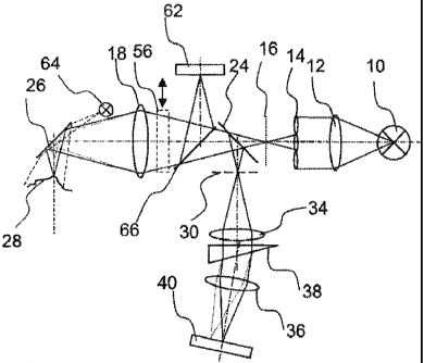
The measuring arrangement comprises among the essential elements one light
source 10
such as a halogen lamp, whose light is collimated by a lens 12. The collimated
light beam
reaches a micro-lens array 14, which images an illumination pattern 16 into
the focal plane
of an objective lens 18 with high chromatic aberration. The illumination
pattern generated
by the micro-lens array 14 can for example possess a size of 20 mm x 15 mm
with
approximately 1600 foci or for example a size of 5 mm x 6.5 mm with
approximately 2000
foci at a spacing of 250 1..tm. Figure 2 illustrates purely as an example the
corresponding
illumination pattern 14, in which as an example two foci carry the reference
labels 20, 22.
The illumination pattern 16 can be designed in such a manner that the
resulting diameter of
CA 02682297 2013-07-24
each of the foci 20, 22 will be approximately 25 1_1111 or approximately 12
jam.
To improve the illumination pattern, the micro-lens array 14 can be combined
with a
matched pinhole array, which is positioned in the object plane of the
objective lens 18. In
this, the holes of the pinhole array correspond with respect to geometry or
position to the
illumination pattern formed by the foci 20, 22.
In accordance with the illustration of figure 1, the light originating from
the light source 10
downstream of the illumination pattern 16 reaches a beam splitter 24, from
which the
transmitted fraction reaches the objective lens 18 with high chromatic
aberration.
In the illustration of the figure, the beam splitter 24 is shown as a plate
with a partially
reflecting layer. But other beam-splitting elements are alternatively
possible. Beam-splitter
cubes should be mentioned as an example. Also envisioned can be ring-shaped
mirrors or
smaller mirrors, whereby the outer or inner beam components, respectively, can
serve for
the detection or illumination to be explained in the following.
The beam passing through the objective lens 18 is imaged via a deflection
device 26 such
as a deflecting mirror onto an object 28 to be measured, such as a tooth. In
this, the distance
between the plane of the illumination pattern 16 and the object 28 is chosen
so that the foci
¨ after deflection by the deflecting device 26 ¨ are imaged onto the surface
of the object 28,
whereby depending on the distance of the object's surface from the objective
lens 18,
different colours, i.e. wavelengths, are imaged sharply in focus. In this, the
size of the
measuring area and the resolution are determined by the chosen image scale.
Part of the radiation, i.e. light, emitted by the object 28, falls back into
the objective lens 18
and after partial reflection at the beam splitter 24 falls onto a first matrix
of holes or pinhole
array 30, whose holes correspond ¨ with respect to the separation of holes and
the size and
overall geometric arrangement ¨ to the arrangement of the illumination pattern
16.
In other words, the axial and lateral position of the pinhole array 30, i.e.
its holes, is chosen
11
CA 02682297 2013-07-24
so that the foci on the surface of the object 28 are imaged confocally in the
holes of the
pinhole array 30. Consequently, each hole of the pinhole array 30 defines the
X and Y
coordinates of the respective focus imaged on the surface of the object 28.
Figure 5 illustrates a configuration of the pinhole array 30, whereby both the
positioning
and extent of the open circles 32 correspond to the pattern of the foci of the
illumination
pattern 16.
Because of the high chromatic aberration of the objective lens 18, in each
case only one
colour will be imaged sharply at the respective measuring point defined by the
position of
the foci of the illumination pattern 16 ¨ depending on its distance from the
object 18 - i.e.
only one wavelength satisfies the confocality condition. Correspondingly, an
intensity
maximum is observed at this wavelength in the spectrum of the light
transmitted through
the respective hole 32 of the pinhole array 30.
As the measuring points become more densely spaced and as the light emission
of the
object 28 increases, an increasing proportion of white light passes through
the hole or
pinhole in addition to the peak wavelength. In order to determine the peak
wavelength
characteristic of this focus to an adequate degree and with the required
accuracy despite this
obstacle, it is intended that behind the pinhole array 30 be arranged a
spectrometric
arrangement corresponding to the illumination pattern 16 and thus the matrix
of holes of the
pinhole array 30, which in the embodiment example consists of the optical
systems 34, 36
and a spectrum-dispersing element, e.g. a prism 38, arranged between them.
The pinhole array 30 is imaged onto a CCD chip sensor, which is used as
detector device
40, by the optical systems 34, 36, which can consist of one or several lenses.
The spectrum-
dispersing element, i.e. the prism 38, effects a lateral spectral spreading of
the maximum-
intensity wavelength region of the light of the focus that appears in the
holes with
maximum intensity, so that consequently every hole of the pinhole array 30 is
imaged onto
a line on the CCD chip sensor 40, i.e. the pixels arranged in an array,
whereby ¨ as in a line
12
CA 02682297 2013-07-24
spectrometer ¨ the position along this line corresponds to a particular
wavelength. In this,
the spectrum-dispersing unit ¨ consisting of the optical systems 34, 36 and
the prism 30 ¨
and the CCD sensor 40 are positioned relative to the pinhole array 30 in a
manner so that
the laterally spread spectral lines from consecutive holes of the pinhole
array 30, which
now form lines on the pixels, contact each other without or nearly without
empty spaces
and without any overlap taking place.
Figure 3 illustrates that all pixels between the measurement points are used
for the spectral
dispersion and thus for the determination of the peak position. The filled
circles 42
represent a measurement point and the arrow 46 extending towards the following
measurement point 44 represents the laterally dispersed spectral lines of the
focus imaged
in the hole of the pinhole array that corresponds to the measurement point 42.
If for example one chooses the above-specified illumination pattern with the
stated
dimensions and a CCD chip or camera chip with a size of 6.4 mm x 4.8 mm and
with 1
million pixels (pixel size 6.7 gm x 6.7 lim), then 186 pixels are available
for the spectral
dispersion per measuring point. Given a line width of 2 pixels which
corresponds to the
pinhole diameter, one obtains for each of the approximately 2000 measuring
points and
approximately 2000 background points a line spectrometer with 93 elements for
spectral
dispersion. After image acquisition, the evaluation of the image information
or
measurement data takes place either on the CCD sensor itself or on an external
unit. For
this, a suitable algorithm is used to determine for every measuring point the
spectral peak
position and from this the distance of each measuring point to the object 28.
In this manner,
one image yields the three-dimensional structure of the object 28 at the nodes
or measuring
points, whereby the resolution depends on the chosen focus distance and the
image scale of
the objective lens 18.
If the separation of nodes is greater than the desired resolution and/or if
the three-
dimensional structure can not be acquired from a viewpoint, the illumination
pattern 16 can
be shifted accordingly. If the measuring arrangement is a manually operated
device, then a
13
CA 02682297 2013-07-24
complete acquisition of the object 28 can be achieved through a continuous
moving of the
measuring arrangement, whereby the resulting individual images are fit
together to form a
complete image in a suitable manner.
Since, as mentioned before, in the case of an object 28 causing scatter, not
only the
wavelength of the sharply imaged focus is imaged in the holes 32 of the
pinhole array 30,
but also a significant amount of white light, it becomes necessary to employ
methods to
eliminate or reduce the background caused by this.
For the purpose of illustrating the importance of the white-light background
during
measuring and evaluation, figure 4 shows a typical measuring signal 48 with
the object 28
being a tooth. The higher the portion of white-light background in the
measuring signal 48
is, the more accurate must be the information on the spectral behaviour of the
background
52 in each measured point to be able to determine the position of the peak 50
that is
characteristic of the wavelength of the focus. For this purpose a method can
be employed
that is schematically illustrated in figure 5. In addition to the holes 32, in
which the foci are
imaged, the pinhole array 30 comprises additional holes 54 that are not
matched to the
illumination pattern 14. In accordance with the preferred arrangement of
figure 5, the holes
54 that do not correspond to the illumination pattern 14 are positioned
between the holes 32
that do correspond to the illumination pattern 14. The spectra in the holes
54, in which no
foci are imaged, thus approximately represent the background signal of the
neighbouring
holes 32, in which foci are imaged and which represent measuring points. In
this, one
alternatively can use the measuring signal of an individual neighbouring hole
54, which
basically contains only white light, or the average of several neighbouring
holes 54, to
determine the background 52. The holes 54 can be referred to as non-
illuminated whereas
the holes 32 can be referred to as illuminated holes or pinholes.
In accordance with the arrangement of the illuminated or first holes 32, in
which foci are
imaged, and the second holes 54 used for determining the background, which can
also be
referred to as non-illuminated holes, only half the number of pixels is
available per
14
CA 02682297 2013-07-24
measuring point for spectral dispersion, compared to the variant, in which the
number of
foci is identical to the number of pinholes or holes 32.
It is further possible to determine the white-light content by using a plane-
parallel plate 56,
which is arranged in the beam path between the first beam splitter 24 and the
beam
deflector 26, in particular between the objective lens 18 and the first beam
splitter 24. The
plane-parallel plate 56 in the optical path effects an axial shifting of the
foci, which in turn
effects a shift of the peak position in the measured signal. By sequentially
acquiring images
at one position with and without the plane-parallel plate 56 one obtains per
measuring point
two spectra with different peak positions but identical background.
Subtracting the two
spectra thus allows eliminating the background. A typical signal curve after
subtraction of
the two spectra is illustrated in figure 8. One recognizes the peaks 58, 60,
which were
determined by the subtraction and whose separation is predetermined by the
plane-parallel
plate 56.
In the further evaluation for determining the unknown object distance, one can
employ
several characteristic quantities, among others the two extrema, i.e. peaks
58, 60 and/or the
spectral position of the zero passage.
Live-image acquisition can be provided as a positioning aid and as an aid for
assigning the
individual images to a complete picture. For this purpose the embodiment
example
possesses an additional camera chip 62, onto which the object 28 is imaged. An
additional
light source 64 can be provided, which preferably illuminates the object 28
via a deflection
device 26. Instead of one light source 64 it is possible to have several light
sources. For
live-image acquisition the light source 64 should emit light in a spectral
region that is
outside of the wavelength range used for the actual measurements. This allows
carrying out
live imaging and measuring independently from each other.
For the purpose of beam splitting one can employ in the beam path between the
objective
lens 18 and the first beam splitter 24 a second beam splitter 66 such as a
dichroic filter,
CA 02682297 2013-07-24
which provides high transmission for the measuring signal and high reflection
for the live-
image signal.
As mentioned above, the objective lens 18 is also used to image the object 28
onto the
camera chip 62, whereby the axial position of the camera chip 62 is chosen so
that the live
image is in focus approximately in the centre of the measuring region.
The size and shape of the measuring arrangement or the measuring device is
particularly
important in the case of intraoral application for measurement of teeth. Thus,
in a
development of the invention, it is possible that only the objective lens 18
and the beam-
deflector 26 are arranged in an intraoral part of a hand-held device, which
can be inserted
into the mouth. The other components can be integrated in an extraoral part of
the hand-
held device or in a separate equipment unit. A compact light source offers the
option of
integration in a hand-held device.
Instead of the halogen lamp 10, one can also envision other light sources,
such as for
example one white-light LED or several LEDs of different colours with a
suitable
collimating optical system.
Alternatively the light source 10 can be integrated in an external unit and
the light is fed
into the hand-held unit via fibres, whereby the output end of the fibre-optic
light guide is
positioned at the focal point of the collimator lens 12, or the output ends of
several
corresponding fibre-optic light guides themselves represent the foci of the
illumination
pattern, instead of the foci of the micro-lens array.
For generating the multi-focal illumination pattern 16 it is also possible to
use ¨ instead of
or in addition to the micro-lens array 14 - a pinhole array that can be
arranged in the plane
of the illumination pattern 16 shown in the figures.
It is essential for an exact measuring process and uncomplicated handling of
the measuring
device that there exists a precise geometric and spatial correspondence
between the micro-
16
CA 02682297 2013-07-24
lens array 14 and the pinhole array 30. To translate this into practice, one
can choose a
monolithic design, which is schematically illustrated in figure 6. Also
integrated into the
monolithic embodiment, which can possess a cube-shaped geometry, is the first
beam
splitter 24.
If the object 28 is not measured or scanned by a single picture but rather by
a multitude of
pictures, i.e. using individual images, the images must have an unambiguous
relation with
respect to each other to facilitate an uncomplicated interpretation. For this
purpose it is
particularly intended that the individual pictures overlap in segments, which
constitute 50%
to 95% of each picture. As an alternative or supplement it is also possible to
use as an aid
for the superposition of the individual images the placing of fixed points on
the object 28.
As an alternative to the manual displacement of the measuring device, which is
preferably
embodied ¨ as mentioned above ¨ as a handheld device, actuators can be
integrated into the
measuring arrangement for the purpose of shifting the measuring points. In
this, the highest
possibly necessary displacement corresponds to the distance between measuring
points
minus the desired resolution, consequently 225 gm in the embodiment example
being
explained (250 gm hole separation - 25 gm resolution).
Figure 7 illustrates an alternative approach for obtaining individual images.
Plane-parallel
plates 70, 72 can be arranged in the measuring beam ¨ in the embodiment
example
upstream of the objective lens 18 -, whereby it is also possible to use a
single plate with two
rotational axes (see arrows 74, 76). In this, the axes of rotation preferably
run in the plane
defined by the plane-parallel plate 70, 72. A further possible variant is the
use of a movable
or rotatable deflection mirror such as the deflection device 26 as a variable
beam-deflection
system.
With respect to the live image it should be mentioned that alternatively the
use of an
additional light source is not necessary, in particular if the illumination
pattern is out of
focus in the spectral region used for the live-image acquisition.
17
CA 02682297 2013-07-24
Furthermore, it is also possible to employ a diffractive element to generate
the high
chromatic aberration, and/or a diffractive element or a grating as dispersing
element, and/or
CMOS detectors instead of CCD chips, and/or LCD modulators or DMDs to generate
the
illumination pattern instead of the micro-lens array.
18