Note: Descriptions are shown in the official language in which they were submitted.
CA 02682317 2014-10-31
ENRICHMENT OF TISSUE-DERIVED ADULT STEM CELLS BASED ON
RETAINED EXTRACELLULAR MATRIX MATERIAL
BACKGROUND
Essentially all developing and adult tissues contain one or more populations
of stem Cells and/or progenitor cells (progenitors) that play a role in the
continued
maintenance of health of the tissue through remodeling activity. Such stem
cell and
progenitor populations also contribute to new tissue formation in the event of
injury,
and represent an essential resource in tissue engineering strategies seeking
to repair,
augment, replace or regenerate tissues that may be lost due to injury,
disease, or
degenerative or aging processes.
For example, bone repair requires osteogenic connective tissue progenitors
(CTP-Os). In settings where the local population CTP-Os is sufficient, they
may be
effectively "targeted" using scaffolds or factors, such as bone morphogenic
proteins
to summon the CTP-Os to where they are needed. However, in settings where the
levels of local CTP-Os are suboptimal, optimizing the bone healing response
requires transplantation of CTP-Os from an alternative source. Many
preclinical
studies demonstrate improved graft performance when CTP-Os are added, even to
small graft sites in young healthy animals, supporting the premise that the
CTP-0
population is suboptimal in virtually all clinical settings and that optimal
performance from any osteoconductive or osteoinductive material may require
augmentation with local CTP-Os.
1
= CA 02682317 2009-09-29
WO 2008/124494
PCT/US2008/059256
As a result of the potential importance of progenitor cell populations in
maintaining or defining the current health of tissue, and as a resource for
cell
therapy strategies, methods for the harvest, isolation, assay,
characterization,
processing and transplantation of progenitor cells have exceptional value, and
are
expected to be the focus of many advances in health care diagnosis and
treatment
modalities.
Data available to date from many organ systems has demonstrated that the
concentration and prevalence of progenitor cells is generally very low, and
varies
widely from tissue to tissue and individual to individual. Investigators have
speculated that the concentration and prevalence of progenitors are a
reflection of
the current state of health of a tissue and may also predict the future health
of a
tissue or individual. As a result, they are likely to have important
implications in
diagnosis and prediction of disease as well as in the treatment of disease.
Adult stem cells present in native tissues tend to be distinctly different
from
the much more numerous population of mature cells in native tissue with
respect to
both morphological as well as chemical and biological properties. Each of
these has
been used in reported methods for progenitor cell isolation. Cell size, cell
density,
and granularity have been used as means of enrichment using density separation
and
countercurrent elutriation. Membrane bound surface markers in the form of
membrane bound protein antigens that are uniquely presented on selected stem
cell
and progenitor populations can be targeted using antibodies. For example, the
presence or absence of CD34, c-kit, Scal and other markers, alone or in
combination, have been used to isolate and fractionate hematopoietic stem
cells
from marrow and other tissues using fluorescent activated cell sorting (FACS),
magnetic separation or affinity columns. adult stem cells also tend to express
novel
markers and patterns of gene expression. Underlying gene expression, while
generally silent, can and has been converted through viral transfection
vectors into
fluorescent reporters that can be used as a basis for isolation. Finally, cell
function,
such as the presence of a selective ABC membrane pumps have been identified as
a
2
CA 02682317 2009-09-29
WO 2008/124494
PCT/US2008/059256
unique feature of several stem cell populations, and have been used to isolate
what
has been referred to as "side population" cells or SP cells, from marrow and
other
tissues.
Many markers have been proposed for positive selection of human
osteogenic connective tissue progenitors, such as STRO-1, STRO-1 with VCAM-1,
and CD antigens 9,10,13,18,29, 44, 49a,54,90,105,146 and 166. See Simmons et
al., Blood (1991) 78(1), p. 55-62. Alkaline phosphatase and osteocalcin are
also
markers of some circulating CTP-Os. However, most of these markers are also
present on other cell populations, limiting their usefulness for positive
selection of
CTP-Os. While positive markers have been elusive, CTP-Os may also be
differentiated from the vast majority of marrow cells based on markers that
they do
not express. For example, CTP-Os are negative/dim for CD45 and many other
hematopoietic markers. Hematopoietic markers therefore provide possible tools
for
CTP-0 enrichment by negative selection or depletion of non-osteogenic cells.
The most common method of isolation of stem cell and progenitor
populations exploits the biological capacity of these cells to proliferate,
and
particularly the capacity of adult stem cells to proliferate under some
conditions in a
manner that exponentially increases their number while at the same time
preserving
one or more desirable biological capacities (e.g. the ability to repopulate
bone
marrow in an animal that has been depleted of hematopoietic stem cells, or the
ability to form new bone tissue in vivo). This strategy of in vitro expansion
and
purification has been used to prepare populations of cells defined variably as
bone
marrow stromal cells (MSCs), mesenchymal stem cells (also "MSCs"),
mesenchymal progenitor cells (MPCs), multipotent adult progenitor cells
(MAPCs),
tissue regenerating cells (TRCs), muscle-derived progenitor cells (MDPCs),
adipose-derived stem cells (ADSC), and others.
It has long been recognized that while culture-expanded stem cell
populations can be prepared that retain desirable biological capabilities,
these
3
CA 02682317 2009-09-29
WO 2008/124494
PCT/US2008/059256
populations differ significantly from the population of adult stem cells that
are
present in native tissue from which they are derived. Differences may be
expressed
in cell size, cell cycle state, expression of markers, and gene expression, as
well as
intrinsic biological behavior such as responses to growth factors.
Furthermore, the
use of culture-expanded cells is associated with the need for delay between
the
harvest of founding cell population and the ultimate use of the resulting
expanded
cell population. This delay adds significantly to the cost and also to the
inconvenience of using culture-expanded cells, because the patient must be
exposed
to separate procedures; first to collect founding cells, and second to implant
cells
after in vitro expansion. In addition, in vitro expansion adds the potentially
significant risks of bacterial or viral contamination of cells while in vitro,
in vitro
selection of cells with undetected undesirable biological properties (e.g.
tumor
= forming cells) and even contamination with other cells or mislabeling
with respect
to the donor of origin.
The rapid isolation and processing of adult stem cells isolated from tissues
of
an individual at the time of a single therapeutic procedure has great
potential value,
and avoids many of the drawbacks of culture expanded cell populations cited
above.
However, rapid processing of freshly isolated cells has itself a number of
drawbacks. First, adult stem cells are typically very few in number. The
prevalence
of progenitor cells (tissue forming cells) within a given tissue can be as
high as one
in 100 cells, but also as low as one in 1,000,000 cells (or less). Second,
stem cells
and progenitor populations in native tissues are generally very heterogeneous,
in
contrast to the relatively homogenous culture-expanded stem cell and
progenitor cell
populations. No one feature or combination of features can define all adult
stem
cells in a given tissue. In fact, one must expect that a given tissue will
provide a
= diverse population of adult stem cells that represent cells from multiple
stem cell
niches within the tissue, each representing a different compartment or niche
for the
= tissue forming cell populations within that tissue. See Muschler et al.,
J. Biomed.
Biotechnol. (2003); 2003(3), p. 170-193.
4
CA 02682317 2009-09-29
WO 2008/124494
PCT/US2008/059256
Rapid processing has two important advantages, however. First, processing
strategies can be designed to take advantage of characteristics of freshly
isolated
cells that may not be preserved when cells are expanded in vitro. Second, due
to the
high potential that adult stem cells have for proliferation, transplantation
of a
relatively small number of adult stem cells into a wound in an environment in
which
cells are likely to survive can result in important and clinically significant
improvement in biological outcome. In fact, removal of competing and non-
tissue
forming cells may be just as important, if not more important, to the
performance of
transplanted progenitor cells as transplanting them in large numbers. For
example,
several recent reports have shown that as little as a 3-4 fold increase in the
concentration of osteogenic connective tissue progenitor cells can result in
significant improvement in bone formation and in union rate in settings of
spinal
fusion and in settings of bone grafting in long bone defects. See U.S. Patent
Nos.
6,049,026 and 6,723,131, issued to Muschler. Removal of competing cells may
eliminate a source of growth factor or signaling molecules that are
maladaptive to
proliferation and new tissue formation, such as inflammatory cytokines that
may
stimulate apoptosis (cell death). Removal of competing non-tissue forming
cells
may also dramatically improve the likelihood that transplanted progenitor
cells will
survive following transplantation, by reducing local consumption of oxygen and
other nutrients. See Muschler et al., J. Bone Joint Surg. Am. (2004) Jul; 86-
A(7), p.
1541-58.
Bone and marrow tissue, including bone marrow harvested using the
minimally invasive method of aspiration contains a heterogeneous population of
cells, including adult stem cells capable of regenerating connective tissues,
blood
cells, blood vessels, bone, cartilage, fat, marrow stroma, muscle, tendons,
ligaments
and other fibrous tissue. These populations include multipotent cells which
are
individually capable of giving rise to progeny along all three germ lines
(i.e.
ectoderm, endoderm and mesoderm), pleuripotent progenitors capable of giving
rise
to progeny that may contribute to multiple mature cell types (e.g. bone,
cartilage,
fat), and mono- or uni-potent progenitors that are committed to progeny of
only one
5
CA 02682317 2009-09-29
WO 2008/124494
PCT/US2008/059256
lineage. These diverse and versatile cell sets are used extensively in
research
settings as well as clinically in bone grafting and tissue engineering
endeavors.
Bone marrow aspirations offer many advantages as a cell source. In particular,
they
result in very low morbidity to the patient and provide cells in single cell
suspension
that can be manipulated and processed using only an anticoagulant, without the
need
for enzymatic digestion that may modify the cell surface.
One method to increase the concentration of bone forming progenitors is
density separation, which is available through use or modification of devices
designed for clinical preparation of platelet rich plasma. See Hernigou et
al., J.
Bone Joint Surg. Am. (2005) Jul; 87(7), p. 1430-1437. Density separation can
increase the CTP concentration 4-8 fold, but is relatively non-selective, and
does not
change osteogenic CTP prevalence.
Focusing on bone forming progenitors in bone marrow, and using the "gold
standard" method for assay of progenitor populations (i.e., the colony forming
unit
(CFU) assay), investigators have found wide variation between individuals and
between individual aspirate samples, but a mean prevalence of osteogenic CTPs
(CTP-Os) of approximately one in every 20,000 cells. Utilizing this CFU assay,
the
unique property of many CTP populations (e.g. CTP-Os) to preferentially and
rapidly adhere to selected surfaces has been investigated, particularly with
regard to
surfaces that can be created or utilized in a porous implantable matrix or
scaffold.
This investigation resulted in the recognition of the process of selective
retention,
which has been used to develop the CellectTM graft preparation device, now
manufactured and marketed by DePuy Spine Inc.
Selective Retention (SR) involves passing a cell suspension through a porous
matrix and uses the intrinsic attachment behavior to retain CTP-Os in the
matrix,
= while non-adherent cells pass through the matrix in the effluent
solution. SR has
been used to enrich CTP-Os as much as 16 fold. See Muschler et al., Clin.
Orthop.
Rel. Res. (2003) 407, p. 102-118. Current scaffolds used for SR (e.g., bone
matrix,
6
CA 02682317 2009-09-29
WO 2008/124494
PCT/US2008/059256
TCP ceramic) will retain 80-90% of CTP-Os and only 20-30% of other nucleated
cells, resulting in a 3-4 fold increase in the CTP concentration and also a 2-
3 fold
increase in CTP prevalence by removing 70-80% of potentially competing cells.
Thus, while effective, the principal limitation of selective retention is the
fact that
many of the vastly more abundant non-progenitor cells also bind to the same
surfaces, and although they are less adherent, they occupy a much larger
fraction of
available surface. SR processing has the advantage of requiring relatively
simple
instrumentation and a minimum of reagents, but is far from being optimized.
Even
in retained "CTP enriched" populations, CTP-Os represent only a small fraction
of
the retained cells (mean ¨ 0.05 %).
Accordingly, there remains a need for a more selective marker and/or
additional separation techniques that can be used to purify or enrich adult
stem cells
from the animal tissues in which they are found.
SUMMARY OF THE INVENTION
One aspect of the invention provides a method for enriching adult stem cells
that includes the steps of obtaining a population of cells including one or
more adult
stem cells from animal tissue; contacting the population of cells with a
recognition
ligand specific for an ECM component retained to the surfaces of adult stem
cells in
said population; and using a separation method to remove cells from said
population
that are not bound to the recognition ligand, thereby enriching adult stem
cells that
are bound to the recognition ligand via said retained ECM component.
In one embodiment of this aspect of the invention, the animal tissue is
connective tissue. In additional embodiments, the separation method can
include
magnetic separation, selective retention using a porous matrix, and/or an
affinity
column method. In another embodiment, the retained ECM component is
hyaluronan. For this embodiment, additional embodiments can provide that the
recognition ligand is hyaluronan binding protein.
7
CA 02682317 2009-09-29
WO 2008/124494
PCT/US2008/059256
In further embodiments of this aspect of the invention, the adult stem cells
can include connective tissue progenitor cells. In another embodiment, the
method
increases the prevalence of adult stem cells by at least about 2-fold. In a
further
embodiment, the method includes delivering the adult stem cells to a tissue in
a
subject that is in need of repair. In embodiments involving tissue repair,
further
embodiments may include adult stem cells that are enriched and delivered to
the
tissue in the subject intraoperatively, and/or the tissue being bone tissue.
In another aspect, the present invention provides a method for detecting
adult stem cells in a cell population that includes the steps of contacting
the cell
population with a recognition ligand specific for an ECM material component
that is
retained to the surfaces of adult stem cells; and detecting adult stem cells
in the cell
population by identifying one or more cells having the recognition ligand
associated
therewith or bound thereto.
In one embodiment of this aspect of the invention, the recognition ligand is a
labeled recognition ligand. In another embodiment, the labeled recognition
ligand is
a labeled antibody. In further embodiments, the adult stem cells are detected
in vivo
or ex vivo. In yet further embodiments, the adult stem cells are detected
using an
immunoassay or flow cytometry.
In other embodiments of the method for detecting adult stem cells, the adult
stem cells are detected in connective tissue. In another embodiment, the adult
stem
cells are connective tissue progenitor cells. In other embodiments, the ECM
component is hyaluronan, and/or the recognition ligand is a hyaluronan binding
protein. In yet another embodiment, the one or more cells having the
recognition
ligand associated therewith or bound thereto are further characterized to
determine
if they have other properties of adult stem cells.
Another aspect of the present invention provides a method of identifying an
ECM component marker associated with a particular cell type that includes the
steps
8
CA 02682317 2009-09-29
WO 2008/124494
PCT/US2008/059256
of obtaining a population of cells including one or more cells of the desired
type
from animal tissue; contacting the population of cells with a recognition
ligand
specific for a particular ECM component; enriching the cells that have the
recognition ligand associated therewith or bound thereto; and determining if
the
enriched cells have the properties of the desired cell type.
In one embodiment of this aspect of the invention, the cells of the desired
type are adult stem cells. In another embodiment, the adult stem cells include
connective tissue progenitor cells. In yet another embodiment, the properties
include proliferation that is different from that of non-adult stem cells.
Yet another aspect of the invention provides a method for enriching adult
stem cells that includes the steps of obtaining a population of cells
including one or
more adult stem cells from animal tissue; contacting the population of cells
with a
recognition ligand specific for an EMC component or antigen that is present to
a
lesser degree on adult stem cells than on other cells in the population; and
using a separation method to remove cells that are bound to a recognition
ligand
from the adult stem cells that are not bound to the recognition ligand.
In one embodiment of this aspect of the invention, the ECM component or
antigen is absent from adult stem cells in the population. In other
embodiments, the
separation method can include magnetic separation, selective retention using a
porous matrix, and/or affinity column methods. In another embodiment, the
adult
stem cells include connective tissue progenitor cells. In a further
embodiment, the
method enriches the prevalence of adult stem cells in said population by at
least
about 2-fold.
Unless otherwise specified, "a," "an," "the," and "at least one" are used
interchangeably and mean one or more than one. Also herein, the recitations of
numerical ranges by endpoints include all numbers subsumed within that range
(e.g., 1 to 5 includes 1, 1.5, 2, 2.75, 3, 3.80, 4, 5, etc.).
9
CA 02682317 2009-09-29
WO 2008/124494
PCT/US2008/059256
The above summary of the present invention is not intended to describe each
disclosed embodiment or every implementation of the present invention. The
description that follows more particularly exemplifies illustrative
embodiments. In
several places throughout the application, guidance is provided through lists
of
examples, which examples can be used in various combinations. In each
instance,
the recited list serves only as a representative group and should not be
interpreted as
an exclusive list.
BRIEF DESCRIPTION OF THE FIGURES
Figure 1 provides a schematic illustration of an adult stem cell that has been
bound by a recognition ligand and has been further bound to a magnetic
particle in
preparation for separation by magnetic separation.
DETAILED DESCRIPTION OF EMBODIMENTS OF THE INVENTION
The invention described herein identifies a new method that can be used to
select, concentrate, enrich, purify, deplete or fractionate populations of
adult stem
cells based upon using unique components of the extracellular matrix (ECM)
retained on their surface as one or more discriminating markers. The invention
is
based in part on the discovery that different stem cell populations can be
located in
tissues within an environment or niche that is characterized by a specific
relationship between the cell and its neighboring cells or extracellular
matrix, and
that this relationship and either cell-cell or cell-matrix interactions may be
instrumental in maintaining the size and biological state and potential of
local adult
stem cells (tissue forming cells). The present invention thus provides a
method for
the rapid enrichment or purification of adult stem cells by targeting ECM
material
retained by those cells.
10
CA 02682317 2009-09-29
WO 2008/124494
PCT/US2008/059256
Accordingly, one aspect of the present invention provides a method for
enriching or purifying adult stem cells. First, a population of cells
including one or
more adult stem cells is obtained from animal tissue. Next, the population of
cells is
contacted with a recognition ligand specific for ECM components retained by an
adult stem cell. Once the recognition ligand has bound to the adult stem
cells, a
separation method is used to remove cells that are not bound to a recognition
ligand
from the adult stem cells bound to the recognition ligand.
Enrichment and/or purification, as used herein, involves increasing the
prevalence of adult stem cells in a cell population as a result of selecting
adult stem
cells and/or depleting non-stem cells from a cell population, and does not
require
that the absolute number of adult stem cells in the population be increased.
Furthermore, enrichment and/or purification, as used herein, refers to an
increase in
the prevalence of adult stem cells in a sample, but is not meant to imply that
all
other cells and/or other materials are excluded from the sample (i.e., a 100%
purification). Rather, enrichment and/or purification represents various
levels of an
increased prevalence of adult stem cells, as further described herein.
Concentration, as used herein, refers to the number of cells in a given
volume of sample. An increased concentration of adult stem cells in a cell
population thus refers to a higher number of cells relative to the total
volume.
While the techniques used to enrich and/or purify the adult stem cells may
result in
a change in concentration, the concentration can be readily modified upwards
or
downwards by changing the volume of the sample.
Adult stem cells, as defined herein, are relatively undifferentiated cells
found
throughout the body after embryonic development that have the capacity to
proliferate and generate progeny that are capable of differentiating to
contribute to
the formation of new tissues. This definition includes not only the most
primitive
undifferentiated cells in adult tissues, but also the progeny or "progenitor
cells"
resident in new tissue that are themselves derived from primitive stem cells
but are
11
CA 02682317 2009-09-29
WO 2008/124494
PCT/US2008/059256
capable of proliferation. While referred to herein as adult stem cells, it is
to be
understood that both stem cells and progenitor cells capable of generating
progeny
that contribute to new tissue formation can be obtained from subjects having a
variety of ages, and not just adults. Adult stem cells and progenitor cells
include
cells derived from a variety of different tissues. For example, adult stem
cells
include connective tissue progenitor cells, adipose-derived adult stem cells,
hematopoietic stem cells, mammary stem cells, mesenchymal stem cells,
endothelial
stem cells, neural stem cells, olfactory adult stem cells, and spermatogonial
progenitor cells. Adult stem cells also include stem cells that have varying
degrees
of potential to differentiate into different tissues. For example, adult stem
cells
include pluripotent stem cells that can differentiate into cells derived from
any of
the three germ layers, multipotent stem cells that produce cells of a closely
related
family of cells (e.g., hematopoietic stem cells), and unipotent cells that can
product
only a single cell type but have the ability to self-renew.
The method for enriching or purifying adult stem cells includes obtaining a
sample comprising a population of cells including one or more adult stem cells
from
animal tissue. The population of cells represents the initial collection of a
variety of
different types of cells found at a tissue site, of which only a small
fraction are
generally adult stem cells. However, in order for the population to be
expected to
include one or more adult stem cells, it is preferred that the population of
cells
obtained should have a size of 100 or more cells, depending on the local
density of
adult stem cells, with much higher populations (e.g. over one million cells)
being
preferred for a typical cell population.
The population of cells may be obtained by aspirating the animal tissue by
various methods known to those skilled in the art. For example, a needle and
syringe may be used to penetrate the tissue and then apply negative pressure
to
withdraw the desired cells. The amount of negative pressure applied should be
sufficient to withdraw the desired cells from the surrounding tissue, and do
so with
sufficient force to obtain one or more adult stem cells that retain
extracellular matrix
12
CA 02682317 2009-09-29
WO 2008/124494
PCT/US2008/059256
material. The size of the needle will vary depending on the type of animal
tissue
involved. For example, when the population of cells is obtained from blood, a
needle with a diameter from about 22 gauge to about 14 gauge may be used. For
bone marrow, a large needle with a diameter from about 1 mm to about 6 mm may
be used. For adipose tissue, an even larger need with a diameter from about 3
mm
to about 12 mm may be used.
The populations of cells typically obtained from a sample of bone marrow
aspirate includes nucleated progenitor cells, nucleated hematopoietic cells,
endothelial cells, and cells derived from peripheral blood, including red
cells and
platelets. Note, however, that there are several other cell types present in
an
aspirate, including stromal cells, pericytes, and reticulocytes. Because a
bone
marrow aspirate contains peripheral blood, it is preferred that the aspirate
be
collected in a syringe containing an anticoagulant. Suitable anticoagulants
include
heparin, sodium citrate, and EDTA. Preferably, a bone marrow aspirate for use
in a
method of the present invention is obtained from the patient who will receive
the
graft (the graftee). Less preferably, the bone marrow aspirate can be obtained
from
another immunologically compatible donor.
The population of cells can be obtained from a variety of different animal
tissues, depending on the type of adult stem cells being sought. Animal
tissue, as
used herein, is tissue obtained from animals including, for example, humans
and
domesticated animals such as farm animals and pets. Animal tissues include a
variety of tissues such as epithelial tissue, connective tissues, muscle
tissue, and
nerve tissue. These categories include a variety of more specialized forms of
tissue.
For example, connective tissue includes blood vessels, lymphatic tissue,
cartilage,
bone, marrow stroma, tendon, and adipose tissue. Animal tissue may be obtained
from the desired tissue site using a variety of methods known to those skilled
in the
art, such as biopsy or aspiration. Animal tissue may contain a variety of
different
types of adult stem cells. For example, bone marrow contains multiple subsets
of
adult stem cells that are capable of contributing to new tissue formation:
13
CA 02682317 2009-09-29
WO 2008/124494
PCT/US2008/059256
hematopoietic (blood) progenitors, vascular progenitors, and bone, fat,
muscle,
fibrous tissues (tendon, ligament, scar), cartilage, and marrow stroma.
Adult stem cells within the heterogeneous population of cells can present a
potentially discriminating array of ECM material retained to their surface,
reflecting
the unique niche where they are naturally present. These ECM materials may be
retained to stem cell surfaces via physical interactions that do not exclude,
or
necessarily include, chemical bonding. In order to utilize this retained ECM
material, the population of cells obtained from animal tissue is contacted
with a
recognition ligand specific for extracellular matrix (ECM) material that is
retained,
or that is retained to a greater degree relative to other cells, by an adult
stem cell. In
order to bring the recognition ligands into contact with the population of
cells, the
recognition ligands can be placed into the solution or a sample containing the
population of cells. The recognition ligand specific for ECM retained by an
adult
stem cell selectively binds to the unique ECM components that have been
retained
on the adult stem cell. That is, in a given sample of animal tissue, it has
been
discovered that adult stem cells may tend to be concentrated in a relatively
higher
prevalence in specific regions of the extracellular matrix (ECM) for that
tissue, as
compared to the tissue and its ECM as a whole. It has also been discovered
that
certain components of the ECM also may be more highly concentrated in these
areas where stem cell prevalence is high, compared to other components of the
ECM for the entire tissue sample or tissue type. As a result, ECM components
that
are more highly concentrated in these specific regions have been found to be
more
likely retained, or retained to a greater degree, to the surfaces of adult
stem cells that
are derived from a sample of the tissue, than to other cells or cell types
that make up
the specific tissue type or sample.
As used herein, the phrase "selectively binds" and other permutations of that
phrase refer to a recognition ligand (e.g., an antibody or binding protein)
that will,
under appropriate (e.g., physiological) conditions, interact with a cell
surface or cell
associated component (e.g., an antigen present on ECM material) preferentially
or to
14
CA 02682317 2009-09-29
WO 2008/124494
PCT/US2008/059256
a greater degree compared to a different or structurally unrelated cell
surface
component or cell associated component. Recognition ligands include antibodies
and other types of proteins, peptides, carbohydrates, lipids, macromolecules,
small
organic molecules, and the like that selectively bind to the desired target
(e.g., ECM
retained by an adult stem cell).
As noted above, the inventors discovered that when cells from native tissue
are isolated they may retain on their surface not only membrane bound
molecules,
which have been the focus of cell isolation and characterization procedures to
date,
but they may also retain on their surface molecules that are derived from the
extracellular matrix niche that they occupied when in vivo. Stem cell
populations
are established in specific anatomic locations referred to as niches that save
stem
cells from depletion while protecting the host from excessive stem-cell
proliferation.
These niches provide a wide variety of inputs to the stem cells to control
stem cell
activity, such as paracrine signaling, humoral input, neural input, metabolic
cues,
cell to cell interactions, and extracellular matrix interactions. See Scadden,
Nature
(2006), Vol. 441, 29 June, p. 1075-1079. In particular with regard to the
present
invention, the unique extracellular matrix material present in adult stem cell
niches
may be retained by adult stem cell and used to aid in their detection and
purification.
For example, ECM molecules can be detected on the surface of the freshly
isolated
bone marrow-derived cells.
The extracellular matrix is the extracellular part of animal tissue that
provides structural support and various other benefits to cells in animal
tissue. As
used herein, ECM retained by adult stem cells refers to ECM material or
component(s) that are associated with the surface of the adult stem cells. In
particular, the retained ECM is extracellular matrix material that remains
associated
with the adult stem cells when they have been removed from tissue by
techniques
such as aspiration, as described herein. The ECM material is retained in
association
with the adult stem cells by the same binding processes which serve to
associate
ECM material with cells in vivo, such as binding by integrin. For example,
CA 02682317 2009-09-29
WO 2008/124494
PCT/US2008/059256
hyaluronan is retained on the surface of adult stem cells by a variety of
hyaluronan
binding proteins and receptors (e.g., CD44) referred to as hyaladherins. The
mechanisms by which cells adhere to the ECM are well known to those skilled in
the art. For further information regarding hyaladherins, see Day et al., J.
Biol.
Chem. (2002) Feb 15; 277(7), p. 4585-8.
The extracellular matrix includes proteoglycan matrix components and non-
proteoglycan matrix components. Proteoglycan matrix components include, for
example, heparin sulfate proteoglycans, chondroitin sulfate proteoglycans, and
keratan sulfate proteoglycans. The non-proteoglycan matrix components include
hyaluronan, collagen, fibronectin, laminin, vitronectin, and elastin. Some ECM
components are relatively ubiquitous molecules that may not be useful for
isolating
the adult stem cells. However, some of the ECM components retained on the
surface of newly obtained cells can be discriminating and valuable in
selection of
adult stem cells, or, in the alternative, depleting a population of non-stem
cells. The
ECM material that are useful to enrich or purify adult stem cells found in one
type
of tissue can be different from the ECM material useful for enriching or
purify adult
stem cells in another tissue. For example, while hyaluronan is useful for
identifying
adult stem cells present in bone marrow, other ECM material may be useful for
identifying adult stem cells present in other tissues such as adipose tissue.
Preferably, the ECM component used for ligand targeting is retained on the
surface of adult stem cells at much higher levels than compared to the other
cell
types in the tissue of interest. For example, it may be found at levels at
least twice
as high, and more preferably at levels at least five times as high on adult
stem cells
in comparison to non-adult stem cells. In further embodiments, the retained
ECM
component is exclusively retained on adult stem cells in a specific tissue
type upon
extraction or aspiration.
In one embodiment, the recognition ligand is an antibody that selectively
binds a unique component of the ECM material that is selectively retained to
the
16
CA 02682317 2009-09-29
WO 2008/124494
PCT/US2008/059256
adult stem cells. For example, the antibody may selectively bind to
hyaluronan.
Hyaluronan (HA) is a large molecular weight polysaccharide molecule that is
present in many adult tissues, particularly in the dermis, in the vitreous of
the eye,
and in articular cartilage. HA also makes of the zona pelucida around the
human
oocyte, through which the sperm must penetrate to come in contact with the
egg.
HA is a repeating linear polymer comprised of D-glucuronic acid and D-N-
acetylglucosamine, linked together via alternating 0-1,4 and 0-1,3 glycosidic
bonds.
Other than the three exceptions in adult tissues mentioned above (cartilage,
dermis, and eye), HA is a relatively minor component of the extracellular
matrix of
most tissues. However, HA is markedly upregulated and a prominent feature in
inflammation and the response of tissues to injury, and the regenerative
environment
that may follow these insults. The present inventors have demonstrated that HA
is
also relatively abundant in the perivascular space around small blood vessels
in the
marrow space, and is also present in the pericellular region around a small
fraction
of fibroblastic or stromal-like cells that are scattered with in the bone
marrow space.
See Midura et al., J. Biol. Chem. (2003) Dec 19; 278(51), p. 51462-8.
Based on these observations, the inventors hypothesized that hyaluronan
may be an important extracellular-matrix component that may relate to the
function
of adult stem cells. Moreover, even if HA was not related to their function
per se, it
was at least hypothesized that HA is more heavily concentrated in niches where
stem-cell prevalence is high, which might suggest a novel method to detect and
isolate them. If HA concentration is relatively higher, compared to other ECM
components, in stem cell niches, HA could be more likely to be retained, or to
be
retained in higher concentrations, by extracted cells. This, in turn, could be
used to
purify stem cells from other cell types.
Experiments to test these hypotheses and also test the hypothesis that cells
that retain HA on their surface would either be more or less likely to exhibit
colony
forming activity (which is indicative of adult stem cells) were carried out.
Briefly,
17
CA 02682317 2009-09-29
WO 2008/124494
PCT/US2008/059256
biotinylated Gl-link protein (a hyaluronan binding protein) was used to label
cells
containing HA on their surface soon after isolation and to link these cells
with
magnetic beads to enable separation in a magnetic field. Further details are
provided in Example 1 herein. These experiments demonstrated not only that
bone
marrow derived cells can be rapidly separated into separate cell populations
that do
and do not present HA retained on their surface, but more importantly that
cells that
present HA on their surface are significantly enriched with respect to the
prevalence
of colony forming units that express an osteoblastic phenotype under
osteogenic
conditions in vitro, both of which indicate that the enriched cells are adult
stem
cells.
In some embodiments, the recognition ligand is an antibody. The term
antibodies, as used herein, includes vertebrate antibodies, hybrid antibodies,
chimeric antibodies, humanized antibodies, altered antibodies, univalent
antibodies,
monoclonal and polyclonal antibodies, Fab proteins and single domain
antibodies.
Preferred types of antibodies used as recognition ligands for the present
invention
include monoclonal and polyclonal antibodies. These types of antibodies are
generally prepared by differing procedures.
If polyclonal antibodies are desired, a selected animal (e.g., mouse, rabbit,
goat, horse or bird, such as chicken) is immunized with the desired
extracellular
matrix material. Serum from the immunized animal is collected and treated
according to known procedures. Serum containing polyclonal antibodies to an
extracellular matrix material can be purified by using an affinity column
method.
Techniques for producing and processing polyclonal antisera are known in the
art
(see for example, Mayer and Walker eds. Immunochemical Methods in Cell and
Molecular Biology (Academic Press, London) (1987), Coligan, et al., Unit 9,
Current Protocols in Immunology, Wiley Interscience (1991), Green et al.,
Production of Polyclonal Antisera, in Immunochemical Protocols (Manson, ed.),
pages 1-5 (Humana Press 1992); Coligan et al., Production of Polyclonal
Antisera in
18
CA 02682317 2009-09-29
WO 2008/124494 S
PCT/US2008/059256
Rabbits, Rats, Mice and Hamsters, in Current Protocols in Immunology, section
2.4.1 (1992)).
Monoclonal antibodies directed against an extracellular matrix material are
readily produced by one skilled in the art. The general methodology for making
monoclonal antibodies by hybridomas is well known. Immortal antibody-producing
cell lines can be created by cell fusion, and also by other techniques such as
direct
transformation of B lymphocyte cells with oncogenic DNA, or transfection with
Epstein-Barr virus (See Monoclonal Antibody Production. Committee on Methods
of Producing Monoclonal Antibodies, Institute for Laboratory Animal Research,
National Research Council; The National Academies Press; (1999), Kohler &
Milstein, Nature, 256:495 (1975); Coligan et al., sections 2.5.1-2.6.7; and
Harlow et
al., Antibodies: A Laboratory Manual, page 726 (Cold Spring Harbor Pub.
1988)).
Panels of monoclonal antibodies produced against extracellular matrix material
can
be screened for various properties such as epitope affinity.
In other embodiments, the recognition ligand is something other than an
antibody. For example, the recognition ligand can be a binding protein. More
specifically, a hyaluronan binding protein can serve as a suitable non-
antibody
recognition ligand. There are a variety of proteins that can be referred to as
hyaluronan binding proteins. For example, crosslinked GI-Link protein is a
synthetic hyaluronan binding protein prepared from aggrecan components that
binds
to hyaluronan with high affinity.
After the population of cells has been contacted with a recognition ligand
specific for ECM retained by an adult stem cell, a separation method is used
to
remove or diminish cells from the sample that are not bound to the recognition
ligand, which will leave behind an enriched (i.e., more prevalent) population
of the
adult stem cells, which are bound to the recognition ligand. Removing cells
that are
not bound to a recognition ligand can have several effects on the population
of cells.
For example, it may represent a method of enriching the percentage of adult
stem
19
CA 02682317 2009-09-29
WO 2008/124494
PCT/US2008/059256
cells bound to a recognition ligand that are left after separation. It may
also
represent a method for increasing the percentage of adult stem cells bound to
a
recognition ligand from the entire, whole population of cells in the tissue-
derived
sample. In addition, it may also represent a method for enriching or purifying
adult
stem cells from a population of cells based on their binding to a suitable
recognition
ligand via the retained ECM component. Finally, for some applications, it can
represent a method for depletion of an undesired population of cells, by
focusing on
the removed cells that are not bound to a recognition ligand.
Separation of cells bound to the recognition ligand from cells that are not
bound to the recognition ligand can be carried out using a wide variety of
techniques. Examples of separation methods that can be used include magnetic
separation, fluorescence activated cell sorting (PACS), density separation,
affinity
column methods, or selective retention using a porous matrix. Multiple
separation
methods can also be combined to achieve higher levels of cell enrichment or
purification. Embodiments of the invention may provide a 2-fold, a 4-fold, or
a 10-
fold enrichment of the population of adult stem cells. Additional embodiments
may
provide a 50-fold or a 100-fold enrichment of adult stem cells.
In one embodiment of the invention, the separation method includes the use
of magnetic separation. Magnetic Separation (MS), which is sometimes referred
to
by the trade name MACS , uses selective cell surface markers to magnetically
label
cells (e.g., microbeads) and separate the labeled and unlabeled cells in a
magnetic
field. Generally an antibody to a surface antigen is linked by a secondary
antibody
to a bead. A cell may be labeled with no beads or multiple beads, in
proportion to
antigen density, and is accelerated in the magnetic field in proportion to the
number
of bound beads. Clinical MS systems (e.g., Dynal MPCTM (Invitrogen), MACSTM
columns (Milthenyi Biotec, Bergisch Gladbach), BD IMagTM (BD Biosciences)
and EasySepTM (StemCell Technologies)) have been used to enrich CD34+
hematopoietic progenitors 10-100 fold. See Lang et al., Blood (2003) 101(4),
p.
CA 02682317 2014-10-31
1630-6. Automation (e.g.,CliniMACSTm) has allowed the use of MS separation in
smaller clinical centers.
Two different strategies are currently available for use in MS; Capture-
Release (CR) and Continuous Magnetophoresis (CM). CR generally involves
placing a container with labeled cells into a magnet (e.g., EasySepTm).
Labeled cells
are retained against the container wall while non-magnetic cells are removed.
In
continuous magnetophoresis (CM), on the other hand, labeled cells are passed
through a laminar flow system where they are continuously separated by pulling
them from an inner to an outer stream path. By eliminating the need for
surface
retention, CM is particularly well suited for high throughput processing, even
using
weakly magnetic, colloidal or molecular reagents (e.g., nano-beads). An
isodynamic field is used, providing a constant radial force, but not a
constant field.
Sorting kinetics are predictable, based on magnetophoretic cell mobility,
field and
gradient, channel geometry, flow rate, and labeling reagents. For additional
details,
see Zborowski et al., Separation Science and Technology (2002) 37, p. 3611-33
and
Moore et al., Anal. Chem. (2004) 76(14), p. 3899-907.
The isolation of adult stem cells including retained hyaluronan using
magnetic beads has an additional desirable feature, in that a simple digestion
step
with Streptomyces hyaluronidase, an endo eliminase that is specific for
hyaluronan,
can be used to remove the magnetic bead/link protein complex, further reducing
the
possibility of an adverse reaction associated with the bead or an immune
response to
the bovine hyaluronate binding protein.
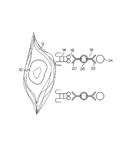
Figure 1 provides a schematic illustration of an adult stem cell that has been
bound by a recognition ligand via a surface-retained ECM component, and has
been
further bound to a magnetic particle in preparation for separation by magnetic
separation. The figure shows an adult stem cell 10 that is surrounded by
retained
ECM 12 (e.g., hyaluronan). A recognition ligand 14 is bound to the retained
ECM
12. The recognition ligand 14 includes a binding antigen 16 (e.g., biotin)
that can
21
CA 02682317 2014-10-31
be recognized by double-sided antibody 18. The double-sided antibody 18
includes
an antigen binding site 20 with affinity for the binding antigen 16, a
particle binding
site 22 with affinity for a magnetic nanobead 24, and a linker molecule 26
that
connects two antibodies to provide the double-sided antibody 18. Note that
while
the figure shows a double-sided antibody 18 that includes two typical
antibodies,
other binding proteins (e.g., streptavidin) can be used to replace one or both
of the
antibodies used in the double-sided antibody 18.
Separation of cells bound to the recognition ligand from cells that are not
bound to the recognition ligand can also be carried out using affinity column
method.. To separate cells bound to the recognition ligand using affinity
column
method, the recognition ligand for ECM component can be bound to the a column
material and a population of cells including adult stem cells retaining ECM
material
can be run through the column which provides a surface on which they will be
preferentially retained by adherence to the recognition ligand. Typically the
recognition ligand is covalently bound to the surface of the column material.
Various types of column material can be used to provide the surface upon which
the
recognition ligand is provided within the affinity column (e.g. glass,
sepharoseTM or
polymeric beads, fibers, or porous matrix). For example, if hyaluronan was
being
used as the target ECM material, a hyaluronan binding protein could be
covalently
bound to glass beads to provide a column matrix. After the population of cells
has
been placed in the affinity column, the column is washed, and then an elution
buffer
or other agent can be to the column in order to release the adult stem cells
that have
been retained by the affinity column. Again, using hyaluronan as an example,
retained cells can be released using hyaluronidase (e.g. Streptomyces
hyaluronidas)
in the elution buffer in order to cleave the hyaluronan to elute the retained
cells. It
should be noted that the use of the term "affinity column method" does not
imply
the need for a traditional physically constrained cylindrical or vertical
column.
While traditional methods for "affinity column" separation often use a surface
that
is fixed and in which gravity flow past a fixed surface is used as the means
of
exposing cells to the surface, affinity methods are not limited to the use of
a fixed
22
CA 02682317 2009-09-29
WO 2008/124494
PCT/US2008/059256
physical column. The affinity surface can be non-fixed, suspended and moved
through the cell suspension to accomplish retention and separation, for
example in a
manner comparable to the use of colloid, beads or resin chemical separation
methods.
A variety of different configurations for separation by use of an affinity
column are available. For example, as is well known to those skilled in the
art,
sandwich techniques in which antibodies are bound to the column that have an
affinity for the recognition ligand itself can also be used, in which case the
recognition ligand is bound to the adult stem cells before passing them
through the
column. An example of an affinity column using a sandwich technique would be
an
affinity column in which streptavidin coated glass beads were used to provide
the
matrix, and the population of cells was contacted with biotinylated hyaluronan
binding protein before running it through the column. Affinity columns
suitable for
use with magnetically labeled particles are also available from suppliers such
as
Miltenyi Biotec Inc.
The separation method can also include multiple iterations of the separation
process in which the cells bound by the recognition ligand are separated from
the
cells that are not bound by a recognition ligand. For example, the separation
method can be carried out twice, three times, four times, or greater than four
times
in order to obtain the desired level of purification. In addition to
encompassing the
use of multiple iterations of the separation process, the separation method
can
include the use of multiple types of separation methods. For example, magnetic
separation can be used together with affinity chromatography or selective
retention
using a porous matrix. ECM marker selection does not exclude or interfere with
other methods of adult stem cell enrichment or purification, and can thus be
readily
combined with other method of cell selection or depletion in a multistep
processing
strategy, if desired.
23
CA 02682317 2014-10-31
Separation of cells bound to the recognition ligand from cells that are not
bound to the recognition ligand can also be carried out by selective retention
using a
porous matrix. Selective retention is similar to affinity column methods in
that
porous matrix has an affinity for adult stem cells or for the recognition
ligands
bound to the adult stem cell via a retained ECM component. However, unlike an
affinity column, the porous matrix of the select retention system is removed
and
delivered to supply adult stem cells, rather than removing the adult stem
cells from
the affinity column by elution before use. The porous matrix used in selective
retention can be a bone matrix or similar material when selective retention is
used to
increase the concentration and/or enrich adult stem cells, and in this
embodiment
includes a combination of particulate and fibrous bone materials. Use of
selective
retention using a porous matrix to enrich a progenitor cell population is
described in
U.S. Patent 6,723,131, issued to Muschler. .
For use in the present invention, selective retention can be used alone, or to
supplement cell separation by other techniques such as magnetic separation or
affinity column methods. When combined with another purification technique, it
is
generally preferable to carry out selective retention as the final step,
because the
adult stem cells are retained in the porous matrix. When used alone, selective
retention in the present invention should be modified to include use of a
retention-
ligand specific for ECM material retained by adult stem cells. The retention-
ligand
is attached to the porous matrix used in the selective retention system in
order to
increase the retention of adult stem cells by the porous matrix. For example,
a
hyaluronan-binding protein can be attached to the porous matrix of a selective
retention system in order to increase the purification of adult stem cells
that include
retained hyaluronan.
An additional method that may be used to supplement the enrichment or
purification of the adult stem cells is negative selection. When using
negative
selection, the process Of using a recognition ligand specific for ECM retained
by
24
CA 02682317 2009-09-29
WO 2008/124494
PCT/US2008/059256
adult stem cells is reversed, and a recognition ligand is used that is
selective for non-
stem cells. Accordingly, in supplemental purification using negative
selection, the
population of cells is mixed with a recognition ligand that binds to non-adult
stem
cells through either retained ECM or cell surface components, and the bound
cells
are then removed, leaving a cell population that is enriched for adult stem
cells.
Negative selection can be carried out using any of the described separation
techniques, such as affinity column methods or magnetic separation. Examples
of
antigens suitable for use as targets for negative selection recognition
ligands include
antigens that are found on differentiated non-stem cells, but not on stem
cells, such
as CD45, CD34, and GLY-A.
Embodiments of the method of enriching or purifying adult stem cells of the
invention can requires less than sixty minutes to complete. Rapid intra-
operative
processing of tissues for progenitor banking programs can involve either
autogenous
cell banking or allograft cell banking strategies. Thus, the adult stem cell
purification can be performed while the source of the cell population (e.g.,
the
patient) is in the operating room, and the cells rapidly delivered back to the
patient.
Accordingly, the number of occasions the patient must undergo invasive
procedures
to receive an infusion of adult stem cells can be reduced using the present
methods.
Rapid processing of tissue-derived adult stem cells using retained ECM surface
markers is expected to reduce cost, time and risk associated with alternative
strategies involving in vitro culture expansion of progenitor cell
populations.
Examples of purification techniques suitable for rapid intra-operative
processing
include a selective retention system including a recognition ligand specific
for ECM
material retained by an adult stem cell or an affinity column that includes a
recognition ligand already bound to the beads of the matrix, or a readily used
sandwich system (e.g., hyaluronan binding protein together with magnetic
particles).
The present invention also provides a method for detecting adult stem cells
in a cell population that includes contacting the cell population with a
recognition
CA 02682317 2009-09-29
WO 2008/124494
PCT/US2008/059256
ligand specific for ECM material retained by an adult stem cell and detecting
the
adult stem cells in the cell population by identifying sample cells bound by
the
recognition ligand. The adult stem cells detected can be any of the types of
adult
stem cells described herein, such as connective tissue progenitor cells.
Detection of
adult stem cells using clinical assays can be used, for example, for research
or to
characterize the health of patients through biopsy and analysis of the adult
stem cell
population present in tissues. Knowledge of adult stem cells levels can be
useful for
evaluating tissue regrowth or identifying adult stem cells and/or adult stem
cell
niches involved in cancer or in other disease processes.
The recognition ligand used to detect adult stem cells can be any of the
recognition ligands described herein for use in enriching or purifying adult
stem
cells. For example, the recognition ligand can be an antibody or a binding
protein
such as a hyaluronan binding protein.
The recognition ligand used is specific for extracellular matrix (ECM)
material retained by adult stem cells. The ECM retained by adult stem cells is
ECM
material that is associated with the surface of the adult stem cells, and
includes
proteoglycan matrix components and non-proteoglycan matrix components, as
described herein. For example, the method may use recognition ligands that
specifically bind to the ECM material hyaluronan.
The recognition ligand specific for ECM material can be either a labeled or
un-labeled recognition ligand, depending on the nature of the method of
detection
being used. For example, if the recognition ligand is used alone (i.e.,
without use of
other types of recognition ligands) the recognition ligand will generally
include a
label in order to detect material that has been bound by the recognition
ligand. As
already described herein, the label should be a compound that facilitates
detection of
the recognition ligand, such as an enzyme (e.g., peroxidase), a radioisotope
(e.g., I-
125), or a fluorescent compound (e.g., fluorescein). Attachment of labels to
26
CA 02682317 2009-09-29
WO 2008/124494
PCT/US2008/059256
recognition ligands can be readily carried out using techniques well known to
those
skilled in the art.
The method of detecting adult stem cells in a cell population can be used to
detect adult stem cells with retained ECM material in vivo. In order to bring
the
recognition ligands into contact with the population of cells, the recognition
ligands
can be administered to the tissue region that includes the cell population
being
studied. The recognition ligand conjugate can be administered to the subject
by
local administration; e.g., by injection into or near the tissue of interest,
such as
bone marrow tissue. The recognition ligand will then bind to adult stem cells
having the corresponding ECM component retained and nearby cells in their
niche,
and can be detected by detecting the associated label, such as a radioisotope
label.
The adult stem cells can be detected in a variety of different tissues, such
as
epithelial tissue, connective tissues (e.g., bone marrow), muscle tissue, and
nerve
tissue.
Alternately, the method of detecting adult stem cells in a cell population
with retained ECM material may be used ex vivo. Contacting the population of
cells
in an ex vivo sample is relatively simple in comparison with in vivo delivery,
and
can be done in the same fashion as described herein for purification of adult
stem
cells. Once the population of cells has been contacted, the cells that have
been
bound by the recognition ligand can be detected. If labeled recognition
ligands are
used, the cells can be detected directly. However, if un-labeled recognition
ligands
are used, the cells can be detected indirectly though a sandwich-type assay in
which
a labeled recognition ligand specific for the ECM-binding recognition ligand
is also
used.
For example, one method of detecting adult stem cells ex vivo is the use of
flow cytometry. Flow cytometry is a precise and versatile means of cell
identification in which cells in a focused stream of water flow past a laser
beam and
one or more fluorescent detectors so that individual cells are evaluated for
various
27
CA 02682317 2009-09-29
WO 2008/124494
PCT/US2008/059256
morphological traits, such as bearing a labeled recognition ligand. For
example,
adult stem cells in a cell population can be identified by flow cytometry by
contacting the cell population with a recognition ligand specific for ECM
material
retained by an adult stem cell and then running the cells through a flow
cytometer in
order to detecting the adult stem cells in the cell population by identifying
cells that
have been bound by the recognition ligand. The recognition ligand can be
readily
detected by using a recognition ligand that bears a fluorescent label.
One variant of flow cytometry is fluorescence-activated cell sorting (FACS),
which can be used to separate cells of interest in addition to identifying
them. For
example, cells labeled with recognition ligands specific for CD34, c-kit, and
CD150
have been applied with some success to accomplish 100-1000 fold enrichment of
hematopoietic stem cells by FACS. See for example Jankowski et al., Hum. Gene
Ther. (2001) 12(6), p. 619-28. In addition to use in detecting adult stem
cells,
FACS can be used to enrich or purify stem cells in a manner similar to that
described above for MACS . However, FACS is limited by cost, issues of
sterility,
burden of reagents, and particularly by throughput limitations (about 25,000
cells/hr), and thus is less preferred than MACS for actual cell purification.
Another method of detecting adult stem cells ex vivo is the use of an
immunoassay. Immunoassays are well known by those skilled in the art, and can
use either labeled recognition ligands or recognition ligands without label.
Those
using labeled reagents can be further divided into homogenous immunoassays and
heterogeneous immunoassays, the latter of which involves a separation step.
Heterogeneous immunoassays can further be competitive, in which ECM material
competes with labeled ECM to bind with antibodies followed by measurement of
the amount of labeled antigen bound to the antibody site, and noncompetitive
"sandwich" immunoassays, in which adult stem cells with retained ECM are bound
to an antibody site and then labeled antibody is bound to the retained ECM,
after
which the amount of labeled antibody on the site is measured. Any suitable
28
CA 02682317 2009-09-29
WO 2008/124494
PCT/US2008/059256
immunoassay technique can be used to detect adult stem cells using recognition
ligands specific for retained ECM material.
For example, adult stem cells in a cell population can be identified by
immunoassay by contacting the cell population with a recognition ligand
specific
for ECM material retained by an adult stem cell in an assay kit and then
providing a
labeled recognition ligand specific for ECM material in order to detecting the
amount of adult stem cells captured by the recognition ligand specific for ECM
material (i.e., use of a noncompetitive immunoassay).
The method of detecting adult stem cells can also include additional steps to
confirm that the cells bound by the recognition ligand are stem cells. For
example,
the one or more cells bound by the recognition ligand are further
characterized to
determine if they have the characteristics of adult stem cells. This can
involve
determining whether the cells can proliferate or differentiate as stem cells,
or other
features associated with stem cells such as cell size or morphology. In
addition, in
some embodiments, one or more additional recognition ligands specific for stem
cells may be used to further characterize the cells being detected by the
method.
The invention also provides a method for tissue repair using adult stem cells.
The method includes the steps of enriching or purifying adult stem cells
obtained
from a subject, as described herein, and then delivering the adult stem cells
to a
tissue in the subject that is in need of repair. The invention thus provides a
method
for cell-based therapy using enriched or purified adult stem cells. Adult stem
cells
purified by the method described herein can be used to treat a variety of
conditions
in which tissue needs repair, such as Parkinson's and Alzheimer's diseases,
spinal
cord injury, stroke, wounds such as burns, heart disease, diabetes,
osteoarthritis, and
rheumatoid arthritis. For example, adult stem cells derived from bone marrow
can
be transplanted into a damaged heart where they generate heart muscle cells
and
successfully repopulate the heart tissue with differentiated myocardial cells.
29
CA 02682317 2009-09-29
WO 2008/124494
PCT/US2008/059256
The adult stem cells used to provide tissue repair can be connective tissue
progenitor cells. Connective tissue progenitor cells can be used to treat a
variety of
types of tissue injury in connective tissues, such as the repair of bone,
cartilage,
tendons, or ligaments. Connective tissue progenitor cells can also for healing
skin
wounds, such as skin wounds caused by burns.
Tissue repair using adult stem cells can be carried out using a variety of
different methods, kits, or devices. For example, adult stem cells purified or
enriched by the invention can be injected locally into a tissue in need of
repair,
where the stem cells will repair the injured tissue. Alternately, the adult
stem cells
can be provided in a cell matrix such as that used in the CellectTM system. In
the
CellectTM system, bone material is used to provide a porous matrix in which
connective tissue progenitor cells are concentrated and/or enriched. The
porous
matrix is then delivered to bone, where it provides an enriched source of stem
cells.
Similar matrices formed of appropriate tissue can be provided for other sites,
thus
allowing adult stem cells to be delivered to repair these tissues in a
suitable
biocompatible matrix. Alternately, rather than providing the adult stem cells
in a
cell matrix, the adult stem cells can be provided in a scaffold, which is a
cell matrix
that has been configured to provide a substrate to encourage the regrowth of a
particular organ or tissue feature such as an ear.
The method of repairing tissue using adult stem cells enriched or purified
according to the present invention can also use adult stem cells that are
enriched or
purified and delivered intraoperatively. In this type of method, a population
of cells
that includes adult stem cells is obtained from the subject and the adult stem
cells
are then purified and returned to the subject within a single treatment
session (e.g.
one procedure in and operating room (OR)). Preferably, the purification and
return
of the adult stem cells to a tissue in need of repair is carried out in 60
minutes or
less. Methods suited to rapid purification of adult stem cells are described
herein.
For example, adult stem cells obtained by aspiration can be run through an
affinity
column method that already includes a recognition ligand bound to the matrix
that is
CA 02682317 2009-09-29
WO 2008/124494
PCT/US2008/059256
specific for ECM material retained by adult stem cells, washed and then
eluted, and
then delivered by injection to a tissue site in need of repair.
The invention also provides a method of identifying an ECM marker
associated with adult stem cells. Adult stem cells found in various different
tissues
can have a variety of different ECM material associated with them, depending
on
the nature of the niche regions used to contain the adult stem cells in the
particular
tissue region. ECM markers associated with adult stem cells identified by this
method can be used to isolate or detect adult stem cells as described herein.
The
method of identifying an associated ECM marker includes obtaining a population
of
cells that includes one or more adult stem cells from animal tissue,
contacting this
population of cells with a recognition ligand specific for ECM; enriching or
= purifying the cells bound to the recognition ligand; and then determining
if the
purified cells have the characteristics of adult stem cells. The method can be
used,
for example, to identify ECM markers associated with connective tissue
progenitor
cells.
The population of cells represents the initial collection of a variety of
different types of cells found at a tissue site, of which only a small
fraction are
generally adult stem cells. The population of cells can be obtained by
aspirating a
tissue site, or by using other methods known to those skilled in the art. The
population of cells can be suspended in a suitable buffer system to maintain
the cells
after they have been obtained.
The population of cells is then contacted with a recognition ligand specific
for particular extracellular matrix components known or believed to be
retained to
or associated with the desired cell types to a greater degree than other cell
types in
the tissue of interest. Recognition ligands include antibodies and other types
of
proteins, peptides, small organic molecules, and the like that selectively
bind to the
desired target (e.g., ECM component retained by an adult stem cell). For
example,
the inventors have used hyaluronan binding protein as a recognition ligand
that
31
CA 02682317 2009-09-29
WO 2008/124494
PCT/US2008/059256
specifically binds to hyaluronan to determine that hyaluronan is associated
with
connective tissue progenitor cells. However, ECM includes a wide variety of
antigens that can be used as a target for recognition ligands. Numerous ECM-
associated antigens are known to those skilled in the art, and further
antigens can be
readily identified using antibodies and inhibition assays, followed by
purification
and characterization of the antigen. See, for example, Varki et al., eds.,
Essentials
of Glycobiology, 1st edition, (2002). Accordingly, the techniques disclosed
herein
can be used to isolate, enrich the prevalence of other cell types within a
heterogeneous population of cells from various tissues once particular ECM
components that are more prevalently retained to or associated with the
desired cell
type have been identified. Methods for identifying particular ECM components
that
may be more prevalently retained to or associated with other cell types
besides adult
stem cells are described immediately above.
The cells bound to the recognition ligand are then enriched or purified, as
described herein. For example, cells may be enriched or purified by magnetic
separation or affinity column method. The enriched or purified cells are then
characterized to determine if they are adult stem cells or any other cell type
of
interest. This is typically accomplished by determining if they have the
characteristics of adult stem cells or other cells of interest (e.g. size,
morphology,
surface markers, gene expression profile, proliferative capacity,
differentiation
behavior) . Stem cells can also be identified as stem cells with the capacity
to
proliferate to produce progeny that then differentiate and contribute to new
tissue
formation in response to appropriate environment or stimuli. Finally, if the
enriched
or purified cells have shown that they have characteristics that identify them
as adult
stem cells, then the ECM material that was used as a target antigen is thereby
identified as being associated with that type of adult stem cell.
An embodiment of aspects of the present invention is illustrated by the
following example. It is to be understood that the particular example,
materials,
32
CA 02682317 2014-10-31
amounts, and procedures are not limiting of the scope of the invention and are
provided only in way of example.
Example 1: Positive Selection of Connective Tissue Progenitors using
Hvaluronan
A study was conducted to evaluate the use of surface-bound hyaluronan
(HA) as a target for positive selection of connective tissue progenitors
(CTPs) from
a fresh bone marrow aspirate. Bone marrow was aspirated from 5 patients from
the
iliac crest in 2 mL aliquots according to approved IRB protocol. Two
sequential
buffy coats were performed to remove the bulk of the red blood cells, and bone
marrow mononuclear cells (BMMNCs) were resuspended in buffer (PBS with 2%
FBS and 1 mM EDTA).
Cells were processed through the EasySepTM Magnetic Separation system
(Stem Cell Technologies Catalog #18543) on the basis of HA expression using a
biotinylated Cl link protein (Sigma #H9910). Cells were resuspended in the
recommended buffer (PBS with 2% FBS and 1 mM EDTA) at 100 million cells per
milliliter in accordance with the manufacturer's protocols. Cells were stained
with
200 microliters (l.tL) of an Fc blocker to prevent nonspecific uptake of
antibodies,
followed by 20 1.1L of biotinylated GI link protein (hyaluronic acid binding
protein,
or HABP) at 0.5 mg/mL for one hour at room temperature. After removing excess
HABP through a washing step, the EasySepTM anti-biotin tetrameric antibody
complex
was added at 2001.IL per mL solution and allowed to incubate for 20 minutes at
room temperature. The magnetic nanobeads were subsequently added at 100 pl.,
per
milliliter solution for 10 minutes. After increasing the total volume to 2.5
mL with
the PBS buffer, the cells were put in the EasySepTM magnet for 5 minutes, and
the
unbound population was decanted. Cells retained in the magnet for 3 sequential
separations were labeled as the purified HA4++ population. Cells that were not
retained were sent back through the magnet for a second pass. Any cells
retained on
the second pass were designated HAP, since the magnetic labeling of these
cells was
not strong enough to retain them during the first round. Cells that were
unbound on
33
CA 02682317 2014-10-31
=
both passes through the magnet were considered HK. Samples from each of these
three fractions were stained with trypan blue for viability, placed in 0.3%
acetic acid
to lyse RBCs, and counted with a haemocytometer.
HA, HA+ and HA- fractions, as well as unselected marrow, were cultured
in osteogenic media consisting of alpha-MEM with bicarbonate and 10% FBS, 1%
Pen/Strep, 104 dexamethasone, and 1% ascorbate. Each fraction was plated at a
density of 1 million cells per LabTekTm slide, cultured at 37 C at 5% CO' with
media
changes on Days 2 and 3. Day 6 cultures were fixed with 1:1 Acetone Methanol
and
stained for their nuclei with DAPI and for oseoblastic activity with Alkaline
Phosphatase (AP).
Culture wells (4.2 cm2) were scanned using a Spot RTSE 9.0 Monochrome6TM
12 bit digital camera (Diagnostic Instruments Inc.) mounted on a Leica DMRBETm
motorized microscope controlled by Metamorph (v6.3)TM imaging software. 540
individual images of the culture well were aquired. A blank image was taken
and
used to background correct each individual image. The individual images were
then
montaged to create a single image of the entire culture well. A region of
interest
was defined to eliminate cell debris around the edges of the LabTekTm gasket.
Cell
nuclei segmentation was done using a global threshold and area calculation.
Lint
and apoptotic debris, as well as glass aberrations, were removed during this
step.
Using a Euclidian distance map, cell nuclei were clustered into colonies
containing eight or more cells where each nucleus was under 142.2 !..1M to its
nearest
neighbor. Each individual colony was quantified; providing a cell count,
colony
area (mm2), colony density (cells/colony area), AP expression (AP area/cell
number) and other morphologic information at a colony-by-colony level. In the
pre-
specified cases of debris detection, skipped colonies, and incomplete colony
detection, the algorithm colony assessment was edited using the ColonizeTm
software system.
34
=
CA 02682317 2009-09-29
WO 2008/124494
PCT/US2008/059256
The observed prevalence of osteogenic CTPs (CTPs-Os) per million
nucleated cells (obsPerp_o) under each condition was calculated based on the
relationship obsPc-rp_o = CTP-0 colonies observed/nucleated cells plated (in
millions). For each colony the number of cells within the colony (nuclei
count),
colony area (mm2) and cell density (cells per colony area) were determined.
For
each patient, the median value of all colonies for each colony level metric
(cells,
area and density) was calculated. The standardization of colony metrics was
done to
ascertain the effect of the treatment on the median colony outcome parameter
and
remove the effect of the known wide variation in CTP prevalence and
performance
between individual subjects.
Colony prevalence, median number of cells per colony, median colony area,
median colony density, and median area fraction of alkaline phosphatase
expression
were summarized as follows. The distribution of each outcome was right skewed
and Normal-theory analyses were conducted using a log base 2 transform. 95%
confidence intervals on the log base 2 means were determined. A back
transformation of the means and confidence intervals provided the geometric
mean
and the 95% confidence interval for the geometric means.
After magnetic separation, cell counts on each fraction (HA+++, HAP, and
HA-) were performed. On average, 3.3 1.2% of the total cells were retained
in the
HA population, 9.9 4.1% were found in the HA+ population, and 86.7 4.5%
were HA-. Within these fractions, the HA+++ cells were significantly enriched
in
CTPs, which an arithmetic average of 3.9-fold enrichment over the unselected
marrow, and a 27.2-fold enrichment over the HA- fraction. The HA+ and HA-
fractions were significantly depleted in progenitors. The average prevalence
of the
unselected marrow control was 56 CTPs per million cells plated. The HA+++
fraction was 168 CTPs per million cells plated, while the HA+ and HA-
fractions
were 43 CTPs per million cells plated and 25 CTPs per million cells plated,
respectively.
CA 02682317 2009-09-29
WO 2008/124494
PCT/US2008/059256
Proliferation was measured by the number of cells per colony. The HA+++
population showed a significant increase in proliferation (2.1-fold increase),
while
the HA- fraction is significantly less proliferative (1.5-fold decrease) than
the
unselected marrow. HA+ colonies show proliferative capabilities similar to the
unselected marrow. Migration, measured by the cell density (number of
cells/colony area), stayed fairly consistent across the unselected marrow, HA,
HAP, and HA- fractions. Alkaline phosphatase (AP) activity was examined to
gauge
the differentiation of the CTPs. The HA+ fraction showed significantly more
differentiation (1.5-fold increase in AP expression), while the HA- fraction
was
significantly less differentiated (2-fold decrease) than the unselected
marrow.
While the HA+++ fraction was also more differentiated (1.4-fold increase),
this result
was not statistically significant.
CTP "accounting" was also performed to access the total partitioning of
CTPs after magnetic separation. The unselected marrow control predicted an
average prevalence of 56 CTPs per million nucleated cells plated. Based on
this
prevalence, a prediction can be made of the total number of CTPs present in
the
starting population before processing through magnetic separation. After
magnetic
separation, the cell count for each fraction, multiplied by that fraction's
prevalence,
gives the total number of CTPs captured in the HA-H-+ , HA+, and HA-
populations,
and also gives a measure of the CTP recovery after processing. For example,
the
HA-14+ fraction had an-average prevalence of 169 CTPs/106 cells plated, and a
mean
of 3.3% of the staring population of cells were retained in the HA+++
fraction.
Calculating the number of CTPs partitioned to this fraction and dividing by
the total
number of predicted CTPs gives the percent of total CTPs found in this HA+++
population. Averaging over the five patients, the results indicated a total
CTP
population capture of 14.3% in the HAm fraction. The HA+ fraction contained an
additional 5.5% of the total CTPs, while the HA- fraction captured 36.0%. A
number of the CTPs are unaccounted for after processing (44%), even though the
majority of BMMNCs are present and viable (per trypan blue viability testing).
This
36
CA 02682317 2014-10-31
suggests that some CTPs are being lost during processing and separation,
possibly
due to their propensity to adhere to plastic surfaces.
The CTP population is enriched when selected for hyaluronan positive cells
by magnetic separation. In comparison to colonies formed by CTPs from
unselected marrow, progeny formed by HA+++ CTPs are significantly more
proliferative, although no significant difference was seen in migration or
differentiation. With regards to the HA- population, the HA+++ fraction was
enriched an average of 27.2-fold. However, it is clear that the HA+++ fraction
selects a subset of all CTPs, and that the HA- population still contains a
majority of
CTPs (as well as the majority of all other bone marrow mononuclear cells).
These
highly proliferative HA+++ cells may still offer superior performance in an in
vivo
graft environment, due to the elimination of the majority of non-essential,
non-
osteogenic cells that compete with CTPs for the limited oxygen and nutrients
available at the graft site. This overwhelming disparity in metabolic demand
limits
the depth at which CTPs can remain viable in the graft, and these competing,
non-
osteogenic cells contribute to persistent inflammation as pro-inflammatory
cytokines and cell debris are released after cell death. On average, 44% of
CTPs are
lost during the staining and magnetic separation process. It is unclear if
some of the
cells lose viability or the ability to attach to the culture slides after
magnetization,
but, given the robust colony formation of the adherent CTPs, this is doubtful.
More
likely, the inherent preference of CTPs to adhere to surfaces is the cause, as
the
staining and magnetization procedure provides ample opportunities for the CTPs
to
attach to the various equipment to which they are exposed. Steps to minimize
the
opportunity for CTPs to adhere include using low-retention pipette tips and
streamlining the protocol by reducing the number of steps necessary as well as
reducing the time required to process cells.
37
CA 02682317 2014-10-31
The foregoing detailed description and
examples have been given for clarity of understanding only. No unnecessary
limitations are to be understood therefrom. The invention is not limited to
the exact
details shown and described, for variations obvious to one skilled in the art
will be
included within the invention defined by the claims.
38