Note: Descriptions are shown in the official language in which they were submitted.
CA 02682397 2009-09-29
WO 2008/134560 PCT/US2008/061641
VNUS.090VPC PATENT
SYSTEMS AND METHODS FOR TREATING HOLLOW ANATOMICAL
STRUCTURES
Cross-Reference to Related Applications
This application claims the benefit under 35 U.S.C. 119(e) of U.S.
Provisional
Applications No. 60/914,660, filed April 27, 2007, titled SYSTEMS AND METHODS
FOR
TREATING HOLLOW ANATOMICAL STRUCTURES; and No. 60/986,577, filed
November 8, 2007, titled SYSTEMS AND METHODS FOR TREATING HOLLOW
ANATOMICAL STRUCTURES, each of which is incorporated herein by reference in
its
entirety and made a part of this specification.
Background
Optical fibers have been used in conjunction with laser systems to treat
venous reflux
for several years. The procedure involves placing an optical fiber in the vein
and transmitting
laser light through the fiber to the vein walls, causing the vein to close. In
current vein
ablation systems, an optical fiber is inserted into the vein, either bare or
through an introducer
sheath. In the latter case, the fiber tip is positioned outside and distal of
the distal end of the
introducer sheath during the procedure. In either case, when laser light is
transmitted to the
fiber, the fiber tip may become very hot, potentially causing its cladding
and/or buffer
material to burn inside the patient's body. In addition, if a hot fiber tip
contacts the vein wall,
it may cause perforations which can result in bruising and patient discomfort.
Summary
The present disclosure includes, in one embodiment, an apparatus for treating
a
hollow anatomical structure. The apparatus comprises a shaft suitable for
insertion into the
hollow anatomical structure. The shaft has an internal lumen, a proximal end
and a distal
end. The apparatus further comprises an optical fiber located in the lumen.
The optical fiber
has a light emitting tip which is located in a distal region of the shaft
lumen and proximal of
the distal end of the shaft.
VNUS.090VPC 1 Knobbe, Martens, Olson & Bear LLP
CA 02682397 2009-09-29
WO 2008/134560 PCT/US2008/061641
At least a portion of a sidewall of the shaft distal of the light emitting tip
can
optionally be transmissive of light. In such a variation the apparatus can
optionally further
comprise a laser light generator coupled to the optical fiber, wherein the
portion of the
sidewall is transmissive of at least one wavelength of light output by the
generator.
The shaft of the apparatus can optionally further comprise an opening in the
distal end
of the shaft, and the distal tip of the optical fiber can optionally be spaced
proximally from
the opening by a distance suitable to substantially prevent buildup of
proteins, coagulum
and/or carbonization on the optical fiber tip, e.g., 2 mm to 20 mm, 2 mm to 10
mm, 2 mm to
8 mm, 2 mm to 5 mm, 2 mm to 4 mm, or 3 mm. The apparatus can further
optionally
comprise a fluid flow space in the shaft between the optical fiber and a
sidewall of the shaft,
and the fluid flow space can be in fluid communication with the opening such
that fluid in the
space can flow distally through the shaft and exit the shaft via the opening.
Such an
apparatus can further optionally comprise a liquid source in fluid
communication with the
fluid flow space and located proximal of the space. Such an apparatus can
further optionally
comprise a flow of liquid proceeding from the liquid source to the fluid flow
space and out
the opening of the shaft. The flow of liquid can optionally have a flow rate
in the fluid flow
space suitable to substantially prevent carbonization and protein buildup on
the distal tip of
the optical fiber; e.g., a flow rate of 5-60 cc/hour, 5-40 cc/hour, 10-30
cc/hour, 15-25 cc/hour,
or about 20 cc/hour. Where employed, the liquid source can be configured to
provide a fixed
and predetermined flow rate, such as any of the flow rates specified above.
In another embodiment, an apparatus for treating a hollow anatomical structure
comprises a cannula suitable for insertion into the hollow anatomical
structure. The cannula
has a distal end and a proximal end, and a lumen therein. The apparatus
further comprises a
light delivery device located at least partially in the cannula. The light
delivery device has a
light emitting portion. The light emitting portion of the light delivery
device is located in the
lumen of the cannula proximal of the distal end of the cannula. The apparatus
further
comprises a light field emanating distally from the light emitting portion of
the light delivery
device.
The cannula can optionally comprise an opening at the distal end of the
cannula, and
the light field can extend through the opening.
VNUS.090VPC 2 Knobbe, Martens, Olson & Bear LLP
CA 02682397 2009-09-29
WO 2008/134560 PCT/US2008/061641
The cannula can optionally comprise a light-transmissive distal portion, and
at least a
portion of the light field can extend through the light-transmissive distal
portion. The light-
transmissive distal portion can optionally be sufficiently transmissive of
light (optionally
including one or more of the wavelengths 810 nm, 940 nm, 980 nm, 1320 nm, or
1470 nm, or
the wavelength ranges 400-3000 nm or 800-1500 nm) to permit heating and
reduction in
diameter of a target hollow anatomical structure such as a vein.
The light delivery device can optionally comprise an optical fiber, and the
light
emitting portion can comprise a tip of the optical fiber. In such an apparatus
the light can
optionally comprise laser light.
The cannula can optionally comprise an opening at the distal end of the
cannula, and
the apparatus can further comprise a flow of liquid proceeding distally
through the cannula,
out the opening, and through at least a portion of the light field. The flow
of liquid can
optionally have a flow rate suitable to substantially prevent carbonization
and protein buildup
on the distal tip of the light delivery device; e.g., a flow rate of 5-60
cc/hour, 5-40 cc/hour,
10-30 cc/hour, 15-25 cc/hour, or about 20 cc/hour. Where employed, a liquid
source can be
configured to provide a fixed and predetermined flow rate in the cannula, such
as any of the
flow rates specified above. In such an apparatus, the light delivery device
can optionally
comprise an optical fiber, and the light emitting portion can comprise a tip
of the optical
fiber. The light can optionally comprise laser light. Such an apparatus can
further optionally
comprise a laser light generator coupled to the optical fiber.
The distal tip of the light delivery device can optionally be spaced
proximally from
the cannula opening by a distance suitable to substantially prevent buildup of
proteins,
coagulum and/or carbonization on the light delivery device tip, e.g., 2 mm to
20 mm, 2 mm
to 10 mm, 2 mm to 8 mm, 2 mm to 5 mm, 2 mm to 4 mm, or about 3 mm.
In another embodiment, an apparatus for treating a hollow anatomical structure
comprises a kit including a sheath and an optical fiber. The sheath has a
distal end suitable
for insertion into the hollow anatomical structure, a reference point located
proximal of the
distal end on a portion of the sheath intended to remain outside the hollow
anatomical
structure during use, and a lumen configured to receive the optical fiber. The
lumen extends
to the distal end of the sheath. The optical fiber has a distal tip suitable
for light emission.
VNUS.090VPC 3 Knobbe, Martens, Olson & Bear LLP
CA 02682397 2009-09-29
WO 2008/134560 PCT/US2008/061641
The optical fiber bears a mark which is positioned along the length of the
fiber such that,
when the mark is aligned with the reference point, the distal tip of the fiber
is located within
the lumen, proximal of the distal end of the sheath.
The distal tip of the fiber can optionally be located 2 mm to 20 mm, 2 mm to
10 mm,
2 mm to 8 mm, 2 mm to 5 mm, 2 mm to 4 mm, or about 3 mm proximal of the distal
end of
the sheath when the mark is aligned with the reference point.
The lumen can optionally extend through a shaft of the sheath, and at least a
distal
portion of the shaft can be transmissive of the wavelength(s) of light emitted
by the apparatus
during use. The distal portion can optionally be sufficiently transmissive of
light (optionally
including one or more of the wavelengths 810 nm, 940 nm, 980 nm, 1320 nm, or
1470 nm, or
the wavelength ranges 400-3000 nm or 800-1500 nm) to permit heating and
reduction in
diameter of a target hollow anatomical structure such as a vein.
The lumen can optionally extend through a shaft of the sheath, and at least a
distal
portion of the shaft can be formed from material which is substantially
transparent or
translucent to visible light.
The kit can optionally be contained in a sterile package.
The sheath can optionally comprise an introducer sheath. In such an apparatus,
the
sheath can optionally comprise a hub and a sidearm connected to the hub, with
the sidearm
being in fluid communication with the lumen of the sheath.
The sheath can optionally have an opening at its distal end.
In another embodiment, an apparatus for treating a hollow anatomical structure
comprises a kit including a sheath and an optical fiber. The sheath has a
distal end suitable
for insertion into the hollow anatomical structure, and a lumen configured to
receive the
optical fiber. The lumen extends to the distal end of the sheath. The lumen
has a sidewall,
and at least a distal portion of the sidewall is transmissive of visible or
infrared light. The
distal portion can optionally be sufficiently transmissive of light
(optionally including one or
more of the wavelengths 810 nm, 940 nm, 980 nm, 1320 nm, or 1470 nm, or the
wavelength
ranges 400-3000 nm or 800-1500 nm) to permit heating and reduction in diameter
of a target
hollow anatomical structure such as a vein.
VNUS.090VPC 4 Knobbe, Martens, Olson & Bear LLP
CA 02682397 2009-09-29
WO 2008/134560 PCT/US2008/061641
At least the distal portion of the sidewall can optionally be substantially
transparent or
translucent to visible light.
The optical fiber can optionally have a distal tip suitable for light
emission. In such
an apparatus the optical fiber can bear a mark which is positioned along the
length of the
fiber such that, when the mark is aligned with a reference point of the
sheath, the distal tip of
the fiber is located within the lumen, proximal of the distal end of the
sheath. In such an
apparatus the distal tip of the fiber can optionally be located 2 mm to 20 mm,
2 mm to 10
mm, 2 mm to 8 mm, 2 mm to 5 mm, 2 mm to 4 mm, or about 3 mm proximal of the
distal
end of the sheath, when the mark is aligned with the reference point.
The kit can optionally be contained in a sterile package.
The sheath can optionally comprise an introducer sheath. In such an apparatus,
the
sheath can optionally comprise a hub and a sidearm connected to the hub,
wherein the
sidearm is in fluid communication with the lumen of the sheath.
The sheath can optionally have an opening at its distal end.
The kit can optionally further comprise a liquid source configured for
connection to
and fluid communication with the lumen of the sheath. The liquid source can be
further
configured to provide a fixed and predetermined liquid flow rate in the
sheath. The fixed and
predetermined liquid flow rate can optionally be suitable to substantially
prevent
carbonization and protein buildup on the distal tip of the optical fiber;
e.g., a flow rate of 5-60
cc/hour, 5-40 cc/hour, 10-30 cc/hour, 15-25 cc/hour, or about 20 cc/hour.
In another embodiment, an apparatus for treating a hollow anatomical structure
comprises a sheath and a light delivery device. The sheath is configured to
receive the light
delivery device, and the sheath has an at least partially optically
transmissive distal region.
The distal region can optionally be sufficiently transmissive of light
(optionally including one
or more of the wavelengths 810 nm, 940 nm, 980 nm, 1320 nm, or 1470 nm, or the
wavelength ranges 400-3000 nm or 800-1500 nm) to permit heating and reduction
in
diameter of a target hollow anatomical structure such as a vein. The light
delivery device has
a light emission portion, and the light emission portion is located in the
distal region of the
sheath, proximal of a distal end of the distal region.
VNUS.090VPC 5 Knobbe, Martens, Olson & Bear LLP
CA 02682397 2009-09-29
WO 2008/134560 PCT/US2008/061641
The distal region of the sheath can optionally comprise a tube.
Such a tube can optionally be formed from a material which is transmissive of
visible or
infrared light, or from a material which is substantially transparent or
translucent to visible
light.
The distal region of the sheath can optionally comprise a plurality of
expandable
members surrounding the light emission portion. The expandable members can
optionally be
spaced apart from each other to permit light to pass therebetween.
The light delivery device can optionally comprise an optical fiber. In such an
apparatus the light emission portion can optionally comprise a distal tip of
the fiber.
The apparatus can optionally further comprise a fluid delivery path in the
sheath,
which fluid delivery path extends distally to and beyond the light emission
portion. The
apparatus can further optionally comprise a flow of liquid proceeding distally
through the
sheath. The flow of liquid can optionally have a flow rate suitable to
substantially prevent
carbonization and protein buildup on the distal tip of the light delivery
device; e.g., a flow
rate of 5-60 cc/hour, 5-40 cc/hour, 10-30 cc/hour, 15-25 cc/hour, or about 20
cc/hour. Where
employed, a liquid source can be configured to provide a fixed and
predetermined flow rate
in the sheath, such as any of the flow rates specified above.
In another embodiment, a method of treating a hollow anatomical structure
comprises
inserting into the hollow anatomical structure an apparatus comprising a
sheath having a
distal end, and a light emission portion disposed in the sheath proximal of
the distal end. The
method further comprises heating a wall of the hollow anatomical structure by
emitting light
from the light emission portion, while the light emission portion is disposed
in the sheath
proximal of the distal end.
The method can optionally further comprise delivering a liquid through the
sheath and
past the light emission portion. In such a method, emitting light can
optionally comprise
passing at least a portion of the light through the liquid, and heating the
liquid with the light.
Such a method can further optionally comprise delivering the heated liquid to
the wall of the
hollow anatomical structure and thereby heating the wall of the hollow
anatomical structure.
Delivering the liquid can further optionally comprise delivering the liquid at
a flow
rate in the sheath suitable to substantially prevent carbonization and protein
buildup on the
VNUS.090VPC 6 Knobbe, Martens, Olson & Bear LLP
CA 02682397 2009-09-29
WO 2008/134560 PCT/US2008/061641
distal tip of the light emission portion; e.g., a flow rate of 5-60 cc/hour, 5-
40 cc/hour, 10-30
cc/hour, 15-25 cc/hour, or about 20 cc/hour. The liquid can be delivered via a
liquid source
can be configured to provide a fixed and predetermined flow rate in the
sheath, such as any of
the flow rates specified above.
In the method, emitting light can optionally comprise passing at least a
portion of the
light through a sidewall of the sheath.
The light emission portion of the apparatus can optionally comprise a tip of
an optical
fiber, with the optical fiber being disposed in the sheath.
In the method, the hollow anatomical structure can optionally comprise a vein
or a
varicose vein.
The method can optionally further comprise preventing, with the sheath, the
light
emission portion from contacting the wall of the hollow anatomical structure
during the
emitting light.
In another embodiment, a method of treating a hollow anatomical structure
comprises
positioning in the hollow anatomical structure a treatment system comprising a
sheath and an
optical fiber with a distal tip located in a lumen of the sheath; and
establishing a liquid flow
of 5-60 cc/hour, 5-40 cc/hour, 10-30 cc/hour, 15-25 cc/hour, or about 20
cc/hour proceeding
distally through the sheath lumen, past the distal tip of the optical fiber.
The method further
comprises: while the distal tip is located in the lumen of the sheath and the
liquid flow is
present, emitting light energy from the optical fiber, and thereby heating a
wall of the hollow
anatomical structure.
The sheath can optionally comprise a distal tip opening and the distal tip of
the optical
fiber can be located 2 mm to 20 mm, 2 mm to 10 mm, 2 mm to 8 mm, 2 mm to 5 mm,
2 mm
to 4 mm, or about 3 mm proximal of the distal tip opening of the sheath, when
emitting the
light energy from the optical fiber.
The method can optionally further comprise reducing the diameter of the hollow
anatomical structure via the heating. The hollow anatomical structure can
optionally
comprise a vein.
VNUS.090VPC 7 Knobbe, Martens, Olson & Bear LLP
CA 02682397 2009-09-29
WO 2008/134560 PCT/US2008/061641
Establishing the liquid flow can comprise establishing the liquid flow with a
liquid
source configured to provide liquid at a fixed and predetermined flow rate.
The flow rate can
be 5-60 cc/hour, 5-40 cc/hour, 10-30 cc/hour, 15-25 cc/hour, or about 20
cc/hour.
The method can optionally further comprise contacting the liquid flow with the
distal
tip of the optical fiber. At least a portion of the distal tip of the optical
fiber can comprise
bare core material of the fiber, and contacting the liquid flow with the
distal tip of the fiber
can comprise contacting the liquid flow with the bare core material.
Establishing the liquid
flow can comprise establishing the liquid flow distally along the length of
the sheath, and
then through a space between the distal tip of the optical fiber and an inner
wall of the sheath.
Establishing the liquid flow can further comprise establishing the flow out an
opening in a
distal region of the sheath. The opening can be located in a distal tip of the
sheath and
oriented transverse to a longitudinal axis of the sheath.
In one variation of the method, emitting light energy from the optical fiber
can
comprise passing at least a portion of the light energy through a sidewall of
the sheath. The
portion of the light energy passing through the sidewall can be sufficient to
reduce the
diameter of the hollow anatomical structure.
In one variation of the method, establishing the liquid flow can comprise
establishing
the liquid flow in a space in the sheath lumen between the optical fiber and
an inner wall of
the sheath.
One variation of the method further comprises minimizing carbonization on the
distal
tip of the optical fiber.
In another embodiment, a method of treating a hollow anatomical structure
comprises
positioning in the hollow anatomical structure a treatment system comprising a
sheath and an
optical fiber with a distal tip located in a lumen of the sheath; inhibiting
carbonization and
protein buildup on the distal tip of the optical fiber by establishing a
liquid flow proceeding
distally through the sheath lumen, past the distal tip of the optical fiber;
and, while the distal
tip is located in the lumen of the sheath and the liquid flow is present,
emitting light energy
from the optical fiber, and thereby heating a wall of the hollow anatomical
structure.
In variations of the method, establishing the liquid flow comprises
establishing a flow
of 5-60 cc/hour, 5-40 cc/hour, 10-30 cc/hour, 15-25 cc/hour, or about 20
cc/hour.
VNUS.090VPC 8 Knobbe, Martens, Olson & Bear LLP
CA 02682397 2009-09-29
WO 2008/134560 PCT/US2008/061641
In variations of the method, the sheath comprises a distal tip opening and the
distal tip
of the optical fiber is located 2 mm to 20 mm, 2 mm to 10 mm, 2 mm to 8 mm, 2
mm to 5
mm, 2 mm to 4 mm, or about 3 mm proximal of the distal tip opening of the
sheath, when
emitting the light energy from the optical fiber.
The method can further optionally comprise reducing the diameter of the hollow
anatomical structure via the heating. The hollow anatomical structure can
optionally
comprise a vein.
The method can further comprise contacting the liquid flow with the distal tip
of the
optical fiber. Optionally, at least a portion of the distal tip of the optical
fiber comprises bare
core material of the fiber, and contacting the liquid flow with the distal tip
of the fiber
comprises contacting the liquid flow with the bare core material. As a further
option,
establishing the liquid flow can comprise establishing the liquid flow
distally along the length
of the sheath, and then through a space between the distal tip of the optical
fiber and an inner
wall of the sheath. Establishing the liquid flow can still further comprise
establishing the
flow out an opening in a distal region of the sheath. The opening can be
located in a distal tip
of the sheath and oriented transverse to a longitudinal axis of the sheath.
In one variation of the method, emitting light energy from the optical fiber
can
comprise passing at least a portion of the light energy through a sidewall of
the sheath.
The portion of the light energy passing through the sidewall can be sufficient
to reduce the
diameter of the hollow anatomical structure.
In one variation of the method, establishing the liquid flow comprises
establishing the
liquid flow in a space in the sheath lumen between the optical fiber and an
inner wall of the
sheath.
Establishing the liquid flow can comprise establishing the liquid flow with a
liquid
source configured to provide liquid at a fixed and predetermined flow rate.
The flow rate can
be 5-60 cc/hour, 5-40 cc/hour, 10-30 cc/hour, 15-25 cc/hour, or about 20
cc/hour.
Another embodiment comprises an apparatus for treating a hollow anatomical
structure. The apparatus comprises a sheath having an inner lumen, the sheath
being sized
and configured for insertion into the hollow anatomical structure; an optical
fiber positioned
in the lumen of the sheath, a distal tip of the fiber being positioned in a
distal portion of the
VNUS.090VPC 9 Knobbe, Martens, Olson & Bear LLP
CA 02682397 2009-09-29
WO 2008/134560 PCT/US2008/061641
sheath; and a liquid flow advancing distally along the lumen of the sheath,
the distal tip of the
fiber contacting the liquid flow, the liquid flow having a flow rate of 5-60
cc/hour, 5-40
cc/hour, 10-30 cc/hour, 15-25 cc/hour, or about 20 cc/hour.
In one variation of the apparatus, at least the distal portion of the sheath
has a sidewall
which is highly transmissive of light. The sidewall can be sufficiently
transmissive of light to
allow heating and reduction in diameter of the hollow anatomical structure.
Additionally the
sidewall can be sufficiently transmissive of light in at least one of the
wavelengths 810 nm,
940 nm, 980 nm, 1320 nm, and 1470 nm, or in at least one of the wavelength
ranges 400-
3000 nm and 800-1500 nm to permit heating and reduction in diameter of the
hollow
anatomical structure.
The sheath can optionally comprise a distal tip opening and the distal tip of
the optical
fiber can be located 2 mm to 20 mm, 2 mm to 10 mm, 2 mm to 8 mm, 2 mm to 5 mm,
2 mm
to 4 mm, or about 3 mm proximal of the distal tip opening of the sheath. The
liquid flow can
optionally advance through the distal tip opening and out the sheath.
The apparatus can optionally further comprise a beam of light emanating from
the
optical fiber, the beam of light having sufficient intensity to facilitate
heating and reduction in
diameter of the hollow anatomical structure. At least a portion of the beam of
light can pass
through a sidewall of the sheath.
The hollow anatomical structure can optionally comprise a vein.
At least a portion of the distal tip of the optical fiber can comprise bare
core material
of the fiber, and the liquid flow can contact the bare core material.
The liquid flow can extend distally within the lumen of the sheath, and
through a
space between the distal tip of the optical fiber and a sidewall of the
sheath. At least a
portion of the sidewall alongside the distal tip of the optical fiber can be
sufficiently
transmissive of light to allow heating and reduction in diameter of the hollow
anatomical
structure. The sidewall portion can be sufficiently transmissive of light in
at least one of the
wavelengths 810 nm, 940 nm, 980 nm, 1320 nm, and 1470 nm, or at least one of
the
wavelength ranges 400-3000 nm and 800-1500 nm to permit heating and reduction
in
diameter of the hollow anatomical structure.
VNUS.090VPC 10 Knobbe, Martens, Olson & Bear LLP
CA 02682397 2009-09-29
WO 2008/134560 PCT/US2008/061641
The apparatus can optionally further comprise a liquid source in fluid
communication
with the lumen of the sheath. The liquid source can be configured to provide a
fixed and
predetermined liquid flow rate in the sheath, e.g., a flow rate of 5-60
cc/hour, 5-40 cc/hour,
10-30 cc/hour, 15-25 cc/hour, or about 20 cc/hour.
Another embodiment comprises an apparatus for treating a hollow anatomical
structure. The apparatus comprises a sheath having an inner lumen, the sheath
being sized
and configured for insertion into the hollow anatomical structure; an optical
fiber positioned
in the lumen of the sheath, a distal tip of the fiber being positioned in a
distal portion of the
sheath; and a liquid flow advancing distally along the lumen of the sheath,
the distal tip of the
fiber contacting the liquid flow, the liquid flow having a flow rate suitable
to inhibit
carbonization and protein buildup on the distal tip of the optical fiber.
The liquid flow rate can optionally be 5-60 cc/hour, 5-40 cc/hour, 10-30
cc/hour, 15-
25 cc/hour, or about 20 cc/hour.
At least the distal portion of the sheath can have a sidewall which is highly
transmissive of light. Such a sidewall can be sufficiently transmissive of
light to allow
heating and reduction in diameter of the hollow anatomical structure. Such a
sidewall can be
sufficiently transmissive of light in at least one of the wavelengths 810 nm,
940 nm, 980 nm,
and 1320 nm, or at least one of the wavelength ranges 400-3000 nm and 800-1500
nm to
permit heating and reduction in diameter of the hollow anatomical structure.
The sheath can optionally comprise a distal tip opening and the distal tip of
the optical
fiber can be located 2 mm to 20 mm, 2 mm to 10 mm, 2 mm to 8 mm, 2 mm to 5 mm,
2 mm
to 4 mm, or about 3 mm proximal of the distal tip opening of the sheath. The
liquid flow can
advance through the distal tip opening and out the sheath.
The apparatus can optionally further comprise a beam of light emanating from
the
optical fiber, the beam of light having sufficient intensity to facilitate
heating and reduction in
diameter of the hollow anatomical structure. At least a portion of the beam of
light can pass
through a sidewall of the sheath.
The hollow anatomical structure can comprise a vein.
In one variation of the apparatus, at least a portion of the distal tip of the
optical fiber
comprises bare core material of the fiber, and the liquid flow contacts the
bare core material.
VNUS.090VPC 11 Knobbe, Martens, Olson & Bear LLP
CA 02682397 2009-09-29
WO 2008/134560 PCT/US2008/061641
In one variation of the apparatus, the liquid flow extends distally within the
lumen of
the sheath, and through a space between the distal tip of the optical fiber
and a sidewall of the
sheath.
In one variation of the apparatus, at least a portion of the sidewall
alongside the distal
tip of the optical fiber is sufficiently transmissive of light to allow
heating and reduction in
diameter of the hollow anatomical structure. The sidewall portion can be
sufficiently
transmissive of light in at least one of the wavelengths 810 nm, 940 nm, 980
nm, 1320 nm,
and 1470 nm, or at least one of the wavelength ranges 400-3000 nm or 800-1500
nm to
permit heating and reduction in diameter of the hollow anatomical structure.
The apparatus can optionally further comprise a liquid source in fluid
communication
with the lumen of the sheath. The liquid source can be configured to provide a
fixed and
predetermined liquid flow rate in the sheath, e.g., a flow rate of 5-60
cc/hour, 5-40 cc/hour,
10-30 cc/hour, 15-25 cc/hour, or about 20 cc/hour.
In another embodiment, an apparatus for treating a hollow anatomical structure
comprises a sheath having an elongate shaft defining an internal lumen. The
shaft has a
sidewall, a proximal portion, and a distal portion, the sidewall being more
transmissive of
therapeutic light energy in the distal portion than in the proximal portion.
The distal portion
of the shaft forms a distal tip of the shaft and has a distal-facing opening
at the distal tip. The
apparatus further comprises an optical fiber disposed within and movable along
the lumen.
The optical fiber has a fiber tip located in the distal portion of the shaft
at a firing position
which is 2-20 mm proximal of the distal tip of the shaft. The apparatus
further comprises a
light propagation path which extends distally from the fiber tip and through
the distal-facing
opening.
The firing position can be a static firing position relative to sheath.
The sidewall can be made from a first material in the proximal portion and
from a
second material in the distal portion, the second material being more
transmissive of
therapeutic light than the first material. In such a variation, the first
material can be more
flexible than the second material. The second material can be one of quartz,
sapphire,
synthetic fused silica, polycarbonate, polyetherethereketone, polysufone,
polyarylethersulfone, polyetherimide, and polyamide-imide. The second material
can
VNUS.090VPC 12 Knobbe, Martens, Olson & Bear LLP
CA 02682397 2009-09-29
WO 2008/134560 PCT/US2008/061641
optionally be transmissive of wavelengths of light from 400 to 3000 nm, or
from 800 to 1500
nm.
The optical fiber can be insertable into the hollow anatomical structure
separately
from the sheath.
The shaft can be sized for insertion into a vein. In such a variation, the
outer diameter
of the shaft can be less than 5 mm.
The apparatus can further comprise a liquid flow advancing distally along the
shaft
lumen and contacting the fiber tip. Such an apparatus can further comprise a
liquid source in
fluid communication with the lumen, the liquid source being configured to
provide the liquid
flow at a fixed and predetermined liquid flow rate. The liquid flow rate can
optionally be 5-
60 cc/hr. In one variation, the liquid source can comprise a saline bag
fluidly coupled to the
shaft lumen through a flow regulator. The flow regulator can comprise a flow
restriction
fluidly coupling the saline bag to the lumen. The flow restriction can
comprise an orifice
having a predetermined effective opening that is sized to provide the
predetermined liquid
flow rate. The liquid source can comprise a liquid reservoir and a liquid flow
path from the
reservoir to the shaft lumen, and the flow restriction comprises an orifice of
a fixed size
positioned in the flow path, the orifice size being smaller than that of the
rest of the liquid
flow path.
The apparatus can further comprise a position limiter configured to limit the
position
of the fiber tip relative to the distal tip of the shaft at the firing
position. In one variation, the
position limiter can comprise a stop configured to limit the distal movement
of the optical
fiber within the shaft lumen when the fiber tip is at the firing position The
stop can comprise
cooperating structures of the optical fiber and the distal shaft portion that
are configured to
limit the relative insertion of the fiber tip within the lumen to the firing
position. The stop
can optionally be located 12 mm from the distal tip of the optical fiber, or
within 10-20 mm
of the distal tip of the optical fiber.
The fiber tip can be optically coupled to the distal-facing opening to form
the light
propagation path.
In another embodiment, a method of treating a hollow anatomical structure
comprises
inserting a sheath with a distal end into the hollow anatomical structure,
inserting an optical
VNUS.090VPC 13 Knobbe, Martens, Olson & Bear LLP
CA 02682397 2009-09-29
WO 2008/134560 PCT/US2008/061641
fiber into the sheath, and positioning a tip of the optical fiber at a firing
position anywhere
from 2-20 mm proximal of the distal end. The method further comprises emitting
light
energy from the fiber tip while the tip is disposed in the sheath proximal of
the distal end and
withdrawing the sheath and optical fiber along the hollow anatomical structure
while emitting
the light energy.
The method can further comprise maintaining the position of the fiber tip in
the firing
position during the emitting and the withdrawing.
The insertion of the optical fiber in the sheath optionally occurs prior to
inserting the
sheath into the hollow anatomical structure. In such a method, the optical
fiber can be
moveable with respect to the sheath after the optical fiber is inserted into
the sheath.
The insertion of the sheath into the hollow anatomical structure optionally
occurs
prior to inserting the optical fiber into the sheath.
The emitting can comprise emitting light energy through a sidewall of the
sheath.
The emitting can comprise emitting light energy through a distal portion of a
sidewall
of the sheath that is more transmissive of light energy than is a proximal
portion of the
sidewall.
The method can further comprise establishing a liquid flow proceeding distally
through the sheath and past the tip of the optical fiber. In such a method,
the establishing can
further comprise providing a predetermined liquid flow rate via a liquid
source. The
predetermined flow rate can be fixed. The predetermined liquid flow rate can
optionally be
provided at 5-60 cc/hour.
The emitting can comprise emitting light energy distally from the fiber tip.
In one
variation, the emitting light energy distally can comprise emitting light
energy through a
distal-facing opening formed in the distal end of the sheath.
The emitting can comprise emitting light energy into a wall of the hollow
anatomical
structure.
In another embodiment, an apparatus for treating a blood vessel comprises a
sheath
defining an inner lumen and having a proximal portion and a distal portion,
with the sheath
configured for insertion into the blood vessel. The apparatus further
comprises an optical
fiber positioned in the lumen and having a distal tip positioned in the distal
portion. The
VNUS.090VPC 14 Knobbe, Martens, Olson & Bear LLP
CA 02682397 2009-09-29
WO 2008/134560 PCT/US2008/061641
apparatus further comprises a liquid flow advancing distally along the lumen
and contacting
the distal tip, and a liquid source in fluid communication with the inner
lumen, the liquid
source configured to provide the liquid flow at a predetermined liquid flow
rate of 5-60
cc/hour.
The predetermined liquid flow rate can be fixed.
The proximal portion of the sheath can be formed from a first material and the
distal
portion of the sheath can be formed from a second material that is more
transmissive of light
energy than the first material. The proximal portion and the distal portion
can have
approximately the same outer diameter.
The apparatus can further comprise a flow path from the liquid source to the
sheath,
the flow path having a flow passage of a predetermined size that restricts the
liquid flow to
provide the predetermined liquid flow rate. In one variation, at least a
portion of the flow
passage can be smaller than the remainder of the flow path from the liquid
source to the
sheath. The flow passage can comprise a channel having a fixed size. The
channel can
optionally comprise a capillary tube. Such an apparatus can further comprise a
flow restrictor
member disposed in the channel. In another variation, the liquid source can be
non-
motorized. Such a liquid source can comprise a liquid reservoir, and the flow
of liquid from
the liquid reservoir can be driven by at least one of gravity and compression
of the liquid
reservoir. The liquid reservoir can comprise a saline bag. The saline bag can
be fluidly
coupled to the inner lumen through a flow regulator. The flow regulator can
comprise a flow
restriction fluidly coupling the saline bag to the lumen. The flow restriction
can comprise an
orifice having a predetermined effective opening that is sized to provide the
predetermined
liquid flow rate.
The optical fiber can be moveable with respect to the sheath.
The distal sheath portion can form a distal tip of the sheath and can have a
distal-
facing opening at the distal tip of the sheath through which the liquid flow
can pass. In one
variation, the distal tip of the optical fiber and the sheath can define a
light propagation path
which extends distally from the distal tip of the optical fiber and through
the distal-facing
opening.
VNUS.090VPC 15 Knobbe, Martens, Olson & Bear LLP
CA 02682397 2009-09-29
WO 2008/134560 PCT/US2008/061641
In another embodiment, a method of treating a hollow anatomical structure
comprises
positioning a treatment system in the hollow anatomical structure, the
treatment system
comprising a sheath having a lumen and an optical fiber with a distal tip
located in the lumen.
The method further comprises establishing a liquid flow at a liquid flow rate
of 5-60 cc/hour
proceeding distally through the lumen and past the distal tip. The method
further comprises
emitting light energy from the optical fiber, thereby causing heating of a
wall of the hollow
anatomical structure, while the distal tip is located in the lumen and the
liquid flow is present.
The method further comprises withdrawing the treatment system along the hollow
anatomical
structure while emitting the light energy.
The establishing can further comprise providing the liquid flow at
predetermined
liquid flow rate. The predetermined liquid flow rate can optionally be fixed.
The providing
can further comprise restricting the liquid flow from a liquid reservoir to
the sheath lumen to
provide the fixed and predetermined liquid flow rate. In one variation, the
restricting can
further comprise flowing liquid through a smaller diameter portion of a flow
passage
coupling the liquid reservoir to the sheath lumen. In another variation, the
restricting can
further comprise flowing liquid through a channel having a fixed size. The
channel can
optionally be rigid. The restricting can further comprise flowing liquid
through a capillary
tube. The restricting can further comprise flowing liquid past a flow
restrictor member
disposed in the channel.
The establishing can further comprise providing the liquid flow rate from a
non-
motorized liquid source. In one variation, the liquid source can comprise a
liquid reservoir
and the providing further comprises driving the flow of liquid from the liquid
reservoir by at
least one of gravity and compression of the liquid reservoir. In another
variation, the
providing can further comprise flowing liquid from a saline bag.
The method can further comprise maintaining the position of the distal fiber
tip
relative to the distal end of the sheath during the emitting and the
withdrawing.
The positioning can comprise sequentially inserting the optical fiber in the
sheath and
inserting the sheath into the hollow anatomical structure.
The positioning can comprise sequentially inserting the sheath into the hollow
anatomical structure and inserting the optical fiber into the sheath.
VNUS.090VPC 16 Knobbe, Martens, Olson & Bear LLP
CA 02682397 2009-09-29
WO 2008/134560 PCT/US2008/061641
The emitting can comprise emitting light energy through a sidewall of the
sheath.
The emitting can comprise emitting light energy through a distal portion of
the sheath.
In such a method, the emitting light energy through a distal portion of the
sheath can
comprise emitting light energy through a portion of the distal portion that is
transmissive of
light energy.
The emitting can comprise emitting light energy distally from the distal tip.
In such a
method, the emitting can comprise emitting light energy through a distal-
facing opening
formed in a distal portion of the sheath.
The emitting can comprises emitting light energy into a wall of the hollow
anatomical
structure.
The emitting can comprise emitting light energy radially from the distal tip.
VNUS.090VPC 17 Knobbe, Martens, Olson & Bear LLP
CA 02682397 2009-09-29
WO 2008/134560 PCT/US2008/061641
Brief Description of the Drawings
Figure 1 is a perspective view of one embodiment of a system for treating a
hollow
anatomical structure.
Figure 2 is a detailed perspective view of a distal portion of a sheath and a
light
delivery device of the system of Figure 1.
Figure 3 is a side sectional view of the distal portion of Figure 2.
Figure 4 is a side sectional view of a distal portion of another embodiment of
the
sheath.
Figure 4A is a schematic view of another embodiment of a system for treating a
hollow anatomical structure having a liquid source.
Figure 4B is a schematic view of a liquid source usable with the systems of
Figures 1-
4A and 5-28.
Figure 4C is a schematic view of a flow regulator usable with the liquid
source of
Figure 4B.
Figure 4D is a detailed view of Figure 4C.
Figure 4E is a schematic view of another embodiment of a flow regulator usable
with
the liquid source of Figure 4B with a bypass controller in a closed position.
Figure 4F is a detailed view of Figure 4E with a bypass controller in an open
position.
Figure 5 is a detailed perspective view of a distal portion of another
embodiment of
the sheath.
Figure 6 is a side sectional view of the sheath of Figure 5.
Figure 7A is a side view of the sheath of Figure 5.
Figure 7B is a side view of the sheath of Figure 5, with expandable members
thereof
in a retracted configuration.
Figure 8A is a side view of another embodiment of the sheath.
Figure 8B is a side view of the sheath of Figure 8A, with an expandable collar
thereof
in the expanded configuration.
Figure 9A is a side view of another embodiment of the sheath.
VNUS.090VPC 18 Knobbe, Martens, Olson & Bear LLP
CA 02682397 2009-09-29
WO 2008/134560 PCT/US2008/061641
Figure 9B is a side view of the sheath of Figure 9A, with an expandable spring
thereof
in the expanded configuration.
Figure l OA is a side view of another embodiment of the sheath.
Figure l OB is a side view of the sheath of Figure 10A, with a balloon thereof
in the
inflated configuration.
Figure 11 is a perspective view of another embodiment of the sheath of Figure
2.
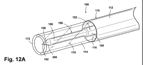
Figure 12A is a perspective view of a distal portion of another embodiment of
the
sheath and another embodiment of the light delivery device.
Figure 12B is a side sectional view of the sheath and the light delivery
device of
Figure 12A.
Figure 13 is a side sectional view of a distal portion of another embodiment
of the
sheath with the light delivery device of Figure 2.
Figure 14A is a side sectional view of a distal portion of another embodiment
of the
sheath with the light delivery device of Figure 2.
Figure 14B is a side sectional view of a distal portion of the embodiment of
the sheath
shown in Figure 14A with a light scattering material located inside the
sheath.
Figure 15 is a side sectional view of a distal portion of another embodiment
of the
sheath with the light delivery device of Figure 12A.
Figure 16 is a side sectional view of a distal portion of another embodiment
of the
sheath with the light delivery device of Figure 12A.
Figure 17A is a perspective view of a distal portion of another embodiment of
the
light delivery device.
Figure 17B is a side sectional view of the light delivery device of Figure
17A.
Figure 18 is a side sectional view of a distal portion of another embodiment
of the
light delivery device.
Figure 19 is a side sectional view of a distal portion of another embodiment
of the
light delivery device.
Figure 20A is a perspective view of a distal portion of another embodiment of
the
light delivery device.
Figure 20B is a side sectional view of the light delivery device of Figure
20A.
VNUS.090VPC 19 Knobbe, Martens, Olson & Bear LLP
CA 02682397 2009-09-29
WO 2008/134560 PCT/US2008/061641
Figure 21A is a side sectional view of a distal portion of another embodiment
of the
light delivery device comprising a position limiter.
Figure 21B is a perspective view of a portion of the position limiter of
Figure 21A.
Figure 22A is a side sectional view of a distal portion of another embodiment
of the
light delivery device comprising a position limiter.
Figure 22B is a perspective view of a portion of the position limiter of
Figure 22A.
Figure 22C is a front view of a portion of the position limiter of Figure 22A.
Figure 23A is a side sectional view of a distal portion of another embodiment
of the
light delivery device comprising a position limiter.
Figure 23B is a perspective view of a portion of the position limiter of
Figure 23A.
Figure 24A is a side sectional view of a distal portion of another embodiment
of the
light delivery device comprising a position limiter.
Figure 24B is a perspective view of a portion of the position limiter of
Figure 24A.
Figure 25A is a side sectional view of a distal portion of another embodiment
of the
light delivery device comprising a position limiter.
Figure 25B is a perspective view of a portion of the position limiter of
Figure 25A.
Figure 25C is a front view of a portion of the position limiter of Figure 25A.
Figure 25D is a top view of a portion of the position limiter Figure 25A.
Figure 26A is a side sectional view of a distal portion of another embodiment
of the
light delivery device comprising a position limiter.
Figure 26B is a perspective view of a portion of the position limiter of
Figure 26A.
Figure 27 is a side sectional view of a distal portion of another embodiment
of the
light delivery device comprising a position limiter.
Figure 28 is a side sectional view of a distal portion of another embodiment
of the
light delivery device comprising a position limiter.
Detailed Description of the Preferred Embodiments
The features of the systems and methods will now be described with reference
to the
drawings summarized above. The drawings, associated descriptions, and specific
implementation are provided to illustrate preferred embodiments of the
invention(s) disclosed
VNUS.090VPC 20 Knobbe, Martens, Olson & Bear LLP
CA 02682397 2009-09-29
WO 2008/134560 PCT/US2008/061641
herein, and not to limit the scope of the patent protection sought in
connection with this
specification.
In addition, methods and functions of treatment systems or devices described
herein
are not limited to any particular sequence, and the acts relating thereto can
be performed in
other sequences that are appropriate. For example, described acts may be
performed in an
order other than that specifically disclosed, or multiple acts may be combined
in a single act.
One embodiment of a system 100 for treating a hollow anatomical structure or
"HAS"
(e.g., a blood vessel, a vein, a varicose vein, a fallopian tube, ovarian
vein, etc.) is depicted in
Figures 1, 2 and 3. The depicted embodiment of the system 100 includes an
introducer
sheath 110 having a preferably tubular and flexible shaft 112, a distal end of
which includes a
protective distal tip portion 114. The sheath 110 preferably further comprises
a hub 120
attached to a proximal end of the shaft 112, and a sidearm 122 which can
include a port 124
to facilitate introduction of fluids into the sidearm 122. In the depicted
embodiment the hub
120 is configured to permit fluid communication between the sidearm 122 and
the shaft 112
such that a fluid introduced into the port 124 of the sidearm 122 can flow
into a lumen 116
(see Figure 3) of the shaft 112. An appropriate connector, such as a Luer
fitting (not shown)
can be included at the port 124 (or on the hub 120 instead of the sidearm 122)
to permit
connection of medical apparatus, fluid sources, etc. to the sidearm 122. The
sheath 110 can
be sized for insertion into a HAS, and can have an outer diameter of 1-5 mm.
The system 100 depicted in Figures 1-3 can further comprise a light delivery
device
150 disposed in the lumen 116 of the shaft 112. In the depicted embodiment the
light
delivery device 150 comprises an optical fiber 152, which can be coupled to a
laser light
generator 154. Where employed, the optical fiber 152 can extend proximally
through the hub
120 of the introducer sheath 110 to the laser light generator 154, to conduct
laser energy
output by the generator 154 through the shaft 112 to the desired treatment
area as will be
discussed in greater detail below. A hemostatic seal or the like can be
provided in the hub
120 to provide a seal around the fiber 152 and prevent fluid in the shaft
lumen 116 from
escaping proximally beyond the hub 120. As an alternative to the depicted
optical fiber 152,
the light delivery device 150 can comprise a small laser light source or other
light source
disposed in the lumen 116 of the shaft 112.
VNUS.090VPC 21 Knobbe, Martens, Olson & Bear LLP
CA 02682397 2009-09-29
WO 2008/134560 PCT/US2008/061641
In the depicted embodiment, the optical fiber 152 comprises a light-conducting
optical core 156 formed from glass, silica or other suitable light-conducting
material(s),
surrounded by cladding 158 made from silica or polymers or the like, to
promote internal
reflection within the core 156. A protective jacket 160 surrounds the cladding
158 and the
core 156. The jacket 160 is optionally stripped back to expose a distal tip
portion of the
cladding 158 and core 156, and this distal tip portion is typically between
about 2mm and
8mm in length. Alternatively, the optical fiber 152 can be employed without
any of the
jacket 160 stripped from the distal fiber tip, e.g. with only the distal face
of the core 156
exposed at the distal tip. The core 156 preferably terminates in an unclad,
distal light
emitting tip 162. In operation, light 170 (e.g. laser light) propagates
distally down the core
156 of the fiber 152, exits the core 156 at the light emitting tip 162 and
advances generally
distally from the tip 162. The tip 162 is preferably a generally flat surface
oriented generally
orthogonal to the longitudinal axis of the fiber 152. Alternatively, however,
the tip 162 can
also be formed, shaped, or ground to create facets, or a spherical or
prismatic tip face to direct
a portion of the light in the radial direction.
The distal tip portion 114 of the shaft 112 is preferably transparent to, or
otherwise
highly transmissive of, the wavelength(s) of light 170 emitted via the tip 162
of the fiber 152
(or other light delivery device 150) during operation of the system 100. Such
wavelengths of
light 170 can optionally range from 400 to 3000 nm, or from 800 to 1500 nm.
The distal tip
portion 114 can also be sufficiently transmissive of such wavelength(s) of
light (or of specific
suitable therapeutic wavelengths such as 810 nm, 940 nm, 980 nm, 1320 nm
and/or 1470 nm)
to permit heating and reduction in diameter of a target hollow anatomical
structure such as a
vein, and/or to avoid melting and/or burning the distal tip portion 114 when
light (optionally
including light in the above-noted wavelength(s) is emanating from the fiber
152 at sufficient
intensity to lead to heating and reduction in diameter of the HAS or vein.
Suitable materials
for use in forming the distal tip portion 114 include, without limitation,
quartz, sapphire,
borosilicate glass (PYREX(tm)), synthetic fused silica, polycarbonate,
polyetheretherketone,
polysulfone, polyarylethersulfone, polyetherimide, and polyamide-im ides. The
distal tip
portion 114 can optionally comprise a tube with a wall thickness of 0.2 - 1.0
mm.
VNUS.090VPC 22 Knobbe, Martens, Olson & Bear LLP
CA 02682397 2009-09-29
WO 2008/134560 PCT/US2008/061641
Some or all of the light 170 can propagate from the tip 162, distally and/or
outwardly
through the sidewalls and/or end of the distal tip portion 114 and to the
desired treatment
area. The fiber tip 162 can therefore remain disposed within the distal tip
portion 114 of the
shaft 112 during treatment, and the distal tip portion 114 can protect the hot
fiber tip 162
from contact with the inner wall of the vein or other target HAS (and vice
versa).
In the depicted embodiment, the fiber tip 162 is spaced proximally from a
distal end
172 of the distal tip portion 114 by a distance X of 2 mm to 20 mm. The distal
tip portion
114 can further optionally include an opening 174 to permit light and/or
liquids to flow from
the tip portion 114, and/or a tapered tip region 176 to facilitate easy and
atraumatic insertion
of the shaft 112 into small-diameter HAS's.
Preferably, the light delivery device 150 and the lumen 116 of the shaft
112/tip
portion 114 are sized so that a fluid delivery space 178 is provided between
the light delivery
device 150 and the inner wall of the shaft 112/tip portion 114. In such an
embodiment, a
liquid such as saline (or any other suitable liquid) can be delivered distally
through the shaft
112 and tip portion 114, and out the opening 174, during delivery of light 170
from the
device 150. The delivered liquid can optionally absorb the wavelength(s) of
light 170
emitted from the device 150, to a sufficient degree to induce heating and/or
boiling of the
delivered liquid as it flows through the delivery space 178 and light 170, and
out the opening
174. The hot/boiling liquid will also tend to heat the tip portion 114. Thus,
this embodiment
of the system 100 can be capable of providing at least three mechanisms of
therapeutic HAS
wall heating: (1) hot or boiling fluid heating of the HAS walls, (2)
conductive heating from
the hot sheath tip 114, and (3) light or laser energy 170 transmitted directly
to the HAS walls.
By controlling the light/laser power, the distance X, liquid flow rate, and
liquid
starting temperature, the HAS heating zone/length can be controlled and an
optimized
thermal therapy can be accomplished. Also, by selecting a preferentially water
absorbing
light/laser wavelength (e.g. 1320nm, etc.) the therapy can be one in which
substantially all of
the light/laser energy is absorbed by the (aqueous) liquid which both flows
from the sheath
opening 174 and heats the sheath tip 114 to create a heat zone for effecting
tissue thermal
therapy. The aforementioned parameters are preferably varied to ensure that
the heating is
maintained at or around 100 C, providing a controlled therapy with minimal
complications
VNUS.090VPC 23 Knobbe, Martens, Olson & Bear LLP
CA 02682397 2009-09-29
WO 2008/134560 PCT/US2008/061641
(e.g., minimizing uncontrolled high temperatures that cause increased depth of
thermal injury
leading in turn to potential pain and bruising; and avoiding fiber tip wall
contact and
perforations that lead to blood extravasations and bruising).
In one embodiment of a method of use of the system 100, the target HAS (e.g. a
vein
such as the greater saphenous vein) can first be accessed by using a suitable
access technique
(e.g. the standard Seldinger technique). A guide wire is passed into the
target HAS, and the
introducer sheath 110 is fed over the guidewire into the target HAS and
advanced to the
desired start location. In the case of the greater saphenous vein, the desired
start location is
just below the sapheno-femoral junction. The guidewire is then withdrawn from
the sheath
110 and the light delivery device 150 is advanced distally through the hub 120
and down the
shaft 112 until the device 150 is appropriately positioned within the sheath
tip 114. Where
the light delivery device 150 comprises the optical fiber 152, the fiber tip
162 is positioned so
that it is proximal of the distal end 172 of the tip 114 by the distance X. An
appropriate mark
(or a projection such as a flange, slidable collar or "donut") can be provided
on a proximal
region of the fiber 152 to facilitate positioning of the fiber tip 162, such
that alignment of the
mark with the proximal edge of the hub indicates that the desired position of
the fiber tip 162
has been reached. A suitable lock, clamp or Touhy-Borst valve can be provided
in the hub
120 to prevent longitudinal movement of the fiber 152 within the sheath, and
this lock or
clamp can be activated after positioning of the fiber tip 162 within the
sheath 110 as
described above. Alternatively, the sheath 110 and light delivery device 150
can be
combined prior to insertion and advanced into the target HAS together, without
need for a
guidewire.
Before or after placement of the optical fiber 152 or other light delivery
device, the
position of the sheath 110 relative to the desired treatment location can be
verified using
appropriate techniques such as ultrasound. In addition, the target HAS can
optionally be
prepared for treatment by using any desired combination of manual compression,
compression bandages, and/or injection of tumescent anesthesia into the
tissues surrounding
the target HAS, to exsanguinate the HAS lumen (in the case of treating blood
vessels) and
reduce the lumenal diameter in preparation for heat treatment.
VNUS.090VPC 24 Knobbe, Martens, Olson & Bear LLP
CA 02682397 2009-09-29
WO 2008/134560 PCT/US2008/061641
If desired, a liquid flow via the sidearm 122, through the sheath 110 and into
the HAS
lumen can be commenced as described above. The light delivery device 150 is
activated,
providing light, such as laser light, at one or more appropriate wavelengths
or wavelength
ranges such as 810 nm, 940 nm, 980 nm, 1320 nm, and/or 1470 nm, and/or 400-
3000 nm or
800-1500 nm, and at an appropriate power level. The assembly of the sheath l
10 and device
150 is slowly withdrawn through the HAS lumen, preferably at a rate about of
0.5-5
millimeters per second. As the assembly is moved along the lumen, therapeutic
heat is
delivered to the HAS walls via one or more of the following: (1) heating of
the HAS walls
via any hot or boiled delivered liquid, (2) conductive heating from the hot
sheath tip 114, and
(3) light or laser energy 170 transmission directly to the HAS walls. After
the desired length
of the target HAS has been treated with the therapeutic heat, the sheath 110
and device 150
can be removed and appropriate post-procedural care can be administered.
In one embodiment of the method of use of the system 100, a liquid flow
suitable to
minimize, inhibit or substantially prevent buildup of proteins, coagulum
and/or carbonization
on the fiber tip 162 (e.g., having a flow rate of 5-60 cc/hour, 5-40 cc/hour,
10-30 cc/hour, 15-
25 cc/hour, or about 20 cc/hour) is established in the sheath 110 during
treatment of a target
HAS. As discussed in further detail below, this liquid flow has also been
found suitable to
minimize, inhibit or substantially prevent perforation of the hollow
anatomical structure
being treated (including veins in particular). When employed with the system
100 depicted
in Figures 1-3, this liquid flow advances distally, along and in contact with
the distal portion
of the fiber 152, in the (typically annular) fluid delivery space 178 between
the distal portion
of the fiber and the inner wall of the distal tip portion 114. Where the fiber
152 of the system
100 includes a stripped distal portion as shown in Figures 2-3, the liquid
flow advances along
and in contact with the cladding 158; and/or the unclad, distal light emitting
tip 162 points or
faces distally toward a portion of the liquid flow located in the sheath tip
114 distal of the tip
162 such that the bare, unclad core material which forms the tip 162 contacts
this distal
portion of the liquid flow. The liquid flow can comprise saline or any other
suitable liquid
disclosed herein.
The method of use of the system 100 can also optionally include positioning
the fiber
tip 162 in the sheath 112 such that the tip 162 is spaced proximally from the
distal end 172
VNUS.090VPC 25 Knobbe, Martens, Olson & Bear LLP
CA 02682397 2009-09-29
WO 2008/134560 PCT/US2008/061641
and/or opening 174 of the distal tip portion 114 by the distance X (see Figure
3) of 2 mm to
20mm,2mmto lOmm,2mmto8mm,2mmto5mm,2mmto4mm,or3mm;or
otherwise by a distance suitable to minimize, inhibit or substantially prevent
buildup of
proteins, coagulum and/or carbonization on the fiber tip 162. This tip spacing
has also been
found suitable to minimize, inhibit or substantially prevent perforation of
the hollow
anatomical structure being treated (including veins in particular).
It has been found that providing an appropriate fluid flow over the distal
portion of
the fiber 152, and/or properly spacing the fiber tip 162 from the distal end
172 and/or opening
174 of the distal tip portion 114 helps to minimize buildup of coagulum and/or
carbonized
blood components on the fiber tip 162. This in turn minimizes perforation of
the treated
hollow anatomical structure, particularly in veins, possibly due to the
elimination of the
enlarged hot carbonized mass often observed on the tip of an optical fiber
used in treatment
of a hollow anatomical structure. Accordingly, a method of minimizing
carbonization on the
fiber tip 162 and/or minimizing HAS/vein perforation (or a step of minimizing
carbonization
and/or HAS/vein perforation, as part of a method of use of the system 100) can
comprise
establishing a liquid flow as specified above, and/or spacing the fiber tip
162 from the distal
end 172 and/or opening 174 as specified above.
In addition, a low-carbonization or no-carbonization (or low-perforation or no-
perforation) system 100 can include the optical fiber 152 disposed within the
sheath 110,
with the distal portion of the fiber 152 (including at least a portion of the
exposed cladding
158, and/or the light emitting tip 162) located in the distal tip portion 114
(which can be
transparent or otherwise highly transmissive of the wavelength(s) of light
emitted from the
fiber tip 162) and surrounded by (and/or in contact with) the liquid flow
specified above. The
fiber tip 162 can be spaced from the distal end 172 and/or distal tip opening
174 (if present)
of the distal tip portion 114, by the distance X specified above. Where both
the fluid flow
and the fiber tip spacing are employed, there can exist a distal portion of
the fluid flow within
the distal tip portion 114 of the sheath I 10, which distal portion of the
fluid flow extends
distally from the fiber tip 162 by the distance X. The distal portion of the
fluid flow
preferably contacts the fiber tip 162; where the fiber tip 162 is an unclad
portion of the fiber
VNUS.090VPC 26 Knobbe, Martens, Olson & Bear LLP
CA 02682397 2009-09-29
WO 2008/134560 PCT/US2008/061641
core material, the distal portion of the fluid flow contacts the fiber core
material at the fiber
tip 162.
Figure 4 depicts an alternative embodiment of the system 100, which can be
similar in
structure, use and function to any of the variations of the system 100 of
Figures 1-3, except as
further described herein. In the system 100 of Figure 4, the distal tip
portion 114 of the shaft
112 of the sheath 110 is substantially non-transparent to the wavelength(s) of
light emitted
from the device 150 during use. The distance X between the tip 162 and the
sheath distal end
172, and the angle 0 through which the light 170 is propagated, can be
selected to ensure that
most or all of the light 170 will not be transmitted to the sheath tip walls,
but will exit
through the opening 174 and be transmitted to the target HAS walls.
As a further variation of the system 100 of Figures 1-3, a light-absorbing
coating can
be applied to the distal tip portion 114. The coating can be selected to
absorb, highly or
completely, the wavelength(s) of light emitted by the device 150. Thus the
emitted light is
converted to heat in the tip portion 114 and any delivered liquid, and energy
is delivered to
the target HAS walls via the hot and/or boiled liquid and/or contact with the
heated tip
portion 114.
As a variation of the systems 100 of Figures 1-4, the shaft 112 of the sheath
110 can
include two, preferably concentric, lumens. In such a sheath 110, the inner
lumen provides
space for the fiber 152 or other light delivery device and the outer lumen
provides a conduit
for any liquid(s) to flow. At the distal end of the shaft 112, the outer lumen
communicates
with the inner lumen and sheath tip 114, allowing saline to flow around the
tip 162 of the
fiber 152 or other device 150.
As another variation of the systems 100 of Figures 1-4, the light delivery
device 150
can be replaced with another energy application device in the form of, e.g.,
an electrically
driven heater wire or heater coil positioned in the sheath tip 114 in a
similar manner as the
stripped portion of the optical fiber 152 depicted in Figures 2-4. Such an
electrically driven
heater wire or coil can be employed to heat the delivered liquid and/or sheath
tip as described
elsewhere herein, and thereby therapeutically heat the walls of the target
HAS.
As another variation of the systems 100 of Figures 1-4, the light delivery
device 150
can be replaced with a thermally insulated conduit for the flow of a pre-
heated liquid (e.g.,
VNUS.090VPC 27 Knobbe, Martens, Olson & Bear LLP
CA 02682397 2009-09-29
WO 2008/134560 PCT/US2008/061641
saline, etc.) out the distal end of the sheath 110 and to treatment site. The
temperature of the
liquid and its flow rate can be controlled to optimize the temperature and
length of the
treatment zone at the sheath tip.
Figure 4A depicts an alternative embodiment of the system 100, which can be
similar
in structure, use and function to any of the variations of the system 100 of
Figures 1-3, except
as described herein. In the system 100 of Figure 4A, a liquid source 300 is
provided which
may be used to facilitate delivery of the liquid flow at a desired flow rate
as discussed above.
The depicted liquid source 300 is in fluid communication with the inner lumen
116 of the
sheath 112 via the sidearm 122 or other suitable connection to the sheath 112.
Figure 4B depicts one embodiment of the liquid source 300. The depicted liquid
source 300 generally comprises a liquid reservoir 310 coupled to a plumbing
network 320
which is operable to control the flow of liquid into and out of the reservoir
310. The liquid
reservoir 310, which optionally can be housed in a suitable housing 312,
preferably
comprises a pressurizable liquid reservoir 310, such as an elastic bladder or
a cylinder with a
spring-loaded piston received therein. Alternatively a non-elastic reservoir
310 can be
employed, which can rely on gravity to drive liquid flow out of the liquid
source 300.
In the depicted embodiment, the plumbing network 320 comprises a primary
passage
322 and a secondary passage 324 which are interconnected by a three-way
stopcock 326. The
primary passage 322 can be coupled to and in fluid communication with the
liquid reservoir
310 via a source connector 328, while the secondary passage 324 terminates in
a fill
connector 330, which preferably comprises a luer fitting but can comprise any
suitable
connector to facilitate connection to a syringe for filling the reservoir 310.
The primary
passage 322 terminates in an outlet 332, which can comprise a luer connector
or other
hardware suitable for facilitating fluid communication between the outlet 332
and the sheath
110 or sidearm 122.
A flow regulator 340 is preferably located on the primary passage 322, and is
operable
to regulate the rate at which liquid flows from the liquid source 300. The
flow regulator
preferably provides a fixed and predetermined flow rate through the primary
passage 322,
e.g., 5-60 cc/hour, 5-40 cc/hour, 10-30 cc/hour, 15-25 cc/hour, or about 20
cc/hour. This can
be implemented via, for example, a restricted passage through the flow
regulator 340 that, in
VNUS.090VPC 28 Knobbe, Martens, Olson & Bear LLP
CA 02682397 2009-09-29
WO 2008/134560 PCT/US2008/061641
combination with the fluid pressure applied by the pressurizable or gravity-
driven liquid
reservoir 310, yields the desired liquid flow rate. In one embodiment, the
flow regulator 340
can provide two or more such fixed and predetermined flow rates with, for
example, a
rotatable disc that can be turned to select and position one of a number of
restricted passages,
provided as holes through the disc, in alignment with the primary passage 322.
The selected
restricted passage thus determines the flow rate through the regulator 340. In
one such
embodiment, the flow regulator can provide one relatively large fixed and
predetermined
flow rate, designated as a "prime" setting, which can be used to quickly prime
the sheath 110
and the rest of the system 100 with liquid before beginning a treatment of a
hollow
anatomical structure. This "prime" flow rate can be larger than any of those
specified herein
for use when treating an HAS. The "prime" flow rate can be provided along with
one or
more "treatment" flow rates.
To use the liquid source 300, the practitioner can first connect the source
300 to the
sheath 100 via the outlet 332 and the sidearm 122 or other apparatus suitable
to provide fluid
communication between the source 300 and the lumen of the sheath 110.
Alternatively, the
connection can be made later in the process. The practitioner charges the
liquid reservoir by
setting the stopcock 326 to provide fluid communication only between the
secondary passage
324 and the reservoir 310, and connecting a syringe or other appropriate
apparatus to the fill
connector 330. Notably, a syringe with a graduated barrel can be employed to
fill the
reservoir 310 with a precise predetermined volume of liquid. The syringe is
operated to
pump a desired volume (e.g. less than 100 cc, or less than 50 cc) of liquid
through the
plumbing network 320 and into the reservoir 310. Where the reservoir 310 is of
the
pressurizable type, the inflow of liquid pressurizes the reservoir 310 (e.g.,
by expanding the
elastic bladder or forcing the piston back against the spring). Once the
reservoir 310 is full,
the practitioner can place the stopcock 326 in the closed position, preventing
any outflow
from the liquid source 300, and if desired remove the syringe or other
apparatus from the fill
connector 330. The sheath 110 can be primed directly from the syringe, or with
the liquid in
the reservoir 310, or from the syringe while still connected to the fill
connector 330 and with
the stopcock 326 at a proper setting. Where suitable, the flow regulator 340
can be placed in
the "prime" setting and the stopcock 326 opened to allow liquid to flow from
the reservoir
VNUS.090VPC 29 Knobbe, Martens, Olson & Bear LLP
CA 02682397 2009-09-29
WO 2008/134560 PCT/US2008/061641
310 to the sheath lumen at the "prime" flow rate until the priming is
complete, and the
stopcock closed. However primed, the system 100 or sheath 110 is inserted into
the target
hollow anatomical structure as disclosed elsewhere herein. At the appropriate
time after
insertion, the stopcock is opened (and the flow regulator 340 set to the
appropriate fixed and
predetermined flow rate) to deliver liquid from the reservoir 310 and into the
lumen of the
sheath l 10 at an appropriate fixed and predetermined treatment flow rate as
discussed herein,
e.g., 5-60 cc/hour, 5-40 cc/hour, 10-30 cc/hour, 15-25 cc/hour, or about 20
cc/hour. The flow
rate is then sustained, either at a constant rate or within a desirable range,
for as long as
necessary during the treatment.
Advantageously, the liquid source 300 and flow regulator 340 can be employed
to
quickly and conveniently provide a liquid flow at a desired flow rate for
treating an HAS. In
contrast, a conventional saline bag and tubing set can require a great deal of
setup and
adjustment before the desired flow rate is achieved. This increases the time
and cost
expended when performing a treatment.
Figures 4C and 4D depict one embodiment of the flow regulator 340 usable with
the
liquid source 300. The flow regulator 340 of Figures 4C-4D comprises a
reservoir chamber
342 having an inlet port 370, a drip chamber 346 having an outlet port 368,
and a flow
restriction 350. The inlet port 370 is in fluid communication with the liquid
reservoir 310
(Figure 4B) and supplies liquid to the reservoir chamber 342, which is in
fluid
communication with the drip chamber 346 via the flow restriction 350. The flow
restriction
350 regulates the flow rate of liquid into the drip chamber 346. From the drip
chamber 346,
liquid is fed via the outlet port 368 to the sidearm 122 (Figure 4A) of the
system 100 via, for
example, a length of tubing (not shown) interconnecting the flow regulator 340
and the
sidearm 122. Thus the flow regulator 340, tubing and sidearm 122 can form a
flow path
between the liquid reservoir 310 and the sheath lumen 116. The flow regulator
340 can be
fabricated from multiple injection-molded pieces which are joined or fixed
together to form
the illustrated flow regulator 340. At least the drip chamber 346 can be
formed from a
transparent material so that a user may visually confirm the presence of
saline in the drip
chamber 346.
VNUS.090VPC 30 Knobbe, Martens, Olson & Bear LLP
CA 02682397 2009-09-29
WO 2008/134560 PCT/US2008/061641
The reservoir chamber 342 defines an internal lumen 344 that is in fluid
communication with an internal lumen 348 defined by the drip chamber 346. The
inlet port
370 can comprise a spike 372 that can be directly coupled to a liquid
reservoir 310 (Figure
4B) such as a saline bag. The spike 372 defines an internal channel 374
forming a flow
passage for liquid between the liquid reservoir 310 and the lumen 344. Other
suitable
connectors, such as luer fittings, can be used in place of the spike 372 in
other embodiments.
The flow restriction 350 can be configured to provide a fixed and
predetermined
liquid flow rate at a desired flow rate for treating an HAS. The flow
restriction 350 can
comprise a restricted passage with an orifice having a predetermined effective
opening that is
sized to provide the desired liquid flow rate. As illustrated, the flow
restriction 350
comprises a channel 352 having an inlet orifice 354 and an outlet orifice 356
and defining a
flow passage 358 between the lumens 344, 348. The flow passage 358 can be
sized to
provide an appropriate fixed and predetermined treatment flow rate as
discussed herein, e.g.,
5-60 cc/hour, 5-40 cc/hour, 10-30 cc/hour, 15-25 cc/hour, or about 20 cc/hour.
For example,
the cross-sectional area of the flow path through the flow regulator 340 can
decrease from
that of the lumen 344 to that of the flow passage 358 to provide a desired
treatment flow rate.
The flow passage 358 through the channel 352 can have a fixed size.
Optionally, the outlet
orifice 356 can have a diameter of approximately 0.5 mm. In another variation,
the channel
352 can be a rigid member such as a rigid tube or a rigid (e.g. glass)
capillary tube.
The flow restriction 350 can further comprise a flow restrictor member 360
positioned
in the channel 352 to provide an appropriate fixed and predetermined treatment
flow rate as
discussed herein, e.g., 5-60 cc/hour, 5-40 cc/hour, 10-30 cc/hour, 15-25
cc/hour, or about 20
cc/hour. As illustrated, the flow restrictor member 360 can comprise a channel
restrictor 362
inserted into the inlet orifice 354 of the channel 352 and extending at least
partially into the
flow passage 358. The channel 352 and channel restrictor 362 can be sized to
provide a gap
therebetween for the passage of fluid. The gap can be on the order of 0.025
mm. The gap
can form an annulus extending around the channel restrictor 362 and bordered
by the
channel. Optionally the annulus can include an annular gap between the channel
restrictor
362 and the channel 352 of approximately 0.025 mm.
VNUS.090VPC 31 Knobbe, Martens, Olson & Bear LLP
CA 02682397 2009-09-29
WO 2008/134560 PCT/US2008/061641
The channel restrictor 362 can comprise a first portion 364 that is inserted
into the
flow passage 358 and a second portion 366 that is bent with respect to the
first portion 364 to
hold the restrictor 362 in place in the flow passage 358. The length of the
first portion 364,
i.e. how far the channel restrictor 362 protrudes into the flow passage 358,
can be selected to
control the flow rate. As a general rule, increasing the length of the first
portion 364 will
decrease the flow rate. The cross-sectional size of the first portion 364 can
also affect the
flow rate. Thus, both the cross-sectional size and length of the first portion
364 of the
channel restrictor 362 may be used to control the flow rate through the flow
passage.
Optionally, the channel restrictor 362 can comprise a wire that is bent to
form the first portion
364 and second portion 366. The first portion 364 can itself be slightly bent
to impart a
springlike characteristic to the first portion that helps to retain the
restrictor 362 in the flow
passage 358.
Figures 4E-F depict an alternate embodiment of the flow regulator 340, which
can be
similar in structure, use and function to the flow regulator 340 of Figures 4C-
D, except as
further described herein. In the flow regulator 340 of Figures 4E-F, a bypass
chamber 376 is
provided for selectively bypassing the flow restriction 350. The bypass
chamber 376 defines
a bypass channel 378 that fluidly communicates with the lumen 344 of the
reservoir chamber
342 via an inlet orifice 380 and with the lumen 348 of the drip chamber 346
via an outlet
orifice 382.
By bypassing the flow restriction 350 using the bypass chamber 376, a higher
flow
rate can be provided than that used during delivery of energy to a HAS to
flush out the system
100. Flushing the system 100 may be done before a treatment procedure to rid
the system
100 of any air bubbles by filling the system 100 with fluid or during a
treatment procedure to
remove a blockage from the system 100.
A bypass controller 384 can be provided that selectively opens one of the
orifices 380,
382 to allow fluid flow through the bypass channel 378 to flush the system
100. As
illustrated in Figure 4F, the bypass controller 384 selectively opens that
orifice 380 to allow
fluid from the reservoir chamber to enter the bypass chamber 376 and pass
through the open
outlet orifice 382 and into the drip chamber 346. The bypass controller 384
can comprise a
spring-biased valve 386 having a valve stem 388 with a push button head 390 at
one end and
VNUS.090VPC 32 Knobbe, Martens, Olson & Bear LLP
CA 02682397 2009-09-29
WO 2008/134560 PCT/US2008/061641
a closure element 392 spaced from the push button head 390. The valve 386 is
biased to a
closed position, shown in Figure 4E, in which the closure element 392 is
seated against the
orifice 380 and prevents fluid flow into the bypass channel 378, by a spring
394 positioned
between the closure element 392 and the channel 352. The valve 386 can be
moved to an
open position, shown in Figure 4F, in which the closure element 392 is spaced
from the
orifice 380, permitting fluid flow into the bypass channel 378, by depressing
the push button
head 390. A sealing element 396 can be placed between the valve stem 388 and
the exterior
wall of the bypass chamber 376 to prevent fluid leakage.
In a variation of the embodiment of Figures 4C and 4D, the channel 352 can
comprise
a capillary tube that utilizes capillary action to pass liquid through the
channel 352. The
channel 352 comprising a capillary tube can be sized to provide an appropriate
fixed and
predetermined treatment flow rate with or without the need for the flow
restrictor member
360.
Figures 5-7B depict another embodiment of the system 100, which can be similar
in
structure, use and function to any of the variations of the systems 100 of
Figures 1-4, except
as further described herein. In the system 100 of Figure 5, the distal tip
portion 114 of the
shaft 112 of the sheath 110 comprises a number of radially expanded or
expandable members
115. The expandable members 115 preferably comprise strips of an appropriate
metallic or
polymeric material having a springlike bias toward a radially expanded
configuration. When
the expandable members 115 are in the expanded configuration (Figures 5-7A),
the members
115 surround and are radially spaced from the emitting tip 162 of the optical
fiber 152 (or
other light delivery device). The tip 162 is preferably spaced proximally from
the distal end
of the expandable members 115 by a distance X of 2 mm to 20 mm. The fiber tip
162 can
therefore remain disposed within the set of expandable members 115 during
treatment, and
the expandable members 115 can protect the hot fiber tip 162 from contact with
the inner
wall of the vein or other target HAS (and vice versa).
As can be seen from Figures 6 and 7B, the members 115 are preferably
retractable
into the shaft 112 by drawing an inner tube assembly 180 proximally into a
surrounding outer
tube 182. The outer tube 182 forces the members 115 radially inward as the
inner tube
assembly 180 is drawn into the lumen of the outer tube 182. As depicted, the
inner tube
VNUS.090VPC 33 Knobbe, Martens, Olson & Bear LLP
CA 02682397 2009-09-29
WO 2008/134560 PCT/US2008/061641
assembly 180 can comprise an inner tube 184, and the expandable members 115,
which are
preferably attached to the distal end of the inner tube 184. The inner tube
184 receives the
optical fiber 152 or other light delivery device within its inner lumen, in a
manner similar to
the lumen 116 of the shaft 112 shown in Figures 1-4. Preferably, the lumen of
the inner tube
184 is sized to accommodate a space for liquid flow between the inner tube 184
and the fiber
152, to facilitate optional delivery of liquid during treatment with the
system 100 of Figures
5-7B, as described above in connection with the embodiments of Figures 1-4.
The system 100 of Figures 5-7B can be used in a manner generally similar to
the
systems 100 of Figures 1-4, except as follows. With the expandable members 115
in the
retracted configuration as shown in Figure 7B, the sheath 110 can be delivered
over a
guidewire (or otherwise) to the desired treatment location. Once the sheath
110 is in
position, the guidewire can be withdrawn and the members 115 can be expanded
by moving
the outer and inner tubes 182, 184 relative to each other such that the
members 115 move
distally beyond the end of the outer tube 182. Free of the constraint of the
outer tube 182, the
members 115 then self-expand to the expanded configuration shown in Figures 5-
7A. The
optical fiber 152 or other light delivery device can then be advanced through
the hub 120 and
down the shaft 112 and positioned so that the tip 162 is disposed within the
members 115,
and spaced proximally by the distance X from the distal ends of the members
115. As
discussed above, the fiber 152 can include a mark (or a projection such as a
flange, slidable
collar or "donut") appropriately spaced from the tip 162 to indicate proper
positioning of the
tip 162 relative to the expanded members 115 upon alignment of the mark with a
reference
point such as the proximal edge of the hub 120.
Once the tip 162 is in position, the treatment can proceed as discussed
elsewhere
herein. After completion of the treatment, the members 115 can be retracted by
drawing the
inner tube assembly 180 into the outer tube 182. The system 100 can then be
withdrawn
from the patient in the usual manner.
Figures 8A-8B depict the distal portion of an alternative embodiment of a
sheath 110
for use with the system 100. The sheath 110 of Figure 8 includes an expandable
collar 190
which is expandable via compression created by interaction of an outer tube
192 and an inner
tube 194. The tubes 192, 194 are slidable relative to each other so that the
collar 190 can be
VNUS.090VPC 34 Knobbe, Martens, Olson & Bear LLP
CA 02682397 2009-09-29
WO 2008/134560 PCT/US2008/061641
compressed (Figure 8B) between the distal end of the outer tube 192 and a
flange 196 fixed
to the distal end of the inner tube 194. The optical fiber 152 or other light
delivery device
can be received in an inner lumen of the inner tube 194. Preferably, during
use, the collar
190 is in the expanded configuration and the light emitting tip 162 of the
fiber 152 is
positioned close to (e.g., about 2 mm to 20 mm proximal of) a distal opening
198 of the inner
tube 194. The expanded collar 190 prevents contact between the hot fiber tip
and the HAS
wall during treatment. If desired, a liquid flow can be provided via the inner
lumen of the
inner tube 194 (around the fiber 152) during application of light/laser
energy, as discussed
elsewhere herein. In various embodiments, the expandable collar 190 can
comprise a fluid
filled annular balloon, or an annular, solid member formed from a compliant
and
compressible polymer material, or the like.
Figures 9A-9B depict another embodiment of a sheath 110 which can be similar
in
structure, function and use to the sheath 110 of Figures 8A-8B, except for the
use of an
expandable coil 191 in place of the expandable collar 190. The coil 191 can
alternatively
comprise a preshaped memory coil which can be deployed by a technique other
than the
compression depicted in Figures 9A-9B, such as by retraction of an overlying
sheath, or a coil
formed from power-induced or resistive-heating-induced memory material such as
Nitinol or
compatible materials, to facilitate expansion of the coil to its "remembered"
expanded
configuration by passing an electrical current through the coil through
electrical leads (not
shown) connected thereto.
Figures l0A-lOB depict another embodiment of a sheath I 10 which can be
similar in
structure, function and use to the sheath 110 of Figures 8A-8B, except that
the expandable
collar 190 is a balloon which is inflatable and deflatable via one or more
inflation passages
(not shown) disposed in the outer tube 192. In this embodiment, the outer tube
192 and inner
tube 194 are preferably not movable relative to each other. In another
embodiment, the collar
190 is a mass of compliant, hydrophilic material (e.g. a sponge) that can be
expanded by
supplying a fluid to it from conduit(s) formed in the sheath 110.
Figure 11 depicts another embodiment of the system 100 which can be similar in
structure, function and use to the systems 100 shown in Figures 1-4, except as
further
discussed below. In this embodiment, the distal tip portion 114 of the sheath
110 contains
VNUS.090VPC 35 Knobbe, Martens, Olson & Bear LLP
CA 02682397 2009-09-29
WO 2008/134560 PCT/US2008/061641
one or more holes 199 in its sidewall. Where employed, the holes 199
communicate hot or
boiling liquid outward to the HAS at location(s) along the length of the
sheath tip 114. The
holes 199 can be arranged in one or more circumferential bands as depicted.
The size of the
holes 199, the number of holes in each band, the number of bands, and the
position of the
bands relative to the distal end of the sheath tip 114 can be varied to
control the length of the
treatment zone. The holes 199 provide a pressure relief of the hot and/or
boiling liquid inside
the sheath 110, to reduce the pressure and velocity of the fluid ejected
through the opening
174 in the distal end of the tip portion 114 during treatment.
Figures 12A and 12B depict another embodiment of the system 100, which can be
similar in structure, function, and use to the systems 100 shown in Figures 1-
3, except as
further discussed below. In this embodiment, the distal tip portion 114 of the
shaft 112
preferably has a generally constant inner and outer diameter and is
transparent to, or
otherwise highly transmissive of, the wavelength of light emitted via the
fiber tip 162. The
fiber tip 162 includes a shaped surface 200 having at least one face or
portion oriented non-
perpendicularly to the longitudinal axis A of the optical fiber 152. The
shaped surface 200
can be configured in accordance with a desired light dispersion pattern. For
example, angle
refraction physics and/or other scientific principles governing light behavior
can be employed
to determine a desired configuration for the shaped surface 200 and, thereby,
control the
emission of light from the light delivery device 150 to the HAS walls, the
sheath distal tip
portion 114, and/or delivered liquid.
In the illustrated embodiment, the shaped surface 200 has a generally conical
configuration that terminates at a point coincident with the distal end of the
optical fiber 152.
The fiber tip 172 with the conical shaped surface 200 provides for more radial
dispersion of
the light as compared to, for example, a blunt end fiber tip 162, such as the
fiber tip 162
shown in Figure 3. In other words, more of the light is directed radially
toward the adjacent
HAS walls rather than axially into the lumen of the HAS located distally of
the system 100.
Other configurations are within the scope of the present disclosure. For
example, the angled
surface 200 can have a generally conical configuration that terminates
proximally of the most
distal end of the optical fiber 152 such that the fiber tip 162 terminates at
a blunt surface
orthogonal to the longitudinal axis of the optical fiber 152 and having a
transverse sectional
VNUS.090VPC 36 Knobbe, Martens, Olson & Bear LLP
CA 02682397 2009-09-29
WO 2008/134560 PCT/US2008/061641
area less than that of portion of the core 156 covered by the jacket 160,
which can sometimes
be referred to as a frustoconical configuration. Other contemplated
configurations include
pyramidal, prismatic, and spherical surfaces.
Factors to consider when determining the configuration of the shaped surface
200
include the configuration and material of the distal tip portion 114 of the
sheath 110. In some
variations of this embodiment, the shape and material of the distal tip
portion 114 can affect
the path of the light emitted from the fiber tip 162. Conversely, the
configuration and
material of the distal tip portion 114 can be selected based on a
predetermined configuration
of the shaped surface 200. The shape and material of the distal tip portion
114 shown in
Figures 12A and 12B are provided for illustrative purposes and are not
intended to limit the
present disclosure. The optical fiber 152 with the tip 162 having the shaped
surface 200 can
be utilized with any suitable sheath 110, including any of the sheaths 110 in
the embodiments
of Figures 1-11.
Figures 13-16 depict other embodiments of the system 100, which can be similar
in
structure, function, and use to the systems 100 shown in Figures 1-3, except
as further
discussed below. Each of the embodiments of Figures 13-16 can comprise a
distally closed
sheath 110 as depicted; differences between each of the embodiments of Figures
13-16 and
Figures 1-3 are described below.
In the embodiment of Figure 13, a plug 210 located at the open distal end 172
of the
distal tip portion 114 effectively closes the distal end of the sheath 110.
The distal tip portion
114 is transparent to, or otherwise highly transmissive of, the wavelength(s)
of light emitted
via the fiber tip 162, while the plug 210 is substantially not transmissive or
substantially
opaque to the wavelength(s) of light. Any suitable material or combination of
materials can
constitute the plug 210, and one suitable material for the plug 210 is a
metal.
The plug 210 includes a reflective body 214 extending proximally toward and
aligned
with the direction of light emission from the fiber tip 162. The reflective
body 214 reflects
the light emitted from the fiber tip 162 and disperses the light radially
outward toward the
HAS walls. Additionally, the reflective body 214 effectively disperses or
spreads the light
beam, thereby decreasing the flux density of the light relative to that of the
light as it exits the
fiber tip 162, which can contribute to minimizing blood boiling effects and
coagulation
VNUS.094VPC 37 Knobbe, Martens, Olson & Bear LLP
CA 02682397 2009-09-29
WO 2008/134560 PCT/US2008/061641
problems, such as deep vein thrombosis. The reflective body 214 can be
configured to reflect
the light radially, radially and proximally, and/or radially and distally. In
the illustrated
embodiment, the reflective body 214 has a generally conical configuration,
which provides
360-degree radial and radial/proximal reflection of the light from the fiber
tip 162. The shape
of the reflective body 214 shown in Figure 13 is provided as an example; other
configurations
of the reflective body 214 are within the scope of the invention. Other
examples of the
reflective body 214 include, but are not limited to, pyramidal, prismatic, and
spherical or
hemispherical bodies. Further, the surface of the reflective body 214 can be
treated, such as
by polishing or coating, to provide a surface texture having a desired
reflectivity.
In the illustrated embodiment, the plug 210 and the distal end 172 of the
distal tip
portion 114 both have a curved distal surface 216, 218 that together form a
rounded distal
end of the sheath. The rounded configuration facilitates insertion of the
system 100 into a
guide sheath or into the HAS.
The system 100 of Figure 13 can employ any suitable light delivery device and
is not
limited to the light delivery device with the optical fiber 152 having the
blunt fiber tip 162.
For example, the system 100 can alternatively use the optical fiber 152 of
Figures 12A and
12B having the fiber tip 162 with the shaped surface 200, which can further
contribute to
dispersion of the light beam. Other optical fibers 152 described and not
described in this
application can be used with the system 100 of Figure 13.
In a variation of the embodiment of the system 100 of Figure 13, the sheath
110 can
include one or more ports, such as a port formed in the distal tip portion
114, for fluidly
communicating the fluid delivery space 178 with the HAS lumen to render the
system 100
suitable for fluid delivery into the HAS lumen. One or more such ports can
additionally or
alternatively be located in the plug 210.
In another variation, the plug 210 or a portion thereof can be formed of a
material at
least partially transmissive of the wavelength of light emitted via the fiber
tip 162 such that a
portion of the light reflects from the reflective body 214 and a portion
transmits through the
plug 210. For example, the reflective body 214 can be made opaque while the
rest of the
plug 210 can be made transmissive.
VNUS.090VPC 38 Knobbe, Martens, Olson & Bear LLP
CA 02682397 2009-09-29
WO 2008/134560 PCT/US2008/061641
In yet another variation of the embodiment of the system 100 of Figure 13, one
or
both of the plug 210 and the distal tip portion 114 can be removable from the
shaft 112.
Optionally, the plug 210 and/or the distal tip portion 114 can be modular or
replaceable with
other types of plug 210 and distal tip portion 114. The removable and
replaceable features of
the distal tip portion 114 and structures associated therewith can be applied
to any of the
embodiments of the system 100 described in this application.
In the embodiment of the system 100 in Figure 14A, the distal tip portion 114
of the
sheath 110 comprises a cylindrical region 220 terminating at a rounded distal
end 222 that
distally closes the sheath 110. The distal tip portion 114 is transparent to,
or otherwise highly
transmissive of, the wavelength(s) of light emitted via the fiber tip 162,
except that the
rounded distal end 222, or at least a portion of the rounded distal end 222,
includes a light
absorbing body 224 arranged within the path of the light emitted from the
fiber tip 162. The
light absorbing body 224 prevents or at least inhibits transmission of light
therethrough, and
the light absorbed by the body 224 heats the body 224, which in turn heats the
HAS walls in
contact with (and/or nearby) the body 224, as described in more detail below.
The light
absorbing body 224 can have any suitable form, such as a material deposited,
coated, or
otherwise attached to the rounded distal end 222 and can be located on a
proximal and/or a
distal surface of the rounded distal end 222. In the illustrated embodiment,
the light
absorbing body 224 is provided on both the proximal and distal surfaces of the
rounded distal
end 222. The material of the light absorbing body 224 is highly absorbent of
the wavelength
of light emitted via the fiber tip 162 such that the light heats the material.
As a result of this
configuration, light emitted from the fiber tip 162 can both be absorbed by
the light absorbing
body 224 and transmit through other areas of the distal tip portion 114. The
light absorbed by
the light absorbing body 224 heats the light absorbing body 224, which can
thereby
conductively heat the HAS walls in contact with the light absorbing body 224
(and/or
otherwise heat the nearby HAS walls). The light transmitted through the distal
tip portion
114 can heat the HAS walls via light energy transmission directly to the HAS
walls.
The fiber 152 and the distal tip portion 114 can have any suitable relative
size and
positioning. As an example, a distance Y of the optical fiber 152 between the
jacket 160 and
the fiber tip 162 can be about 1-2 mm, the distance X between the fiber tip
162 and the most
VNUS.090VPC 39 Knobbe, Martens, Olson & Bear LLP
CA 02682397 2009-09-29
WO 2008/134560 PCT/US2008/061641
distal portion of the rounded distal end 222 can be about 3-5 mm, and a
distance Z
corresponding to the length of the distal tip portion 114 can be about 5-10
mm.
The system 100 of Figure 14A can employ any suitable light delivery device and
is
not limited to the light delivery device with the optical fiber 152 having the
blunt fiber tip
162. For example, the system 100 can alternatively use the optical fiber 152
of Figures 12A
and 12B having the fiber tip 162 with the shaped surface 200. In such a
variation, the light
absorbing body 224 can be adapted according to the direction of light emission
from the
shaped surface 200 of the fiber tip 162. For example, a portion of the light
absorbing body
224 can be extended to the cylindrical region 220 of the distal tip portion
114. Other optical
fibers 152 described and not described in this application can be used with
the system 100 of
Figure 14A.
In a variation of the embodiment of the system 100 of Figure 14A, a light
scattering
material located in the space 178 between the optical fiber 152 and the distal
tip portion 114,
as illustrated in Figure 14B, can facilitate transmission of the light to the
light absorbing body
224 and through the distal tip portion 114. The light scattering material can
be a liquid, a
solid, or combination of a liquid and a solid. For example, the light
scattering material can be
a translucent liquid with a reflective solid particulate suspended in the
liquid. For such a
light scattering material, the liquid transmits the light for reflection by
the solid particulate.
The aggregate effect of the suspended particulate is to scatter the light
incident on the
scattering material from the fiber 152. This can include scattering the light
radially, radially
and distally, or radially and proximally.
In another variation of the embodiment of the system 100 of Figure 14A, the
light
absorbing body 224 can be a body separate from and closing the distal tip
portion 114,
similar to the manner in which the plug 210 closes the distal tip portion 114
in the
embodiment of Figure 13. Further, the light absorbing body 224 and the rounded
distal end
222 need not be rounded; other configurations, such as blunt and tapered, are
within the
scope of the invention.
In yet another variation of the embodiment of the system 100 of Figure 14A,
the
sheath 110 can include one or more ports, such as a port formed in a sidewall
of the distal tip
portion 114, for fluidly communicating the fluid delivery space 178 with the
HAS lumen.
VNUS.090VPC 40 Knobbe, Martens, Olson & Bear LLP
CA 02682397 2009-09-29
WO 2008/134560 PCT/US2008/061641
One or more ports can additionally or alternatively be located in the light
absorbing body
224.
In the embodiment of the system 100 in Figure 15, the distal tip portion 114
of the
sheath 110 comprises the cylindrical region 220 terminating at the rounded
distal end 222
that distally closes the sheath l 10, similar to the sheath 110 in the
embodiment of the device
100 shown in Figure 14A. The distal tip portion 114 of the sheath 110 in
Figure 15,
however, lacks the light absorbing body 224 of Figure 14A. Instead, the entire
distal tip
portion 114 can transmit the light emitted from the fiber tip 162. The
cylindrical region 220
and the rounded distal end 222 can be integrally formed, as illustrated, or
formed of separate
bodies joined in any suitable manner. In one variation, the rounded distal end
222 can be
formed as a separate body removably coupled to the cylindrical region 220.
The system 100 of Figure 15 includes the light delivery device 150 having the
shaped
surface 200 at the fiber tip 162 shown in the embodiment of Figures 12A and
12B and
described above in detail. The system 100 of Figure 15, however, can employ
any suitable
light delivery device and is not limited to the light delivery device with the
optical fiber 152
having the fiber tip 162 with the shaped surface 200. For example, the system
100 can
alternatively use the optical fiber 152 having the blunt fiber tip 162. Other
optical fibers 152
described and not described in this application can be used with the system
100 of Figure 15.
As described above for variations of the embodiment of Figure 14A, variations
of the
embodiment of the system 100 in Figure 15 can include other features,
including a light
scattering material in the space 178 between the optical fiber 152 and the
distal tip portion
114 and/or one or more ports in the distal end or sidewall of the distal tip
portion 114. In
another variation, the rounded distal end 222 can have a configuration other
than rounded, as
discussed below with respect to the embodiment of the system 100 in Figure 16.
The embodiment of the system 100 in Figure 16 can be similar to the embodiment
of
the system 100 in Figure 15, except that the distal end 222 has a generally
conical
configuration rather than a rounded configuration. The distal end 222 shown in
Figure 16
comprises a tapered region 230 that terminates at a closed tip 232. The
tapered region 230
can be configured to transmit the light emitted from the fiber tip 162 in a
desired pattern. For
example, the tapered region 230 can be designed to refract the light distally
and radially,
VNUS.090VPC 41 Knobbe, Martens, Olson & Bear LLP
CA 02682397 2009-09-29
WO 2008/134560 PCT/US2008/061641
completely radially, or proximally and radially. In the illustrated
embodiment, the shape of
the tapered region 230 effectively redirects the light emitted from the fiber
tip 162 to provide
more radial transmission of the light to the adjacent HAS walls, as indicated
by the radially
oriented arrows in Figure 16, than would be present without the tapered region
230. The
tapered region 230 can have any suitable configuration, and, as one example
and as
illustrated, the tapered region 230 can be angled differently than the shaped
surface 200 of the
fiber tip 162. As another example, the tapered region 230 can be angled at the
same angle
employed with the shaped surface 200 of the fiber tip 162.
The system 100 of Figure 16 includes one suitable manner of joining the distal
tip
portion 114 and the shaft 112 of the sheath 110 different from that shown in
the previous
embodiments. While the distal end portion 114 and the shaft 112 can be joined
in any
suitable manner, a heat shrinkable sleeve 240 joins the distal tip portion 114
and the shaft 112
of the system 100 illustrated in Figure 16. An adhesive, such as an epoxy, can
be employed
independently or in combination with the sleeve 240 to facilitate joining the
distal tip portion
114 and the shaft 112.
The system 100 of Figure 16 includes the light delivery device 150 having the
shaped
surface 200 at the fiber tip 162 shown in the embodiment of Figures 12A and
12B and
described above in detail. The system 100 of Figure 16, however, can employ
any suitable
light delivery device and is not limited to the light delivery device with the
optical fiber 152
having the fiber tip 162 with the shaped surface 200. For example, the system
100 can
alternatively use the optical fiber 152 having the blunt fiber tip 162. Other
optical fibers 152
described and not described in this application can be used with the system
100 of Figure 16.
In a variation of the embodiment of the system 100 of Figure 16, the tip 232
of the
tapered region 230 can be opened rather than closed. The opened tip 232 can
facilitate
delivery of fluid from the fluid delivery space 178 while still redirecting
the light emitted
from the fiber tip 162 and preventing contact of the fiber tip 162 with the
HAS walls. As an
alternative or addition, the distal tip portion 114 can include one or more
fluid ports in the
distal end or sidewall of the tip portion 114, as described above with respect
to other
embodiments. In another variation of the system 100 of Figure 16, the system
100 can
VNUS.090VPC 42 Knobbe, Martens, Olson & Bear LLP
CA 02682397 2009-09-29
WO 2008/134560 PCT/US2008/061641
include a light scattering material in the space 178 between the optical fiber
152 and the
distal tip portion 114, as described above for the embodiment of Figure 14A.
Figures 17A-20B depict other embodiments of the light delivery device 150,
which
can be similar in structure, function, and use to the light delivery devices
150 shown in
Figures 1-16, except as further discussed below.
The light delivery device 150 of Figures 17A and 17B can be similar to the
light
delivery device 150 in the embodiment of Figures 12A and 12B, except that the
light delivery
device 150 of Figures 17A and 17B includes a lumen 250 formed in the optical
core 156 of
the optical fiber 152 and terminating at a distal opening 252 at the fiber tip
162. While the
lumen 250 can have any suitable size and cross-sectional shape, in one
example, the optical
fiber 152 can have an outer diameter in a range of about 300-1000 m, and the
lumen 250
can have a circular cross-section with an inner diameter in a range of about
300-600 m.
Further, the fiber tip 162 of the light delivery device 150 can include any
desired
configuration for the shaped surface 200 and is not limited to the generally
conical shape
shown in Figures 17A and 17B. For example, the fiber tip 162 can be prismatic,
rounded,
etc., according to a desired light emission pattern. Alternatively, the fiber
tip 162 can be
blunt.
In one variation of the embodiment, the lumen 250 can be fluidly coupled to
the
sidearm 122 (Figure 1) or other fluid source such that fluid supplied to the
lumen 250 via the
fluid source flows through the lumen 250 and exits the lumen 250 at the distal
opening 252
for delivery to the HAS. Internal reflection of the light in the lumen 250 can
heat the fluid as
it flows through the lumen 250.
In another variation of the light delivery device 150 of Figures 17A and 17B,
the
internal surface of the optical core 156 forming the lumen 250 can be coated
with a material
to prevent internal reflection of the light in the lumen 250. Such a coating
can be beneficial
when using the lumen 250 for fluid delivery if heating of the fluid is not
desired.
The light delivery device 150 of Figures 17A and 17B can be employed with any
suitable sheath, including any of the sheaths 110 shown with respect to the
embodiments of
Figures 1-16 and other sheaths not illustrated or described in this
application, and used in a
manner generally similar to that described above for the system 100 of Figures
1-3.
VNUS.090VPC 43 Knobbe, Martens, Olson & Bear LLP
CA 02682397 2009-09-29
WO 2008/134560 PCT/US2008/061641
Embodiments of the light delivery device 150 illustrated in Figures 18-20B can
be
employed without a sheath to treat an HAS as described elsewhere herein. These
embodiments are designed to prevent direct contact between the HAS walls and
the fiber tip
162. Each of these embodiments, particularly the differences between them and
the
embodiments of the light delivery devices 150 previously presented are
described below.
The embodiment of the light delivery device 150 shown in Figure 18 can be
similar to
the light delivery device 150 of the embodiment in Figures 1-3, except that
the light delivery
device 150 of Figure 18 includes a distal tip portion 260 extending from the
jacket 260 to a
distal end 262 projecting beyond the fiber tip 162 a predetermined distance.
The distal tip
portion 260, similar to the distal tip portion 114 of the shaft 112 in
previous embodiments, is
transparent to, or otherwise highly transmissive of, the wavelength of light
emitted via the
fiber tip 162. Extension of the distal end 262 beyond the fiber tip 162
prevents contact
between the HAS walls and the fiber tip 162. The distal end 262 can be blunt,
as illustrated,
or otherwise configured for a desired light emission pattern. The distal tip
portion 260 can be
coupled to the jacket 160 in any suitable manner, such as by a heat shrinkable
sleeve 264
optionally combined with an adhesive, including epoxy adhesives.
The embodiment of the light delivery device 150 in Figure 19 provides an
example of
modifying the distal end 262 of the distal tip portion 260. The light delivery
device 150 of
Figure 19 can be otherwise similar to that of Figure 18. The distal end 262
shown in Figure
19 has a rounded configuration and includes an annular projection 266
extending radially
inward distally of the fiber tip 162. The projection 266 inhibits inadvertent
distal movement
of the optical core 156 and the cladding 158 relative to the jacket 160 and
the distal tip
portion 260 beyond the position shown in Figure 19, and the rounded
configuration facilitates
smooth insertion of the light delivery device into the HAS. While the rounded
configuration
can provide such a benefit, it is within the scope of the present disclosure
for the distal end
262 and/or the projection 266 to be shaped otherwise.
Referring now to Figures 20A and 20B, another embodiment of the light delivery
device 150 comprises the optical fiber 152 having the optical core 156, the
cladding 158, and
the jacket 160 as described above for the other embodiments of the light
delivery device 150
and further includes a distal body 270 enclosing a distal portion of the
optical fiber 152
VNUS.090VPC 44 Knobbe, Martens, Olson & Bear LLP
CA 02682397 2009-09-29
WO 2008/134560 PCT/US2008/061641
including at least the fiber tip 162. In the illustrated embodiment, the
distal body 270
encloses the portion of the optical core 156 not covered by the cladding 158
and the jacket
160. In a variation, the cladding 158 can extend along the portion of the
optical core 156
enclosed by the distal body 270, except for the fiber tip 162. The distal body
270 can assume
any suitable shape and is shown by way of example as having a tubular
configuration with a
rounded distal end 272.
The distal body 270 prevents direct contact between the fiber tip 162 and the
HAS
walls and can be transparent to, highly transmissive of, or absorbing of the
wavelength of
light emitted from the fiber tip 162. In one variation, the distal body 270
can contain a
material, such as a fluid, a solid, or a combination fluid and solid, that
absorbs the
wavelength of light emitted by the fiber tip 162 such that the light energy
heats the distal
body 270. The heated distal body 270 conductively heats the HAS walls when in
contact
therewith. Alternatively or additionally, the heated distal body 270 can heat
fluid in the HAS
lumen, including fluid delivered by the system 100. In another variation, the
distal body 270
can contain a material, such as a fluid, a solid, or a combination fluid and
solid, at least
partially transmissive of the light emitted from the fiber tip 162 such that
the light travels
through the distal body 270 to the HAS walls, thereby heating the HAS walls
via light energy
transmission. Optionally, the material can include reflective/scattering
particles to facilitate
in the dispersion of light to the HAS walls.
As stated above, the light delivery devices 150 of Figures 18-20B can be
employed
without a sheath. Each of these embodiments can include an element that
precludes direct
contact between the HAS walls and the fiber tip 162. For the embodiments of
Figures 18 and
19, the projection of the distal end 262 beyond the fiber tip 162 inhibits
contact between the
HAS walls and the fiber tip 162. In the embodiment of Figures 20A and 20B, the
distal body
270 provides a physical barrier between the HAS walls and the fiber tip 162.
The manner of
using the light delivery devices 150 of these embodiments without a sheath is
substantially
the same as described above for the embodiments of Figures 1-3, except that
the process can
be adapted slightly to accommodate the absence of the sheath. For example, a
guide sheath
can be inserted along the guide wire for purposes of introducing the light
delivery device 150
and then withdrawn once the light delivery device 150 is situated in the HAS.
Alternatively,
VNUS.090VPC 45 Knobbe, Martens, Olson & Bear LLP
CA 02682397 2009-09-29
WO 2008/134560 PCT/US2008/061641
the light delivery device 150 can be adapted for insertion along the guide
wire such that a
guide sheath or similar element is not needed. As still another alternative,
the light delivery
devices 150 of Figures 18-20B can be employed to treat an HAS (such as a vein)
as described
elsewhere herein, but without use of a guidewire or a sheath.
Alternatively, the embodiments of the light delivery devices 150 in Figures 18-
20B
can be used with a sheath, including the sheaths 110 shown with respect to the
embodiments
of Figures 1-16 and other sheaths not illustrated or described in this
application. In such a
case, the systems 100 with the light delivery device 150 of any of Figures 18-
20B.-can be used
in a manner generally similar to that described above for the system 100 of
Figures 1-3.
Figures 21A-28 depict an alternative embodiment of the system 100, which can
be
similar in structure, use and function to the systems 100 shown in Figures 1-
4A and 11-16,
except as further discussed below. For each of the embodiments of Figures 21A-
28, the
system 100 is provided with a position limiter 400 which is configured to
limit the position of
the fiber tip 162 to a predetermined position suitable for emitting light from
the optical fiber
152, which can also be termed a firing position. The firing position can
comprise a position
proximal of the distal end 172 of the distal tip portion 114. The position
limiter 400 can be
configured to assist the user in placing the fiber tip in the firing position
by spacing the fiber
tip 162 from the distal end 172 by the distance X of 2 mm to 20 mm, 2 mm to 10
mm, 2 mm
to 8 mm, 2 mm to 5mm, 2 mm to 4 mm, or 3 mm; or otherwise by a distance
suitable to
minimize, inhibit, or substantially prevent buildup of proteins, coagulum
and/or
carbonization on the fiber tip 162. The spacing can also be suitable to
minimize, inhibit, or
substantially prevent perforation of the HAS being treated (including veins in
particular).
The position limiter 400 is advantageous when the optical fiber 152 is
inserted into the sheath
I10 after the sheath 100 has been positioned in a HAS because the position
limiter 400
provides tactile feedback to a user who is not able to visually determine when
the fiber tip
162 reaches the firing position and further prevents the fiber tip 162 from
being advanced
distally beyond the firing position and into the HAS.
The position limiter 400 can be located anywhere along the length of the
optical fiber
152 or the introducer sheath 110. It can be beneficial to place the position
limiter nearer the
fiber tip 162 or the distal tip portion 114, respectively, than the proximal
end of either. Thus
VNUS.090VPC 46 Knobbe, Martens, Olson & Bear LLP
CA 02682397 2009-09-29
WO 2008/134560 PCT/US2008/061641
the distance between the position limiter 400 and the fiber tip 162 is
minimized, which
facilitates manufacture by minimizing the dimension requiring control during
assembly of the
position limiter to the fiber. With a smaller distance between the position
limiter 400 and the
fiber tip 162, that distance can be manufactured to a greater degree of
precision and with less
expense.
As illustrated, the position limiter 400 can comprise a stop configured to
limit the
relative movement of the optical fiber 152 within the lumen 116 when the fiber
tip 162 is at
the firing position or distance X. The stop can comprise cooperating
structures on the optical
fiber 152 and the sheath 110 that are configured to prevent the insertion or
distal movement
of the distal tip of the optical fiber 152 into the lumen 116 beyond the
firing position. As
illustrated, the cooperating structure on the optical fiber 152 can include a
tube 402 or other
protrusion at least partially surrounding the jacket 160 of the optical fiber
152 and having a
fixed position relative to the fiber tip 162. The cooperating structure on the
sheath 110 can
include a shoulder 404 formed in a portion of the shaft 112 by inserting a
distal end of the
shaft 112 into the distal tip portion 114 to create a narrowed portion of the
lumen 116 which
tapers in the distal direction toward the distal tip portion 114. The outer
diameter of the shaft
112 proximal of the shoulder 404 can be approximately equal to the outer
diameter of the
distal tip portion 114, which can optionally be approximately 1.75 mm. The
wall of the shaft
112 can optionally be approximately 0.005 mm thick.
In the embodiment of Figures 21A and 21B, the tube 402 comprises an open-ended
hollow cylinder having an annular sidewall 405 with a distal face 406, a
proximal face 408,
and a channel 410 extending between the distal and proximal faces 406, 408.
The tube 402 is
mounted to the optical fiber 152 with the optical fiber 152 extending through
the channel 410
and the fiber tip 162 spaced a predetermined distance from the distal face 406
selected such
that upon insertion of the optical fiber 152 into the lumen 116, the distal
face 406 will
cooperate with the shoulder 404 to prevent movement of the fiber tip 162
beyond the
predetermined firing position. The distal face 406 can optionally be located
within 10-20 mm
of the fiber tip 162, or 12 mm from the fiber tip 162.
In the embodiment of Figures 22A-22C, the tube 402 comprises an open-ended
hollow cylinder similar to the tube 402 of Figures 21A and 21B, with the
exception that the
VNUS.090VPC 47 Knobbe, Martens, Olson & Bear LLP
CA 02682397 2009-09-29
WO 2008/134560 PCT/US2008/061641
sidewall 405 comprises one or more recesses 412 formed adjacent the distal
face 406. The
recesses 412 provide flow passages that permit the passage of liquid from a
liquid source, for
example, liquid source 300 (Figures 4A and 4B) distally past the junction of
the cooperating
structures 402, 404 and into the fluid delivery space 178. The size of the
recesses 412 can be
selected to provide a fixed and predetermined liquid flow rate so that the
tube 402 (or the
cooperating structures 402, 404) function(s) as a liquid flow regulator in
addition to a
position limiter or stop. In this case, the flow rate of fluid to the fluid
delivery space 178 can
be controlled without the need for or use of a flow regulator (Figures 4B-4F)
upstream of the
sheath 112.
In the embodiment of Figures 23A and 23B, the tube 402 comprises an open-ended
hollow cylinder similar to the tube 402 of Figures 21A and 218, with the
exception that the
tube 402 is fabricated from a porous material providing pores 416 through the
tube 402
through which the liquid can flow. The pore size can be selected to provide a
fixed and
predetermined liquid flow rate through the tube walls so that the porous tube
402 can
function as both a liquid flow regulator and a position limiter or stop. In
this case, the flow
rate of fluid to the fluid delivery space 178 can be controlled without the
use of or need for a
flow regulator 340 (Figures 4B-4F) upstream of the sheath 112. Suitable porous
materials
include ceramics and polymers such as UHMWPE, HDPE, LDPE, PP, PC, EVA, PVDF,
and
TPU. With this configuration, the fluid may enter the sidewall 405 or proximal
face 408 and
pass through the pores 416 to exit through the distal face 406. This
configuration does not
require the discrete flow paths through or around the tube 402 as found in the
embodiment of
Figures 22A-22C.
In the embodiment of Figures 24A and 24B, the tube 402 is similar to the
porous tube
402 of Figures 23A and 23B, with the exception that the sidewall 405 comprises
a distal
conical section 418 tapering toward the distal face 406 and a proximal conical
section 420
tapering toward the proximal face 408. The taper of the distal conical section
418 can be
generally complementary to the taper of the shoulder 404, as illustrated, so
that the distal
conical section 418 will match the shoulder 404 when the fiber tip 162 is in
the
predetermined firing position. With this configuration, the fluid may enter
the sidewall 405,
proximal face 408, or proximal conical section 420 and pass through the pores
416 to exit
VNUS.090VPC 48 Knobbe, Martens, Olson & Bear LLP
CA 02682397 2009-09-29
WO 2008/134560 PCT/US2008/061641
through the distal face 406. Alternately, if the taper of the distal conical
section 418 is not
complementary to the taper of the should 404, fluid may also exit through the
distal conical
section 418. Either configuration does not require the discrete flow paths
through or around
the tube 402 as found in the embodiment of Figures 22A-22C. The tube 402 can
optionally
be approximately 5mm long, with an outer diameter of 1.2 mm. The channel 410
can
optionally have an inner diameter of 0.8 mm.
In the embodiment of Figures 25A-25D, the tube 402 comprises an open-ended
hollow cylinder similar to the tube 402 of Figures 21A and 21B, with the
exception that the
tube 402 comprises two angled faces 422 cut through the distal face 406 and
the sidewall 405
at an angle with respect to the longitudinal axis A of the optical fiber 152.
The angled faces
422 form two spaces 424 that permit the passage of liquid from a liquid
source, for example,
liquid source 300 (Figures 4A and 4B) between the angled faces 422 distally
past the junction
of the cooperating structures 402, 404 and into the fluid delivery space 178.
The size of the
spaces 424 can be selected to provide a fixed and predetermined flow rate so
that the tube
402 (or the cooperating structures 402, 404) can function as both a liquid
flow regulator and a
position limiter or stop. In this case, the flow rate of fluid to the fluid
delivery space 178 can
be controlled without the need for or use of a flow regulator (Figures 4B-4F)
upstream of the
sheath 110. The size of the spaces 424 can be selected by changing the angle
of the angled
faces 422 with respect to the longitudinal axis A. The tube 402 can optionally
have an outer
diameter of 1.2 mm and an inner diameter of 0.85 mm. The angled face 422 can
optionally
extend approximately 3 mm proximally from the distal face 406 and be formed at
an angle of
30-45 degrees to the longitudinal axis A.
In the embodiment of Figures 26A-26D, the tube 402 comprises an open-ended
hollow cylinder similar to the tube 402 of Figures 21A and 21B, with the
exception that the
distal face 406 formed at an angle with respect to the longitudinal axis A of
the optical fiber
152. The angled distal face 406 comprises a distal-most tip 414 that will
cooperate with the
shoulder 404 to prevent movement of the fiber tip 162 beyond the predetermined
firing
position. The angled distal face 406 recedes proximally from the distal-most
tip 414 to
provide a space between the angled distal face 406 and the shoulder 404 that
permits the
passage of liquid from a liquid source, for example, liquid source 300
(Figures 4A and 4B)
VNUS.090VPC 49 Knobbe, Martens, Olson & Bear LLP
CA 02682397 2009-09-29
WO 2008/134560 PCT/US2008/061641
into the fluid delivery space 178. The tube 402 can optionally have an outer
diameter of 1.2
mm and an inner diameter of 0.85 mm. The angled distal face 406 can optionally
extend
approximately 3 mm along the longitudinally axis A and be formed at an angle
of 20-45
degrees to the longitudinal axis A.
While the embodiments of Figures 21A-26D illustrate various position limiters
400
comprising tubes 402 cooperating with a portion of the shaft 112 to limit the
position of the
fiber tip 162, it is also understood that the distal tip portion 114 can be
configured to
cooperate with the tubes 402 of Figures 21A-26D to limit the position of the
fiber tip 162. In
the embodiment of Figure 27, the cooperating structure on the optical fiber
152 comprises the
open-ended tube 402 of Figures 21 A and 21 B, although any of the tubes 402
shown herein
could be used, and the cooperating structure on the sheath 110 comprises a
shoulder 426
formed in the distal tip portion 114 that creates a narrowed portion of the
lumen 116 which
tapers in the distal direction toward the distal end 172. The distal face 406
of the tube 402
will cooperate with the shoulder 426 to prevent movement of the fiber tip 162
beyond the
predetermined firing position.
In the embodiment of Figure 28, the cooperating structure on the optical fiber
152
comprises the open-ended tube 402 of Figures 21A and 21B, although any of the
tubes 402
shown herein could be used, and the cooperating structure on the sheath 100
comprises a
proximal face 428 of the distal tip portion 114. The distal face 406 of the
tube 402 will
cooperate with the proximal face 428 of the distal tip portion 114 to prevent
movement of the
fiber tip 162 beyond the predetermined firing position.
Additional embodiments comprise methods of sterilization. Certain such methods
can comprise sterilizing, either terminally or sub-terminally, any of the
apparatus disclosed
herein that are intended for insertion into (or other contact with) the
patient or that are
intended for use at or near the surgical field during treatment of a patient.
Any suitable
method of sterilization, whether presently known or later developed, can be
employed.
Accordingly, certain methods comprise sterilizing, either terminally or sub-
terminally,
any of the embodiments of the system 100 or any of the components or
subsystems thereof
disclosed herein, including but not limited to any of the embodiments of the
sheath 110 or
light delivery device 150 disclosed herein. Any suitable method of
sterilization, whether
VNUS.090VPC 50 Knobbe, Martens, Olson & Bear LLP
CA 02682397 2009-09-29
WO 2008/134560 PCT/US2008/061641
presently known or later developed, can be employed. For example, the method
can
comprise sterilizing any of the above-listed apparatus with an effective dose
of a sterilant
such as cyclodextrin (Cidex(TM)), ethylene oxide (EtO), steam, hydrogen
peroxide vapor,
electron beam (E-beam), gamma irradiation, x-rays, or any combination of these
sterilants.
The sterilization methods can be performed on the apparatus in question while
the
apparatus is partially or completely assembled (or partially or completely
disassembled); thus,
the methods can further comprise partially or completely assembling (or
partially or
completely disassembling) the apparatus before applying a dose of the selected
sterilant(s).
The sterilization methods can also optionally comprise applying one or more
biological or
chemical indicators to the apparatus before exposing the apparatus to the
sterilant(s), and
assessing mortality or reaction state of the indicator(s) after exposure. As a
further option,
the sterilization methods can involve monitoring relevant parameters in a
sterilization
chamber containing the apparatus, such as sterilant concentration, relative
humidity, pressure,
and/or apparatus temperature.
In view of the foregoing discussion of methods of sterilization, further
embodiments
comprise sterile apparatus. Sterile apparatus can comprise any of the
apparatus disclosed
herein that are intended for insertion into (or other contact with) the
patient or that are
intended for use at or near the surgical field during treatment of a patient.
More specifically,
any one or combination of the following can be provided as a sterile
apparatus: any of the
embodiments of the system 100 or any of the components or subsystems thereof
disclosed
herein, including but not limited to any of the embodiments of the sheath 110
or light
delivery device 150 disclosed herein.
While certain embodiments have been described, these embodiments have been
presented by way of example only, and are not intended to limit the scope of
the disclosure or
of the patent protection sought in connection with this specification. Indeed,
the novel
methods and systems described herein may be embodied in a variety of other
forms;
furthermore, various omissions, substitutions and changes in the form of the
methods and
systems described herein may be made without departing from the spirit of the
disclosure.
VNUS.090VPC 51 Knobbe, Martens, Olson & Bear LLP