Note: Descriptions are shown in the official language in which they were submitted.
CA 02683056 2014-05-22
52281-16
-1-
Inventors: Rudolph Jaenisch, Jacob Hanna,
Marius Wernig and Christopher J.
Lengner
Attorney's Docket No.: WIBR-101-W01
REPROGRAMMING OF SOMATIC CELLS
CROSS-REFERENCE TO RELATED APPLICATIONS
The present application claims priority to U.S.
Provisional Application No. 61/036,065, filed March 12,
2008; U.S. Provisional Application No. 60/959,341, filed
July 12, 2007; and U.S. Provisional Application No.
60/922,121, filed April 7, 2007.
GOVERNMENT SUPPORT
The invention was supported, in whole or in part, by
grants 5-R01-HD045022, 5-R37-cA084198 and 5-R01-CA087869
from the National Institutes of Health. The U.S. Government
has certain rights in the invention.
BACKGROUND OF THE INVENTION
Embryonic development and cellular differentiation are
considered unidirectional pathways because cells undergo a
progressive loss of developmental potency during cell fate
specification. Two categories of pluripotent stem cells are
CA 02683056 2009-10-06
WO 2008/124133 PCT/US2008/004516
- 2 -
known to date: embryonic stem cells and embryonic germ
cells. Embryonic stem cells are pluripotent stem cells that
are derived directly from an embryo. Embryonic germ cells
are pluripotent stem cells that are derived directly from
the fetal tissue of aborted fetuses. For purposes of
simplicity, embryonic stem cells and embryonic germ cells
will be collectively referred to as "ES" cells herein.
The success of somatic cell nuclear transfer (SCNT)
experiments in mammalian species provided proof that the
epigenetic state of adult differentiated cells is not fixed
but remains pliable for reprogramming by factors present in
the oocyte cytoplasm (Byrne et a/., 2007; Jaenisch and
Young, 2008; Wakayama and Yanagimachi, 2001). However, the
inefficiency and ethical concerns associated with attempting
to clone human somatic cells have spurred the field to
search for alternative methods to achieve nuclear
reprogramming without using oocytes (Jaenisch and Young,
2008). Indeed, fusion of somatic cells to embryonic
carcinoma cells or embryonic stem (ES) cells results in
epigenetic resetting of the somatic genome but involves the
generation of 4N pluripotent cells, limiting the potential
therapeutic use of such cells (Cowan et a/., 2005; Tada et
al., 2001).
Nevertheless, the reprogramming of somatic cells by
fusion with ES cells suggested that ES cells, similar to the
oocyte cytoplasm, contain factors that can induce nuclear
reprogramming. An important breakthrough was achieved by
Yamanaka and colleagues, who succeeded in directly
reprogramming fibroblasts into induced pluripotent stem
(iPS) cells by transduction of the four transcription
factors 0ct4, Sox2, Klf4 and c-Myc (Takahashi and Yamanaka,
CA 02683056 2009-10-06
WO 2008/124133 PCT/US2008/004516
-3-
2006). Although the initially obtained iPS cells were not
normal, several groups have since advanced the direct
reprogramming technique by generating iPS cells that are
epigenetically and developmentally indistinguishable from
embryo-derived ES cells (Maherali, 2007; Meissner et a/.,
2007; Okita et al., 2007; Wernig et a/., 2007). Moreover,
transgenic expression of c-Myc was found to be dispensable
for reprogramming, though it accelerated and enhanced the
efficiency of reprogramming (Nakagawa et al., 2008; Wernig
et a/., 2008). Finally, it has also been shown that human
iPS cells can be generated by transduction of defined
factors into somatic cells (Park et al., 2008; Takahashi et
al., 2007; Yu et a/., 2007).
Despite the work that has been done to date, it remains
unknown whether terminally differentiated cells can be
reprogrammed to pluripotency with defined factors, or
whether only less differentiated cells such as somatic stem
cells can undergo nuclear reprogramming to pluripotency.
Moreover, it is unclear whether progressive differentiation
of the donor cells affects the efficiency of in vitro
reprogramming.
SUMMARY OF THE INVENTION
The present invention provides engineered somatic
cells, in which one or more endogenous pluripotency gene(s)
is operably linked to a selectable marker in such a manner
that the expression of the selectable marker substantially
matches the expression of the endogenous pluripotency gene
to which the marker is linked. The invention also provides
transgenic mice containing these engineered somatic cells.
CA 02683056 2009-10-06
WO 2008/124133 PCT/US2008/004516
-4-
The present invention also provides methods for
reprogramming somatic cells to a less differentiated state.
In certain of the methods, engineered somatic cells of the
invention are treated with an agent. Cells that express the
selectable marker are then selected, and assessed for
pluripotency characteristics. The treatment with an agent
may be contacting the cells with an agent which alters
chromatin structure, or may be transfecting the cells with
at least one pluripotency gene, or both.
The present invention further provides methods for
identifying an agent that reprograms somatic cells to a less
differentiated state. In certain of the methods, the
engineered somatic cells described above are contacted with
a candidate agent. Cells that express the selectable marker
are then selected, and assessed for pluripotency
characteristics. The presence of at least a subset of
pluripotency characteristics indicates that the agent is
capable of reprogramming somatic cells to a less-
differentiated state. The agents identified by the present
invention can then by used to reprogram somatic cells by
contacting somatic cells with the agents.
The present invention also provides methods for
identifying a gene that causes the expression of at least
one endogenous pluripotency gene in somatic cells. In
certain of the methods, the engineered somatic cells are
transfected with a cDNA library prepared from a pluripotent
cell, such as an ES cell. The cells that express the
appropriate selectable marker are then selected, and the
expression of the appropriate endogenous pluripotency gene
is examined. The expression of an endogenous pluripotency
gene indicates that the cDNA encodes a protein whose
CA 02683056 2009-10-06
WO 2008/124133
PCT/US2008/004516
-5-
expression in the cell results in, directly or indirectly,
expression of the endogenous pluripotency gene.
The invention provides methods of deriving reprogrammed
somatic cells from somatic cells that have not been
genetically modified. The invention provides methods of
deriving reprogrammed somatic cells without use of genetic
selection or, in some embodiments, without use of chemical
selection. Reprogrammed somatic cells are derived from non-
engineered somatic cells according to the invention by, for
example, introducing reprogramming agents into non-
engineered somatic cells and/or expressing such agents
therein and selecting reprogrammed cells by any of a variety
of methods that do not require presence of exogenous genetic
material within the cells.
In some embodiments, the methods employ morphological
criteria to identify reprogrammed somatic cells from among a
population of somatic cells that are not reprogrammed. In
some embodiments, the methods employ morphological criteria
to identify somatic cells that have been reprogrammed to an
ES-like state from among a population of cells that are not
reprogrammed or are only partly reprogrammed to an ES-like
state.
In some embodiments, the methods employ complement-
mediated lysis to eliminate at least some non-reprogrammed
somatic cells from a population of cells that contains at
least some reprogrammed somatic cells.
The present invention further provides methods for
treating a condition in an individual in need of such
treatment. In certain embodiments, somatic cells are
obtained from the individual and reprogrammed by the methods
of the invention under conditions suitable for the cells to
CA 02683056 2009-10-06
WO 2008/124133 PCT/US2008/004516
-6-
develop into cells of a desired cell type. The reprogrammed
cells of a desired cell type are then harvested and
introduced into the individual to treat the condition. In
certain further embodiments, the somatic cells obtained from
the individual contain a mutation in one or more genes. In
these instances, in certain embodiments the methods are
modified so that the somatic cells obtained from the
individual are first treated to restore the one or more
normal gene(s) to the cells such that the resulting cells
carry the normal endogenous gene, which are then introduced
into the individual.
In certain further embodiments, the somatic cells
obtained from the individual are engineered to express one
or more genes following their removal from the individual.
The cells may be engineered by introducing a gene or
expression cassette comprising a gene into the cells. The
gene or a portion thereof may be flanked by sites for a
site-specific recombinase.
The gene may be one that is useful for purposes of
identifying, selecting, and/or generating a reprogrammed
cell. In certain embodiments the gene encodes an expression
product that causes a reduction in DNA methylation in the
cell. For example, the gene may encode an RNA that
interferes with expression of a DNA methyltransferase, e.g.,
DNA methyltransferase 1, 3a, or 3b (Dnmtl, 3a, 3b). The RNA
may be a short hairpin RNA (shRNA) or microRNA precursor.
In certain embodiments the RNA is a precursor that is
processed intracellularly to yield a short interfering RNA
(siRNA) or microRNA (miRNA) that inhibits expression of
Dnmtl, 3a, or 3b. In certain embodiments the gene encodes a
CA 02683056 2009-10-06
WO 2008/124133 PCT/US2008/004516
-7-
marker that is usable for positive and for negative
selection.
In certain embodiments the gene is one that contributes
to initiating and/or maintaining the reprogrammed state. In
certain embodiments the gene is one whose expression product
contributes to initiating the reprogrammed state (and in
certain embodiments is necessary for maintaining the
reprogrammed state) but which is dispensable for maintaining
the reprogrammed state. In these instances, in certain
embodiments the methods include a step of treating the
engineered cells after reprogramming in order to reduce or
eliminate expression of the gene. In methods in which the
reprogrammed cells are differentiated in vitro or in vivo
after reprogramming, the treatment to reduce or eliminate
expression of the gene may occur before or after the
reprogrammed cells differentiate. The treatment may
comprise causing excision of at least a portion of the
introduced gene, e.g., by introducing or expressing a
recombinase in the cells. In
certain embodiments the gene
is one whose expression product contributes to maintaining
the reprogrammed state (and in certain embodiments is
necessary for maintaining the reprogrammed state) but which
is dispensable once the reprogrammed cells have
differentiated into a desired cell type. In these
embodiments the methods may include a step of treating the
engineered reprogrammed cells after their differentiation so
as to reduce or eliminate expression of the gene.
In certain other embodiments, methods of the invention
can be used to treat individuals in need of a functional
organ. In the methods, somatic cells are obtained from an
individual in need of a functional organ, and reprogrammed
CA 02683056 2009-10-06
WO 2008/124133 PCT/US2008/004516
-8-
by the methods of the invention to produce reprogrammed
somatic cells. Such reprogrammed somatic cells are then
cultured under conditions suitable for development of the
reprogrammed somatic cells into a desired organ, which is
then introduced into the individual. The methods are useful
for treating any one of the following conditions: a
neurological, endocrine, structural, skeletal, vascular,
urinary, digestive, integumentary, blood, autoimmune,
inflammatory, or muscular condition.
The present invention also provides methods for
producing a cloned animal. In the methods, a somatic cell
is isolated from an animal having desired characteristics,
and reprogrammed using the methods of the invention to
produce one or more reprogrammed pluripotent somatic cell
("RPSC"). The RPSCs are then inserted into a recipient
embryo, and the resulting embryo is cultured to produce an
embryo of suitable size for implantation into a recipient
female, which is then transferred into a recipient female to
produce a pregnant female. The pregnant female is
maintained under conditions appropriate for carrying the
embryo to term to produce chimeric animal progeny, which is
then bred with a wild type animal to produce a cloned
animal.
In certain embodiments, the RPSCs may alternatively be
cryopreserved for future cloning uses. In certain other
embodiments, genetic modification, such as a targeted
mutation, may be introduced into the RPSCs prior to its
insertion into a recipient embryo.
The present invention also provides methods for
producing a cloned avian. In the methods, a somatic cell is
isolated from an avian having desired characteristics, and
CA 02683056 2009-10-06
WO 2008/124133 PCT/US2008/004516
-9-
reprogrammed using the methods of the invention to produce
one or more reprogrammed pluripotent somatic cell ("RPSC").
The RPSCs are then inserted into eggs that are unable to
develop into an embryo, and the resulting eggs are then
incubated to produce avian offspring having the genotype of
the RPSC, thereby producing a cloned avian.
It is contemplated that all embodiments described above
are applicable to all different aspects of the invention. It
is also contemplated that any of the above embodiments can
be freely combined with one or more other such embodiments
whenever appropriate.
As described herein, transgenic and inducible
expression of four transcription factors (0ct4, Sox2, K1f4,
and c-Myc) was used to reprogram mouse B lymphocytes. These
factors were sufficient to convert non-terminally
differentiated B cells that have undergone partial B cell
receptor rearrangements to a pluripotent state.
Reprogramming of mature B cells required additional ectopic
expression of a myeloid transcription factor CCAAT/enhancer-
binding-protein-a (C/EBPa), known for its ability to
interrupt the transcriptional state maintaining B cell
identity. Multiple iPS lines were clonally derived from both
non-fully and fully differentiated mature B lymphocytes, and
gave rise to adult chimeras, to late term embryos when
injected into tetraploid blastocysts, and contributed to the
germline. Work described herein provides definitive proof
for the direct nuclear reprogramming of terminally
differentiated adult cells to pluripotency.
Accordingly, in one embodiment the invention relates to
a method of reprogramming a differentiated somatic cell to a
pluripotent state, comprising the steps of contacting a
CA 02683056 2009-10-06
WO 2008/124133 PCT/US2008/004516
-10-
differentiated somatic cell with at least one reprogramming
agent that contributes to reprogramming of said cell to a
pluripotent state; maintaining said cell under conditions
appropriate for proliferation of the cell and for activity
of the at least one reprogramming agent for a period of time
sufficient to begin reprogramming of the cell; and
functionally inactivating the at least one reprogramming
agent.
In another embodiment the invention relates to a method
of reprogramming a differentiated somatic cell to a
pluripotent state, comprising the steps of providing a
differentiated somatic cell that contains at least one
exogenously introduced factor that contributes to
reprogramming of said cell to a pluripotent state;
maintaining the cell under conditions appropriate for
proliferation of the cell and for activity of the at least
one exogenously introduced factor for a period of time
sufficient to activate at least one endogenous pluripotency
gene; and functionally inactivating the at least one
exogenously introduced factor.
In a further embodiment the invention pertains to a
method of selecting a differentiated somatic cell that has
been reprogrammed to a pluripotent state, comprising the
steps of providing a differentiated somatic cell that
contains at least one exogenously introduced factor that
contributes to reprogramming of the cell to a pluripotent
state; maintaining the cell under conditions appropriate for
proliferation of the cell and for activity of the at least
one exogenously introduced factor for a period of time
sufficient to activate at least one endogenous pluripotency
gene; functionally inactivating the at least one exogenously
CA 02683056 2009-10-06
WO 2008/124133 PCT/US2008/004516
-11-
introduced factor; and differentiating or distinguishing
between cells which display one or more markers of
pluripotency and cells which do not. In one embodiment
differentiating or distinguishing between cells which
display one or more markers of pluripotency and cells which
do not comprises selection or enrichment for cells
displaying one or more markers of pluripotency and/or
selection against cells which do not display one or more
markers of pluripotency.
In some embodiments of the invention the differentiated
somatic cell is partially differentiated. In other
embodiments of the invention the differentiated somatic cell
is fully differentiated.
In some embodiments of the invention the differentiated
somatic cell is cell of hematopoetic lineage; in some
embodiments the differentiated somatic cell is obtained from
peripheral blood. In one embodiment of the invention the
differentiated somatic cell is an immune system cell. In
one embodiment the differentiated somatic cell is a
macrophage. In one embodiment the differentiated somatic
cell is a lymphoid cell. In other embodiments of the
invention the differentiated somatic cell is a B cell, such
as an immature (e.g., pro-B cell or pre-B cell) or mature
(e.g., non-naive) B-cell.
In some embodiments of the invention the at least one
exogenously introduced factor is a polynucleotide. In other
embodiments the at least one exogenously introduced factor
is a polypeptide. In one embodiment the at least one
exogenously introduced factor is selected from the group
consisting of 0ct4, Sox2, Klf-4, Nanog, Lin28, c-Myc and
combinations thereof. In particular embodiments of the
CA 02683056 2009-10-06
WO 2008/124133 PCT/US2008/004516
-12-
invention the differentiated somatic cell contains
exogenously introduced 0ct4, Sox2, and Klf-4 exogenously
introduced 0ct4, Sox2, K1f-4 and c-Myc.
In one embodiment of the invention the at least one
exogenously introduced factor is selected from the group
consisting of 0ct4, Sox2, Klf-4, c-Myc and combinations
thereof and the differentiated somatic cell further contains
at least one exogenously introduced factor (e.g., a
polynucleotide or polypeptide) capable of inducing
dedifferentiation of the differentiated somatic cell. In
some embodiments the factor capable of inducing
dedifferentiation of said differentiated somatic cell is
selected from the group consisting of at least one
polynucleotide which downregulates B cell late specific
markers, at least one polynucleotide which inhibits
expression of Pax5, at least one polypeptide which
downregulates B cell late specific markers, at least one
polypeptide which inhibits expression of Pax5, and
combinations thereof. In one embodiment of the invention
the factor capable of inducing dedifferentiation of said
differentiated somatic cell is C/EBPa or a human homolog of
C/EBPa.
In particular embodiments of the invention the at least
one exogenously introduced factor is introduced using a
vector, e.g., an inducible vector or a conditionally
expressed vector. In one aspect the at least one
exogenously introduced factor is introduced using a vector
which is not subject to methylation-mediated silencing. In
yet another embodiment the at least one exogenously
introduced factor is introduced using a viral vector such as
a retroviral or lentiviral vector.
CA 02683056 2009-10-06
WO 2008/124133 PCT/US2008/004516
-13-
In one embodiment of the invention the differentiated
somatic cell is maintained in the presence of hematopoetic
cytokines and growth factors or is cultured on media
comprising bone marrow stromal cells.
In some embodiments of the present invention the
endogenous pluripotency gene is selected from the group
consisting of Nanog, Oct4, Sox2 and combinations thereof.
In other embodiments the endogenous pluripotency gene is co-
expressed with a selectable marker, such as an antibiotic
resistance gene or luminescent marker. In particular
embodiments the differentiated somatic cell further
comprises at least one polynucleotide encoding a selectable
marker operably linked to expression control elements that
regulate expression of said at least one endogenous
pluripotency gene. In specific embodiments the
differentiated somatic cell comprises a selectable gene in
the 0ct4 locus, the Nanog locus, or both the 0ct4 and Nanog
loci. In a certain embodiment the at least one exogenously
introduced factor is introduced using an inducible vector
and wherein functionally inactivating said at least one
exogenously introduced factor comprises rendering the
conditions under which said cell is maintained unsuitable
for inducible expression of said vector.
In some embodiments of the invention, markers of
pluripotency are selected from the group consisting of
expression of a pluripotency gene, expression of a gene
whose expression is a direct or indirect result of
expression of a pluripotency gene, expression of alkaline
phosphatase, expression of SSEA1, expression of SSEA3,
expression of SSEA4, expression of TRAF-60, expression of
Nanog, expression of 0ct4, expression of Fxb15, morphology
CA 02683056 2009-10-06
WO 2008/124133 PCT/US2008/004516
-14-
characteristic of an ES cell or an ES cell colony, ability
to participate in formation of chimeras that survive to
term, ability to differentiate into cells having
characteristics of endoderm, mesoderm and ectoderm when
injected into SCID mice, presence of two active X
chromosomes, resistance to DNA methylation, and combinations
thereof.
The invention also relates to an isolated pluripotent
cell derived from a reprogrammed differentiated somatic cell
in accordance with methods of the invention. In particular
the invention relates to a purified population of somatic
cells comprising at least 70% pluripotent cells derived from
reprogrammed differentiated somatic cells.
The invention further relates to an isolated
pluripotent cell produced by a method comprising (a)
providing a differentiated somatic cell that contains at
least one exogenously introduced factor that contributes to
reprogramming of said cell to a pluripotent state; (b)
maintaining said cell under conditions appropriate for
proliferation of said cell and for activity of said at least
one exogenously introduced factor for a period of time
sufficient to activate at least one endogenous pluripotency
gene; (c) functionally inactivating said at least one
exogenously introduced factor; and (d) differentiating cells
which display one or more markers of pluripotency from cells
which do not.
The invention also relates to a purified population of
somatic cells comprising at least 70% pluripotent cells
derived from reprogrammed differentiated somatic cells
produced by a method comprising (a)providing a
differentiated somatic cell that contains at least one
CA 02683056 2009-10-06
WO 2008/124133 PCT/US2008/004516
-15-
exogenously introduced factor that contributes to
reprogramming of said cell to a pluripotent state; (b)
maintaining said cell under conditions appropriate for
proliferation of said cell and for activity of said at least
one exogenously introduced factor for a period of time
sufficient begin reprogramming of said cell or to activate
at least one endogenous pluripotency gene; (c) functionally
inactivating said at least one exogenously introduced
factor; and (d) differentiating cells which display one or
more markers of pluripotency and cells which do not.
In another aspect the invention relates to a method of
producing a pluripotent cell from a somatic cell, comprising
the steps of (a) providing one or more somatic cells that
each contain at least one exogenously introduced factor that
contributes to reprogramming of said cell to a pluripotent
state, wherein said exogenously introduced factor is
introduced using an inducible vector which is not subject to
methylation-induced silencing; (b) maintaining said one or
more cells under conditions appropriate for proliferation of
said cells and for activity of said at least one exogenously
introduced factor for a period of time sufficient begin
reprogramming of said cell or to activate at least one
endogenous pluripotency gene; (c) functionally inactivating
said at least one exogenously introduced factor; (d)
selecting one or more cells which display a marker of
pluripotency; (e) generating a chimeric embryo utilizing
said one or more cells which display a marker of
pluripotency; (f) obtaining one or more somatic cells from
said chimeric embryo; (g) maintaining said one or more
somatic cells under conditions appropriate for proliferation
of said cells and for activity of said at least one
CA 02683056 2009-10-06
WO 2008/124133
PCT/US2008/004516
-16-
exogenously introduced factor for a period of time
sufficient to begin reprogramming said cell or to activate
at least one endogenous pluripotency gene; and (h)
differentiating between cells which display one or more
markers of pluripotency and cells which do not. In a
particular embodiment the method yields a purified
population of somatic cells comprising at least 70%
pluripotent cells derived from reprogrammed differentiated
somatic cells
The invention also relates to an isolated pluripotent
cell produced by a method comprising (a) providing one or
more somatic cells that each contain at least one
exogenously introduced factor that contributes to
reprogramming of said cell to a pluripotent state, wherein
said exogenously introduced factor is introduced using an
inducible vector which is not subject to methylation-induced
silencing; (b) maintaining said one or more cells under
conditions appropriate for proliferation of said cells and
for activity of said at least one exogenously introduced
factor for a period of time sufficient to begin
reprogramming said cell or to activate at least one
endogenous pluripotency gene; (c) functionally inactivating
said at least one exogenously introduced factor; (d)
selecting one or more cells which display a marker of
pluripotency; (e) generating a chimeric embryo utilizing
said one or more cells which display a marker of
pluripotency; (f) obtaining one or more somatic cells from
said chimeric embryo; (g) maintaining said one or more
somatic cells under conditions appropriate for proliferation
of said cells and for activity of said at least one
exogenously introduced factor for a period of time
CA 02683056 2009-10-06
WO 2008/124133 PCT/US2008/004516
-17-
sufficient to activate at least one endogenous pluripotency
gene; and (h) differentiating cells which display one or
more markers of pluripotency and cells which do not.
In preferred embodiments of the invention the methods
yield a purified population of somatic cells comprising at
least 70% (e.g., 70%, 75%, 80%, 85%, 90%, 95%, 99%)
pluripotent cells derived from reprogrammed differentiated
somatic cells. In particular embodiments the pluripotent
cells are genetically homogenous.
The invention also pertains to a purified population of
somatic cells comprising at least 70% pluripotent cells
derived from reprogrammed differentiated somatic cells
produced by a method comprising (a) providing one or more
somatic cells that each contain at least one exogenously
introduced factor that contributes to reprogramming of said
cell to a pluripotent state, wherein said exogenously
introduced factor is introduced using an inducible vector
which is not subject to methylation-induced silencing; (b)
maintaining said one or more cells under conditions
appropriate for proliferation of said cells and for activity
of said at least one exogenously introduced factor for a
period of time sufficient to begin reprogramming of said
cell or to activate at least one endogenous pluripotency
gene; (c) functionally inactivating said at least one
exogenously introduced factor; (d) selecting one or more
cells which display a marker of pluripotency; (e) generating
a chimeric embryo utilizing said one or more cells which
display a marker of pluripotency; (f) obtaining one or more
somatic cells from said chimeric embryo; (g) maintaining
said one or more somatic cells under conditions appropriate
for proliferation of said cells and for activity of said at
CA 02683056 2009-10-06
WO 2008/124133 PCT/US2008/004516
-18-
least one exogenously introduced factor for a period of time
sufficient to begin reprogramming said cell or to activate
at least one endogenous pluripotency gene; and (h)
differentiating cells which display one or more markers of
pluripotency and cells which do not.
The invention also encompasses a method of
reprogramming a differentiated immune cell to a pluripotent
state, comprising the steps of (a) providing a
differentiated immune cell that contains exogenously
introduced 0ct4, Sox2, Klf-4 and c-Myc, each under the
control of an inducible vector, and further contains
exogenously introduced C/EBPa; (b) maintaining said cell
under conditions appropriate for proliferation of said cell
and for activity of 0ct4, Sox2, Klf-4, c-Myc and C/EBPa for
a period of time sufficient to activate endogenous Nanog
and/or 0ct4; and (c) functionally inactivating exogenously
introduced 0ct4, Sox2, K1f-4 and c-Myc. In one embodiment
of the method said inducible vector is not subject to
methylation-derived silencing.
The invention also relates to a purified population of
immune cells comprising at least 7096 pluripotent cells
derived from reprogrammed differentiated immune cells
produced by a method comprising the steps of (a) providing a
differentiated immune cell that contains exogenously
introduced 0ct4, Sox2, K1f-4 and c-Myc, each under the
control of an inducible vector, and further contains
exogenously introduced C/EBPa; (b) maintaining said cell
under conditions appropriate for proliferation of said cell
and for activity of 0ct4, Sox2, Klf-4, c-Myc and C/EBPa for
a period of time sufficient to activate endogenous Nanog
CA 02683056 2009-10-06
WO 2008/124133 PCT/US2008/004516
-19-
and/or 0ct4; and (c) functionally inactivating exogenously
introduced 0ct4, Sox2, K1f-4 and c-Myc.
The invention also relates to a method of identifying a
reprogramming agent comprising (a) providing one or more
somatic cells that each contain at least one exogenously
introduced factor that contributes to reprogramming of said
cell to a pluripotent state, wherein each of said
exogenously introduced factors is introduced using an
inducible vector which is not subject to methylation-induced
silencing and the expression of which is controlled by
regulatory elements induced by distinct inducers; (b)
maintaining said one or more cells under conditions
appropriate for proliferation of said cells and for activity
of said at least one exogenously introduced factor for a
period of time sufficient to reprogram said cell or to
activate at least one endogenous pluripotency gene; (c)
functionally inactivating said at least one exogenously
introduced factor; (d)selecting one or more cells which
display a marker of pluripotency; (e) generating a chimeric
embryo utilizing said one or more cells which display a
marker of pluripotency; (f) obtaining one or more somatic
cells from said chimeric embryo; (g) maintaining said one or
more somatic cells under conditions appropriate for
proliferation of said cells and for activity of said at
least one exogenously introduced factor wherein activity of
said at least one exogenously introduced factor is
insufficient by itself to activate at least one endogenous
pluripotency gene; (h) contacting the somatic cell of (g)
with one or more candidate reprogramming agents; and (i)
identifying cells contacted with said one or more candidate
reprogramming agents which display one or more markers of
' 81622459
- 20 -
pluripotency, wherein candidate reprogramming agents which
induce the somatic cell of (g) to display one or more markers
of pluripotency are identified as reprogramming agents.
The present invention as claimed relates to:
- a purified preparation of isolated pluripotent
reprogrammed mammalian somatic cells, wherein the cells
(a) express endogenous 0ct4 and Nanog; (b) differentiate into
tissues having the characteristics of endoderm, mesoderm, and
ectoderm when injected into SCID mice; (c) do not contain a
selectable marker operably linked to an endogenous pluripotency
gene; and (d) are reprogrammed by introducing to the somatic
cells exogenous factors comprising 0ct4, Sox2, and K1f4, but
not c-Myc; wherein 0ct4, Sox2 and K1f4 are encoded by one or
more polynucleotides;
- a method of generating a reprogrammed mammalian
somatic cell comprising: introducing into a mammalian somatic
cell one or more exogenous polynucleotides encoding 0ct4, Sox2,
and Klf4, but not c-Myc, that contribute to reprogramming said
cell to a pluripotent state, wherein said cell has been
reprogrammed to a pluripotent state in which the cells (i)
express endogenous 0ct4; (ii) do not contain a selectable
marker operably linked to an endogenous pluripotency gene; and
(iii) differentiate into tissues having the characteristics of
endoderm, mesoderm, and ectoderm when injected into SCID mice;
- a method of reprogramming a differentiated mammalian
somatic cell to a pluripotent state, comprising the steps of:
(a) contacting a differentiated mammalian somatic cell with at
CA 2683056 2019-04-09
' 81622459
- 20a -
least one reprogramming agent comprising 0ct4, Sox2, and Klf4,
but not c-Myc, wherein said cell does not contain a selectable
marker operably linked to an endogenous pluripotency gene; and
(b) maintaining said cell under conditions appropriate for
proliferation of said cell and for activity of said at least one
reprogramming agent for a period of time sufficient to begin
reprogramming of said cell, thereby reprogramming the cell to a
pluripotent state;
- use of the purified preparation of cells as
described herein in the preparation of a medicament comprising
the purified preparation of cells or cells differentiated
therefrom;
- use of cells produced according to the method as
described herein in the preparation of a medicament comprising
the purified preparation of cells or cells differentiated
therefrom; and
- a composition comprising cells produced according
to the method as described herein.
CA 2683056 2019-04-09
CA 02683056 2014-05-22
52281-16
- 20b -
BRIEF DESCRIPTION OF THE DRAWINGS
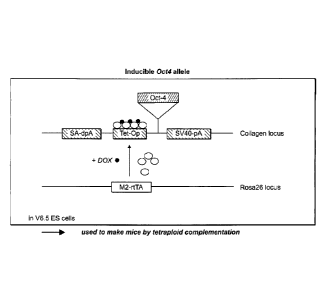
Fig. 1 is a schematic illustration of an inducible 0ct4
allele. The first integration vector, inducible 0ct4
integration vector, contains an 0ct4 gene driven by a
tetracycline-inducible promoter (Tet-Op). The Tet-Op-0ct4
cassette is flanked by a splice-acceptor double poly-A
signal (SA-dpA) at its 5' end and a SV40 polyA tail (SV40-
pA) at its 3' end. The second integration vector,
tetracycline activator integration vector, contains a mutant
form of tetracycline activator, M2-rtTA, which is more
responsive to doxycycline (Dox) induction than the wild type
activator (Urlinger et al., Proc Natl Acad Sci USA
97(14):7963 2000)).
Figs. 2A-23 show the generation of 0ct4- and Nanog-
selected iPS cells. As illustrated in Fig. 2A, an IRES-
GfpNeo fusion cassette was inserted into the Bell site
downstream of 0ct4 exon 5. Correctly targeted ES cell clones
were screened by Southern analysis of NcoI digested DNA
using a 5' external probe. The Nanog gene was targeted as
described in Mitsui et al., Cell 1/3(5):631 (2003). Fig. 2B
shows the total number (left scale) and percentages (right
scale) of AP- and strong SSEAl-positive colonies of 0ct4-
and Nanogneo MEFs 4 weeks after infection and neo selection.
Fig. 3 shows the transgenic inducible expression of
0CT4, Sox2, Klf4 and c-Myc in the mouse B cell lineage, in
particular a schematic drawing representing the strategy
CA 02683056 2009-10-06
WO 2008/124133 PCT/US2008/004516
- 2 1 -
used in this study for reprogramming cells from the B cell
lineage.
Fig. 4 shows a schematic representation of experiments
attempting to measure reprogramming efficiency. 3*10^6
CD19+ adult B cells were infected with retrovirus encoding
C/EBPa-NeoR construct, and after 24 hours we sorted IgM+IgD+
mature adult B cells and plated them as single cells in 96-
well plates preplated with 0P9 stromal cell line. Cells were
grown in conditioned medium + Dox + LIF throughout the
experiment. On day 6, culture wells were subjected to
puromycin and neomycin selections for 5 days, which allowed
only the growth of transgenic B cells infected with C/EBPa.
On day 20, the wells containing drug resistant cells were
screened for Nanog-GFP expression by FACS analysis. Wells
that scored positive were subsequently passaged on MEFs in
ES media and grown into iPS cell lines.
DETAILED DESCRIPTION OF THE INVENTION
Nuclear reprogramming, which pertains to the concept of
rewiring the epigenetic state of a somatic nucleus to that
of another cell type, can be achieved by different
approaches. The most recently established strategy to
reprogram somatic cells to pluripotency involves direct
ectopic expression of defined transcription factors in
somatic cells (Okita et al., 2007; Takahashi and Yamanaka,
2006; Wernig et a/., 2007). This enforced factor expression
appears to initiate a sequence of stochastic events
occurring over a relatively extended time period in culture
that eventually leads to generation of a small fraction of
cells that have acquired a stable pluripotent state
(Jaenisch and Young, 2008). The transduced factors are
CA 02683056 2009-10-06
WO 2008/124133 PCT/US2008/004516
-22-
required for an initial period of time in the reprogramming
process (Brambrink et al., 2008; Stadtfeld et al., 2008),
during which they may interact with endogenous pluripotency
genes (Boyer et a/., 2005; Loh et a/., 2006) and gradually
induce epigenetic changes that subsequently sustain a stable
epigenetic state that is indistinguishable from that of
inner cell mass-derived ES cells. During this process, the
de novo methyltransferases Dnmt3a and Dnmt3b also become
activated and in turn methylate and silence the virally
transduced factors. Silencing of the exogenous factors is
crucial for subsequent differentiation of the iPS cells
(Brambrink et al., 2008; Takahashi et a/., 2007; Wernig et
al., 2007; Stadfeld et al., 2008).
In the development of cells along the B cell lineage,
sequential intrinsic genetic DNA rearrangements in the heavy
and light chain immunoglobulin loci genetically mark the
different consecutive stages of B cell maturation (Jung et
a/., 2006). Cells at the Pro-B stage of development
initiate immunoglobulin rearrangements, a process involving
the assembly of V (variable), D (diversity) and J (joining)
gene segments. Assembly of the heavy chain locus (IgH)
precedes that of the light chain loci (IgL) (Jung et a/.,
2006). In addition, the rearrangements of the IgH locus are
sequential, with DH to JH joining occurring on both alleles
prior to VH to DHJH segment rearrangement (Papavasiliou et
al., 1997). The productive assembly of VH-DHJH variable
gene region indirectly signals differentiation to the next
stage, in which IgL chains are assembled with Igic
rearrangement generally preceding that of IgA (Papavasiliou
et a/., 1997). Productive IgL chain generation eventually
leads to the association of functional light and heavy chain
CA 02683056 2009-10-06
WO 2008/124133 PCT/US2008/004516
- 23 -
proteins, which together form the B cell receptor on the
cell surface. The resulting B cells can migrate to the
periphery where, upon encountering a cognate antigen, they
can exert proper immunological functions (Schlissel, 2003).
Work described herein used cells from this highly
ordered developmental pathway that carry distinct,
sequentially-acquired, genetic "fingerprints" that would
allow accurate retrospective assessment of the developmental
stage of the donor B cell nucleus that was able to generate
the respective monoclonal iPS line. In particular, as
described herein, iPS cells were generated from pro- and
pre-B cells by transduction with the reprogramming factors
0ct4, Sox2, c-Myc and Klf4 and from mature B cells by the
additional over expression of C/EBPa, a well-characterized
myeloid transcription factor. This work shows that the
reprogrammed cells carried the genetic rearrangements
characteristic of donor non-terminally differentiated and
mature terminally differentiated B cells and were able to
generate adult chimeric mice and contribute to germline.
These results indicate that specific combinations of
reprogramming factors can reset the genome of terminally
differentiated cells to a pluripotent state.
The work described herein provides conclusive evidence
that terminally differentiated mature B cells obtained from
adult mice can be directly reprogrammed into ES-like cells
in vitro. The donor B cell population that eventually
underwent successful reprogramming had completed a complex
differentiation pathway involving epigenetic and genetic
changes: an initial commitment to the hematopoietic and
subsequently to the B cell lineage; acquisition of
productive heavy and light chain rearrangements; egression
CA 02683056 2009-10-06
WO 2008/124133 PCT/US2008/004516
- 2 4 -
f rom the bone marrow to repopulate peripheral lymphoid
organs in adult mice, and as observed in one of the cell
lines obtained, acquisition of somatic hypermutations in
variable region of B cell receptor genes in response to
antigen stimuli. Thus, robust ectopic expression of 0ct4,
Sox2, Klf-4, c-Myc and C/EBPa transcription factors induced
reprogramming of fully differentiated lymphoid cells to
pluripotency with a relatively high efficiency of -1 in 30
cells.
Importantly, results described herein demonstrate that
under similar induction levels of 0ct4, Sox2, Klf4 and c-Myc
transgenes in the B cell lineage, non-terminally
differentiated and terminally differentiated B cells respond
differently to these factors. Robust reprogramming of fully
differentiated mature B lymphocytes to pluripotency was
achieved when the C/EBPa transcription factor, which
normally plays a role in granulocyte cell fate
specification, was initially over-expressed (Ramji and Foka,
2002). Thomas Graf and colleagues (Xie et a/., 2004) have
shown that overexpression of C/EBPa converted B cells into
macrophage-like cells by downregulating B cell late specific
markers (e.g., CD19) through inhibition of Pax5 functions
and facilitating extinction of the early B cell regulators,
EBF1 and E2A transcription factors. In addition, C/EBPa
induced up-regulation of components of a myeloid
transcriptional network (Laiosa et al., 2006; Xie et al.,
2004). These observations are relevant for understanding the
mechanisms of reprogramming and suggest a crucial role for
C/EBPa in inducing the reprogramming process of mouse mature
B lymphocytes. This suggests a number of mutually non-
exclusive possibilities:
CA 02683056 2009-10-06
WO 2008/124133
PCT/US2008/004516
- 2 5 -
1) C/EBPa may cross-antagonize key regulators of the B
cell transcriptional network that maintain the mature B cell
identity. This may facilitate the dedifferentiation of B
cells to a less differentiated state, allowing 0ct4, Sox2,
Klf4 and c-Myc transgene-induced reprogramming. This
explanation is consistent with observations that the
differentiation state of the donor cells is known to
influence the efficiency of reprogramming by nuclear
transplantation, as neural and keratinocyte stem cells were
more efficiently reprogrammed than other more differentiated
cells obtained from the same lineage (Blelloch et a/., 2006;
Li et a/., 2007). As conditional deletion of Pax5 in mature
B cells resulted in their dedifferentiation and loss of
several mature B cell markers (Cobaleda et al., 2007a), it
may be that deletion of Pax5 would also sensitize mature B
cells to reprogramming to pluripotency by 0ct4, Sox2, Klf4
and c-Myc.
2) C/EBPa may convert mature B cells into macrophage-
like cells (Xie et al., 2004) which have a different
epigenetic state that possibly allows enhanced accessibility
to target genes of 0ct4, Sox2, Kit 4, and/or c-Myc that would
facilitate the efficient induction of the endogenous auto-
regulatory loop governing the pluripotent state (Boyer et
al., 2005; Loh et a/., 2006).
3) C/EBPa-mediated overexpression may enable mature B
cells to transition from a state of growing in suspension to
become adherent cells in the presence of 0P9 cells, which
might be a rate-limiting event in their reprogramming.
4) Finally, other combinations of factors than those
used in the examples may be able to reprogram mature B
lymphocytes under different culture conditions.
CA 02683056 2009-10-06
WO 2008/124133 PCT/US2008/004516
-26 -
Applicants have devised novel methods of reprogramming
somatic cells, e.g., partially or fully differentiated
somatic cells, to generate pluripotent cells or multipotent
cells. It should be noted that the methods of the invention
are not intended to encompass prior art methods including,
but not limited to, somatic cell nuclear transfer. That is,
it is not within the scope of the invention to reprogram a
somatic cell by contacting the nucleus of said cell with the
intact cytoplasm of an oocyte, i.e., by transferring the
nucleus of said cell into an enucleated oocyte. While some
embodiments of the invention encompass methods of
reprogramming a nucleus of a somatic cell which has been
isolated from the cytoplasm in which it is ordinarily
contained, and optionally subsequently transferring said
nucleus to an enucleated cell of the same or different cell
type, these embodiments do not encompass methods in which
the reprogramming agent is an enucleated oocyte. Applicants
have also devised novel methods to identify agents that,
alone or in combination with other factors and/or
conditions, reprogram somatic cells.
Certain of the methods of the invention make use of
characteristics that differ between ES cells (e.g., ES cells
generated using conventional methods described in the
Background) and somatic cells. These characteristics
distinguish ES cells from somatic cells that have not been
reprogrammed and are used as a basis to identify
reprogrammed cells (induced pluripotent cells) in certain of
the methods.
One such characteristic is the increased ability of ES
cells to survive demethylation of genomic DNA relative to
CA 02683056 2009-10-06
WO 2008/124133 PCT/U
S2008/004516
- 2 7 -
somatic cells. Somatic cells are treated in any of a
variety of ways that may result in reprogramming, and the
cells are subjected to a procedure that results in DNA
demethylation. In certain embodiments of the invention,
somatic cells that are able to survive the procedure are
identified as being reprogrammed or having an increased
likelihood of being reprogrammed relative to cells which are
not able to survive the procedure. In certain embodiments of
the invention a candidate reprogramming agent, e.g., a
treatment or factor, that has resulted in at least a portion
of the cells becoming resistant to DNA demethylation (i.e.,
able to survive under conditions of DNA methylation) is
identified as an agent useful for reprogramming a somatic
cell.
Another characteristic of ES cells that distinguishes
them from somatic cells is that ES cells contain two
transcriptionally active X chromosomes, whereas in somatic
cells'one X chromosome is normally largely or completely
transcriptionally inactive (see Avner, P. and Heard, E.,
Nature Reviews Genetics, 2: 59-67, 2001; Eggan, K., et al.,
Science, 290(5496):1578-81, 2000). According to one
embodiment of the invention, somatic cells are treated in
any of a variety of ways that may result in reprogramming.
The treatment can be, for example, contacting the cells with
a candidate reprogramming agent, e.g., a treatment or
factor. In certain embodiments of the invention, cells in
which both X chromosomes are transcriptionally active are
identified as reprogrammed or having an increased likelihood
of being reprogrammed relative to cells in which only one X
chromosome is transcriptionally active. In certain
embodiments of the invention a candidate reprogramming
CA 02683056 2009-10-06
WO 2008/124133 PCT/US2008/004516
-28-
agent, e.g., a treatment or factor, that has resulted in at
least a portion of the cells having two transcriptionally
active X chromosomes is identified as a treatment useful for
reprogramming a somatic cell. In some embodiments, one step
of the method involves selecting for cells that have only
one transcriptionally active X chromosome, and a subsequent
step of the method comprises selecting for cells that have
activated their inactive X chromosome.
Certain other of the methods take advantage of the
engineered somatic cells designed by Applicants, in which an
endogenous gene typically associated with pluripotency
("pluripotency gene") is engineered to be operably linked to
a selectable marker in a manner that the expression of the
endogenous pluripotency gene substantially matches the
expression of the selectable marker. Because pluripotency
genes are generally expressed only in pluripotent cells and
not in somatic cells, the expression of an endogenous
pluripotency gene(s) is an indication of successful
reprogramming. Having a selectable marker operably linked
to an endogenous pluripotency gene gives one a powerful
mechanism to select for potentially reprogrammed somatic
cells, which may be a rare occurrence. The resulting cells
may be alternatively or additionally assessed for other
pluripotency characteristics to confirm whether a somatic
cell has been successfully reprogrammed to pluripotency.
Accordingly, in one embodiment the invention relates to
a method of reprogramming one or more somatic cells, e.g.,
partially differentiated or fully/terminally differentiated
somatic cells, to a less differentiated state, e.g., a
pluripotent or multipotent state. In general the method
comprises the steps of contacting the somatic cell or
CA 02683056 2009-10-06
WO 2008/124133 PCT/US2008/004516
-29-
isolated somatic cell nucleus with at least one
reprogramming agent that contributes to reprogramming of the
cell to a pluripotent state; maintaining the cell under
conditions appropriate for proliferation of the cell and for
activity of the reprogramming agent for a period of time
sufficient to activate an endogenous pluripotency gene, and
functionally inactivating the reprogramming agent, e.g.,
inactivating or removing the reprogramming agent. In further
embodiments the invention also relates to reprogrammed
somatic cells produced by methods of the invention and to
uses of said cells.
Generating pluripotent or multipotent cells by using
the methods of the present invention has at least two
advantages. First, the methods of the present invention
allow one to generate autologous pluripotent cells, which
are cells specific to a patient. The use of autologous
cells in cell therapy offers a major advantage over the use
of non-autologous cells, which are more likely to be subject
to immunological rejection. In contrast, autologous cells
are less likely to elicit significant immunological
responses. Second, the methods of the present invention
allow one to generate pluripotent cells without using
embryos, oocytes and/or nuclear transfer technology.
Terminology
The articles "a", "an" and "the" as used herein, unless
clearly indicated to the contrary, should be understood to
include the plural referents. Claims or descriptions that
include "or" between one or more members of a group are
considered satisfied if one, more than one, or all of the
group members are present in, employed in, or otherwise
CA 02683056 2009-10-06
WO 2008/124133 PCT/US2008/004516
-30-
relevant to a given product or process unless indicated to
the contrary or otherwise evident from the context. It
should it be understood that, in general, where the
invention, or aspects of the invention, is/are referred to
as comprising particular elements, features, etc., certain
embodiments of the invention or aspects of the invention
consist, or,consist essentially of, such elements, features,
etc. For purposes of simplicity those embodiments have not
in every case been specifically set forth in haec verba
herein. It should also be understood that any embodiment of
the invention, e.g., any embodiment found within the prior
art, can be explicitly excluded from the claims, regardless
of whether the specific exclusion is recited in the
specification. For example, any agent may be excluded from
the set of candidate reprogramming agents, and any gene can
be excluded from the set of pluripotency genes.
Where ranges are given herein, the invention includes
embodiments in which the endpoints are included, embodiments
in which both endpoints are excluded, and embodiments in
which one endpoint is included and the other is excluded.
It should be assumed that both endpoints are included unless
indicated otherwise. Furthermore, it is to be understood
that unless otherwise indicated or otherwise evident from
the context and understanding of one of skill in the art,
values that are expressed as ranges can assume any specific
value or subrange within the stated ranges in different
embodiments of the invention, to the tenth of the unit of
the lower limit of the range, unless the context clearly
dictates otherwise. It is also understood that where a
series of numerical values is stated herein, the invention
includes embodiments that relate analogously to any
CA 02683056 2009-10-06
WO 2008/124133 PCT/US2008/004516
-31-
intervening value or range defined by any two values in the
series, and that the lowest value may be taken as a minimum
and the greatest value may be taken as a maximum. Numerical
values, as used herein, include values expressed as
percentages. For any embodiment of the invention in which a
numerical value is prefaced by "about" or "approximately",
the invention includes an embodiment in which the exact
value is recited. For any embodiment of the invention in
which a numerical value is not prefaced by "about" or
"approximately", the invention includes an embodiment in
which the value is prefaced by "about" or "approximately".
"Approximately" or "about" generally includes numbers that
fall within a range of 1% or in some embodiments 5% of a
number in either direction (greater than or less than the
number) unless otherwise stated or otherwise evident from
the context (except where such number would impermissibly
exceed 100% of a possible value).
Furthermore, it is to be understood that the invention
encompasses all variations, combinations, and permutations
in which one or more limitations, elements, clauses,
descriptive terms, etc., from one or more of the listed
claims is introduced into another claim dependent on the
same base claim (or, as relevant, any other claim) unless
otherwise indicated or unless it would be evident to one of
ordinary skill in the art that a contradiction or
inconsistency would arise. Where elements are presented as
lists, e.g., in Markush group or similar format, it is to be
understood that each subgroup of the elements is also
disclosed, and any element(s) can be removed from the group.
Certain claims are presented in dependent form for the
sake of convenience, but any dependent claim may be
CA 02683056 2009-10-06
WO 2008/124133 PCT/U S2008/004516
- 3 2 -
rewritten in independent format to include the limitations
of the independent claim and any other claim(s) on which
such claim depends, and such rewritten claim is to be
considered equivalent in all respects to the dependent claim
(either amended or unamended) prior to being rewritten in
independent format. It should also be understood that,
unless clearly indicated to the contrary, in any methods
claimed herein that include more than one act, the order of
the acts of the method is not necessarily limited to the
order in which the acts of the method are recited, but the
invention includes embodiments in which the order is so
limited. It is contemplated that all embodiments described
above are applicable to all different aspects of the
invention. It is also contemplated that any of the above
embodiments can be freely combined with one or more other
such embodiments whenever appropriate.
Somatic Cells
Somatic cells of the invention may be primary cells
(non-immortalized cells), such as those freshly isolated
from an animal, or may be derived from a cell line
(immortalized cells). The cells may be maintained in cell
culture following their isolation from a subject. In certain
embodiments the cells are passaged once or more than once
(e.g., between 2-5, 5-10, 10-20, 20-50, 50-100 times, or
more) prior to their use in a method of the invention. In
some embodiments the cells will have been passaged no more
than 1, 2, 5, 10, 20, or 50 times prior to their use in a
method of the invention. They may be frozen, thawed, etc.
In certain embodiments of the invention the somatic cells
are obtained from a female. The somatic cells used may be
CA 02683056 2009-10-06
WO 2008/124133 PCT/US2008/004516
- 3 3 -
native somatic cells, or engineered somatic cells, i.e.,
somatic cells which have been genetically altered.
Somatic cells of the present invention are typically
mammalian cells, such as, for example, human cells, primate
cells or mouse cells. They may be obtained by well-known
methods and can be obtained from any organ or tissue
containing live somatic cells, e.g., blood, bone marrow,
skin, lung, pancreas, liver, stomach, intestine, heart,
reproductive organs, bladder, kidney, urethra and other
urinary organs, etc. Mammalian somatic cells useful in the
present invention include, but are not limited to, sertoli
cells, endothelial cells, granulosa epithelial, neurons,
pancreatic islet cells, epidermal cells, epithelial cells,
hepatocytes, hair follicle cells, keratinocytes,
hematopoietic cells, melanocytes, chondrocytes, lymphocytes
(B and T lymphocytes), erythrocytes, macrophages, monocytes,
mononuclear cells, cardiac muscle cells, and other muscle
cells, etc. The term "somatic cells", as used herein, also
includes adult stem cells. An adult stem cell is a cell
that is capable of giving rise to all cell types of a
particular tissue. Exemplary adult stem cells include
hematopoietic stem cells, neural stem cells, and mesenchymal
stem cells.
In some embodiments cells are selected based on their
expression of an endogenous marker known to be expressed
only or primarily in a desired cell type. For example,
vimentin is a fibroblast marker. Other useful markers
include various keratins, cell adhesion molecules such as
cadherins, fibronectin, CD molecules, etc. The population
of somatic cells may have an average cell cycle time of
between 18 and 96 hours, e.g., between 24-48 hours, between
CA 02683056 2009-10-06
WO 2008/124133 PCT/US2008/004516
-34-
48-72 hours, etc. In some embodiments, at least 90%, 95%,
98%, 99%, or more of the cells would be expected to divide
within a predetermined time such as 24, 48, 72, or 96 hours.
Methods of the invention may be used to reprogram one
or more somatic cells, e.g., colonies or populations of
somatic cells. In some embodiments a population of cells of
the present invention is substantially uniform in that at
least 90% of the cells display a phenotype or characteristic
of interest. In some embodiments at least 95%, 96%, 97%,
98%, 99%, 99.5%, 99.8%, 99.9, 99.95% or more of the cells
display a phenotype or characteristic of interest. In
certain embodiments of the invention the somatic cells have
the capacity to divide, i.e., the somatic cells are not
post-mitotic. The cells may, for example, have an average
cell cycle time (i.e., time required for a cell to complete
a single cell division cycle) of between 18-72 hours, e.g.,
between 24-48 hours when maintained in culture under
standard culture conditions known in the art.
Differentiated somatic cells of the invention are
partially or completely differentiated. Differentiation is
the process by which a less specialized cell becomes a more
specialized cell type. Cell differentiation can involve
changes in the size, shape, polarity, metabolic activity,
gene expression and/or responsiveness to signals of the
cell. For example, hematopoietic stem cells differentiate
to give rise to all the blood cell types including myeloid
(monocytes and macrophages, neutrophils, basophils,
eosinophils, erythrocytes, megakaryocytes/platelets,
dendritic cells) and lymphoid lineages (T-cells, B-cells,
NK-cells). During progression along the path of
differentiation, the ultimate fate of a cell becomes more
CA 02683056 2009-10-06
WO 2008/124133 PCT/US2008/004516
- 3 5 -
fixed. As shown by work described herein, both partially
differentiated somatic cells (e.g., immature B cells such as
pre-B cells and pro-B cells) and fully differentiated
somatic cells (e.g., mature B cells, non-naive mature B
cells) can be reprogrammed as described herein to produce
multipotent or pluripotent cells (also known as "induced
pluripotent cells").
Reprogramming and Pluripotent Cells
Differentiation status of cells is a continuous
spectrum, with a terminally differentiated state at one end
of this spectrum and de-differentiated state (pluripotent
state) at the other end. Reprogramming, as used herein,
refers to a process that alters or reverses the
differentiation status of a somatic cell, which can be
either partially or terminally differentiated.
Reprogramming includes complete reversion, as well as
partial reversion, of the differentiation status of a
somatic cell. In other words, the term "reprogramming", as
used herein, encompasses any movement of the differentiation
status of a cell along the spectrum toward a less-
differentiated state. For example, reprogramming includes
reversing a multipotent cell back to a pluripotent cell, and
reversing a terminally differentiated cell back to either a
multipotent cell or a pluripotent cell. In one embodiment,
reprogramming of a somatic cell turns the somatic cell all
the way back to a pluripotent state. In another embodiment,
reprogramming of a somatic cell turns the somatic cell back
to a multipotent state. The term "less-differentiated
state", as used herein, is thus a relative term and includes
CA 02683056 2009-10-06
WO 2008/124133 PCT/U S2008/004516
- 3 6 -
a completely de-differentiated state and a partially
differentiated state.
A pluripotent cell is a cell that has the potential to
divide in vitro for a long period of time (e.g., greater
than one year) and has the unique ability to differentiate
into cells derived from all three embryonic germ layers--
endoderm, mesoderm and ectoderm. Pluripotent cells have the
potential to differentiate into the full range of daughter
cells having distinctly different morphological, cytological
or functional phenotypes unique to a specific tissue. By
contrast, descendants of pluripotent cells are restricted
progressively in their differentiation potential, with some
cells having only one fate. A multipotent cell is a cell
that is able to differentiate into some but not all of the
cells derived from all three germ layers. Thus, a
multipotent cell is a partially differentiated cell. Adult
stem cells are also multipotent or partially differentiated
cells. Known adult stem cells include, for example,
hematopoietic stem cells and neural stem cells.
Treatment of Somatic Cell(s) with Reprogramming Agent
As described herein, one or more (e.g., a population or
colony) somatic cells, e.g., differentiated somatic cells,
is treated or contacted with at least one reprogramming
agent or factor that contributes to reprogramming of said
cell. The terms "contact", "contacting", "treat",
"treating", etc., are used interchangeably herein and
include subjecting a cell to any kind of process or
condition or performing any kind of procedure on the cell.
The treatment can be, by way of non-limiting example,
contacting the cells with a known or candidate reprogramming
CA 02683056 2009-10-06
WO 2008/124133 PCT/U S2008/004516
agent (e.g., an agent which alters the chromatin structure
of the cell, an agent which decreases DNA methylation, an
agent which decreases histone acetylation) transfecting the
cells with a polynucleotide encoding a reprogramming agent,
and/or culturing the cells under conditions that differ from
standard culture conditions in which such cells are
typically maintained. For example, the temperature or pH
could be varied. Multiple known or candidate reprogramming
agents may be used concurrently/simultaneously or
sequentially. In another embodiment, methods of the
invention may further include repeating the steps of
treating the cells with an agent or factor. The agent used
in the repeating treatment may be the same as, or different
from, the one used during the first treatment.
Reprogramming agents of the invention can be
polynucleotides, polypeptides, small molecules, etc.
The cells may be contacted with a reprogramming factor
or agent for varying periods of time. In some embodiments
the cells are contacted with the agent for a period of time
between 1 hour and 30 days. In some embodiments the cells
are contacted with the agent for a period of time sufficient
to reprogram the cell or to activate an endogenous
pluripotency gene. For example, the period may be 1 day, 5
days, 7 days, 10 days, 12 days, 14 days or 20 days. The
reprogramming agent may be removed or inactivated prior to
performing a selection to enrich for pluripotent cells or
assessing the cells for pluripotency characteristics.
According to some embodiments of the invention, after
the somatic cell(s) are contacted with the reprogramming
agent or factor, they are maintained under conditions
appropriate for proliferation of the cell and for activity
CA 02683056 2009-10-06
WO 2008/124133 PCT/US2008/004516
- 3 8 -
of the reprogramming agent or factor for a time sufficient
to reprogram the cell or to activate at least one endogenous
pluripotency gene. Cells may be maintained in culture for
varying periods of time while reprogramming takes place,
prior to selection of or enrichment for reprogrammed cells.
Thus in certain methods, somatic cells which have been
contacted with a reprogramming agent or factor are
maintained in culture for more than 3 days prior to
identifying or selecting for reprogrammed cells. In some
methods, said cells are maintained in culture for at least
4, 5, 6, 7, 8, 9, 10, 11, 12, 13, 14, 15, 16, 17, 18, 19,
20, 21 or more days (e.g., between 3-5 weeks) prior to
identifying or selecting for reprogrammed cells.
In addition, in particular embodiments of the
invention the somatic cells which have been contacted with
one or more reprogramming agents according to the described
methods are maintained under conditions appropriate for
proliferation of said cells. Conditions appropriate for the
maintenance and proliferation of particular cell types will
be apparent to the skilled artisan. Specialized culture
medium may be obtained from commercial sources, or factors
necessary or desirable for enhancing the proliferation may
be added to standard culture medium. Additional factors and
agents may also be added to culture medium, for example, to
induce expression of inducible elements in said cells or to
inhibit growth of cells which are sensitive to particular
agents.
By way of non-limiting example, DNA methylation
inhibitors and histone deacelyation inhibitors are two
classes of agents that may be used in the methods of the
invention; exemplary agents include 5-aza-cytidine, TSA and
CA 02683056 2009-10-06
WO 2008/124133 PCT/U S2008/004516
- 3 9 -
valproic acid. As described herein, DNA methylation
inhibitors are also of use to identify cells that have been
reprogrammed, regardless of whether a DNA methylation
inhibitor contributes to the reprogramming. Thus in some
embodiments of the invention the reprogramming agent is not
a DNA methylation inhibitor, e.g., it has no detectable
effect on DNA methylation or reduces DNA methylation by less
than 1%. In some embodiments the reprogramming agent reduces
DNA methylation by less than 5% and/or inhibits DNMT1, 3a,
and/or 3b activity by less than 1% or less than 5%.
In certain embodiments of the invention the
reprogramming agent or factor is exogenously introduced to
the cell. "Exogenously introduced" is used consistently
with its meaning in the art to refer to a polynucleotide (or
other substance including but not limited to a small
molecule or protein) which has been introduced into a cell
or an ancestor of the cell from outside the cell (typically
by a process that involves the hand of man) and/or is either
not found in nature in cells of that type or is found in a
different sequence, context and/or in different amounts.
In some embodiments, reprogramming agents are
introduced into cells using viral transduction, e.g.,
retroviral or lentiviral transduction. In particular
embodiments the vector used is not subject to methylation-
induced silencing. In some embodiments the vector is a non-
replicating vector, and in some embodiments the vector is a
non-integrating vector. In particular embodiments the
vector is an integrating vector which is able to be excised
from the cell's genome, e.g., able to be excised such that
the cell's genome after excision is substantially similar or
identical to the genome of the cell prior to integration of
CA 02683056 2009-10-06
WO 2008/124133 PCT/U S2008/004516
- 4 0 -
the vector. In some embodiments, reprogramming agents are
introduced into cells using protein transduction or
transient transfection of a nucleic acid construct that
encodes a protein effective either by itself or in
combination with other reprogramming agent(s) to reprogram
the cells. Optionally cells are subjected to an electric
field and/or contacted with an agent that enhances cell
permeability to increase uptake of the reprogramming agent.
In some embodiments, at least one of 0ct4, Sox2, Klf4,
Nanog, Lin28 and c-Myc may be exogenously introduced into
somatic cells using such methods. In one embodiment 0ct4,
Sox2 and Klf4 are introduced into the cell(s), while in
another embodiment 0ct4, Sox2, Klf4 and c-Myc are introduced
into the cells(s). In another embodiment 0ct4, Sox2, Nanog
and Lin28 are introduced into the cell(s).
Genes that affect the pluripotent state of a cell and
thus are candidate reprogramming agents include pluripotency
genes, genes involved in chromatin remodeling, and genes
that are important for maintaining pluripotency, such as
LIF, BMP, and PD098059 (Cell, 115: 281-292 (2003); Philos
Trans R Soc Lond B Biol Sci. 2003 Aug 29;358(1436):1397-
402). Thomson et a/. used 0ct4, Sox2, Nanog, and Lin28 using
a lentiviral system to reprogram adult human cells (Thomson
et a1., Science 5854: 1224-1225 (11/23/2007)). Other genes
that can affect whether or not a cell is pluripotent include
certain oncogenes, such as c-myc. Other genes include
telomerase, e.g., the gene encoding the catalytic subunit of
telomerase. Yet other genes include Soxl, Sox2, Sox3, Sox
15, Sox18, FoxD3, Stat3, N-Myc, L-Myc, Klfl, Klf2, Klf4 and
Klf5. Other genes of interest include those encoding
microRNA precursors that have been associated with
CA 02683056 2009-10-06
WO 2008/124133 PCT/US2008/004516
-41-
multipotency or pluripotency and/or that is naturally
expressed in multipotent or pluripotent cells. Optionally
the gene is downregulated as the cells differentiate and/or
is not expressed in adult somatic cells. Other
polynucleotides of interest include those encoding RNAi
agents such as shRNAs targeted to a gene that is a target of
an endogenous microRNA that is naturally expressed in
multipotent or pluripotent cells.
In addition, additional factors may be overexpressed or
exogenously expressed in the somatic cell to facilitate
reprogramming. For example, factors which assist in
inducing the cell to assume a less differentiated state may
be expressed in the cell. As described herein, C/EBPa has
been shown to assist in the reprogramming of mature B cells.
Other members of the C/EBPa family, such as human homologs
of C/EBPa may be similarly useful.
It will be understood that throughout the embodiments
of the invention, encoded polypeptides may be exogenously
introduced into a cell instead of or in addition to
exogenous introduction of a polynucleotide encoding said
polypeptide unless otherwise indicated or implied from
context. In addition, it will be understood that reference
to a "gene" herein is intended to encompass the coding
sequence of the gene with or without the endogenous
regulatory elements of the gene and with or without intronic
sequence elements unless otherwise indicated or implied from
context.
Expression of an exogenously introduced polynucleotide
may be carried out in several ways. In one embodiment, the
exogenously introduced polynucleotide may be expressed from
a chromosomal locus different from the endogenous
CA 02683056 2009-10-06
WO 2008/124133 PCT/U S2008/004516
- 4 2 -
chromosomal locus of the polynucleotide. Such chromosomal
locus may be a locus with open chromatin structure, and
contain gene(s) dispensible for a somatic cell. In other
words, the desirable chromosomal locus contains gene(s)
whose disruption will not cause cells to die. Exemplary
chromosomal loci include, for example, the mouse ROSA 26
locus and type II collagen (Col2a1) locus (See Zambrowicz et
al., 1997). The exogenously introduced polynucleotide may be
expressed from an inducible promoter such that its
expression can be regulated as desired.
In an alternative embodiment, the exogenously
introduced polynucleotide may be transiently transfected
into cells, either individually or as part of a cDNA
expression library. In one embodiment the cDNA expression
library can be prepared from pluripotent cells, including
but not limited to embryonic stem cells, oocytes,
blastomeres, inner cell mass cells, embryonic germ cells,
embryoid body (embryonic) cells, morula-derived cells,
teratoma (teratocarcinoma) cells, and multipotent partially
differentiated embryonic stem cells taken from later in the
embryonic development process. Candidate reprogramming
agents may be identified from such libraries.
The cDNA library is prepared by conventional
techniques. Briefly, mRNA is isolated from an organism of
interest. An RNA-directed DNA polymerase is employed for
first strand synthesis using the mRNA as template. Second
strand synthesis is carried out using a DNA-directed DNA
polymerase which results in the cDNA product. Following
conventional processing to facilitate cloning of the cDNA,
the cDNA is inserted into an expression vector such that the
cDNA is operably linked to at least one regulatory sequence.
CA 02683056 2009-10-06
WO 2008/124133 PCT/US2008/004516
-43-
The choice of expression vectors for use in connection with
the cDNA library is not limited to a particular vector. Any
expression vector suitable for use in mouse cells is
appropriate. In one embodiment, the promoter which drives
expression from the cDNA expression construct is an
inducible promoter. The term regulatory sequence includes
promoters, enhancers and other expression control elements.
Exemplary regulatory sequences are described in Goeddel;
Gene Expression Technology: Methods in Enzymology, Academic
Press, San Diego, CA (1990). For instance, any of a wide
variety of expression control sequences that control the
expression of a DNA sequence when operatively linked to it
may be used in these vectors to express cDNAs. Such useful
expression control sequences, include, for example, the
early and late promoters of SV40, tet promoter, adenovirus
or cytomegalovirus immediate early promoter, the lac system,
the trp system, the TAC or TRC system, T7 promoter whose
expression is directed by T7 RNA polymerase, the major
operator and promoter regions of phage lambda , the control
regions for fd coat protein, the promoter for 3-
phosphoglycerate kinase or other glycolytic enzymes, the
promoters of acid phosphatase, e.g., Pho5, the promoters of
the yeast a-mating factors, the polyhedron promoter of the
baculovirus system and other sequences known to control the
expression of genes of prokaryotic or eukaryotic cells or
their viruses, and various combinations thereof. It should
be understood that the design of the expression vector may
depend on such factors as the choice of the host cell to be
transformed and/or the type of protein desired to be
expressed. Moreover, the vector's copy number, the ability
to control that copy number and the expression of any other
CA 02683056 2009-10-06
WO 2008/124133 PCT/U
S2008/004516
- 4 4 -
prote in encoded by the vector, such as antibiotic markers,
should also be considered.
The exogenously introduced polynucleotide may be
expressed from an inducible promoter. The term "inducible
promoter", as used herein, refers to a promoter that, in the
absence of an inducer (such as a chemical and/or biological
agent), does not direct expression, or directs low levels of
expression of an operably linked gene (including cDNA), and,
in response to an inducer, its ability to direct expression
is enhanced. Exemplary inducible promoters include, for
example, promoters that respond to heavy metals (CRC Boca
Raton, Fla. (1991), 167-220; Brinster et al. Nature (1982),
296, 39-42), to thermal shocks, to hormones (Lee et al.
P.N.A.S. USA (1988), 85, 1204-1208; (1981), 294, 228-232;
Klock et al. Nature (1987), 329, 734-736; Israel and
Kaufman, Nucleic Acids Res. (1989), 17, 2589-2604),
promoters that respond to chemical agents, such as glucose,
lactose, galactose or antibiotic (e.g., tetracycline or
doxycycline).
A tetracycline-inducible promoter is an example of an
inducible promoter that responds to an antibiotic. See
Gossen et al., 2003. The tetracycline-inducible promoter
comprises a minimal promoter linked operably to one or more
tetracycline operator(s). The presence of tetracycline or
one of its analogues leads to the binding of a transcription
activator to the tetracycline operator sequences, which
activates the minimal promoter and hence the transcription
of the associated cDNA. Tetracycline analogue includes any
compound that displays structural homologies with
tetracycline and is capable of activating a tetracycline-
inducible promoter. Exemplary tetracycline analogues
CA 02683056 2009-10-06
WO 2008/124133 PCT/US2008/004516
-45-
includes, for example, doxycycline, chlorotetracycline and
anhydrotetracycline. Also of use are tetracycline-
repressible promoters.
The aforementioned methods may be used to express any
of the exogenously introduced polynucleotides described
herein in a somatic cell. For example, they may be used to
express a ploynucleotide that encodes an RNAi agent targeted
to an endogenous DNA methyltransferase or may be used to
express a site-specific recombinase.
Applicant discovered that the exogenously introduced
factors may be dispensable for maintenance of the
pluripotent phenotype. For example, expression of
exogenously introduced polynucleotides 0ct4, Sox2 and Klf4
are dispensable for maintenance of the pluripotent
phenotype. The invention therefore comprises the recognition
that reprogrammed somatic cells can be modified after being
reprogrammed so as to render one or more introduced
factor(s), e.g., polynucleotides, nonfunctional while
retaining the ES-like phenotype of the cells.
In certain embodiments of the invention, rendering an
introduced polynucleotides nonfunctional reduces potential
concerns associated with introducing oncogenes into cells.
Thus the invention comprises introducing one or more
polynucleotides into a somatic cell, wherein said one or
more polynucleotides at least in part reprogram the cell to
an ES-like state, identifying a cell that has been
reprogrammed to an ES-like state, and functionally
inactivating one or more of the introduced polynucleotides.
The cells may be maintained in culture for a suitable time
period before inactivating the introduced polynucleotide(s).
In one embodiment the time period may be selected to be
CA 02683056 2009-10-06
WO 2008/124133 PCT/US2008/004516
-46-
sufficient for the cells to begin displaying a marker or
characteristic of pluripotency, to begin expressing an
endogenous pluripotency gene, e.g., Oct-4 and/or Nanog, or
to begin expressing a downstream target of an endogenous
pluripotency gene. In certain embodiments the exogenously
introduced polynucleotide is regulated by an inducible
regulatory element and functional inactivation is achieved
by removal of the inducer of said element.
Functional inactivation is also intended to encompass
removal or excision of the introduced polynucleotide. In
certain embodiments at least a portion of the one or more
introduced polynucleotides is flanked by sites for a site-
specific recombinase. The introduced polynucleotide can be
functionally inactivated by expressing the recombinase in
the cell or introducing the recombinase into the cell. The
resulting reprogrammed somatic cell may lack any exogenously
introduced coding sequences and/or regulatory elements. The
cell may be identical to a non-engineered somatic cell
except that it contains one or more sites that remain
following recombination.
Markers of Pluripotency
Somatic cells which have been treated with one or more
reprogramming agents are maintained in culture for a period
of time sufficient to begin reprogramming of the cell.
Populations of treated cells may be analyzed in a variety of
ways to identify the occurrence or non-occurrence of
reprogramming. That is, a population of treated cells can
be further treated or analyzed to select for or enrich for
cells which have begun the reprogramming process or to
select against or decrease cells which have not begun the
CA 02683056 2009-10-06
WO 2008/124133 PCT/US2008/004516
-47-
reprogramming process. Populations of treated somatic cells
can be assessed to identify cells which do or do not display
one or more markers or characteristics of reprogrammed,
e.g., pluripotent, cells. For example, said cell
populations can be assessed to identify phenotypic,
functional or genetic markers of reprogramming, including
expression of one or more pluripotency genes and expression
of one or more genes whose expression is activated directly
or indirectly as a result of expression of the pluripotency
gene. By way of non-limiting example, a population of cells
can be assessed to identify expression of alkaline
phosphatase, expression of SSEA1, expression of SSEA3,
expression of SSEA4, expression of TRAF-60, expression of
Nanog, expression of 0ct4, expression of Fxb15, morphology
characteristic of an ES cell or an ES cell colony, ability
to participate in formation of chimeras that survive to
term, ability to differentiate into cells having
characteristics of endoderm, mesoderm and ectoderm when
injected into SCID mice, presence of two active X
chromosomes, resistance to DNA methylation, and combinations
thereof. A population of cells can also be assessed to
identify the absence of any of the markers of reprogramming
to identify cells which have not undergone reprogramming.
The term "pluripotency gene", as used herein, refers to
a gene that is associated with pluripotency. The expression
of a pluripotency gene is typically restricted to
pluripotent cells, e.g., pluripotent stem cells, and is
crucial for the functional identity of pluripotent cells.
It will be appreciated that the protein encoded by a
pluripotency gene may be present as a maternal factor in the
oocyte, and the gene may be expressed by at least some cells
CA 02683056 2009-10-06
WO 2008/124133 PCT/US2008/004516
-48 -
of the embryo, e.g., throughout at least a portion of the
preimplantation period and/or in germ cell precursors of the
adult.
In some embodiments the pluripotency gene is one whose
average expression level in ES cells of a mammal is at least
5, 10, 20, 50, or 100-fold greater than its average per cell
expression level in somatic cell types present in the body
of an adult mammal of that type (e.g., mouse, human, farm
animal). In some embodiments the pluripotency gene is one
whose average expression level in ES cells is at least 5,
10, 20, 50, or 100-fold greater than its average expression
level in those terminally differentiated cell types present
in the body of an adult mammal (e.g., mouse, human, farm
animal). In some embodiments the pluripotency gene is one
that is essential to maintain the viability or pluripotent
state of ES cells derived using conventional methods. Thus
if the gene is knocked out or inhibited (i.e., eliminated or
reduced), the ES cells die or, in some embodiments,
differentiate. In some embodiments the pluripotency gene is
characterized in that inhibiting its expression in an ES
cell (resulting in, e.g., a reduction in the average steady
state level of RNA transcript and/or protein encoded by the
gene by at least 20%, 30%, 40%, 50%, 60%, 70%, 80%, 90%,
95%, or more) results in a cell that is viable but no longer
pluripotent. In some embodiments the pluripotency gene is
characterized in that its expression in an ES cell decreases
(resulting in, e.g., a reduction in the average steady state
level of RNA transcript and/or protein encoded by the gene
by at least 50%, 60%, 70%, 80%, 90%, 95%, or more) when the
cell differentiates into a terminally differentiated cell.
CA 02683056 2009-10-06
WO 2008/124133 PCT/US2008/004516
- 4 9 -
The transcription factor Oct-4 (also called Pou5f1,
Oct-3, 0ct3/4) is an example of a pluripotency gene. Oct-4
has been shown to be required for establishing and
maintaining the undifferentiated phenotype of ES cells and
plays a major role in determining early events in
embryogenesis and cellular differentiation (Nichols et al.,
1998, Cell 95:379-391; Niwa et al., 2000, Nature Genet.
24:372-376). Oct-4 is down-regulated as stem cells
differentiate into specialised cells.
Nanog is another example of a pluripotency gene. Nanog
is a divergent homeodomain protein that directs propagation
of undifferentiated ES cells. Nanog mRNA is present in
pluripotent mouse and human cell lines, and absent from
differentiated cells. In pre-implantation embryos, Nanog is
restricted to founder cells from which ES cells can be
derived. Endogenous Nanog acts in parallel with cytokine
stimulation of Stat3 to drive ES cell self-renewal.
Elevated Nanog expression from transgene constructs is
sufficient for clonal expansion of ES cells, bypassing Stat3
and maintaining 0ct4 levels. (See Chambers et al., 2003,
Cell 113: 643-655; Mitsui et al., Cell. 2003, 113(5):631-
42). Other exemplary pluripotency genes include Sox2 and
Stella (see Imamura et al., BMC Developmental Biology 2006,
6:34, Bortvin et al. Development. 2003, 130(8):1673-80;
Saitou et al., Nature. 2002, 418 (6895):293-300).
In certain embodiments of the invention the endogenous
pluripotency gene is co-expressed with a selectable marker.
For example, the endogenous pluripotency gene can be linked
to a polynucleotide (e.g., DNA) encoding a selectable marker
in such a manner that the selectable marker and the
endogenous pluripotency gene are co-expressed. As used
CA 02683056 2009-10-06
WO 2008/124133 PCT/US2008/004516
- 5 0 -
herein co-expression is intended to mean that expression of
the selectable marker substantially matches the expression
of the endogenous pluripotency gene. In one embodiment, the
differentiated somatic cells of the present invention
comprise a first endogenous pluripotency gene linked to DNA
encoding a first selectable marker in such a manner that the
expression of the first selectable marker substantially
matches the expression of the first endogenous pluripotency
gene. The differentiated somatic cells may also be
engineered to comprise any number of endogenous pluripotency
genes respectively linked to a distinct selectable marker.
Thus, in another embodiment, the differentiated somatic
cells of the present invention comprise two endogenous
pluripotency genes, each of which is linked to DNA encoding
a distinct selectable marker. In a further embodiment, the
differentiated somatic cells of the present invention
comprise three endogenous pluripotency genes, each of which
is linked to DNA encoding a distinct selectable marker. The
differentiated somatic cells may be further engineered to
have one or more pluripotency gene(s) expressed as a
transgene under an inducible promoter.
In one embodiment, somatic cells used in the methods
comprise only one endogenous pluripotency gene linked to a
first selectable marker, and the selection step is carried
out to select for the expression of the first selectable
marker. In an alternative embodiment, the somatic cells
used in the methods comprise any number of endogenous
pluripotency genes, each of which is linked to a distinct
selectable marker respectively, and the selection step is
carried out to select for at least a subset of the
selectable markers. For example, the selection step may be
CA 02683056 2009-10-06
WO 2008/124133 PCT/US2008/004516
-51-
carried out to select for all the selectable markers linked
to the various endogenous pluripotency genes.
In one embodiment, somatic cells used in the method
comprise a selectable marker linked to an endogenous
pluripotency gene and an additional pluripotency gene
expressed as a transgene under an inducible promoter. For
these cells, the method of reprogramming may comprise
inducing the expression of the pluripotency transgene and
select for the expression of the selectable marker. The
method may further comprise contacting the somatic cells
with an agent that alters chromatin structure.
For purposes of the present invention, it is not
necessary that the expression level of the endogenous
pluripotency gene and the selectable marker is the same or
even similar. It is only necessary that the cells in which
an endogenous pluripotency gene is activated will also
express the selectable marker at a level sufficient to
confer a selectable phenotype on the reprogrammed cells.
For example, when the selectable marker is a marker that
confers resistance to a lethal drug (a "drug resistance
marker"), the cells are engineered in a way that allows
cells in which an endogeneous pluripotency gene is activated
to also express the drug resistance marker at a sufficient
level to confer on reprogrammed cells resistance to lethal
drugs. Thus, reprogrammed cells will survive and
proliferate whereas non-reprogrammed cells will die.
In certain embodiments of the invention the selectable
marker is operably linked to expression control elements
that regulate transcription from the endogenous pluripotency
gene. The DNA encoding a selectable marker may be inserted
downstream from the end of the open reading frame (ORF)
CA 02683056 2009-10-06
WO 2008/124133 PCT/US2008/004516
- 5 2 -
encoding the desired endogenous pluripotency gene, anywhere
between the last nucleotide of the ORF and the first
nucleotide of the polyadenylation site. An internal
ribosome entry site (IRES) may be placed in front of the DNA
encoding the selectable marker. Alternatively, the DNA
encoding a selectable marker may be inserted anywhere within
the ORF of the desired endogenous pluripotency gene,
downstream of the promoter, with a termination signal. An
internal ribosome entry site (IRES) may be placed in front
of the DNA encoding the selectable marker. In further
embodiments the DNA encoding the selectable marker may be
inserted anywhere within a gene whose expression is
activated directly or indirectly as a result of expression
of the pluripotency gene. In some embodiments the DNA
encoding a selectable marker is inserted into an intron. In
some embodiments, the endogenous pluripotency gene into
which the DNA has been inserted expresses a functional
pluripotency gene product while in other embodiments it does
not. The selectable marker may be inserted into only one
allele, or both alleles, of the endogenous pluripotency
gene. In certain other embodiments an exogenous
polynucleotide including a selectable marker operably linked
to expression control elements that regulate transcription
from the endogenous pluripotency gene is inserted into the
cellular genome at a location external to the locus of an
endogenous pluripotency gene such that conditions
appropriate to activate expression of the endogenous
pluripotency gene also activate expression of the exogenous
polynucleotide.
A selectable marker, as used herein, is a marker that,
when expressed, confers upon recipient cells a selectable
CA 02683056 2009-10-06
WO 2008/124133 PCT/US2008/004516
- 5 3 -
phenotype, such as antibiotic resistance, resistance to a
cytotoxic agent, nutritional prototrophy, or expression of a
surface protein. Other proteins whose expression can be
readily detected such as a fluorescent or luminescent
protein or an enzyme that acts on a substrate to produce a
colored, fluorescent, or luminescent substance are also of
use as selectable markers. The presence of a selectable
marker linked to an endogenous pluripotency gene makes it
possible to identify and select reprogrammed cells in which
the endogenous pluripotency gene is expressed. A variety of
selectable marker genes can be used, such as neomycin
resistance gene (neo), puromycin resistance gene (puro),
guanine phosphoribosyl transferase (gpt), dihydrofolate
reductase (DHFR), adenosine deaminase (ada), puromycin-N-
acetyltransferase (PAC), hygromycin resistance gene (hyg),
multidrug resistance gene (mdr), thymidine kinase (TK),
hypoxanthine -guanine phosphoribosyltransferase (HPRT), and
hisD gene. Other markers include green fluorescent protein
(GFP) blue, sapphire, yellow, red, orange, and cyan
fluorescent proteins and variants of any of these.
Luminescent proteins such as luciferase (e.g., firefly or
Renilla luciferase) are also of use. Systems based on
enzyme reporters such as beta-galactosidase, alkaline
phosphatase, chloramphenicol acetyltransferase, etc., are
also of use. In some embodiments the marker is a secreted
enzyme. As will be evident to one of skill in the art, the
term "selectable marker" as used herein can refer to a gene
or to an expression product of the gene, e.g., an encoded
protein.
In some embodiments the selectable marker confers a
proliferation and/or survival advantage on cells that
CA 02683056 2009-10-06
WO 2008/124133 PCT/US2008/004516
-54-
express it relative to cells that do not express it or that
express it at significantly lower levels. Such
proliferation and/or survival advantage typically occurs
when the cells are maintained under certain conditions,
i.e., "selective conditions". To ensure an effective
selection, a population of cells can be maintained for a
under conditions and for a sufficient period of time such
that cells that do not express the marker do not proliferate
and/or do not survive and are eliminated from the population
or their number is reduced to only a very small fraction of
the population. The process of selecting cells that express
a marker that confers a proliferation and/or survival
advantage by maintaining a population of cells under
selective conditions so as to largely or completely
eliminate cells that do not express the marker is referred
to herein as "positive selection", and the marker is said to
be "useful for positive selection". Markers useful for
positive selection are of particular interest in embodiments
of the invention in which an endogenous pluripotency gene is
linked to a selectable marker.
Negative selection and markers useful for negative
selection are also of interest in certain of the methods
described herein. Expression of such markers confers a
proliferation and/or survival disadvantage on cells that
express the marker relative to cells that do not express the
marker or express it at significantly lower levels (or,
considered another way, cells that do not express the marker
have a proliferation and/or survival advantage relative to
cells that express the marker). Cells that express the
marker can therefore be largely or completely eliminated
CA 02683056 2009-10-06
WO 2008/124133 PCT/US2008/004516
-55-
from a population of cells when maintained in selective
conditions for a sufficient period of time.
Certain markers of interest herein are useful for
positive and negative selection depending on the particular
selective conditions employed. Thus under certain sets of
conditions cells that express the marker have a
proliferation and/or survival advantage relative to cells
that do not express the marker while under other sets of
conditions cells that express the marker have a
proliferation and/or survival disadvantage relative to cells
that do not express the marker. Two examples of such
markers that are suitable for use in the invention are
hypoxanthine phosphoribosyl transferase (HPRT), an enzyme
that catalyzes certain reactions in which purine-type
compounds are synthesized and/or interconverted, and
thymidine kinase (TK), which catalyzes certain reactions in
which pyrimidine-type compounds are synthesized and/or
interconverted. Under typical culture conditions DNA
synthesis in mammalian cells proceeds through a main (de
nova) pathway in which glutamine and aspartate are used as
initial substrates for a series of reactions leading to
synthesis of purine-type (e.g., dATP and dGTP) and
pyrimidine-type (e.g., dCTP and dTTP) nucleotides. When the
de novo pathway is blocked, mammalian cells must utilize
alternative pathways to synthesize the needed nucleotides.
The purine salvage pathway involves converting hypoxanthine
to IMP, a reaction catalyzed by HPRT,. The second pathway
converts thymidine to-dTMP, a reaction catalyzed by TK. Thus
cells lacking HPRT expression (e.g., cells lacking a
functional copy of the HPRT gene) or lacking TK expression
(e.g., cells lacking a functional copy of the TK gene) can
CA 02683056 2009-10-06
WO 2008/124133 PCT/US2008/004516
-56 -
grow in standard culture medium but die in HAT medium, which
contains aminopterin, hypoxanthine, and thymidine. In cells
lacking HPRT endogenous expression, HPRT can be used as
selectable marker whose expression may be selected for in
HAT medium. Similarly, in cells lacking endogenous TK
expression, TK can be used as a selectable marker whose
expression may be selected for in HAT medium.
In addition to the ability to select for cells that
express HPRT or TK, it is also possible to select for cells
that lack expression of functional HPRT and/or TK, e.g.,
cells that do not express one or both of these enzymes.
HPRT converts certain otherwise non-toxic compounds
including a variety of purine analogs such as 8-azaguanine
(8-AZ) and 6-thioguanine (6-TG) into cytotoxic compounds. TK
converts certain pyrimidine analogs such as 5-
bromodeoxyuridine and trifluoro-methyl-thymidine into
cytotoxic compounds. The cytotoxic compounds may have
deleterious effects on cells e.g., by inhibiting enzymes
involved in nucleic acid synthesis and/or becoming
incorporated into DNA, leading to mismatches and mutations.
Thus in culture medium containing 8-AZ, 6-TG, etc., cells
that express HPRT are at a growth disadvantage relative to
cells that do not express HPRT or that express it at lower
levels insufficient to fully support nucleic acid synthesis.
It is therefore possible to use these selective conditions
to select for cells that lack HPRT activity. Similarly, in
medium containing bromodeoxyuridine or trifluoro-methyl-
thymidine, cells that express TK are at a growth
disadvantage relative to cells that lack TK expression or
express a lower and insufficient level of TK. It is
CA 02683056 2009-10-06
WO 2008/124133 PCT/US2008/004516
-57-
therefore possible to use these selective conditions to
select for cells that lack TK activity.
In some embodiments of the invention, a population of
differentiated somatic cells which have been treated with
one or more reprogramming agents or factors and then
maintained for a suitable period of time are assayed to
identify cells which display a marker of pluripotency.
As described herein, differentiated somatic cells for
use in the invention may the engineered differentiated
somatic cells can be obtained from a transgenic mouse
comprising such engineered somatic cells. Such transgenic
mouse can be produced using standard techniques known in the
art. For example, Bronson et al. describe a technique for
inserting a single copy of a transgene into a chosen
chromosomal site. See Bronson et al., 1996. Briefly, a
vector containing the desired integration construct (for
example, a construct containing a selectable marker linked
to a pluripotency gene) is introduced into ES cells by
standard techniques known in the art. The resulting ES cells
are screened for the desired integration event, in which the
knock-in vector is integrated into the desired endogenous
pluripotency gene locus such that the selectable marker is
integrated into the genomic locus of the pluripotency gene
and is under the control of the pluripotency gene promoter.
The desired ES cell is then used to produce a transgenic
mouse in which all cell types contain the correct
integration event. Desired types of cells may be selectively
obtained from the transgenic mouse and maintained in vitro.
In one embodiment, two or more transgenic mice may be
created, each carrying a distinct integration construct.
These mice may then be bred to generate mice that carry
CA 02683056 2009-10-06
WO 2008/124133 PCT/US2008/004516
-58-
multiple desired integration constructs. For example, one
type of transgenic mouse may be created to carry an
endogenous pluripotency gene linked to a selectable marker,
while a second type of transgenic mouse may be created to
carry a pluripotency gene expressed as a transgene under an
inducible promoter. These two types of mice may then be
bred to generate transgenic mice that have both a selectable
marker linked to an endogenous pluripotency gene and an
additional pluripotency gene expressed as a transgene under
an inducible promoter. These two pluripotency genes may or
may not be the same. Many variables are contemplated: the
identity of the endogenous pluripotency gene linked to
marker, the identity of the pluripotency gene expressed as a
transgene, and the number of the endogenous pluripotency
gene linked to a selectable marker, and the number of
pluripotency gene expressed as a transgene. The present
invention encompasses all possible combinations of these
variables. In other embodiments one of the mice carries an
endogenous pluripotency gene linked to a selectable marker
and one of the mice carries a DNA that encodes an RNAi agent
targeted to a DNMT gene (thereby capable of inhibiting the
expression of the DNMT gene) as discussed further below.
Alternatively, engineered differentiated somatic cells
of the present invention may be produced by direct
introduction of the desired construct into somatic cells. A
DNA construct may be introduced into cells by any standard
technique known in the art, such as viral transfection
(e.g., using an adenoviral system) or liposome-mediated
transfection. Any means known in the art to generate
somatic cells with targeted integration can be used to
produce somatic cells of the invention, e.g., cells in which
CA 02683056 2014-05-22
52281-16
-59-
a selectable marker is operably linked to an endogenous
pluripotency gene or cells in which an endogenous gene is
rendered conditional by introducing a conditional promoter
or sites for a site-specific recombinase into or near the
gene.
In mammalian cells, homologous recombination occurs at
much lower frequency compared to non-homologous
recombination. To facilitate the selection of homologous
recombination events over the non-homologous recombination
events, at least two enrichment methods have been developed:
the positive-negative selection (PNS) method and the
"promoterless" selection method (Sedivy and Dutriaux, 1999).
Briefly, PNS, the first method, is in genetic terms a
negative selection: it selects against recombination at the
incorrect (non-homologous) loci by relying on the use of a
negatively selectable gene that is placed on the flanks of a
targeting vector. On the other hand, the second method, the
"promoterless" selection, is a positive selection in genetic
terms: it selects for recombination at the correct
(homologous) locus by relying on the use of a positively
selectable gene whose expression is made conditional on
recombination at the homologous target site.
As described herein, differentiated somatic cells which
have been contacted with at least one reprogramming agent
are assessed to distinguiah cells which have been
reprogrammed to multipotency or pluripotency from cells
which have not. This may be done by distinguishing cells
which demonstrate one or more pluripotency characteristics
or display one or more markers of pluripotency from cells
which do not.
CA 02683056 2009-10-06
WO 2008/124133 PCT/US2008/004516
-60-
The term "pluripotency characteristics", as used
herein, refers to many characteristics associated with
pluripotency, including, for example, the ability to
differentiate into all types of cells and an expression
pattern distinct for a pluripotent cell, including
expression of pluripotency genes, expression of other ES
cell markers, and on a global level, a distinct expression
profile known as "stem cell molecular signature" or
"sternness."
Thus, to assess reprogrammed somatic cells for
pluripotency characteristics, one may analyze such cells for
different growth characteristics and ES cell-like
morphology. Cells may be injected subcutaneously into
immunocompromised SCID mice to induce teratomas (a standard
assay for ES cells). ES-like cells can be differentiated
into embryoid bodies (another ES specific feature).
Moreover, ES-like cells can be differentiated in vitro by
adding certain growth factors known to drive differentiation
into specific cell types. Self-renewing capacity, marked by
induction of telomerase activity, is another plutipotency
characteristic that can be monitored. One may carry out
functional assays of the reprogrammed somatic cells by
introducing them into blastocysts and determine whether the
cells are capable of giving rise to all cell types. See
Hogan et al., 2003. If the reprogrammed cells are capable
of forming a few cell types of the body, they are
multipotent; if the reprogrammed cells are capable of
forming all cell types of the body including germ cells,
they are pluripotent.
One may also examine the expression of an individual
pluripotency gene in the reprogrammed somatic cells to
CA 02683056 2009-10-06
WO 2008/124133 PCT/US2008/004516
-61-
assess their pluripotency characteristics. Additionally,
one may assess the expression of other ES cell markers.
Stage-specific embryonic 1 5 antigens-1, -3, and -4 (SSEA-1,
SSEA-3, SSEA-4) are glycoproteins specifically expressed in
early embryonic development and are markers for ES cells
(Solter and Knowles, 1978, Proc. Natl. Acad. Sci. USA
75:5565-5569; Kannagi et al., 1983, EMBO J 2:2355-2361).
Elevated expression of the enzyme Alkaline Phosphatase (AP)
is another marker associated with undifferentiated embryonic
stem cells (Wobus et al., 1 984, Exp. Cell 152:212-219;
Pease et al., 1990, Dev. Biol. 141:322-352). Other stem
/progenitor cells markers include the intermediate
neurofilament nestin (Lendahl et al., 1990, Cell 60:585-595;
Dah-Istrand et al., 1992, J. Cell Sci. 103:589-597), the
membrane glycoprotein prominin/AC133 (Weigmann et al., 1997,
Proc. Natl. Acad. USA 94:12425-12430; Corbeil et al., 1 998,
Blood 91:2625-22626), the transcription factor Tcf-4
(Korinek et al, 1998, Nat. Genet. 19: 379-383; Lee et al.,
1999, J. Biol. Chem. 274.1 566-1 572), and the transcription
factor Cdxl (Duprey et al., 1 988, Genes Dev. 2:1647-1654;
Subramania'n et al., 1998, Differentiation 64:11-1 8).
Additional ES cell markers are described in Ginis, I., et
al., Dev. Biol., 269: 369-380, 2004. For example, REX-1,
TERT, UTF-1, TRF-1, TRF-2, connexin43, connex1n45, FGFR-4,
ABCG-2, and Glut-1 are of use.
One may additionally conduct expression profiling
analyses of the reprogrammed somatic cells to assess their
pluripotency characteristics. Pluripotent cells, such as
embryonic stem cells, and multipotent cells, such as adult
stem cells, are known to have a distinct pattern of global
gene expression profile. This distinct pattern is termed
CA 02683056 2009-10-06
WO 2008/124133 PCT/US2008/004516
- 62 -
"stem cell molecular signature", or "stemness". See, for
example, Ramalho-Santos et al., Science 298: 597-600 (2002);
Ivanova et al., Science 298: 601-604. One may assess the
epigenetic state of cellular DNA. One may assess the
resistance of the cells to global DNA demethylation. One
may assess the developmental potential of the cells. In
some embodiments, cells that are able to form teratomas
containing cells having characteristics of endoderm,
mesoderm, and ectoderm when injected into SCID mice and/or
possess ability to participate (following injection into
murine blastocysts) in formation of chimeras that survive to
term are considered pluripotent.
Engineered Somatic Cells and Transgenic Mice Comprising Such
Cells
The present invention further provides engineered
somatic cells in which DNA methylation can be regulated.
"DNA methylation" is used herein consistently with it use in
the art to refer to the modification of eukaryotic DNA by
attachment of a methyl group to a cytosine. As known in the
art, cytosine methylation of DNA plays important roles in
epigenetic gene regulation and the maintenance of genomic
integrity. Mammalian cells possess several different DNA
methyltransferases that are responsible for transfer of a
methyl group to cytosine present in DNA (Goll, G, and
Bestor, T., Annu Rev. Biochemistry, 74: 481-514, 2005). As
least three genes are involved in establishing and
maintaining genomic methylation in mammalian cells, i.e.,
those encoding the de novo methyltransferases DNMT3a and
DNMT3b and the maintenance enzyme DNmTi which methylates
hemimethylated DNA but also exhibits the ability to
CA 02683056 2009-10-06
WO 2008/124133 PCT/US2008/004516
- 6 3 -
methylate unmethylated DNA. Mutational analysis in mice has
demonstrated that these three genes are essential, with
lethality occurring soon after gastrulation in Dnmtl-null
embryos and at later time points in the case of embryos
lacking functional Dnmt3a or Dnmt3b genes (Li, 1992; Okano,
et al., Cell, 99(3):247-57, 1999). Aberrant regulation of a
number of genes has been observed in these embryos. The
data are consistent with a requirement for DNA methylation
for the transcriptional silencing that occurs in many cell
types during mammalian development and is likely necessary
for the proper cell differentiation.
The invention provides cells in which expression of an
endogenous DNA methyltransferase (DNMT) gene such as Dnmtl,
Dnmt3a, or Dnmt3b can be regulated and/or in which
expression of an endogenous Dnmt gene is altered relative to
nonengineered somatic cells. In certain embodiments the
somatic cells contain an exogenously introduced gene that
encodes an RNA that interferes with expression of an
endogenous DNA methyltransferase (DNMT) gene such as Dnmtl,
Dnmt3a, or Dnmt3b. In some embodiments the RNA interferes
with expression of an endogenous DNA methyltransferase gene
by RNA interference (RNAi). "RNAi" is used herein
consistently with its meaning in the art to refer to a
phenomenon whereby double-stranded RNA (dsRNA) triggers the
sequence-specific degradation or translational repression of
a corresponding mRNA having complementarity to one strand of
the dsRNA. It will be appreciated that the complementarity
between the strand of the dsRNA and the mRNA need not be
100% but need only be sufficient to mediate inhibition of
gene expression (also referred to as "silencing" or
"knockdown"). For example, the degree of complementarity is
CA 02683056 2009-10-06
WO 2008/124133 PCT/US2008/004516
- 6 4 -
such that the strand can either (i) guide cleavage of the
mRNA in a protein complex called the RNA-induced silencing
complex (RISC); or (ii) cause translational repression of
the mRNA. In certain embodiments the double-stranded
portion of the RNA is less than about 30 nucleotides in
length, e.g., between 17 and 29 nucleotides in length. In
mammalian cells, RNAi may be achieved by introducing an
appropriate double-stranded nucleic acid into the cells or
expressing a nucleic acid in cells that is then processed
intracellularly to yield dsRNA therein.
For purposes of the present invention an at least
partly double-stranded RNA that is capable of triggering
sequence-specific inhibition of gene expression, optionally
after undergoing intracellular processing, is referred to as
an "RNAi agent". Exemplary nucleic acids capable of
mediating RNAi are a short hairpin RNA (shRNA), a short
interfering RNA (siRNA), and a microRNA precursor. These
terms are well known and are used herein consistently with
their meaning in the art. siRNAs typically comprise two
separate nucleic acid strands that are hybridized to each
other to form a duplex. They can be synthesized in vitro,
e.g., using standard nucleic acid synthesis techniques.
They can comprise a wide variety of modified nucleosides,
nucleoside analogs and can comprise chemically or
biologically modified bases, modified backbones, etc. Any
modification recognized in the art as being useful for RNAi
can be used. Some modifications result in increased
stability, cell uptake, potency, etc. In certain
embodiments the siRNA comprises a duplex about 19
nucleotides in length and one or two 3' overhangs of 1-5
nucleotides in length, which may be composed of
CA 02683056 2009-10-06
WO 2008/124133 PCT/US2008/004516
-65-
deoxyribonucleotides. shRNA comprise a single nucleic acid
strand that contains two complementary portions separated by
a predominantly non-self complementary region. The
complementary portions hybridize to form a duplex structure
and the non-self complementary region forms a loop connecting
the 3' end of one strand of the duplex and the 5' end of the
other strand. shRNAs undergo intracellular processing to
generate siRNAs.
MicroRNAs (miRNAs) are small, non-coding, single-
stranded RNAs of about 21-25 nucleotides (in mammalian
systems) that inhibit gene expression in a sequence-specific
manner. They are generated intracellularly from precursors
having a characteristic secondary structure comprised of a
short hairpin (about 70 nucleotides in length) containing a
duplex that often includess one or more regions of imperfect
complementarity. Naturally occurring miRNAs are only
partially complementary to their target mRNA and typically
act via translational repression. As used herein the term
"shRNA" encompasses RNAi agents modelled on endogenous
microRNA precursors. In some embodiments, a sequence
encoding the stem portion of a stem-loop structure or
encoding a complete stem-loop can be inserted into a nucleic
acid comprising at least a portion of an endogenous microRNA
primary transcript, e.g., in place of the sequence that
encodes the endogenous microRNA or minimum (-70 nucleotide)
microRNA hairpin.
One of skill in the art will be able to identify an
appropriate RNAi agent to inhibit expression of a gene.
Such an RNAi agent is referred to as being "targeted to" the
gene and the encoded mRNA. The RNAi agent may inhibit
expression sufficiently to reduce the average steady state
CA 02683056 2009-10-06
WO 2008/124133 PCT/US2008/004516
- 6 6 -
level of the RNA transcribed from the gene (e.g., mRNA) or
its encoded protein by, e.g., by at least 50%, 60%, 70%,
80%, 90%, 95%, or more). The RNAi agent may contain a
sequence between 17-29 nucleotides long, e.g., 19-23
nucleotides long that is 100% complementary to the mRNA or
contains up to 1, 2, 3, 4, or 5 nucleotides, or up to about
10-30% nucleotides, that do not participate in Watson-Crick
base pairs when aligned with the mRNA to achieve the maximum
number of complementary base pairs. The RNAi agent may
contain a duplex between 17-29 nucleotides long in which all
nucleotides participate in Watson-Crick base pairs or in
which up to about 10-30% of the nucleotides do not
participate in a Watson-Crick base pair. One of skill in
the art will be aware of which sequence characteristics are
often associated with superior siRNA functionality and
algorithms and rules by which such siRNAs can be designed
(see, e.g., Jagla, B., et al, RNA, 11(6):864-72, 2005). The
methods of the invention can employ siRNAs having such
characteristics, although the range of useful sequences is
not limited to those that satisfy these rules. In some
embodiments the sequence of either or both strands of the
RNAi agent is/are chosen to avoid silencing non-target
genes, e.g., the strand(s) may have less than 70%, 80%, or
90% complementarity to any mRNA other than the target mRNA.
In some embodiments multiple different sequences are used.
The tables below list the Gene IDs of the human and mouse
genes encoding DNMT1, 3a, and 3b and antisense sequences of
exemplary siRNAs for silencing these genes. Similar
information is included also for HPRT. One of skill in the
art can readily find the Gene ID, accession numbers, and
sequence information for any gene of interest in publicly
CA 02683056 2014-05-22
52281-16
-67-
available databases. One of skill in the art can readily
design siRNAs and shRNAs to silence these genes or others.
It will be appreciated that the sequences may be varied
and/or extended by incorporating additional nucleotides at
either or both ends. Furthermore, if multiple isoforms
exist, one can design siRNAs or shRNAs targeted against a
region present in all of the isoforms expressed in a given
cell type or organism of interest.
Table A: siRNA sequences targeting Human genes
Gene Gene ID siRNA sequences
Dnmtl 1786 GGAAGAAGAGUUACUAUAA (SEQ. ID. NO: 11)
GAGCGGAGGUGUCCCAAUA (SEQ. ID. NO: 12)
GGACGACCCUGACCUCAAA (SEQ. ID. NO: 13)
GAACGGUGCUCAUGCUUAC (SEQ. ID. NO: 14)
UUUCUCCCUCAGACACUC (SEQ ID NO: 15)
Dnmt3a 1788 GCACAAGGGUACCUACGGG (SEQ. ID. NO: 16)
CAAGAGAGCGGCUGGUGUA (SEQ. ID. NO: 17)
GCACUGAAAUGGAAAGGGU (SEQ. ID. NO: 18)
GAACUGCUUUCUGGAGUGU (SEQ. ID. NO: 19)
Dnmt3b 1789 GAAAGUACGUCGCUUCUGA (SEQ. ID. NO: 20)
ACAAAUGGCUUCAGAUGUU (SEQ. ID. NO: 21)
GCUCUUACCUUACCAUCGA (SEQ. ID. NO: 22)
UUUACCACCUGCUGAAUUA (SEQ. ID. NO: 23)
HPrt 3251 CCAGUUUCACUAAUGACACAA (SEQ ID NO: 24)
CA 02683056 2014-05-22
52281-16
-68-
Table B: siRNA Targeting Mouse genes
Gene Gene ID siRNA sequences
Dnmtl 13433 GGAAAGAGAUGGCUUAACA (SEQ. ID. NO: 25)
GCUGGGAGAUGGCGUCAUA (SEQ. ID. NO: 26)
GAUAAGAAACGCAGAGUUG (SEQ. ID. NO: 27)
GGUAGAGAGUUACGACGAA (SEQ. ID. NO: 28)
Dnmt3a 13435 CGCGAUUUCUUGAGUCUAA (SEQ. ID. NO: 29)
CGAAUUGUGUCUUGGUGGA (SEQ. ID. NO: 30)
AAACAUCGAGGACAUUUGU (SEQ. ID. NO: 31)
CAAGGGACUUUAUGAGGGU (SEQ. ID. NO: 32)
Dnmt3b 13436 GCAAUGAUCUCUCUAACGU (SEQ. ID. NO: 33)
GGAAUGCGCUGGGUACAGU (SEQ. ID. NO: 34)
UAAUCUGGCUACCUUCAAU (SEQ. ID. NO: 35)
GCAAAGGUUUAUAUGAGGG (SEQ. ID. NO: 36)
Hprt 15452 CCAGUUUCACUAAUGACACAA (SEQ ID NO: 37)
To express an RNAi agent in somatic cells, a nucleic
acid construct comprising a sequence that encodes the RNAi
agent, operably linked to suitable expression control
elements, e.g., a promoter, can be introduced into the cells
as known in the art. For purposes of the present invention
a nucleic acid construct that comprises a sequence that
encodes an RNA or polypeptide of interest, the sequence
being operably linked to expression control elements such as
a promoter that direct transcription in a cell of interest,
is referred to as an "expression cassette". The promoter
can be an RNA polymerase I, IT, or III promoter functional
in mammalian cells. In certain embodiments the promoter is
one that is functional when introduced into somatic cells.
In certain embodiments expression of the RNAi agent is
conditional. In some embodiments expression is regulated by
CA 02683056 2009-10-06
WO 2008/124133 PCT/US2008/004516
- 6 9 -
placing the sequence that encodes the RNAi agent under
control of a regulatable (e.g., inducible or repressible)
promoter.
In some embodiments regulation of expression of a DNA
methyltransferase is dependent on a site-specific
recombinase. Site-specific recombinases and methods of use
thereof for achieving controlled and reversible expression
of genes are known in the art. Such recombinases are
proteins that recognize specific nucleic acid sequences and
mediate insertion into or excision of sequences located
between these sites. Recombinase systems include the Cre-
Lox and Flp-Frt systems, among others. In some embodiments
at least a portion of the coding sequence for the RNAi agent
is positioned between sites for the recombinase. Expression
of the recombinase (e.g., Cre) in the cell or its exogenous
introduction into a cell causes excision of the portion of
the coding sequence located between the sites, permanently
turning off expression of the gene. In some embodiments
expression of a gene in a cell is prevented due to presence
of a "stopper" sequence located between a promoter element
and the transcription start site or between different
portions of a promoter element (e.g., between a TATA box and
a second portion of a promoter element). The stopper
sequence is flanked by sites for a recombinase, which sites
are also located between the promoter and the transcription
start site or between different portions of a promoter
element. Expression or introduction of the recombinase into
the cell causes excision of the stopper sequence, thereby
bringing the promoter into operable association with the
transcription start site or reconstituting a functional
promoter, thereby allowing transcription to proceed. In
CA 02683056 2009-10-06
WO 2008/124133 PCT/US2008/004516
-70-
some embodiments, the cells comprise an expression cassette
in which expression of the recombinase is under control of
inducible expression control elements such as an inducible
promoter. Expression of the recombinase is induced, e.g.,
by administering an appropriate inducing agent such as a
small molecule (e.g., tetracycline or an analog thereof, a
hormone such as estrogen or a glucocorticoid, a metal, etc.)
to cells or to an organism or by introducing an expression
vector that encodes the recombinase into the cell or
organism. See, e.g., U.S. Pat. No. 6,995,011 and Ventura,
et al. (reference 13 of reference set 2). In one embodiment
the promoter is a U6 promoter, and a Lox-Stop-Lox sequence
is inserted between the proximal sequence element (PSE) and
the TATA box or between the TATA box and the transcription
start site. In some embodiments the TATA box in a promoter
(e.g., the U6 promoter) is replaced by a bifunctional lox
site (TATAlox) that retains the ability to undergo Cre-
mediated recombination and contains a functional TATA box is
used, so that after recombination the spacing between the
PSE, TATA, and transcriptional start site is not altered
(Ventura, et. Al, 2004).
In some embodiments the invention provides a cell that
comprises a first copy of a Dnmt gene that is functional but
can be rendered nonfunctional by expressing in or
introducing a first recombinase into the cell and a second
copy of the Dnmt gene that is nonfunctional but can be
rendered functional by expressing in or introducing a second
recombinase into the cell. The first copy of the gene or an
essential portion thereof may, for example, be flanked by
sites for the first recombinase so that when the first
recombinase is present the gene or a portion thereof is
CA 02683056 2009-10-06
WO 2008/124133 PCT/US2008/004516
-71-
excised and the gene is rendered nonfunctional. The second
copy of the gene may, for example, comprise a stopper
sequence located between sites for the second recombinase.
The stopper sequence prevents synthesis of a functional DNMT
protein. For example, the stopper may be present between
the promoter and the transcriptional start site and prevent
transcription or it may result in an insertion into the DNMT
protein that renders the protein non-functional. When the
second recombinase is present the stopper sequence is
excised, and a functional DNMT is produced. In some
embodiments, a gene is considered "nonfunctional" if it is
not detectably transcribed or, if transcribed, the level of
transcription is reduced by at least 100-fold. In some
embodiments, a "nonfunctional" gene encodes a DNMT protein
that lacks at least 90% of its catalytic domain and/or at
least 90% of its localization domain. One of skill in the
art will be able to generate non-functional Dnmtl, 3a,
and/or 3b genes. Genes can be tested to determine whether
they encode a functional protein using standard in vitro
assays or by determining whether the gene is able to rescue
the lethality of a Dnmtl, 3a, or 3b knockout. In some
embodiments, a "nonfunctional gene" encodes a protein whose
DNA methylating activity in vitro against a suitable
substrate (e.g., hemimethylated DNA in the case of DNMT1)
using a standard assay known in the art is reduced by at
least 95%, 98%, 99% or more. In some embodiments a non-
functional gene is one that, when present as the sole source
of DNMT protein in a somatic cell of interest such as a
primary mammalian fibroblast, does not encode a protein
capable of allowing the cell to survive for a period of 10
days in standard culture conditions. DNA methylation can be
CA 02683056 2009-10-06
WO 2008/124133 PCT/US2008/004516
- 72 -
regulated in the cell as follows. First, DNA methylation is
inhibited by introduction or expression of the first
recombinase, thereby eliminating expression of functional
DNMT1. The cells are maintained in culture. In the absence
of a functional DNMT1, DNA demethylation occurs over time
(either spontaneously or as a result of active
demethylation) and hemimethylated DNA is not remethylated
after cell division. When it is desired to restore DNA
methylation, the second recombinase is introduced or
expressed in cells, causing removal of the stopper sequence
and allowing production of functional DNMT1. In some
embodiments this approach is applied to render expression of
DNMT1, 3a, 3b, or any combination thereof conditional.
In some embodiments the recombinase is expressed
transiently, e.g., it becomes undetectable after about 1-2
days, 2-7 days, 1-2 weeks, etc. Transient expression can be
achieved by transient transfection or by expression from a
regulatable promoter.
In some embodiments the recombinase is introduced from
external sources. Optionally the recombinase in these
embodiments comprises an amino acid sequence (also referred
to as a "protein transduction domain") that enhances
cellular uptake of polypeptides. Such uptake-enhancing
amino acid sequences are found, e.g., in HIV-1 TAT protein,
the herpes simplex virus 1 (HSV-1) DNA-binding protein VP22,
the Drosophila Antennapedia (Antp) homeotic transcription
factor, and others. Synthetic peptides, e.g., having a high
basic amino acid content (Lys and Arg) are also of use. See
U.S. Patent Pub. No. 20060148104 for additional useful
sequences. In some embodiments expression of the
recombinase is achieved by infecting cells with a vector,
CA 02683056 2009-10-06
WO 2008/124133 PCT/US2008/004516
- 7 3 -
e . g . , a virus vector (e.g., a lentivirus, adenovirus, or
adeno-associated virus vector) containing an expression
cassette containing the sequence encoding the recombinase
operably linked to a promoter. The vector may be one that
results in transient expression of the recombinase, e.g.,
that does not stably integrate into the cell's genome or
result in a stably inherited episome.In certain embodiments
of the invention the engineered somatic cells contain a
functional p53 pathway (see Harris, S., and Levine, A,
Oncogene, 24: 2899-2908, 2005 for description of p53
pathways). Such cells contain a functional p53 gene and are
able to undergo p53-dependent cell cycle arrest and/or cell
death in response to various stresses such as DNA damage,
hypoxia, and/or exposure to various chemotherapeutic agents
(e.g., microtubule inhibitors) known in the art to induce
p53-dependent apoptosis in somatic cells. In some
embodiments the p53-dependent pathway leads to apoptosis.
In some embodiments the p53-dependent pathway leads to cell
senescence. One of skill in the art will be able to
determine whether cells have a functional p53 pathway, e.g.,
by exposing cells to conditions known to induce p53-
dependent cell cycle arrest or death and determining whether
the cells respond in a manner consistent with existence of a
functional p53-dependent pathway. In general, noncancerous
somatic cells obtained from a mammalian subject are expected
to possess functional p53 pathways.
In certain embodiments of the invention the somatic
cells are sensitive to DNA demethylation. As used herein, a
cell is "sensitive to" DNA demethylation if it displays
decreased ability to survive or proliferate under conditions
of reduced DNA methylation. DNA methylation is required
CA 02683056 2009-10-06
WO 2008/124133 PCT/US2008/004516
-74-
for survival of a variety of different somatic cell types,
particularly those that are proliferating. For example,
when the Dnmtl gene is rendered nonfunctional in
proliferating fibroblasts by Cre-mediated recombination, the
cells exhibit progressive DNA demethylation between 3-5 days
following introduction of a construct from which Cre is
expressed, and die between 5 and 6 days following
introduction of the construct (Jackson-Grusby, et al.). DNA
demethylation is a property shared by proliferating somatic
cells of diverse types, consistent with the fact that Dnmtl,
3a, and 3b are essential genes. In contrast, ES cells are
able to survive and proliferate in the absence of functional
DNMT1 unless induced to differentiate.
In certain embodiments a population of cells of the
present invention is characterized in that on average the
number of methylated cytosines in the genomic DNA of the
cells is reduced by at least 5% relative to the level that
would exist under "standard conditions". In some
embodiments the population of cells is subjected to
conditions such that the number of methylated cytosines in
genomic DNA is reduced on average by between 5% and 10%,
between 10% and 25%, between 25% and 50%, between 50% and
75%, between 75% and 95%, or by between 95% and 100%,
relative to the level that exists under standard conditions.
In certain embodiments of the invention the average amount
of methylation (i.e., the average number of methylated
cytosines) of at least 10, 20, 50, or 100 genes and/or
genetic elements such as IAP, Li, LINE, or SINE elements or
endogenous retroviral elements is reduced in the population
of cells relative to an otherwise identical population of
cells that has not been subjected to demethylating
CA 02683056 2009-10-06
WO 2008/124133
PCT/US2008/004516
-75-
conditions. In certain embodiments the average expression
level of Dnmt mRNA, e.g., Dnmtl mRNA, in the population of
cells is less than 50% of its normal level. In some
embodiments the average expression level of DNMT protein,
e.g., DNMT1 protein, in the population of cells is less than
50% of its normal level.
A cell is said to be "resistant to DNA demethylation"
if it is able to survive and to proliferate when DNA
methylation is reduced to a level that would result in cell
cycle arrest or cell death in a proliferating somatic cell
such as a primary fibroblast. In certain embodiments of
the invention a "proliferating cell" is one that would be
expected to divide within 96 hours if maintained under
appropriate culture conditions. In certain embodiments the
proliferating cell would be expected to divide within 72
hours if maintained under appropriate culture conditions.
In some embodiments the cell would be expected to divide
within 48 hours, or within 24 hours. In other words, if the
cell (and its progeny) is/are maintained in culture under
appropriate conditions the total number of cells would
double within 24, 48, 72, or 96 hours. "Appropriate culture
conditions" refers to standard culture conditions known in
the art as being suitable for a somatic cell type of
interest to survive and proliferate. See,
e.g., Masters,
J. (ed.) Animal Cell Culture: A Practical Approach, 3rd ed.,
Oxford University Press, 2000; Freshey, I., et al., Culture
of Animal Cells: A Manual of Basic Technique, 5th ed.,
Wiley-Liss, 2005.
Reduced DNA methylation can be achieved by (a)
inhibiting expression or activity of an endogenous DNA
methyltransferase or otherwise inhibiting DNA methylation by
CA 02683056 2009-10-06
WO 2008/124133 PCT/US2008/004516
-76-
an endogenous DNA methyltransferase, e.g., by contacting a
cell with an agent that inhibits expression or activity of
the endogenous DNA methyltransferase or otherwise inhibits
DNA methylation; (b) expressing an agent that inhibits
expression or activity of an endogenous DNA
methyltransferase or otherwise inhibits DNA methylation by
the DNMT in the cell; (c) inhibiting expression or activity
of an endogenous protein other than a DNA methyltransferase,
which protein is needed for any step of a biochemical
pathway that provides a substrate for the transfer of a
methyl group to cytosine by a DNA methyltransferase; (d)
expressing in the cell an agent that inhibits expression or
activity of an endogenous protein other than a DNA
methyltransferase, which protein is needed for any step of a
biochemical pathway that provides a substrate for the
transfer of a methyl group to cytosine by a DNA
methyltransferase; and/or (e) culturing the cell under
conditions in which it is deprived of nutrients needed for
synthesis of a substrate for the transfer of a methyl group
to cytosine by a DNA methyltranferase (but in the presence
of sufficient nutrients to otherwise support cell
viability). Expressing an agent in the cell as described in
(b) or (d) can be achieved by contacting the cell with a
agent that induces or derepresses such expression or
otherwise causes such expression (e.g., by a recombinase-
mediated mechanism). A cell that has been treated in any of
the afore-mentioned ways (or any other way known in the art)
to reduce DNA demethylation is said to have been subjected
to "DNA demethylating conditions". For example, a cell that
has been contacted with an agent that induces expression of
an RNAi agent that inhibits DNMT expression or has been
CA 02683056 2009-10-06
WO 2008/124133 PCT/US2008/004516
- 77 -
contacted with a DNA methyltransferase inhibitor or a
recombinase that inactivates a DNMT gene has been subjected
to DNA demethylating conditions.
The DNA methyltransferase can be DNMT, 3a, and/or 3b.
In some embodiments expression and/or activity of only DNMT1
is inhibited. In other embodiments expression and/or
activity of DNMT1 and either DNMT3a or 3b is inhibited. In
some embodiments expression and/or activity of DNMT1, 3a,
and 3b are inhibited. In some embodiments the endogenous
protein other than a DNMT is an endogenous transporter or
enzyme needed for any step of a biochemical pathway that
provides a substrate for the transfer of a methyl group to
cytosine by a DNA methyltransferase in the cell. In some
embodiments a combination of conditions is used, e.g., at
least one DNMT is inhibited and cells are cultured in
conditions lacking at least some of the nutrients needed for
DNA methylation. In another embodiment cells are contacted
with a small molecule that inhibits DNA methylation (such as
5'aza-cytidine) and an RNAi agent that inhibits expression
of DNMT1, 3a, or 3b is expressed in the cells. In certain
embodiments a cell that is sensitive to DNA demethylation
undergoes cell cycle arrest or death when DNA methylation is
reduced and/or when DNA methyltransferase activity is
inhibited, leading to DNA demethylation.
A variety of DNA methylation inhibitors are known in
the art and are of use in the invention. See, e.g., Lyko,
F. and Brown, R., JNCI Journal of the National Cancer
Institute, 97(20):1498-1506, 2005. Inhibitors of DNA
methylation include nucleoside DNA methyltransferase
inhibitors such as 5-azacytidine, 5-azadeoxycytidine, and
zebularine, non-nucleoside inhibitors such as the polyphenol
CA 02683056 2009-10-06
WO 2008/124133 PCT/U S2008/004516
- 7 8 -
( - ) - epigal 1 oca t e chi n - 3 - gal 1 at e (EGCG) and the small molecule
RG108 (2-(1,3-dioxo-1,3-dihydro-2H-isoindo1-2-y1)-3-(1H-
indo1-3-yl)propanoic acid), compounds described in
W02005085196 and phthalamides, succinimides and related
compounds as described in W02007007054. Three additional
classes of compounds are: (1) 4-Aminobenzoic acid
derivatives, such as the antiarrhythmic drug procainamide
and the local anesthetic procaine; (2) the psammaplins,
which also inhibits histone deacetylase (Pina, I.C., J Org
Chem., 68(10):3866-73, 2003); and (3) oligonucleotides,
including siRNAs, shRNAs, and specific antisense
oligonucleotides, such as MG98. DNA methylation inhibitors
may act by a variety of different mechanisms. The
nucleoside inhibitors are metabolized by cellular pathways
before being incorporated into DNA. After incorporation,
they function as suicide substrates for DNMT enzymes. The
nonnucleoside inhibitors procaine, epigallocatechin-3-
gallate (EGCG), and RG108 have been proposed to inhibit DNA
methyltransferases by masking DNMT target sequences (i.e.,
procaine) or by blocking the active site of the enzyme
(i.e., EGCG and RG108). In some embodiments of the
invention combinations of DNA methylation inhibitors are
used. In some embodiments the concentrations are selected
to minimize toxic effects on cells. In some embodiments
agents that incorporate into DNA (or whose metabolic
products incorporate into DNA) are not employed. In certain
embodiments of the invention DNA methylation in a cell is
considered "reduced", and the DNA of the cell is considered
at least in part "demethylated", if the number of methylated
cytosines in the cell's genomic DNA is reduced by at least
5% relative to the level that would exist under "standard
CA 02683056 2009-10-06
WO 2008/124133 PCT/US2008/004516
- 7 9 -
conditions", by which are meant conditions within the body
of a mammalian subject or appropriate cell culture
conditions known and routinely used in the art for cells of
a particular type of interest. In some embodiments the
number of methylated cytosines in genomic DNA is reduced by
between 5% and 10%, between 10% and 25%, between 25% and
50%, between 50% and 7596, between 75% and 95%, or by between
9596 and 100%, relative to the level that exists under
standard conditions, e.g., prior to administration or
induction of expression of an inhibitor of a DNA
methyltransferase. In certain embodiments of the invention
DNA methylation in a cell is considered "reduced", and the
DNA of the cell is considered at least in part
"demethylated", if the number of methylated CpG sequences in
the cell's genomic DNA is reduced by at least 5% relative to
the level that would exist under "standard conditions", by
which are meant conditions within the body of a mammalian
subject or appropriate cell culture conditions known and
routinely used in the art for cells of a particular type of
interest. In some embodiments the number of methylated CpG
sequences in genomic DNA is reduced by between 5% and 10%,
between 10% and 25%, between 25% and 50%, between 50% and
75%-, between 75% and 95%, or by between 95% and 100%,
relative to the level that exists under standard conditions,
e.g., prior to administration or induction of expression of
an inhibitor of a DNA methyltransferase. In certain
embodiments the cell is subjected to global DNA
demethylation. "Global DNA demethylation" refers to DNA
demethylation that occurs at many locations in the genome as
opposed to at one or a few specific loci. In certain
embodiments of the invention global DNA demethylation
CA 02683056 2009-10-06
WO 2008/124133 PCT/US2008/004516
-80-
reduces the methylation (i.e., the number of methylated
cytosines) of at least 10, 20, 50, or 100 genes and/or
genetic elements such as IAP, Li, LINE, or SINE elements or
endogenous retroviral elements. One of skill in the art
will readily be able to determine qualitatively whether the
cell's DNA is demethylated and/or to determine the extent of
demethylation. For example, one of skill in the art could
make use of the fact that certain restriction enzymes and/or
DNA cleaving agents recognize only methylated DNA. In
certain embodiments bisulfite sequencing is employed. In
one embodiment bisulfite treatment followed by PCR
amplification of DNA repetitive elements is employed (Yang,
A.S., et al., Nucl. Acids Res., 32(3): e38, 2004). In
certain embodiments HPLC or nearest neighbor analysis is
used to quantify the amount of 5-methylcytosine.
In some embodiments cell cycle arrest or death occurs
within 30 days or less following subjecting the cells to
demethylating conditions, e.g., within 15 days or less,
within 10 days, within 5 days, etc. In some embodiments
cell cycle arrest or death occurs within 5-6 days following
subjecting the cells to demethylating conditions. In some
embodiments, cell cycle arrest or death occurs within 10
times the time required for the cell to complete 10 cell
cycles under non-demethylating conditions, e.g., between 5-
10 cell cycle times or between 2-5 cell cycle times
following subjecting the cells to demethylating conditions.
In some embodiments cell cycle arrest or death occurs within
days or less following inducing expression of an RNAi
agent targeted to a Dnmt gene in the cells, e.g., within 15
30 days or less, within 10 days, within 5 days, etc. In some
embodiments cell cycle arrest or death occurs within 5-6
CA 02683056 2009-10-06
WO 2008/124133 PCT/US2008/004516
- 81 -
days following inducing expression of an RNAi agent targeted
to a Dnmt gene in the cells. In some embodiments, cell
cycle arrest or death occurs within 10 times the time
required for the cell to complete 10 cell cycles under
conditions in which the Dnmt gene is expressed normall,
e.g., between 5-10 cell cycle times or between 2-5 cell
cycle times following inducing the expression of an RNAi
agent targeted to a Dnmt gene in the cells. In some
embodiments cell cycle arrest or death occurs after 30 days
or less during which the average expression level of Dnmt
mRNA, e.g., Dnmtl mRNA, is less than 50% of its normal
level, e.g., after 15 days or less, after 10 days or less,
or after 5 days or less. In some embodiments cell cycle
arrest or death occurs after 30 days or less during which
the average expression level of DNMT protein, e.g., DNMT1
protein, is less than 50% of its normal level, e.g., within
15 days or less, after 10 days, after 5 days, etc. In some
embodiments cell cycle arrest or death occurs after 30 days
or less during which the average methyltransferase activity
level of DNMT protein, e.g., DNMT1 proteinõ is less than
50% of its normal level, e.g., after 15 days or less, after
10 days or less, or after 5 days or less.
It will be appreciated that the methods of the
invention are often practiced using populations of somatic
cells. A population of somatic cells is said to be
sensitive to DNA demethylation if at least 90% of the cells
are sensitive to DNA demethylation. In some embodiments at
least 95%, 96%, 97%, 98%, 99%, 99.5%, 99.8%, 99.9, 99.95% or
more of the cells are sensitive to DNA demethylation. Thus
when the cells are subjected to demethylating conditions, at
least at least 95%, 96%, 97%, 98%, 99%, 99.5%, 99.8%, 99.9%,
CA 02683056 2009-10-06
WO 2008/124133 PCT/US2008/004516
- 82 -
99.95% or more of the cells undergo cell cycle arrest or die
within a selected time period, e.g., within 30 days, within
15 days, within 10 days, etc. The population of cells may
be of a single type and may be substantially free of other
cell types. "Substantially free" as used herein refers to
at least about 80% pure, preferably 85%, 90%, 95%, 99% or
more pure population of the desired cells in the whole cell
population. In some embodiments the cells are cultured in
medium that supports growth of only a desired cell type for
a period of time, thereby resulting in a population of cells
substantially free of other cell types.
In certain embodiments of the invention, reprogrammed
somatic cells are identified by a method that comprises
selecting for cells that are resistant to DNA demethylation.
The invention provides a method of identifying a somatic
cell that has been at least in part reprogrammed to an ES-
like state, the method comprising steps of: (a) providing
somatic cells, at least some of which have been at least in
part reprogrammed to an ES-like state; and (b) selecting a
cell that is resistant to DNA demethylation, thereby
identifying a cell that has an increased likelihood of
having been reprogrammed, e.g., reprogrammed to an ES-like
state. In some embodiments at least some of the cells
identified using the method have been reprogrammed to an ES-
like state. In some embodiments at least some of the cells
have been at least in part reprogrammed to an ES-like state,
such that they are more susceptible to reprogramming to a
pluripotent state when subjected to one or more additional
treatments than cells that are not resistant to DNA
demethylation.
CA 02683056 2009-10-06
WO 2008/124133 PCT/US2008/004516
- 83 -
[ 0 002 ] The method makes use of the fact that
many or most somatic cell types are sensitive to DNA
demethylation, i.e., they cannot survive or proliferate for
prolonged periods of time without the ability to maintain
sufficient methylation of their genomic DNA. In contrast,
ES cells are resistant to DNA demethylation and can survive
in the absence of endogenous DNA methyltransferase. In some
embodiments a population of somatic cells is subjected to
conditions under which at least 70%, at least 80%, or at
least 90% of unreprogrammed somatic cells of that cell type
would be expected to cease proliferating or to die within 30
days after being subjected to the conditions. In some
embodiments at least 90% of unreprogrammed somatic cells of
that type would be expected to cease proliferating or die
within 20 days after being subjected to the conditions. In
some embodiments a population of somatic cells is subjected
to conditions under which at least 95% of unreprogrammed
somatic cells of that cell type would be expected to cease
proliferating or to die within 15 days after being subjected
to the conditions. In another embodiment a population of
somatic cells is subjected to conditions under which at
least 99% of unreprogrammed somatic cells of that cell type
would be expected to cease proliferating or to die within 10
days after being subjected to the conditions. In some
embodiments the cells are human cells. In some embodiments
the somatic cells are proliferating cells. In some
embodiments the somatic cells are fibroblasts. In certain
embodiments the somatic cells express an exogenously
introduced reprogramming factor. In certain embodiments the
cells have been contacted with a reprogramming agent. In
some embodiments the cells are subjected to conditions under
CA 02683056 2009-10-06
WO 2008/124133 PCT/U S2008/004516
- 8 4 -
which DNA is demethylated. In certain embodiments the
somatic cells reversibly express an RNAi agent targeted to
an endogenous DNA methyltransferase. In certain
embodiments, the method further comprises after the cell is
selected, inhibiting (i.e., reducing or eliminating) the
expression of the RNAi agent in the selected somatic cell,
thereby allowing the genomic DNA of the selected somatic
cell to become methylated. Thus DNA methylation can occur
as the cell is maintained in culture and/or as its progeny
are induced to differentiate.
The invention further provides a method of identifying
a somatic cell that has been at least in part reprogrammed
to a pluripotent state, the method comprising providing
somatic cells that are sensitive to DNA demethylation;
contacting the cells with one or more factors capable of
reprogramming somatic cells; treating the cells so as to
reduce methylation of genomic DNA; maintaining the cells in
culture for a time period; and identifying a cell that is
alive after said time period, thereby identifying a cell
that has an increased likelihood of having been at least in
part reprogrammed to a pluripotent state. In some
embodiments at least some of the cells identified using the
method have been reprogrammed to an ES-like state. In some
embodiments at least some of the cells have been at least in
part reprogrammed to an ES-like state, such that they are
more susceptible to reprogramming to a pluripotent state
when subjected to one or more additional treatments than
cells that are not resistant to DNA demethylation. In
certain embodiments of the invention the cells are then
subjected to such additional treatment(s). One of skill in
the art will be able to test a population of somatic cells
CA 02683056 2009-10-06
WO 2008/124133 PCT/US2008/004516
- 8 5 -
to determine the conditions and the time period needed such
that a desired fraction of the cells in a population will
not survive when subjected to demethylating conditions.
For example, one may culture the cells after inducing
expression of an RNAi agent targeted to the Dnmtl gene and
count the number of viable cells at different time points to
determine the length of time ("X" hours or days) needed for
at least 80%, at least 90%, at least 95%, or at least 99% of
the cells to be killed as a consequence of reduced DNA
methylation. When practicing the inventive methods, cells
that have been treated with an agent capable of
reprogramming cells and are viable after X hours or days are
potentially reprogrammed. It will be appreciated that not
all viable cells may be reprogrammed. For example, some of
the cells may not express the RNAi agent at levels
sufficient to kill unreprogrammed cells. The cells may be
subjected to one or more additional selections or tests to
determine whether they are reprogrammed or to select from
the potentially reprogrammed cells those that are
reprogrammed. For example, cells that are viable after time
X may be subjected to a screen or selection for cells that
have two transcriptionally active X chromosomes (in the case
of cells derived from a female), and/or may be screened or
selected for cells that express one or more markers
characteristic of ES cells, etc.
The invention further provides a method of identifying
a differentiated somatic cell that has been reprogrammed to
a pluripotent state, the method comprising providing a
population of cells, at least some of which have been
reprogrammed to a pluripotent state, wherein said cell
comprises a polynucleotide encoding a selectable marker
CA 02683056 2009-10-06
WO 2008/124133 PCT/U S2008/004516
- 8 6 -
operably linked to expression control elements that regulate
expression of an endogenous pluripotency gene in such a
manner that expression of the selectable marker
substantially matches expression of the endogenous
pluripotency gene, and identifying a cell that expresses the
selectable marker, thereby identifying a somatic cell that
has an increased likelihood of having been reprogrammed to a
pluripotent state (relative to cells that do not express the
selectable marker). In some embodiments, the endogenous
pluripotency gene is Oct-4 or Nanog. In some embodiments
the method further comprises selecting a cell or colony of
cells having a morphology characteristic of an ES cell or ES
cell colony. Morphological criteria known in the art can be
used to select such cells or colonies.
In a further embodiment of the invention, reprogrammed
somatic cells are identified by selecting for cells that
contain two transcriptionally active X chromosomes. In one
embodiment the invention provides a method of identifying a
somatic cell that has an increased likelihood of having been
reprogrammed to an ES-like state, the method comprising
providing somatic cells that contain two X chromosomes, one
of which is inactive; subjecting the cells to one or more
treatments that reprogram somatic cells; and identifying a
cell in which the inactive X chromosome has become active,
thereby identifying a cell that has an increased likelihood
of having been reprogrammed, e.g., reprogrammed to an ES-
like state. In some embodiments at least some of the cells
identified using the method have been reprogrammed to an ES-
like state. In some embodiments at least some of the cells
have been at least in part reprogrammed to an ES-like state,
such that they are more susceptible to reprogramming to a
CA 02683056 2009-10-06
WO 2008/124133 PCT/U S2008/004516
- 8 7 -
pluripotent state when subjected to one or more additional
treatments than cells that do not have two transcriptionally
active X chromosomes. In certain embodiments of the
invention the cells are then subjected to such additional
treatment(s).
In certain embodiments the somatic cells contain two X
chromosomes, one of which is inactive, wherein one of the X
chromosomes contains a functional allele of a selectable
marker gene and the other X chromosome does not contain a
functional allele of said selectable marker gene. In
certain embodiments the selectable marker gene is an
endogenous gene normally present on the X chromosome. In
certain embodiments the somatic cells contain two X
chromosomes, one of which is inactive, wherein both of the X
chromosomes contain a functional allele of a selectable
marker gene.
In certain embodiments the somatic cells contain two X
chromosomes, one of which is inactive, wherein both of the X
chromosomes contain a functional allele of a selectable
marker gene that is useful for both positive and negative
selection, and the method comprises: (a) selecting cells
that do not express the selectable marker gene, thereby
obtaining a population of cells in which a first X
chromosome is transcriptionally inactive; (b) subjecting the
cells to one or more treatments that reprogram the cells;
(c) functionally inactivating the selectable marker gene on
said first x chromosome; and (d) selecting cells that
express the selectable marker gene, thereby selecting cells
in which the second X chromosome is transcriptionally
active. In certain embodiments the selectable marker gene is
CA 02683056 2009-10-06
WO 2008/124133 PCT/U S2008/004516
- 8 8 -
an endogenous gene normally present on the X chromosome,
e.g., the Hprt gene.
The invention further provides a method of identifying
a somatic cell having an increased likelihood of having been
reprogrammed to an ES-like state, the method comprising
steps of: (a) providing somatic cells that have an active X
chromosome that lacks a functional allele of a selectable
marker and an inactive X chromosome that contains a
functional allele of said selectable marker; (b) subjecting
the cells to one or more treatments capable of reprogramming
somatic cells; and (c) selecting cells that express the
selectable marker gene, thereby selecting cells in which the
inactive X chromosome has become transcriptionally active.
Such cells have an increased likelihood of having been
reprogrammed to an ES-like state relative to cells in which
the inactive X chromosome has not become transcriptionally
active.
The invention further provides a method of identifying
a somatic cell that has an increased likelihood of having
been reprogrammed to an ES-like state, the method
comprising: (a) providing somatic cells that contain two X
chromosomes, one of which is inactive, wherein one of the X
chromosomes contains a functional allele of a selectable
marker gene and the other X chromosome does not contain a
functional allele of the selectable marker gene; (b)
selecting cells that do not express the selectable marker
gene, thereby selecting cells in which the inactive X
chromosome contains a functional allele of the selectable
marker gene; (c) subjecting the cells to one or more
treatments capable of reprogramming somatic cells; and (d)
selecting cells that express the selectable marker gene,
CA 02683056 2009-10-06
WO 2008/124133 PCT/U S2008/004516
- 8 9 -
thereby selecting cells in which the inactive X chromosome
has become transcriptionally active. Such cells have an
increased likelihood of having been reprogrammed to an ES-
like state relative to cells in which the inactive X
chromosome has not become transcriptionally active.
Somatic cells that have a first X chromosome that lacks
a functional allele of a selectable marker can be prepared
in a variety of ways. For example, homologous recombination
could be used to delete all or part of the allele. Cells in
which the allele was successfully inactivated can be
selected using conventional methods. Alternatively, the
cells may not be genetically engineered but may instead
harbor a mutation that inactivates the gene. The cells may
have been exposed to a mutagen or condition such as UV
radiation to increase the proportion of cells having such a
mutation or the mutation may spontaneously arise under
selective pressure. In one embodiment, the selectable marker
is one that is usable for positive and negative selection
such as Hprt. In such embodiments cells in which one X
chromosome lacks a functional allele of the gene are
selected under conditions that select against cells that
express the marker. For example in the case of Hprt, cells
may be selected by culturing them in medium containing
thioguanine. After subjecting the cells to a treatment
capable of reprogramming somatic cells, cells that express
the marker are selected, e.g., by culturing in HAT medium.
Such cells will have reactivated the inactive X chromosome
and thus have an increased likelihood of having been
reprogrammed. At least some of the cells identified using
the method are reprogrammed somatic cells.
CA 02683056 2014-05-22
52281-16
-90-
Certain methods of the invention include a step of
selecting cells that express a marker that is expressed by
multipotent or pluripotent cells. The marker may be
specifically expressed in such cells. One could culture
potentially reprogrammed cells in the presence of
antibodies that have a detectable label attached thereto to
and use flow cytometry (e.g., fluorescence activated cell
sorting) to separate cells that express the marker
(indicative of a reprogrammed state) from cells that do not.
In other embodiments, an affinity-based separation method is
used to separate reprogrammed cells from cells that are not
reprogrammed. In one embodiment, reprogrammed somatic cells
are selected by contacting the cells with a solid or semi-
solid support that has a binding agent that specifically
binds to an ES cell surface marker attached thereto. The
support has a surface to which a binding agent can be bound.
The surface could comprise, e.g., plastic (polypropylene,
polyvinyl chloride, polyvinylidene chloride,
polytetrafluorethylene, polyethylene, polyamides), glass,
metal (e.g., silicon), agarose, etc. Useful supports
include agarose or agarose-based matrices (e.g., agarose or
sepharose beads), particles that consist at least in part of
a magnetic material, particles comprising polymers such as
styrene or latex, tissue culture vessels or plates, tubes
(e.g., microfuge tubes), membranes, etc. In some
embodiments the support is'a population of particles such as
magnetic beads. Such particles are often under 100 microns
in longest axial dimension, e.g., between 1 and 10 microns,
and often approximately spherical. Magnetic beads and
methods of using them for cell separation are known in the
art and are commercially available from many sources. For
CA 02683056 2014-05-22
52281-16
-91-
'TM
example, Dynabeads (Dynal Biotech, Norway) are
superparamagnetic polymer beads that have a dispersion of
magnetic material throughout with a thin polymer shell.
Binding agents can be covalently or noncovalently attached
to the surface using conventional methods. The binding
agent could be a naturally occurring or artificial peptide
or polypeptide, small molecule, nucleic acid (e.g., an
aptamer), that specifically binds to the ES cell surface
marker. In one embodiment the binding agent is an antibody
or antibody fragment. In another embodiment the binding
agent is a ligand for a receptor. In some embodiments cells
are incubated in a liquid medium in the presence of magnetic
beads that have a binding agent attached thereto. A
magnetic force is used to retrieve the beads from the
medium. For example, the beads may be attracted to the side
of a vessel and the medium removed. Cells are recovered
from the beads, or the beads are removed from the cells,
using standard methods such as competition with the binding
agent or by contacting the beads with an affinity reagent
that binds to a molecule present on the surface of the beads
but does not bind to the cells.
Alternately or additionally, one could select cells
that do not express markers characteristic of somatic cells
from which the potentially reprogrammed cells were derived
and which are not expressed in ES cells generated using
conventional methods. For example, one could incubate cells
in the presence of a first binding agent (e.g., an antibody)
that binds to a marker characteristic of a somatic cell and
not found on a pluripotent cell. If the binding agent is
labeled, flow cytometry could be used to isolate cells that
do not have the antibody attached thereto. In another
CA 02683056 2009-10-06
WO 2008/124133 PCT/US2008/004516
- 92 -
embodiment, a second binding agent that binds to the first
the binding agent is used to remove cells that have the
first binding agent bound thereto. In another embodiment
the first binding agent is crosslinked and precipitated to
remove cells that express a marker characteristic of somatic
cells. Other methods of separating cells may utilize
differences in average cell size or density that may exist
between pluripotent cells and somatic cells. For example,
cells can be filtered through materials having pores that
will allow only certain cells to pass through.
The methods of the invention may be combined in any
order. In some embodiments cell that express a first ES
cell marker are selected, and the cells are then assessed
for an additional pluripotency characteristic such as
expression of a second ES cell marker, resistance to DNA
methylation, having two transcriptionally active X
chromosomes, etc. In some embodiments cell that are
resistant to DNA methylation and/or have two
transcriptionally active X chromosomes are selected, thereby
providing a population of cells enriched for reprogrammed
cells. The cells are then subjected to an additional
enrichment step comprising selecting cells that express a
first ES cell marker. Optionally the cells are then tested
to determine whether they express a second ES cell marker.
Any number of markers may be used to enrich for ES-like
cells and/or their expression assessed.
The invention thus allows the artisan to prepare a
purified preparation of pluripotent reprogrammed somatic
cells. Somatic cells may be reprogrammed to gain either a
complete set of the pluripotency characteristics and are
thus pluripotent. Alternatively, somatic cells may be
CA 02683056 2009-10-06
WO 2008/124133 PCT/US2008/004516
-93-
reprogrammed to gain only a subset of the pluripotency
characteristics. In another alternative, somatic cells may
be reprogrammed to be multipotent.
The instant specification provides a number of methods
to identify and/or select reprogrammed cells, wherein the
cells have a genetic modification usable for such purposes
and/or wherein a chemical or genetic selection based on such
genetic modification is employed. However, as described
herein, somatic cells that have been reprogrammed to an ES-
like state can be identified without use of such chemical or
genetic selection. Thus the invention further provides
methods of deriving reprogrammed somatic cells from somatic
cells that are not genetically modified, and further
provides reprogrammed somatic cells derived using the
inventive methods. In some embodiments somatic cells that
are not genetically modified can be obtained from a variety
of species. For example, suitable cells can be obtained from
mice, rats, rabbits, farm animals (e.g., sheep, goats,
horses, cows and the like), companion animals (e.g., dogs,
cats and the like), primates and humans and used to derive
ES-like pluripotent or multipotent cells. In some
embodiments the methods employ morphological criteria to
identify colonies containing reprogrammed somatic cells from
a population of cells. The colonies are subcloned and/or
passaged once or more in certain embodiments, thereby
obtaining a population of cells enriched for ES-like cells.
The enriched population may contain at least 80%, 85%, 90%,
95%, 96%, 97%õ 98%, 99% or more, e.g., 100% ES-like cells.
The invention provides cell lines of somatic cells that have
been stably and heritably reprogrammed to an ES-like state.
CA 02683056 2009-10-06
WO 2008/124133 PCT/US2008/004516
- 9 4 -
"Genetic selection" encompasses methods in which
genetic material (e.g., DNA) is introduced into cells,
wherein introduced genetic material allows desired cells
(e.g., cells having one or more desired characteristics) to
be distinguished from other cells. For example, an
endogenous pluripotency gene linked to DNA encoding a
detectable marker such as a fluorescent protein, would allow
genetic selection. "Chemical selection" encompasses methods
that involve exposing cells to a chemical agent that exerts
negative selective pressure on undesired cells, e.g., kills
them or reduces their rate of proliferation and/or allows
only desired cells to survive and/or proliferate. For
example, an endogenous pluripotency gene linked to DNA
encoding a drug resistance marker such as neo, would allow
chemical selection by culturing cells in the presence of a
chemical agent (e.g., G418) that kills cells not expressing
the drug resistance marker. Such selection would also be
considered a genetic selection since it makes use of
introduced genetic material. In some embodiments, a
chemical selection method is employed, but the method does
not depend on the presence of genetic material not naturally
found in the cell. For example, the chemical selection may
be directed against a naturally occurring cell product,
e.g., a cell surface marker. In some embodiments the
chemical selection method does not employ an antibiotic.
The invention provides methods of deriving reprogrammed
somatic cells from somatic cells without requiring genetic
modification of the cells that are to be reprogrammed. In
some embodiments, the reprogrammed somatic cells do not
contain exogenous genetic material introduced into the
genome of said cells (or ancestors of said cells) by the
CA 02683056 2009-10-06
WO 2008/124133 PCT/US2008/004516
- 9 5 -
hand of man. In some embodiments the reprogrammed somatic
cells do not contain genetic material introduced either
transiently into the cells or introduced stably into the
genome of said cells (or ancestors of said cells) by the
hand of man. In some embodiments, cells are transiently
transfected with a construct that encodes a protein that
contributes to reprogramming, wherein the construct encodes
a drug resistance marker or other selectable marker.
Selective pressure is maintained for a sufficient period of
time for the cells to become reprogrammed. Subsequently,
after a sufficient period of time for the cells to become at
least in part reprogrammed and/or to activate endogenous
pluripotency gene(s) such as 0ct4, a second selection is
applied to select cells that have lost the construct. In
some embodiments the reprogrammed somatic cells do contain
exogenously introduced genetic material in their genome, but
such genetic material is introduced for purposes of (i)
inducing the reprogramming process and/or (ii) correcting a
genetic defect in such cells or enabling such cells to
synthesize a desired protein for therapeutic purposes and,
in either case, is not used to select reprogrammed cells.
It will be appreciated that genetic modifications performed
in order to induce reprogramming are distinct from genetic
modifications whose purpose is to allow selection of
reprogrammed cells and does not itself contribute to
reprogramming.
In some embodiments, the methods employ morphological
criteria to identify reprogrammed somatic cells from among a
population of somatic cells that are not reprogrammed. In
some embodiments, the methods employ morphological criteria
to identify somatic cells that have been reprogrammed to an
CA 02683056 2014-05-22
52281-16
-96-
ES-like state from among a population of cells that are not
reprogrammed or are only partly reprogrammed to an ES-like
state. "Morphological criteria" is used in a broad sense to
refer to any visually detectable aspect of the size, shape,
structure, organization, and/or physical form of the cells
or colonies. Identification based on morphological is
distinct from identification based on visually detectable
expression of a particular selectable marker (e.g., a
fluorescent protein) by the cells. Morphological criteria
include, e.g., the shape of the colonies, the sharpness of
colony boundaries (with sharp boundaries characterizing
colonies of ES-like cells), the density of the cells in the
colonies (with increased density characterizing colonies of
ES-like cells), and/or the small size and distinct shape of
the reprogrammed cells relative to non-reprogrammed cells,
etc. The invention encompasses identifying and, optionally,
isolating colonies (or cells from colonies) wherein the
colonies display one or more such characteristics depicted
in these figures.
The reprogrammed somatic cells may be identified as
colonies growing in a first tissue culture dish, and the
colonies, or portions thereof, transferred to a second
tissue culture dish, thereby isolating reprogrammed somatic
cells. "Tissue culture dish" as used herein refers to any
vessel, plate, receptacle, container, etc., in which living
cells can be maintained in,vitro. The bottom of the tissue
culture dish may be at least in part coated with a
substrate, e.g., a protein or mixture thereof such as
TM
gelatin, Matrigel, fibronectin or other cell adhesion
molecule, collagen, protein-based or non-protein based
hydrogel, etc., on which the cells are disposed. In some
CA 02683056 2009-10-06
WO 2008/124133 PCT/US2008/004516
- 97 -
embodiments the dish contains a feeder cells (optionally
irradiated), which may at least in part coat the bottom of
the dish.
In some embodiments, the methods employ complement-
mediated lysis to eliminate at least some non-reprogrammed
somatic cells from a population of cells that contains at
least some reprogrammed somatic cells. In one embodiment, a
population of somatic cells is contacted with a complement-
fixing antibody (e.g., an IgG or IgM antibody) that binds to
a cell surface marker that is not detectably expressed by
pluripotent cells, e.g., ES cells (or is expressed at much
lower, e.g., insignificant levels by such cells) but is
expressed by unreprogrammed somatic cells (e.g.,
unreprogrammed fibroblasts). Such lower levels may be,
e.g., less than 20%, less than 10%, less than 5%, or less
than 1% the average level of expression found in
unreprogrammed cells in various embodiments of the
invention, or such level as will not be sufficient to
support complement-mediated lysis of a majority of the
cells. The cells are further contacted with complement
components ("complement") sufficient to mediate lysis of the
cells to which the antibody is bound. In one embodiment the
cells are contacted with serum, e.g., mouse or human serum
containing complement. In one embodiment the cells are
contacted with recombinant complement components (e.g.,
complement components sufficient to mediate the classical
pathway such as Cl, C2, C3, C4, C5, and C6-C9). Cells that
survive in the presence of complement and the antibody are
identified as having an increased likelihood of being
reprogrammed. The method is of use to enrich or select for
reprogrammed cells. In one embodiment, the cell surface
CA 02683056 2009-10-06
WO 2008/124133 PCT/US2008/004516
- 98 -
marker is an MHC Class I antigen ("MHC"). For example, as
shown in Example 9, mouse cells that have been reprogrammed
to an ES-like state (iPS cells) turn off MHC. Cells picked
randomly after transduction with factors that result in
reprogramming and sorted for 0ct4 activation are MHC
negative. Furthermore, MHC negative cells in a cell
population transduced with the factors are more likely to be
reprogrammed. Infected cells sorted for MHC negative: many
more colonies than in high MHC population. Complement-
mediated depletion (killing of un-reprogrammed cells) leads
to enrichment of SSEA1 positive cells. Complement-mediated
selection leads to much higher number of colonies exhibiting
morphological features indicative of reprogramming.
In some embodiments of the invention two or more
methods, neither of which employs genetic or chemical
selection, are employed. For example, the invention
provides a method of deriving reprogrammed cells comprising
steps of: (i) providing a population of non-genetically
modified cells, at least some of which are partly or fully
reprogrammed to an ES-like state; (ii) enriching for partly
or fully reprogrammed cells using complement-mediated lysis
to eliminate at least some unreprogrammed cells; and (iii)
identifying reprogrammed cells or colonies comprising such
cells using morphological criteria.
Any of the methods of the invention that relate to
generating, selecting, or isolating a reprogrammed somatic
cell may include a step of obtaining a somatic cell or
obtaining a population of somatic cells from a donor in need
of cell therapy. Reprogrammed somatic cells are generated,
selected, or identified from among the obtained cells or
cells descended from the obtained cells. Optionally the
CA 02683056 2009-10-06
WO 2008/124133 PCT/U S2008/004516
- 9 9 -
cell(s) are expanded in culture prior to generating,
selecting, or identifying reprogrammed somatic cell(s)
genetically matched to the donor.
Methods for Screening for an Agent that Reprograms Somatic
Cells
The present invention also provides methods for
identifying an agent that reprograms somatic cells to a
less-differentiated state, as well as the agents thus
identified. In one embodiment, the methods comprise
contacting the engineered or selected somatic cells of the
invention with a candidate agent, selecting for cells that
express the appropriate selectable marker. The presence of
cells that express the appropriate selectable marker
indicates that the agent reprograms somatic cells. Such an
agent is referred to as a "reprogramming agent" or "an agent
that reprograms cells" for purpose of this application. In
some embodiments of the invention the reprogramming agent is
not Sox2, 0ct4, c-myc, Klf4 or Nanog.
In a further embodiment, the methods comprise
contacting the engineered somatic cells of the invention
with a candidate agent, selecting for cells that express the
appropriate selectable marker, and assessing the cells so
selected for pluripotency characteristics. The presence of
a complete set of pluripotency characteristics indicates
that the agent reprograms somatic cells to become
pluripotent.
In a further embodiment the invention provides a method
of identifying an agent that reprograms somatic cells to a
less differentiated state, the method comprising steps of:
CA 02683056 2009-10-06
WO 2008/124133 PCT/US2008/004516
- 100 -
(a) contacting somatic cells with a candidate reprogramming
agent, wherein the somatic cells are sensitive to reduced
DNA methylation; and (b) determining whether more of the
cells are resistant to reduced DNA methylation than would be
expected if the agent does not reprogram somatic cells,
wherein the candidate reprogramming agent is identified as a
reprogramming agent if more of the cells are resistant to
reduced DNA methylation than would be expected if the
candidate reprogramming agent does not reprogram somatic
cells. In certain embodiments the method comprises
maintaining the cells in culture under conditions of reduced
DNA methylation and determining whether more of the cells
survive than would be expected if the agent does not
reprogram somatic cells. In certain embodiments of the
invention the cells are proliferating cells, i.e., they are
not post-mitotic.
In a further embodiment the invention provides a method
of identifying an agent that reprograms somatic cells to a
less differentiated state, the method comprising steps of:
(a) contacting somatic cells with a candidate reprogramming
agent, wherein the somatic cells are sensitive to reduced
DNA methylation; and (b) determining the amount of cells
that are resistant to reduced DNA methylation, wherein an
increased amount of cells that are resistant to reduced DNA
methylation, as compared to a control, is indicative of the
candidate agent being a reprogramming agent. The control may
be a parallel sample that has not been treated with the
candidate agent, or which has been treated with a candidate
having a known effect (e.g., a positive effect, a negative
effect, or no effect). Alternatively, the control may be a
predetermined value for a particular assay.
CA 02683056 2009-10-06
WO 2008/124133 PCT/US2008/004516
- 10 1 -
The cells may be treated so as to reduce methylation of
genomic DNA, e.g., by inhibiting expression of a DNA
methyltransferase and/or by contacting the cells with an
agent that inhibits DNA methyltransferase activity or
otherwise inhibits any step in the pathway leading to DNA
methylation. Suitable methods and agents are described
above. In one embodiment, DNA methylation is reduced by
reversibly inducing expression of an interfering RNA in the
cells, wherein the interfering RNA inhibits expression of a
DNA methyltransferase such as DNMT1. In some embodiments,
expression of Dnmt (e.g., Dnmtl) mRNA is reduced in the
cells on average by at least 50%, at least 90%, or more. In
some embodiments, expression of a DNMT protein, e.g., a
DNMT1 protein, is reduced in the cells on average by at
least 50%, at least 90%, or more. Engineered somatic cells
useful for practicing the methods are described above.
The cells may be maintained in culture for a period of
time after being contacted with the candidate reprogramming
agent but before subjecting the cells to conditions under
which DNA demethylation occurs. For example, the cells may
be maintained in the presence of the candidate reprogramming
agent for between 1 and 12 hours, between 12 and 24 hours,
between 24 and 48 hours, between 48 and 72 hours, etc.,
prior to subjecting the cells to DNA demethylating
conditions. Alternately the cells can be contacted with the
agent after the DNA demethylating conditions have been
imposed, e.g., up to 1, 2, 5, or 10 days after DNA
demethylating conditions have been imposed. The candidate
reprogramming agent may, but need not be, present while the
cells are subjected to conditions under which DNA
demethylation occurs. The cells may be maintained in
CA 02683056 2009-10-06
WO 2008/124133 PCT/US2008/004516
- 102 -
culture under conditions of reduced DNA methylation, e.g.,
under conditions in which expression of one or more
endogenous DNMT proteins is reduced. If cells are able to
survive and/or proliferate under such conditions in greater
numbers than would be expected if the cells are not
reprogrammed, then the agent is identified as one that
reprograms somatic cells. The cells may be maintained in
culture for, e.g., at least 5 days, up to 10 days, up to 15
days, up to 30 days, etc., under conditions of reduced DNA
methylation. In some embodiments the agent is identified as
an agent that reprograms cells if there are at least 2, 5,
or 10 times as many viable cells after said time period if
the cells have been contacted with the candidate agent than
if the cells have not been contacted with the agent.
The presence of living cells can be assessed using any
method known in the art for assessing cell viability. For
example, the ability of the cells to exclude a dye, ability
of cells to carry out an enzymatic reaction, MTT assay,
measuring incorporation of a labeled substrate, or visual
observation under a microscope are examples of methods that
can be used to determine whether there are living cells and
to quantify them. In some embodiments viable cells produce
a fluorescent or luminescent signal. In some embodiments,
the assay comprises determining whether the cells are
undergoing apoptosis. For example, expression of genes that
induce or participate in apoptosis such as caspases can be
assessed, or an assay that examines DNA fragmentation can be
used.
In some embodiments the method further comprises
determining whether the cells have an intact p53 pathway,
such that the cells could under p53-dependent apoptosis
CA 02683056 2009-10-06
WO 2008/124133 PCT/US2008/004516
-103 -
and/or cell cycle arrest. In some embodiments cells that
are resistant to DNA demethylation but are still able to
undergo p53-dependent apoptosis are selected. Thus in
certain embodiments the candidate agent is not one that
inhibits p53 or a gene required for cells to undergo p53-
dependent apoptosis.
The invention further provides a method of identifying
an agent that reprograms somatic cells to a less
differentiated state, the method comprising steps of: (a)
providing somatic cells containing two X chromosomes, one of
which is inactive; (b) contacting the somatic cells with a
candidate reprogramming agent; (c) maintaining the cells in
culture; (d) determining whether more of the cells
reactivate their inactive X chromosome while in culture than
would be expected if the candidate agent does not reprogram
somatic cells, wherein the candidate agent is identified as
a reprogramming agent if more of the cells reactivate their
inactive X chromosome than would be expected if the
candidate reprogramming agent does not reprogram somatic
cells. In one embodiment the method comprises steps of: (a)
providing somatic cells containing two X chromosomes, one of
which is inactive, wherein one of the X chromosomes contains
a functional allele of a selectable marker gene and the
other X chromosome does not contain a functional allele of
said selectable marker gene; (b) selecting cells that do not
express the selectable marker, thereby selecting cells in
which the x chromosome that contains the selectable marker
gene is inactive; (c) contacting the somatic cells selected
in step (b) with a candidate reprogramming agent; (d)
determining whether more of the cells express the selectable
marker than would be expected if the X chromosome that
CA 02683056 2009-10-06
WO 2008/124133 PCT/US2008/004516
- 104 -
contains the functional allele of the selectable marker gene
remains inactive, thereby determining whether more of the
cells reactivated their inactive X chromosome than would be
expected if the candidate reprogramming agent does not
reprogram somatic cells; and (e) identifying the candidate
agent as a reprogramming agent if more of the cells
reactivate their inactive X chromosome than would be
expected if the candidate reprogramming agent does not
reprogram somatic cells. In one embodiment the afore-
mentioned method comprises steps of: selecting cells that
express a functional form of the selectable marker after
contacting the cells with the candidate reprogramming agent,
thereby selecting for cells that have reactivated their
inactive X chromosome. In certain embodiments of the
invention the selectable marker is suitable for positive
selection and negative selection. In certain embodiments
the method comprises maintaining the cells under conditions
in which cells that express a functional form of the
selectable marker substantially do not survive; and after
treating the cells with a candidate reprogramming agent
maintaining cells under conditions in which cells that do
not express a functional form of the selectable marker
substantially do not survive. In certain embodiments the
gene is an endogenous gene present on the X chromosome,
e.g., the gene encodes hypoxanthine-guanine
phosphoribosyltransferase (HPRT). In certain embodiments
the X chromosome that lacks a functional allele of said gene
contains an engineered genetic modification that inactivates
the gene. In certain embodiments the method comprises steps
of:
CA 02683056 2009-10-06
WO 2008/124133 PCT/US2008/004516
- 105 -
( a ) providing somatic cells containing two X chromosomes,
one of which is inactive, wherein one of the X chromosomes
contains a functional allele of a first selectable marker
gene whose expression can be selected against and a
functional form of a second selectable marker gene whose
expression can be selected for, and wherein the other X
chromosome lacks a functional allele of each of said genes;
(b) selecting cells that do not express a functional form of
the first selectable marker, thereby selecting cells in
which the X chromosome that contains the functional allele
is inactive; (c) contacting the somatic cells with a
candidate reprogramming agent; (d) selecting cells that
express a functional form of the second selectable marker,
thereby selecting for cells that have reactivated their
inactive X chromosome; (e) determining whether more of the
cells reactivated the inactive X chromosome than would be
expected if the candidate reprogramming agent does not
reprogram somatic cells; and (f) identifying the candidate
agent as a reprogramming agent if more of the cells
reactivated their inactive X chromosome than would be
expected if the candidate reprogramming agent does not
reprogram somatic cells.
Candidate agents used in the invention encompass
numerous chemical classes, though typically they are organic
molecules, including small organic compounds (e.g.,
compounds having a molecular weight equal to or less than
1500 daltons and multiple carbon-carbon bonds). Candidate
agents are also found among biomolecules including peptides,
saccharides, fatty acids, steroids, purines, pyrimidines,
nucleic acids and derivatives, structural analogs or
combinations thereof.
CA 02683056 2009-10-06
WO 2008/124133 PCT/US2008/004516
- 1 0 6 -
Candi date agents may be naturally arising, recombinant
or designed in the laboratory. The candidate agents may be
isolated from microorganisms, animals, or plants, or may be
produced recombinantly, or synthesized by chemical methods
known in the art. In some embodiments, candidate agents are
isolated from libraries of synthetic or natural compounds
using the methods of the present invention. For example,
numerous means are available for random and directed
synthesis of a wide variety of organic compounds and
biomolecules, including expression of randomized
oligonucleotides and oligopeptides. Alternatively,
libraries of natural compounds in the form of bacterial,
fungal, plant and animal extracts are available or readily
produced. Additionally, natural or synthetically produced
libraries and compounds are readily modified through
conventional chemical, physical and biochemical means, and
may be used to produce combinatorial libraries. Known
pharmacological agents may be subjected to directed or
random chemical modifications, including acylation,
alkylation, esterification, amidification, to produce
structural analogs.
There are numerous commercially available compound
libraries, including, for example, the Chembridge DIVERSet.
Libraries are also available from academic investigators,
such as the Diversity set from the NCI developmental
therapeutics program.
The screening methods mentioned above are based on
assays performed on cells. These cell-based assays may be
performed in a high throughput screening (HTS) format, which
has been described in the art. For example, Stockwell et
al. described a high-throughput screening of small molecules
CA 02683056 2014-05-22
52281-16
-107-
in miniaturized mammalian cell-based assays involving post-
translational modifications (Stockwell et al., 1999).
Likewise, Qian et al. described a leukemia cell-based assay
for high-throughtput screening for anti-cancer agents (Qian
et al., 2001).
A reprogramming agent may belong to any one of many
different categories. For example, a reprogramming agent
may be a chromatin remodeling agent. A chromatin remodeling
agent may be a protein involved in chromatin remodeling or
an agent known to alter chromatin toward a more open
structure, such as a DNA methylation inhibitor or a histone
deacelyation inhibitor. Exemplary compounds include 5-aza-
cytidine, TSA and valproic acid. For another example, such
an agent may be a pluripotency protein, including, for
example, Nanog, Oct-4 and Stella. Such an agent may also be
a gene essential for pluripotency in at least some contexts,
including, for example, Sox2, FoxD3, and LIF, and Stat3.
See Smith et al. 1988, William et al., 1988, Ihle, 1996,
Avilion et al., 2003, and Hanna et al., 2002). It will be
appreciated that the candidate reprogramming agent is
typically one that is not present in standard culture
medium, or if present is present in lower amounts.
It will also be appreciated that a useful reprogramming
agent or other form of reprogramming treatment need not be
capable of reprogramming all types of somatic cells and need
not be capable of reprogramming all somatic cells of a given
cell type. If the treatment results in a population
enriched for reprogrammed cells relative to the untreated
population (i.e., has a higher proportion of reprogrammed
cells than the starting population), it is of use in the
CA 02683056 2009-10-06
WO 2008/124133 PCT/US2008/004516
- 10 8 -
present invention. For example, and without limitation, a
reprogramming treatment that reprograms between .000001% and
100% of the treated cells is of use. Also, methods that
provide a population of somatic cells that is enriched for
reprogrammed cells are of use even if a substantial fraction
of the cells are not reprogrammed. Cells in such a
population have an increased likelihood of being
reprogrammed cells relative to an otherwise equivalent
population of cells that has not been subjected to the
method. Without limitation, and by way of example, a
screen or selection that results in a population of cells in
which at least 5% of the cells are reprogrammed is of use.
Without limitation, a method that results in a population
that is enriched for reprogrammed cells by a factor of 2, 5,
10, 50, 100 or more (i.e., the fraction of reprogrammed
cells in the population is 2, 5, 10, 50, or 100 times more
than present in a starting population) is of use. Multiple
selection and/or screening procedures can be employed to
provide populations of cells that are increasingly enriched
for reprogrammed cells.
In one embodiment of the invention, induced pluripotent
cells for use in screening for candidate reprogramming
agents are prepared by a method comprising providing one or
more somatic cells that each contain at least one
exogenously introduced factor that contributes to
reprogramming of said cell to a pluripotent state, wherein
each of said exogenously introduced factors is introduced
using an inducible vector which is not subject to
methylation-induced silencing and the expression of which is
controlled by regulatory elements induced by distinct
inducers (i.e., each exogenously introduced factor is
CA 02683056 2009-10-06
WO 2008/124133 PCT/U S2008/004516
- 1 0 9 -
separately inducible); (b) maintaining said one or more
cells under conditions appropriate for proliferation of said
cells and for activity of said at least one exogenously
introduced factor for a period of time sufficient to
reprogram said cell or to activate at least one endogenous
pluripotency gene; (c) functionally inactivating said at
least one exogenously introduced factor; (d) selecting one
or more cells which display a marker of pluripotency; (e)
generating a chimeric embryo utilizing said one or more
cells which display a marker of pluripotency; (f) obtaining
one or more somatic cells, e.g., differentiated somatic
cells, from said chimeric embryo; (g) maintaining said one
or more somatic cells under conditions appropriate for
proliferation of said cells and for activity of said at
least one exogenously introduced factor for a period of time
sufficient to activate at least one endogenous pluripotency
gene; and (h) differentiating between cells which display
one or more markers of pluripotency and cells which do not.
In a preferred embodiment the exogenously introduced factors
are sufficient for reprogramming in combination but
insufficient if less than the combination is expressed.
Subcombinations of the exogenously introduced factors can be
inducibly expressed, and candidate reprogramming agents,
e.g., libraries of agents, can be screened for their ability
to substitute for the missing factor(s).
In some embodiments of the invention the reprogramming
agent is selected from genes encoding Oct-4, Sox-2, c-Myc,
and Klf4 and/or the proteins themselves. In some
embodiments at least 2, 3, or all of said agents are
introduced into somatic cells. One aspect of the invention
comprises method of identifying alternate reprogramming
CA 02683056 2009-10-06
WO 2008/124133 PCT/US2008/004516
- 110 -
agents. For example, 3 of said agents can be introduced
into cells, thereby rendering such cells susceptible to
reprogramming. The cells are then used in an inventive
screening method to identify a fourth agent or combination
of agents that reprograms the cells to an ES-like state. In
one embodiment, the method is used to identify an agent that
substitutes for c-myc. In one embodiment, the method is
used to identify an agent that substitutes for Klf4. In one
embodiment, the method is used to identify an agent that
substitutes for Sox2. In one embodiment, the method is used
to identify an agent that substitutes for Oct-4. In some
embodiments the methods are practiced using human cells and
human analogs of the relevant factors are expressed. In
some embodiments the cells are the engineered cells of the
present invention that contain an endogenous pluripotency
gene linked to a selectable marker.
Methods for Gene Identification
The present invention provides methods for identifying
a gene that activates the expression of an endogenous
pluripotency gene in somatic cells. The methods comprise:
transfecting the somatic cells of the present invention with
a cDNA library prepared from ES cells or oocytes, selecting
for cells that express the first selectable marker, and
assessing the expression of the first endogenous
pluripotency gene in the transfected cells that express the
first selectable marker. The expression of the first
endogenous pluripotency gene indicates that the cDNA encodes
a gene that activates the expression of an endogenous
pluripotency gene in somatic cells.
CA 02683056 2009-10-06
WO 2008/124133 PCT/US2008/004516
-ill-
The methods are applicable for identifying a gene that
activates the expression of at least two endogenous
pluripotency genes in somatic cells. The somatic cells used
in the methods further comprise a second endogenous
pluripotency gene linked to a second selectable marker. The
methods are modified to select for transfected cells that
express both selectable markers, among which the expression
of the first and the second endogenous pluripotency genes
are assessed. The expression of both the first and the
second endogenous pluripotency genes indicates that the cDNA
encodes a gene that activates the expression of at least two
pluripotency genes in somatic cells.
The methods are further applicable for identifying a
gene that activates the expression of at least three
endogenous pluripotency genes in somatic cells. The somatic
cells used in the methods further comprise a third
endogenous pluripotency gene linked to a third selectable
marker. The methods are modified to select for transfected
cells that express all three selectable markers, among which
the expression of all three endogenous pluripotency genes
are assessed. The expression of all three endogenous
pluripotency genes indicates that the cDNA encodes a gene
that activates the expression of at least three pluripotency
genes in somatic cells.
The practice of the present invention will employ,
unless otherwise indicated, conventional techniques of mouse
genetics, developmental biology, cell biology, cell culture,
molecular biology, transgenic biology, microbiology,
recombinant DNA, and immunology, which are within the skill
of the art. Such techniques are described in the
literature. See, for example, Current Protocols in Cell
CA 02683056 2009-10-06
WO 2008/124133 PCT/US2008/004516
-112 -
Biology, ed. by Bonifacino, Dasso, Lippincott-Schwartz,
Harford, and Yamada, John Wiley and Sons, Inc., New York,
1999; Manipulating the Mouse Embryos, A Laboratory Manual,
3rd Ed., by Hogan et al., Cold Spring Contain Laboratory
Press, Cold Spring Contain, New York, 2003; Gene Targeting:
A Practical Approach, IRL Press at Oxford University Press,
Oxford,1993; and Gene Targeting Protocols, Human Press,
Totowa, New Jersey, 2000.
Reprogrammed Somatic Cells and Their Uses
The invention thus provides a number of significant
advances that facilitate therapeutic uses of reprogrammed
somatic cells including the following: (i) the ability to
reprogram somatic cells lacking genetic modification to an
ES-like state and select such reprogrammed cells from a
population of cells that are not reprogrammed or are only
partly reprogrammed to an ES-like state; and (ii) the
recognition that stable reprogramming can be achieved by
transient presence of reprogramming agents rather than
requiring stable and ongoing expression or exposure to such
agents. The first advance allows, among other things, the
efficient derivation of ES-like cells from donor-specific
somatic cells without requiring genetic modification for
purposes of selection. The second advance allows, among
other things, reprogramming using methods such as transient
transfection (e.g., of nucleic acid constructs encoding a
protein that contributes to reprogramming), protein
transduction, and other methods of introducing agents into
cells that neither require modification of the genome or the
introduction of stably heritable genetic elements into the
somatic cells. In summary, these advances open the
CA 02683056 2009-10-06
WO 2008/124133 PCT/US2008/004516
- 113 -
pos s ibi lity of obtaining donor-specific ES-like cells by
reprogramming somatic cells without the use of genetic
modification.
The present invention also provides reprogrammed
somatic cells (RSCs), including reprogrammed pluripotent
somatic cells (RPSCs), produced by the methods of the
invention. These methods, useful for the generation of
cells of a desired cell type, have wide range of
applications. For one example, these methods have
applications in livestock management, involving the precise
genetic manipulation of animals for economic or health
purposes. For another example, these methods have medical
application in treating or preventing a condition.
Accordingly, the invention provides methods for the
treatment or prevention of a condition in a mammal. In one
embodiment, the methods start with obtaining somatic cells
from the individual, reprogramming the somatic cells so
obtained by methods of the present invention to obtain
RPSCs. The RPSCs are then cultured under conditions
suitable for development of the RPSCs into cells of a
desired cell type. The developed cells of the desired cell
type are harvested and introduced into the individual to
treat the condition. In an alternative embodiment, the
methods start with obtaining somatic cells from the
individual, reprogramming the somatic cells so obtained by
methods of the present invention. The RPSCs are then
cultured under conditions suitable for development of the
RPSCs into a desired organ, which is harvested and
introduced into the individual to treat the condition. The
condition may be any condition in which cell or organ
function is abnormal and/or reduced below normal levels.
CA 02683056 2009-10-06
WO 2008/124133 PCT/US2008/004516
-114-
Thus the invention encompasses obtaining somatic cells from
a donor in need of cell therapy, subjecting the cells to a
reprogramming agent such as contacting the cells with a
reprogramming agent, selecting reprogrammed somatic cells
according to a method of the invention. A donor in need of
cell therapy may suffer from any condition, wherein the
condition or one or more symptoms of the condition can be
alleviated by administering cells to the donor and/or in
which the progression of the condition can be slowed by
administering cells to the donor.
The RPSCs in certain embodiments of the present
invention are ES-like cells, and thus may be induced to
differentiate to obtain the desired cell types according to
known methods to differentiate ES cells. For example, the
RPSCs may be induced to differentiate into hematopoietic
stem cells, muscle cells, cardiac muscle cells, liver cells,
pancreatic cells, cartilage cells, epithelial cells, urinary
tract cells, nervous system cells (e.g., neurons) etc., by
culturing such cells in differentiation medium and under
conditions which provide for cell differentiation. Medium
and methods which result in the differentiation of embryonic
stem cells are known in the art as are suitable culturing
conditions.
For example, Palacios et al., Proc. Natl. Acad. Sci.,
USA, 92: 7530-37 (1995) teaches the production of
hematopoietic stem cells from an embryonic cell line by
subjecting stem cells to an induction procedure comprising
initially culturing aggregates of such cells in a suspension
culture medium lacking retinoic acid followed by culturing
in the same medium containing retinoic acid, followed by
.* 81622459
-115-
transferral of cell aggregates to a substrate which provides
for cell attachment.
MOreover, Pedersen, J. Reprod. Fertil. Dev., 6: 543-52
(1994) is a review article which references numerous
articles disclosing methods for in vitro differentiation of
embryonic stem cells to produce various differentiated cell
types including hematopoietic cells, muscle, cardiac muscle,
nerve cells, among others.
Further, pain et al., Dev. Biol., 168:342-357 (1995)
teaches in vitro differentiation of embryonic stem cells to
produce neural cells which possess neuronal properties. '
These references are exemplary of reported methods for
obtaining differentiated cells from embryonic or stem-like
cells. These references and in particular the disclosures
therein relating to methods for differentiating embryonic
stem cells are specifically referred to.
Thus, using known methods and culture medium, one
skilled in the art may culture the subject embryonic or
stem-like cells to obtain desired differentiated cell types,
e.g., neural cells, muscle cells, hematopoietic cells, etc.
In addition, the use of inducible Bc1-2 or Poi-xi might be
useful for enhancing in vitro development of specific cell
lineages. In vivo, BcI-2 prevents many, but not all, forms
of apoptotic cell death that occur during lymphoid and
neural development. A thorough discussion of how Bc1-2
expression might be used to inhibit apoptosis of relevant
cell lineages following transfection of donor cells is
disclosed in U.S. Patent No. 5,646,008.
CA 2683056 2019-04-09
CA 02683056 2009-10-06
WO 2008/124133 PCT/US2008/004516
-116-
The subject RPSCs may be used to obtain any desired
differentiated cell type. Therapeutic usages of such
differentiated human cells are unparalleled. For example,
human hematopoietic stem cells may be used in medical
treatments requiring bone marrow transplantation. Such
procedures are used to treat many diseases, e.g., late stage
cancers such as ovarian cancer and leukemia, as well as
diseases that compromise the immune system, such as AIDS.
Hematopoietic stem cells can be obtained, e.g., by fusing
adult somatic cells of a cancer or AIDS patient, e.g.,
epithelial cells or lymphocytes with an enucleated oocyte,
e.g., bovine oocyte, obtaining embryonic or stem-like cells
as described above, and culturing such cells under
conditions which favor differentiation, until hematopoietic
stem cells are obtained. Such hematopoietic cells may be
used in the treatment of diseases including cancer and AIDS.
The methods of the present invention can also be used
to treat, prevent, or stabilize a neurological disease such
as Alzheimer's disease, Parkinson's disease, Huntington's
disease, or ALS, lysosomal storage diseases, multiple
sclerosis, or a spinal cord injury. For example, somatic
cells may be obtained from the individual in need of
treatment, and reprogrammed to gain pluripotency, and
cultured to derive neurectoderm cells that may be used to
replace or assist the normal function of diseased or damaged
tissue.
For the treatment or prevention of endocrine
conditions, RPSCs that produce a hormone, such as a growth
factor, thyroid hormone, thyroid-stimulating hormone,
parathyroid hormone, steroid, serotonin, epinephrine, or
norepinephrine may be administered to a mammal.
CA 02683056 2009-10-06
WO 2008/124133 PCT/US2008/004516
- 117 -
Additionally, reprogrammed epithelial cells may be
administered to repair damage to the lining of a body cavity
or organ, such as a lung, gut, exocrine gland, or urogenital
tract. It is also contemplated that RPSCs may be
administered to a mammal to treat damage or deficiency of
cells in an organ such as the bladder, brain, esophagus,
fallopian tube, heart, intestines, gallbladder, kidney,
liver, lung, ovaries, pancreas, prostate, spinal cord,
spleen, stomach, testes, thymus, thyroid, trachea, ureter,
urethra, or uterus.
The present invention has the potential to provide an
essentially limitless supply of isogenic or syngeneic human
cells suitable for transplantation. Such a supply would
obviate the significant problem associated with current
transplantation methods, i.e., rejection of the transplanted
tissue which may occur because of host versus graft or graft
versus host rejection. Conventionally, rejection is
prevented or reduced by the administration of anti-rejection
drugs such as cyclosporin. However, such drugs have
significant adverse side-effects, e.g., immunosuppression,
carcinogenic properties, as well as being very expensive.
The present invention may eliminate, or at least greatly
reduce, the need for anti-rejection drugs, such as
cyclosporine, imulan, FK-506, glucocorticoids, and
rapamycin, and derivatives thereof.
RPSCs may also be combined with a matrix to form a
tissue or organ in vitro or in vivo that may be used to
repair or replace a tissue or organ in a recipient mammal.
For example, RPSCs may be cultured in vitro in the presence
of a matrix to produce a tissue or organ of the urogenital
system, such as the bladder, clitoris, corpus cavernosum,
CA 02683056 2009-10-06
WO 2008/124133 PCT/US2008/004516
-118-
kidney, testis, ureter, uretal valve, or urethra, which may
then be transplanted into a mammal (Atala, Curr. Opin. Urol.
9(6):517-526, 1999). In another transplant application,
synthetic blood vessels are formed in vitro by culturing
reprogrammed cells in the presence of an appropriate matrix,
and then the vessels are transplanted into a mammal for the
treatment or prevention of a cardiovascular or circulatory
condition. For the generation of donor cartilage or bone
tissue, RPSCs such as chondrocytes or osteocytes are
cultured in vitro in the presence of a matrix under
conditions that allow the formation of cartilage or bone,
and then the matrix containing the donor tissue is
administered to a mammal. Alternatively, a mixture of the
cells and a matrix may be administered to a mammal for the
formation of the desired tissue in vivo. Preferably, the
cells are attached to the surface of the matrix or
encapsulated by the matrix. Examples of matrices that may be
used for the formation of donor tissues or organs include
collagen matrices, carbon fibers, polyvinyl alcohol sponges,
acrylateamide sponges, fibrin-thrombin gels, hyaluronic
acid-based polymers, and synthetic polymer matrices
containing polyanhydride, polyorthoester, polyglycolic acid,
or a combination thereof (see, for example, U.S. Pat. Nos.
4,846,835; 4,642,120; 5,786,217; and 5,041,138).
The RPSCs produced according to the invention may be
used to produce genetically engineered or transgenic
differentiated cells. Essentially, this will be effected by
introducing a desired gene or genes, or removing all or part
of an endogenous gene or genes of RPSCs produced according
to the invention, and allowing such cells to differentiate
into the desired cell type. A preferred method for achieving
CA 02683056 2009-10-06
WO 2008/124133 PCT/US2008/004516
-119 -
such modification is by homologous recombination because
such technique can be used to insert, delete or modify a
gene or genes at a specific site or sites in the stem-like
cell genome.
This methodology can be used to replace defective
genes, e.g., defective immune system genes, cystic fibrosis
genes, or to introduce genes which result in the expression
of therapeutically beneficial proteins such as growth
factors, lymphokines, cytokines, enzymes, etc. For example,
the gene encoding brain derived growth factor maybe
introduced into human embryonic or stem-like cells, the
cells differentiated into neural cells and the cells
transplanted into a Parkinson's patient to retard the loss
of neural cells during such disease. Examples of mutations
that may be rescued using these methods include mutations in
the cystic fibrosis gene; mutations associated with
Dunningan's disease such as the R482W, R482Q, and R584H
mutations in the lamin A gene; and mutations associated with
the autosomal-dominant form of Emery Deyfuss muscular
dystrophy such as the R249Q, R453W, and Q6STOP mutations in
the lamin A gene. In the Q6STOP mutation, the codon for G1n6
is mutated to a stop codon.
Previously, cell types transfected with BDNF varied
from primary cells to immortalized cell lines, either neural
or non-neural (myoblast and fibroblast) derived cells. For
example, astrocytes have been transfected with BDNF gene
using retroviral vectors, and the cells grafted into a rat
model of Parkinson's disease (Yoshimoto et al., Brain
Research, 691:25-36, (1995)). This ex vivo therapy reduced
Parkinson's-like symptoms in the rats up to 45% 32 days
after transfer. Also, the tyrosine hydroxylase gene has been
CA 02683056 2014-05-22
=
52281-16
-120-
placed into astrocytes with similar results (Lundberg et
al., Develop. Neurol., 139:39-53 (1996)).
However, such ex vivo systems have problems. In
particular, retroviral vectors currently used are down-
regulated in vivo and the transgene is only transiently
expressed (review by Mulligan, Science, 260: 926-932
(1993)). Also, such studies used primary cells, astrocytes,
which have finite life span and replicate slowly. Such
properties adversely affect the rate of transfection and
impede selection of stably transfected cells. Moreover, it
is almost impossible to propagate a large population of gene
targeted primary cells to be used in homologous
recombination techniques.
By contrast, the difficulties associated with
retroviral systems should be eliminated by the use of RPSCs
of the present invention, which are ES-like cells. Using
known methods to introduced desired genes/mutations into ES
cells, RPSCs may be genetically engineered, and the
resulting engineered cells differentiated into desired cell
types, e.g., hematopoietic cells, neural cells, pancreatic
cells, cartilage cells, etc. Genes which may be introduced
into the RPSCs include, for example, epidermal growth
factor, basic fibroblast growth factor, glial derived
neurotrophic growth factor, insulin-like growth factor (I
and II), neurotrophin3, neurotrophin-4/5, ciliary
neurotrophic factor, AFT- 1, cytokine genes (interleukins,
interferons, colony stimulating factors, tumor necrosis
factors (alpha and beta), etc.), genes encoding therapeutic
enzymes, collagen, human serum albumin, etc.
CA 02683056 2009-10-06
WO 2008/124133 PCT/US2008/004516
-121-
In addition, it is also possible to use one of the
negative selection systems now known in the art for
eliminating therapeutic cells from a patient if necessary.
For example, donor cells transfected with the thymidine
kinase (TK) gene will lead to the production of embryonic
(e.g., ES-like) cells containing the TK gene.
Differentiation of these cells will lead to the isolation of
therapeutic cells of interest which also express the TK
gene. Such cells may be selectively eliminated at any time
from a patient upon gancyclovir administration. Such a
negative selection system is described in U.S. Patent No.
5,698,446, and is herein incorporated by reference. In
other embodiments the cells are engineered to contain a gene
that encodes a toxic product whose expression is under
control of an inducible promoter. Administration of the
inducer causes production of the toxic product, leading to
death of the cells. Thus any of the somatic cells of the
invention may comprise a suicide gene, optionally contained
in an expression cassette, which may be integrated into the
genome. The suicide gene is one whose expression would be
lethal to cells. Examples include genes encoding diphtheria
toxin, cholera toxin, ricin, etc. The suicide gene may be
under control of expression control elements that do not
direct expression under normal circumstances in the absence
of a specific inducing agent or stimulus. However,
expression can be induced under appropriate conditions,
e.g., (i) by administering an appropriate inducing agent to
a cell or organism or (ii) if a particular gene (e.g., an
oncogene, a gene involved in the cell division cycle, or a
gene indicative of dedifferentiation or loss of
differentiation) is expressed in the cells, or (iii) if
CA 02683056 2009-10-06
WO 2008/124133 PCT/U S2008/004516
- 12 2 -
expre s s ion of a gene such as a cell cycle control gene or a
gene indicative of differentiation is lost. See, e.g., U.S.
Pat. No. 6,761,884. In some embodiments the gene is only
expressed following a recombination event mediated by a
site-specific recombinase. Such an event may bring the
coding sequence into operable association with expression
control elements such as a promoter. The recombinase may be
a different recombinase to that used to induce expression of
the RNAi agent targeted to a DNA methyltransferase.
Expression of the suicide gene may be induced if it is
desired to eliminate cells (or their progeny) from the body
of a subject after the cells (or their ancestors) have been
administered to a subject. For
example, if a reprogrammed
somatic cell gives rise to a tumor, the tumor can be
eliminated by inducing expression of the suicide gene. In
some embodiments tumor formation is inhibited because the
cells are automatically eliminated upon dedifferentiation or
loss of proper cell cycle control.
Examples of diseases, disorders, or conditions that may
be treated or prevented include neurological, endocrine,
structural, skeletal, vascular, urinary, digestive,
integumentary, blood, immune, auto-immune, inflammatory,
endocrine, kidney, bladder, cardiovascular, cancer,
circulatory, digestive, hematopoeitic, and muscular
diseases, disorders, and conditions. In addition,
reprogrammed cells may be used for reconstructive
applications, such as for repairing or replacing tissues or
organs.
With respect to the therapeutic methods of the
invention, it is not intended that the administration of
RPSCs to a mammal be limited to a particular mode of
CA 02683056 2009-10-06
WO 2008/124133 PCT/US2008/004516
- 123 -
administration, dosage, or frequency of dosing; the present
invention contemplates all modes of administration,
including intramuscular, intravenous, intraarticular,
intralesional, subcutaneous, or any other route sufficient
to provide a dose adequate to prevent or treat a disease.
The RPSCs may be administered to the mammal in a single dose
or multiple doses. When multiple doses are administered, the
doses may be separated from one another by, for example, one
week, one month, one year, or ten years. One or more growth
factors, hormones, interleukins, cytokines, or other cells
may also be administered before, during, or after
administration of the cells to further bias them towards a
particular cell type.
The RPSCs of the present invention may be used as an in
vitro model of differentiation, in particular for the study
of genes which are involved in the regulation of early
development. Differentiated cell tissues and organs using
the RPSCs may be used in drug studies.
Furthermore, the RPSCs produced according to the
invention maybe introduced into animals, e.g., SCID mice,
cows, pigs, e.g., under the renal capsule or intramuscularly
and used to produce a teratoma therein. This teratoma can be
used to derive different tissue types. Also, the inner cell
mass produced by X-species nuclear transfer may be
introduced together with a biodegradable, biocompatible
polymer matrix that provides for the formation of 3-
dimensional tissues. After tissue formation, the polymer
degrades, ideally just leaving the donor tissue, e.g.,
cardiac, pancreatic, neural, lung, liver. In some instances,
it may be advantageous to include growth factors and
proteins that promote angiogenesis. Alternatively, the
CA 02683056 2009-10-06
WO 2008/124133 PCT/US2008/004516
- 124 -
formation of tissues can be effected totally in vitro, with
appropriate culture media and conditions, growth factors,
and biodegradable polymer matrices.
Applications of the Somatic Cell Reprogramming Methods and
RPSCs in Animals
The reprogramming methods disclosed herein may be used
to generate RPSCs for a variety of animal species. The
RPSCs generated can be useful to produce desired animals.
Animals include, for example, avians and mammals as well as
any animal that is an endangered species. Exemplary birds
include domesticated birds (e.g., quail, chickens, ducks,
geese, turkeys, and guinea hens) as well as other birds such
as birds of prey (e.g., hawks, falcons, ospreys, condors,
etc.), endangered birds (e.g., parrots, California condor,
etc.), ostriches etc. Exemplary mammals include murine,
caprine, ovine, bovine, porcine, canine, feline and primate.
Of these, preferred members include domesticated animals,
including, for examples, cattle, buffalo, pigs, horses,
cows, rabbits, guinea pigs, sheep, and goats.
RPSCs generated by the reprogramming methods of the
present invention allows one, for the first time, to
genetically engineer animals for which ES cells are not
available through other means. RPSCs are ES-like cells, and
are thus amenable to genetic manipulation. To date, no ES
cells are available for a wide variety of animals. As a
result, for these animals, it is currently practically
impossible to create genetically modified animals having
targeted mutations. The ES-cell like RPSCs can be
manipulated to introduce desired targeted genetic
modifications. The resulting engineered RPSCs can then be
CA 02683056 2009-10-06
WO 2008/124133 PCT/US2008/004516
-125 -
used to generate a cloned animal with the desired genetic
modifications in its germ line, using methods described for
ES cells in mouse. See Capecchi and Thomas, U.S. Patent
5,487,992, 5,627,059, 5,631,153, and 6,204,061. Genetic
engineering in animals has potentially great applications in
a variety of animals, especially farm animals.
The somatic cell reprogramming methods of the present
invention provides at least two methods for delivering
optimized farm animals. In the first, somatic cell
reprogramming can be used to capture the best available
phenotype for a farm animal stock. The current technologies
used to deliver optimized farm animals are based on
selective breeding, and expansion from preferred breeding
stocks. Animals that have been selected on the basis of
superior characteristics, including, for example, meat
content, egg production (in the case of poultry), feed
conversion ratio, are used to breed large numbers of animals
that are in turn used in the human food supply. This
traditional process has profound inherent inefficiencies.
The phenotype observed in an individual animal is often only
partially transmitted in the progeny of that animal.
Therefore, traditional breeding schemes are inefficient in
capturing the very best phenotype in all of the progeny
animals. In contrast, the reprogramming methods of the
present invention provides a controlled and efficient way to
achieve the same goal, by generating RPSCs from somatic
cells of an animal with the desired characteristics. The
RPSCs generated may be used immediately to generate cloned
animals derived from the RPSCs. Known methods for
generating mice from ES cells can be used for this
CA 02683056 2014-05-22
52281-16
-126-
procedure. Alternatively, the RPSCs generated may be
cryopreserved and thawed in response to a grower's needs.
In the second method, somatic cells from an animal with
the desired characteristics are reprogrammed to produce
RPSCs. The RPSCs are further genetically engineered to
introduce desired genetic modification(s), before being
placed into a recipient embryo to produce desired progeny.
The reprogramming methods can also be used to rescue
endangered species. Somatic cell reprogramming provides an
efficient method to generate RPSCs from somatic cells of an
endangered animal. The resulting RPSCs can be used
immediately to expand the numbers of the endangered animal.
Alternatively, the RPSCs can be cryopreserved to generate a
RPSC stock for the endangered species, as a safeguard
measure against extinction of the endangered species.
The subject invention will be more particularly
described with reference to the following non-limiting
examples.
30
CA 02683056 2014-05-22
52281-16
-127-
EXAMPLES
EXAMPLE 1
METHODS
Cell culture, MEF isolation and viral infections
ES and iPS cells were cultivated on irradiated MEFs in DME
containing 15%. fetal calf serum, Leukemia Inhibiting Factor
(LIF), penicillin/streptomycin, L-glutamine, and non-
essential amino acids. All cells were depleted of feeder
cells for two passages on 0.2% gelatin before RNA, DNA or
protein isolation. Transgenic MEFs were isolated and
selected in 2pg/m1 puromycin (Sigma) from E13.5 chimeric
embryos following blastocyst injection of 0ct4-inducible KH2
ES cells (Hochedlinger et al., Cell /2/(3):465 (2005)) which
had been previously targeted with either 0ct4-IRES-GfpNeo or
Nanog-neo constructs (Mitsui et al., Cell 113(5):631
(2003)). 2x10⁵ MEFs at passage 3-4 were infected overnight
with pooled viral supernatant generated by transfection of
HEK293T cells (Fugene, Roche) with the Moloney-based
retroviral vector pLIB (Clontech) containing the cDNAs of
0ct4, Sox2, K1f4 and c-Myc together with the packaging
plasmid pCL-Eco (Naviaux et al., J Virol 70(8):5701 (1996)).
Southern blot, methylation and chromatin analysis
To assess the levels of DNA methylation, genomic DNA was
digested with HpaII, and hybridized to pMR150 as a probe for
the minor satellite repeats (Chapman et al., Nature 284
(1984)), or with an IAP-probe (Walsh et al., Nat Genet
20(2):116 (1998)). Bisulfite treatment was performed with
TM
the Qiagen EpiTect Kit. For the methylation status of 0ct4
and Nanog promoters bisulfite sequencing analysis was
CA 02683056 2009-10-06
WO 2008/124133 PCT/US2008/004516
- 128 -
performed as described previously (Blelloch et al., Stem
Cells 24(9):2007 (2006)). 10-20 clones of each sample were
sequenced in both directions. For imprinted genes, a COBRA
assay was performed. PCR primers and conditions were as
described previously (Lucifero et a/., Genomics 79(4):530
(2002)). PCR products were gel purified, digested with BstUI
or HpyCH4 IV and resolved on a 2% agarose gel. The status of
bivalent domains was determined by chromatin
immunopreciptation followed by quantitative PCR analysis as
described before (Boyer et a/., Nature 441:349 (2006)).
Expression analysis
50 ng of total RNA isolated using TRIzol reagent
(Invitrogen) was reverse transcribed and quantified using
QuantTtect SYBR green RT-PCR Kit (Qiagen) on a 7000 ABI
detection system. Western blot and immunofluorescence
analysis was performed as described (Hochedlinger et a/.,
Cell 12/(3):465 (2005); Wernig et al., J. Neurosci
24(22):5258 (2004)). Primary antibodies included 0ct4
(monoclonal mouse, Santa Cruz), Nanog (polyclonal rabbit,
Bethyl), actin (monoclonal mouse, Abcam), SSEA1 (monoclonal
mouse, Developmental Studies Hybridoma Bank). Appropriately
labeled secondary antibodies were purchased from Jackson
Immunoresearch. Microarray targets from 2pg total RNA were
synthesized and labeled using the Low RNA Input Linear Amp
Kit (Agilent) and hybridized to Agilent whole mouse genome
oligo arrays (G4122F). Arrays were scanned on an Agilent
G2565B scanner and signal intensities were calculated in
Agilent FE software. Datasets were normalized using a R
script and clustered as previously described (Brambrink et
=
CA 02683056 2009-10-06
WO 2008/124133 PCT/U S2008/004516
- 12 9 -
al., Proc Natl Acad Sci USA /03(4):933 (2006)). Microarray
datasets were submitted to the ArrayExpress database.
RESULTS
0ct4-induced fibroblasts are more susceptible to
reprogramming than uninduced fibroblasts as demonstrated by
nuclear transfer experiment
A. Generation of transgenic mouse carrying an inducible
0ct4 transgene
An inducible 0ct4 allele was constructed as follows: first,
two integration vectors are constructed. The first
integration vector, inducible 0ct4 integration vector,
contains an 0ct4 gene driven by a tetracycline-inducible
promoter (Tet-Op). The Tet-Op-Oct4 cassette is flanked by a
splice-acceptor double poly-A signal (SA-dpA) at its 5' end
and a SV40 polyA tail (SV40-pA) at its 3' end. The second
integration vector, tetracycline activator integration
vector, contains a mutant form of tetracycline activator,
M2-rtTA, which is more responsive to doxycycline (Dox)
induction than the wild type activator. (Urlinger et a/.,
Proc Natl Acad Sci USA 97(14):7963 (2000)).
The two integration vectors are introduced into V6.5 ES
cells: the inducible 0ct4 integration vector and the
tetracycline activator integration vector are introduced
into the Collagen locus and the Rosa26 locus respectively
via site-specific integration, as shown in Fig. 1. The
resulting ES cells are used to make 0ct4-inducible mice by
tetraploid complementation.
CA 02683056 2009-10-06
WO 2008/124133 PCT/US2008/004516
-130-
B. Expression of the inducible 0ct4 transgene
Fibroblasts derived from tail biopsies of the 0ct4-inducible
mice were cultured. A fraction of the cultured fibroblasts
were induced with doxycycline for 3 days (at 2
microgram/ml), and 0ct4 expression was detected by Northern
blot and Western blot analysis. The 0ct4 expression level
in fibroblasts treated with doxycycline is comparable to the
0ct4 expression level in ES cells, and undetectable in
fibroblasts not treated with doxycycline. The expression
results demonstrate that the inducible 0ct4 transgene is
expressed as planned.
C. Nuclear transfer experiment
Nuclear transfer was performed on fibroblasts derived from
tail biopsies of mice that carry the inducible 0ct4
transgene. Dox induction was for 24 hours prior to nuclear
transfer. Cloned embryos were then activated and cultured
to the blastocyst stage to derive ES cells as described
previously (Hochedlinger and Jaenisch, Nature 4/5:1035
(2002)). On average, blastocyst formation and ES cell
derivation (as measured as a fraction of eggs with
pronucleus formation) is more efficient from 0ct4-induced
fibroblast than from uninduced fibroblasts. This result
demonstrated that induced 0ct4 expression in somatic cells
such as fibroblasts make these cells more susceptible to
reprogramming.
Selection of ES-like Cells by Stringent Criteria
Using homologous recombination in ES cells, we generated
mouse embryonic fibroblasts (MEFs) that carried a neomycin
resistance marker inserted into either the endogenous 0ct4
CA 02683056 2009-10-06
WO 2008/124133 PCT/US2008/004516
- 13 1 -
(Oct4-neo) or Nanog locus (Nanog-neo) (Fig. 2A). These
cultures were sensitive to G418, indicating that the 0ct4
and Nanog loci were, as expected, silenced in somatic cells.
Five days after infection with 0ct4-, Sox2-, c-Myc- and
K1f4- expressing retroviral vectors the cells were passaged,
and G418 was added to the cultures to select for drug
resistant cells. Resistant colonies appeared in both the
Nanog-neo and the 0ct4-neo cultures, though with a very
different efficiency: the number of drug resistant colonies
in the Nanog-neo cultures was 35 fold higher than in the
0ct4-neo cultures (Fig. 2B). When the colonies were stained
for alkaline phosphatase (AP) or SSEA1, a significantly
higher fraction of the 0ct4-neo colonies was positive and
showed an ES cell like morphology. This suggests that
although the Nanog locus was easier to activate, a higher
fraction of the drug resistant colonies in 0ct4-neo cultures
were reprogrammed to a pluripotent state. Consistent with
this notion, out of 12 randomly picked 0ct4-neo colonies,
ten continued to proliferate and maintain an ES-like
phenotype, and three of these displayed strong AP activity
and SSEA1 expression. In contrast, all nine continuously
proliferating Nanog-neo colonies had a flat or small and
round-shaped appearance, and the rare ES cell-resembling
colonies were only partially labeled with SSEA1 antibodies.
However, after careful morphological selection of colonies
from both selection strategies based on criteria known in
the art to be characteristic of ES cells, we were able to
propagate ES-like clones (designated as iPS cells for
"induced pluripotent cells") which displayed homogenous
Nanog, SSEA1 and AP expression and formed undifferentiated
CA 02683056 2009-10-06
WO 2008/124133 PCT/U S2008/004516
- 1 3 2 -
colonies when seeded at clonal density on gelatin-coated
dishes.
Characterization of Gene Expression and DNA Methylation in
iPS Cells
To characterize the reprogrammed cells on a molecular level,
we used quantitative RT-PCR (qRT-PCR) to measure expression
of ES cell and fibroblast-specific genes. 0ct4-neo-selected
iPS cells expressed endogenous Nanog and 0ct4 at similar
levels as ES cells, whereas MEFs did not express either
gene. Using specific primers to distinguish endogenous from
viral Sox2 transcripts showed that the vast majority of Sox2
transcripts originated from the endogenous locus. In
contrast, HoxA9 and Zfpm2 were highly expressed in MEFs but
at very low levels in iPS or ES cells. Western analysis
showed similar Nanog and 0ct4 protein levels in iPS and ES
cells. Finally, we used microarray technology to compare
gene expression patterns on a global level. The iPS cells
clustered with ES cells in contrast to wild type or donor
MEFs.
To investigate the DNA methylation level of 0ct4 and
Nanog promoters, we performed bisulfite sequencing and COBRA
analysis with DNA isolated from ES cells, iPS cells and
MEFs. Both loci were demethylated in ES and iPS cells and
fully methylated in MEFs. To assess whether the maintenance
of genomic imprinting was compromised, we assessed the
methylation status of four imprinted genes H19, Pegl, Peg3
and Snrpn. Bands corresponding to an unmethylated and
methylated allele were detected for each gene in MEFs, iPS
cells and tail tip fibroblasts. In contrast, EG cells, which
have erased all imprints (Labosky et a/., Development
CA 02683056 2009-10-06
WO 2008/124133 PCT/U S2008/004516
- 1 3 3 -
/ 2 0 ( 1 1 ) : 3 1 9 7 (1994)), were unmethylated. Our results
indicate that the epigenetic state of the 0ct4 and Nanog
genes was reprogrammed from a transcriptionally repressed
(somatic) state to an active (embryonic) state and that the
pattern of somatic imprinting was maintained in iPS cells.
Recently, downstream target genes of 0ct4, Nanog and
Sox2 have been defined in ES cells by genome wide location
analyses (Boyer et a/., Nat Genet 38(4):431 (2006)). These
targets include many important developmental regulators, a
proportion of which are also bound and repressed by the PcG
complexes PRC1 and PRC2 (Lee et a/., Cell /25(2):301 (2006);
Boyer et a/., Nature 44/(7091):349 (2006)). Notably, the
chromatin at many of these non-expressed target genes adopt
a bivalent conformation in ES cells, carrying both the
"active" histone H3 lysine 4 (H3K4) methylation mark and the
"repressive" histone H3 lysine 27 (H3K27) methylation mark
(Bernstein et a/., Cell 125(2): 315 (2006); Azuara et a/.,
Nat Cell Biol 8(5):532 (2006)). In differentiated cells,
those genes tend to instead carry either H3K4 or H3K27
methylation marks depending on their expression state.
We used chromatin immunoprecipitation (ChIP) and real-
time PCR to quantify H3K4 and H3K27 methylation for a set of
genes reported to be bivalent in pluripotent ES cells
(Bernstein et a/., Cell 125(2): 315 (2006)). In the MEFs,
the expressed genes Zfpm2 and HoxA9 carry strong H3K4
methylation, but weaker or no H3K27 methylation, whereas
Nkx2.2, Soxl, Lbxlh, Pax5 and Evxl predominantly carry H3K27
methylation. When analyzing 0ct4-neo iPS cells, however, we
found at each of these genes a bivalent conformation with
both histone modifications like in normal ES cells
(Bernstein et a/., Cell 125(2): 315 (2006)). Identical
CA 02683056 2009-10-06
WO 2008/124133 PCT/US2008/004516
- 134 -
results were obtained in several iPS clones selected from
0ct4-neo and Nanog-neo fibroblasts.
iPS Cells are Resistant to Global Demethylation
Tolerance of genomic demethylation is a unique property of
ES cells as somatic cells undergo rapid apoptosis upon loss
of the methyltransferase. We investigated whether iPS cells
would be resistant to global demethylation after Dnmtl
inhibition and would be able to re-establish global
methylation patterns after restoration of Dnmtl activity. To
this end, we utilized a conditional lentiviral vector
containing a Dnmtl targeting shRNA and a GFP reporter gene
(Ventura et al., Proc Natl Acad Sci USA 101(28):10380
(2004)). Infected iPS cells were plated at low density and
GFP-positive colonies were picked and expanded. Southern
analysis using HpaII digested genomic DNA showed that global
demethylation of infected iPS cells was similar to Dnmtl-/-
ES in contrast to uninfected iPS cells or MEFs, which
displayed normal methylation levels.
Morphologically, the GFP-positive cells were
indistinguishable from the parental line or from uninfected
sister subclones indicating that iPS cells tolerate global
DNA demethylation. In a second step, the Dnmtl shRNA was
excised through Cre-mediated recombination and normal DNA
methylation levels were restored as has been reported
previously for ES cells (Holm et al., Cancer Cell 8(4):275
(2005)). These observations show the functional reactivation
of the de novo methyltransferases Dnmt3a/b in iPS cells
(Okano et al., Cell 99:247 (1999)). As expected, the
imprinted genes Snrpn and Peg3 were unmethylated and
resistant to remethylation.
CA 02683056 2009-10-06
WO 2008/124133 PCT/US2008/004516
- 1 3 5 -
Retroviral Vectors are Silenced by de novo Methylation in
iPS Cells
Southern analysis indicated that the 0ct4-neo iPS clone 18
carried 4-6 copies of the 0c14, c-Myc and Klf4 and only 1
copy of Sox2 retroviral vectors. Because these four factors
were under the control of the constitutively expressed
retroviral LTR, it was unclear in a prior study why iPS
cells could be induced to differentiate (Takahashi and
Yamanaka, Cell 126 (4):663 (2006)). To address this
question, we designed primers specific for the 4 viral-
encoded factor transcripts and compared expression levels by
qRT-PCR in MEFs 2 days after infection, in iPS cells, in
embryoid bodies (EB) derived from iPS cells and in
demethylated and remethylated iPS cells. Although the MEFs
represented a heterogenous population composed of uninfected
and infected cells, viral dependent 0ct4, Sox2, c-Myc and
K1f4 RNA levels were 5-fold lower in iPS cells than in the
infected MEFs, suggesting silencing of the viral LTR by de
novo methylation upon reprogramming of the MEFs. Consistent
with this conclusion is the fact that the total Sox2 and
0ct4 RNA levels in iPS cells was similar to that in wild
type (wt) ES cells and that the Sox2 transcripts in iPS
cells were mostly, if not exclusively, transcribed from the
endogenous gene. Upon differentiation to EBs, both viral and
endogenous transcripts were downregulated. Importantly, all
viral Sox2, 0ct4 and Klf4 transcripts were about 2-fold
upregulated in Dnmtl knock down iPS cells and again
downregulated following restoration of Dnmtl activity. In
contrast, transcript levels of c-Myc were about 20-fold
lower in iPS cells than in infected MEFs and did not change
CA 02683056 2009-10-06
WO 2008/124133 PCT/U S2008/004516
- 1 3 6 -
upon differentiation of demethylation. Our results suggest
that the retroviral vectors are subject to silencing by de
novo methylation upon reprogramming of the fibroblasts.
iPS Cells Have Similar Developmental Potential as ES Cells
We determined the developmental potential of iPS cells by
teratoma and chimera formation. Histological analysis of
tumors formed 3 weeks following subcutaneous injection of
iPS cells into SCID mice revealed that the cells had
differentiated into various cell types representing all
three embryonic germ layers. Importantly, 0ct4 and Nanog
were only expressed in cells that appeared undifferentiated
but were silenced in differentiated cells as in teratomas
resulting from the injection of wt ES cells. To more
stringently assess the developmental potential of iPS cells,
GFP-labeled subclones were injected into diploid (2N) or
tetraploid (4N) blastocysts. Injection of cells into 4N
blastocysts is the most rigorous test for developmental
potency, as the resulting embryo is composed only of the
injected donor cells ("all ES embryo"). iPS cells derived
from 0ct4-neo and Nanog-neo MEFs could generate "all iPS
embryos." Injection of iPS cells into 2N blastocysts
efficiently generated high-contribution prenatal and viable
postnatal chimeras. These findings indicate that iPS cells
can contribute to all lineages of the embryo and thus have a
similar developmental potential as ES cells.
The results presented in Example 1 confirm that the
four transcription factors 0ct4, Sox2, c-Myc and Klf4 can
induce epigenetic reprogramming of a somatic genome to an
embryonic state though with low efficiency. These four
CA 02683056 2009-10-06
WO 2008/124133 PCT/U S2008/004516
- 1 3 7 -
factors were initially identified based on their ability to
induce expression of the Fbx15 gene in somatic cells. Fbx15
is specifically expressed in mouse ES cells and early
embryos but is dispensable for maintenance of pluripotency
and mouse development (Takahashi and Yamanaka, Cell
/26(4):663 (2006)). In contrast to cells selected based on
their expression of Fbx15, fibroblasts that had reactivated
the endogenous 0ct4 (0ct4-neo) or Nanog (Nanog-neo) loci
grew feeder independently, expressed normal 0ct4, Nanog and
Sox2 RNA and protein levels, were epigenetically identical
to ES cells by a number of criteria and were able to
generate viable chimeras. Transduction of the 4 factors
generated 35-fold more drug resistant cells from Nanog-neo
than from 0ct4-neo fibroblasts but a higher fraction of
0ct4-selected cells exhibited all characteristics of
pluripotent ES cells that were assessed.
The data presented above suggests that the pluripotent
state of iPS cells is induced by the virally-transduced
factors but is largely maintained by the activity of the
endogenous pluripotency factors including 0ct4, Nanog and
Sox2 because the viral controlled transcripts, though
expressed highly in MEFs, become mostly silenced in iPS
cells. The total levels of 0ct4, Nanog and Sox2 were similar
in iPS and wt ES cells. Consistent with the conclusion that
the pluripotent state is maintained by the endogenous
pluripotency genes is the fact that the 0ct4 and the Nanog
genes become hypomethylated in iPS as in ES, and that the
bivalent histone modifications of developmental regulators
was reestablished. Importantly, iPS cells were resistant to
global demethylation induced by inactivation of Dnmt1
similar to ES cells and in contrast to somatic cells. Re-
CA 02683056 2014-05-22
52281-16
-138-
expression of Dnmtl in the hypomethylated ES cells resulted
in global remethylation indicating that the iPS cells had
also reactivated the de novo methyltransferases Dnmt3a/b.
All these observations are consistent with the conclusion
that the iPS cells have gained an epigenetic state that is
similar to that of normal ES cells.
Expression of the 4 factors proved to be a robust
method to induce reprogramming of somatic cells to a
pluripotent state. One object of the present invention is to
provide new ways to identify small molecules that reprogram
cells without gene transfer of potentially harmful genetic
material.
EXAMPLE 2
METHODS
Cell culture, MEF isolation and viral infections
ES and iPS cells were cultivated on irradiated MEFs in DME
containing 15% fetal calf serum, Leukemia Inhibiting Factor
(LIF), penicillin/streptomycin, L-glutamine, beta-
mercaptoethanol and non-essential amino acids. All cells
were depleted of feeder cells for two passages on 0.2%-
gelatin before RNA, DNA or protein isolation. 2x10⁵ MEFs at
passage 3-4 were infected overnight with pooled viral
supernatant generated by transfection of HEK293T cells
TM
(Fugene, Roche) with the Moloney-based retroviral vector
pLIB (Clontech) containing the cDNAs of 0ct4, Sox2, Klf4 and
c-Myc together with the packaging plasmid pCL-Eco (Naviaux
et al., J Virol 70:5701 (1996)).
CA 02683056 2009-10-06
WO 2008/124133 PCT/US2008/004516
-139-
Blastocyst injection
Diploid or tetraploid blastocysts (94-98 hours post HCG
injection) were placed in a drop of DMEM with 15% FCS under
mineral oil. A flat tip microinjection pipette with an
internal diameter of 12-15 mm was used for ES cell
injection. A controlled number of ES cells were injected
into the blastocyst cavity. After injection, blastocysts
were returned to KSOM media and placed at 37 C until
transferred to recipient females.
Recipient females and caesarean sections
Ten to fifteen injected blastocysts were transferred to each
uterine horn of 2.5 days postcoitum pseudopregnant 36D2F1
females. To recover full-term ES or chimeric pups, recipient
mothers were sacrifized at 19.5 days postcoitum. Surviving
pups were fostered to lactating BALB/c mothers.
Viral integrations
Genomic DNA was digested with SpeI overnight, followed by
electrophoresis and transfer. The blots were hybridized to
the respective radioactively labeled cDNAs.
Immunohistochemistry
Cells were fixed in 4% paraformaldehyde for 10 min at room
temperature, washed 3 times with PBS and blocked for 15 min
with 5% FBS in PBS containing 0.1% Triton. After incubation
with primary antibodies against Sox2 (monoclonal mouse, R&D
Systems), 0ct4 (monoclonal mouse, Santa Cruz), c-myc
(polyclonal rabbit, Upstate), Nanog (polyclonal rabbit,
Bethyl) and SSEA1 (monoclonal mouse, Developmental Studies
Hybridoma Bank) for 1 hour cells were washed 3 times with
CA 02683056 2014-05-22
52281-16
-140-
PBS and incubated with fluorophore-labeled apropriate
secondary antibodies purchased from Jackson Immunoresearch.
Specimen were analyzed on an Olympus Fluorescence microscope
TM
and images were acquired with a Zeiss Axiocam camera.
RESULTS
Reprogramming of Somatic Cells Without Genetic or Chemical
Selection
As described above, in vitro reprogramming of somatic cells
into a pluripotent ES cell-like state has been achieved
through retroviral transduction of 0ct4, Sox2, c-myc and
Klf4 into murine fibroblasts. In these experiments the rare
"induced Pluripotent Stem" (iPS) cells were isolated by
stringent selection for activation of a neomycin resistance
gene inserted into the endogenous 0ct4 or Nanog loci.
Direct isolation of pluripotent cells from cultured somatic
cells is of potential therapeutic interest but in order to
translate such methods to non-murine, e.g., human, systems
it would be desirable to develop alternatives to the
requirement for transgenic donors used in the iPS isolation
protocol described above. Here we demonstrate for the first
time that reprogrammed pluripotent cells can be isolated
from genetically unmodified somatic donor cells solely based
upon morphological criteria. Thus, for example, genetically
unmodified somatic donor cells can be obtained from a mouse,
a rat, a rabbit, a farm animal, a companion animal, a
primate or a human, and reprogrammed pluripotent cells can
be derived from these donor cells.
Somatic cell nuclear transfer and cell fusion with
embryonic stem (ES) cells have been well-established
approaches to achieve reprogramming of somatic nuclei into a
CA 02683056 2009-10-06
WO 2008/124133 PCT/US2008/004516
- 141 -
pluripotent state. Direct in vitro isolation of pluripotent
ES-like cells from cultured somatic cells was achieved
recently by transduction of the four transcription factors
0ct4, Sox2, Klf4 and c-myc (below referred to as "factors")
into genetically modified fibroblasts. The selection for the
rare reprogrammed induced Pluripotent Stem (iPS) cells was
based upon the reactivation of the Fbx15 (Takahashi and
Yamanaka, Cell 126:663 (2006)) or the 0ct4 or Nanog genes,
all of which carried a drug resistance marker inserted into
the respective endogenous loci by homologous recombination
or a transgene containing the Nanog promoter. While iPS cell
isolation based upon Fbx15 activation yielded cells that
were pluripotent, they differed from ES cells at the
molecular level and were unable to generate live chimeras.
In these experiments selection was initiated at 3 days after
viral transduction. In contrast, selection for 0ct4 or Nanog
activation produced pluripotent iPS cells that were
epigenetically and biologically indistinguishable from
normal ES cells. Reprogramming to pluripotency was, however,
a slow and gradual process involving the sequential
activation of the ES cell markers alkaline phosphatase (AP),
SSEA1 and Nanog over a period of 2-4 weeks after factor
transduction. Thus, when G418 was added to cultures of 0ct4-
neo or Nanog-neo fibroblasts at 3 days after factor
transduction, no drug resistant colonies were formed,
whereas addition of drug at 1 week generated a few and
addition at 2 weeks significantly more drug resistant and
reprogrammed colonies. The inverse relationship between the
time of drug selection after factor transduction and the
number of drug resistant iPS cells is consistent with the
notion that the process of reprogramming involves multiple
CA 02683056 2009-10-06
WO 2008/124133 PCT/US2008/004516
- 14 2 -
stochastic events that convert the epigenetic state of a
somatic to that of a pluripotent cell.
In the present Example we show that pluripotent iPS
cells can be derived from normal, genetically unmodified
donor cells. In the first set of experiments we used a GFP
marker inserted into the 0ct4 locus to monitor the
reprogramming process. Mouse embryonic fibroblasts (MEFs)
carrying an IRES-EGFP cassette in the 0ct4 locus were
transduced with the four factors 0ct4, Sox2, c-myc and Klf4
by retrovirus-mediated gene transfer as described before.
Three days after infection the fibroblasts became
morphologically more diverse than uninfected control cells
and foci of increased growth appeared. On day 6, small
tightly packed and sharp-edged colonies developed resembling
ES cell colonies. During the following days these colonies
continued to grow into large and more heterogeneous cell
aggregates with some sectors resembling ES cell-like growth
while more small and tight colonies continued to appear.
Eight of these large colonies were picked on day 11 and
ten additional colonies were picked on day 16 based solely
upon their morphology. When examined under the fluorescence
microscope no GFP expression was detectable at day 11 and
only one of the ten colonies picked on day 16 showed weak
GFP expression. One of the eight colonies picked on day 11
and four of the ten colonies picked on day 16 gave rise to
homogenous, ES-like cell lines. All five lines initiated
0ct4-EGFP expression within 1-3 passages (Table 1) and
displayed homogenous AP activity as well as SSEA1 and Nanog
expression as would be expected for fully reprogrammed iPS
cells.
CA 02683056 2009-10-06
WO 2008/124133 PCT/U S2008/004516
-14 3 -
Of the remaining colonies that had been picked
initially based on morphological criteria, ten gave rise to
heterogeneous cultures containing mainly fibroblast-like
cells interspersed with a few ES-like colonies (Table 1). We
investigated whether these heterogeneous cultures would
yield additional iPS cell lines upon further passaging. For
this we picked three ES-like colonies from each of five
mixed cultures derived from the initial outgrowths and
successfully established five additional iPS cell lines
within 2-3 passages (Tables 1 and 2). In order to test
whether the observed heterogeneity was a result of partly
incomplete reprogramming or a contamination of not
reprogrammed fibroblasts, we FACS-sorted the GFP positive
and negative cells from clone #5 and the heterogenous
subclone #5.2 and compared proviral integration patterns
using southern blot analysis. The results indicated that the
two cell populations are derived from the same parental cell
indicating the requirement of further epigentic events.
From the picked subclones that did not generate secondary
iPS lines, three subclones (6.1, 6.2 and 6.3; see Table 1
and 2) displayed an altered morphology (small cells, tightly
grown colonies) but remained 0ct4-GFP negative over multiple
passages and displayed no staining for AP, SSEA1 or Nanog,
suggesting that these cells were not pluripotent. The
occurrence of ES marker negative cells was rare and these
cells displayed subtle morphological differences from ES or
true iPS cells such as the shape of colony boundaries.
Because the cells were infected with all four retroviruses,
it is possible that the four factors may not have been
expressed at the right levels, giving rise to transformed
rather than pluripotent cells. For example, high c-myc/K1f4
CA 02683056 2009-10-06
WO 2008/124133 PCT/US2008/004516
- 144 -
and insufficient 0ct4/Sox2 expression may lead to rapidly
growing non-iPS cells consistent with the notion that the
role of 0ct4 and Sox2 in the reprogramming process may be
the suppression of the c-myc and Klf4 transformed phenotype
(Yamanaka, Stem Cells /:39-49 (2007)).
All iPS cell lines tested showed GFP intensity
comparable to the 0ct4-GFP ES cells consistent with our
previous observation that 0ct4 protein levels were similar
in different iPS cell lines (Wernig, 2007). To analyze
whether the iPS cells isolated by morphological criteria
remained phenotypically stable over time, GFP fluorescence
was monitored after multiple passages. These results show
that the iPS cells exhibited non-variable and robust 0ct4-
GFP expression up to at least nine passages. These data
clearly demonstrate that stable iPS lines can be efficiently
derived without relying on drug selection.
We used the fraction of virus infected input cells and
the number of ES cell-like colonies to estimate the
efficiency of reprogramming. In a typical experiment about
100,000 cells were exposed to virus. Using staining for
Sox2, 0ct4 and c-myc as criterion we estimated about 10.2%%
of the cells were infected with all four virus generating
115 ES cell-like colonies. The efficiency for deriving iPS
cells from the number of picked colonies was 44%. Thus, the
overall efficiency of reprogramming was extrapolated to be
about 0.5%.
Finally, we evaluated the developmental potency of non-
selected iPS cells by teratoma formation, and injections
into diploid (2N) and tetraploid (4N) blastocysts (Table 3)
(Eggan et al., Proc Natl Acad Sci USA 98:6209-6214 (2001)).
Three weeks after subcutaneous injection into SCID mice,
CA 02683056 2009-10-06
WO 2008/124133 PCT/US2008/004516
- 14 5 -
lines 8.1 and 14 developed tumors which contained tissue
types from all three germ layers determined by histological
analysis. Following injection into 2N blastocysts, we
generated live postnatal animals with high coat color
chimerism. Importantly, when injected into 4N blastocysts,
which is the most stringent test for developmental potency,
live E14.5 embryos could be recovered (Table 4). These data
demonstrate that screening for iPS cells based upon
morphological criteria rather than selection for drug
resistance can generate pluripotent iPS cells that display a
similar biological potency as ES cells.
Derivation of iPS Cells from Genetically Unmodified Donor
Cells
In the experiments described above, the 0ct4-GFP marker was
used to monitor the reprogramming process but not to screen
for reprogrammed iPS cells. To assess whether iPS cells can
be derived from genetically unmodified donor cells, we
generated wild type MEFs from Balb/c and 129SvJae/C57B16(F1)
mice and adult tailtip fibroblasts from 129SvJae/C57B16
(F1) and C57B16/DBA (F1) 2-3 month old mice. The cells were
infected with retroviruses encoding the four factors and
large colonies were picked at day 16 or later as described
above. As in the previous experiments ES-like colonies
became visible within one passage after picking of the
primary colonies. Upon continued passaging or through
subcloning we readily established homogenous cell lines with
ES cell morphology and growth properties.
Assuming that reprogrammed cells outgrow the donor
fibroblasts, we attempted to generate iPS cells by passaging
CA 02683056 2009-10-06
WO 2008/124133 PCT/U S2008/004516
- 1 4 6 -
the entire plate instead of picking colonies following
morphological criteria. Many small colonies perfectly
resembling ES cell colonies appeared within several days
after the first passage of infected cell populations and 5
out 6 picked colonies grew into stable iPS lines (Table 3).
After 2-3 passages using either direct picking or passaging
the whole plate followed by picking of individual colonies
we established one or more iPS lines from each background
(Table 3). All genetically unmodified iPS lines expressed
AP, SSEA1 and Nanog. In addition we generated chimeric
embryos from Balb/c and 129/B6 MEF derived iPS lines,
demonstrating that iPS cells from genetically unmodified
fibroblasts are pluripotent (Table 4). It should be noted,
however, that passaging of the factor transduced cell
populations, while representing a simplified isolation
protocol, cannot exclude that individual iPS cell lines may
have been derived from the same reprogrammed parental cell.
Our results suggest that in vitro reprogramming of
fibroblasts occurs frequently enough be detected in cultures
of non-transgenic donor cells and is stable without
selective pressure to express 0ct4 or Nanog. Thus, the four
factor-induced reprogramming can be applied to wild type
cells. Without being bound by any theory, it appears that
ectopic expression of 0ct4, Sox2, c-myc and Klf4 initiates a
gradual reprogramming process in multiple infected cells
that ultimately leads to pluripotency over a time period of
several weeks. Using 0ct4 GFP MEFs to monitor reactivation
of the endogenous 0ct4 locus we found that all colonies but
one were GFP-negative at the time of picking (see Table 2)
and became GFP positive only after several passages. This
suggests that reprogramming is a slow process involving the
CA 02683056 2009-10-06
WO 2008/124133 PCT/US2008/004516
-14 7 -
sequential activation of ES cell markers such as AP, SSEA1
and Nanog with 0ct4 activation representing one of the last
epigenetic events in the process. Also, these observations
are consistent with our previous finding that the numbers of
reprogrammed colonies were lower when drug selection for
0ct4 activation was applied early after viral transduction,
but was significantly higher when drug selection was
initiated later. Finally, the slow reprogramming process
induced by factor transduction may explain why the drug
selection for Fbx15 activation as early as 3 days after
infection as used in the initial iPS isolation protocol
yielded only cells that had undergone incomplete epigenetic
reprogramming. Our results predict that selection for Fbx15
activation at later times would generate iPS cells that are
similar to iPS cells selected for 0ct4 activation or
isolated based on morphological criteria.
EXAMPLE 3
METHODS
Cell culture and viral infections.
ES and established iPS cells were cultured on irradiated
MEFs in DME containing 15% FCS, leukemia inhibiting factor
(LIF), penicillin/streptomycin, L-glutamine, beta-
mercaptoethanol and nonessential amino acids. MEFs used to
derive primary iPS lines by infections with inducible
lentiviruses were harvested at 13.5dpc from Fl matings
between ROSA26-M2rtTA mice (Beard et al., 2006) and Nanog-
GFP mice (Brambrink et al., 2008). Mouse C/EBPa cDNA was
cloned into EcoRI cloning site of pLib, MSCV-Neo and pMig
retroviral vectors. pMXs vectors encoding ES pluripotency
genes were previously described (Takahashi and Yamanaka,
CA 02683056 2009-10-06
WO 2008/124133 PCT/U
S2008/004516
- 14 8 -
2006). Lentiviral preparation and infection with
Doxycycline-inducible lentiviruses encoding 0ct4, Klf4, c-
Myc and Sox2 cDNA driven by the TetO/CMV promoter were
previously described (Brambrink, 2008). Retrovirus stocks
were prepared by transient transfection of Phoenix-Eco cells
using Fugene (Roche), and supernatants were harvested 48 hr
later. For infection, purified B cell subsets were
resuspended in IMDM with 15% FCS as well as IL-4, IL-7, Flt-
3L, SCF (long/m1 each, Peprotech), anti-CD40 (0.1pg/ml, BD-
Biosciences), LPS (long/ml, Sigma-Aldrich) and Dox (4pg/m1).
Then, 2 ml aliquots were plated onto a 24-well plate
precoated with retronectin (Takara) followed by 2 ml of
retrovirus supernatant to which polybrene (Sigma) was added
(8 pg/ml). The plates were incubated at 37 C for 2 hours,
and afterward 1 ml of viral supernatant was replaced with B
cells resuspended in the cytokine-conditioned media
described above. Plates were centrifuged for 90 min at
900RPM and then incubated 24 hours at 37 C 5%CO2. Infected
cells were then transferred onto 0P9 bone marrow stromal
cells line (ATCC) in fresh cytokine and Dox-supplemented
media. After 14 days on Dox, colonies were picked and
cultured on MEF feeder cells in ES media (without
hematopoietic cytokines or Dox) and in the presence of
puromycin (2 pg/ml) to eliminate any remaining 0P9 cells.
V(D)J rearrangement analysis.
IgH, Igx and IgA rearrangements were amplified by PCR using
degenerate primer sets as previously described (Chang et
al., 1992; Cobaleda et al., 2007a; Schlissel et al., 1991)
(Table 2). To characterize individual V-DJ rearrangements,
the PCR fragments were cloned in TOPO vector, and at least 5
CA 02683056 2009-10-06
WO 2008/124133 PCT/US2008/004516
-14 9 -
clones corresponding to the same PCR fragment were
sequenced. Obtained sequences were analyzed with DNAPLOT
search engine (found at www.dnaplot.de). V-DJ and D-J
rearrangements at the Igh locus were detected by Southern
blot analysis on genomic DNA of the indicated iPS lines
digested with EcoRI and using a 3'JH4 probe (1.6-kb HindIII-
EcoRI fragment of plasmid JH4.3) (Alt et a/., 1981). VK-JK
rearrangements at the Igk locus were determined by Southern
blot analysis of BamHI-digested genomic DNA using a 3'JK5
probe (1-kb XbaI-EcoRV fragment of plasmid pBS-JKMAR) (Lewis
et al., 1982).
DNA methylation and histone marks analysis.
For the methylation status of 0ct4 and Nanog promoters,
bisulphite sequencing analysis was performed as described
previously (Wernig et al., 2007). A total of 10-20 clones of
each sample was sequenced in both directions. The status of
H3K4 and H3K27 bivalent domains was determined by chromatin
immunopercipitation followed by quantitative PCR analysis,
as previously described (Bernstein et a/., 2006).
Blastocyst injections and teratoma formation.
Diploid or tetraploid blastocysts (94-98 h after HCG
injection) were placed in a drop of DMEM with 15% FCS under
mineral oil. A flat-tip microinjection pipette with an
internal diameter of 12-15 mm was used for iPS cell
injection (using a Piezo micromanipulator 34). A controlled
number of cells was injected into the blastocyst cavity.
After injection, blastocysts were returned to KSOM media and
placed at 37 C until transferred to recipient females. Ten
to fifteen injected blastocysts were transferred to each
CA 02683056 2009-10-06
WO 2008/124133 PCT/US2008/004516
-150 -
uterine horn of 2.5 days post coitum pseudo-pregnant B6D2F1
females. To recover full-term pups, recipient mothers were
killed at 19.5 days post coitum. Surviving pups were
fostered to lactating BALB/c mothers. For teratoma
generation, 2*10^6 cells were injected subcutaneously into
both flanks of recipient SCID mice, and tumors were
harvested for sectioning 3-6 weeks after initial injection.
Immunofluorescence staining.
Cells were fixed in 4% paraformaldehyde for 20 minutes at
25 C, washed 3 times with PBS and blocked for 15 min with 5%
FBS in PBS containing 0.1% Triton-X. After incubation with
primary antibodies against Nanog (polyclonal rabbit, Bethyl)
and SSEA1 (monoclonal mouse, Developmental Studies Hybridoma
Bank) for 1 h in 1% FBS in PBS containing 0.1% Triton-X,
cells were washed 3 times with PBS and incubated with
fluorophore- labeled appropriate secondary antibodies
purchased from Jackson Immunoresearch. Specimens were
analyzed on an Olympus Fluorescence microscope and images
were acquired with a Zeiss Axiocam camera.
Quantitative RT-PCR.
Bone marrow B cells were grown on 0P9 cells in media
supplemented with IL-7, SCF, Flt3, while spleen B cells were
grown with IL-4, anti-CD40 and LPS. 0P9 cells were depleted
by pre-plating on gelatin-coated plates before the cells
were harvested for mRNA preparation. Puromycin was added to
fibroblast (2pg/m1) and B cell (0.3.1g/ml) cultures to
eliminate non-transgenic cells. Total RNA was isolated using
Rneasy Kit (Qiagen). Three micrograms of total RNA was
treated with DNase I to remove potential contamination of
CA 02683056 2014-05-22
52281-16
-151-
genomic DNA using a DNA Free RNA kit (Zymo Research, Orange,
CA). One microgram of DNase I-treated RNA was reverse
transcribed using a First Strand Synthesis kit (Invitrogen)
and ultimately resuspended in 100 ul of water. Quantitative
PCR analysis was performed in triplicate using 1/50 of the
reverse transcription reaction in an ABI Prism 7000 (Applied
Biosystems, Foster City, CA) with Platinum SYBR green qPCR
TM
SuperMix-UDG with ROX (Invitrogen). Primers used for
amplification were as follows: c-Myc: F, 5'-
ACCTAACTCGAGGAGGAGCTGG-3' (SEQ ID NO: 1) and R, 5'-
TCCACATAGCGTAAAAGGAGC-3 (SEQ ID NO: 2); Klf4: F, 5'-
ACACTGTCTTCCCACGAGGG-3' (SEQ ID NO: 3) and R, 5'-
GGCATTAAAGCAGCGTATCCA-3' (SEQ ID NO: 4); Sox2: F, 5'-
CATTAACGGCACACTGCCC-3' (SEQ ID NO: 5) and R, 5'-
GGCATTAAAGCAGCGTATCCA-3' (SEQ ID NO: 6); Oct4: F, 5'-
AGCCTGGCCTGTCTGTCACTC-3' (SEQ ID NO: 7) and R, 5'-
GGCATTAAAGCAGCGTATCCA-3' (SEQ ID NO: 8). To ensure equal
loading of cDNA into RT reactions, GAPDH mRNA was amplified
using the following primers: F, 5'-TTCACCACCATGGAGAAGGC-3'
(SEQ ID NO: 9); and R, 5'-CCCTTTTGGCTCCACCCT-3' (SEQ ID NO:
10). Data were extracted from the linear range of
amplification. All graphs of qRT-PCR data shown represent
samples of RNA that were DNase treated, reverse transcribed,
and amplified in parallel to avoid variation inherent in
these procedures. Gene expression analysis for ES markers
was performed by PCR using,previously published primers
(Takahashi and Yamanaka, 2006).
Flow cytometry analysis and cell sorting.
The following fluorescently conjugated antibodies (PE, FITC,
Cy-Chrome or APC labeled) were used for FACS analysis and
CA 02683056 2009-10-06
WO 2008/124133 PCT/US2008/004516
-152 -
cell sorting: anti-SSEA1 (RnD systems), anti-Igic, anti-
IgA1,2,3, anti-CD19, anti-3220, anti-c-Kit, anti-CD25, anti-
sIgM, anti-sIgD (all obtained from BD-Biosciences). Cell
sorting was performed by using FACS-Aria (BD-Biosciences),
and consistently achieved cell sorting purity of >97%. For
isolation of mature IgM+ IgD+ B cells from spleen and lymph
nodes, cells were depleted of. Lin-s- non-B cells by MACS
sorting after staining with lineage markers antibodies
(CD3E, CD4, CD8, CD11c, Grl, c-Kit, Mad l and Ter119) prior
to sorting.
RESULTS
Inducible expression of reprogramming factors in the B cell
lineage
Initially work described herein sought to determine
whether 0ct4, Sox2, Klf4 and c-Myc transcription factors,
which were shown to be sufficient to reprogram mouse and
human fibroblast cultures (Meissner et a/., 2007; Okita et
a/., 2007; Takahashi et a/., 2007; Takahashi and Yamanaka,
2006; Wernig et a/., 2007) and mouse liver- and stomach-
derived cell cultures (Aoi et al., 2008), were capable of
reprogramming cells of the B cell lineage. Because of the
relatively low infectivity of mouse lymphocytes with
viruses, we established a system that allowed inducible
transgenic expression of the four reprogramming factors in B
cells.
To this end, we have recently shown that doxycycline-
inducible (Dox) lentiviral vectors encoding the 0ct4, Sox2,
c-Myc and Klf4 transcription factors are able to reprogram
mouse embryonic fibroblasts (MEFs) into stable iPS cells
that maintain their pluripotency after Dox withdrawal
CA 02683056 2009-10-06
WO 2008/124133 PCT/US2008/004516
-153 -
( Brambr ink et al., 2008). When injected into blastocysts
these cells were capable of generating postnatal chimeras
which contain clonal populations of somatic cells carrying
the identical proviral copies that generated the "primary"
iPS cells (Brambrink et a/., 2008). We reasoned that B cells
derived from these chimeras, when exposed to Dox under
appropriate culture conditions, might activate the proviral
copies that induced the primary iPS cells and thus might
facilitate reprogramming and the generation of "secondary"
iPS cells (Fig. 3).
MEFs carrying a constitutively expressed reverse
tetracycline trans-activator driven by the ROSA26 promoter
(R26-M2rtTA) and a knock-in of GFP into the endogenous Nanog
locus (Nanog-GFP) were infected with the Dox-inducible
lentiviral vectors encoding 0ct4, Sox2, c-Myc and Klf4 genes
(Brambrink et al., 2008). Large macroscopic colonies
appearing after 12 days of Dox treatment were picked and
propagated without Dox to establish Nanog-GFP+ iPS lines,
which expressed pluripotency markers alkaline phosphatase
(AP), SSEA1 antigen and 0ct4. The MEF-derived primary iPS
cells were injected into blastocysts to generate embryonic
and adult chimeras. Pro-B (B220+c-Kit+) and Pre-B cells
(B220+CD25+) (Cobaleda et a/., 2007a) were isolated from the
bone marrow, and mature IgM+IgD+ B cells were purified from
the spleen of 8 week old adult chimeric mice and grown in
media supplemented with hematopoietic cytokines and Dox for
7 days. A functional puromycin resistance gene had been
inserted into the ROSA26 locus as part of the targeting
strategy of M2rtTA (Brambrink et a/., 2008) and allowed
elimination of host-derived non-transgenic B cells by
puromycin selection (0.3pg/m1).
CA 02683056 2009-10-06
WO 2008/124133 PCT/US2008/004516
- 154 -
Chimeras derived from MEF-iPS-#1 cell line were chosen
for further study, as donor B cells from chimeras induced
high expression levels of the 4 factors in the presence of
Dox. Adult tail tip fibroblasts derived from MEF-iPS #1 line
also yielded Nanog-GFP+ iPS lines following addition of Dox,
though the expression levels of the four factors following
addition of Dox were lower than those observed in B cells
derived from the same chimera.
Reprogramming of non-terminally differentiated B cells
Initial attempts failed to reprogram bone marrow-
derived B cells and spleen B cells that had been cultured on
irradiated feeder cells in ES media supplemented with LIF
and Dox, as the cells died within five days in culture. We
reasoned that addition of cytokines might be necessary to
allow for an initial proliferation of the B cells that would
ensure a sufficient number of cell divisions necessary to
initiate epigenetic reprogramming by expression of the four
factors. Therefore, we optimized culture conditions that
would support immature and mature B cell growth as well as
that of ES cells to ensure viability during the
reprogramming process from B to iPS cells.
Cells were grown on 0P9 bone marrow stromal cells in
media supplemented with LIF which is required for ES cell
growth, with IL-7, SCF and F1t-3L which support B cell
development (Milne et a/., 2004), and with IL-4, anti-CD40
and LPS which are important for proliferation of mature B
cells (Hayashi et a/., 2005). In initial experiments we
detected AP-positive colonies in cultures of sorted Pre- and
Pro-B cell subsets derived from 8 week old adult chimera
bone marrow after 14 days of Dox treatment. Small flat
CA 02683056 2009-10-06
WO 2008/124133 PCT/US2008/004516
-155 -
colonies appeared 3 days after Dox induction that
subsequently underwent robust expansion. Around day 11 after
Dox induction smooth ES-like small colonies embedded within
the granulated large colonies were observed which became
Nanog-GFP+ at day 14. Colonies were picked 14 days after Dox
induction from 3 independent experiments and grown on MEF
feeders in ES media without Dox. Within 3 passages over 90%
of the picked colonies grew into homogenous ES-like Nanog-
GFP+ iPS cells. In the following we will refer to these cell
lines as iB-iPS cells (for iPS cells derived from "immature"
non-fully differentiated B cells including Pre- and Pro-B
cells)
Genomic DNA harvested from established iB-iPS cell
lines was analyzed by PCR for heavy and light chain
rearrangements. We used previously described degenerate
primers that recognize the majority of rearrangements
involving three major families of V segments of the heavy
chain locus (VHQ52, VH7183-DJ, VHGam3.8) DH-JH heavy chain
rearrangements, and Igic and IgA light chain rearrangements
(Chang et a/., 1992; Cobaleda et al., 2007a) (Table 2).
Representative cell lines reprogrammed from the B220+c-Kit+
Pro-B cell subpopulation showed that some iPS lines carried
DH-JH rearrangements (lines #1,2,7,9), whereas others did
not show evidence for any IgH rearrangements (lines #3,4,6),
as would be expected for rearrangements in the donor B cell
subset at the Pro-B cell stage of development. Cell lines
established from the adult bone marrow-derived 3220+CD25+
Pre-B cells carried at least one VH-DJH rearrangement and an
additional DH-JH or VH-DJH rearrangement (lines #5,8), both
genetic rearrangements of the IgH locus typically observed
in such B cell populations (Jung et al., 2006). IgH
CA 02683056 2009-10-06
WO 2008/124133 PCT/US2008/004516
- 15 6 -
rearrangements in the iB-iPS were verified by Southern blot
analysis.
For subsequent analysis, we focused on cell lines that
contained genetic evidence for IgH rearrangements, as only
those can be definitively traced to cells committed to the B
cell lineage. All iB-iPS cell lines stained positive for
the ES markers AP, SSEA1 and 0ct4, and all cell lines tested
(# 5, 7, 8, 9) generated differentiated teratomas when
injected into immunedeficient mice. Furthermore, we obtained
adult chimeras from several iB-iPS cell lines (Table 1).
Representative Southern blots of tail DNA from an iB-iPS#8
cell line-derived chimera showed a heavy chain rearrangement
pattern identical to the donor iB-iPS cell line, thus
confirming that the chimera was derived from the respective
iB-iPS cell line and not from contaminating ES- or MEF-
derived iPS cells. A chimera derived from iB-iPS line #9
produced 100%- germline transmission as demonstrated by the
agouti coat color of all mice obtained. As expected,
Southern blot analysis confirmed segregation of the
rearranged IgH allele found in the donor iB-iPS line in some
of the mice. These results demonstrate that cells committed
to the B cell lineage carrying DH-JH or 141-130-14
rearrangements, although not fully differentiated, can be
reprogrammed to a pluripotent ES-like state by the induction
of the 4 transcription factors 0ct4, Sox2, Klf4 and c-Myc.
Reprogramming of terminally differentiated B cells
We failed to generate any reprogrammed AP+ colonies from
mature spleen IgM+IgD+ cells or bone marrow derived IgK+
cells in 5 independent experiments. This was puzzling given
that IgM+IgD+ mature transgenic B cells could be maintained
CA 02683056 2009-10-06
WO 2008/124133 PCT/U S2008/004516
- 1 5 7 -
in our culture conditions for up to 6 weeks and continued to
express B cell markers. It appeared possible that the
transgenic B cells were able to proliferate in conditioned
media with Dox for a relatively extended period due to
induction of c-Myc, which is known to promote B cell growth
and is a key player in B cell transformation (Zhu et a/.,
2005). We tested, therefore, the hypothesis that additional
pluripotency factors might be needed to achieve
reprogramming of mature B cells. Adult IgM+IgD+ spleen B
cells were infected with combinations of retroviruses
encoding 20 different pluripotency factors that were
originally generated to screen for fibroblast reprogramming
(Takahashi and Yamanaka, 2006). Yet, these experiments
repeatedly yielded negative results.
As an alternative approach, we aimed to "sensitize" the
B cells to respond to Dox-dependent 4-factor induction by
altering their mature B cell identity. It has been shown
that over-expression of the myeloid transcription factor
CCAAT/enhancer-binding protein-a (C/EBPa) is able to
reprogram B cells into macrophage-like cells (Xie et a/.,
2004) by disrupting the function of Pax5, a transcription
factor that is a master regulator of mature B cell
development and immunological function (Cobaleda et al.,
2007b). In these experiments the C/EBPa transduced B cells
had been grown on bone marrow stromal cells in the presence
of myeloid cytokines and had differentiated into functional
macrophages (Xie et a/., 2004). We tested, therefore,
whether transduction with C/EBPa would facilitate
reprogramming of mature B cells.
Adult spleen B cells derived from 10 week-old chimeras
were transduced with a retrovirus encoding C/EBPa and/or the
CA 02683056 2009-10-06
WO 2008/124133 PCT/US2008/004516
- 15 8 -
IL7-Ra subunit and cultured on 0P9 cells in the presence of
Dox to induce the four factors 0ct4, Sox2, Klf4 and c-Myc.
AP positive colonies appeared after 14 days in culture in
cells transduced with C/EBPa or with C/EBPa and IL7-Ra but
not in cells transduced with IL7-Ra alone. After 3 days of
growth on 0P9, small adherent colonies were formed which
continued to grow into denser granulated colonies. Similarly
to Dox-induced Pre- and Pro-B cell cultures, small round ES-
like colonies appeared within the large dense granulated
colonies, and Nanog-GFP+ foci were readily detected at
approximately day 14.
Plating on 0P9 bone marrow stromal cells was critical
for recovering iPS cells, as no iPS cells were detected when
the cells were cultured on MEF feeders or gelatin coated
plates. Colonies isolated at day 14 were passaged on MEF
feeder cells without hematopoietic cytokines or Dox and
within 3 passages all lines assumed an ES-like morphology
and were positive for the Nanog-GFP marker.
We performed FACS analysis to measure kinetics of SSEA1
and Nanog pluripotency marker activation in Dox induced bone
marrow B220+ B cell populations and mature spleen IgM+IgD+ B
cells infected with C/EBPa retrovirus. This assay showed
similar reprogramming kinetics in which SSEA1+ cells were
initially detected at day 7 and became abundant at the day
11 after Dox addition. Nanog expression was detected at day
15 similar to the sequential appearance of pluripotency
markers during reprogramming of MEFs (Brambrink et al.,
2008). Our results suggest that transduction with C/EBPa can
sensitize mature B cells to respond to the expression of
0ct4, 50x2, c-Myc and Klf4 and re-express pluripotency
markers.
CA 02683056 2009-10-06
WO 2008/124133 PCT/US2008/004516
- 15 9 -
We established 120 independent iPS lines that were
picked from independent tissue culture wells containing
IgM+IgD+ B cells from adult spleen and lymph nodes at 14
days after Dox addition and C/EBPa transduction, and 9 cell
lines were randomly selected for in depth characterization
(Lines 1-6 obtained from adult spleen and 7-9 from adult
lymph nodes). In the following we will refer to these cell
lines as "B-iPS" cells (iPS cells derived from mature "B"
cells).
Next, we characterized marker expression, DNA
methylation and histone marks of the B-iPS cell lines.
Immunoflorescence staining showed that all B-iPS cell lines
uniformly expressed ES cell markers AP, SSEA1 antigen, 0ct4
protein and were positive for Nanog-GFP. Gene expression
analysis by RT-PCR showed that B-iPS and ES cells, but not
primary B cells, expressed comparable levels of Nanog,
Ecatl, Rexl, Zfp296 and GDF3 genes. Bisulphite sequencing
was performed to determine the methylation status of 0ct4
and Nanog gene promoters for iB-iPS and B-iPS cell lines. As
expected, fibroblast and B cell control samples displayed
extensive methylation at both promoters, whereas 3-iPS and
iB-iPS lines showed widespread demethylation of these
regions similar to that seen in ES cells.
To assess the chromatin state of the cells, chromatin
immunopercipitation (ChIP) and real time PCR were performed
to quantify 'active' histone H3 lysine 4 trimethylation
(H3K4me3) and 'repressive' histone H3 lysine 27
trimethylation (H3K27me3) methylation marks on a selected
set of genes known to be bivalent (carry both active and
repressive methylation marks) in ES cells (Bernstein et al.,
2006). As cells differentiate, such genes can become
CA 02683056 2009-10-06
WO 2008/124133 PCT/US2008/004516
- 16 0 -
"monovalent" and carry either H3K4me3 or H3K27me3 marks,
depending on their expression. The promoter region for the B
cell transcription factor gene Pax5 displayed strong
enrichment for H3K4me3 methylation in the donor mature B
cells, whereas H3K27me3 methylation predominated at the
silent genes Zfpm2 and Irx2. Conversely, in B-iPS and ES
cells all these genes carry equivalent enrichment for both
histone modifications, consistent with the notion that these
bivalent domains were re-established during reprogramming.
In summary, our results indicate that the chromatin
configuration of the B-iPS cells had been converted from a
configuration typical of terminally differentiated adult
mature B cells to one that is characteristic for ES cells
(Bernstein et al., 2006).
Rearrangements of immunoglobulin loci in B-iPS cells confirm
mature B cell identity of the donor cells
In order to characterize the genomic rearrangements of
the Ig loci in the B-iPS cells, genomic DNA from MEF-
depleted iPS cell lines grown on gelatin was analyzed for
IgH, Igic and IgA rearrangements by complementary approaches
that included Southern blotting, PCR and sequencing of
individual PCR fragments (Alt et al., 1981; Chang et al.,
1992; Cobaleda et al., 2007a; Lewis et al., 1982; Schlissel
et a/., 1991) (Table 6). All cell lines contained 2 heavy
chain rearrangements: one was a productive in-frame V-DJ
rearrangement whereas the other was either a frozen D-J
rearrangement or a non-productive V-DJ rearrangement. These
results are consistent with the well established observation
that adult mature B cells in the periphery have 2 rearranged
heavy chain loci (Jung et a/., 2006).
CA 02683056 2009-10-06
WO 2008/124133 PCT/US2008/004516
- 161 -
As predicted for mature B cells, the light chain loci
had one productive in-frame Igk or IgX light chain
rearrangement (Jung et al., 2006). Though 95% of IgA+ B
cells in mice are known to carry unproductive Igic
rearrangements, B-iPS cell line #9 was derived from a minor
B cell subpopulation with a rearranged productive IgA chain
and kappa locus that was retained in the germline
configuration (Nadel et a/., 1990; Oberdoerffer et a/.,
2003). Finally, sequences obtained from heavy and light
chain rearrangements from B-iPS cell line #4 provided
conclusive evidence that the donor B cell nucleus that
yielded this cell line had undergone somatic hypermutation,
a process that occurs after antigen encounter in vivo and
involves acquiring a high rate of somatic mutations at
"hotspots" located throughout the DNA encoding the
immunoglobulin variable region (Teng and Papavasiliou,
2007). This directed hypermutation allows for the selection
of B cells that express immunoglobulin receptors possessing
an enhanced ability to recognize and bind a specific foreign
antigen. The abundance of mostly non-silent mutations in the
variable region of the productive rearrangements in this
cell line shows that non-naive B cells that have already
encountered antigen in vivo are also amenable to direct
reprogramming.
B-iPS#4 cell line likely arose from a contaminating
IgM+IgD- cell during the cell sorting process because
IgM+IgD+ B cells had been selected for reprogramming and
this selection would be expected to yield only naive mature
B cells as cells that undergo antigen encounter and somatic
hypermutation downregulate the IgD antigen (Matthias and
Rolink, 2005). Finally, the C/EBPa viral transgene was
CA 02683056 2009-10-06
WO 2008/124133 PCT/US2008/004516
- 162 -
detected in genomic DNA from all B-iPS cell lines analyzed.
The genomic analyses described above provide unequivocal
evidence that the iPS cell lines were derived from
terminally differentiated adult mature B cells which had
completed their maturation in the bone marrow, carried the
expected functional heavy and light chain rearrangements,
and populated peripheral lymphoid organs.
Developmental potential of B-iPS cells
As an initial test for developmental potency we
injected 8 B-iPS cell lines subcutaneously into the dorsal
flanks of immunodeficient (SCID) mice. Six weeks after
injection, macroscopic teratomas were observed in all
injected mice. Histological examination showed that the
teratomas contained cell types representing all three
embryonic germ layers, including gut-like epithelial tissues
(endoderm), striated muscle (mesoderm), cartilage
(mesoderm), neural tissues (ectoderm), and keratin-
containing epidermal tissues (ectoderm). To assess more
stringently their developmental potential, individual B-iPS
cell lines were injected into diploid (2N) blastocysts
resulting in the generation of viable, high-contribution
chimaeras from all 4 B-iPS cell lines tested (Table 5).
Southern blot analysis of genomic DNA isolated from B-iPS
#4- and #1-derived chimeras revealed the presence of genomic
fragments corresponding to rearranged Igm alleles identical
to those observed in the donor injected B-iPS cell lines.
Importantly, B-iPS line #1 contributed to the germline as
was evident by the derivation of offspring carrying a
constitutively expressed lentiviral transgene EGFP vector
CA 02683056 2009-10-06
WO 2008/124133 PCT/US2008/004516
- 163 -
that was used for transducing B-iPS line #1 prior to
blastocyst injections.
The generation of mice by tetraploid complementation,
which involves injection of pluripotent cells in 4N host
blastocysts, represents the most rigorous test for
developmental potency because the resulting embryos are
derived only from injected donor cells (Eggan et al., 2001).
Both B-iPS lines tested (#4 and #9) were able to generate
mid- and late-gestation 'all B-iPS embryos' after injection
into 4N blastocysts. Sensitive PCR analysis for the
detection of a 2Kb germ-line region from the B cell receptor
heavy chain locus that is lost upon initiation of genetic
rearrangement (Chang et al., 1992) shows that genomic DNA
from B-iPS #4 cell line embryos derived by tetraploid
complementation had lost the germ line band and carried only
the D-J rearrangement as predicted from the repertoire in
the donor nucleus. This conclusion was confirmed by Southern
blot analysis of genomic DNA from a day E14.5 tetraploid
embryo obtained from B-iPS#4 demonstrating two rearranged
IgK locus alleles, without any evidence for a germline
allele. This is in contrast to DNA obtained from 2N chimeras
that yielded the same 2 rearranged IgK alleles and a
germline band originating from host blastocyst derived
cells.
We next tested the ability of reprogrammed mature B
cells to generate monoclonal B cells in vivo as a result of
the restrictions imposed by their pre-rearranged IgH and IgL
loci (Hochedlinger and Jaenisch, 2002; Inoue et al., 2005;
Oberdoerffer et a/., 2003). To facilitate the isolation of
B-iPS derived cells in chimeric mice, B-iPS lines #4 and 9
were labeled with the GFP marker by lentiviral vector-
CA 02683056 2009-10-06
WO 2008/124133 PCT/US2008/004516
- 16 4 -
mediated transduction prior to blastocyst injection. Surface
expression of Igx and IgX light chain proteins expressed on
CD19+ cells purified from peripheral blood was evaluated by
FACS staining. All GFP+ B cells in B-iPS #4-derived chimeras
expressed Igic chain, but not IgX protein, consistent with
the genetic analysis that showed a functional Igic light
chain rearrangement in this cell line. In contrast, B-iPS #9
cell line-derived B cells carried only a functional IgX
light chain rearrangement.
Finally, we established two B-iPS cell lines that were
generated by direct infection of genetically unmodified
mature B cells with the Oct-4, Klf4, Sox2, c-Myc and C/EBPa
grown in the same culture conditions described in our study
and were capable of generating adult chimeras. In summary,
our results provide unequivocal molecular and functional
proof that mature B cell donor nuclei that contain
functional light and heavy chain rearrangements were
reprogrammed to pluripotency. The cell lines carried
productive heavy and light chain rearrangements, expressed
pluripotency markers, generated live chimeras and
contributed to the germ line.
Efficiency of reprogramming mature adult B cells to
pluripotency
To estimate the efficiency of reprogramming of mature
adult B cells to pluripotency, a large starting pool
(3*10^6) of CD19+ B cells isolated from the spleen of adult
chimeras was infected with a C/EBPa encoding retrovirus
carrying a neomycin resistance gene. After 24 hours,
IgM+IgD+ B cells were plated as single cells in 96-well
plates on 0P9 stromal cells in cytokine conditioned medium
CA 02683056 2009-10-06
WO 2008/124133 PCT/US2008/004516
- 16 5 -
in the presence of Dox and LIF. Five days after plating
puromycin and neomycin were added to the culture medium in
order to select for transgenic B cells that had also been
infected with C/EBPa (Fig. 4). At day 20 wells that showed
cell growth were screened by FACS for detection of Nanog-
GFP+ cells. Wells that scored positive were expanded, and
Nanog-GFP iPS cells appeared within 3 passages. PCR analysis
of B-iPS lines obtained confirmed that all cell lines
obtained from two independent experiments originated from
C/EBPa-infected mature B cells that had distinct B cell
receptor rearrangements. Based on these data, we were able
to calculate the efficiency of reprogramming by dividing the
number of GFP+ wells obtained (output) by the number of
C/EBPa-infected transgenic B cell-containing wells
(puromycin and neomycin double resistant wells = input).
This calculation suggested that the relative efficiency for
direct reprogramming of mature B cells was approximately 1
in 27-34 cells. We attribute the relatively high efficiency
of reprogramming to the strong Dox-mediated induction of 4
out of the 5 ectopically expressed factors that did not rely
on retroviral vector infection and random proviral
integrations.
CA 02683056 2009-10-06
WO 2008/124133
PCT/US2008/004516
-166-
Table 1. Summary of primary iPS lines.
Initial GFP Growth primary GFP
outgrowth expression post iPS line positive
(day picked) picking
1 (11) + -
2 (11) + -
3 (11)
4 (11) -
(11) + -
6 (11) + -
7 (11) + + 18
8 (11) + -
9 (16) + + 20
(16) + + 20
11 (16) + + + 18
12 (16) -
13 (16) + -
14 (16) + + 26
(16) + -
16 (16) _
17 (16) + -
18 (16) + _
5 Table 2. Summary of secondary iPS lines.
subclones GFP Growth seconda GFP
picked on express post ry positive
day 16 ion picking iPS
line
1.1 - + + 28
1.2 - + - -
1.3 - + + 30
4.1 - + - -
4.2 - -
4.3 - + - -
5.1 - + + 32
5.2 - -
5.3 _ - ,
6.1 - + - _***
6.2 - + - _***
6.3 - + - _***
8.1 - + + 28
8.2 - + + 36
8.3 - -
CA 02683056 2009-10-06
WO 2008/124133 PCT/US2008/004516
-167-
Table 3 Summary of genetically unmodified iPS
derivation
Background/ type Picked # Prim/sec iPS
on day expanded lines
16
129/36 Fl/ MEFs 8 3 2
129/B6 Fl/ TT 8 3 1
Balb/ MEFs 8 3 2
36/DBA Fl/ TT 8 3 1
Whole plate 129/B6 Fl/ MEF 5 5
Table 4. Summary of blastocyst injections.
2N injections 4N injections
Cell Injected Live Chimerism Injected Dead Live
blast. chimeras (%) blast. embryos embryos
line (arrested) (analyzed)
OG-7 25 2 15-60 74 4 (Ell-
15)*
0G-7.3 18 1 40 - -
0G-8.1 16 3 30-60 - -
OG-9 nd - - 14 1
(E12.5)**
0G-10 18 3 20-40 - -
0G-14 nd - - 42 4 (E11-14) 3
(E14.5)
129/86 18 1*** *** - -
Fl/
MEFs
Balb/c 22 3*** *** _ _
MEFs
CA 02683056 2009-10-06
WO 2008/124133 PCT/US2008/004516
-168-
Table 5. Summary of blastocyst injections.
The extent of chimerism was estimated on the basis of coat
color or EGFP expression. ND, not determined. 4N injected
blastocysts were analyzed between day E10.5 and E14.5.
'Analyzed' indicates the day of embryonic development
analyzed; 'arrested' indicates the estimated stage of
development of dead embryos.
2N injections 4N injections
Cell Injected Live chimerism Germ- Injected Dead embryos
Live
line blastocysts chimeras
line blastocysts (arrested) embryos
(analyzed)
iB-iPS 36 1 10-30 ND ND ND ND
#1
iB-iPS 95 5 40-70 Yes ND ND ND
#4
iB-iPS 20 2 50-70 No ND ND ND
#8
B-iPS 40 3 20-60 Yes ND ND ND
#1
B-iPS 24 2 30-50 No ND ND ND
#2
B-iPS 135 6 30-80 ND 115 7 (E10-14.5)
3 (E12.5)
#4
2 (E14.5)
B-iPS 95 8 30-80 ND 90 5 (E9-12.5)
5 (E12.5)
#9
B-iPS 46 3 30-60 ND ND ND ND
#121
- = -81622459
- 169 -
Table 6... Primers used for PCR analysis-of lg
rearrangements.
K: G Or T, M: A or C, 3: C or.G, R: A or G, W: A oil, Y: C or T.
Sense Oligonucleotides
Igh = VHJ558 CGAGCTCTCCARCACAGCCTVVCATGCARCTCARC
locus
V.7183 CGGTACCAAGAASAMCCTGTWCCTGCAAATGASC
=
V.Q52 CGGTACCAGACTGARCATCASCAAGGACAAYTCC
VnG a m3.8 CAAG G GA CGGTTTGCCTICTCTITG GAA
D SF AGGGATCCTTGTGAAGGGATCTACTACTGTG
lgL V1 GCCATTTCCCCAGGCTGTTGTGACTCAGG
loci
V x GGCTGCAGSTTCAGTGGCAGTGGRTCWGGRAC
Antisense Oligonucleotides
Igh J4 TCTCAGCCGGCTCCCTCAGGG
locus
3.4 AAAGACCTGCAGAGGCCATICTTACC
(used with
DSF primer)
Igl. J7s.1,3 ACTCACCTAGGACAGTCAGCTTGGTrCC
/cc! ________________________________________________________________
J x5 ATGCGACGTCAACTGATAATGAGCCCTCTCC
CA 2683056 2019-07-09
CA 02683056 2014-05-22
52281-16
-170-
REFERENCES
The following references are cited herein.
Alt, F., et al., Organization and reorganization of
immunoglobulin genes in A-MULV-transformed cells:
rearrangement of heavy but not light chain genes. Cell 27,
381-390 (1981).
15 Avilion, J., et al., Nat. Biotechnol. 20 : 1240-45 (2003).
Azuara, V., et al., Nat Cell Biol 8 (5), 532 (2006).
Bernstein, B.E., et al., A bivalent chromatin structure
marks key developmental genes in embryonic stem cells. Cell
125, 315-326 (2006).
Blelloch, R., et al., Reprogramming efficiency following
somatic cell nuclear transfer is influenced by the
differentiation and methylation state of the donor nucleus.
Stem cells (Dayton, Ohio) 24, 2007-2013 (2006).
Boyer, L.A., et al., Core transcriptional regulatory
circuitry in human embryonic stem cells. Cell 122, 947-956
(2005).
CA 02683056 2009-10-06
WO 2008/124133 PCT/US2008/004516
-171-
Boyer, L.A., et al., Nature 441 (7091), 349 (2006).
Brambrink, T., et al., Sequential Expression of Pluripotency
Markers during Direct Reprogramming of Mouse Somatic Cells.
Cell Stem Cell 2, 151-159 (2008).
Brambrink, T., et al., Proc Natl Acad Sci U S A 103 (4), 933
(2006).
Byrne, J.A., et al., Producing primate embryonic stem cells
by somatic cell nuclear transfer. Nature 450, 497-502
(2007).
Chang, Y., et al., Enumeration and characterization of DJH
structures in mouse fetal liver. The EMBO journal 11, 1891-
1899 (1992).
Chapman, V., et al., Nature, 284 (1984).
Cobaleda, C., et al., (2007a). Conversion of mature B cells
into T cells by dedifferentiation to uncommitted
progenitors. Nature 449, 473-477.
Cobaleda, C., et al., (2007b). Pax5: the guardian of B cell
identity and function. Nature immunology 8, 463-470.
Cowan, C.A., et al., Nuclear reprogramming of somatic cells
after fusion with human embryonic stem cells. Science (New
York, NY 309, 1369-1373 (2005).
Eads, C.A. and P. W. Laird, Methods Mol Biol 200, 71 (2002).
CA 02683056 2009-10-06
WO 2008/124133
PCT/US2008/004516
- 172 -
Eggan, K., et al., Hybrid vigor, fetal overgrowth, and
viability of mice derived by nuclear cloning and tetraploid
embryo complementation. Proceedings of the National Academy
of Sciences of the United States of America 98, 6209-6214
(2001).
Eggan, K., et al., Mice cloned from olfactory sensory
neurons. Nature 428, 44-49 (2004).
Gossen M. et al., Transcriptional activation by
tetracyclines. in mammalian cells, Science 268: 1766-1769
(1995).
Grompe, M. (2005). The origin of hepatocytes.
Gastroenterology 128, 2158-2160.
Gurdon, J.B. (2006). From nuclear transfer to nuclear
reprogramming: the reversal of cell differentiation. Annual
review of cell and developmental biology 22, 1-22.
Hanna, J., et al., (2007). Treatment of sickle cell anemia
mouse model with iPS cells generated from autologous skin.
Science 318, 1920-1923.
Hanna, L.A., et al., Genes Dev. 16: 2650-61 (2002).
Hayashi, E.A., et al., (2005). Role of TLR in B cell
development: signaling through TLR4 promotes B cell
maturation and is inhibited by TLR2. J Immunol 174, 6639-
6647.
CA 02683056 2009-10-06
WO 2008/124133
PCT/US2008/004516
- 17 3 -
Hochedlinger, K., and Jaenisch, R. (2002). Monoclonal mice
generated by nuclear transfer from mature B and T donor
cells. Nature 415, 1035-1038.
Hochedlinger, K., and Jaenisch, R., Nature 441 (7097), 1061
(2006).
Hochedlinger, K., et al., Cell 121 (3), 465 (2005).
Hochedlinger, K. & Jaenisch, R. Nuclear reprogramming and
pluripotency. Nature 441, 1061-1067 (2006).
Hogan et al., Cold Spring Contain Laboratory Press, Cold
Spring Contain, New York, 2003.
Holm, T.M., et al., Cancer Cell 8 (4), 275 (2005).
Ihle, J. H., Cell 84 : 331-334 (1996).
Inoue, K., et al., (2005). Generation of cloned mice by
direct nuclear transfer from natural killer T cells. Curr
Biol 15, 1114-1118.
Jaenisch, R., Young (2008). Stem Cells, the Molecular
Circuitry of Pluripotency and Nuclear Reprogramming. Cell
132.
Jaenisch, R., N Engl J Med 351 (27), 2787 (2004).
Jackson-Grusby, L., et al., Nat. Genet. 27 (1), 31 (2001).
CA 02683056 2014-05-22
52281-16
-174-
Jung, D., et al., (2006). Mechanism and control of V(D)J
recombination at the immunoglobulin heavy chain locus.
Annual review of immunology 24, 541-570.
Labosky, P.A., et al., Development 120 (11), 3197 (1994).
Laiosa, C.V., et al., (2006). Reprogramming of committed T
cell progenitors to macrophages and dendritic cells by C/EBP
alpha and PU.1 transcription factors. Immunity 25, 731-744.
Lee, T.I., et al., Cell 125 (2), 301 (2006).
Lengner, C. et al. 0ct4 expression is not required for mouse
somatic stem cell self-renewal. Cell Stem Cells (2007)
October 11; 1(4):403-415.
Lewis, S., et al., (1982). Continuing kappa-gene
rearrangement in a cell line transformed by Abelson murine
leukemia virus. Cell 30, 807-816.
Li, E., et al., Cell 69, 915 (1992).
Li, J., et al., (2007). Mice cloned from skin cells.
Proceedings of the National Academy of Sciences of the
United States of America 104, 2738-2743.
Li, J., et al., (2004). Odorant receptor gene choice is
reset by nuclear transfer from mouse olfactory sensory
neurons. Nature 428, 393-399.
CA 02683056 2014-05-22
52281 -16
-175-
Loh, Y.H., et al., (2006). The 0ct4 and Nanog transcription
network regulates pluripotency in mouse embryonic stem
cells. Nature genetics 38, 431-440.
Lucifero, D., et al., Genomics 79 (4), 530 (2002).
Maherali, N. et al. (2007). Directly Reprogrammed
Fibroblasts Show Global Epigenetic Remodeling and Widespread
Tissue Contribution. Cell Stem Cells 1, 55-70.
15 Matthews, V.B., et al., (2004). Genetic manipulations
utilizing albumin and alpha-fetoprotein promoter/enhancers
affect both hepatocytes and oval cells. Hepatology 40, 759-
760.
Matthias, P., and Rolink, A.G. (2005). Transcriptional
networks in developing and mature B cells. Nature reviews 5,
497-508.
Meissner, A., et al., (2007). Direct reprogramming of
genetically unmodified fibroblasts into pluripotent stem
cells. Nature biotechnology 25, 1177-1181.
Meissner, A., et al., Nucleic Acids Res 33 (18), 5868
(2005).
CA 02683056 2009-10-06
WO 2008/124133
PCT/US2008/004516
- 17 6 -
Milne, C.D., et al., (2004). IL-7 does not prevent pro-
B/pre-B cell maturation to the immature/sIgM(+) stage.
European journal of immunology 34, 2647-2655.
Mitsui, K., et al., Cell 113 (5), 631 (2003).
Munsie M.J., et al., Curr. Biol. 10 : 989 (2000).
Nadel, B., et al., (1990). Murine lambda gene
rearrangements: the stochastic model prevails over the
ordered model. The EMBO journal 9, 435-440.
Nakagawa, M., et al., (2008). Generation of induced
pluripotent stem cells without Myc from mouse and human
fibroblasts. Nature biotechnology 26, 101-106.
Naviaux, R.K., et al., The pCL vector system: rapid
production of helper-free, high-titer, recombinant
retroviruses. J Virol 70, 5701-5705 (1996).
Oberdoerffer, P., et al., (2003). Expression of a targeted
lambda 1 light chain gene is developmentally regulated and
independent of Ig kappa rearrangements. The Journal of
experimental medicine 197, 1165-1172.
Okano, M., et al., Cell 99, 247 (1999).
Okita, K., et al., (2007). Generation of germline-competent
induced pluripotent stem cells. Nature 448, 313-317.
CA 02683056 2009-10-06
WO 2008/124133 PCT/US2008/004516
-177-
Papavasiliou, F., et al., (1997). V(D)J recombination in
mature B cells: a mechanism for altering antibody responses.
Science 278, 298-301.
Park, I.H., et al., (2008). Reprogramming of human somatic
cells to pluripotency with defined factors. Nature 451, 141-
146.
Peitz, M., et al., Proc Natl Acad Sci U S A 99 (7), 4489
(2002).
Postic, C., et al., (1999). Dual roles for glucokinase in
glucose homeostasis as determined by liver and pancreatic
beta cell-specific gene knock-outs using Cre recombinase.
The Journal of biological chemistry 274, 305-315.
Ramji, D.P., and Foka, P. (2002). CCAAT/enhancer-binding
proteins: structure, function and regulation. The
Biochemical journal 365, 561-575.
Rountree, C.B., et al., (2007). A CD133-expressing murine
liver oval cell population with bilineage potential. Stem
cells (Dayton, Ohio) 25, 2419-2429.
Schlissel, M.S. (2003). Regulating antigen-receptor gene
assembly. Nature reviews 3, 890-899.
Schlissel, M.S., et al., (1991). Virus-transformed pre-B
cells show ordered activation but not inactivation of
immunoglobulin gene rearrangement and transcription. The
Journal of experimental medicine 173, 711-720.
CA 02683056 2014-05-22
52281-16
-178-
Shmblott, M.J., et al., Derivation of pluripotent stem cells
from cultured human primordial germ cells. Proc. Natl. Acad.
Sci. USA 95: 13726-13731 (1998).
Smith A.G., et al. Nature 336: 688-690 (1988).
Tada, M., et al., (2001). Nuclear reprogramming of somatic
cells by in vitro hybridization with ES cells. Curr Biol 11,
1553-1558.
Takahashi, K., et al., (2007). Induction of pluripotent stem
cells from adult human fibroblasts by defined factors. Cell
131, 861-872.
Takahashi, K., and Yamanaka, S. (2006). Induction of
pluripotent stem cells from mouse embryonic and adult
fibroblast cultures by defined factors. Cell 126, 663-676.
Tan, D.S., et al., S.L. J. Am. Chem. Soc. 120, 8565-8566 =
(1998).
Teng, G., and Papavasiliou, F.N. (2007). Immunoglobulin
somatic hypermutation. Annual review of genetics 41, 107-
120.
CA 02683056 2009-10-06
WO 2008/124133 PCT/US2008/004516
-179-
Thomson, J.A., et al., Embryonic stem cell lines derived
from human blastocysts. Science, 282: 1145-1147 (1998).
Urlinger S., et al., Proc. Natl. Acad. Sci. U S A.
97(14):7963-8 (2000). Exploring the sequence space for
tetracycline-dependent transcriptional activators: novel
mutations yield expanded range and sensitivity.
Ventura, A., et al., Proc Natl Acad Sci U S A 101 (28),
10380 (2004).
Wakayama, T., and Yanagimachi, R. (2001). Mouse cloning with
nucleus donor cells of different age and type. Molecular
reproduction and development 58, 376-383.
Walsh, C.P., et al., Nat. Genet. 20 (2), p116 (1998).
Wang, X., et al., (2003). The origin and liver repopulating
capacity of murine oval cells. Proceedings of the National
Academy of Sciences of the United States of America 100
Suppl 1, 11881-11888.
Wang, Z. & Jaenisch, R. At most three ES cells contribute to
the somatic lineages of chimeric mice and of mice produced
by ES-tetraploid complementation. Dev Biol 275, 192-201
(2004).
Wernig, M., et al., (2007). In vitro reprogramming of
fibroblasts into a pluripotent ES-cell-like state. Nature
448, 318-324.
CA 02683056 2009-10-06
WO 2008/124133 PCT/US2008/004516
- 1 8 0 -
Wernig, M.A., et al., (2008). c-Myc Is Dispensable for
Direct Reprogramming of Mouse Fibroblasts. Cell Stem Cell 2,
10-12.
Wernig, M., et al., J Neurosci 24 (22), 5258 (2004).
William R.L., et al., Nature 336 : 684-687 (1988)
Wilmut, I., et al., (1997). Viable offspring derived from
fetal and adult mammalian cells. Nature 385, 810-813.
Xie, H., et al., (2004). Stepwise reprogramming of B cells
into macrophages. Cell 117, 663-676.
Yamada, Y., et al., (1990). Regulation of the collagen II
gene in vitro and in transgenic mice. Ann. New York Acad.
Sci. 580, 81-87.
Yamanaka, S. Strategies and New Developments in the
Generation of Patient-Specific Pluripotent Stem Cells. Cell
Stem Cells 1, 39-49 (2007).
Yang, X. et al. Nuclear reprogramming of cloned embryos and
its implications for therapeutic cloning. Nat Genet 39, 295-
302 (2007).
Yu, J., et al. (2007). Induced pluripotent stem cell lines
derived from human somatic cells. Science (New York, NY 318,
1917-1920.
CA 02683056 2009-10-06
WO 2008/124133 PCT/US2008/004516
- 181 -
Zambrowicz B.P. et al., Disruption of overlapping
transcripts in the ROSA bgeo 26 gene trap strain leads to
widespread expression of b-galatosidase in mouse embryos and
hematopoietic cells. Proc. Natl. Acad. Sci. USA 94: 3789-
3794 (1997).
Zhu, D., et al., (2005). Deregulated expression of the Myc
cellular oncogene drives development of mouse "Burkitt-like"
lymphomas from naive B cells. Blood 105, 2135-2137.
CA 02683056 2013-08-07
= 181a
SEQUENCE LISTING IN ELECTRONIC FORM
In accordance with Section 111(1) of the Patent Rules, this
description contains a sequence listing in electronic form in ASCII
text format (file: 52281-16 Seq 06-08-13 vl.txt).
A copy of the sequence listing in electronic form is available from
the Canadian Intellectual Property Office.
The sequences in the sequence listing in electronic form are
reproduced in the following table.
SEQUENCE TABLE
<110> Whitehead Institute for Biomedical Research
Jaenisch, Rudolf
Hanna, Yaqub
Wernig, Marius
Lengner, Christopher J.
Meissner, Alexander
Erambrink, Oliver Tobias
Welstead, G. Grant
Foreman, Ruth
<120> REPROGRAMMING OF SOMATIC CELLS
<130> WIBR-101-CA1
<140> 2,683,056
<141> 2008-04-07
<150> PCT/US08/004516
<151> 2008-04-07
<150> 60/922,121
<151> 2007-04-07
<150> 60/959,341
<151> 2007-07-12
<150> 61/036,065
<151> 2008-03-12
<160> 48
<170> PatentIn version 3.5
<210> 1
<211> 22
<212> DNA
<213> Artificial Sequence
CA 02683056 2013-08-07
181b=
<220>
<223> c-Myc forward primer
<400> 1
acctaactcg aggaggagct gg 22
<210> 2
<211> 21
<212> DNA
<213> Artificial Sequence
<220>
<223> c-Myc reverse primer
<400> 2
tccacatagc gtaaaaggag c 21
<210> 3
<211> 20
<212> DNA
<213> Artificial Sequence
<220>
<223> Klf4 forward primer
<400> 3
acactgtctt cccacgaggg 20
<210> 4
<211> 21
<212> DNA
<213> Artificial Sequence
<220>
<223> Klf4 reverse primer
<400> 4
ggcattaaag cagcgtatcc a 21
<210> 5
<211> 19
<212> DNA
<213> Artificial Sequence
<220>
<223> Sox2 forward primer
<400> 5
19
cattaacggc acact_gccc
CA 02683056 2013-08-07
181c
<210> 6
<211> 21
<212> DNA
<213> Artificial Sequence
<220>
<223> Sox2 reverse primer
<400> 6
ggcattaaag cagcgtatcc a 21
<210> 7
<211> 21
<212> DNA
<213> Artificial Sequence
<220>
<223> 0ct4 forward primer
<400> 7
agcctggcct gtctgtcact c 21
<210> 8
<211> 21
<212> DNA
<213> Artificial Sequence
<220>
<223> 0ct4 reverse primer
<400> 8
ggcattaaag cagcgtatcc a 21
<210> 9
<211> 20
<212> DNA
<213> Artificial Sequence
<220>
<223> GAPDH forward primer
<400> 9
ttcaccacca tggagaaggc 20
<210> 10
<211> 18
<212> DNA
<213> Artificial Sequence
<220>
<223> GAPDH reverse primer
CA 02683056 2013-08-07
181d
<400> 10
cccttttggc tccaccct 18
<210> 11
<211> 19
<212> RNA
<213> Artificial Sequence
<220>
<223> Human Dnmtl-taigeting siRNA
<400> 11
ggaagaagag uuacuauaa 19
<210> 12
<211> 19
<212> RNA
<213> Artificial Sequence
<220>
<223> Human Dnmtl-targeting siRNA
<400> 12
gagcggaggu gucccaaua 19
<210> 13
<211> 19
<212> RNA
<213> Artificial Sequence
<220>
<223> Human Dnmtl-targeting siRNA
<400> 13
ggacgacccu gaccucaaa 19
<210> 14
<211> 19
<212> RNA
<213> Art'_ficial Sequence
<220>
<223> Human Dnmtl-targeting siRNA
<400> 14
gaacqqugcu cauqcuuac 19
<210> 15
<211> 18
<212> RNA
<213> Artificial Sequence
CA 02683056 2013-08-07
181e
<220>
<223> Human Dnmtl-targeting siRNA
<400> 15
uuucucccuc agacacuc 18
<210> 16
<211> 19
<212> RNA
<213> Artificial Sequence
<220>
<223> Human Dnmt3a-targeting siRNA
<400> 16
gcacaagggu accuacggg 19
<210> 17
<211> 19
<212> RNA
<213> Artificial Sequence
<220>
<223> Human Dmrt3a-targeting siRNA
<400> 17
caagagagcg gcuggugua 19
<210> 18
<211> 19
<212> RNA
<213> Artificial Sequence
<220>
<223> Human Dnmt3a-targeting siRNA
<400> 18
gcacugaaau ggaaagggu 19
<210> 19
<211> 19
<212> RNA
<213> Artificial Sequence
<220>
<223> Human Dnmt3a-targeting siRNA
<400> 19
gaacugcuuu cuggagugu 19
CA 02683056 2013-08-07
= 181f
<210> 20
<211> 19
<212> RNA
<213> Artificial Sequence
<220>
<223> Human Dnmt3b-targeting siRNA
<400> 20
qaaaquacqu cqcuucuqa 19
<210> 21
<211> 19
<212> RNA
<213> Artificial Sequence
<220>
<223> Human Dumt3b-targeting siRNA
<400> 21
acaaauggcu ucagauguu 19
<210> 22
<211> 19
<212> RNA
<213> Artificial Sequence
<220>
<223> Human Dnmt3b-targeting siRNA
<400> 22
gcucuuaccu uaccaucga 19
<210> 23
<211> 19
<212> RNA
<213> Artificial Sequence
<220>
<223> Human DumL3b-LargeLing siRNA
<400> 23
uuuaccaccu gcugaauua 19
<210> 24
<211> 21
<212> RNA
<213> Artificial Sequence
<220>
<223> Human Hprt-targeting siRNA
CA 02683056 2013-08-07
181g
<400> 24
ccaguuucac uaaugacaca a 21
<210> 25
<211> 19
<212> RNA
<213> ArLificial Sequence
<220>
<223> Mouse Dnmtl-targeting siRNA
<400> 25
ygaaagagau ggcuuaaca 19
<210> 26
<211> 19
<212> RNA
<213> Artificial Sequence
<220>
<223> Mouse Dnmtl-targeting siRNA
<400> 26
gcugggagau ggcgucaua 19
<210> 27
<211> 19
<212> RNA
<213> Artificial Sequence
<220>
<223> Mouse Dnmtl-targeting siRNA
<400> 27
gauaagaaac gcagaguug 19
<210> 28
<211> 19
<212> RNA
<213> Artificial Sequence
<220>
<223> Mouse Dnmtl-targeting siRNA
<400> 28
gguagagagu uacgacgaa 19
<210> 29
<211> 19
<212> RNA
<213> Artificial Sequence
CA 02683056 2013-08-07
181h=
<220>
<223> Mouse Dumt3a-targeting siRNA
<400> 29
cgcgauuucu ugagucuaa 19
<210> 30
<211> 19
<212> RNA
<213> Artificial Sequence
<220>
<223> Mouse Dnmt3a-targeting siRNA
<400> 30
cgaauugugu cuuggugga 19
<210> 31
<211> 19
<212> RNA
<213> Artificial Sequence
<220>
<223> Mouse Dumt3a-targeting siRNA
<400> 31
aaacaucgag gacauuugu 19
<210> 32
<211> 19
<212> RNA
<213> Artificial Sequence
<220>
<223> Mouse Dumt3a-targeting siRNA
<400> 32
caagggacuu uaugagggu 19
<210> 33
<211> 19
<212> RNA
<213> Artificial Sequence
<220>
<223> Mouse Dnmt3b-targeting siRNA
<400> 33
gcaaugaucu cucuaacgu 19
CA 02683056 2013-08-07
= 181i
<210> 34
<211> 19
<212> RNA
<213> ArLificial Sequence
<220>
<223> Mouse Dnmt3b-targeting siRNA
<400> 34
ggaaugcgcu ggcuacaqu 19
<210> 35
<211> 19
<212> RNA
<213> Artificial Sequence
<220>
<223> Mouse Dumt3b-targeting siRNA
<400> 35
uaaucuggcu accuucaau 19
<210> 36
<211> 19
<212> RNA
<213> Artificia] Sequence
<220>
<223> Mouse 9rimt3b-targeting siRNA
<400> 36
gcaaagguuu auaugaggg 19
<210> 37
<211> 21
<212> RNA
<213> Artificial Sequence
<220>
<223> Mouse Hort-targeting siRNA
<400> 37
ccaguuucac uaaugacaca a 21
<210> 38
<211> 34
<212> DNA
<213> Artificial
<220>
<223> sense oligonucleotide
CA 02683056 2013-08-07
= 181]
<220>
<221> misc_feature
<222> (12)..(12)
<223> R = A or G
<220>
<221> misc_feature
<222> (21)..(21)
<223> W - A or T
<220>
<221> misc_feature
<222> (27)¨(27)
<223> R = A or G
<220>
<221> misc_feature
<222> (33)..(33)
<223> R = A or G
<400> 38
cgagctctcc arcacagcct wcatgcarct carc 34
<210> 39
<211> 34
<212> DNA
<213> Artificial
<220>
<223> sense oligonucleotide
<220>
<221> misc_feature
<222> (13)..(13)
<223> S = C or G
<220>
<221> misc_fealufe
<222> (15)..(15)
<223> M = A or C
<220>
<221> misc_feature
<222> (21)..(21)
<223> W = A or T
<220>
<221> misc_feature
<222> (33)..(33)
<223> M - C or G
<400> 39
cggtaccaag aasamcctgt wcctgcaaat gasc 34
CA 02683056 2013-08-07
A 181k
<210> 40
<211> 34
<212> DNA
<213> Artificial
<220>
<223> sense oligonucleotide
<220>
<221> misc_feature
<222> (15)..(15)
<223> R = A or G
<220>
<221> misc_feature
<222> (21)..(21)
<223> S = C or G
<220>
<221> misc_feature
<222> (30)..(30)
<223> Y = C or T
<400> 40
cggtaccaga ctgarcatca scaaggacaa ytcc 34
<210> 41
<211> 28
<212> DNA
<213> Artificial
<220>
<223> sense oligonucleotide
<400> 41
caagggacgg tttgccttct ctttggaa 28
<210> 42
<211> 31
<212> DNA
<213> Artificial
<220>
<223> sense oligonucleotide
<400> 42
agggatcctt gtgaagggat ctactactgt g 31
<210> 43
<211> 29
<212> DNA
<213> Artificial
CA 02683056 2013-08-07
1811
<220>
<223> sense oligonuclectide
<400> 43
gccatttccc caggctgttg tgactcagg 29
<210> 44
<211> 32
<212> DNA
<213> Artificial
<220>
<223> sense oligonucleotide
<220>
<221> misc_feature
<222> (9)..(9)
<223> S = C or C
<220>
<221> misc_feaLure
<222> (24)..(24)
<223> R = A or
<220>
<221> misc feature
<222> (27)..(27)
<223> W = A or T
<220>
<221> misc_feature
<222> (30)..(30)
<223> R = A or G
<400> 44
ggctgcagst tcagtggcag tggrtcwggr ac 32
<210> 45
<211> 21
<212> DNA
<213> Artificial
<220>
<223> antisense oligonucleotide
<400> 45
tctcagccgg ctccctcagg g 21
<210> 46
<211> 26
<212> DNA
<213> Artificial
CA 02683056 2013-08-07
181m
<220>
<223> antisense oligonucleotide
<400> 46
aaagacctgc agaggccatt cttacc 26
<210> 47
<211> 28
<212> DNA
<213> Artificial
<220>
<223> antisense oligonucleotide
<400> 47
actcacctag gacagtcagc ttggttcc 28
<210> 48
<211> 31
<212> DNA
<213> Artificial
<220>
<223> antisense oligonucleotide
<400> 48
atgcgacgtc aactgataat gagccctctc c 31