Note: Descriptions are shown in the official language in which they were submitted.
CA 02683142 2009-10-05
WO 2008/122975 PCT/IL2008/000472
1
MEANS AND METHODS FOR DETECTING BACTERIA IN A SAMPLE
FIELD OF THE INVENTION
The present invention relates to the field of spectroscopic medical
diagnostics of
specific bacteria within a sample. More particularly, the present invention
provides a
means and methods for detecting different kinds of bacteria in a sample by
using
spectroscopic measurements. The detection can be used for both medical and non-
medical applications, such as detecting bacteria in water, beverages, food
production,
sensing for hazardous materials in crowded places etc.
BACKGROUND OF THE INVENTION
The identification of microorganisms is clearly of great importance in the
medical
fields. Furthermore, in recent years the need for efficient and relatively
rapid
identification techniques has become even more pressing owing to the
remarkable
expansion of environmental and industrial microbiology. One field in which it
there is
an urgent need for a rapid and accurate identification of bacteria is in the
respiratory
diseases.
Respiratory disease is an umbrella term for diseases of the lung, bronchial
tubes,
trachea and throat. These diseases range from mild and self-limited (coryza -
or
common cold) to being life-threatening, (bacterial pneumonia, or pulmonary
embolism for example).
Respiratory diseases can be classified as either obstructive or restrictive.
Obstructive
is a condition which impede the rate of flow into and out of the lungs (e.g,
asthma);
and restrictive is a condition which cause a reduction in the functional
volume of the
lungs (e.g., pulmonary fibrosis).
Respiratory disease can be further classified as either upper.or lower
respiratory tract
(most commonly used in the context of infectious respiratory disease),
parenchymal
and vascular lung diseases.
Infectious Respiratory Diseases are, as the name suggests, typically caused by
one of
many infectious. agents able to infect the mammalian respiratory system, the
etiology
can be viral or bacterial (for example the bacterium Streptococcus
pneumoniae).
CA 02683142 2009-10-05
WO 2008/122975 PCT/IL2008/000472
2
A patient who suffers from infectious respiratory diseases will usually endure
sore
throat and have trouble swallowing. However, these sympthoms might indicate
also a
flu.
Usually a throat culture is taken from the patient, that is suspected to have
strep, in
order to correctly diagnose the infection and to give the proper treatment.
The throat culture and bacterial analysis will usually take about three days.
Moreover,
the test causes some inconvience to the patient.
The bacterial analysis will determine what is the desired and correct
treatment and
medication.
Another kind of tests are the "rapid" strep tests. In these tests a throat
swab is inserted
into a reagent and the presence of the bacteria is determined according to the
chemical
reaction between the bacteria and the reagent. Although these test give fast
results (10
to 30 minutes) their sensitivity is very poor and they are not user friendly.
Therfore
they are not commonly used by the medical stuff.
Usually the physician desires to know if the bacteria is present and then
perscribe
antibiotics. Therefore, it will be beneficial for the doctor and the patient
alike to get an
immidiate response for the throat sample:
An immindiate response might be obtained by sampling the exhaled debrit
(exhaled
gases and micro fluids) of coughing or other human fluids (saliva, mucos etc.)
and
optically characterizing their content. Optically characterizing the sample
will likely
be more convinient for the patient than the usual throat culturing.
Some spectroscopic techniques already known in the art. For example, PCT No.
WO
98/41842 to NELSON, Wilfred discloses a system for the detection of bacteria
antibody complexes. The sample to be tested for the presence of bacteria is
placed in a
medium which contains antibodies attached to a surface for binding to specific
bacteria to form an antigen - antibody complex. The medium is contacted with
an
incident beam of light energy. Some of the energy is emitted from the medium
as a
lower resonance enhanced Raman backscattered energy. The detection of the
presence
or absence of the microorganism is based on the characteristic spectral peak
of said
microorganism. In other words PCT No. WO 98/41842 uses UV resonance Raman
spectroscopy.
US patent No. 6,599,715 to Laura A. Vanderberg relates to a process for
detecting the
presence of viable bacterial spores in a sample and to a spore detection
system. The
CA 02683142 2009-10-05
WO 2008/122975 PCT/IL2008/000472
3
process includes placing a sample in a germination medium for a period of time
sufficient for commitment of any present viable bacterial spores to occur.
Then the
sample is mixed with a solution of a lanthanide capable of forming a
fluorescent
complex with dipicolinic acid. Lastly, the sample is measured for the presence
of
dipicolinic acid.
US patent No. 4,847,198 to Wilfred H. Nelson; discloses a method for the
identification of a bacterium. Firstly, taxonomic markers are excited in a
bacterium
with a beam of ultra violet energy. Then, the resonance enhance Raman back
scattered energy is collected substantially in the absence of fluorescence.
Next, the
resonance enhanced Raman back scattered energy is converted into spectra which
corresponds to the taxonomic markers in said bacterium. Finally, the spectra
are
displayed and thus the bacterium may be identified.
US patent No. 6,379,920 to Mostafa A. El-Sayed discloses a method to analyze
and
diagnose specific bacteria in a biologic sample by using spectroscopic means.
The
method includes obtaining the spectra of a biologic sample of a non-infected
patient
for use as a reference, subtracting the reference from the spectra of an
infected
sample, and comparing the fingerprint regions of the resulting differential
spectrum
with reference spectra of bacteria. Using this diagnostic technique, patent
6,379,920
claims to identify specific bacteria without culturing.
Naumann et al had demonstrated bacteria detection and classification in dried
samples
using FTIR spectroscopy [Naumann D. et al., "Infrared spectroscopy in
microbiology", Encyclopedia of Analytical Chemistry, R.A. Meyers (Ed.) pp. 102-
131, John Wiley & Sons Ltd, Chichester, 2000.]. Marshall et al had identifies
live
microbes using FTIR Raman spectroscopy [Marshall et al " Vibrational
spectroscopy
of extant and fossil microbes: Relevance for the astrobiological exploration
of Mars",
Vibrational Spectroscopy 41 (2006) 182-189]. Others methods involve
fluorescence
spectroscopy of a combination of the above.
None of the prior art literature discloses means and method that can quickly
(without
culturing) and accurately detect bacteria from a sample, and none demonstrates
identification within a wet sample. Furthermore, non of the prior art
literature
discloses means and method that can eliminate the water influence from the
sample so
as to better detect the bacteria. Moreover all of the above require a skilled
operator
and/or, the use of reagents or a complicated sample preparation for the
detection of
bacteria.
CA 02683142 2009-10-05
WO 2008/122975 PCT/IL2008/000472
4
Thus, there is a long felt need for means and method for an accurate bacteria
identification from an uncultured sample especially wet samples without the
use of
reagents and/or complicated sample preparation.
SUMMARY OF THE INVENTION
It is one object of the present invention to provide a method for detecting
and/or
identifying specific bacteria within an uncultured sample. The method
comprises
steps selected inter alia from:
a. obtaining an absorption spectrum (AS) of said uncultured sample;
b. acquiring the n dimensional volume boundaries for said specific
bacteria by
i. obtaining at least one absorption. spectrum (AS2) of samples
containing said specific bacteria;
ii. extracting x features from said AS2 selected from a group
consisting of peaks wavelength, peaks height and widths,
different peaks' intensity ratios or any combination thereof; said
x is an integer higher or equal to one;
iii. calculating at least one derivative of said AS2;
iv. dividing said AS2 into several segments according to said x
features;
v. calculating the y statistical correlation of each of said segment;
said y is an integer higher or equal to one;
vi. defining n dimensional space; n equals the sum of said x
features and said y statistical correlations;
vii. assigning each one of said x feature and each one of said. y
correlation to said specific bacteria;
viii. calculating the Gaussian distribution for each of said x feature
and/or for each of said y statistical correlations; said Gaussian
distributions defined the n dimensional volume in said n
dimensional space;
ix. determining said boundaries of said n dimensional volume by
using technique selected from a group consisting of quadratic
CA 02683142 2009-10-05
WO 2008/122975 PCT/IL2008/000472
Gaussian classifier, k nearest neighbor, Bayesian classification
or any combination thereof;
c. data processing said AS;
i. noise reducing by using different smoothing techniques
selected from a group consisting of running average savitzky-
golay or any combination thereof;
ii. extracting m features from said AS selected from a group
consisting of peak's width, intensity, the ratio width/intensity,
peak's wavelength, different peaks' intensity ratios, or any
combination thereof; said m is an integer higher or equal to
one;
iii. dividing said AS into several segments according to said m
features;
iv. calculating the mi statistical correlation of each of said
segment; said ml is an integer higher or equal to one; and,
d. detecting and/or identifying said specific bacteria if said mi statistical
correlation and/or said m features are within said n dimensional
volume.
It is another object of the present invention to provide the method as defined
above,
wherein said step (c) of data processing said AS additionally comprising steps
of:
i. calculating at least one of the o`h derivative of said AS; said o is
an integer greater than or equals 1;
ii. extracting m2 features from said o`h derivative selected from a
group consisting of peak's width, intensity, the ratio
width/intensity, different peaks' intensity ratios, peak's
wavelength or any combination thereof;
iii. dividing said o`h derivative into several segments according to
said mz features;
iv. calculating the m3 statistical correlation in each of said
segments; and,
v. detecting and/or identifying said specific bacteria if said ml
and/or m3 statistical correlation and/or said m and/or said mZ
features are within said n dimensional volume.
CA 02683142 2009-10-05
WO 2008/122975 PCT/IL2008/000472
6
It is another object of the present invention to provide the method as defined
above,
wherein said step of calculating the statistical correlation of each of said
segment is
performed by using Pearson's correlation coefficient.
It is another object of the present invention to provide the method as defined
above,
additionally comprising the step of selecting said specific bacteria selected
from a
group consisting of Streptococcus Pyogenes, Group C and G beta-hemolytic
streptococci, Corynebacterium haemolyticum, Diphtheria and Ulcerans, Neisseria
Gonorrhoeae, Mycoplasma Pneumoniae, Yersinia Enterocolitica, Mycobacterium
tuberculosis, Chlamydia Trachomatiss and Pneumoniae, Bordetella Pertussis,
Legionella spp, Pneumocystis Carinii, Nocardia, Histoplasma Capsulatum,
Coccidioides Immitis, Haemophilus influenza group A beta hemolytic and
staphylococcus Aureus.
It is another object of the present invention to provide the method as defined
above,
wherein said step of obtaining the AS additionally comprising steps of:
a. providing at least one optical cell accommodates said uncultured
sample;
b. providing p light source selected from a group consisting of laser,
lamp, LEDs tunable lasers, monochrimator, p is an integer equal or
greater than 1; said p light source are adapted to emit light to said
optical cell;
c. providing detecting means for receiving the spectroscopic data of said
sample;
d. emitting light from said light source at different wavelength to said
optical cell; and,
e. collecting said light exiting from said optical cell by said detecting
means; thereby obtaining said AS.
It is another object of the present invention to provide the method as defined
above,
wherein said step of emitting light is performed at the wavelength range of
UV,
visible, IR, mid-IR, far-IR and terahertz.
It is another object of the present invention to provide the method as defined
above,
additionally comprising the step of detecting said bacteria by analyzing said
AS in the
region of about 3000-3300 cm 1 and/or about 850-1000 cm 1 and/or about 1300-
1350
cm .
CA 02683142 2009-10-05
WO 2008/122975 PCT/IL2008/000472
7
It is another object of the present invention to provide a method for
detecting and/or
identifying specific bacteria within an uncultured sample. The method
comprises
steps selected inter alia from:
a. obtaining an absorption spectrum (AS) of said uncultured sample; said
AS containing water influence;
b. acquiring the n dimensional volume boundaries for said specific
bacteria by
i. obtaining at least one absorption spectrum (AS2) of samples
containing said specific bacteria;
ii. extracting x features from said AS2 selected from a group
consisting of peaks wavelength, peaks height and widths,
different peaks' intensity ratios or any combination thereof; said
x is an integer higher or equal to one;
iii. calculating at least one derivative of said AS2;
iv. dividing said AS2 into several segments accordirig to said x
features;
v. calculating the y statistical correlation of each of said segment;
said y is an integer higher or equal to one;
vi. defining n dimensional space; n equals the sum of said x
features and said y statistical correlations;
vii. assigning each one of said z feature and each one of said y
correlation to said specific bacteria;
viii. calculating the Gaussian distribution for each of said x feature
and/or for each of said y statistical correlations; said Gaussian
distributions defined the n dimensional volume in said n
dimensional space;
ix. determining said boundaries of said n dimensional volume by
using technique selected from a group consisting of quadratic
Gaussian classifier, k nearest neighbor, Bayesian classification
or any combination thereof;
c. eliminating said water influence from said AS;
i. providing the absorption intensity at each of wavenumber (x)
within said AS (Sigivfth water(x));
ii. dividing said AS into at least two wavenumber ranges;
CA 02683142 2009-10-05
WO 2008/122975 PCT/IL2008/000472
8
iii. calculating the correction factors (CF) at each wavenumber (x)
within said least two ranges (CF(x));
iv. acquiring from said AS at least one absorption intensity that is
mainly influenced by said water Sigwater only(x1) and the
corresponding wavenumbers (xl);
v. calculating at least one correction factor of said water (CFwater
only (xl)) at said at least one wavenumber (xl);
vi. dividing said at least one Sigwater only(x1) by said at least one
CFwater (i.e., Sigtivater only(xl )/ CFwater only (xl )) at said at least one
wavenumber (xl);
vii. calculating the average of step (vi) (AVG[Sigwater only(xl) /
CFwater only (xl )] );
viii. multiplying said AVG[Sigwater only(xl )/ CFwater only] (xl) by said
CF(x) for each of said wavenumber (x) within said AS; and,
ix. subtracting the result of step (viii) from said (Sigwith water(x)) at
each of said wavenumber(x) within said AS;
d. data processing said AS without said water influence by
i. noise reducing by using different smoothing techniques
selected from a group consisting of running average savitzky-
golay or any combination thereof;
ii. extracting m features from said AS selected from a group
consisting of peak's width, intensity, the ratio width/intensity,
peak's wavelength, different peaks' intensity ratios, or any
combination thereof; said m is an integer higher or equal to
one;
iii. dividing said AS into several segments according to said m
features;
iv. calculating the m, statistical correlation of each of said
segment; said ml is an integer higher or equal to one; and,
e. detecting and/or identifying said specific bacteria if said mi statistical
correlation and/or said m features are within said n dimensional
volume.
CA 02683142 2009-10-05
WO 2008/122975 PCT/IL2008/000472
9
It is another object of the present invention to provide the method as defined
above,
wherein said step (c) of data processing said AS without said water influence,
additionally comprising steps of:
i. calculating at least one of the oh derivative of said AS; said o is
an integer greater than or equals 1;
ii. extracting mz features from said oh derivative selected from a
group consisting peak's width, intensity, the ratio
width/intensity, different peaks' intensity ratios, peak's
wavelength or any combination thereof;
iii. dividing said o`h derivative into several segments according to
said m2 features;
iv. calculating the m3 statistical correlation in each of said
segments; and,
v. detecting and/or identifying said specific bacteria if said ml
and/or m3 statistical correlation and/or said m and/or said m2
features are within said n dimensional volume.
It is another object of the present invention to provide the method as defined
above,
wherein said step of calculating the statistical correlation in each of said
segments is
performed by using Pearson's correlation coefficient.
It is another object of the present invention to provide the method as defined
above,
additionally comprising the step of selecting said specific bacteria selected
from a
group consisting of Streptococcus Pyogenes, Group C and G beta-hemolytic
streptococci, Corynebacterium haemolyticum , Diphtheria and Ulcerans,Neisseria
Gonorrhoeae, Mycoplasma Pneumoniae, Yersinia Enterocolitica, Mycobacterium
tuberculosis, Chlamydia Trachomatiss and Pneumoniae, Bordetella
Pertussis,Legionella spp, Pneumocystis Carinii, Nocardia, Histoplasma
Capsulatum,
Coccidioides Immitis, Haemophilus influenza group A beta hemolytic and
staphylococcus Aureus.
It is another object of the present invention to provide the method as defined
above,
wherein said step of obtaining the AS additionally comprising steps of:
a. providing at least one optical cell accommodating said uncultured
sample;
b. providing p light source selected from a group consisting of laser,
lamp, LEDs tunable lasers, monochrimator, p is an integer equal or
CA 02683142 2009-10-05
WO 2008/122975 PCT/IL2008/000472
greater than 1; said p light source are adapted to emit light to said
optical cell;
c. providing detecting means for receiving the'spectroscopic data of said
sample;
d. emitting light from said light source at different wavelength to said
optical cell;
e. collecting said light exiting from said optical cell by said detecting
means; thereby obtaining said AS.
It is another object of the present invention to provide the method as defined
above,
wherein said step of emitting light is performed at the wavelength range of
UV,
visible, IR, mid-IR, far IR and terahertz.
It is another object of the present invention to provide a system 1000 adapted
to detect
and/or identify specific bacteria within a sample. The system comprises:
a. means 100 for obtaining an absorption spectrum (AS) of said sample;
b. statistical processing means 200 for acquiring the n dimensional
volume boundaries for said specific bacteria; said means 200 are
characterized by
i. means 201 for obtaining at least one absorption spectrum (AS2)
of samples containing said specific bacteria;
ii. means 202 for extracting x features from said AS2 selected
from a group consisting of peaks wavelength, peaks height and
widths, different peaks' intensity ratios or any combination
thereof; said x is an integer higher or equal to one;
iii. means 203 for calculating at least one derivative of said AS2;
iv. means 204 for dividing said AS2 into several segments
according to said x features;
v. means 205 for calculating the y statistical correlation of each of
said segment; said y is an integer higher or equal to one;
vi. means 206 for defining n dimensional space; n equals the sum
of said x features and said y statistical correlations;
vii. means 207 for assigning each one of said x feature and each
one of said y correlation to said specific bacteria;
viii. means 208 for calculating the Gaussian distribution for each of
said x feature and/or for each of said y statistical correlations;
CA 02683142 2009-10-05
WO 2008/122975 PCT/IL2008/000472
11
said Gaussian distributions defined the n dimensional volume
in said n dimensional space;
ix. means 209 for determining said boundaries of said n
dimensional volume by using. technique selected from a group
consisting of quadratic Gaussian classifier, k nearest neighbor,
Bayesian classification or any combination thereof;
c. means 300 for data processing said AS; said means 300 are
characterized by -
i. means 301 for noise reducing by using different smoothing
techniques selected from a group consisting of running average
savitzky-golay or any combination thereof;
ii. means 302 for extracting m features from said AS selected from
a group consisting of peak's width, intensity, the ratio
width/intensity, peak's wavelength, different peaks' intensity
ratios, or any combination thereof; said m is an integer higher
or equal to one;
iii. means 303 for dividing said AS into several segments
according to said m features;
iv. means 304 for calculating the m, statistical correlation of each
of said segment; said mi is an integer higher or equal to one;
and,
d. means 400 for detecting and/or identifying said specific bacteria if said
mi statistical correlation and/or said m features are within said n
'dimensional volume.
It is another object of the present invention to provide the system as defined
above,
wherein said means 300 for data processing said AS additionally characterized
by:
i. means 305 for calculating at least one of the o`h derivative of
said AS; said o is an integer greater than or equals 1;
ii. means 306 for extracting mz features from said o" derivative
selected from a group consisting of peak's width, intensity, the
ratio width/intensity, different peaks' intensity ratios, peak's
wavelength or any combination thereof;
iii. means 307 for dividing said o`h derivative into several segments
according to said m2 features;
CA 02683142 2009-10-05
WO 2008/122975 PCT/IL2008/000472
12
iv. means 308 for calculating the mj statistical correlation in each
of said segments; and,
v. means 309 for detecting and/or identifying said specific
bacteria if said ml and/or m3 statistical correlation and/or said m
and/or said mz features are within said n dimensional volume.
It is another object of the present invention to provide the system as defined
above,
wherein said means 308 or 304 for calculating the statistical correlation is
selected
from a group consisting of Pearson's correlation coefficient.
It is another object of the present invention to provide the system as defined
above,
wherein said specific bacteria is selected from a group consisting of
Streptococcus
Pyogenes, Group C and G beta-hemolytic streptococci, Corynebacterium
haemolyticum, Diphtheria and Ulcerans, Neisseria Gonorrhoeae, Mycoplasma
Pneumoniae, Yersinia Enterocolitica, Mycobacterium tuberculosis, Chlamydia.
Trachomatiss and Pneumoniae, Bordetella Pertussis, Legionella spp,
Pneumocystis
Carinii, Nocardia, Histoplasma Capsulatum, Coccidioides Immitis, Haemophilus
influenza group A beta hemolytic and staphylococcus Aureus.
It is another object of the present invention to provide the system as
defined_ above,
wherein said means 100 for obtaining an absorption spectrum (AS) of said
sample
additionally comprising:
a. at least one optical cell for accommodating said uncultured sample;
b. p light source selected from a group consisting of laser, lamp, LEDs
tunable lasers, monochrimator, p is an integer equal or greater than 1; said
p light source are adapted to emit light at different wavelength to said
optical cell; and,
c. detecting means for receiving the spectroscopic data of said sample exiting
from said optical cell.
It is another object of the present invention to provide the system as defined
above,
wherein said p light source are adapted to emit light at wavelength range
selected
from a group consisting of UV, visible, IR, mid-IR, far-IR and terahertz.
It is another object of the present invention to provide a system 2000 adapted
to detect
and/or identify specific bacteria within an uncultured sample; wherein said
system
2000 comprising:
a. means 100 for obtaining an absorption spectrum (AS) of said uncultured
sample; said AS containing water influence;
CA 02683142 2009-10-05
WO 2008/122975 PCT/IL2008/000472
13
b. statistical processing means 200 for acquiring the n dimensional.volume
boundaries for said specific bacteria; said means 200 are characterized
by
i. means 201 for obtaining at least one absorption spectrum (AS2)
of samples containing said specific bacteria;
ii. means 202 for extracting x features from said AS2 selected
from a group consisting of peaks wavelength, peaks height and
widths, different peaks' intensity ratios or any combination
thereof; said x is an integer higher or equal to one;
iii. means 203 for calculating at least one derivative of said AS2;
iv. means 204 for dividing said AS2 into several segments
according to said x features;
v. means 205 for calculating the y statistical correlation of each of
said segment; said y is an integer higher or equal to one;
vi. means 206 for defining n dimensional space; n equals the sum
of said x features and said y statistical correlations;
vii. means 207 for assigning each one of said x feature and each
one of said y correlation to said specific bacteria;
viii. means 208 for calculating the Gaussian distribution for each of
said x feature and/or for each of said y statistical correlations;
said Gaussian distributions defined the n dimensional volume
in said n dimensional space;
ix. means 209 for determining said boundaries of said n
dimensional volume by using technique selected from a group
consisting of quadratic Gaussian classifier, k nearest neighbor,
Bayesian classification or any combination thereof;
c. means 300 for eliminating said water influence from said AS; said means
300 having:
i. means 301 for providing the absorption intensity at each of
wavenumber (x) within said AS (Sigwith water(x));
ii. means 302 for dividing said AS into at least two wavenumber
ranges;
iii. means 303 for calculating the correction factors (CF) at each
wavenumber (x) within said least two ranges (CF(x));
CA 02683142 2009-10-05
WO 2008/122975 PCT/IL2008/000472
14
iv. means 304 for acquiring from said AS at least one absorption
intensity that is mainly influenced by said water Sigwater only(x1)
and the corresponding wavenumbers (xl);
v. means 305 for calculating at least one correction factor of said
water (CFwater only (xl)) at said at least one wavenumber (xl);
vi. means 306 for dividing said at least one Sigwater only(xl ) by said
at least one CFwater (i.e., Sigwater only(xl )/ CFwater only (xl)) at said
at least one wavenumber (xl);
vii. means 307 for calculating the average of step (vi) (AVG[Sigwater
only(X]) / CFwater only (xl )] );
viii. means 308 for multiplying said AVG[Sigwater only(xl) / CFwater
only] (xl) by said CF(x) for each of said wavenumber (x) within
said AS; and,
ix. means 309 for subtracting the result of step (viii) from said
(Sigwith water(x)) at each of said wavenumber(x) within said AS;
d. means 400 for data processing said AS without said water influence; said
means 400 are characterized by
i. means 401 for noise reducing by using different smoothing
techniques selected from a group consisting of running average
savitzky-golay or any combination thereof;
ii. means 402 for extracting m features from said AS selected from
a group consisting of peak's width, intensity, the ratio
width/intensity, peak's wavelength, different peaks' intensity
ratios, or any combination thereof; said m is an integer higher
or equal to one;
iii. mearis 403 for dividing said AS into several segments
according to said m features;
iv. means 404 for calculating the mi statistical correlation of each
of said segment; said ml is an integer higher or equal to one;
and,
e. means 500 for detecting and/or identifying said specific bacteria if said
m,
statistical correlation and/or said m features are within said n dimensional
volume.
CA 02683142 2009-10-05
WO 2008/122975 PCT/IL2008/000472
It is another object of the present invention to provide the system as defined
above,
wherein said means 400 for data processing said AS without said water
influence
additionally comprising:
i. means 405 for calculating at least one of the o`h derivative of
said AS; said o is an integer greater than or equals 1;
ii. means 406 for extracting m2 features from said oh derivative
selected from a group consisting peak's width, intensity, the
ratio width/intensity,' different peaks' intensity ratios, peak's
wavelength or any combination thereof;
iii. means 407 for dividing said o`h derivative into several segments
according to said m2 features;
iv. means 408 for calculating the mj statistical correlation in each
of said segments; and,
v. means 409 for detecting and/or identifying said specific
bacteria if said m, and/or m3 statistical correlation and/or said- m
and/or said m2 features are within said n dimensional volume.
It is another object of the present invention to provide the system as defined
above,
wherein said means 408 and/or 404 for calculating the statistical correlation
in each of
said segments is selected form a group consisting of Pearson's correlation
coefficient.
It is another object of the present invention to provide the system as defined
above,
wherein said specific bacteria is selected from a group consisting of
Streptococcus
Pyogenes, Group C and G beta-hemolytic streptococci, Corynebacterium
haemolyticum, Diphtheria and Ulcerans, Neisseria Gonorrhoeae, Mycoplasma
Pneumoniae, Yersinia Enterocolitica, Mycobacterium tuberculosis, Chlamydia
Trachomatiss and Pneumoniae, Bordetella Pertussis,Legionella spp, Pneumocystis
Carinii, Nocardia, Histoplasma Capsulatum, Coccidioides Immitis, Haemophilus
influenza group A beta hemolytic and staphylococcus Aureus.
It is still an object of the present invention to provide the system as
defined above,
wherein said means 100 for obtaining an absorption spectrum (AS) of said
sample
additionally comprising:
a. at least one optical cell for accommodating said uncultured sample;
b. p light source selected from a group consisting of laser, lamp, LEDs
tunable lasers, monochrimator, p is an integer equal or greater than 1;
CA 02683142 2009-10-05
WO 2008/122975 PCT/IL2008/000472
16
said p light source are adapted to emit light at different wavelength to
said optical cell; and,
c. detecting means for receiving the spectroscopic data of said sample
exiting from said optical cell.
It is lastly an object of the present invention to provide the system as
defined above,
wherein said p light source are adapted to emit light at wavelength range
selected
from a group consisting of UV, visible, IR, mid-IR, far-IR and terahertz.
BRIEF DESCRIPTION OF THE FIGURES
In order to understand the invention and to see how it may be implemented in
practice, a plurality of embodiments will now be described, by way of non-
limiting
example only, with reference to the accompanying drawings, in which
Figs. 1-2 illustrate a system 1000 and 2000 adapted to detect and/or identify
bacteria
within a sample according to preferred embodiments of the present invention.
Figs. 3-4 illustrate anabsorption spectrum prior to the water correction
(figure 3) and
after the water correction (figure 4).
Fig. 5 is an example of the absorption peaks of streptococcus.
Fig. 6 illustrates the absorption signal of a dry sample containing 100%
streptococcus
prior to and after the noise was reduced.
Fig. 7 illustrates the first derivative of the absorption signal in a dry
sample containing
100% streptococcus prior to and after the noise was reduced.
Figs. 8-12 illustrate the absorption spectrum of a sample and a reference
sample at
wavenumber range of 950 cm 1 to 1200 cm"1 and the corresponding statistical
correlation. Figures 8-12 also present the first derivative of the spectrum at
the same
range and the corresponding statistical correlation.
Figs. 13-17 illustrate the absorption spectrum of a sample and a reference
sample at
wavenumber range of 1220 cm 1 to 1380 cm"1 and the corresponding statistical
correlation. Figures 13-17 also present the first derivative of the spectrum
at the same
range and the corresponding statistical correlation.
Fig 18 schematically illustrates the boundaries of a two dimensions area that
identifies
the bacteria within a dry sample.
CA 02683142 2009-10-05
WO 2008/122975 PCT/IL2008/000472
17
Fig. 19 illustrates the absorption signal of a sample containing 100%
streptococcus
prior to and after the noise was reduced.
Fig. 20 illustrates the first derivative of the absorption signal in a sample
containing
100% streptococcus prior to and after the noise was reduced.
Figs. 21-23 illustrate the first derivative of the absorption spectrum of a
reference
sample containing 100% Streptococcus, a sample containing 100% Streptococcus
and
the corresponding statistical correlations.
Figs. 24-26 illustrate the first derivative of the absorption spectrum of a
reference
sample containing 100% Streptococcus and a solution containing 100%
Staphylococcus and the correlation coefficient between them.
Figs. 27-29 illustrate the first derivative of the absorption spectrum of a
reference
sample 100% Streptococcus and a solution containing 50% Staphylococcus and 50%
Streptococcus and the correlation coefficient between them.
Figs. 30-32 illustrate the first derivative of the absorption spectrum of a
reference
sample 100% Streptococcus and a solution containing 25% Staphylococcus and 75%
Streptococcus and the correlation coefficient between them.
Figs. 33-35 illustrate the first derivative of the absorption spectrum of a
reference
sample 100% Streptococcus and a solution containing 75% Staphylococcus and 25%
Streptococcus and the correlation coefficient between them.
I
Fig. 36 schematically illustrates the boundaries of a two dimensions area that
identifies the bacteria within a solution.
DETAILED DESCRIPTION OF THE PREFERRED EMBODIMENTS
The following description is provided, alongside all chapters of the present
invention,
so as to enable any person skilled in the art to make use of said invention
and sets
forth the best modes contemplated by the inventor of carrying out this
invention.
Various modifications, however, will remain apparent to those skilled in the
art, since
the generic principles of the present invention have been defined specifically
to
provide means and methods for detecting bacteria within a sample by using
Spectroscopic measurements.
CA 02683142 2009-10-05
WO 2008/122975 PCT/IL2008/000472
18
Spectroscopic measurements, whether absorption fluorescence Raman, and
scattering
are the bases for all optical sensing devices. In order to identify a
hazardous material
(for example a bacteria) a sample that might contain the material is placed
inside a
spectrometer and the absorption spectrum of the sample is then analyzed to
verify
whether the spectral signature of the hazardous material is recognized.
The present invention provides means and methods for detection or
identification of
bacteria by analyzing the absorption spectra of a sample which might contain
bacteria.
The term "sample" refers herein to either an aerosol sample or a liquid
sample. The
present invention provides detection means that enable the detection of
bacteria in
liquids as well as in aerosol. The detection means can be used for medical or
non-
medical applications. Furthermore, the detection means can be used, for
example, in
detecting bacteria in water, beverages, food production, sensing for hazardous
materials in crowded places etc.
The term "Pearson's correlation coefficient" refers hereinafter to the
correlation
between two variables that reflects the degree to which the variables are
related.
Pearson's correlation reflects the degree of linear relationship between two
variables.
It ranges from +1 to -1. A correlation of -1 means that there is a perfect
negative
linear relationship between variables. A correlation of 0 means there is no
linear
relationship between the two variables. A correlation of 1 means there is a
complete
linear relationship between the two variables.
A commonly used formula for computing Pearson's correlation coefficient r is
the
following one:
YXY-~xy, Y
r= N
j(X2- (I:xj2 Y,2_ (Iy~ )
11~ N
The term "about" refers hereinafter to a range of 25% below or above the
referred
value.
The term "segments" refers hereinafter to wavelength ranges within the
absorption
spectrum.
CA 02683142 2009-10-05
WO 2008/122975 PCT/IL2008/000472
19
The term "n dimensional volume" refers hereinafter to a volume in an n
dimensional
space that is especially adapted to identify the bacteria under consideration.
The n
dimensional volume is constructed by extracting features and statistical
correlations
from the absorption spectrum or its derivatives.
The term "n dimensional space" refers hereinafter to a space where each
coordinate
is a feature or a statistical correlation extracted from the bacteria spectral
signature or
a calculated statistical correlation calculated out of the spectrum and its
derivatives or
from a segment of the spectrum and/or its derivatives
The term "n dimensional volume boundaries" refers hereinafter to a range that
includes about 95% of the bacteria under consideration possible features and
correlation values.
Methods and means for bacteria detection adapted to utilize the unique
spectroscopic
signature of microbes/bacteria/hazardous materials and thus enables the
detection of
the microbes/bacteria/hazardous materials within a sample are provided by the
present
invention.
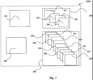
Reference is now made to figure 1, illustrating a system 1000 adapted to
detect and/or
identify specific bacteria within a sample according to one preferred
embodiment of
the present invention. System 1000 comprises:
a. means 100 for obtaining an absorption spectrum (AS) of the sample;
b. statistical processing means 200 for acquiring the n dimensional volume
boundaries for the specific bacteria, having:
i. means 201 for obtaining at least one absorption spectrum (AS2)
of samples containing the specific bacteria;
ii. means 202 for extracting x features from the AS2 selected from
a group consisting of peaks' wavelength, peaks height and
widths, different peaks' intensity ratios or any combination
thereof; x is an integer higher or equal to one;
iii. means 203 for calculating at least one derivative of the AS2;
iv. means 204 for dividing the AS2 into several segments
according to the x features;
v. means 205 for calculating the y statistical correlation of each of
the segment; y is an integer higher or equal to one;
vi. means 206 for defining n dimensional space; n equals the sum
of the x features and the y statistical correlations;
CA 02683142 2009-10-05
WO 2008/122975 PCT/IL2008/000472
vii. means 207 for assigning each of the x feature and the y
correlation to the specific bacteria;
viii. means 208 for calculating the Gaussian distribution for each of
the x feature and/or for each of the y statistical correlations; the
Gaussian distributions defined the n dimensional volume in the
n dimensional space;
ix. means 209 for determining the boundaries of the n dimensional
volume by using technique selected from a group consisting of
quadratic Gaussian classifier, k nearest neighbor, Bayesian
classification or any combination thereof;
c. means 300 for data processing the AS, having:
i. means 301 for noise reducing by using different smoothing
techniques selected from a group consisting of running average
savitzky-golay or any combination thereof;
ii. means 302 for extracting m features from the AS selected from
a group consisting of peak's width, intensity, the ratio
width/intensity, peak's wavelength, different peaks' intensity
ratios, or any combination thereof; m is an integer higher or
equal to one;
iii. means 303 for dividing the AS into several segments according
to the m features;
iv. means 304 for calculating the m, statistical correlation of each
of the segment; ml is an integer higher or equal to one; and,
d. means 400 for detecting and/or identifying the specific bacteria if the
mi statistical correlation and/or the m features are within the n
dimensional volume.
According to another embodiment of the present invention, means 300 (in system
1000) for data processing the AS additionally characterized by:
i. means 305 for calculating at least one of the o derivative of
the AS; o is an integer.greater than or equals 1;
ii. means 306 for extracting mZ features from the oth derivative
selected from a group consisting of peak's width, intensity, the
ratio width/iritensity, different peaks' intensity ratios, peak's
wavelength or any combination thereof;
CA 02683142 2009-10-05
WO 2008/122975 PCT/IL2008/000472
21
iii. means 307 for dividing the o derivative into several segments
according to the mz features;
iv. means 308 for calculating the m3 statistical correlation in each
of the segments; and,
v. means 309 for detecting and/or identifying the specific bacteria
if the ml and/or m3 statistical correlation and/or the m and/or the
m2 features are within the n dimensional volume.
According to another embodiment of the present invention, means 308 or 304 (in
system 1000) for calculating the statistical correlation is selected from a
group
consisting of Pearson's correlation coefficient.
According to yet another embodiment of the present invention, the specific
bacteria to
be identified by system 1000 is selected from a group consisting of
Streptococcus
Pyogenes, Group C and G beta-hemolytic streptococci, Corynebacterium
haemolyticum, Diphtheria and Ulcerans, Neisseria Gonorrhoeae, Mycoplasma
Pneumoniae, Yersinia Enterocolitica, Mycobacterium tuberculosis, Chlamydia
Trachomatiss and Pneumoniae, Bordetella Pertussis, Legionella spp,
Pneumocystis
Carinii, Nocardia, Histoplasma Capsulatum, Coccidioides Immitis, Haemophilus
influenza group A beta hemolytic and staphylococcus Aureus.
According to another embodiment of the present invention, the means 100 for
obtaining an absorption spectrum (AS) of the sample (in system 1000),
additionally
comprising:
a. at least one optical cell for accommodating the sample;
b. p light source selected from a group consisting of laser, lamp, LEDs
tunable lasers, monochrimator, p is an integer equal or greater than 1; the
p light source are adapted to emit light at different wavelength to the
optical cell; and,
c. detecting means for receiving the spectroscopic data of the sample
exiting from the optical cell.
According to yet another embodiment of the present invention, the p light
source (in
system 1000) are adapted to emit light at wavelength range selected from a
group
consisting of UV, visible, IR, mid-IR, far-IR and terahertz.
Reference is now made to figure 2, illustrating a system 2000 adapted to
detect and/or
identify specific bacteria within a sample, according to another preferred
embodiment
of the present invention. System 2000 comprises:
CA 02683142 2009-10-05
WO 2008/122975 PCT/IL2008/000472
22
a. means 100 for obtaining an absorption spectrum (AS) of the sample;
the AS containing water influence;
b. statistical processing means 200 for acquiring the n dimensional
volume boundaries for the specific bacteria, having:
i. means 201 for obtaining at least one absorption spectrum (AS2)
of samples containing the specific bacteria;
ii. means 202 for extracting x features from the AS2 selected from
a group consisting of peaks wavelength, peaks height and
widths, different peaks' intensity ratios or any combination
thereof; x is an integer higher or equal to one;
iii. means 203 for calculating at least one derivative of the AS2;
iv. means 204 for dividing the AS2 into several segments
according to the x features;
v. means 205 for calculating the y statistical correlation of each of
the segment; y is an integer higher or equal to one;
vi. means 206 for defining n dimensional space; n equals the sum
of the x features and the y statistical correlations;
vii. means 207 for assigning each of the x feature and the y
correlation to the specific bacteria;
viii. means 208 for calculating the Gaussian distribution for each of
the x feature and/or for each of the y statistical correlations; the
Gaussian distributions defined the n dimensional volume in the
n dimensional space;
ix. means 209 for determining the boundaries of the n dimensional
volume by using technique selected from a group consisting of
quadratic Gaussian classifier, k nearest neighbor, Bayesian
classification or any combination thereof;
c. means 300 for eliminating the water influence from the AS,
comprising:
i. means 301 for providing the absorption intensity at each of
wavenumber (x) within said AS (Sigwtth water(x));
ii. means 302 for dividing said AS into at least two wavenumber
ranges;
CA 02683142 2009-10-05
WO 2008/122975 PCT/IL2008/000472
23
iii. means 303 for calculating the correction factors (CF) at each
wavenumber (x) within said least two ranges (CF(x));
iv. means 304 for acquiring from said AS at least one absorption
intensity that is mainly influenced by said water Sigwater only(xl )
and the corresponding wavenumbers (xl);
v. means 305 for calculating at least one correction factor of said
water (CFwate, only (xl)) at said at least one wavenumber (xl);
vi. means 306 for dividing said at least one Sigwater only(xl ) by said
at least one CFwater (i.e., Sigwater only(xl )/ CFwater only (xl)) at said
at least one wavenumber (xl);
vii. means 307 for calculating the average of step (vi) (AVG[Sigwater
only(xl ) / CFwater only (xl )] );
viii. means 308 for multiplying said AVG[Sigwater only(xl) / CFwater
onty] (xl) by said CF(x) for each of said wavenumber (x) within
said AS; and,
ix. means 309 for subtracting the result of step (viii) from said
(Sjgwith water(x)) at each of said wavenumber(x) within said AS;
d. means 400 for data processing the AS without the water influence,
characterized by:
i. means 401 for noise reducing by using different smoothing
techniques selected from a group consisting of running average
savitzky-golay or any combination thereof;
ii. means 402 for extracting m features from the AS selected from
a group consisting of peak's width, intensity, the ratio
width/intensity, peak's wavelength, different peaks' intensity
ratios, or any combination thereof; m is an integer higher or
equal to one;
iii. means 403 for dividing the AS into several segments according
to the m features;
iv. means 404 for calculating the mi statistical correlation of each
of the segment; ml is an integer higher or equal to one; and,
e. means 500 for detecting and/or identifying the specific bacteria if the
mi statistical correlation and/or the m features are within the n
dimensional volume.
CA 02683142 2009-10-05
WO 2008/122975 PCT/IL2008/000472
24
According to another embodiment of the present invention, means 400 (in system
2000) for data processing the AS without the water influence additionally
comprising:
i. means 405 for calculating at least one of the o derivative of the AS; o
is an integer greater than or equals 1;
ii. means 406 for extracting m2 features from the o derivative selected
from a group consisting peak's width, intensity, the ratio
width/intensity, different peaks' intensity ratios, peak's wavelength or
any combination thereof;
iii. means 407 for dividing the o~' derivative into several segments
according to the m1 features;
iv. means 408 for calculating the mj statistical correlation in each of the
segments; and,
v. means 409 for detecting and/or identifying the specific bacteria if the
mI and/or m3 statistical correlation and/or the m and/or the m2 features
are within the n dimensional volume.
According to another embodiment of the present invention, means 408 and/or 404
within system 2000, for calculating the statistical correlation in each of
said.segments
is selected form a group consisting of Pearson's correlation coefficient.
According to another embodiment of the present invention, the specific
bacteria (in
system 2000) is selected from a group consistirig of Streptococcus Pyogenes,
Group
C and G beta-hemolytic streptococci, Corynebacterium haemolyticum, Diphtheria
and Ulcerans, Neisseria Gonorrhoeae, Mycoplasma Pneumoniae, Yersinia
Enterocolitica, Mycobacterium tuberculosis, Chlamydia Trachomatiss and
Pneumoniae, Bordetella Pertussis,Legionella spp, Pneumocystis Carinii,
Nocardia,
Histoplasma Capsulatum, Coccidioides Immitis, Haemophilus influenza group A
beta
hemolytic and staphylococcus Aureus.
According to another embodiment of the present invention, means 100 for
obtaining
an absorption spectrum (AS) of the sample additionally comprising:
a. at least one optical cell for accommodating the sample;
b. p light source selected from a group consisting of laser, lamp, LEDs
tunable lasers, monochrimator, p is an integer equal or greater than 1; p
light source are adapted to emit light at different wavelength to the
optical cell; and,
CA 02683142 2009-10-05
WO 2008/122975 PCT/IL2008/000472
c. . detecting means for receiving the spectroscopic data of the sample
exiting from the optical cell.
According to yet another embodiment of the present invention, the.p light
source are
adapted to emit light at wavelength range selected from a group consisting of
UV,
visible, IR, mid-IR, far-IR and terahertz.
Yet another object of the present invention is to provide a method for
detecting and/or
identifying specific bacteria within a sample. The method comprises step
selected
inter alia from:
a. obtaining an absorption spectrum (AS) of the sample;
b. acquiring the n dimensional volume boundaries for the specific
bacteria by:
i. obtaining at least one absorption spectrum (AS2) of samples
containing the specific bacteria;
ii. extracting x features from the AS2 selected from a group
consisting of peaks wavelength, peaks height and widths,
different peaks' intensity ratios or any. combination thereof; x is
an integer higher or equal to one;
iii. calculating at least one derivative of the AS2;
iv. dividing the AS2 into several segments according to the x
features;
v. calculating the y statistical correlation of each of the segment; y
is an integer higher or equal to one;
vi. defining n dimensional space; n equals the sum of the x features
and the y statistical correlations;
vii. assigning each of the x feature and the y correlation to the
specific bacteria;
viii. calculating the Gaussian distribution for each of the x feature
and/or for each of the y statistical correlations; the Gaussian
distributions defined the n dimensional volume in the n
dimensional space;
ix. determining the boundaries of the n dimensional volume by
using technique selected from a group consisting of quadratic
Gaussian classifier, k nearest neighbor, Bayesian classification
or any combination thereof.
CA 02683142 2009-10-05
WO 2008/122975 PCT/IL2008/000472
26
c. data processing the AS;
i. noise reducing by using different smoothing techniques
selected from a group consisting of running average savitzky-
golay or any combination thereof;
ii. extracting m features from the AS selected from a group
consisting of peak's width, intensity, the ratio width/intensity,
peak's wavelength, different peaks' intensity ratios, or any
combination thereof; m is an integer higher or equal to one;
iii. dividing the AS into several segments according to the m
features;
iv. calculating the mi statistical correlation of each of the segment;
ml is an integer higher or equal to one; and,
d. detecting and/or identifying the specific bacteria if the mi statistical
correlation and/or the m features are within the n dimensional volume.
It should be pointed out that in each of the systems as described above
(either 1000 or
2000), the statistical processing means 200 is used only once for each
specific
bacteria. Once the boundaries were provided by the statistical processing
means 200
the determination whether the specific bacteria is present in a sample is
performed by
verifying whether the ml and/or m3 statistical correlation and/or the m and/or
m2
features are within the boundaries. Furthermore, once the boundaries were
provided,
there exists no need for the statistical processing of the same specific
bacteria again.
Yet another object of the present invention is to provide a method for
detecting and/or
identifying specific bacteria within a sample. The method comprises steps
selected
inter alia from:
a. obtaining an absorption spectrum (AS) of the sample; the AS
containing water influence;
b. acquiring the n dimensional volume boundaries for the specific
bacteria by:
i. obtaining at least one absorption spectrum (AS2) of samples
containing the specific bacteria;
ii. extracting x features from the AS2 selected from a group
consisting of peaks wavelength, peaks height and widths,
CA 02683142 2009-10-05
WO 2008/122975 PCT/IL2008/000472
27
different peaks' intensity ratios or any combination thereof; x is
an integer higher or equal to one;
iii. calculating at least one derivative of the AS2;
iv. dividing the AS2 into several segments according to the x
features;
v. calculating the y statistical correlation of each of the segment; y
is an integer higher or equal to one;
vi. defining n dimensional space; n equals the sum of the x features
and the y statistical correlations;
vii. assigning each of the x feature and the y correlation to the
specific bacteria;
viii. calculating the Gaussian distribution for each of the x feature
and/or for each of the y statistical correlations; the Gaussian
distributions defmed the n dimensional volume in the n
dimensional space;
ix. determining the boundaries of the n dimensional volume by
using technique selected from a group consisting of quadratic
Gaussian classifier, k nearest neighbor, Bayesian classification
or any combination thereof;
c. eliminating the water influence from the AS by:
i. providing the absorption intensity at each of wavenumber (x)
within said AS (Sigwih water(x));
ii. dividing said AS into at least two wavenumber ranges;
iii. calculating the correction factors (CF) at each wavenumber (x)
within said least two ranges (CF(x));
iv. acquiring from said AS at least one absorption intensity that is
mainly influenced by said water Sigwater onry(xl ) and the
corresponding wavenumbers (xl);
v. calculating at least one correction factor of said water (CFwater
,ly (xl)) at said at least one wavenumber (xl);
vi. dividing said at least one Sigwater oniy(xl ) by said at least one
CFwater (i.e., Sigwater only(xl )/ CFwater only (xl )) at said at least one
wavenumber (xl);
CA 02683142 2009-10-05
WO 2008/122975 PCT/IL2008/000472
28
vii. calculating the average of step (vi) (AVG[Sigwater onty(x1). /
CFwater only (xl )] );
viii. multiplying said AVG[Sigwater only(xl )/ CFwater onry] (xl) by said
CF(x) for each of said wavenumber (x) within said AS; and,
ix. subtracting the result of step (viii) from said (Sigwfrh water(x)) at
each of said wavenumber(x) within said AS;
d. data processing the AS without the water influence by:
i. noise reducing by using different smoothing techniques
selected from a group consisting of running average savitzky-
golay or any combination thereof;
ii. extracting m features from the AS selected from a group
consisting of peak's width, intensity, the ratio width/intensity,
peak's wavelength, different peaks' intensity ratios, or any
combination thereof; m is an integer higher or equal to one;
iii. dividing the AS into several segments according to the m
features;
iv. calculating the mi statistical correlation of each of the segment;
ml is an integer higher or equal to one; and,
e. detecting and/or identifying the specific bacteria if the m, statistical
correlation and/or the m features are within the n dimensional volume.
In each of the methods as described above, the statistical processing is used
only once
for each specific bacteria. Once the boundaries were provided by the
statistical
processing the determination whether the specific bacteria is present in a
sample is
performed by, verifying whether the m, statistical correlation and/or said m
features
are within the boundaries. Furthermore, once the boundaries were provided,
there
exists no need for the statistical processing of the same specific bacteria
again.
According to another embodiment of the present invention step (c) of data
processing
the AS, in the methods as described above, additionally comprising steps of:
i. calculating at least one of the oth derivative of the AS; o is an integer
greater than or equals 1;
ii. extracting mz features from the oth derivative selected from a group
consisting of peak's width, intensity, the ratio width/intensity, different
peaks' intensity ratios, peak's wavelength or any combination thereof;
CA 02683142 2009-10-05
WO 2008/122975 PCT/IL2008/000472
29
iii. dividing the o`h derivative into several segments according to the m1
features;
iv. calculating the m3 statistical correlation in each of the segments; and,
v. detecting and/or identifying the specific bacteria if the ml and/or m3
statistical correlation and/or the m and/or the mZ features are within the
n dimensional volume.
According to another embodiment of the present invention, the step of
calculating the
statistical correlation of each of said segment, in the methods as described
above, is
performed by using Pearson's correlation coefficient.
According to another embodiment of the present invention, the methods as
described
above, additionally comprising the step of selecting the specific bacteria
selected from
a group consisting of Streptococcus Pyogenes, Group C and G beta-hemolytic
streptococci, Corynebacterium haemolyticum, Diphtheria and Ulcerans, Neisseria
Gonorrhoeae, Mycoplasma Pneumoniae, Yersinia Enterocolitica, Mycobacterium
tuberculosis, Chlamydia Trachomatiss and Pneumoniae, Bordetella Pertussis,
Legionella spp, Pneumocystis Carinii, Nocardia, Histoplasma Capsulatum,
Coccidioides Immitis, Haemophilus influenza group A beta hemolytic and
staphylococcus A ureus.
According to another embodiment of the present invention, the step of
obtaining the
AS, in the methods as described above, additionally comprising the following
steps:
a. providing at least one optical cell accommodates the sample;
b. providing p light source selected from a group consisting of laser,
lamp, LEDs tunable lasers, monochrimator, p is an integer equal or
greater than 1; p light source are adapted to emit light to the optical
cell;
c. providing detecting means for receiving the spectroscopic data of the
sample;
d. emitting light from the light source at different wavelength to the
optical cell; and,
e. collecting the light exiting from the optical cell by the detecting means;
thereby obtaining the AS.
According to another embodiment of the present invention, the step of emitting
light
is -performed at the wavelength range of UV, visible, IR, mid-IR, far-IR and
terahertz.
CA 02683142 2009-10-05
WO 2008/122975 PCT/IL2008/000472
According to another embodiment of the present invention, the methods as
defined
above, additionally comprising the step of detecting the bacteria by analyzing
the AS
in the region of about 3000-3300 cm 1 and/or about 850-1000 cm I and/or about
1300-
1350 cm"1.
According to yet another embodiment of the present invention, the absorption
spectra,
in any of the systems (1000 or 2000) or for any of the methods as described
above, is
obtained using an instrument selected from the group consisting of a Fourier
transform infrared spectrometer, a fluorometer and a Raman spectrometer.
According to yet another embodiment of the present invention, the uncultured
sample,
in any of the systems (1000 or 2000) or for any of the methods as described
above, is
selected from fluid originated from the human body such as blood, saliva,
urine, bile,
vaginal secretions, middle ear aspirate, pus, pleural effusions, synovial
fluid,
abscesses, cavity swabs, and serum.
In the foregoing description, embodiments of the invention, including
preferred
embodiments, have .been presented for the purpose of illustration and
description.
They are not intended to be exhaustive or to limit the invention to the
precise.form
disclosed. Obvious modifications or variations are possible in light of the
above
teachings. The embodiments were chosen and described to provide the best
illustration of the principals of the invention and its practical application,
and to
enable one of ordinary. skill in the art to utilize the invention in various
embodiments
and with various modifications as are suited to the particular use
contemplated. All
such modifications and variations are within the scope of the invention as
determined
by the appended claims when interpreted in accordance with the breadth they
are
fairly, legally, and equitably entitled.
EXAMPLES
Examples are given in order to prove the embodiments claimed in the present
invention. The examples describe the manner and process of the present
invention and
set forth the best mode contemplated by the inventors for carrying out the
invention,
but are not to be construed as limiting the invention.
EXAMPLE 1 - Water influence
CA 02683142 2009-10-05
WO 2008/122975 PCT/IL2008/000472
31
One of the major problems in identifying bacteria from a fluid sample's
spectrum (and
especially an aerosol spectrum) is the water influence (i.e., the water noise
which
masks the desired spectrum by the water spectrum).
The water molecule may vibrate in a number of ways. In the gas state, the
vibrations
involve combinations of symmetric stretch (vl), asymmetric stretch (v3) and
bending
(v2) of the covalent bonds. The water molecule has a very small moment of
inertia on
rotation which gives rise to rich combined vibrational-rotational spectra in
the vapor
containing tens of thousands to millions of absorption lines. The water
molecule has
three vibrational modes x, y and z. The following table (table 1) illustrates
the water
vibrations, wavelength and the assignment of each vibration:
Table 1: water vibrations, wavelength and the assignment of each vibration
Wavelength cm-1 Assignment
0.2~mm 50 intermolecular bend
55 n1 183.4 intermolecular stretch 25 m 395.5 L=1, librations
15 uni 686.3 L2, librations 6.08 ni 164~ v2. bend
4.65 1n 2150 v2 + L2 b
3.05 Eun 3277 v 1. wmmetric- stretch 2.87 hrn 3490 v3. asymnletric
`* stretch t.
1900 nni 5260 av t+ v2 + bv3;
a+b=1 1470 nm 6800 avl T bv3; a+b=21200 nni 8330avl .+ v2 -bv3;
a+b=2
970 nm 10310 av 1+ bv3; a-=b=3
836 nm 11960 av1 v2 + bv3;'a+b+3
739 nm 1353.0 avl + bv3; a+b=4
660 nm 15150 avl + _v2 bv3;
a+b=4
606 nrn 16500 avl bv3; a+b=5514 iun 19460 a,,-1 bv3; a+b=ba and ba.re
integers;> 0 ms.
The present invention provides a method for significantly reducing and even
eliminating the water influence within the absorption spectra.
CA 02683142 2009-10-05
WO 2008/122975 PCT/IL2008/000472
32
Reference is now made to figures 3 and 4 which illustrate an absorption
spectrum of a
sample with and without the water influence. Example for a spectrum without
the
water influence is given in figure 4. Figure 3 represents a spectrum prior to
the water
correction.
The method for eliminating the water influence contains the following steps:
First the absorption spectrum was divided in several segments (i.e, wavelength
ranges). The spectrum was divided to segments (wavenumber ranges) of about
1800
cm"1 to about 2650 cm i, about 1400 cm'1 to about 1850 cm'1, about 1100 cm"1
to
about 1450 cm 1, about 950 cm 1 to about 1100 cm 1, about 550 cm 1 to about
970
cml:
The segments were determined according to (i) different intensity peaks within
the
water's absorption spectrum; and, (ii) the signal's trends.
Next, each segment was eliminated from the water influence in the following
manner:
(a) providing the absorption intensity at each of wavenumber (x) within the
absorption spectrum (refers hereinafter as Sigwih water(x));
(b) calculating the correction factors (CF) at each wavelength (refers
hereinafter
as x) within each segment (refers hereinafter as CF(x));
(c) acquiring from the absorption spectrum, at least one absorption intensity
that
is mainly influenced by water (refers hereinafter as Sigwater on1y(x1)) at the
corresponding wavenumbers (xl);
(d) calculating at least one correction factor of the water (CFwate, anty
(xl)) at said
at least one wavenumber (xl);
(e) dividing at least one Sigwater only(xl ) by at least one CFwater (i.e.,
Sigwater only(xl )
/ CFwater only (xl )) at said at least one wavenumber (xl);
(f) calculating the average of the results of step (e) (refers hereinafter as
AVG[Sigwater only(XI ) / CFwater only (XI A );
(g) multiplying the AVG[Sigwater only(x1) / CFwater only] (xl) by CF(x) for
each
wavenumber (x); and,
(h) Subtracting each result of step (g) from Sigwith water(x) per each (x).
In other words, each absorption intensity within the spectrum is eliminated
from the
water influence according to the following equation:
Slgwith water(x)-( CF(x) * A VG [Sigwater only(XI )/ CFwater only (XI A )
Calculating the correction factors
CA 02683142 2009-10-05
WO 2008/122975 PCT/IL2008/000472
33
The correction factors (CF) depends on the wavelength range, the water
absorption
peak's shape at each wavelength, peak's width, peak's height, absorption
spectrum
trends and any combination thereof. The following series were used as a
correction
factor (x - denote the wavenumber in cm I)
1. Wavelength range 1846 cm 1 to 2613 cm"1
Coefficients:
all = 137.2;
bll = 2170;
cll = 224.3;
a21 = 19.02;
b21 = 2063;
c21 = 37.53;
a31 = 0.7427;
b31 = 2224;
c31= 13;
a41 = 98.33;
b41 = 2124;
c41= 109.8;
a51 = -4.988;
b51= 2192;
c51 = 33.87;
a61 = 20.19;
b61 = 1998;
c61 = 40.22;
a71 = 228.3;
b71 =. 1496;
c71 = 1329;
a81 = 6.751e+012;
b81 = -1226;
c81 = 592.1;
CF(x) = al l* e(-((z-bl l)/cll)=) + a21 * e(-((z-bzl)/czl)z) + a31 * e(-((z-
b31)/c31)=) +a41 * e(-((r-b41)/c41)=) + a51 * e(-((x-b51)/c51)=) +
a61 * e(-((z-b61)/c6l)2) + a71 * e(-((z-b71)1c71)Z) + ag 1* e(-((z-b81)/c81)=)
2. Wavelength range 1461 cm 1 to 1846 cm"1
a12 = -300.2;
b12 = 1650;
c12 = 13.65;
a22 = -51.65;
b22 = 1665;
c22 = 6.48;
a32 = 142.4;
b32 = 1623;
c32 = 7.584;
a42 = 1450;
b42 = 1649;
CA 02683142 2009-10-05
WO 2008/122975 PCT/IL2008/000472
34
c42 = 32.62;
a52 = 96.34;
b52 = 1617;
c52 = 2.387;
a62 = 608;
b62 = 1470;
c62 = 369.3;
a72 = 0;
b72 = 1873;
c72 = 2.625;
a82 = 1037;
b82 = 1644.;
c82 = 76.21;
CF(x) = a12 * g(-((s-6l2)/c21)=) +a22 * e(-((z-b22)/c22)=) + a32 * C(-((z-
b32)1c32)=) + a42 * g(-((z-ba2)/c42)2) + a52 * C(-((z-b52)/c52)=) +
a62 * C(-((x-b62)/c62)')+ a72 * e(-((r-b72)1c72)=) + a82 * Q(-((z-b82)/c82)=)
3. Wavelength range 1111 cm-1 to 1461 cm 1
a13 = 1368;
b13 = 2167;
c13 = 767;
a23 = 80.67;
b23 = 1356;
c23 = 68.83;
a33 = 36.85;
b33 = 1307;
c33 = 33.79;
a43 = 142.5;
b43 = 1244;
c43 = 67.19;
a53 = 260.4;
b53 = 1130;
c53 = 88.91;
a63 = 66.54;
b63 = 1093;
c63 = 31;
a73 = 7.126;
b73 = 1345;
c73 = 20.9;
a83 = 4.897;
b83 = 1280;
c83 = 11.05;
CF(x) = a13 s e(-((x-nls)/cls)') +a23 s e(-((:-a2s)/c23)') + a33 * e(-((x-
b33)lCsa)=) + a43 s e(-((:-na3)/cas)=) + a53 * e(-((:-as3)/css) ) +
a63'R e(-((z-b63)/c63)=) + a73 * C(-((x-b73)/c73)=) + a83 71 g(-((i-
b83)/c8J)=)
4. Wavelength range 961 cm-1 to 1111 cm-1
a14 = 692.6;
b14 = 952;
CA 02683142 2009-10-05
WO 2008/122975 PCT/IL2008/000472
c14 = 31.04;
a24 = 48.46;
b24 = 983.2;
c24 = 15.72;
a34 = 287.5;
b34 = 994.6;
c34 = 27.98;
a44 = 434.9;
b44 = 1032;
c44 = 40.86;
a54 = 17.05;
b54 = 1052;
c54 = 13.55;
a64 = 48.61;
b64 = 1068;
c64 = 16.56;
a74 = 70.71;
b74 = 1086;
c74 = 21.23;
a84 = 497.3;
b84 = 1124;
c84 = 64.42;
CF(x) = a14 * e(-((=-ala)roi4)') + a24 * e(-((=-nz4)/cz4)2) +a34 * e(-((x-
nsa)lcsa)') + a44 * e(-((x-a44)lcaa)') + a54 * e(-((=-as4)/cs4)') +
a64 * e(-((r-b64)1c64)=) + a74 * e(-((x-b74)lc74)=) + a84 * e(-((z484)/c84)2)
Wavelength range 570 cm 1 to 961 cm 1
a15 = -2877;
b15 = 36.23;
c15 = 29.09;
a25 = 0;
b25 = -124.3;
c25 = 22.09;
a35= -190.7;
b35 = 18.97;
c35 = 16.45;
a45 = 1.589e+004;
b45 = -3.427;
c45 = 56.25;
a55 = -1.352e+004;
b55 = -5.861;
c55 = 40.75;
a65 = 476.7;
b65 = 82.38;
c65 = 17.29;
a75 = 1286;
b75 = 62.29;
c75 = 180.3;
a85 = 802.9;
b85 = 102.8;
CA 02683142 2009-10-05
WO 2008/122975 PCT/IL2008/000472
36
c85 = 18.79;
CF(x) = a15 * ec-(cz-b15>1c15)=) +a25 * ec-ccx-bz5>1cz5)=) +a35 * ec-(cx-
b35>1c35)=) +a45 * ec-(cx-b45)1c45)2) +a55 * Qc-(cz-b55>1c55)Z) +
a65 * ec-(cz-b65>/c65)2) +a75 * e(-ccz-b75>/c75)=).+ a85ec-(cz-bs5)lcs5)=)
Absorption intensity mainly influenced by water
Reference is made again to figure 3 which illustrate the absorption spectrum
prior to
eliminating the water influence.
As can be seen from the figure, the absorption intensity that is mainly
influenced by
the water is the wavenumber region of 2000 cm -1 and above. The intensity at
that
region is about 0.2 absorption units. In the present example, xl is 2000 and
Sigwoter
only(x1) is 0.2.
Reference is made again to figure 4, which illustrate the absorption spectrum
of a
sample after the influence of the water was eliminated.
It should be pointed out that for the purpose of obtaining a better resolution
both
graphs (3 and 4) are normalized to 2 (i.e., multiplied by 2).
EXAMPLE 2 - Bacteria's absorption spectrum
Each type of bacteria has a unique spectral. signature. Although many types of
bacteria
have similar spectral signatures there are still some spectral differences
that are due to
different proteins on the cell membrane and differences in the DNA/ RNA
structure.
The following table, table 2, lists the wavelengths in the IR region that can
be used to
identify a bacterium.
Table 2: bacterium wavelengths in the IR region
Wavelength Wavenumber Assignment
[nm] [cm']
2857 3500 0-H stretch etch of hydroxyl groups
3125 3200 N-H stretch (amide A) of proteins
3379 2959 C-H stretch (asymmetric) of -CH3
3408 2934 C-H stretch (asymmetric) of >CH2
3423 2921 C-H stretch (asymmetric) of >CH2 in fatty acids
3450 2898 C-H stretch of C-H methine
3481 2872 C-H stretch (symmetric) of CH3
3506 2852 C-H stretch (symmetric) of >CH2 in fatty acids
5743 1741 >C=0 stretch of esters
5830 1715 >C=0 stretch of esters, RNA/DNA, OH-C=0
5899 1695 Amide I band components
5934 1685 resulting from antiparallel
5970 1675 pleated sheets and b-turns of proteins
6042 1655 Amide I of a-helical stretch uctures
CA 02683142 2009-10-05
WO 2008/122975 PCT/IL2008/000472
37
6108 1637 Amide I of b-pleated sheet stretch uctures
6459 1548 Amide II
6600 1515 "Tyrosine" band
6811 1468 C-H def of >CH2
7142 1400 C=O stretch (symmetric) of COO"
7633-8064 1310-1240 Amide III band components of proteins
8000-8190 1250-1220 P=0 stretch (asymmetric) of >P02 hosphodiesters
8333-11111 1200-900 C-O-C, C-O dominated by ring vibrations of
carbohydrates C-O-P, P-O-P
9216 1085 P=O stretch (symmetric) of >POz
13888 720 C-H rocking of >CH2
11111- 900-600 Specific bacteria bonds
16666
The table was taken from Naumann D., "Infrared spectroscopy in microbiology",
Encyclopedia ofAnalytical Chemistry, R.A. Meyers (Ed.) pp. 102-131, John Wiley
&
Sons Ltd, Chichester, 2000.
The following table, table 3, and figure 5 is an example of the absorption
peaks of
streptococcus payogenous. Some of the peaks could be related to the peaks in
table 2.
Others, such as peak number 1, 2, 9, 12, 13 and 14 are specific streptococcus
payogenous peaks discovered in the present invention and are used for the
detection
and identification.
Table 3: streptococcus' absorption peaks,
Peak Wavenumber Absorbanc Weightin Assignment.
numbe [cm"1] e g factor
r
1 3286 0.3638 0.09 Specific bacteria characteristics
2 3077 0.2852 0.1 Specific bacteria characteristics
3 2962 0.2875 0.1 C-H stretch (asymmetric) of -
CH3
4 2933 0.2785 0.09 C-H stretch (asymmetric) of
>CH2
1648 0.5245 0.01 Amide I of a-helical stretch
uctures
6 1548 0.4029 0.01 Amide II
7 1452 0.2235 0.15 C-Hdefof>CH2
8 1405 0.2920 0.15 C=O stretch (symmetric) of
COO"
9 1348 0.1763 0.15 Specific bacteria characteristics
1245 0.1589 0.15 P=0 stretch (asymmetric) of
>POZ
11 1076 0.2159 0.15 P=0 stretch (symmetric) of
>POz
12 989 0.0957 0.05 Specific bacteria characteristics
13 925 0.0821 0.05 Specific bacteria characteristics
CA 02683142 2009-10-05
WO 2008/122975 PCT/IL2008/000472
38
14 850 0.0828 0.05 Specific bacteria characteristics
EXAMPLE 3 - Distin ug ishingbetween two bacteria in a dry mixture
The. following ex-vivo example provides a method to distinguish between two
bacteria within a dry mixture of - Streptococcus payogenes and Staphylococcus
aureus and to identify and/or determine whether Streptococcus is present
within the
sample.
Five mixtures of - Streptococcus payogenes and Staphylococcus aureus were
prepared
according to the following protocol:
1. The following solutions of Strep. P hemolytic (cat number-ATCC 19615) and
Staphylococcus aureus (Cat. Number ATCC 25923) were prepared:
Total Strep Pyogenes Staph Aurous
volume CFU/160,uL CFU/160 fcL
60 12 0
60 9 3
60 6 6
60 3 9
60 0 12
Total 150 L 150 L
2. 30 L of each solution was placed on one marked slot on an optical plate
(ZnSe). Another slot contained 30 g1 ddHZO for reference.
3. The plate was placed in a desiccator (Dessicator 250 mm polypropylene,
Yavin Yeda, Israel) in. the presence of several petri plates having a
desiccant
agent (Phosphorus Pentoxide cat #79610 Sigma Aldrich).
4. The desiccator was vacuumed for about 30 minutes.
Then the samples were placed inside a FTIR spectrometer (Bruker) and the
spectral
response of the samples was acquired (spectral range 4000cm'1 to 400cm 1).
The absorption spectra of each sample were applied with Blackman-Harris 3-term
apodization function (see the following equation):
CA 02683142 2009-10-05
WO 2008/122975 PCT/IL2008/000472
39
`~ 1~72 47i '?2 6 ~fiZ
(~'~~) = ~0 - c~l eQS - 1) (~T + c~~ cos - 1) - ~3 cos (1'F - 1)
1~
a0. = 0.35875; ai = 0.48829; a,) = 0.14128; a3 = 0.01168
In whichu'(n) is the Blackman-Harris 3-term apodization; n is integer between
0 to N-
1; and, N is the range at which the Blackman-Harris 3-term apodization
function was
applied.
Next, the sample was scanned sufficient number of times (64 scans) in order to
reach
a signal to noise ratio greater than 3000:1.
The identification and/or detection of specific bacteria was as follows:
(a) the noise in each of the absorption spectra was reduced by using Savitzky-
Golay smoothing;
(b) the signal's first derivative was calculated;
(c) m features such as, but not limited to, peaks wavelength, peaks height and
widths, peaks height ratio etc. were extracted from the spectra. A total of m
features were extracted. m is an integer higher or equals 1;
(d) the signal and/oi its first derivative were divided into several regions
(segments, i.e., several wavenumber regions) according to said m features;
(e) mi statistical correlation were calculated for (i) the spectral signal at
each
region; and for (ii) the signal derivatives at each region. A total of ml
statistical correlations were extracted. mi is an integer higher or equals 1.
The
statistical correlation for the each region (the signal's and its derivatives)
was
calculated by using Pearson's correlation coefficient;
(f) the m features and the mi statistical correlation were examined and
checked
whether they are within the n dimensional volume boundaries (which acquired
by the statistical processing);
(g) the identification of the specific bacteria was determined as positive if
the m
features and/or the mi statistical correlation were within the n dimensional
volume boundaries.
Statistical processing
The statistical processing is especially adapted to provide the n dimensional
volume
boundaries. For each specific bacterium the statistical processing was
performed only
once, for obtaining the boundaries. Once the boundaries were provided, the
CA 02683142 2009-10-05
WO 2008/122975 PCT/IL2008/000472
determination whether the specific bacteria is present in a sample was as
explained
above (i.e., verifying whether the features or correlation are within the
boundaries).
Furthermore, once the boundaries were provided, there exists no need for the
statistical processing of the same specific bacteria again.
The statistical processing for each specific bacterium is performed in the
following
manner:
(a) obtaining several absorption spectrum (AS2) of samples containing the
specific bacteria;
(b) extracting x features such as, but not limited to, peaks wavelength, peaks
height and widths, different peaks' intensity ratios etc. were extracted from
the
spectra. A total of x features. x is an integer higher or equals 1;
(c) calculating the signal's first derivative;
(d) dividing the signal and/or its first derivative into several regions
(segnients)
according to said x features;
(e) Calculating the y correlation for the different segments within the
absorption
spectrums;
(f) Defining n dimensional space. n equals the sum of the x features and the y
statistical correlations;.
(g) Assigning and/or interlinking each one of the x feature, and each one of
the y
correlation to the specific bacteria which its identification is required;
(h) Calculating the Gaussian distribution for (i) each of the x feature;
and/or for
(ii) each of the y statistical correlations. All the calculated Gaussian
distributions constitute an n dimensional volume in the n dimensional space;
and,
(i) Determining the boundaries of each volume by using quadratic Gaussian
classifier or similar method (for example k nearest neighbor, Bayesian
classification et cetera).
If the features and correlation (extracted from the spectrum) are within the n
dimensional volume boundaries, the specific bacteria are identified. Otherwise
the
bacteria are not identified.
Alternatively or additionally, each of the x feature and/or for the y
statistical
correlations is given a weighting factor. The weighting factor is determined
by the
CA 02683142 2009-10-05
WO 2008/122975 PCT/IL2008/000472
41
examining how each feature or correlation improves the bacteria detection
prediction
(for example by using maximum likelihood or Bayesian estimation). Once the
weighting factor is assigned to each one of the x feature and the y
statistical
correlations, the boundaries are determined for the features and/or the
statistical
correlations having the most significant contribution to the bacteria
prediction.
Alternatively or additionally, the AS2 is smoothed by reducing the noise. The
noise
reduction is obtained by different smoothing techniques. selected from a group
consisting of running average savitzky-golay or any combination thereof.
We'll first present an example for the smoothing technique. For that the
entire signal
and its first derivative prior to and after the noise are presented.
Then will give examples for the deviation of the spectrum. And lastly an
example of
the boundaries is given. It should be pointed put that for demonstrating
purposes the
boundaries were calculated according - to the features and/or statistical
correlations
which had the most significant contribution are given for the samples.
Furthermore, the object of the following examples is to identify
streptococcus.
Smoothing of the spectrum
Reference is now made to figure 6 illustrating the absorption signal of a
sample
containing 100% streptococcus prior to and after the noise was reduced
(recorded
signal vs. smoothed signal).
Reference is now made to figure 7 illustrating the signal's first derivative
of a sample
containing 100% streptococcus prior to and after the noise was reduced
(recorded
signal vs. smoothed signal).
Dividing the absorption'spectrum and its first derivative
The absorption spectrum of different samples (having different amount of
streptococcus and Staphylococcus) and the first derivative are given. The
absorption
spectrum and the first derivative are given as example in two selected
segments (950
cm 1 to 1200 cm"1; and, 1230 cm"1 to 1360 cm 1).
The entire segments (as divided both for the signal and the first derivative)
are listed
in table 3 and 4 along with the correlations.
CA 02683142 2009-10-05
WO 2008/122975 PCT/IL2008/000472
42
Reference is now made to figures 8-12 which illustrate the absorption spectrum
of a
sample (solid line) and a reference sample (dotted line) at wavenumber range
of 950
cm 1 to 1200 cm'1 and the corresponding statistical correlation. Figures 8-12
also
present the first derivative of the spectrum at the same range and the
corresponding
statistical correlation.
Each of the absorption spectra was smoothed (i.e., noise reduction) by using
different
techniques (such as running average, Savitzky-Golay etcetera).
Figure 8 represents a reference sample having 100% streptococcus (dotted line)
and a
sample having 100% streptococcus (solid line);
Figure 9 represents a reference sample having 100% streptococcus (dotted line)
and a
sample having 100% Staphylococcus (solid line);
Figure 10 represents a reference sample having 100% streptococcus (dotted
line) and
a sample having 50% streptococcus and 50% Staphylococcus (solid line);
Figure 11 represents a reference sample having 100% streptococcus (dotted
line) and
a sample having 75% streptococcus and 25% Staphylococcus (solid line);
Figure 12 represents a reference sample having 100% streptococcus (dotted
line) and
a sample having 25% streptococcus and 75% Staphylococcus (solid line).
Reference is now made to figures 13-17 which illustrate the first derivative
of an
absorption spectrum of a sample (solid line) and a reference sample (dotted
line) at
wavenumber range of 1220 cm'1 to 1380 cm'1 and the corresponding statistical
correlation. Figures 13-17 also present the first derivative of the spectrum
at the same
range and the corresponding statistical correlation.
Each of the absorption spectra was smoothed (i.e., noise reduction) by using
different
techniques (such as running average, Savitzky-Golay etcetera).
Figure 13 represents the first derivative of a reference sample having 100%
streptococcus (dotted line) and the first derivative of a sample having 100%
streptococcus (solid line);
Figure 14 represents the first derivative of a reference sample having 100%
streptococcus (dotted line) and the first derivative of a sample having 100%
Staphylococcus (solid line);
Figure 15 represents the first derivative of a reference sample having 100%
streptococcus (dotted line) and the first derivative of a sample having 50%
streptococcus and 50% Staphylococcus (solid line);
CA 02683142 2009-10-05
WO 2008/122975 PCT/IL2008/000472
43
Figure 16 represents the first derivative of a reference sample having 100%
streptococcus (dotted line) and the first derivative of a sample having 75%
streptococcus and 25% Staphylococcus (solid line);
Figure 17 represents the first derivative of a reference sample having 100%
streptococcus (dotted line) and the first derivative of a sample having 25%
streptococcus and 75% Staphylococcus (solid line).
The m features extracted from the spectrum
The following features were extracted peaks wavelength, peaks height and
widths,
different peaks' intensity ratios, peaks height ratio. The signal and the
signal's first
derivative were divided to the above mentioned segments according to said
features
due to the fact that in that region there were differences between the
specific bacteria
to be detected (i.e., streptococcus) and other bacteria (e.g.,
Staphylococcus).
The mi statistical correlation of each segment and the weighting factor
The following table, table 4 illustrates the mi statistical correlation of the
signal's first
derivative for each segment. The wavenumber ranges are in cm 1 and are
mentioned in
the brackets. Table 4 also presents'the weighting factor for each.
Table 4: signal's first derivative correlation table
Cori-elationleorrelation 2 C'otrelatie~n 3 Correlaiion 1 CorreLation 5
sam le 11 190:990]~ 11363:1235 11650:15-501 [1780:1720] 1299----):28361
Strep. 0.9966 0.988 0.9987 0.5004 0.9987
100%
Staph. 0.7845 0.7467 0.996 0.3735 0.9826
1000 % =Strep. 0.9167 0.978 0.998 0.8102 0.9892
75 ~
Strep:50% 0.9753 0.9016 0.9993 0.7703 0.9888
Strep. 0.9646 0.8246 0.9979 0.4362 0.9901
25g,o
e1,-, htinb 0.31 0.3 0.02 0.001 0.019
factor The following table, table 5 illustrates the mi statistical correlation
of the signal for
each segment. The wavenumber ranges are in cm'1 and are mentioned in the
brackets.
Table 5 also presents the weighting factor for each.
CA 02683142 2009-10-05
WO 2008/122975 PCT/IL2008/000472
44
Table 5: signal correlation table
Correlationl Correlation 2 Correlation 3 Correlation 4 Correlation 5
sample [1190:990] [1363:1235] [1650:1550] [1780:1720] [2995:2836]
Strep. 0.99887 0.99417 0.99985 0.99697 0.99987
100%
Staph. 0.8719 0.97454 0.9978 0.98574 0.99703
100%
Strep. 0.91777 0.98977 0.99901 0.99817 0.99775
75%
Strep.50% 0.99542 0.987 0.99982 0.99563 0.99697
Strep. 0.98933 0.98857 0.99739 0.98584 0.99598
25%
weighting 0.18 0.09 0.04 0.02 0.02
factor
The weighting factors of each feature or correlation was determined by the
maximum
likelihood method.
As can be seen from the tables (4 & 5) correlation 1 and correlation 2 have
the largest
weighting factor both in the signal and its first derivative. Hence, we will
illustrate the
calculated boundaries for those correlations.
Boundaries calculation
As explained above, the boundaries are calculated according to the features
and/or
statistical correlations which had the most significant contribution for the
specific.
bacteria identification in the sample.
Reference is now made to figure 18 which illustrate the boundaries of a two
dimensions area which enable the identification of bacteria. The boundaries
were
calculated based on the two features or correlation having the significant
contribution
to the bacteria prediction - correlation 1(for the wavenumber ranges of 990 cm
1-
1190 cm 1) and correlation 2 (for the wavenumber ranges of 1235 cm 1-1363 cm
1)
calculated from the first derivative. The specific bacteria to be identified
are
streptococcus.
As can be seen from figure 18, when streptococcus is present in the sample, it
is
possible to optically determine and identify its presence within the sample.
CA 02683142 2009-10-05
WO 2008/122975 PCT/IL2008/000472
Verification whether the features or correlation are within the boundaries
Once. a sample for detection is obtained (for example, a sample containing 50%
strep),
the absorption signal is read, the first derivative is calculated and data
processed.
Then, according to the correlations and/or features one can determine whether
strep. is
present in the sample. The correlations presented in figure 18 are the l St
and the 2nd
correlation.
As can be seen from figure 15, the 2 nd correlation (the correlation
calculated from the
wavenumber range of 1235 cm i- 1363 cm 1) of the first derivative is 0.9016,
the lst
correlation (the correlation calculated from the wavenumber range of 990 cm"1-
1190
cm 1) of the first derivative is 0.9753 (figure 10).
Referring again to figure 18, it can be seen that the point (0.9016, 0.9753)
is in the
Strep. region - and hence we can inform the patient that strep. is present in
the
sample.
Let us'look at another sample - 100% Staph (i.e., no streptococcus).
As can be seen from figure 14, the 2nd correlation (the correlation calculated
from the
wavenumber range of 1235 cm"1 - 1363 cm"1) of the first derivative is 0.7467,
the lsc
correlation (the correlation calculated from the wavenumber range of 990 cm- 1-
1190
cm 1) of the first derivative is 0.7845 (figure 9).
And from figure 18, one can observe that the point (0.7467, 0.7845) is in the
Staph.
region -hence we can inform the patient that strep. is not present in the
sample.
Interlinking between the m feature and the ml correlation to the specific
bacteria
The following feature and correlations were linked to streptococcus:
peaks 1, 2, 9, 12 13 and 14 from table 3 and the first derivative correlations
in the
range 990cm'1 to 1190cm"1 and 1235cm 1 to 1363cm I.
It should be pointed out that the present invention detects bacteria as whole
and not
just single proteins on the membrane.
EXAMPLE 4 - Distinguishing between two bacteria in a solution
The following ex-vivo example provides a method to distinguish between two
bacteria within a solution. The solution contained a mixture of -
Streptococcus
payogenes and Staphylococcus aureus. Furthermore, the ex-vivo example provides
a
CA 02683142 2009-10-05
WO 2008/122975 PCT/IL2008/000472
46
method to identify and to determine whether Streptococcus is present within
the
sample.
The following example provides a method to distinguish between two bacteria in
a
solution. The solution contained a mixture of - Streptococcus payogenes and
Staphylococcus aureus.
Five mixtures of - Streptococcus payogenous and Staphylococcus aurus were
prepared. The first derivative of the sample's spectroscopic absorption
spectrum was
then analyzed in different wavelength . regions by using Pearson's correlation
coefficient as described above.
The mixtures were prepared as follows:
Strep. P hemolytic ( lot number 6919) and Staphylococcus Aureus ( lot number
6985)
after third transfer (note: two extra transfers are acceptable before
mutations) were
purchased (from HY Labs ( 08-9366475. www.hylabs.co.il))
Next, two eppendorf tubes were weighted. Then, one stoke solution per brand
was
prepared. The total volume was of 160 l + 10 swipes with a quadloop from each
plat
in the two measured plates.
Next, 30 L of the solutions was put on one marked slot on an optical plate
(ZnSe):
one slot for the 30 l Strep and one slot for 30 1 ddH2O. Then, 30 L of the
solution
was put on one marked slot on another optical plate (ZnSe): one slot for the
30 1
Staph and one slot for 30 1 ddH2O.
Next, the plates were placed in a desiccator (Dessicator 250 mm polypropylene,
Yavin Yeda) in the presence of several petri plates with a desiccant agent
(Phosphorus
Pentoxide cat #79610 Sigma Aldrich) and vacuum was used for 30 minutes.
Next, the two tubes were centrifuged at 14,0000RPM (HSIANGTAI CNM2000) for
minutes. And the supernatant was aspirated. Then, the tubes were weighted and
an
even concentration was adjusted by diluting the dry bacteria with ddH2O.
The following calculations were recorded:
Weight Weight Bacteria Water
empty tube tube+bacteria weight mg added
Streptococcus 1.05167 1.05814 6.47 150 1
Pyogenes
CA 02683142 2009-10-05
WO 2008/122975 PCT/IL2008/000472
47
Staphylococcus 1.06035 1.07268 12.33 300 1
Aureus
Next, 5 solutions were prepared according to the following table:
Total Strep Pyogenes % Staph Aurous
volume l (amount to take from CFU/160 fcL
master solution in Nl) (amount to take from
master solution)
60 100% (60 L) 0% (0 L)
60 75% (45gL) 25% (15 L)
60 50% (30gL) 50% (30 L)
60 25%(15gL) 75% (45 L)
60 0 (O L) 0 (O L)
Total gL 150 L 150
Then, the tubes were centrifuged at 140,000RPM (HSIANGTAI CNM2000) for 5
minutes; and the supernatant was aspirated. Next, 2-3 1 L droplets were
placed on
both sides of an optical plate and the spectral signature was read.
The identification and/or detection of specific bacteria was as follows:
(a) the noise in each of the absorption spectra was reduced by using Savitzky-
Golay smoothing;
(b) the signal's first derivative was calculated;
(c) m features such as, but not limited to, peaks wavelength, peaks height and
widths, peaks height ratio etc. were extracted from the spectra. A total of m
features were extracted. m is an integer higher or equals 1;
(d) the signal and/or its first derivative were divided into several regions
(segments) according to said m features;
(e) mi statistical correlation were calculated for (i) the spectral signal at
each
region; and for (ii) the signal derivatives at each region. A total of ml
statistical correlations were extracted. ml is an integer higher or equals 1.
The
statistical correlation for the each region (the signal's and its derivatives)
was
calculated by using Pearson's correlation coefficient;
CA 02683142 2009-10-05
WO 2008/122975 PCT/IL2008/000472
48
(f) the m features and the mi statistical correlation were examined and
checked
whether they are within the n dimensional volume boundaries (which acquired
by the statistical processing);
(a) the identification of the specific bacteria was determined as positive if
the m
features and/or the mi statistical correlation were within the n dimensional
volume boundaries.
Statistical processing
The statistical processing is especially adapted to provide the n dimensional
volume
boundaries. For each specific bacterium the statistical processing was
performed only
once,. for obtaining the boundaries. Once the boundaries were provided, the
determination whether the specific bacteria is present in a sample was as
explained
above (i.e., verifying whether the features or correlation are within the
boundaries).
Furthermore, once the boundaries were provided, there exists no need for the
statistical processing of the same specific bacteria again.
The statistical processing for each specific bacterium is performed in the
following
manner:
(a) obtaining several absorption spectrum (AS2) of samples containing the
specific bacteria;
(b) extracting x features, from said AS2, such as, but not limited to, peaks
wavelength, peaks height and widths, different peaks' intensity ratios etc.
were
extracted from the spectra. A total of x features. x is an integer higher or
equals 1;
(c) calculating the signal's first derivative;
(d) dividing the signal and/or its first derivative into several regions
(segments)
according to said x features;
(e) Calculating the y correlation for the different segments within the
absorption
spectrums;
(f) Defining n dimensional space. n equals the sum of the x features and the y
statistical correlations;.
(g) Assigning and/or interlinking each one of the x feature, and each one of
the y
correlation to the specific bacteria which its identification is required;
(h) Calculating the Gaussian distribution for (i) each of the x feature;
and/or for
(ii) each of the y statistical correlations. All the calculated Gaussian
CA 02683142 2009-10-05
WO 2008/122975 PCT/IL2008/000472
49
distributions constitute an n dimensional volume in the n dimensional space;
and,
(i) Determining the boundaries of each volume by using quadratic Gaussian
classifier or similar method (for example k nearest neighbor, Bayesian
classification et cetera).
If the features and correlation (extracted from the spectrum) are within the n
dimensional volume boundaries, the specific bacteria are identified. Otherwise
the
bacteria are not identified.
Alternatively or additionally, each of the x feature and/or for the y
statistical
correlations is given a weighting factor. The weighting factor is determined
by the
examining how each feature or correlation improves the bacteria detection
prediction
(for example by using maximum likelihood or Bayesian estimation). Once the
weighting factor is assigned to each one of the x feature and the y
statistical
correlations, the boundaries are determined for the features and/or the
statistical
correlations having the most significant contribution to the bacteria
prediction.
Alternatively or additionally, the AS2 is smoothed by reducing the noise. The
noise
reduction is obtained by different smoothing techniques selected from a group
consisting of running average savitzky-golay or any combination thereof.
We'll first present an example for the smoothing technique. For that the
entire signal
and its first derivative prior to and after the noise are presented.
Then will give examples for the deviation of the spectrum. And lastly an
example of
the boundaries is given. It should be pointed put that for demonstrating
purposes the
boundaries were calculated according to the features and/or statistical
correlations
which had the most significant contribution are given for the samples.
Furthermore, the object of the following examples is to identify
streptococcus.
Smoothing of the spectrum
Reference is now made to figure 19 illustrating the absorption signal of a
sample
containing 100% streptococcus prior to and after the noise was reduced
(recorded
signal vs. smoothed signal).
CA 02683142 2009-10-05
WO 2008/122975 PCT/IL2008/000472
Reference is now made to figure 20 illustrating the signal's first derivative
of a sample
containing 100% streptococcus prior to and after the noise was reduced
(recorded
signal vs. smoothed signal).
Dividing the absorption spectrum and its first derivative
The first derivative of an absorption spectrum of different samples (having
different
amount of streptococcus and Staphylococcus) are given. The first derivatives
are
given as example in three selected segments (950 cm"1 to 1200 cm 1; 1220 cm"1
to
1380 cm 1; and 1710 cm'1 to 1780 cm 1).
The entire segments (as divided both for the signal and the first derivative)
are listed
in table 5 and 6 along with the correlations.
Reference is now made to figures 21-23 which illustrate the first derivative
of the
absorption spectrum of a reference sample containing 100% Streptococcus
(dotted
line), a sample containing 100% Streptococcus(solid line), and the
corresponding
statistical correlations.
Figure 21 illustrates the reference sample's and sample's first derivative in
the first
range region of 950 cm"1 to 1200 cm'1. Figure 22 illustrates the reference
sample's and
the sample's first derivative in the second region of 1220 cm"1 to 1380 cm 1.
Figure 23
illustrates the reference sample's and the sample's first derivative in the
third region of
1710 cm 1 to 1780 cm"1.
Reference is now made to figures 24-26 which illustrate the first derivative
of the
absorption spectrum of a reference sample containing 100% Streptococcus
(dotted
line) and a sample containing 100% Staphylococcus (solid line). The figures
also
present the corresponding statistical correlations.
Figure 24 illustrates the reference sample's and the sample's first derivative
in the first
range region of 950 cm"1 to 1200 cm- 1.
Figure 25 illustrates the reference sample's and the sample's first derivative
in the
second region of 1220 cm"1 to 1380 cm- 1.
Figure 26 illustrates the reference sample's and the sample's first derivative
in the
third region of 1710 cm 1 to 1780 cm i.
Reference is now made to figures 27-29 which illustrate the first derivative
of the
absorption spectrum of a reference sample containing 100% Streptococcus
(dotted
CA 02683142 2009-10-05
WO 2008/122975 PCT/IL2008/000472
51
line) and a sample containing 50% Staphylococcus and 50% Streptococcus (solid
line). The figures also present the corresponding statistical correlations. .
Figure 27 illustrates the reference sample's and the sample's first derivative
in the first
range region of 950 cm"1 to 1200 cm 1.
Figure 28 illustrates the reference sample's and the sample's first derivative
in the
second region of 1220 cm 1 to 1380 cm 1.
Figure 29 illustrates the reference sample's and the sampl'e's first
derivative in the
third region of 1710 cmto 1780 cm"1.
Reference is now made to figures 30-32 which illustrate the first derivative
of the
absorption spectrum of a reference sample containing 100% Streptococcus
(dotted
line) and a sample containing 25% Staphylococcus and 75% Streptococcus (solid
line). The figures also present the corresponding statistical correlations.
Figure 30 illustrates the reference sample's and the sample's first derivative
in the first
range region of 950 cm 1 to 1200 cm 1.
Figure 31 illustrates the reference sample's and the sample's first derivative
in the
second region of 1220 cm 1 to 1380 cm 1.
Figure 32 illustrates the reference sample's and the sample's first derivative
in the
third region of 1710 cm 1 to 1780 cm l.
Reference is now made to figures 33-35 which illustrate the first derivative
of the
absorption spectrum of a reference sample containing 100% Streptococcus
(dotted
line) and a sample containing 75% Staphylococcus and 25% Streptococcus (solid
line). The figures also present the corresponding statistical correlations.
Figure 33 illustrates the reference sample's and the 'sample's first
derivative in the first
range region of 950 cm"1 to 1200 cm'1.
Figure 34 illustrates the reference sample's and the sample's first derivative
in the
second region of 1220 cm 1 to 1380 cm"1.
Figure 35 illustrates the reference sample's and the sample's first derivative
in the
third region of 1710 cm i to 1780 cm'1.
The m features extracted from the spectrum
The following features were extracted peaks wavelength, peaks height and
widths,
different peaks' intensity ratios, peaks height ratio. The signal and the
signal's first
derivative were divided to the above mentioned segments according to said
features
CA 02683142 2009-10-05
WO 2008/122975 PCT/IL2008/000472
52
due to the fact that in that region there. were differences between the
specific bacteria
to be detected (i.e., streptococcus) and other bacteria (e.g.,
Staphylococcus).
The mi statistical correlation of each segment and the weighting factor
The following table, table 6 illustrates the m, statistical correlation of the
signal's first
derivative for each segment. The wavenumber ranges are in cm 1 and are
mentioned in
the brackets. Table 6 also presents the weighting factor for each.
Table 6: signal's first derivative correlation table
-- ---
C'orrelationl C'orrelation 2 Correlation 3 C_orretation 4 Correlatipn 5
sample [1190:990] [1363:1235] [1650:1550] [1780:1,720112995:28361
Strep. 0.99719 0.99517 0.98354 0.99716 0.99794 -
100% Staph. 0.89313 0.91064 0.89163 -0.43181 0.98581
100%
Strep. 0.98223 0.98223 0.98703 0.86995 0.99619
7J /"o
Strep.50% 0.91633 0.95679 0.83732 -0.1747 0.98709
Strep. 0.90358 0.93308 0.83065 -0.19636 0.99509
25%
%veichting 0.27 0.18 0.01 0.3 0.05
factor
The following table, table 7 illustrates the mi statistical correlation of the
signal for
each segment. The wavenumber ranges are in cm 1 and are mentioned in the
brackets.
Table 7 also presents the weighting factor for each.
~urrtlationl Correlation 2 C'orrelati<~n 3 C'orrelation 4 C'orreiation5
sample 1190:9901 1363:1235 1630:1 0 117K0:1720 12995:28361
Strep. 0.99097 0.99954 0.99237 0.99997 0.99923
100%
Staph. 0.97118 0.99099 0.90992 0.99441 0.99516
100%
Strep. 0.98197 0.9858 0.93361 0.99329 0.99692
75%Strep.50 '~ 0.98696 0.99762 0.99266 0.99921 0.99983
Strep. 0.96841 0.98883 0.85486 0.99385 0.98444
25%
INeightin~ 0.09 0.005 0.005 0.01 0.09
factor
CA 02683142 2009-10-05
WO 2008/122975 PCT/IL2008/000472
53
The weighting factors of each feature or correlation was determined by the
maximum
likelihood method.
As can be seen from the tables (6 & 7) correlation 1 and correlation 4 have
the largest
weighting factor both in the signal and its first derivative. Hence, we will
illustrate the
calculated boundaries for those correlations.
Boundaries calculation
As explained above, the boundaries are calculated according to the features
and/or
statistical correlations which had the most significant contribution for the
specific
bacteria identification in the samples.
Reference is now made to figure 36 which illustrate the boundaries of a two
dimensions area which enable the identification of bacteria. The boundaries
were
calculated based on the two features or correlation having the significant
contribution
to the bacteria prediction - correlation 1(for the wavenumber ranges of 990 cm-
1 -
1190 cm 1) and correlation 2 (for the wavenumber ranges of 1235 cm i-1363 cm
1)
calculated from the first derivative. The specific bacteria to be identified
are
streptococcus.
As mentioned, the boundaries of the correlation values as well as for the
other
features are determined by the quadratic Gaussian classifier or similar
method:
Again, as can be seen from figure 36 and as was demonstrated in the dry
samples,
when streptococcus is present in the sample, it is possible to optically
determine and
identify its presence within the sample.
Verification whether the features or correlation are within the boundaries
Once a sample for detection is obtained (for example, a sample containing 25%
strep),
the absorption signal is read, the first derivative is calculated and data
processed.
Then according to the correlations and/or features one can determine whether
strep. is
present in the sample. The correlations presented in figure 36 are the 1St and
the 4th
correlation.
As can be seen from figure 35, the 4th correlation (the correlation calculated
from the
wavenumber range of 1720 cm 1- 1780 cm 1) of the first derivative is -0.1936,
the 1St
CA 02683142 2009-10-05
WO 2008/122975 PCT/IL2008/000472
54
correlation (the correlation calculated from the wavenumber range of 990 cm"1-
1190
cm"1) of the first derivative is 0.90358 (figure 33).
Referring again to figure 36, it can be seen that the point (-0.1936, 0.90358)
is in the
Strep. region - and hence we can inform the patient that strep. is present in
the
sample.
Let us look at another sample - 100% Staph (i.e., no streptococcus).
As can be seen from figure 26, the 4th correlation (the correlation calculated
from the
wavenumber range of 1720 cm 1- 1780 cm"1) of the first derivative is -0.43181,
the lsc
correlation (the correlation calculated from the wavenumber range of 990 cm 1-
1190
cm 1) of the first derivative is 0.89313 (figure 24).
And from figure 36, one can observe that the point (-0.43181, 0.89313) is in
the
Staph. region -hence we can inform the patient that strep. is not present in
the sample.
Interlinking between the m feature and the ml correlation to the specific
bacteria
The following feature and correlations were linked to streptococcus peaks 1,
2, 9, 12
13 and 14 from table 3 and the first derivative correlations in the range
990cm"1 to
1190cm"1 and 1235cm'1 to 1363cm 1.
It should be pointed out that the present invention detects bacteria as whole
and not
just single proteins on the membrane.
Furthermore, it should be pointed out that in this specific example the water
influence
was not eliminated. And hence, it is within the scope of the present invention
to
identify bacteria without eliminating the water influence.