Note: Descriptions are shown in the official language in which they were submitted.
CA 02683968 2009-10-14
WO 2008/130904 PCT/US2008/060307
VISUAL OBTURATOR WITH HANDLE
CROSS-REFERENCE TO RELATED APPLICATIONS
This application claims the benefit of and priority to U.S. Provisional Patent
Application No. 60/925,108, filed April 17, 2007, the entire disclosure of
which is
incorporated by reference herein.
BACKGROUND
1. Technical Field
The present disclosure relates to an apparatus for penetrating body tissue.
More particularly, the present disclosure relates to a trocar system including
an obturator
capable of receiving an endoscope to provide visual observation during
penetration of the
peritoneum or other body tissue.
2. Description of the Related Art
Endoscopic~surgical procedures, i.e., surgical procedures performed
through tubular sleeves or cannulas have been utilized for many years.
Initially,
endoscopic surgical procedures were primarily diagnostic in nature. More
recently as
endoscopic technology has advanced, surgeons are performing increasingly
complex and
innovative endoscopic surgical procedures. In endoscopic procedures, surgery
is
performed in any hollow viscus of the body through a small incision or through
narrow
endoscopic tubes (cannulas) inserted through small entrance wounds in the
skin. In
laparoscopic procedures surgery is performed in the interior of the abdomen.
Laparoscopic procedures generally utilize instrumentation that is
internally sealed to inhibit gases from entering or exiting the body through
the
laparoscopic or endoscopic incision. This is particularly true in surgical
procedures in
1
CA 02683968 2009-10-14
WO 2008/130904 PCT/US2008/060307
which the surgical region is insufflated. Moreover, laparoscopic and
endoscopic
procedures often require the surgeon to act on organs, tissues and vessels far
removed
from the incision, thereby requiring that any instruments to be used in such
procedures be
of sufficient size and length to permit remote operation. Typically, after the
surgical
region is insufflated, trocars are used to puncture the body cavity and
include a cannula
which remains in place for use during endoscopic procedures. Generally,
trocars used
during such procedures include a stylet having a sharp tip for penetrating the
body cavity
positioned coaxially within protective tubes to protect a patient or surgeon
from
inadvertent contact with the tip. An example of a known trocar is described in
commonly
assigned, U.S. Pat. No. 4,601,710 to Moll. Most currently used trocars rely on
protective
tubes or relative retraction of the tip to prevent inadvertent contact with
tissue.
The present disclosure relates to a trocar system for observing the
penetration of the peritoneum or other body portions.
SUMMARY
A trocar system includes an obturator handle defining a handle axis and
having an axial bore and an obturator cartridge adapted for releasable
mounting to the
obturator handle. The obturator cartridge includes a cartridge frame and an
elongate
obturator extending from the cartridge frame and at least partially
positionable within the
axial bore of the obturator handle. The elongate obturator includes an image
transmitting
member and having an obturator blade mounted adjacent the image transmitting
member.
The obturator blade is adapted for movement relative to the image transmitting
member
between an initial condition and a deployed position. A trigger is mounted to
the
2
CA 02683968 2009-10-14
WO 2008/130904 PCT/US2008/060307
obturator handle and adapted for releasable operative coupling to the
obturator blade.
The trigger is movable to cause movement of the obturator blade from at least
the initial
condition to the deployed position thereof. The obturator cartridge includes a
longitudinal opening extending through the cartridge frame and the elongate
obturator.
The longitudinal opening is adapted to receive an endoscope.
The cartridge frame of the obturator cartridge includes a firing member
operatively engageable with the obturator blade and with the trigger. The
firing member
is normally biased in a firing direction corresponding to the deployed
condition of the
obturator blade. The trigger includes a latch adapted to restrain the firing
member in a
first position corresponding to the initial condition of the obturator blade
and wherein
movement of the trigger causes release of the latch from the firing member to
thereby
permit the firing member to move in the firing direction toward a second
position thereof.
The cartridge frame may include a firing spring operatively couplable with the
firing
member and adapted to bias the firing member in the firing direction. A return
spring is
disposed within the cartridge frame, and positioned to engage the firing
member upon
movement of the firing member in the firing direction to the second position
thereof. The
return spring is adapted to bias the firing member in a return direction
opposed to the
firing direction and to the first position of the firing member. The latch of
the trigger is
adapted to releasably couple with the firing member upon return thereof to the
first
position.
BRIEF DESCRIPTION OF THE DRAWINGS
3
CA 02683968 2009-10-14
WO 2008/130904 PCT/US2008/060307
The preferred embodiments of the invention are described hereinbelow
with reference to the drawings wherein:
FIG. 1 is a perspective view of a trocar system in accordance with the
principles of the present disclosure illustrating the cannula assembly and the
obturator
assembly positioned within the cannula assembly;
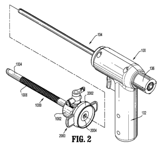
FIG. 2 is a perspective view of the trocar system illustrating the obturator
assembly removed from the cannula assembly;
FIG. 3 is a perspective view of the obturator assembly illustrating the
obturator handle and the obturator cartridge removed from the obturator
handle;
FIG. 4 is a perspective view with parts separated of the handle of the
obturator assembly;
FIG. 5 is a perspective view in partial cross-section illustrating the
obturator cartridge mounted within the obturator handle of the obturator
assembly;
FIG. 6 is a perspective view with parts separated of the obturator cartridge
of the obturator assembly;
FIG. 7 is an enlarged view of the area of detail depicted in FIG. 5;
FIG. 8 is an enlarged view of the area of detail depicted in FIG. 5;
FIG. 9 is an enlarged view of the area of detail depicted in FIG. 6;
FIG. 10 is an enlarged view of the image transmitting member and the
obturator blade;
FIG. 11 is an enlarged view of the area of detail depicted in FIG. 5;
FIG. 12 is a perspective view of the obturator cartridge prior to mounting
within the obturator handle;
4
CA 02683968 2009-10-14
WO 2008/130904 PCT/US2008/060307
FIG. 13 is a perspective view illustrating the obturator cartridge mounted
within the obturator handle;
FIG. 14 is a side cross-sectional view of the trocar system illustrating an
endoscope positioned within the obturator assembly;
FIG. 15 is a side cross-sectional view illustrating the trigger of the
obturator handle in an initial position;
FIG. 16 is a side cross-sectional view illustrating the trigger of the
obturator handle in an actuated position;
FIG. 17 is a side cross-sectional view illustrating the hammer of the
obturator cartridge released to deploy the obturator blade;
FIG. 18 is an enlarged perspective view illustrating the obturator blade
deployed relative to the image transmitting member;
FIG. 19 is a perspective view illustrating an alternate embodiment of the
obturator assembly; and
FIG. 20 is a perspective view in cross-section of the obturator assembly of
FIG. 19.
DETAILED DESCRIPTION OF THE PREFERRED EMBODIMENTS
Referring now in detail to the drawing figures, in which, like references
numerals identify similar or identical elements, there is illustrated, in
FIGS. 1 and 2, a
trocar system constructed in accordance with a preferred embodiment of the
present
disclosure and designated generally by reference numeral 10. Trocar system 10
is
particularly adapted for use in minimally invasive surgical procedures such as
endoscopic
CA 02683968 2009-10-14
WO 2008/130904 PCT/US2008/060307
or laparoscopic procedures. Trocar system 10 is adapted to penetrate body
tissue, e.g.,
the abdominal wall, and to provide a simultaneous forward directional view of
the body
tissue. Generally, trocar system 10 includes two principal subassemblies,
namely,
obturator assembly 100 and cannula assembly 1000.
Cannula assembly 1000 may be any cannula assembly suitable for use in a
laparoscopic surgical procedure. In one preferred embodiment, cannula assembly
1000
includes cannula housing 1002 and cannula sleeve 1004 extending from the
cannula
housing 1002. Either or both cannula housing 1002 and cannula sleeve 1004 may
be
transparent in part, or in whole, and may be fabricated from biocompatible
metal or
polymeric material. Cannula assembly 1000 may include an internal seal such as
a duck-
bill valve or other zero closure valve adapted to close in the absence of a
surgical
instrument to prevent passage of insufflation gases through the cannula
assembly 1000.
Cannula sleeve 1004 may include a plurality of annular ribs 1006 to facilitate
retention of
the cannula sleeve 1004 within tissue.
Trocar system 10 may also include a seal assembly 2000 which is
preferably releasably mounted to cannula housing 1002. Means for releasably
connected
seal assembly 2000 to cannula housing 1002 may include a bayonet coupling,
threaded
connection, latch, friction fit, tongue and groove arrangements, snap-fit,
etc. Seal
assembly 2000 includes seal housing 2002 and at least one internal seal 2004
which is
adapted to form a fluid tight seal about an instrument inserted through the
seal assembly
2000. One suitable seal may be the fabric seal disclosed in commonly assigned
U.S.
Patent No. 6,702,787, which issued March 9, 2004, the entire contents of which
are
incorporated herein by reference. The seal disclosed in the `630 patent may be
a flat
6
CA 02683968 2009-10-14
WO 2008/130904 PCT/US2008/060307
septum seal having a first layer of resilient material and a second fabric
layer juxtaposed
relative to the first layer. Further details of the seal may be ascertained by
reference to
the "787 patent. Seal assembly 2000 may or may not be a component of cannula
assembly 1000. For example, the seal assembly may be a separate, removable
assembly.
In the alternative, the seal assembly may comprise an integral part of the
cannula
assembly 1000 and not be removable.
Referring to FIGS. 2 -4, in conjunction with the cross-sectional view of
FIG. 5, obturator assembly 100 includes obturator handle 102 and obturator
cartridge 104
which is adapted for releasable mounting to the obturator handle 102.
Obturator handle
102 includes handle half sections 102a, 102b connected to each other via screw
means
106, adhesives, cements or the like. Obturator handle 102 defines pistol or
hand grip 108
and barrel 110. Obturator handle 102 further includes trigger 112 which is
mounted
within recess 114 defined within the obturator handle 102. Trigger 112 is
adapted for
reciprocal longitudinal movement relative to obturator handle 102. Trigger 112
includes
latch 116 which is adapted to pivot about fulcrum 118. Latch 116 includes cam
pin 120
which is received beneath cam shelf 122 of obturator handle 102. Upon rearward
or
proximal movement of trigger 112, cam pin 120 rides beneath cam shelf 122
causing
latch 116 to pivot downwardly about fulcrum 118. Trigger 112 is normally
biased in a
distal direction by coil spring 124. At its proximal end, coil spring 124
engages interior
surface 126 of obturator handle 102 and, at its distal end, is received about
spring mount
128 of trigger 112. Barrel 110 of obturator handle 102 defines longitudinal
bore 130
extending therethrough.
7
CA 02683968 2009-10-14
WO 2008/130904 PCT/US2008/060307
Referring now to FIGS. 3, 5 and 6 obturator cartridge 104 will be
discussed. Obturator cartridge 104 is adapted for at least partial insertion
through
longitudinal bore 130 of obturator handle 102, and provides penetration and
visual
capabilities to trocar system 10. Obturator cartridge 104 may be intended for
disposal, or
possibly, for resterilization, subsequent to its use, and, according is
releasably mountable
to obturator handle 102. Moreover, obturator cartridge 104 may be intended to
be
removed from obturator handle 102 whereby after use another obturator
cartridge 104
may be mounted to obturator handle 102 for subsequent use.
Obturator cartridge 104 includes cartridge frame 132 and elongate
obturator member 134 extending from the cartridge frame 132. Cartridge frame
132
includes proximal collar 136 and cylindrical portion 138 extending from the
proximal
collar 136. Proximal collar 136 includes longitudinal ribs 140 for
facilitating engagement
by the clinician. Cylindrical portion 138 includes at least one longitudinal
slot 142,
preferably, two longitudinal slots 142 in diametrical opposed relation, in its
outer wall.
Cartridge frame 132 further defines external locking lugs 144 in diametrical
opposed
relation to facilitate releasable mounting of cartridge frame 132 to obturator
handle 102
as will be discussed.
Referring to FIGS. 5 and 6, cartridge frame 132 of obturator cartridge 104
includes, from proximal to distal, scope retainer 146, hammer spring 148,
hammer 150,
stop 152, driver holder 154, return spring 156. Scope retainer 146 is mounted
within
cartridge frame 132 and incorporates 0-ring spring 158. In particular, 0-ring
spring 158
is received within peripheral slot 160 of scope retainer 146. Scope retainer
146 defines a
plurality of proximal legs 162 separated by slots 164. Proximal legs 162 may
move
8
CA 02683968 2009-10-14
WO 2008/130904 PCT/US2008/060307
radially outwardly to receive an instrument, e.g., an endoscope, in frictional
relation
therewith. 0-ring spring 158 is adapted to bias proximal legs 162 of scope
retainer 146
radially inwardly to the position shown in FIG. 5. In this position, the
interior surfaces of
proximal legs 162 may establish a frictional relation with the inserted
object.
Hammer 150 of obturator cartridge 104 is adapted for reciprocal
longitudinal movement within cartridge frame 132. Hammer 156 includes
diametrically
opposed tabs 166 which extend through longitudinal slots 142 of cartridge
frame 132, and
are each adapted to engage latch 116 of trigger 112 when in an initial
position of the
trigger 112 depicted in FIG. 5. Hammer 150 is biased in a distal direction
through
hammer spring 148. Specifically, hammer spring 148 engages interior surface
168 of
cartridge frame 132 and engages proximal end face 170 of hammer 150.
Stop 152 of obturator cartridge 104 is fixed within cartridge frame 132 and
defines central opening 172 for at least partial reception of driver holder
154. Stop 152
limits the range or degree of longitudinal movement of hammer 150. Stop 152 is
coaxially mounted about driver holder 154 in a manner to permit the driver
holder 154 to
advance and retract within bore 172 of the stop 152. Driver holder 154 defines
a central
cylinder 174 having a pair of opposed slots 176 at least partially extending
through the
wall of the central cylinder 174. Driver holder 154 is normally biased in a
proximal
direction by driver return spring 156 which engages both an interior surface
of cartridge
frame 132 and distal collar 154c of driver holder 154. Cartridge frame 132
further
includes port seal opener 178 extending distally from the cartridge frame 132.
In one
embodiment, port seal opening 178 includes proximal flange 180 which is
received
within corresponding mounting recess 182 of cartridge frame 132 as best
depicted in FIG.
9
CA 02683968 2009-10-14
WO 2008/130904 PCT/US2008/060307
7. Post seal opening 178 is adapted to penetrate and/or open seal 2004 of seal
assembly
2000 of cannula assembly 1000.
With continued reference to FIGS. 5 and 6, elongated obturator member
134 of obturator cartridge 104 is securely mounted within cartridge frame 132.
In one
method, obturator member 134 includes diametrical openings 184 adjacent its
proximal
end. Openings 184 receive internal locking tabs 186 of cartridge frame 132,
e.g., in snap
relation therewith to secure the two components. Obturator member 134 may be a
scope
tube and defines a longitudinal passage for accommodating in endoscope.
Obturator
member 134 may define a pair of opposed longitudinal grooves 188 in its outer
surface
extending to the distal end of the obturator member 134. Obturator member 134
has
image transmitting member 190 mounted to its distal end. In one embodiment,
image
transmitting member 190 is adhered to obturator member 134. Image transmitting
member 190 may be a transparent optical window fabricated from a variety of
materials
such as polystyrene, polymethyl-methylaceylate (PMMA), polyurethane,
transparent
epoxies and/or glass or transparent materials. Image transmitting member 190
may
define a semi-hemispherical configuration. Alternately, image transmitting
member 190
may be an image directing member in the form of, e.g., a lens, an optical
prism, an
optical mirror, or like image directing medium.
Referring now to FIGS. 5-6 and 8-10, obturator cartridge 104 includes a
tissue penetrating assembly in the form of blade drivers 192 and cutting blade
194 which
is mounted to the distal end of the blade drivers 192. In one embodiment, two
blade
drivers 192 extend along obturator member 134 and are received within
longitudinal
grooves 188 of the obturator member 134. As best depicted in FIG. 8, the
proximal ends
CA 02683968 2009-10-14
WO 2008/130904 PCT/US2008/060307
of blade drivers 192 include offset portions 196 which are received within
driver holder
154, specifically, within grooves 176 of the driver holder 154, thereby
securing the blade
drivers 192 to the driver holder 154. Accordingly, longitudinal movement of
driver
holder 154 causes corresponding longitudinal movement of blade drivers 192 and
cutting
blade 194.
Referring now to FIGS. 9-11, cutting blade 194 is secured to blade drivers
through corresponding reception of mounting hooks 198 of blade drivers 192
within u-
shaped recesses 200 of cutting blade 194. Cutting blade 194 is at least
partially
accommodated within groove 202 of image transmitting member 190 and moves
within
the groove 202 between a non-deployed position and a deployed position.
Cutting blade
194 may define a sharpened cutting edge or alternatively, may be relatively
blunt to be
atraumatic to tissue. Cutting blade 194 may be formed of a suitable rigid
material such as
stainless steel or titanium, or alternatively, may be fabricated of a suitable
polymeric
material. Cutting blade 194 is preferably centered with respect to the outer
surface of the
image transmitting member 190 as shown. Thus, in visualization, cutting blade
194 is
seen as a thin line through the center, i.e. bisecting, the viewing field so
as not to obstruct
viewing of the body.
FIGS. 12-13 illustrate one arrangement for releasably mounting obturator
cartridge 104 within obturator handle 102 is illustrated. As indicated
hereinabove,
cartridge frame 132 includes external locking lugs 144 arranged in diametrical
opposed
relation. Obturator handle 102 includes corresponding lug receiving recesses
204
adjacent its proximal face. Upon assembly of obturator cartridge 104 within
obturator
handle 102, locking lugs 144 are aligned with locking recesses 204 and
advanced
11
CA 02683968 2009-10-14
WO 2008/130904 PCT/US2008/060307
therewithin. Thereafter, cartridge frame 132 is rotated in a clockwise
direction where
locking lugs 144 are received within locking slots 206 of obturator handle 102
to
releasably secure the two components (FIG. 5). Obturator cartridge 104 is
symmetrically
arranged about its longitudinal axis "k". Accordingly, obturator cartridge 104
may be
introduced within obturator handle 102 without concern of alignment of a
particular
locking lug 144 of the cartridge 104 with a locking recess 204 of the handle
102.
Upon securing obturator cartridge 104 within obturator handle 102,
hammer 150 is engaged by trigger 112, e.g., latch 116 of the trigger 112 in
the
aforedescribed manner. Moreover, either tab 166 of hammer 150 (depending on
the
rotational orientation of cartridge frame 132) will be engaged by latch 116 of
trigger 112
through a corresponding longitudinal slot 142 of cartridge frame 132.
FIG. 14 illustrates trocar system fully assembled and in cross-section with
an endoscope 300 positioned within obturator handle 102 and within obturator
cartridge
104. One suitable endoscope 300 which may be used with trocar system 10 is
disclosed
in commonly assigned U.S. Patent No. 5,412,504 to Leiner, the entire contents
of which
disclosure are hereby incorporated by reference. Endoscope 300 may be any
conventional scope suitable for endoscopic applications including, e.g., a
laparoscope,
arthroscope, colonoscope, etc. Endoscope 300 incorporates an optical train or
lens
arrangement which is capable of transmitting an image of an object from the
distal or
objective lens through the eyepiece or monitor for viewing by the surgeon.
Further
details of endoscope 300 may be ascertained by reference to the `504 patent.
In operation and with initial reference to FIG. 15, trigger 112 is illustrated
in the initial position with latch 116 of the trigger 112 engaging hammer 150
thereby
12
CA 02683968 2009-10-14
WO 2008/130904 PCT/US2008/060307
preventing longitudinal movement of the hammer 150. This is the position
achieved
upon positioning of obturator cartridge 104 within handle 102. When it is
desired to
advance trocar system 10 within abdominal cavity, image transmitting member
190 of the
trocar system is pressed against the tissue. Trigger 112 is depressed or
retracted in the
proximal direction as depicted in FIG. 16 which compresses hammer spring 148.
Such
movement of trigger 112 also causes latch 116 to pivot downwardly through
movement
of cam pin 120 against cam shelf 122 to thereby release hammer 150.
With reference to FIG. 17, hammer 150, which is no longer constrained by
latch 116, is driven in a distal or firing direction by the linear force of
compressed
hammer spring 148 to contact driver holder 154 and correspondingly advance the
driver
holder 154. Distal advancement of driver holder 154 advances blade drivers 192
to
advance relative to obturator member 134 which advances cutting blade 194
relative to
image transmitting member 190. Advancing movement of driver holder 154 is
limited by
stop 152. In this advanced position depicted in FIG. 18, cutting blade 194 is
positioned
to penetrate, incise or cut through the tissue. Trigger 112 is released and
returned to its
initial position by trigger return spring 124.
Concurrently with advancing movement of driver holder 154, return
spring 156 is caused to assume a compressed condition. The linear force of
return spring
thereby causes subsequent return of driver holder 154, blade drivers 192 and
cutting
blade 194 to the initial position of FIG. 15. During movement toward the
initial position,
driver holder 154 forces hammer 150 in a proximal direction and into
engagement with
latch 116 of trigger 112 to assume the initial condition. Proximal cam surface
206 of
13
CA 02683968 2009-10-14
WO 2008/130904 PCT/US2008/060307
hainmer 150 enable locking tabs 166 to ride along the corresponding cam
surface 208 of
latch 116 to facilitate securement of the locking tab 160 relative to the
latch 116.
During penetration of the body tissue the surgeon either observes such
penetration through the eyepiece of the endoscope 300, or in instances where a
video
system is utilized the surgeon simply observes the penetration of the body
tissue via any
known video monitor.
In operation, the surgeon may also more selectively deploy the cutting
blade 194 during penetration. That is, the surgeon may insert the trocar
assembly and
bluntly penetrate the body tissue until reaching thicker tissue, such as
muscle. At this
point, the blade can be deployed to penetrate (cut through) this thick tissue,
then retracted
to provide blunt penetration until thick tissue is again encountered where
once again the
blade can be deployed.
After penetration into the body cavity, endoscope 300 may be removed
and the obturator assembly 100 may be removed from the cannula assembly 1000,
leaving the cannula assembly 1000 in the body for insertion of desired
instrumentation
therethrough.
FIGS. 19-20 illustrate another embodiment of the obturator assembly.
This obturator assembly is similar to the embodiment of FIGS. 1-18; however,
in
accordance with this embodiment, obturator cartridge 104 and obturator handle
102 are a
single unit, i.e., obturator cartridge 104 is not releasably mountable to
obturator handle
102. Accordingly, obturator assembly 100 may be disposed of as a single unit
or
sterilized subsequent to its uses.
It will be understood that various modifications can be made to the
14
CA 02683968 2009-10-14
WO 2008/130904 PCT/US2008/060307
embodiments of the present invention herein disclosed without departing from
the spirit
and scope thereof. For example, various diameters for the cannula assembly,
the
obturator assembly, as well as various diameter endoscopes are contemplated.
Also,
various modifications may be made in the configuration of the parts.
Therefore, the above
description should not be construed as limiting the invention but merely as
exemplifications of preferred embodiments thereof. Those skilled in the art
will envision
other modifications within the scope and spirit of the present invention as
defined by the
claims appended hereto.