Note: Descriptions are shown in the official language in which they were submitted.
CA 02683974 2009-10-14
WO 2008/130624 PCT/US2008/005011
PROTEIN-MODIFIED NANO-DROPLETS, COMPOSITIONS
AND METHODS OF PRODUCTION
CROSS-REFERENCE TO RELATED APPLICATION
This application claims priority to U.S. Provisional Application No.
60/907,824 filed
April 18, 2007, the entire contents of which are hereby incorporated by
reference.
BACKGROUND
1. Field of Invention
This application relates to nanodroplets, and more particularly to protein-
modified
nanodroplets and compositions, and methods of production.
2. Discussion of Related Art
The contents of all references, including articles, published patent
applications and
patents referred to anywhere in this specification are hereby incorporated by
reference.
Pure viral capsid protein can be self assembled around nanoscale objects,
(Bancroft, J. B.;
Hiebert, E. Formation of an Infectious Nucleoprotein from Protein and Nucleic
Acid Isolated
from a Small Spherical Virats, Virology 1967, 32, 354-356; Bancroft, J. B.;
Hills, G. J.;
Markham, R. A Stucly of the Self-Assembly Process in a Small Spherical Virus.
Formation of
Organized Structures from Protein S-ubunits in Vitro. Virology 1967, 31, 354-
379; Hiebert, E.;
Bancroft, J. B.; Bracker, C. E. The Assembly in Vitro of Some Sinall Spherical
Viruses, Hybrid
Viruses, and Other Nucleoproteins, Virology 1968, 34, 492-508) enclosing them
in protein shells
through a process known as "encapsidation". (Douglas, T.; Strable, E.;
Willits, D.; Aitouchen,
A.; Libera, M.; Young, M. Protein Engineering of a Viral Cage for Constrainecl
Nanoinaterials
Synthesis, Adv. Mater. 2002, 14, 415-418; Douglas, T.; Young, M. Host-Guest
Enecrpsulatiaz of
Mcrterials by Asseinbled Virus Protein Cages, Ncrture 1998, 393, 152-155;
Douglas, T.; Young,
M. Virus Particles (is Teinplates for Mciterials Synthesis, Aclv. Mater. 1999,
11, 679-681;
Dragnea, B.; Chen, C.; Kwak, E. S.; Stein, B.; Kao, C. C. Gold Ncinoparticles
as Specti-oscopic
Enhancers for in Vitro Stciclies on Single Viruses, J. Ani. Chein. Soc. 2003,
125, 6374-6375.)
1
CA 02683974 2009-10-14
WO 2008/130624 PCT/US2008/005011
By displaying viral protein, encapsidated nanomaterials can potentially be
endowed with
a desirable viral functionality: preferential localization in specific tissues
that could be useful for
cell targeting (Uchida, M.; Klem, M. T.; Allen, M.; Suci, P.; Flenniken, M.;
Gillitzer, E.;
Varpness, Z.; Liepold, L. 0.; Young, M.; Douglas, T. Biological Containers:
Protein Cages as
Multificnctional Nanoplatforms, Adv. Mater. 2007, 19, 1025-1042). In a classic
demonstration
of encapsidation, an infectious virus was assembled in vitro by combining pure
capsid protein
with pure RNA and dialyzing to change pH and ionic strength (Bancroft, J. B.;
Hiebert, E.
Virology 1967, 32, 354-356). Likewise, synthetic polymers (Bancroft, J. B.;
Hiebert, E.;
Bracker, C. E. The Effects of Various Polyanions on Shell Formation of Some
Spherical Viruses,
Virology 1969, 39, 924-930), parapolyoxometalate particles (Douglas, T.;
Young, M. Host-Guest
Encapsulation of Materials by Assembled Virus Protein Cages, Nature 1998, 393,
152-155),
solid gold nanocrystals (Dragnea, B.; Chen, C.; Kwak, E. S.; Stein, B.; Kao,
C. C. Gold
Nanoparticles as Spectroscopic Enhancers for in Vitro Studies on Single
Viruses, J. Am. Chein.
Soc. 2003, 125, 6374-6375; Chen, C.; Daniel, M. C.; Quinkert, Z. T.; De, M.;
Stein, B.;
Bowman, V. D.; Chipman, P. R.; Rotello, V. M.; Kao, C. C.; Dragnea, B.
Nanoparticle-
Templated Assembly of Viral Protein Cages, Nano Lett. 2006, 6, 611-615; Sun,
J.; DuFort, C.;
Daniel, M.-C.; Murali, A.; Chen, C.; Gopinath, K.; Stein, B.; De, M.; Rotello,
V. M.;
Holzenburg, A.; et al. Core-Controlled Polymorphism in Virus-Like Particles,
Proc. Natl. Aca(l.
Sci. U.S.A. 2007, 104, 1354-1359), and quantum dots (Dixit, S. K.; Goicochea,
N. L.; Daniel,
M.-C.; Murali, A.; Bronstein, L.; De, M.; Stein, B.; Rotello, V. M.; Kao, C.
C.; Dragnea, B.
Quantum Dot Encapsulation in Viral Capsicls, Nano Lett. 2006, 6, 1993-1999)
have been
encapsidated to create virus-like particles (VLPs) similar in size to the
native virus. For such
small VLPs, electron microscopy indicates that the protein shell assembles
from individual
subunits in a manner reminiscent of micelle formation (McPherson, A. Micelle
Formation ancl
Crystallization as Paradigms for Virus Assembly, BioEss(iys 2005, 27, 447-458)
into ordered
structures characteristic of icosahedral viruses (Zandi, R.; Reguera, D.;
Bruinsnia, R. F.; Gelbart,
W. M.; Rudnick, J. Origin ofIcosahedral Symmetry in Viruses, Proc. Ncitl.
Acacl. Sci. U.S.A.
2004, 101, 15556-15560), including protruding ring-like multimers, or
"capsomers", that have
five-fold and six-fold symmetry (Caspar, D. L.; Klug, A. Physical Principles
in the Constiziction
of Regular Viruses, Cold Spring Harb. Symp. Quant. Biol. 1962, 27, 1-24).
However, the prior
2
CA 02683974 2009-10-14
WO 2008/130624 PCT/US2008/005011
art encapsidation materials and techniques have been of limited utility to
date. There thus
remains a need for improvements.
SUMMARY
A protein-modified droplet according to an embodiment of the current invention
includes
a droplet comprising a liquid material, and a protein structure formed to at
least partially enclose
the droplet. The protein structure comprises a plurality of protein molecules
having an affinity to
at least a region of the droplet during formation of the protein structure,
and the droplet has a
maximum dimension of at least about 1 nm and less than about 1000 nm. A
composition
according to an embodiment of the current invention comprises a plurality of
protein-modified
droplets according an embodiment of the current invention dispersed in an
aqueous solution.
A method of producing protein-modified droplets according to an embodiment of
the
current invention includes supplying first and second immiscible liquid
materials; adding a
stabilizing agent to at least one of the first and second immiscible liquid
materials; emulsifying
-.the first and second liquid materials to form a plurality of droplets of the
second liquid material
in the first liquid material that are stabilized by the stabilizing agent,
each droplet of the plurality
of droplets having a maximum dimension of at least about 1 nm and less than
about 100 nm;
adding protein molecules at least one of prior to or after said emulsifying;
and allowing a protein
structure to form to at least partially enclose each of the plurality of
droplets. The stabilizing
agent and the protein molecules added are of types that have mutual
electrostatic attractions to
each other when the stabilizing agent is attached to the droplets.
BRIEF DESCRIPTION OF THE DRAWINGS
The invention is better understood by reading the following detailed
description with
reference to the accompanying figures in which:
3
CA 02683974 2009-10-14
WO 2008/130624 PCT/US2008/005011
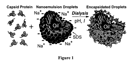
Figure I is a schematic illustration showing the encapsidation of an oil
droplet stabilized
by anionic sodium dodecyl sulfate (SDS) surfactant in water by purified capsid
protein from
cowpea chlorotic mottle virus (CCMV) according to an embodiment of the current
invention.
This is one example of a protein-modified droplet. By adjusting the pH and
ionic strength I
using dialysis, the capsid protein in bulk solution can be induced to condense
and assemble
around the negatively charged surfaces of the nanoemulsion droplets.
Figures 2(a) and 2(b) show capsid protein structures observed by negatively
stained TEM
according to an embodiment of the current invention. Figure 2(a) shows
individual nanoscale
droplets as a function of pH and ionic strength I of NaC1 after mixing and
dialyzing SDS-
stabilized nanoemulsions with purified CCMV protein. Buffers are: RNA-
reassembly (R) (pH =
7.2, I= 0.1 M); hexagonal sheet (H) (pH = 6.2, I= 0.1 M); dimer (D) (pH = 6.2,
I= 1.0 M);
multi-shell (M) (pH = 4.8, I= 0.1 M); and empty shell (E) (pH = 4.8, I= 1.0
M). Inset (upper
riglit): Fluorescence optical micrograph of FITC-labeled CCMV protein (green)
covering the
surfaces of microscale silicone oil droplets stabilized by SDS after dialysis
with R buffer. Figure
*2(b) shows nanodroplets encapsidated by 1, 2, and 3 concentric protein shells
are observed after
dialysis with M buffer. Scale bar = 20 nm (all images).
Figure 3 shows representative examples of CCMV protein structures observed as
a
function of the droplet diameter, cl (italic numbers), on a single side of
individual encapsidated
oil nanodroplets after dialysis using RNA-reassembly buffer according to an
embodiment of the
current invention. TEM images have been background subtracted and Fourier
filtered to enhance
the protein structures on the droplet surfaces. Complete protein `capsomers'
(white rings) are
found more often on the surfaces of smaller nanodroplets that have sizes
closer to that of the
native virus. Ring-like capsomers can order into six-fold arrangements locally
(dark circle).
Extended dark trough-like `scars' (dark circle), defected capsomers, and
hexagonal web-like
networks of capsid protein (dark circle) are more frequently seen on larger
droplets. Allowed
triangulation numbers T and predicted outer diameters of nanodroplets that
could be encapsidated
by perfect icosaliedra of ordered capsomers are shown in the lower scale. The
outer diameters (in
nm) are estimated using: cl(T) z 28(T/3)1/2, consistent with cl = 28 nm for
CCMV, a T= 3 virus.
4
CA 02683974 2009-10-14
WO 2008/130624 PCT/US2008/005011
Figures 4(a) - 4(c) show local protein structures observed on the surfaces of
nanodroplets
(enlarged from dark circles in Fig. 3) have different degrees of order and
disorder. Figure 4(a)
shows six-fold coordinated capsomers (dots at center) represent a high degree
of order seen
mostly on smaller droplets (left side). An example of a trough-like scar that
consists of an
elongated dark region (arrow) surrounded by a protruding white region
(middle). Hexagonal web
structure, typically seen on larger droplets, consists of dark spots (dots)
surrounded by an
interconnected white network of protein protruding from the interface (right
side). Figure 4(b)
shows probabilities p, and pW versus distance, r, between centers of dark
regions for hexagonal
capsomers and web, respectively. The average spacing between the dark spots of
the web (4.7
nm) is roughly half of the distance between the centers of capsomers (9.5 nm).
Figure 4(c) shows
a web-like structure (right side) can be made by packing hexagonal capsomers
(lower left) of
hand-in-glove protein dimers (upper left) on a flat surface. Regions of low
protein density are
marked in one hexagonal cell with black dots.
DETAILED DESCRIPTION
In describing embodiments of the present invention illustrated in the
drawings, specific
terminology is employed for the sake of clarity. However, the invention is not
intended to be
limited to the specific terminology so selected. It is to be understood that
each specific element
includes all technical equivalents which operate in a similar manner to
accomplish a similar
purpose.
According to some embodiments of the current invention, we provide a process
for
creating nanoemulsion droplets modified by and/or covered by protein. In some
embodiments,
the protein can effectively provide a capsule or container which can be loaded
witli selected
materials. Such containers can provide a drug delivery structure in some
embodinients of the
current invention. However, the broad concepts of the invention are not
limited to only drug
delivery. In addition, a protein capsule containing a liquid droplet therein
is only one example of
a protein-modified droplet according to an enibodinient of the current
invention. For instance, a
5
CA 02683974 2009-10-14
WO 2008/130624 PCT/US2008/005011
protein capsule could contain a nanoporous polymeric gel particle that is
loaded with selected
materials.
In native viruses, the viral coat protein of the virus serves as a barrier to
protect its
interior contents, the nucleic acid RNA or DNA, which is necessary for self-
propagation and
genomic reproduction. Viruses have the ability to readily penetrate specific
cells, so some
embodiments of the current invention may include targeting delivery of
particular drugs to
certain cells by tailoring the type of viral coating on the surface of the
droplets. Thus, some
embodiments of the current invention can provide a capsule that mimics some
aspects of the
natural virus. This may include, in some embodiments, providing a capsule that
can penetrate
cell barriers and deliver the contents inside the cell.
In one embodiment, we obtained viral capsid protein through a standard method
of
growing the virus, disassembling it, and separating the protein from the
genetic material (RNA or
DNA). However, the broader concepts of the invention are not limited to only
such techniques
and those particular proteins. In an alternative, the capsid protein can be
obtained in larger
quantities through bacterial expression of the viral RNA. Next, we prepared
microscale
;emulsions or nanoscale emulsions (nanoemulsions) of hydrophobic oil in water.
The
'hydrophobic drug molecules readily dissolve in the oil, yet the oil is not so
low in molecular
weight that the emulsion destabilizes through Ostwald ripening. The
concentration of the drug
molecules is fixed in the oil, and then the drug-laden oil is used as a feed
for the next step, i.e.,
the production of oil-in-water emulsions through shear emulsification. The
extreme
emulsification process used to make nanoemulsions in one example involved
using a commercial
high-pressure microfluidic device. An ultrasonic device and other methods can
also be used in
accordance with the invention.
Droplets comprised of liquid can be encapsulated with viral proteins, yielding
a
dispersion of viral protein-coated droplets of one liquid in a different
immiscible liquid through
several different methods according the various embodiments of the current
invention. Some
metliods according the current invention include the following: (1) adding oil
of the desired type
to an aqueous dispersion of viral capsid protein while controlling the droplet
stabilization
througli type and concentration of stabilizing agents (e.g. surfactants,
particles, or polyniers) and
also controlling the pH, ionic content (e.g. types of salts or buffers), and
ionic strengtli (e.g.
6
CA 02683974 2009-10-14
WO 2008/130624 PCT/US2008/005011
concentrations of salts or buffers) and applying a mechanical shear or
otherwise inducing a flow
that can cause bigger droplets to break down into smaller droplets; (2)
combining an existing oil-
in-water emulsion or nanoemulsion (stabilized by charge surfactant, particles,
or polymers) to an
aqueous dispersion of viral capsid protein at an appropriate pH, ionic
content, and ionic strength
and mixing in a manner that does not cause droplet break-up but does
distribute the components
by convection; and (3) combining an existing oil-in-water emulsion or
nanoemulsion with an
aqueous dispersion of viral capsid protein and then dialyzing using a semi-
permeable membrane
to change the pH, ionic content, and ionic strength in order to cause the
adsorption of the protein
onto the surfaces of the droplets.
The liquid material of the droplets can include one or more of the following
materials: an
oil, a silicone oil, a hydrocarbon oil, a petroleum oil, a fuel oil, a wax, a
fat, a fluorinated oil, a
non-volatile oil, a volatile oil, an aromatic oil, an oil derived from a plant
material, an oil derived
from an animal material, an oil derived from a natural source, a distilled
oil, an extracted oil, a
cooking oil, a food oil, a lubricant, a reactive material that is
predominantly hydrocarbon in
composition, an epoxy material, an adhesive material, a polymerizable
material, a thermotropic
liquid crystal, a lyotropic liquid crystal, an acidic oil, a basic oil, a
neutral oil, a natural oil, a
~polymer oil, and a synthetic oil.
Biologically active agents according to some embodiments of the current
invention can
include, but are not limited to, drug molecules, anti-cancer molecules,
therapeutic molecules,
hormone molecules, agonist molecules, antagonist molecules, inhibitor
molecules, suppressor
molecules, sensitizer molecules, antidepressant molecules, antiviral
molecules, antifungal
molecules, antibacterial molecules, bioavailability enhancer molecules, toxin
molecules, dye
niolecules, fluorescent molecules, biomolecules, nutrients, vitamins, flavors,
enzymes,
nanoparticles, and imaging contrast enhancement agents.
A surfactant, such as negatively charged sodium dodecyl sulfate (SDS) can be
added to
give the emulsion droplets stability against subsequent coalescence after they
are created tlirough
flow-induced rupturing of bigger droplets into smaller droplets.
Alternatively, commercial
mixers, blenders, colloid mills, or flow-focusing microfluidic devices could
be used to create the
emulsions or nanoemulsions out of the oil containing the drug molecules.
Existing niethods of
7
CA 02683974 2009-10-14
WO 2008/130624 PCT/US2008/005011
extreme flow are capable of creating droplets down to about 5-10 nm in radius,
so that only a
very small number of drug molecules may be in a given droplet. These smaller
nanodroplets
themselves can penetrate cellular and intestinal membranes more readily
through enhanced
diffusion and penetration of pores, and the viral coating gives them a
sturdiness and active means
of traversing membranes through protein triggering of cellular uptake. Since
the droplets can be
produced in large quantities in some embodiments of the current invention, the
viral protein often
being a limiting ingredient, we typically do not emulsify with the protein
present, although this
can be done in some embodiments of the current invention. Instead, we obtain
the droplets,
dilute them and fix the surfactant concentration, and then add the
disassembled viral capsid
protein in an embodiment of the current invention. By then changing the ionic
strength of the
solution and/or the pH, we can cause the protein to become attracted to the
droplet surfaces and
assemble a coating on the droplets. In some embodiments, we use an anionic
surfactant to
stabilize the droplets, and this causes the droplets to have a negative charge
on their surfaces.
This mimics RNA and DNA, which are also negatively charged in solution. Then,
we add
disassembled capsid protein and change the ionic strength and pH of the
solution to cause the
- viral shell to form on the surfaces of the droplets. To demonstrate this
principle, we have
performed the first viral encapsulation experiments of nanodroplets using
silicone nanoemulsions
coated with an anionic surfactant, sodium dodecylsulfate (SDS), and capsid
protein obtained
from cowpea chlorotic mottle virus (CCMV), a plant virus. No specific drug
molecules were
added to the oil in that example. In other examples, we have added other oil-
soluble molecules
into our nanodroplets, such as fluorescent dyes. Our transmission electron
micrograph images
show the successful assembly of the viral protein on the surfaces of the
droplets. One can
optimize pH and ionic strength to fully coat the droplets without causing
empty virus shells to
form. These empty shells waste the protein, so they are not typically
desirable. Under certain
conditions of coniposition and assembly, we have also observed that several
inner droplets can be
enclosed within a single outer shell of protein that forms around them.
Overall, we describe
methods that can be used to create emulsion and nanoemulsion droplets of a
very wide range of
sizes that are coated by the viral protein and can have enhanced ability to
trigger rapid
penetration, targeting, and delivery. By controlling the size distribution of
the droplets in some
embodiments, one can control the release of the drug since larger droplets
will penetrate more
8
CA 02683974 2009-10-14
WO 2008/130624 PCT/US2008/005011
slowly than smaller droplets. Alternatively, other proteins synthesized or
purified by known
methods could be used to coat the droplets.
EXAMPLE 1
In an example according to one embodiment of the current invention, we use the
capsid
protein from the CCMV (Cowpea Chlorotic Mottle Virus), which self-assembles at
the surface of
nanoemulsion dropets due to electrostatic interactions. In the native virus,
the positively-charged
interior of the virus interacts with one or more negatively-charged polyanions
of RNA. Since the
nanoemulsion droplets have negatively-charged surfactant head groups on the
exterior of the
droplets, the viral proteins assemble at the exterior interface of the oil
droplet.
Procedure for obtaining capsid protein:
We adopt Rao's procedure for purification of the CCMV protein (Choi, Y. G.;
Rao, A. L.
N., Molecular Studies on Bromovirus Capsid Protein: VII. Selective Packaging
of BMV RNA4
by Specific n-Ten-ninal Arginine Residues. Virology 2000, 275, 207-217). We
start first with
:wild-type CCMV at a concentration of - 4 mg/mL in suspension buffer. The CCMV
is dialyzed
"in disassembly buffer for 24 hours in order to dissociate the CCMV into
protein dimers and
RNA. The disassembled CCMV is removed from the buffer and centrifuged for 30
minutes at
14,000 rpm (Eppendorf Centrifuge 580 4R) to precipitate the RNA. The protein
in the
supernatant is extracted and then further dialyzed in RNA assembly buffer for
24 hours in order
to assemble around RNA left in the supernatant. Finally, the supernatant is
centrifuged for 1:40
hours at 100,000 rpm (Beckman TLA 110 UC) and the upper 3/4 of the
supernatant, which
contains the pure CCMV protein, is used for further study. The purity and
concentration of the
resultant protein is measured using UV-visible spectroscopy. All work is done
at and 4 C.
Procedure for making nanoemulsion droplets:
Nanoemulsions, droplets of one liquid phase stabilized in another inimiscible
liquid phase
by surfactant, with diameters less than 100 nm, were created using extreme
shear with a
microfluidic injection system. The size of the nanoemulsion droplets is
dependent upon the
amount and type of surfactant used, the pressures at which the liquids are
injected into the
9
CA 02683974 2009-10-14
WO 2008/130624 PCT/US2008/005011
microfluidic system, and the viscosities of the liquids. The nanoemulsions
were then centrifuged
and fractionated in order to obtain a specific size distribution of the
droplets (Mason, T. G., J. N.
Wilking, K. K. Meleson, C. B. Chang, and S. M. Graves. 2006. Nanoemulsions:
formation,
structure, and physical properties, Journal of Physics: Condensed Matter 18:
R635-R666;
Meleson, K., S. Graves, and T. Mason. 2004. Formation of Concentrated
Nanoemulsions by
Extreme Shear. Soft Materials 2: 109-123). We typically make oil-in-water
nanoemulsion
droplets, the size of which can be controlled through the microfluidic device
and other
compositional parameters. Thus, this embodiment is for packaging hydrophobic
drugs inside a
droplet that is in turn inside a viral capsid shell.
Assembly conditions (combining viral proteins with nanoemulsion droplets):
We have used various assembly conditions to assemble viral proteins around
nanoemulsion droplets. By varying pH and ionic strength of the solutions
against which the
nanoemulsion droplets and viral proteins are dialyzed, one can create droplets
that have a single
coat of viral protein on the outside, a double coat, or multiple coats (see
Fig. 2(b)).
Procedure for taking EM images:
Copper grids of 400-mesh size (Ted Pella Inc., Redding, CA) were prepared
using
support films of parlodoin, and then carbon-coated. The grids are glow-
discharged by using
high-voltage, alternating current, immediately before sample deposition.
Sample deposition
steps consisted of placing 5pL of the sample directly on to the grid for 1
minute, wicking with
Whatman 4 filter paper, immediately staining with 1% uranyl acetate for 1
minute, wicking
again, and air-drying. Samples were viewed under a Hitachi H-7000 electron
microscope at an
accelerating voltage of 75kV. Negatives were developed and scanned using a
Minolta Dimage
Scan MultiPro scanner for image analysis.
Discussion of Results:
Advantages of this niethod for producing droplets covered by viral protein
according to
some embodiments of the current invention can include the ability to fine-tune
the size of the
nanoeniulsion, which is the template for viral assembly. Thus, we are able to
vary the diameter
CA 02683974 2009-10-14
WO 2008/130624 PCT/US2008/005011
of this protein container from about 10 nm to 100 nm, for example, below 1/10
of a micron,
allowing size-specific variants for future applications. The adsorption of the
viral capsid protein
onto the surfaces of the droplets can be controlled by the affinity of the
protein for the oil and
surfactant on the surfaces of the droplets, not by the droplet size.
Therefore, it is possible for us
to also make sub-micron, microscale, and even larger virally encapsulated
droplets, if these
would be desired.
Some embodiments of this invention can provide methods to produce protein-
modified
droplets for delivering biologically active contents (hydrophobic drug) into
the interior of an
organism through ingestion, injection, inhalation, or through the skin.
Molecules that contain
radioactive species or high atomic number elements could be inserted into the
nanodroplets for
cancer treatment or imaging enhancement. Thus, some embodiments of this
invention could
have potential applications in both medical imaging and drug delivery. In
medical imaging, one
application can be the use of the container in tracing pathways of transport
within the cell. In
drug delivery, one application can be the use of therapeutic agents
encapsulated in the
,nanoemulsion and subsequently delivered upon entry of cancerous cell to treat
cancer.
EXAMPLE 2
This example is the encapsidation of incompressible spherical nanodroplets, or
`nanoemulsions', that can have a continuous range of sizes extending
significantly beyond the
wild-type core and are stabilized by adsorbed anionic surfactant molecules. We
show that it is
possible to force the capsid protein to self-assemble into spherical shells
without the perfect
symmetry and discrete sizes of ideal icosahedra dictated by the Caspar-Klug
hierarchy (Caspar,
D. L.; Klug, A., Physical Principles in the Construction of Regular Viruses.
Col(i Spri,rg Harb.
Synip. Qua,it. Biol. 1962, 27, 1-24), which requires special integral
multiples (e.g. 1,3,4,7,...) of
60 proteins. Silicone oil (poly-dimethylsiloxane)-in-water nanoemulsions
stabilized by sodium
dodecyl sulfate (SDS) are made by high-pressure homogenization (Meleson, K.;
Graves, S.;
Mason, T. G., Formation of Concentrated Nanoemulsions by Extreme Shear. Soft
Hateri ls
2004, 2, 109-123), mixed with pure cowpea chlorotic mottle vinis (CCMV) capsid
protein (Choi,
Y. G.; Rao, A. L. N., Molecular Studies on Broniovirus Capsid Protein: VII.
Selective Packaging
11
CA 02683974 2009-10-14
WO 2008/130624 PCT/US2008/005011
of BMV RNA4 by Specific n-Terminal Arginine Residues. Virology 2000, 275, 207-
217), and
dialyzed to reduce the divalent cation concentration, causing the protein to
self-assemble
(Adolph, K. W.; Butler, P. J. G., Reassembly of a Spherical Virus in Mild
Conditions. Nature
1975, 255, 737-738). Over a wide range of pH and ionic strength, the re-
assembly creates virus-
like droplets (VLDs) coated by a single protein shell. We also explore a broad
range of pH and
ionic strength to control the number of concentric shells formed by the capsid
protein around the
nanodroplets. In the limit of low pH and ionic strength, where empty multi-
shell structures have
been formed (Adolph, K. W.; Butler, P. J., Studies on the Assembly of a
Spherical Plant Virus. I.
States of Aggregation of the Isolated Protein. J. Mol. Biol. 1974, 88, 327-
341), droplets can be
encapsidated inside two or more protein shells.
For VLDs coated by single shells, transmission electron microscopy (TEM)
reveals that
the protein has self-assembled on the curved surfaces not only into ordered
capsomers but also
into a variety of other structures. As the droplet surface curvature is
reduced, ordered capsomer
structures become less prevalent, and other protein structures appear:
defected capsomers,
hexagonal webs, and trough-like scars. Some of these structures appear to be
due to jamming
(Liu, A. J.; Nagel, S. R., Jamming Is Not Just Cool Any More. Nczture 1998,
396, 21-22) of the
protein on the curved surface and are reminiscent of defects found on
macroscopic droplets
stabilized by solid microscopic particles (Bausch, A. R.; Bowick, M. J.;
Cacciuto, A.; Dinsmore,
A. D.; Hsu, M. F.; Nelson, D. R.; Nikolaides, M. G.; Travesset, A.; Weitz, D.
A., Grain
Boundary Scars and Spherical Crystallography. Science 2003, 299, 1716-1718;
Bowick, M.;
Cacciuto, A.; Nelson, D. R.; Travesset, A., Crystalline Order on a Sphere and
the Generalized
Thomson Problem. Phys. Rev. Lett. 2002, 89, Art. No. 185502 pp. 1-4; Tarimala,
S.; Dai, L. L.,
Structure of Microparticles in Solid-Stabilized Emulsions. Langmuir 2004, 20,
3492-3494).
However, other structures, such as the hexagonal web, arise from special rules
associated with
attractive protein-protein and protein-surface interactions. The overall
reduction in the
population of ordered capsomers on larger droplets implies that the three
different conformations
of the protein (Speir, J. A.; Munshi, S.; Wang, G.; Baker, T. S.; Johnson, J.
E., Structures of the
Native and Swollen Forms of Cowpea Chlorotic Mottle Virus Determined by X-Ray
Crystallography and Cryo-Electron Microscopy. Structui-e 1995, 3, 63-78) in
the capsid of
CCMV are not present in the sanle proportions when the protein assembles on
incorr-pressible
12
CA 02683974 2009-10-14
WO 2008/130624 PCT/US2008/005011
surfaces that have lower curvature. Thus, surface curvature plays an important
role in setting
protein conformation and profoundly influences the structure of assembled
proteins that
encapsidate incompressible objects.
METHODS
Protein Purification
Following Choi and Rao's procedure (Choi, Y. G.; Rao, A. L. N., Molecular
Studies on
Bromovirus Capsid Protein: VII. Selective Packaging of BMV RNA4 by Specific n-
Terminal
Arginine Residues ), we isolate and purify capsid protein from CCMV. CCMV has
a single
capsid protein, so any reference to `CCMV protein' therefore specifies CCMV's
single unique
capsid protein. Purified CCMV is dialyzed for 24 hours in 1.0 L of disassembly
buffer (0.5 M
CaC12, 50 mM Tris-HCI at pH 7.5, 1.0 mM EDTA, 1.0 mM DTT, 0.5 mM PMSF). The
dissociated virus is centrifuged for 30 minutes at 14,000 RPM to precipitate
the RNA. The
protein supernatant is extracted and dialyzed for 24 hours in 1.0 L of RNA
reassembly buffer (50
mM NaCI, 50 mM Tris-HCI, pH 7.2, 10 mM KC1, 5.0 mM MgCIZ, 1.0 mM DTT). The
solution
is then centrifuged for 100 minutes at 100,000 RPM, and the protein
supernatant is extracted.
The concentration and purity of the protein have been measured using UV-
visible spectroscopy.
All work has been performed at 4 C.
Nanoemulsion Preparation and Fractionation
Nanoemulsions are created using extreme flow with a high-pressure microfluidic
device
(Meleson, K.; Graves, S.; Mason, T. G., Formation of Concentrated
Nanoemulsions by Extreme
Shear. Soft Materials 2004, 2, 109-123). Polydisperse emulsions are size-
fractionated using
ultracentrifugation to achieve better droplet uniformity and to set the SDS
concentration CsDs=
Prior to mixing with protein and dialyzing, the nanoemulsions have CSDS = 1 mM
SDS, well
below the critical niicelle concentration, and 0 = 0.05. The PDMS oil (10 cSt
viscosity, supplied
by Gelest) has a low vapor pressure, so it does not evaporate over the time
scale of these
microscopy measurements, even when capsid protein is not present.
Dialysis Buffers
13
CA 02683974 2009-10-14
WO 2008/130624 PCT/US2008/005011
RNA reassembly buffer (Adolph, K. W.; Butler, P. J., Assembly of a Spherical
Plant Virus.
Philos. Trans. R. Soc. Loncl. B 1976, 276, 113-122): Tris-HCI buffer at pH =
7.2, I= 0.10 M
NaCI, 10 mM KCI, 5.0 mM MgC12, and 1.0 m.M DTT. Empty shell buffer: 50 mM
sodium
acetate buffer at pH = 4.8 and I = 1.0 M NaCI. Dimer buffer: 50 mM of sodium
phosphate buffer
at pH = 6.2 and I= 1.0 M NaCI. Multi-shell buffer: 50 mM sodium acetate buffer
at pH = 4.8 and
I = 0.1 M NaCI. Hexagonal sheet buffer: 50 mM sodium phosphate buffer at pH =
6.2 and I = 0.1
M NaCI. The last four buffers also contain 1.0 mM EDTA and 1.0 mM DTT.
Encapsidation Procedure
A 10 ftL aliquot of stock nanoemulsion at 1.0 mM SDS and 0 = 0.05 is added to
purified CCMV
protein at 0.15 ,ug/mL to give a total reaction volume of 200 fiL. The mixture
is dialyzed in 1.0 L
of the appropriate buffer for 24 hours at 4 C. The SDS concentration after
dilution and dialysis
is roughly 10-5 M, so binding of SDS-protein interaction in the bulk solution
is minimized while
still maintaining droplet stability. The sulfate head-group of SDS remains
negatively charged
over the entire range of pH we access. After dilution, the charge density of
SDS on the oil-water
interfaces is estimated to be roughly -0.1 e/nm2.
Transmission Electron Microscopy: Staining and Analysis
Pelco copper grids of 400 mesh size and 3.0 mrn OD (Ted Pella, Inc.) are
coated with a thin film
of parlodion and carbon. The grids are glow-discharged using high-voltage,
alternating current,
immediately before sample deposition. We place 5,uL of the sample directly
onto the grid for I
minute, then wick with Whatman 4 filter paper, and immediately stain with a 1%
solution of
uranyl acetate in water for 1 minute. The samples are air-dried and viewed
under a Hitachi H-
7000 electron microscope at an accelerating voltage of 75 kV. Negatives were
developed and
scanned using a Minolta Dimage Scan MultiPro scanner for image analysis. Adobe
Photoshop is
used to flatten the image background by subtracting a strongly blurred image.
Cross-correlation
Fourier-transform iniage analysis is applied using a correlation kernel that
has a dark center,
corresponding to the size of the capsomer's dark dimple and a white outer
ring.
Fluorescence Microscopy
14
CA 02683974 2009-10-14
WO 2008/130624 PCT/US2008/005011
We have made stock solutions of FITC and 5(6)-FAM, SE in DMSO at 1.0 mg/mL. An
aliquot
of the 5(6)-FAM, SE stock is added to the dissociated CCMV protein in RNA
reassembly buffer
at pH 7.2. Another aliquot of the FITC stock solution is added to dissociated
CCMV protein and
equilibrated in 50 mM phosphate buffer at a pH of 8.2. The protein and dye are
mixed, and, after
8 hours, the FITC-labeled protein is dialyzed into RNA reassembly buffer,
lowering the pH. Both
sets of the fluorescently-labeled protein are mixed with 10 ,uL of microscale
emulsions at 1.0
mM concentrations of either SDS (or CTAB) and 0 = 0.05 in a total reaction
volume of 200 ,uL
and dialyzed using RNA reassembly buffer. Fluorescence micrographs reveal the
presence of
labeled protein at the droplet surfaces through strong fluorescence at the
edges of the droplets.
Microscale emulsions in the absence of labeled protein do not show this
fluorescence. Therefore,
droplets that are much larger than the native virus can be coated by a dual-
layer consisting of a
first inner layer of anionic surfactant and a second outer layer of virus
protein. The adsorption of
the protein likely inhibits the equilibrium exchange of surfactant to and from
the droplet
interfaces. This protein adsorption is typically irreversible for neutral and
acidic conditions of pH
over a wide range of ionic strength in the solution surrounding the protein-
modified droplet.
After assembly, the protein-modified droplet can be disassembled by causing
the solution
conditions to enter a region that would cause the disassembly of native
virions.
Results and Discussion
Anionically stabilized nanodroplets provide incompressible, charged templates
that offer
a wide range of curvatures upon which capsid protein can be assembled. Through
extreme
emulsification, we make oil-in-water nanoemulsions comprised of spherical
droplets that can be
as small as CCMV (inner diameter of 21 nm and outer diameter of 28 nm)( Mason,
T. G.;
Wilking, J. N.; K. Meleson, K.; Chang, C. B.; Graves, S. M., Nanoemulsions:
Formation,
Structure, and Physical Properties. J. Phys.: Condens. Matter 2006, 18, R635-
R666). Relying
upon differences in creaming rates, ultracentrifugal size-fractionation
provides uniform model
nanoemulsions liaving droplet radii between 10 nm < a < 100 nm (Meleson, K.;
Graves, S.;
Mason, T. G., Formation of Concentrated Nanoemulsions by Extreme Shear. Soft
Materi'als
2004, 2, 109-123). In addition, the droplet volume fraction 0 and surfactant
concentration Csi)s
can be set independently. The Laplace pressure, corresponding to the stress
necessary to
CA 02683974 2009-10-14
WO 2008/130624 PCT/US2008/005011
overcome surface tension and deform a droplet, is typically above 10 atm, so
droplets are
spherical at dilute 0. To inhibit Ostwald ripening (Taylor, P., Ostwald
Ripening in Emulsions:
Estimation of Solution Thermodynamics of the Disperse Phase. Adv. Colloid
Interface Sci. 2003,
106, 261-285), which can lead to unwanted growth of the droplets through
molecular diffusion,
the dispersed liquid is chosen to be very insoluble in the continuous liquid
phase.
We create virus-like droplets by mixing pure, disassembled CCMV capsid protein
with
an oil-in-water nanoemulsion and changing the pH and NaCI ionic strength, I,
of the buffer
through dialysis, causing the protein to assemble on the droplet surfaces (see
Figure 1).
Transmission electron microscopy (TEM) of negatively stained VLDs reveals the
presence of
both ordered protein structures, including ring-like capsomers, on the
surfaces of individual
droplets. To probe a diversity of structures, we have encapsidated SDS-coated
nanodroplets at
five different buffer conditions, corresponding to the known phase behavior of
the protein
(Adolph, K. W.; Butler, P. J., Studies on the Assembly of a Spherical Plant
Virus. I. States of
Aggregation of the Isolated Protein. J. Mol. Biol. 1974, 88, 327-341; Adolph,
K. W.; Butler, P.
J., Assembly of a Spherical Plant Virus. Philos. Trans. R. Soc. Lond. B 1976,
276, 113-122;
Bancroft, J. B.; Hills, G. J.; Markham, R., A Study of the Self-Assembly
Process in a Small
Spherical Virus. Formation of Organized Structures from Protein Subunits in
Vitro. Virology
1967, 31, 354-379): `RNA-reassembly' (pH = 7.2, I= 0.1 M), `hexagonal sheet'
(pH = 6.2, I=
0.1 M), `dimer' (pH = 6.2,1= 1.0 M), `multi-shell' (pH = 4.8,1= 0.1 M), and
`empty shell' (pH
= 4.8,1= 1.0 M). We show TEM images of negatively stained VLDs for these
buffers in Figure
2a. Protein-coated nanodroplets can be distinguished from empty capsid shells
because the
uranyl acetate staining does not penetrate into the core of the coated
droplets, so they appear
noticeably brighter in the center. A darker ring around the edges of the
droplets exists due to the
trapping of the stain as the water contact line recedes during the evaporation
process. This
staining and drying process yields TEM images that provide excellent views of
only one-half of
the surface of each droplet. Because the inlages do not contain a significant
signal from the
protein on the other half, it is possible to identify and interpret the
protein surface structure on
incliviclual droplets, rather than having to rely on reconstruction methods
that presume an ordered
structure.
16
CA 02683974 2009-10-14
WO 2008/130624 PCT/US2008/005011
For all five buffers, CCMV protein encapsidates nanodroplets, regardless of
their size
(Figure 2a). Dimer buffer and RNA-reassembly buffer create VLDs efficiently
without any loss
of protein into empty shells. For the multi-shell buffer, we observe
nanodroplets coated with
single-, double- (dominant), and triple-shells (Figure 2b). For the empty- and
multi-shell buffer
conditions, due to the slight excess of protein beyond what is required to
coat the droplets, we
observe encapsidated droplets and also empty shells.
To confirm that the protein is not simply deposited on the droplet surfaces
during drying
but actually assembles around the droplets while in solution, we have examined
fluorescein
isothiocyanate (FITC)-labeled CCMV capsid protein on microscale silicone oil
droplets after
dialyzing with RNA-assembly buffer. Strong fluorescence emanates from the
surfaces of the
droplets (Figure 2a inset), indicating that they are coated with the labeled
protein. By contrast,
when droplets coated with cationic cetyl-trimethylammonium bromide (CTAB)
surfactant are
mixed with FITC-labeled CCMV proteins and dialyzed in the same manner, no
surface
fluorescence is observed, indicating that cationic surfactants are typically
not suitable for creating
protein-modified droplets with this particular protein.
We have also examined the structures and relative degree of order and disorder
of protein
on nanodroplets having different curvatures for RNA-reassembly conditions
(Figure 3). To
enhance the images of the structures on the surfaces of individual VLDs, we
remove the
background and then reduce high-frequency noise using Fourier filtering. We
identify complete
capsomers as white rings that have an internal dark central spot and also a
dark external trough
surrounding the bright ring. The brighter regions indicate a higher density of
proteins that project
outward from the surfaces and the darker regions generally indicate a lower
density of protein
where stain becomes more highly concentrated in the local depressions. As the
droplets become
progressively larger than CCMV, complete capsomers become less prevalent and
several other
protein structures are observed on the less curved incompressible surfaces. In
particular,
imperfect capsomers, linear scar-like defects, and hexagonal web-like
structures are seen, in
sharp contrast to perfect icosahedral order on wild-type CCMV. Based on energy
minimization,
a greater relative coverage of the droplets by ordered capsomers might be
expected when droplet
sizes correspond to allowed integral triangulation numbers T(Bruinsma, R. F.;
Gelbart, W. M.;
Reguera, D.; Rudnick, J.; Zandi, R., Viral Self-Assembly as a Thennodynaniic
Process. Phys.
17
CA 02683974 2009-10-14
WO 2008/130624 PCT/US2008/005011
Rev. Lett. 2003, 90, Art. No. 248101 pp. 1-4; Zandi, R.; Reguera, D.;
Bruinsma, R. F.; Gelbart,
W. M.; Rudnick, J., Origin of Icosahedral Symmetry in Viruses. Proc. Nat.
Acad. Sci. 2004, 101,
15556-15560) (see Figure 3-lower scale), yet this assumes that the structure
and size of the
capsomers will not be influenced by the underlying curvature and degree of
compressibility of
the core. Although our experiments at all buffer conditions do not reveal a
higher degree of
capsomer order on droplets that correspond to allowed T, this might occur at
different pH and I
than we have yet explored.
For smaller droplets closer to the size of the native virus, we have
identified local
hexagonal packing of capsomers (Figure 4a-left), as can be seen on the native
virus. Although we
find numerous examples of six capsomers surrounding a central capsomer, five-
fold coordinated
capsomers without defects have not been observed on droplets significantly
larger than CCMV.
The distribution of center-to-center distances between neighboring six-fold
capsomers is shown
in Figure 4b, and the average distance of 9.5 nm is in excellent agreement
with that known from
native CCMV (Speir, J. A.; Munshi, S.; Wang, G.; Baker, T. S.; Johnson, J. E.,
Structures of the
Native and Swollen Forms of Cowpea Chlorotic Mottle Virus Determined by X-Ray
Crystallography and Cryo-Electron Microscopy. Structatre 1995, 3, 63-78).
On a number of larger droplets, we observe a hexagonal web-like structure of
protein:
regions of dark dots surrounded by interconnected white boundaries, or `web'
(Figure 4a-right).
Dark outer troughs characteristic of capsomers are absent. Although this
protein web usually has
local six-fold hexagonal order, in general, it can be disordered due to
defects. The average
distance between nearest-neighbor dots in the web is only 4.7 nm, about half
the distance
between the centers of capsomers (Figure 4b). This is consistent with capsid
protein self-
assembling in a different manner on flatter, incompressible surfaces than on
more highly curved,
compressible surfaces (Bancroft, J. B.; Hills, G. J.; Markham, R., A Study of
the Self-Assembly
Process in a Small Spherical Virus. Formation of Organized Structures from
Protein Subunits in
Vitro. Virology 1967, 31, 354-379).
We propose that the mechanism of the formation of the hexagonal web structure
of
protein can be understood by considering the underlying symmetry and dense
packing of self-
assembled protein sub-units on incompressible surfaces of lower curvature.
CCMV capsid
protein is known to self-assemble into hexagonal capsomers of hand-in-glove
dimmers (Tang, J.;
18
CA 02683974 2009-10-14
WO 2008/130624 PCT/US2008/005011
Johnson, J. M.; Dryden, K. A.; Young, M. J.; Zlotnick, A.; Johnson, J. E., The
Role of Subunit
Hinges and Molecular'Switches' in the Control of Viral Capsid Polymorphism. J.
Struct. Biol.
2006, 154, 59-67; Adolph, K. W.; Butler, P. J., Studies on the Assembly of a
Spherical Plant
Virus. I. States of Aggregation of the Isolated Protein. J. Mol. Biol. 1974,
88, 327-341) that have
been identified by x-ray cystallography. These dimers are energetically
favored over monomers
in many buffer conditions; a protruding arm of one protein is inserted into
the folded region of its
partner and is held by an attraction, and vice-versa. Six hand-in-glove dimers
can come together
to form a capsomer that has six protruding arms in a structure resembling a
gear (Figure 4c-
lower left). Such capsomer structures are also energetically favored over a
random assembly of
dimers. When these gear-like hexagonal capsomers of dimers self-assemble and
then densely
pack to cover a flat surface, they can create hexagonal arrays of capsomers
that have regions that
are depleted of protein at half of the center-to-center distance between
neighboring capsomers.
This packed-gear structure would give the appearance of the web that we
observe: a hexagonal
array of dark spots where the protein density is lower and an interconnected
hexagonal network
of bright web where the protein density is higher. For self-assembly of
protein on a flat surface, it
,is reasonable to assume that the folded capsid protein and dimers exist in
only a single
conformation and do not distort into the three known conformations that are
required for
assembling five-fold coordinated capsomers on core structures that have higher
curvature
comparable to that of the wild-type virus.
In addition to the ordered web, we also observe elongated protein scars that
are dark
troughs surrounded by white rims (Figure 4a-middle). These protein scars bear
some
resemblance to scar defects found in the packing of monodisperse solid spheres
on the surfaces
of curved liquid droplets (Bausch, A. R.; Bowick, M. J.; Cacciuto, A.;
Dinsmore, A. D.; Hsu, M.
F.; Nelson, D. R.; Nikolaides, M. G.; Travesset, A.; Weitz, D. A., Grain
Boundary Scars and
Spherical Crystallography. Science 2003, 299, 1716-1718; Bowick, M.; Cacciuto,
A.; Nelson, D.
R.; Travesset, A., Crystalline Order on a Sphere and the Generalized Thomson
Pi-oblem. Phys.
Ren. Lett. 2002, 89, Art. No. 185502 pp. 1-4), a controlled variety of
`Pickering
ernulsions'(Tarimala, S.; Dai, L. L., Structure of Microparticles in Solid-
Stabilized Emulsions.
Laungmuir 2004, 20, 3492-3494; Pickering, S. U., Emulsions. J. Chem. Soc.
Trans. Lontl. 1907,
91, 2001-2021; Subramaniani, A. B.; Abkarian, M.; Stone, H. A., Controlled
Assembly of
19
CA 02683974 2009-10-14
WO 2008/130624 PCT/US2008/005011
Jammed Colloidal Shells on Fluid Droplets. Nat. Mater. 2005, 4, 553-556), yet
capsid protein
scars are distinctly different. Protein scars do not consist simply of line
defects between fully
formed hexagonal ring-like capsomers on incommensurately sized droplets;
instead they indicate
disorder of the protein at a smaller scale than even the capsomer unit itself.
Several mechanisms
combine to produce the scar defects: incommensurate sizes of the droplets
relative to those
corresponding to allowed T-numbers for icosahedral structures, and surface
jamming of the
protein that strongly inhibits protein reorientation and rearrangement once
the surface is
completely covered.
Discussion of Results
A variety of protein structures, including dimers, partial capsomers, and
complete
capsomers, may become jammed into locally disordered states(Liu, A. J.; Nagel,
S. R., Januning
Is Not Just Cool Any More. Nature 1998, 396, 21-22) on incompressible
spherical surfaces that
have reduced curvature in a manner reminiscent of out-of-equilibrium glasses
and gels.
Additional defects may arise because protein adsorbed at high densities may
not be able to
change conformation and reorganize into lowest-energy ordered states, as when
forming around
RNA. Controlling the relative protein coverage and examining the kinetics of
the process of
encapsidation will provide greater insight into how ordered and disordered
protein structures
arise on the surfaces of VLDs. By adjusting the pH and ionic strength, it may
be possible to
encapsidate droplets, nanoparticles, and synthetic polymers with a controlled
number of capsid
shells.
By interpreting TEM images of individual encapsidated nanodroplets, we have
revealed a
range of new structures, including defected capsomers, hexagonal web, and
scars, on the surfaces
of the droplets. The discovery of these structures provides significant new
insight into the nature
of protein conformations on curved surfaces. Moreover, it shows that non-
equilibrium protein
structures can exist on encapsidated nanoscale objects due to surface jamming
on an
incompressible charged template and that the picture of thermodynamic self-
assembly of perfect
icosahedral shells may be correct only in certain limiting cases.
CA 02683974 2009-10-14
WO 2008/130624 PCT/US2008/005011
Proteins useful for making protein-modified droplets may be obtained from
viruses that
are members of the following families of viruses: Adenoviridae, Anellovirus,
Arenaviriclae,
Arteriviriclae, Ascoviridae, Asfarviriclae, Astroviriclae, Asunviroidae,
Baculoviridae,
Barnaviriclae, Benyvirus, Birnaviridae, Bornaviriclae, Bromoviriclae,
Btmyaviridae,
Cctlieiviriclae, Caulimoviridae, Chercavirus, Chrysoviridae, Cireoviridae,
Closteroviridae,
Cornoviriclae, Coronaviridae, Corticoviridae, Cystoviriclae, Deltavirus,
Dieistroviriclae,
Endornavirus, Filoviridae, Flaviviriclae, Flexiviriclae, Furovirus,
Fuselloviriclae, Geminiviriclae,
Guttaviridae, Hepadnaviridae, Hepeviridae, Herpesviriclae, Horcleivirus,
Hypoviriclae, Iflavirus,
Inoviridae, Iridoviridae, Leviviriclae, Lipothrixviriclae, Luteoviridae,
Marnaviriclae, Metaviridae,
Microviriclae, Mimivirus, Myoviridcte, Nanoviridae, Narnaviridae, Nimaviridae,
Nodaviridae,
Ophiovirus, Orthomyxoviriclae, Ourmiavirus, Papillomaviridae, Paramyxoviridae,
Partitiviridae, Parvoviriclae, Pecluvirus, Phycodnaviridae, Picornaviridae,
Plasmaviridae,
Podoviridae, Polyclnaviridae, Polyomaviriclae, Pospiviroidae, Potyviridae,
Poxviriclcie,
Pseudoviridae, Reoviridae, Retroviridae, Rhabdoviridae, Rhizidiovirus,
Roniviriclae,
Rudiviridae, Saclwavirus, Salterprovirus, Sequiviridae, Siphoviridae,
Sobemovirus, Teetiviridae,
Tenuivirus, Tetraviridae, Tobamovirus, Tobravirtts, Togaviridae, Tomb-
usviridae, Totiviriclae,
Tytnoviriclae, Umbravirus, and Varicosavirus. Proteins useful for making
protein-modified
droplets may also be obtained from members of other families of viruses not in
this list and also
from families of viruses yet to be discovered and studied.
In addition to proteins from viruses, proteins taken from bacteria, fungi,
plants, animals,
and sponges can be used to make protein-modified droplets if such proteins can
be effectively
isolated, separated, and manipulated in a manner that brings them into
proximity with the
surfaces of droplets in a manner that is created by an attractive interaction
of the protein with the
droplet surface.
Proteins useful for making protein-modified droplets may have a variety of
functions,
including but not limited to: structural protein, non-structural protein, coat
protein, capsid
protein, core protein, envelope protein, matrix protein, transniembrane
protein, membrane
21
CA 02683974 2009-10-14
WO 2008/130624 PCT/US2008/005011
associated protein, non-structural protein, nucleocapsid protein, filamentous
protein, capping
protein, crosslinking protein, glycoprotein, and motor protein.
The examples of protein-modified droplets we have provided are of a single
capsid
protein purified from Cowpea Chlorotic Mottle Virus (CCMV), a member of the
family of plant
viruses, Brornoviriclae.
Viruses can have more than one type of capsid protein. In the particular
examples we
have shown, we have substituted a polyanionic droplet in place of polyanionic
genetic material as
a template for protein assembly, a wide variety of viral capsid proteins can
be effectively
attracted to the surface of the charged droplet. Thus, for viruses having two
or more capsid
proteins, an appropriate stoichiometric ratio of different protein types would
certainly provide
sufficient structural features to modify and/or enclose the droplets.
Moreover, for viruses that
naturally produce two or more types of capsid proteins, even a single type of
capsid protein that
has been purified is sufficient to modify and/or enclose the droplets; having
all different types of
capsid proteins present from a particular virus is not necessary. The main
requirement is that the
charge on the surface of the droplet, the pH of the solution, and the ionic
composition and ionic
strength of the solution must be adjusted such that the protein experiences an
attractive
interaction with the droplet surface and thereafter remains proximate to said
droplet surface.
Once the protein coating has been formed around the droplet it can be
advantageous in
certain applications to also form a lipid coating, a lipoprotein coating, or a
lipid-protein coating
that surrounds the protein layer, thereby creating a total structure
resembling an enveloped virus.
Thus, this structure contains an inner droplet core, a surface active agent
typically adsorbed to the
core, a layer of protein surrounding the droplet core with surface active
agent, and a layer of
lipid, lipoprotein, or lipid-protein.
The selective uptake and localization of specific viruses, both with and
without the
enveloped layer, by specific tissues and organs in higher-level organisms is
well known and is
discussed in a book such as "Basic Virology", 2nd edition, by E.K. Wagner and
M.J. Hewlett,
22
CA 02683974 2009-10-14
WO 2008/130624 PCT/US2008/005011
Blackwell Publishing (2004). Protein-modified droplets present the same
protein structures to
biological organisms as do naturally occurring virions that contain genetic
material, so the
preferential uptake and localization of protein-modified droplets will occur
in the same tissues
and organs as is found for natural virions that display the same proteins.
The current invention is not limited to the specific embodiments of the
invention
illustrated herein by way of example, but is defined by the claims. One of
ordinary skill in the art
would recognize that various modifications and alternatives to the examples
discussed herein are
possible without departing from the scope and general concepts of this
invention.
23