Note: Descriptions are shown in the official language in which they were submitted.
CA 02685772 2009-10-30
WO 2008/135082 PCT/EP2007/054317
"A COUPLING DEVICE FOR PERFORMING ENDOLUMINAL AND/OR
TRANSLUMINAL SURGERY"
DESCRIPTION
The present invention relates, in general, to devices
and methods for surgically modifying organs and vessels
and more particularly to a surgical coupling device and
method for performing an endoluminal and or transluminal
surgery, e.g. an anastomosis, particularly of the
digestive tract, such as gastro-jejunostomy, jejuno-
jejunostomy or similar interventions as for example
colo-proctostomy, jejuno-colostomy or anastomoses
involving the Chole duct, by applying an anastomotic
ring device comprising two snap-connectable rings.
The known surgical methods and instruments for
performing the above mentioned surgery, e.g. anastomoses
by applying anastomotic ring devices, involve
traditional open surgery or laparoscopic surgical
techniques, which are rather invasive and require the
use of quite complex and cumbersome surgical devices. As
a result the risk of post-operative complications is
undesirably high.
Moreover, the known devices and methods are not suitable
to perform the anastomosis by a pure or at least
partially endoscopic or endolumenal approach.
Such an endolumenal approach, if assisted by a suitable
CA 02685772 2009-10-30
WO 2008/135082 PCT/EP2007/054317
2
instrumentation, would sensibly reduce the disadvantages
of the traditional open surgery and laparoscopic
methods. Particularly, endolumenal operations would
reduce the invasiveness of the intervention, thereby
reducing the risk of complications and shortening the
post operative course of the patient.
Even though the endoscopic and endolumenal approach to
surgery in general and to anastomosis in particular is
very promising, it is presently very difficult to use
such an approach in practice, e.g. for performing an
anastomosis of the digestive tract, since neither the
instrumentation nor the methods are mature enough to
exclude operative and post operative complications.
One of the weaknesses of the endoscopic and endoluminal
surgery is the coupling of surgical devices and
instruments inside the body of the patient. Such a
coupling of instruments can be e.g. desirable during the
creation of a guide wire loop across natural ducts in
order to transport instruments to the operational site
and to enable the surgeon to apply reciprocal forces to
devices, e.g. to compression rings in order to perform
an anastomosis.
The importance of reliable and simple coupling systems
becomes even more evident if one considers that
endoscopic and endoluminal surgery involves very often
CA 02685772 2009-10-30
WO 2008/135082 PCT/EP2007/054317
3
balloon catheters and balloon dilators whose flexibility
allows generally an easy access to most of the anatomic
structures but makes it difficult to connect to other
instruments and to transmit push-pull forces.
The object of the present invention is therefore to
provide a reliable and preferably releasable coupling
device which can be used also in connection with balloon
dilators and which can be both engaged and disengaged
inside the body of the patient during a surgical
intervention.
A further object of the invention is to provide a
surgical method for performing an endoluminal or
transluminal anastomosis, particularly of the digestive
tract, by an endoluminal or endoscopic approach which
involves the use of the surgical coupling device.
These and other objects are achieved by a surgical
coupling device according to claim 1 and by the method
described in the following description. Advantageous
embodiments are claimed in the dependent claims.
For better understanding the invention and appreciating
the related advantages, a detailed and non limiting
description of embodiments is provided with reference to
the accompanying drawings, in which:
- Figure 1 is a perspective view of an inflatable
coupling portion of a coupling device in a disengaged
CA 02685772 2009-10-30
WO 2008/135082 PCT/EP2007/054317
4
configuration according to an embodiment of the
invention;
- Figure 2 is a perspective view of a catching portion
of the coupling device in a disengaged configuration
according to an embodiment of the invention;
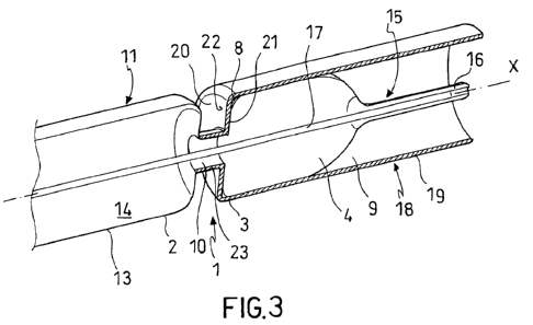
- Figure 3 is a perspective, sectioned view of the
coupling device in an engaged configuration;
- Figure 4 is a sectioned view of a catching portion of
the coupling device according to a further embodiment of
the invention;
- Figure 5 is a sectioned view of a catching portion of
the coupling device according to a further embodiment of
the invention;
- Figure 6 is a sectioned view of the coupling device
implemented in an endoluminal anastomotic ring applier
according to an embodiment of the invention;
- Figure 7 is a perspective view of a detail of the
endoluminal anastomotic ring applier in figure 6;
- Figure 8 is a sectioned view of the detail in figure
7;
- Figure 9 is a perspective view of a further detail of
the endoluminal anastomotic ring applier in figure 6;
- Figure 10 is a perspective view of the inflatable
coupling portion of the coupling device implemented in
an endoluminal anastomotic ring carrier device according
CA 02685772 2009-10-30
WO 2008/135082 PCT/EP2007/054317
to an embodiment of the invention;
- Figure 11 is a perspective view of an anastomotic
compression ring device intended to be deployed by means
of an anastomotic applier coupled by the coupling device
5 according to the invention;
- Figures 12, 13 and 14 illustrate a series of steps of
a method for performing a transluminal anastomosis by
means of a compression ring deployment system in figures
6 to 11 and the coupling device according to the
invention;
- Figure 15 is a perspective view of the coupling device
implemented in an endoluminal anastomotic ring applier
according to a further embodiment of the invention;
- Figure 16 is a sectioned view of a portion of the
endoluminal anastomotic ring applier in figure 15;
- Figure 17 is a sectioned view of a detail of the
endoluminal anastomotic ring applier in figure 15;
- Figure 18 is a sectioned perspective view of a further
detail of the endoluminal anastomotic ring applier in
figure 15;
- Figures 19, 20 and 21 illustrate a series of steps of
a method for performing a transluminal anastomosis by
means of the compression ring deployment system in
figures 15 to 18 and the coupling device according to
the invention;
CA 02685772 2009-10-30
WO 2008/135082 PCT/EP2007/054317
6
- Figure 22 is a schematic illustration of the general
functional units of an anastomotic compression ring
deployment system;
- Figure 23 illustrates a creation of a guide wire loop
during in preparation of an anastomosis (gastro-
jejunostomy);
- Figure 24 illustrates a creation of an anastomosis
(gastro-jejunostomy);
Turning to the figures, a coupling device for coupling a
first surgical device to a second surgical device during
endoluminal and transluminal surgery is denoted by
reference numeral 1. The coupling device 1 comprises a
coupling portion 2 and a catching portion 3 configured
to trap and hold the coupling portion 2.
The coupling portion 2 comprises a first connector 5 for
connecting the coupling portion 2 to the first surgical
device and an inflatable anchoring head 4, as well as an
activating device 6 connected to the anchoring head 4
and suitable to deform (insufflate and deflate) the
anchoring head 4 such that it can take on an expanded
configuration (fig. 1) and a retracted configuration
(fig. 10).
The catching portion 3 comprises a second connector 58,
7 for connecting the catching portion 3 to the second
surgical device which is intended to be coupled with the
CA 02685772 2009-10-30
WO 2008/135082 PCT/EP2007/054317
7
first surgical device. The catching portion 3 further
comprises an anchoring seat 8 which defines a receiving
space 9 for receiving at least part of the anchoring
head and an access aperture 10 through which the
anchoring head 4 can be inserted from outside the
catching portion 3 into the receiving space 9. The
access aperture 10 is configured to allow insertion and
withdrawal of the anchoring head 4 when it is retracted
and to lock the at least partially inserted anchoring
head 4 to the catching portion 3 when it is expanded.
In accordance with an embodiment, the anchoring head 4
comprises a balloon catheter or balloon dilator 11 which
can be actuated by a fluid pump device which is
connected in fluid communication with the balloon
dilator 11 through a preferably flexible pressure fluid
duct 12. As illustrated in figures 1 and 10
respectively, the balloon dilator 11 can assume a
substantially elongate filiform retracted shape and, if
subject to fluid pressure, a substantially elongate
cylindrical expanded shape.
In accordance with an embodiment, the balloon dilator 11
has an internal inflatable chamber 14 defined by a
flexible (in the sense of bending deformation) but
inextensible wall 13 such that it can adapt to the shape
of the anchoring seat 8 even and particularly if it is
CA 02685772 2009-10-30
WO 2008/135082 PCT/EP2007/054317
8
only partially inserted into the receiving space 9. The
balloon dilator 11 has a tapered distal (from a surgeons
point of view) insertion end 15 which is preferably
substantially convex and narrowed towards the end, e.g.
ogive shaped, and which can further have a central pin
shaped tip 16 or needle in order to facilitate the
insertion of the anchoring head through tissue portions
and into the access aperture 10 of the catching portion
3.
According to an embodiment, the first connector 5 of the
coupling portion 2 comprises a longitudinal channel 17
which is configured to receive for instance a guide wire
88 of the first surgical device. The guide wire channel
17 is advantageously coaxial to the cylindrical expanded
shape of the balloon dilator 11 and can be directly
glued to the guide wire 88 or, alternatively, the
balloon dilator 11 can be latched to the guide wire 88
by clamping devices such as clips etc.
Turning again to the catching portion 3, in accordance
with an embodiment of the invention the catching portion
3 comprises a housing 18 with a preferably but not
necessarily cylindrical side wall 19 and a distal end
wall 20 which define together at least part of the
receiving space 9. The end wall 20 delimits the above
said access aperture 10 through which the inflatable
CA 02685772 2009-10-30
WO 2008/135082 PCT/EP2007/054317
9
anchoring head 4 can be at least partially introduced
into the receiving space 9 and the edge of which
constricts the anchoring head 4 after it has been
expanded.
In accordance with an embodiment, the end wall 20 has an
internal locking surface 21 which faces into the
receiving space 9 and which is substantially
perpendicular to the direction of insertion (X) of the
anchoring head 4 or defines a circumferential shoulder
inclined towards the inside of the receiving space 9 so
that it locks the partially introduced, expanded and
constricted anchoring head 4 in order to prevent
slippage of the latter once it undergoes traction
forces.
In accordance with an embodiment, the internal locking
surface 21 defines an undercut for preventing the
insufflated anchoring head 4 from slipping out of
engagement with the catching portion 3.
An external surface 22 of the distal end wall 20 defines
a guide for facilitating the insertion of the anchoring
head 4 into the anchoring seat 8.
In accordance with an embodiment, the external surface
22 is substantially funnel shaped around the preferably
circular central access aperture 10 and its radially
internal edge forms a constriction portion 23 which is
CA 02685772 2009-10-30
WO 2008/135082 PCT/EP2007/054317
narrower than the receiving space 9.
In a radial cross-section the external surface 22 has
preferably a double curvature with a concave portion 24
near the outer circumference and a convex portion 25
5 near the constriction portion 23, so that the external
surface 22 converges at least approximately to a
longitudinal direction parallel to the insertion
direction X both approaching the outer circumference and
the constriction edge 23 (Fig. 5).
10 This particular shape of the external surface 22 makes
it possible that the housing of the catching portion 3
together with the portion of the insufflated balloon
dilator 11 which protrudes outside the catching portion
3 (dashed line in figure 5) defines a approximately
continuous cylindrical guide body for e.g. a guided
approximation of anastomotic compression rings.
As is the case with the coupling portion 2, also the
catching portion 3 is connected or connectable to a
surgical device or instrument.
In accordance with an embodiment, the housing of the
catching portion 3 comprises or forms portions which
enable the catching portion 3 to be snap connected,
friction connected, interference connected or shape
connected to the second surgical instrument, for
instance a crown of elastic snapper teeth 26 formed at a
CA 02685772 2009-10-30
WO 2008/135082 PCT/EP2007/054317
11
proximal end of the catching portion 3 and configured to
connect the catching portion to a visualization device
such as a gastroscope 89 (Fig. 2).
In accordance with one embodiment, the catching portion
can be partially housed or encapsulated within the
surgical device intended to be coupled to another
instrument.
In accordance with a further embodiment, the catching
portion 3 is at least partially transparent in order to
enable visualization by means of a visualization device
(e.g. endoscope, laparoscope) from inside or outside the
catching portion 3 across its transparent housing.
Specific advantages of the above described features will
become apparent from the following description of
exemplary embodiment applications of the coupling device
in an endoluminal anastomosis procedure.
For the sake of clarity and for better evidencing the
technical effect of the features of the surgical
coupling device and its interaction with the particular
environment of application, the detailed description of
embodiment applications will first deal with a surgical
method thought up by the inventors and subsequently
describe the anastomotic device and surgical instruments
for carrying out the method.
Overall procedure to perform the anastomosis
CA 02685772 2009-10-30
WO 2008/135082 PCT/EP2007/054317
12
A method for performing an endoluminal or transluminal
anstomosis, such as e.g. a gastro-jejunostomy, a jejuno-
jejunostomy, a colo-proctostomy, a jejuno-colostomy or
anastomoses of the chole duct, comprises generally the
following steps:
- Creating a loop of guide wire means by placing guide
wire means 88 in the body of a patient in a way that the
guide wire means 88 extend from an extracorporeal
proximal end 88' into the body where it goes through a
proximal first tissue portion 45 and through a distal
second tissue portion 44 which are planned to be joined
in anastomosis and out of the body up to an
extracorporeal distal end 88''.
In the following, if not otherwise specified, the terms
"proximal" and "distal" are referred to the directions
along the guide wire loop and to the above defined
proximal 88' and distal ends 88'' thereof.
- Fixing a proximal first ring 36 of an anastomotic ring
device to the proximal end 88' of the guide wire means
88 and delivering the proximal first ring 36 to the
proximal first tissue portion 45 by pulling the distal
extracorporeal end 88'' of the guide wire means 88 in a
distal direction until the proximal first ring 36
reaches the proximal first tissue portion 45,
- Slidably connecting a distal second ring 39 of the
CA 02685772 2009-10-30
WO 2008/135082 PCT/EP2007/054317
13
anastomotic ring device to the distal end 88" of the
guide wire means 88 and pushing it proximally along the
guide wire means 88 until it reaches the distal second
tissue portion 44.
- Contemporaneously pulling the proximal first
compression ring 36 distally and pushing the second
distal compression ring 39 proximally to approximate the
proximal and distal rings, thereby tearing the first and
second tissue portions 45, 44 situated upon the guide
wire means 88 between the first and second rings 36, 39
in compression contact to another,
- Connecting the first and second rings 36, 39, thereby
clamping the first and second tissue portions between
them,
- Widening the tissue internally overhanging the
anastomotic ring device to open the anastomotic lumen,
- Pulling the proximal end 88' of the guide wire means
88 to remove the guide wire means 88 from the body.
The loop of the guide wire means 88 starts and ends
either in natural orifices, like mouth, nose, anus or,
alternatively, in artificially created openings in the
body, such as colostomy, abdominal incisions, wound or
fistulas. Preferably, the guide wire means enters and
exits the body through natural ducts (e.g. mouth, Figs.
23 and 24).
CA 02685772 2009-10-30
WO 2008/135082 PCT/EP2007/054317
14
While the guide wire means can comprise one or more
single flexible guide wires, it is preferable to provide
only one single guide wire which penetrates the proximal
and distal tissue portion.
Creation of the loop of the guide wire means
The loop of the guide wire means 88 can be created by
means of the following procedural steps:
- Transluminally (e.g. transorally) introducing an
elongate insertion device through the proximal inlet
port (e.g. mouth) for the guide wire means and pushing
the insertion device from outside the body distally
towards the proximal tissue portion (e.g. a jejunal
anstomotic site),
- Transporting the distal end of the guide wire means to
the proximal tissue portion through one or more guide
wire canals formed in the insertion device and
perforating the proximal tissue portion with the guide
wire end or needle guide wire end. Alternatively, the
proximal tissue portion is perforated by a distinct
radio frequency needle device having a sheath which is
insertable along the guide wire canal and which defines
two or more internal canals (one for the RF needle and
one for the guide wire).
In this way the distal guide wire end protrudes distally
from the proximal tissue portion (e.g. into the
CA 02685772 2009-10-30
WO 2008/135082 PCT/EP2007/054317
previously C02 insufflated abdominal space),
- Removing the insertion device from the body by pulling
it proximally out of the proximal inlet port (e.g.
mouth) and leaving the guide wire means in place,
5 - Transluminally introducing a grasping device through
the distal inlet port for the guide wire means which
might but need not coincide with the proximal inlet port
(e.g. mouth) and pushing the grasping device from
outside the body proximally (with reference to the loop
10 direction) towards the distal tissue portion, e.g. the
gastric wall tissue,
- Creating a hole in the distal tissue portion, e.g. by
means of a radiofrequency needle or equivalent
penetration devices and, preferably, widening the hole
15 with a balloon catheter which is preferably transported
to the distal tissue portion through a working channel
of the grasping device.
- Passing the grasping device through the previously
widened hole (gastrotomy) of the distal tissue portion
into the same space where the distal guide wire end lays
(e.g. in the previously C02 insufflated abdominal space)
and pushing it towards the distal end of the guide wire
means.
- Inserting a snare through the working or instrument
delivery canal formed in the grasping device and
CA 02685772 2009-10-30
WO 2008/135082 PCT/EP2007/054317
16
advancing the snare proximally until it exits from the
grasping device and reaches the distal guide wire end.
- Feeding the snare over the distal guide wire end and
closing or tightening it around the guide wire means in
order to catch it, subsequently pulling the snare
together with the distal end of the guide wire means
distally through the perforation of the distal tissue
portion (e.g. gastric wall) and distally withdrawing the
grasping device together with the distal guide wire end
through the distal inlet port (e.g. transorally) out of
the body.
Advantageously, the transluminal introduction of the
grasping device and the snare is performed by a pure
endoscopic or endolumenal approach. Alternatively, this
procedural step is performed under laparoscopic
supervision. In this case the hole does not need to be
widened and only the snare (preferably a radio frequency
snare adapted to pierce through the gastric wall)
creates the hole and is passed through it into the
abdominal space. The distal guide wire end is then
laparoscopically fed into the snare hole to be caught by
the latter.
In case of a gastro-jejunostomy, the insertion device
needs to be advanced transorally through the esophagus
and the stomach and across the pylorus into the
CA 02685772 2009-10-30
WO 2008/135082 PCT/EP2007/054317
17
duodenum, which is not always easy to point at. During
this step of the procedure a protective or guiding tube
might be pushed through the patients mouth down the
esophagus into the stomach and the insertion device can
be advantageously guided inside the protective or
guiding tube up to and across the pylorus.
Delivering the proximal ring of the anastomotic ring
device to the anastomotic site and approximation of the
proximal and distal tissue portions
The proximal first ring 36 is delivered to the
anastomotic site by means of the following procedural
steps:
- Detachably connecting the proximal ring with a,
preferably distinct, carrier member outside the body of
the patient.
- Extracorporeal attachment of the carrier member (which
holds the proximal ring) to the proximal end 88' of the
guide wire means 88, e.g. by inserting it over the
single guide wire, and locking the carrier member in a
manner to prevent it from sliding along the guide wire
means. The carrier member is preferably locked by means
of locking elements, such as clips which can be clamped
around the guide wire and which are adapted to provide
an obstacle against sliding of the carrier member. After
having connected the carrier member to the proximal
CA 02685772 2009-10-30
WO 2008/135082 PCT/EP2007/054317
18
guide wire end, it is introduced in the proximal
endoscopic inlet port and pulled to the anastomotic
site, i.e. to the proximal first tissue portion (e.g.
intestinal wall 45) by pulling the distal guide wire end
88'' distally.
Alternatively, the locking of the carrier member to the
guide wire might be obtained by locking features, for
instance clamping portions, integrated in the carrier
member. According to a yet further embodiment, the
carrier member has been fabricated and distributed
together with the guide wire or previously connected to
it in a manner that, after completion of the guide wire
loop, it is sufficient to introduce the carrier member
together with the proximal ring into the proximal inlet
port (e.g. mouth and esophagus) and deliver it to the
proximal tissue portion 45 by pulling the distal guide
wire end 88' distally.
Alternatively, the carrier member can be slid along the
guide wire up to the proximal tissue layer, wherein the
guide wire acts as a guide rail for the carrier member.
- Subsequently, approximating the proximal tissue
portion 45 (e.g. intestinal tissue) towards the distal
tissue portion 44 (e.g. gastric wall) until they are in
intimate contact by pulling the distal guide wire end
88'' distally so that the carrier member urges the
CA 02685772 2009-10-30
WO 2008/135082 PCT/EP2007/054317
19
proximal ring, particularly a distal tissue contact
surface thereof, against the proximal tissue portion and
moves the latter distally towards and in contact with
the distal tissue portion.
Delivering the distal ring of the anastomotic ring
device to the anastomotic site and connection of the
distal and proximal rings
The distal ring is delivered to the anastomotic site by
means of the following procedural steps:
- Inserting the distal second compression ring 39 over
the distal guide wire end 88'' and pushing it proximally
along the guide wire means 88 towards the anastomotic
site and, hence, towards the proximal first compression
ring 36. Thanks to the guide wire loop, the first and
second compression rings 36, 39, together with the first
and second tissue portions 45, 44 are axially aligned in
the anastomotic site.
According to an embodiment, the distal compression ring
is pushed along the guide wire by means of an apposite
anastomotic applier which is advantageously adapted to
perform an angular positioning, if required, of the
distal compression ring with respect to the proximal
compression ring in order to enable their mutual
connection. Alternatively, the compression rings are
provided with connecting features which enable their
CA 02685772 2009-10-30
WO 2008/135082 PCT/EP2007/054317
mutual connection in different angular positions or
independently from their reciprocal angular position.
- Connecting the compression rings and, hence the tissue
portions clamped between them, by pushing the distal
5 compression ring proximally in engagement with the
proximal compression ring. To this end, the anastomotic
device includes the coupling device 1 according to the
present invention and an approximation device 37. In
this way it is possible to couple (preferably in or near
10 the operational site) the carrier of the first
compression ring to the approximation device 37 (which
in turn is connected to the second compression ring) in
order to apply a reciprocal pushing or pulling force on
the compression rings.
15 In accordance with an embodiment, the compression rings
are connected by applying a distinct locking device
adapted to engage both compression rings and to maintain
them connected at a desired reciprocal distance.
Advantageously, one of the compression rings is firstly
20 pushed against the other compression ring and
successively the locking device is pushed in connecting
engagement with both the compression rings to connect
them.
The method of connecting the compression rings to
another can additionally comprise the step of fixating
CA 02685772 2009-10-30
WO 2008/135082 PCT/EP2007/054317
21
the tissue portions to the compression rings in order to
effectively prevent the tissue from radially slipping
out of the grip engagement with the compression rings.
This additional step might be performed before or
contemporaneously with the application of the locking
device. This additional fixation step is preferably
performed by compressing the tissue portions between the
distal and proximal ring device and by driving a
plurality of needles into and across the ring device and
the tissue portions so that the needles provide the
above said additional fixation.
As the delivery of the locking device and the group of
needles (if provided) to the anastomotic site is
concerned, according to an embodiment of the method, the
locking device is inserted extracorporeally over the
distal guide wire end 88' and pushed proximally along
the guide wire 88 towards the anastomotic site and
towards the proximal compression ring. Preferably, both
the distal compression ring and the distinct locking
device (and, if provided, the needle group) are arranged
on a distal end (here the term "distal" is referred to
the surgeons viewpoint and not to the guide wire loop)
of the anastomotic applier and pushed by means of the
latter to the anastomotic site and in engagement with
the proximal compression ring.
CA 02685772 2009-10-30
WO 2008/135082 PCT/EP2007/054317
22
Alternatively, and inversely with respect to the above
described embodiment both the proximal first compression
ring and the distinct locking device (and, if provided,
the needle group) are fixed to the proximal ring carrier
device and transported to the anastomotic site and in
engagement with the distal compression ring.
Widening the tissue at the anastomotic site
The distal and proximal tissue portions intended to be
joined in anastomosis are widened by means of the
following procedural steps:
- widening the passage apertures of the tissue portions
through which the guide wire means, preferably a single
guide wire, passes by cutting or forced tissue extension
after the tissue portions have been approximated in
mutual contact but, preferably, before the distal and
proximal compression rings are definitely connected.
According to an advantageous embodiment, the passage
openings of the tissue portions are widened by the same
balloon dilator 11 which acts as expandable anchoring
head 4 of the coupling device 1. The balloon dilator 11
can be inserted over the guide wire 88 and delivered
towards and at least partially across the passage
openings in the tissue portions. Successively, the
balloon dilator 11 is insufflated to an extent that it
fills approximately a central aperture of the distal
CA 02685772 2009-10-30
WO 2008/135082 PCT/EP2007/054317
23
compression ring, thereby widening the anastomotic lumen
to substantially the same extent and at the same time
performing the coupling action between the anchoring
head and the catching portion.
In accordance with an advantageous embodiment, prior to
or after the inflation of the balloon dilator 11 of the
coupling device 1, the proximal first compression ring
(the locking device together with the needle group is
preferably preassembled with one of the compression
rings) is inserted over a portion of the balloon dilator
11 such that the inflated balloon dilator provides a
guide for the successive approximation and alignment of
the compression rings.
Removing the instrumentation and guide wire means from
the body of the patient
After the connection of the compression rings, the
balloon dilator is deflated and detached from the
catching portion 3. The anastomotic applier with the
catching portion 3 are withdrawn from the body of the
patient. Also the carrier member with the coupling
portion 2 are detached from the proximal first
compression ring and removed, preferably together with
the guide wire 88, by pulling the proximal guide wire
end 88' in proximal direction. As the proximal first
ring is definitely locked to the distal ring, during
CA 02685772 2009-10-30
WO 2008/135082 PCT/EP2007/054317
24
proximal traction of the guide wire 88 the carrier
member (which received the proximal ring by a weaker
connection, e.g. by friction fit) detaches from the
proximal compression ring which rests connected to the
distal compression ring.
Even though the method of performing an anastomosis has
been described and illustrated with reference to a
gastro-jejunostomy which is performed starting from the
jejunum, such a gastro-jejunostomy might also be
performed starting from the stomach. In this case, it
might be advantageous to introduce a shielding device
into the jejunum in order to prevent piercing of the
intestine wall opposite the one intended to be
anastomized.
Possibly, the tightness of the anastomosis can be tested
using methylene blue.
The above described method can be advantageously
performed endolumenally or partially endolumenally
during a gastro-jejunostomy. In case the above described
method forms part of a procedure for creating a gastro-
intestinal bypass, it is possible to proceed with a
performance of a further anastomosis involving the small
intestine (e.g. a jejuno-jejunostomy or an ileo-
jejunostomy), possibly by applying the above described
steps in analogous manner also to the further
CA 02685772 2009-10-30
WO 2008/135082 PCT/EP2007/054317
anastomosis.
Turning now again to the surgical coupling device 1
according to the invention, when implemented or
incorporated in an anastomotic applier and used during
5 an anastomosis procedure as described above in general
terms, such a coupling device can improve among others
the ring approximation function, the ring guidance
function and also the loop creation. This will become
apparent from the following description of the surgical
10 instrumentation for performing the anastomosis by means
of the coupling device according to the invention.
General description of the anastomotic ring applier
system
With reference to figure 22, an anastomotic ring applier
15 system 30 comprises a first compression ring carrier
device 31 (dashed line --- ) and an anastomotic applier
32 (dotted dashed line -.-). The first compression
ring carrier device 31 comprises a first (e.g. jejunal)
ring carrier 33 adapted to hold a first compression ring
20 36 and connectable or connected to the guide wire device
88, preferably to a single guide wire, by means of a
locking portion 35 which is configured to prevent the
first (jejunal) compression ring 36 from sliding along
the guide wire device in a direction opposite the ring
25 approximation direction.
CA 02685772 2009-10-30
WO 2008/135082 PCT/EP2007/054317
26
The first ring carrier 33 can be connected to a balloon
dilator 11 which is built up around the guide wire and
which can be connected to the guide wire outside the
body of the patient or near an inflatable head of the
balloon dilator, e.g. by means of a discrete gluing
point (or more generally a locking portion 35) which
doesn't hinder the fluid communication from the fluid
duct of the balloon dilator to its inflatable head.
According to an embodiment of the invention, the first
compression ring carrier device 31 can be coupled to and
uncoupled from the ring approximation device 37 of the
anastomotic applier 32 (which will be described below)
by means of the surgical coupling device 1 (double
dotted dashed line in figure 22) according to the
invention.
The anastomotic applier 32 further comprises a second
(gastric) ring carrier 38 adapted to hold a second
compression ring 39 for instance by means of a ring
connecting portion 40 which is configured to engage the
second (gastric) compression ring 39 by snap-fit or
friction-fit or press-fit.
The anastomotic applier 32 further comprises a ring
approximation device 37 with a first portion 42
connectable by means of the coupling device 1 to the
first ring carrier device 31 and a second portion 43
CA 02685772 2009-10-30
WO 2008/135082 PCT/EP2007/054317
27
connected or connectable to the second ring carrier
device 38.
The first and second portions 42, 43 of the ring
approximation device 37 are movable to one another in a
way to approximate the first and second ring carrier
devices 31, 38 thereby approximating the two tissue
layers (e.g. gastric tissue layer 44 and jejunal tissue
layer 45) held between the two compression rings 36, 39
and interconnecting the compression rings 36, 39 to
complete the anastomosis.
In accordance with an embodiment, the inflatable
anchoring head 4 of the coupling device 1, particularly
the balloon dilator 11 has, in its expanded
(insufflated) configuration an approximately cylindrical
shape, except of the constriction caused by the access
aperture 10 of the catching portion 3. Thanks to this
feature, the anchoring head 4 is adapted not only to
connect the first ring carrier device 31 to the
anastomotic applier 32, but also to widen the
anastomotic lumen in the tissue layers 44, 45 and to
provide guidance for both compression rings 36, 39
during approximation and deployment.
Moreover, thanks to the fact that the coupling device 1
connects the first compression ring carrier device 31
and the anastomotic applier 32 at or near the
CA 02685772 2009-10-30
WO 2008/135082 PCT/EP2007/054317
28
anastomotic site, the ring approximation device 37 can
be advantageously arranged inside the anastomotic
applier 32, thereby obviating an extracorporeal guide
wire pulling and counter-pushing with the anastomotic
applier.
In accordance with an aspect of the invention, the ring
approximation device 37 of the anastomotic applier 32
can be embodied as a guide wire pulling device, such as
a guide wire slidably received in a working channel of
an endoscope, e.g. a gastroscope, wherein the pulling
action can be exerted either by pulling the guide wire
with respect to the scope or by pulling the entire scope
together with the guide wire and contemporaneously
counter-pushing the support structure of the second ring
carrier.
In this example, the second (gastric) ring carrier
device 38 can be embodied by means of a ring connector
portion which is arranged at a distal end (surgeons
point of view) of a flexible tube, such as a gastric
protection tube. The preferably transparent tube in turn
defines an internal working space which can house the
gastroscope. Alternatively, the second (gastric) ring
carrier device 38 comprises a ring connector portion
formed at or connected to a distal end portion of the
gastroscope.
CA 02685772 2009-10-30
WO 2008/135082 PCT/EP2007/054317
29
In both cases, the surgical coupling device 1 can be at
least partially arranged within the second (gastric)
ring carrier device 38.
In accordance with a further embodiment, the ring
approximation device 37 of the anastomotic applier 32
comprises a screw drive device 46, wherein the first
portion 42 forms a first thread 47 and the second
portion 43 forms a second thread 48 meshing the first
thread 47 and a rotary device 49 cooperates with at
least one of the first and second portions 42, 43 to
rotate them to one another. The relative rotation of the
first and second portions 42, 43 causes them to
translate to one another and to approximate the first
and second ring carrier devices 31, 38 and the two
tissue layers (e.g. gastric tissue layer 44 and jejunal
tissue layer 45) held between the two compression rings
36, 39 and to interconnect the compression rings 36, 39.
In accordance with an embodiment (Figs. 6 to 14, the
terms "proximal" and "distal refer to the surgeons point
of view), the first portion 42 of the approximation
device 37 is embodied as a rotor assembly 50 with an
externally threaded outer housing 51 which defines a
distal opening 52 and a proximal connecting portion 53.
A torque connector 54 connects a torsional cable or
rotary rod 55 to the proximal connecting portion 53. The
CA 02685772 2009-10-30
WO 2008/135082 PCT/EP2007/054317
torsional cable or rotary rod 55 can be passed through a
working channel of an endoscope (e.g. gastroscope) and
has a proximal end extending outside the patients body
where an appropriate torque and rotary movement can be
5 applied to it by the surgeon. The torque connector 54
can comprise a connecting cap or plug (Fig. 7, 8)
connected by radial connecting pins 56 to a tubular ring
wall 57 of the outer housing 51 so that it can transmit
torque and traction from the torsional cable or rotary
10 rod 55 to the threaded outer housing 51.
In this embodiment, the catching portion 3 of the
coupling device 1 is arranged inside the outer housing
51 and free to rotate with respect to the latter. The
access aperture 10 of the catching portion 3 is arranged
15 in alignment with a distal end surface and preferably
coaxial to the distal opening 52 of the outer housing
51.
In order to facilitate the free rotation of the outer
housing 51 of the rotor assembly with respect to the
20 catching portion 3 and, hence, with respect to the
trapped anchoring head 4 of the coupling device 1, a
rotational bearing 58 can be interposed between the
outer housing 51 and the catching portion 3.
In accordance with an embodiment, the access aperture 10
25 of the catching portion 3 is defined by a short tubular,
CA 02685772 2009-10-30
WO 2008/135082 PCT/EP2007/054317
31
preferably cylindrical collar having a diameter which is
smaller than the diameter or transversal extension of
the receiving space 9. The tubular collar has an outer
cylindrical surface which engages an internal surface of
the rotational bearing 58 which, in turn, is connected
to the outer housing 51 near its distal opening 52
(compare Figs. 6 and 8).
In this embodiment, the second portion 43 of the ring
approximation device 37 is embodied as a jackscrew
device 59. The jackscrew device 59 comprises a tubular
support structure 60, e.g. a rigid or at least partially
flexible tube, with a proximal grounding portion 61, a
distal ring connector portion 64 and an internal thread
48 which is configured to mesh with the external thread
47 of the rotor assembly 50.
The grounding portion 61 is adapted to connect the
support structure 60 to an endoscope (e.g. gastroscope
not illustrated in the figures) so that the endoscope
can transmit push and/or pull forces and torque to the
support structure 60. Alternatively, the support
structure 60 can extend outside the body of the patient
and the grounding portion 61 can be extracorporeally
torsionally and axially held or grounded.
The distal ring connector portion 64 is configured to
hold the second (e.g. gastric) compression ring 39, e.g.
CA 02685772 2009-10-30
WO 2008/135082 PCT/EP2007/054317
32
through a snap-fit, press-fit or friction-fit action
which can be provided by a crown of elastic teeth, as
has been illustrated in Fig. 9 and 12.
The screw drive device 46 makes it possible to firmly
couple the rotor assembly 50 to the first ring carrier
device 33 by partially inserting and subsequently
insufflating the anchoring head 4 inside the catching
portion 3 and contemporaneously providing both a tissue
widening means and a cylindrical guide (expanded
anchoring head - balloon dilator 11) for the
compression rings during approximation and snap-
connection. After having coupled the first ring carrier
means to the rotor assembly, a rotary movement of the
torque cable can rotate the outer housing of the rotor
assembly with respect to the axially and torsionally
locked support structure 60, thereby causing the rotor
assembly 50 together with the catching portion 3 to
translate and, hence, to approximate and interconnect
the anastomotic compression rings 36, 39.
In accordance with a further embodiment, the tubular
support structure 60 is at least partially, preferably
completely transparent in order to enable a
visualization by the endoscope (e.g. gastroscope) also
across the tubular wall of the support structure.
In accordance with a yet further embodiment, the ring
CA 02685772 2009-10-30
WO 2008/135082 PCT/EP2007/054317
33
approximation device 37 of the anastomotic applier 32
comprises a fluid pressure device 65. In this
embodiment, the first portion 42 and the second portion
43 of the ring approximation device 37 are connected to
one another by a piston-cylinder thrust unit 66. A
pressure chamber 67 of the piston-cylinder thrust unit
66 is connected through a fluid duct 68 with a fluid
pump (not illustrated). In this way, operation of the
fluid pump raises the fluid pressure inside the pressure
chamber 67 and causes the first and second portions 42,
43 to translate to one another, thereby approximating
the first and second ring carrier devices 31, 38 and the
two tissue layers (e.g. gastric tissue layer 44 and
jejunal tissue layer 45) held between the two
compression rings 36, 39 and interconnecting the
compression rings 36, 39.
In accordance with an embodiment (Figs. 15 to 21, the
terms "proximal" and "distal refer to the surgeons point
of view), the first portion 42 of the approximation
device 37 is embodied as a cylinder device 69. The
cylinder device 69 forms a tubular support structure 70,
e.g. a rigid or at least partially flexible tube, with a
proximal grounding portion 71, the catching portion 3 of
the coupling device 1 and a distal pressure chamber 67
in fluid communication with the fluid duct 68 and
CA 02685772 2009-10-30
WO 2008/135082 PCT/EP2007/054317
34
configured to slidably receive a piston portion 72 of
the second portion 43 (which will be described below).
The grounding portion 71 is adapted to connect the
support structure 70 to an endoscope (e.g. gastroscope
not illustrated in the figures) so that the endoscope
can transmit push and/or pull forces to the support
structure 70. Alternatively, the support structure 70
can extend outside the body of the patient and the
grounding portion 71 can be extracorporeally held or
grounded.
As already described with reference to the embodiment of
figures 9 and 12, the tubular support structure 70 can
be at least partially, preferably completely transparent
in order to enable a visualization by the endoscope
(e.g. gastroscope) also across the tubular wall of the
support structure.
The second portion 43 of the approximation device 37 is
embodied as a piston device 73 (Figs. 16, 17).
The piston device 73 comprises a distal ring connector
portion 74 and the above mentioned proximal piston
portion 72.
The ring connector portion 74 is configured to hold the
second (e.g. gastric) compression ring 39, e.g. through
a snap-fit, press-fit or friction-fit action which can
be provided by a crown of elastic snapper teeth, as has
CA 02685772 2009-10-30
WO 2008/135082 PCT/EP2007/054317
been illustrated in Fig. 15 and 17.
The piston portion 72 is slidably housed in the pressure
chamber 67 of the cylinder device 69 so that a fluid
pressure increase inside the pressure chamber 67
5 (generated by the fluid pump) causes the piston device
73 to translate with respect to the catching portion 3,
thereby approximating the first and second ring carrier
devices 33, 38.
In accordance with an embodiment, the pressure chamber
10 67 has a generally annular shape and is delimited by an
outer tubular wall 75, an inner tubular wall 76 and a
proximal base wall 77 which define a distally open cup
shaped pressure chamber 67 in which the complementary
shaped proximal tubular piston portion 72 of the piston
15 device 73 is slidably received to hermetically close the
pressure chamber 67. Preferably but not necessarily,
both the tubular walls 75, 76 and the tubular piston
portion 72 have circular ring shaped cross sections.
In accordance with an embodiment, the proximal base wall
20 77 is embodied as a circular ring and defines a
circumferential distally facing connecting slot 78 which
receives a proximal ring shaped edge 79 of the catching
portion 3 of the coupling device 1, so that the
cylindrical side wall 19 (Fig. 5) of the catching
25 portion 3 acts as the above said inner tubular wall 76.
CA 02685772 2009-10-30
WO 2008/135082 PCT/EP2007/054317
36
In this case, the catching portion 3 can be embodied as
previously described with reference to fig. 5. This
makes it possible to use the distal end portion of the
catching device 3 together with the insufflated
anchoring head 4 (balloon dilator 11) as a guide for the
anastomotic rings during approximation and snap
connection.
In order to enable a good visualization of the
endoluminal coupling and anastomosis procedure, the
catching portion 3 and particularly the receiving space
9 can be proximally open so that the partially
introduced balloon dilator 11 can be directly visualized
by the endoscope (e.g. gastroscope).
The fluid duct 68 which is not necessarily but
preferably integrated in or arranged along a side wall
of the tubular support structure 70 can be put in
communication with the pressure chamber 67 by means of a
passage 80 defined in the proximal base wall 77.
For the sake of completeness of disclosure and with
reference to figure 11, an illustrative embodiment of an
anastomotic ring device and locking device will be
described, which can be deployed by means of the
anastomotic applier comprising the coupling device
according to the invention.
Figure 11 is a perspective view of an anastomotic ring
CA 02685772 2009-10-30
WO 2008/135082 PCT/EP2007/054317
37
device with the first (jejunal) ring 36 and the second
(gastric) ring 39 which is not only but particularly
adapted to perform a gastro-jejunostomy according to the
previously described method. The first compression ring
36 (or "bowel ring" or "intestinal ring" 36) is adapted
to bear against the first tissue portion 45 (jejunal
tissue), while the second compression ring 39 (or
"gastric ring" 39) is adapted to bear against the second
tissue portion 44 (gastric tissue) opposite the first
tissue portion 45 and the first ring 36.
The first and second rings 36, 39 are adapted to be
latched together in at least one, preferably two or more
locking positions corresponding to different compression
rates acting on the tissue portions 44, 45 clamped
between the rings.
Both the first 36 and second compression ring 39 have a
preferably closed circular annular shape (Fig. 11, 12)
in order to enable their mutual alignment and connection
independently from their relative angular position.
The pressure surfaces of the first and second rings 36,
39 destined to contact and clamp the first and second
tissue portions 45, 44 are substantially plane or
circumferentially wavy in order to increase the
circumference of the anastomotic lumen with respect to
the external ring circumference (not shown in the
CA 02685772 2009-10-30
WO 2008/135082 PCT/EP2007/054317
38
figures). Advantageously, the pressure surfaces are
roughened or locally profiled in order to prevent the
tissue 45, 44 to squeeze radially out of the ring device
in response to the axial pressure.
In accordance with a preferred embodiment, the second
compression ring 39 (gastric ring) comprises a shape or
a specific interaction portion suitable to be engaged
(e.g. by snap-fit, friction-fit, shape-fit or press-fit)
by the second ring carrier 38.
Both compression rings are advantageously at least
partially made of bio-absorbable or bio- fragmentable
material.
In order to provide an additional fixation of the tissue
portions to the compression rings 36, 39 to prevent the
tissue from slipping out of engagement with the ring
device, a group of needles 81 or staples is provided
which is anchored in at least one of the compression
rings and adapted to pierce through the tissue clamped
therebetween.
The needles 81 can be directly fixed to a compression
ring or to a ring locking device 82 or, as shown in
figure 11, the group of needles 81 comprise a self
supporting needle ring 83 which can be e.g. received by
an apposite seat defined at the locking device 82 in
order to anchor the needles 81 reliably to the ring
CA 02685772 2009-10-30
WO 2008/135082 PCT/EP2007/054317
39
device during and after its deployment.
Both the needles and the needle ring are advantageously
but not necessarily made of bio-absorbable or bio-
fragmentable material.
Turning now to the locking device itself, in accordance
with an embodiment (e.g. fig. 11, Fig. 12), the locking
device 82 comprises an annular proximal shoulder 84 with
a distal engaging surface 85 suitable to engage the
proximal end surface of the first (jejunal) compression
ring 36 and a tubular longitudinal portion 86 which
protrudes distally from the proximal shoulder 84. The
longitudinal portion 86 has a shape adapted to be
inserted through the central openings of both the first
and second compression rings 36, 39 and forms one or
more elastically supported snapper teeth 87 adapted to
snap engage a corresponding edge or surface of the
second (gastric) compression ring 39 (in the description
of the locking device 82 the terms "proximal" and
"distal" are referred to the direction of deployment).
The provision of a distinct locking device allows the
compression rings to be designed specifically to meet
the requirements of the delivery and anastomosis,
thereby avoiding compromises between these requirements
and those of the connection of the compression rings.
The tubular shape of the locking device 82 stabilizes
CA 02685772 2009-10-30
WO 2008/135082 PCT/EP2007/054317
the shape of the anastomotic lumen and provides a
protective lining which isolates the tissue portions 45,
44 clamped between the compression rings from fluids and
contents passing through the anastomotic lumen at least
5 during the period of wound healing.
The locking device 82 is advantageously at least
partially made of bio-absorbable or bio- fragmentable
material.
As can be appreciated from the foregoing description,
10 the surgical coupling device 1 and its embodiment
applications in an anastomotic applier and in
anastomotic ring approximating devices has many
advantages. The coupling device allows to couple and
uncouple surgical devices endoluminally and in
15 particular near the operational site. It can act at the
same time as tissue widening device and as guide device,
e.g. in endoluminal and transluminal anastomoses.
Although preferred embodiments of the invention have
been described in detail, it is not the intention of the
20 applicant to limit the scope of the claims to such
particular embodiment, but to cover all modifications
and alternative constructions falling within the scope
of the invention.