Note: Descriptions are shown in the official language in which they were submitted.
CA 02686122 2009-11-03
WO 2008/135712 PCT/GB2008/001390
IMAGING TECHNIQUE
The present invention relates to methods for imaging the lungs and in
particular to the
application of a compartmental model to (but not limited to) Oxygen-Enhanced
Magnetic Resonance Imaging (OE-MRI).
Nuclear magnetic resonance (NMR) involves applying a magnetic field that acts
on
the nuclei of atoms with fractional spin quantum numbers and thereby polarizes
them.
During measurements, radio-frequency pulses of given resonance energy are
applied
that flip the nuclear spins and disturb the orientation distribution. The
nuclei then
return (relax) to the initial state in a time dependent exponential fashion
and thereby
give a signal that may be electronically processed into recordable data. When
the
signals are spatially differentiated and of sufficient level, the data can be
organized
and displayed as images on a screen. For instance, computing the signals
generated by
the protons of water within organic tissues makes it possible to construct
magnetic
resonance images (MRI) allowing direct visualization of internal organs in
living
beings. NMR is therefore a powerful tool in diagnostics, medical treatment and
surgery.
It will be appreciated that a clinician will wish to test lung function for a
number of
reasons. By way of example, it can be informative to characterise lung
ventilation
because such ventilation can be affected by a range of obstructive pulmonary
disorders. Currently, standard lung function tests can assess a wide range of
global
variables describing lung physiology but cannot be used to investigate disease
regionally. Scintigraphy is used for functional imaging but the technique
necessitates
the inhalation of radioactive substances; is limited by low spatial
resolution; and is not
tomographic.
Until relatively recently, MRI has been limited in its application to the lung
because
of the intrinsically low proton density, the large susceptibility differences
and
respiratory and cardiac motion. Hyperpolarized gas MRI using 3He or 129Xe has
shown the possibility for detailed regional assessment of lung function but
the high
costs and specialized equipment involved have limited its use in a clinical
setting.
1
CA 02686122 2009-11-03
WO 2008/135712 PCT/GB2008/001390
OE-MRI has been demonstrated in both healthy volunteers and patients with
pulmonary disease as an alternative, indirect method to visualize lung
ventilation.
Molecular oxygen is paramagnetic and so acts as an NMR contrast agent when
dissolved in parenchymal water due to its effect on TI. (TI is known to those
skilled in
the art of NMR as the named spin-lattice relaxation time and is the time
constant in
the z-direction, which is taken to be parallel with the applied magnetic
field).
Breathing 100% oxygen results in an increase in the concentration of dissolved
oxygen in the lung tissue producing a corresponding decrease in T, which can
be
detected as a regional signal intensity increase in a Tl-weighted image. Pixel-
by-pixel
analysis is made difficult by the change in size and shape of the lungs from
one image
to the next due to breathing. Breath-holding has been used in some studies but
in
patients with lung disease this can be uncomfortable and, as a result,
difficult to
perform in a reproducible manner. It may also be argued that breath-holding
interferes
with the phenomena being assessed since it requires large static inhalations
which
may lead: to spurious interpretation of normal breathing diffusing capacity.
Accordingly.~image registration methods have been developed to correct for
breathing
motion (e.g. see Naish et al. (2005) Magnetic Resonance in Medicine 54:464-
469).
Such methods allow registration of a lung outline and subsequent application
of the
registration to lung images leads to a significant improvement in the
determination of
regional oxygen-induced changes in T, and the time course of regional signal
intensity
change during oxygen wash-in and wash-out.
Naish et al (supra) and others (Ohno Y et al, (2002) Magnetic Resonance
Medicine,
47, 1139, Jakob PM et al, ,(2004) Magnetic Resonance Medicine, 51,1009-1016)
have
demonstrated that OE-MRI may be used to assess lung function by measuring the
enhancement ratio between breathing air and 100% oxygen, and also by measuring
the time to saturation of the increased signal effect (i.e. the oxygen wash-in
rate, and
also the oxygen wash-out rate). However, these constants give non-specific
information on lung ventilation, diffusion arid perfusion, leading to
difficulties in
interpretation of any differences in uptake characteristics in terms of
physiologically-
relevant processes. It therefore remains a problem that conventional OE-MRI is
only
able to provide limited information that may help a clinician give a reasoned
diagnosis
or prognosis.
2
CA 02686122 2009-11-03
WO 2008/135712 PCT/GB2008/001390
Published US patent application US 5,694,934A describes a method of acquiring
images of the lungs for direct interpretation by a radiologist. The method
uses the
weakly paramagnetic effect of dissolved oxygen on the TI relaxation time of
lung
tissue, as detected by an MR imager. When oxygen is ventilated into the lungs,
the
paramagnetic effect of the Oxygen enhances the signal in a T, weighted MR scan
of
areas of the lungs into which the Oxygen has dissolved. According to the
method
described in US 5,694,934A therefore, a first T, weighted image is generated
before
the oxygen has been absorbed into the lungs and a second Tl weighted image is
generated after the oxygen has been absorbed into the lungs. The two images
are then
interpreted and compared by a medical professional. Areas in the second,
oxygen
enhanced, image in which the signal does not appear to have been enhanced are
interpreted by the medical professional as not having been ventilated, which
indicates
a problem in that area of the lung.
The two images produced by the method described in US 5,694,934A relate only
to
the presence or absence of dissolved oxygen at locations within the lungs.
This
information alone is of limited diagnostic value because it is only an
indication of
whether, and to what extent, oxygen has dissolved into each area of the lungs.
As
described in US 5,694,934A, the information is merely a novel indicator of
lung
ventilation and may be used in place of other tests which would indicate lung
ventilation.
Published European Patent Application EP1588180A1 describes a method of
obtaining dynamic data sets from NMR spectroscopy scans of the lungs of a
subject
by introducing hyperpolarised IZ9Xe as a contrast agent by causing the subject
to
inhale the polarised 129Xe. The method involves directly detecting
hyperpolarised
I29Xe in each of a gaseous, a water-dissolved and a blood-dissolved state
within the
lungs. The three types of detected data are analysed so as to create dynamic
data sets,
which may contain information about, for example, tissue thickness, blood
compartment thickness, perfusion or alveolar radius. Unfortunately the method
involves the inhalation of a noble gas and a breath hold, which has well
documented
potential risks to the patient, particularly patients with suspected lung
pathology.
Breath hold techniques are also known to be a cause of errors in the
measurement of
lung function, because the lungs are not functioning as they normally would at
the
3
CA 02686122 2009-11-03
WO 2008/135712 PCT/GB2008/001390
time of measurement. Also, a subject does not usually breathe hyperpolarised
129Xe
so results from this kind of scan do not show dynamic datasets of the lungs
under
normal working conditions, i.e. breathing a gas mix which contains oxygen.
Use of a hyperpolarised gas incurs costs in creation, transport, storage and
administration of the gas to the patient. 129Xe that is not hyperpolarised
produces little
or no signal in an NMR spectroscopy scan. The process for polarising 129Xe
commonly includes mixing the 129Xe with an alkali vapour, which would be
damaging
to a live subject. The alkali vapour must therefore be removed before the
hyperpolarised 129Xe may be administered to the subject at a cost to ease and
efficiency. Also, because hyperpolarised 129Xe depolarises over time, the
substance
has a limited shelf-life after which time it becomes useless for the purpose
described
in EP 1588180 Al. Storage of the 129Xe so as to prolong the polarisation
period is
also costly and inefficient, as is the re-polarisation of 129Xe which has
depolarised.
These factors add to the great inconvenience associated with this kind of
scanning.
It is therefore an object of the present invention to overcome problems
associated with
prior art scanning methods (e.g. OE-MRI methods) and provide a technique that
will
provide clinically significant information about lung function and physiology
in the
health and diseased states.
According to a first aspect of the present invention there is provided a
method of
characterising lung function in a subject in need of such characterisation
comprising:
performing an imaging technique, on a voxel defined within a lung space of
interest,
wherein image data is generated over a time period during which the subject
inhales gases with at least two different partial pressures of a paramagnetic
gas,
and applying a compartmental model algorithm to the image data generated
for the voxel to provide information on ventilation, diffusion and perfusion
of a lung.
The imaging technique may be any appropriate imaging technique known to the
skilled person. For instance it may be any form of MRI, CT scanning, X-ray
etc.
However it is preferred that the imaging technique is MRI.
4
CA 02686122 2009-11-03
WO 2008/135712 PCT/GB2008/001390
The paramagnetic gas may be any appropriate paramagnetic gas although it is
preferred that the paramagnetic gas is Oxygen.
When the imaging technique is MRI it is preferred that the paramagnetic gas is
Oxygen. Alternatively, when MRI is used, the paramagnetic gas may be an
aerosol or
other contrast media such as gadolinium-based aerosols that cause a signal
change in
the lung parenchyma when observed with MRI.
It is most preferred that the imaging technique is Oxygen-Enhanced Magnetic
Resonance Imaging (OE-MRI).
It is preferred that the image data provides information in respect of
ventilation,
diffusion across the alveolar membrane and/or perfusion of a lung.
The method of the first aspect of the invention allows lung function to be
evaluated
and provides important data that is useful for making a diagnosis and also
giving a
prognosis for subjects with lung damage or disease (e.g. subjects with
pulmonary
fibrosis, subjects with obstructive lung conditions, smokers, asthmatics and
the like)
or those who are predisposed to such damage or disease (e.g. from
environmental
causes or for genetic reasons).
By the term "voxel" we mean a volume element in a grid defined by 3-
dimensional
space within the lung volume. The size of a voxel is scalable and may comprise
the
whole lung. However, in the present invention, it is preferred that each lung
is divided
into a matrix of voxels that are each typically a few cubic millimetres.
The present invention is based upon the inventors' knowledge in the field of
MRI, and
particularly OE-MRI, and image processing. They have appreciated that OE-MRI
is
useful for visualising the lung because molecular oxygen is effectively non
MRI-
visible in gaseous form when using 'H MR imaging (e.g. in the bronchi or the
alveolar
space) but when in an aqueous environment (e.g. in the interstitial fluid,
inside cells or
in plasma) will interact with protons in water and therefore result in an
altered NMR
signal. The present invention was made when the inventors were considering
whether
or not these MRI properties of molecular oxygen would make it possible to
obtain
CA 02686122 2009-11-03
WO 2008/135712 PCT/GB2008/001390
meaningful data relating to pulmonary function from OE-MRI. They realised that
the
difference in MRI-visibility between molecular oxygen in the gaseous and
aqueous/lipid phase may allow them to use OE-MRI to measure the rates at which
oxygen was removed from the alveoli gaseous space into to the fluid of the
alveolar
membrane, interstitial spaces and alveolar capillaries, and finally removed
from the
alveolar capillaries when taken into the body via the blood stream. Such data
would
be of great value because they would provide a clinician with informative data
regarding the health status of a subject's lungs. A clinician will appreciate
that there
are numerous situations (e.g. obstructive pulmonary disease) where either the
efficiency of ventilation along the airways to the alveoli, or diffusion of
oxygen at the
alveoli, or perfusion of the lung is compromised (or indeed any combination of
these),
and that a technique for visualising areas of the lung suffering impairment in
any of
these aspects of function would be very powerful for making a diagnosis or
prognostic
assessment.
The inventors further realised that OE-MRI could!.be a powerful technique
because
the voxel size could be set quite sniall and NMR used to visualise the whole
of the
lung by detecting an NMR signal from a matrix of voxels that fill the whole
space of
the lung or a proportion thereof. Accordingly the method of the invention
preferably
involves conducting OE-MRI on "n" voxels forming a matrix within the lung
space.
The efficiency of gaseous exchange can be measured for each voxel and a
clinician
may then be presented with specific information on ventilation, diffusion and
perfusion in discrete areas of a lung.
The inventors appreciated that the best way of calculating the rate of oxygen
transfer
would be to analyse the transfer of oxygen from the alveolar gaseous space (a
first
compartment) into the tissues (a second compartment) by continuous dynamic
acquisition of NMR data from alveoli while the gas supply was switched between
gas
mixtures of varying partial pressures of oxygen, resulting in a gradual
variation in the
concentration of gaseous oxygen arriving at the alveoli. In principle, this
may be
achieved by requiring a subject to breathe in at least two different.
concentrations of
oxygen. The MRI data collected when the lungs are ventilated with the
different
concentrations of oxygen can be used to calculate the rate of oxygen
ventilation
6
CA 02686122 2009-11-03
WO 2008/135712 PCT/GB2008/001390
through the airways and transfer across the alveolar membranes using the
algorithm
discussed in more detail below.
A further important factor that contributed to the realisation of the
invention is that the
inventors appreciated that much of the oxygen that is transferred across the
alveolar
walls is rapidly transported way from the lungs via the venous network and
ultimately
in the pulmonary vein. Accordingly this effect is factored into the algorithm
used
according to the invention.
Subjects tested according to the method of the invention may be any subject
for which
it is desirable to test lung function. The subject is preferably a mammal
(although the
methodology is also generally applicable to any organism with a lung, such as
birds,
reptiles, amphibians) and the method is particularly suitable for testing lung
function
in animals of veterinary importance (e.g. horses, cattle, dogs or cats), or
animals
important in therapeutic (including but not limited to pharmacological)
development
work (e.g. mice or rats). However it will be appreciated that:the subject is
preferably a
human.
The method is particularly useful for testing human subjects with conditions
such as
asthma, chronic obstructive lung disease, fibrotic lung diseases, emphysema,
bronchitis, alphal-antitrypsine deficiency and bronchiectasis or in the case
of airway
constriction or alveolar damage caused by smoking or environmental factors.
Subjects to be tested should be placed in an MRI machine typically but not
necessarily at 1.5 Tesla magnetic field strength. As the method requires
little
specialist equipment it should be possible to use OE-MRI in any MRI machine
designed for human or animal use. A Ti-weighted imaging protocol should be
chosen
which is suitable for lung imaging, i.e. which can overcome the problems
caused by
low proton density in the lung and the magnetic field in homogeneity induced
by the
many air-tissue interfaces of the lung, and one which is also sufficiently
sensitive to
the signal changes induced by changes in inhaled oxygen concentration, e.g. an
Inversion Recovery Half Fourier Single-Shot Turbo Spin-Echo (IR-HASTE)
sequence, or an Inversion Recovery Snapshot Fast Low Angle-Shot (IR Snapshot
7
CA 02686122 2009-11-03
WO 2008/135712 PCT/GB2008/001390
FLASH) sequence. Gases are typically delivered at a rate of 10-15 1/min. Most
preferred NMR parameters are provided in the methods section of Example 1.
The subject inhaling gases with at least two different partial pressures of a
paramagnetic gas may be fitted with a mask or breathing apparatus for gas
delivery in
order that different gases may be inhaled while the MRI scans are performed.
When
the gas is oxygen room air may be used as one of the partial pressures of
oxygen in
which instance the subject would breathe normally without the use of any
apparatus.
It is preferred that the subject inhales two gases - a first gas has a
relatively low
concentration of oxygen (e.g. 10%-35%) and the other gas contains a relatively
high
concentration of oxygen (e.g. 45%-100%). It is most preferred that the first
gas is air
(comprising approximately 21% oxygen) and the other is a gas comprising an
oxygen
content of 90%-100%. It will be appreciated that the choice of gases used may
depend
on the health status of the subject.
Before the beginning of a scan using dissolved oxygen as a contrast agent, the
concentration of dissolved oxygen within the lungs of a live subject is always
greater
than zero because the subject has been continuously breathing air. This is
different to
imaging techniques in which artificial contrast agents such as hyperpolarized
129Xe
are used because hyperpolarized 1Z9Xe is not a naturally occurring substance,
so the
concentration of 129Xe in the lungs of a subject before a scan can be assumed
to be
zero. Providing a first gas, of a first concentration of oxygen, allows
signals to be
detected for dissolved oxygen concentration within the lungs. Providing
another gas,
of a different concentration, during scanning allows the changes in dissolved
oxygen
concentration to be detected during a transition period in which the alveolar
spaces fill
with the other gas and the increased concentration of oxygen within the other
gas is
dissolved into the lungs. Further measurements may then be made during
breathing
of this gas.
The subject may revert back to breathing the first gas. In this event,
measurements
are preferably made which detect the change in concentration of dissolved
oxygen
within the lungs during this further transition period. Transitions between
each gas
may be repeated as needed. This method provides a more accurate measurement of
8
CA 02686122 2009-11-03
WO 2008/135712 PCT/GB2008/001390
local concentrations of oxygen within the lung than can be obtained simply by
measuring dissolved oxygen concentration for a single gas.
The time taken for a transition from a lower to a higher concentration of
oxygen in the
alveolar spaces is known as the "wash in" time. The time taken for a
transition from a
higher to a lower concentration of oxygen in the alveolar spaces is known as
the
"wash out" time. The length of wash in time and wash out time are
approximately
equal for a single subject during a single scanning period and, accordingly,
the
approximate length in seconds of the wash in and wash out times for a single
subject
during a single scanning period is indicated herein by a single value (TvENr)=
It is preferred that OE-MRI data is recorded for each voxel by starting a
subject on a
low concentration of oxygen; swapping the inhaled gas to one with a high
oxygen
concentration for a period of time; and then returning the subject to inhaling
the low
,`oxygen concentration gas again. The method of the invention most preferably
generates OE-MRI data from a subject wherein 100% oxygen is washed-in and
'washed-out when individuals are breathing normal air (e.g. medical air
comprising
21% oxygen) before and after the 100% oxygen is inhaled. The differing
concentrations of the oxygen, acting as a contrast medium, then influence the
NMR
signal detected from protons (primarily from water or lipids in the pulmonary
tissue)
if proton NMR is being employed but potentially other NMR-visible nuclei if
non-
proton MRI is being employed, and this OE-MRI data is then used to create the
input
for the algorithm used according to the invention. Most preferred regimens are
described in the examples.
In individuals with healthy lung function the Oxygen-Enhanced MRI signal of
the
lung will have increased and reached saturation within approximately 5 min.
The time
for the signal to decrease to its normal baseline value when the gases are
switched
back to air is also within the same time frame of approximately 5 min.
Typically the
subject will be required to breathe a gas mixture or mixtures with a higher
concentration of oxygen for a maximum period of approximately 10 minutes.
Adverse
effects from breathing higher concentrations of oxygen have only been noted
after
approximately 24 hours exposure, and therefore this length of exposure is
deemed
safe and without any detrimental effects for the majority of subjects.
9
CA 02686122 2009-11-03
WO 2008/135712 PCT/GB2008/001390
A major challenge using NMR in the lungs is the problem caused by the movement
and expansion of the lungs during breathing and also by the movement caused by
the
beating of the heart. This causes a technical challenge when an MRI signal
needs to
be measured from a single voxel over time. It is therefore preferred that
image
registration techniques are applied to ensure that measurements can be made
from the
same volume of tissue. It is preferred that the image registration techniques
developed
by Naish et al. are used according to the method of the invention (Naish et
al. (2005)
Magnetic Resonance in Medicine 54:464-469).
The invention has been based on the realisation that a compartmental modelling
approach may be applied to OE-MRI to allow the extraction of parameters from
the
enhancement information that give more specific information on local
ventilation,
diffusion and perfusion. The compartmental model may be based on a first
compartment which is the alveolus space (containing non-MRI visible gaseous
oxygen): and a second compartment (including the alveolus membrane,
interstitial
space between the membrane and pulmonary capillaries and the plasma within the
capillaries), containing oxygen dissolved in water with a combined oxygen
concentration, that will determine the NMR signal from the voxel which
contains the
alveolus (see Fig. 1).
The model applied to the data is preferentially a compartmental model although
it will
be appreciated that similar methods, such as distributed parameter models, may
also
be used. Accordingly similar methods, such as distributed parameter models,
fall
within the definition of "a compartmental model algorithm" as used herein. For
instance the methods according to the invention may utilise a distributed
parameter
model, according to principles described in Johnson JA, Wilson TA. A model for
capillary exchange. Am J Physiol 1966;210(6):1299-1303 instead of the
compartmental models discussed below.
It will be appreciated that the development of such a model represented
considerable
technical hurdles. The inventors therefore applied considerable inventive
endeavour to
develop a compartmental model for OE-MRI of the lung that allows the
calculation of
CA 02686122 2009-11-03
WO 2008/135712 PCT/GB2008/001390
parameters describing efficiency of lung ventilation, the rate of diffusion
across the
alveolar membranes and the rate of blood flow through pulmonary capillaries.
One particular realisation of the inventors has been that the compartment
model may
be used to calculate such parameters in the absence of detected signal values
in at
least one of the compartments, namely the value of the concentration of oxygen
within the alveolar space (CA). This is in direct contrast to prior uses of
compartmental models for interpreting scan data, in which the values of
individual
compartments are read and the function of the compartmental model is merely to
infer
transmission parameters between the compartments based on the changes in value
of
each compartment. In general an advantage of this method is the ability to
infer the
contents of a compartment of the model from the contents of the other
compartments.
Specifically, in the case of OE-MRI, an advantage is that gaseous oxygen which
is not
visible in the alveolar space, may be inferred from the oxygen that is
delivered in a
controlled manner to the subject by a breathing mask and the lag time for the
oxygen
to be transferred,to, the alveolar space may be inferred from the values of
oxygen
which has dissolved into the tissues and blood surrounding the alveolar space.
The method according to the invention is preferably a two-compartment model
based
on known physiological parameters for rate of oxygen diffusion across alveoli
membranes and pulmonary blood flow. Such a compartmental model preferably
models the combined oxygen concentration of a second compartment.comprising
the
alveolus membrane, interstitial space between the membrane and pulmonary
capillaries and the plasma within the capillaries (CP) obtained from the
changing
NMR signal values. This may be achieved by calculating the change in T, spin-
lattice
relaxation time, which causes the signal change, and by converting the change
in TI
time through known constants of proportionality to the change in dissolved
oxygen
concentration.
It is also preferred that the compartmental model takes into account one or
more of the
following parameters, or facilitates the calculation of such parameters: the
fractional
volume of blood plasma and tissue water per MRI visible tissue (Vp); diffusing
capacity of the alveolar membrane (KoX); the extraction fraction of oxygen
from the
tissue water and capillaries (E); the rate of blood flow in the capillaries
(Fb) and also
11
CA 02686122 2009-11-03
WO 2008/135712 PCT/GB2008/001390
the parameters describing the shape of the input function which defines the
predicted
oxygen concentration arriving at the alveolus (i.e. the time-lag between
inhalation of
an elevated level of oxygen and the maximum input oxygen concentration within
the
alveolus, or ventilation time).
It is particularly preferred that the compartmental model takes into account
the
amount of Oxygen in the alveolar space, the amount of dissolved Oxygen in the
area
of the lungs (indicated by the detected signal by the imaging scan), the rate
at which
oxygen is dissolved into the tissues and blood within the lungs, and also the
rate at
which the dissolved oxygen is removed from the area of the lungs by the blood.
The
model preferably indirectly accounts for the amount of Oxygen in the alveolar
space
as this value cannot be determined directly. The realisation that this part of
the
compartmental model does not need to be detected by an imaging scan was a
major
technical hurdle which was overcome during the development of a compartmental
model for use with oxygen enhanced MRI.
The model allows the calculation of these parameters using any algorithm (such
as the
Levenberg Marquardt non-linear least squares fitting algorithm) that allows
the fitting
of the functional form described by the compartmental model Cp (see equation
II) to
the dynamic oxygen concentration dataset calculated from the changing NMR
signals
in the pulmonary water.
The model used according to the invention may be based on a number of
compartmental models, such as a three compartment model which again assigns
the
gaseous space of the alveolus as the first compartment, but this time the
alveolus
membrane and interstitial fluid comprises the second and the plasma within the
alveolar capillaries is assigned as a third separate compartment.
It is preferred that the compartmental model is an adaptation of the equations
developed by Kety (Kety, SS (1951) Pharmacological Reviews. 3: 1-41) who
described the rate of diffusion of gases across the alveolus membrane to
pulmonary
capillary blood.
12
CA 02686122 2009-11-03
WO 2008/135712 PCT/GB2008/001390
Therefore the method of the first aspect of the invention preferably applies a
compartmental model algorithm based on the Kety two compartment model. The
algorithm is applied to OE-MRI data obtained by washing-in and washing-out
inhaled
gases with at least two different partial pressures of oxygen. Preferably MRI
measurements will be made on a subject who starts breathing normal air (21%
oxygen); 100% oxygen is then washed-in and maintained for defined time period
(e.g.
minutes); and the 100% oxygen is then washed-out by returning to breathing
normal
air (21% oxygen). The differing concentrations of the oxygen, acting as a
contrast
medium, then influence the NMR signal detected from protons (primarily from
water
in the pulmonary tissue) and this OE-MRI data is then used as a function to be
fitted
by a two-compartment model according to the invention.
It will be appreciated that a number of different algorithms may be developed
for use
according to the method of the first aspect of the invention. It will be
further
appreciated that one reason for an inventive,step of the method of the
invention is that
the inventors were the first to appreciate that a compartmental model could be
applied
to OE-MRI data from the lung (despite the problems associated with such
techniques).
In a preferred embodiment of the invention, the inventors developed an
algorithm by
applying the following proof:
The first compartment is the alveolus space and the oxygen concentration may
be
denoted by CA, and the second compartment includes the alveolus membrane,
interstitial space between the membrane and pulmonary capillaries and the
plasma
within the capillaries, with a combined oxygen concentration denoted by Cp (
see Fig.
1).
The inventors then developed a model by assuming near saturation of oxygen in
arterial haemoglobin during air breathing, which would result in extra oxygen
being
carried mainly in the plasma when 100% oxygen breathing occurs. In lung the
increased signal occurs in the parenchymal water and capillary blood, and
therefore
the measured increased concentration can be considered equivalent to
13
CA 02686122 2009-11-03
WO 2008/135712 PCT/GB2008/001390
Cp C r( 7'oZ )_1 - (Air )'_1<02 _ RAir 1 Using these approximations the
inventors
developed equation (I):
dCP - (I)
VP dt Kar(CA-CP)-EFbCP
where Vp is the fractional volume of blood plasma and tissue water per MRI
visible
tissue, Kox is a term describing the diffusing capacity of the alveolar
membrane, E is
the extraction fraction of oxygen from the tissue water and capillaries, and
Fb is the
rate of blood flow in the capillaries. Based on these calculations the
inventors realised
that it would be possible to solve Cp (i.e. the combined oxygen concentration
of the
second -comparhnent comprising the alveolus membrane, interstitial space
between
the membrane and pulmonary capillaries and the plasma within the capillaries,
calculated as described above) using equation II:
CP = ~oz (Ca(T)eXP KoXV EFb (t-z).dr - (~)
P J P
Clinically meaningful information may be attached to values for EFb and KoX
and the
lag time to maximum oxygen concentration in the gaseous alveolar space and
data is
present from pulmonary regions and also as parameter maps from a coronal
section of
the lung in the examples.
The method of the present invention is particularly useful for both prognostic
and
diagnostic purposes in respiratory conditions. However, in a preferred
embodiment
the method will be of particular use in prognostics and in the development and
monitoring of drug therapies. Prognostic use could also include the
identification of
patients who are more or less likely to respond to a given treatment option,
which
could enhance patient selection criteria for therapy.
This technique of measuring regional lung function will allow the measurement
of
ventilation impairment, damage of the alveoli membrane or structure and also
14
CA 02686122 2009-11-03
WO 2008/135712 PCT/GB2008/001390
assessment of pulmonary perfusion and a broad variety of diseases and
conditions
including but not limited to emphysema, bronchitis, asthma, chronic
obstruction
pulmonary disease, bronchiectasis, byssinosis, bronchiolitis, asbestosis,
fibrosis,
hypersensitivity pneumonitis, smoking induced lung damage and pneumonia as
well
as pulmonary vascular conditions such as pulmonary embolisms.
It will be appreciated that the method of the invention has many advantages
over prior
art techniques. Prior to this invention, other workers, such as the inventors
of US
5,694,934 A, analysed the OE-MRI signal by simplistic comparisons of the
magnitude
of signal change achieved at varying oxygen concentrations and/or the time
taken for
the signal to achieve maximum enhancement or the time for the signal to fall
back to
baseline. These simplistic approaches did not take into account the complex
underlying interaction of lung ventilation, alveolar diffusion and pulmonary
perfusion
which combined to create the enhanced signals measured.
A major advantage of the invention is that a clinician does not need to
conduct any
time consuming conventional tests to obtain data relating to lung function,
such as
spirometry measurements or diffusing capacity (DLCO) tests, which are noted to
be
sometimes not very reliable or reproducible and cannot in any case provide
regional
information. The method enables a person conducting the test to perform quick,
relatively standard MRI (albeit the subject needs to wear a mask for supply of
the first
and further gases containing different concentrations of oxygen) and can very
rapidly-
generate an image of the lung function.
It should be noted that the concept of a compartmental model applied to
imaging of
lung function is also applicable to other gases or aerosols that may be
breathed by the
patient and that cause a subsequent change in the signal observed in the lung
parenchyma.
It will be appreciated that the use of a compartmental model, in conjunction
with
measurements of the concentration of dissolved parenchymal oxygen and an input
function, allows the derivation of physiological parameters that have values
that are
independent of the scanning machine or data acquisition method (although it is
acknowledged that these factors may affect the quality of the derived
parameters).
CA 02686122 2009-11-03
WO 2008/135712 PCT/GB2008/001390
This is an advantage over methods that seek to measure oxygen enhancement
ratios or
wash-in rates based on NMR signal or T, values, each of which can be dependent
upon the choice of field strength, the nature of the gas or aerosol, and NMR
data
acquisition technique.
A further advantage of using oxygen as a contrast agent is that it is non-
toxic and
requires no specialist preparation beyond the provision of a supply of pure
oxygen.
Other possible contrast media that could be used in a compartmental model are
generally of a specialist nature (for example gadolinium-based aerosols),
making them
a less practical option than oxygen. Additionally, oxygen may be breathed
comfortably for many minutes without any practical or physiological
complications.
Other possible media (for example gadolinium-based aerosols) are generally
limited
to a single breath administration, which would limit their practical utility.
It will be appreciated that the methods according to the first aspect of the
invention
(described above) may be adapted to evaluate lung function using any imaging
technique that allows the assessment of the quantities of any gas, aerosol or
other
contrast medium that can be delivered to the lungs in breathable form and
becomes
measurable only once it has dissolved in the pulmonary fluid, and that is
subsequently
washed away by the pulmonary blood supply. This may include any other
paramagnetic gases, aerosols and other contrast media such as gadolinium-based
aerosols that cause a signal change in the lung parenchyma when=observed with
MRI.
This may also involve other imaging modalities with appropriate contrast media
that
are only detectable once dissolved in the pulmonary water.
According to a second aspect of the invention there is provided a computer
apparatus
for generating data concerning lung function, the apparatus comprising:
a memory storing processor readable instructions; and
a processor configured to read and execute instructions stored in said memory;
wherein said processor readable instructions comprise instructions controlling
said processor to apply the algorithm defined in the first aspect of the
invention to
lung image data.
16
CA 02686122 2009-11-03
WO 2008/135712 PCT/GB2008/001390
The apparatus according to the second aspect of the invention may comprise
computational hardware and a display device required to calculate and display
the
outputs following the application of the algorithm. The hardware and display
device
may either be separate entities to the scanning device used in the method
(e.g. an MRI
scanner) or may be integrated within the scanner, as is the case for many
biomedical
digital imaging systems such as an MRI scanner. Therefore the computer
apparatus
may be part of a scanning apparatus.
It will be appreciated that computer software may apply the algorithm required
to fit
the model to the raw OE-MRI data and convert the output parameters to
histograms or
maps of lung function, or to regional average values. Such histograms and maps
are
routinely generated for MRI (e.g. see Figures 4 or 5). The manipulation of OE-
MRI
data with such software has the advantage that data from large numbers of
voxels can
be quickly manipulated, without user input, to provide a detailed image of
function
across the whole lung or a region thereof.
The algorithm of the invention may be embodied within computer software and
may
be implemented using a computational hardware and display device that is
separate to
the imaging device or integral to it. Such software represents a further
aspect of the
invention and according to a third aspect of the invention there is provided a
carrier
medium carrying computer readable program code configured to cause a computer
to
carry out a method of applying an algorithm as defined in the first aspect of
the
invention.
It will be appreciated that a computer program embodying the invention may be
provided in any desirable manner. Such a computer program in any form
represents a
further aspect of the invention and according to a fourth aspect of the
invention there
is provided a computer program configured to cause a computer to carry out a
method
of applying an algorithm as defined by the first aspect of the invention.
Software according to the fourth aspect of the present invention may be
provided in
any desirable programming language including Java TM (Sun Microsystems, Inc.
901
San Antonio Road Palo Alto, CA 94303, USA), C++ (One Microsoft Way Redmond,
17
CA 02686122 2009-11-03
WO 2008/135712 PCT/GB2008/001390
WA 98052-6399, USA) or Matlab (The MathWorks, Inc. P.O. Box 845428 Boston,
MA, USA).
A user of software in accordance with the present invention would preferably
obtain
the software and install the software on an appropriate computer system which
is
configured to receive suitable MR image data, such as OE-MRI data.
Embodiments of the invention will now be further described, by way of example
only,
with reference to the following example and figures in which:-
Fig. 1: illustrates a two-compartment model for transfer of oxygen in the lung
using
OE-MRI: The first compartment is the alveolus with an effective oxygen
concentration CA which is proportional to the gaseous partial pressure
concentration
PAO2 and within which the oxygen has negligible effect on the NMR signal. A
constant Kox describes the rate of diffusion to and from the second
compartment
comprising of the membrane, interstitial space and the blood plasma within the
alveolar capillaries, within which the oxygen alters the NMR signal. Oxygen is
extracted from the second compartment at a rate defined by the extraction
fraction E
times rate of blood flow Fb
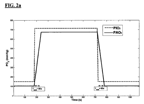
Fig. 2a: represents predicted wash in and wash out times (TVENT) for a given
change
in partial pressure of Oxygen breathed by a subject (P02).
Fig. 2b: illustrates a compartmental model fit to the estimated average tissue
P02
(difference above baseline air-breathing) of a region of interest at the top
of the right
lung of a subject as discussed in Example 1. The alveolar oxygen input
function is
also illustrated.
Fig. 3: represents maps of the compartmental model parameters obtained in the
right
lung of one individual as discussed in Example 1. Map (a) illustrates with a
rainbow
colourmap the extraction fraction multiplied by blood flow and demonstrates a
regional variation over the lung, with lower values towards the top of the
lung and at
the periphery. Map (b) illustrates the results of Map (a) using a greyscale
colourmap.
If E is assumed equal to 1, the Fb values are consistent with those reported
in the
18
CA 02686122 2009-11-03
WO 2008/135712 PCT/GB2008/001390
literature. Map (c) shows the distribution of KoX, which is mainly uniform
apart from
apparent clusters of higher ventilation in the centre of the lung using the
rainbow
colourmap. Map (d) shows the results of Map (c) using a greyscale colourmap.
Map
(e) shows the variation in time to achieve maximum P02 in the alveoli, with
central
areas achieving times close to the predicted value of 1 minute, while at the
lung
periphery values of up to 2 minutes were obtained. Map (f) shows the results
of Map
(e) using a greyscale colourmap.
Fig. 4: represents maps and histograms of the compartmental model parameters
obtained in the right lung of (a) a non-smoker; and (b) a smoker as discussed
in
Example 2.
Fig. 5: represents histograms of averaged data of the compartmental model
parameters obtained in the right lung of (a) a group of non-smokers; and (b) a
group
of smokers as discussed in Example 2.
Fig. 6: represents modeled values for the concentration of Oxygen in the
second
compartment (Cp) in a particular exemplary embodiment. Detected signal values
for
concentrations of Oxygen Ce are also shown.
Fig. 7: represents a box plot of results from an analysis of Kox of a number
of test
subjects in the third example.
Fig. 8: represents a box plot of results from an analysis of EFb of a number
of test
subjects in the third example.
Fig. 9: represents a box plot of results from an analysis of the wash in time
TvENr to
reach a maximum concentration of Oxygen in the lung tissues and blood of a
number
of test subjects in the third example.
Fig. 10: represents image maps of values for a) Kox, b) EFb and c) TvENT for
the lung
of a healthy non-smoking subject.
19
CA 02686122 2009-11-03
WO 2008/135712 PCT/GB2008/001390
Fig. 11: represents image maps of values for a) Kox, b) EFb and c) TVENT for
the lung
of a smoking subject with a healthy spirometry.
Fig. 12: represents image maps of values for a) Kox, b) EFb and c) TVENT for
the lung
of an unhealthy smoking subject.
Fig. 13: represents a correlation plot between values of Kox modelled by a
method
according to the present invention and PS generated from standard Dynamic
Contrast
Enhanced MRI (DCE-MRI). Each data point is signified as belonging to a smoker
(square points) or a non-smoker (circular points).
Fig. 14: represents a correlation plot between values of EFb modelled by a
method
according to the present invention and PS generated from standard DCE-MRI.
Each
data point is signified as belonging to a smoker (square points) or a non-
smoker
(circular points).
CA 02686122 2009-11-03
WO 2008/135712 PCT/GB2008/001390
EXAMPLE 1
The method of the first aspect of the invention was developed and applied to
measure
lung function of a group of normal individuals.
1.1 Methods
(a) Image Acquisition
The images used in this study were obtained from five normal consented
volunteers,
(two males, three females, ages 30-39), using a 1.5 T Philips Gyroscan NT
Intera MR
system (Philips Medical Systems, Best, Netherlands). Subjects breathed medical
air or
100 % oxygen through an MR compatible Bain breathing system (Intersurgical
Ltd.,
Wokingham, UK) and tightly fitting mask. A standard anesthesia trolley (10
Umin
capability) was used. A first set of images was acquired in order to measure
T, during
air-breathing. A half Fourier single shot turbo spin-echo (HASTE) sequence was
used
with 68 phase encoding steps and inter-echo spacing of 4ms, effective echo
time
16ms, 128 x 128 matrix with field of view 450 x 450 mm2, coronal section with
slice
thickness 10 mm. TI measurements were performed using a saturation recovery
HASTE sequence with saturation times (TSAT) of 100, 200, 400, 800, 1200, 1700,
2300, 3000, 3500 ms. Five images were collected for each saturation time to
enable
averaging over the cardiac cycle. Saturation recovery (SR) was chosen here to
give a
shorter total imaging time. Next, dynamic image acquisitions were performed
using
an IR HASTE sequence with an inversion time of 720 ms (chosen to approximately
null the signal from the lungs while breathing air). The gas supply was
switched from
medical air to 100 % oxygen after the tenth image in the series. A set of Tl
measurement SR images was acquired while the subject continued to breathe 100
%
oxygen. Finally a second series of dynamic images was acquired with the gas
supply
being switched back to medical air after the tenth image.
(b) Compartment Model
The first compartment is the alveolus space and the oxygen concentration may
be
denoted by CA, and the second compartment includes the alveolus membrane,
interstitial space between the membrane and pulmonary capillaries and the
plasma
within the capillaries, with a combined oxygen concentration denoted by Cp
(see Fig.
1).
21
CA 02686122 2009-11-03
WO 2008/135712 PCT/GB2008/001390
The inventors then developed a model by assuming near saturation of oxygen in
arterial haemoglobin during air breathing would result in extra oxygen being
carried
mainly in the plasma when 100% oxygen breathing occurs. In lung the increased
signal occurs in the parenchymal water and capillary blood, and therefore the
measured increased concentration can be considered equivalent to
Cp(ar(7o )~_(Ta.) ~_~~ _~~.11. Using these approximations the inventors
developed
lL~ ~ - J)
equation (I):
dC P _ / (I)
VP dt Kar(CA - CP) - EFbCp,
where Vp is the fractional volume of blood plasma and tissue water per MRI
visible
tissue, Kox is a term describing the diffusing capacity of the alveolar
membrane, E is
the extraction fraction of oxygen from the tissue water and capillaries, and
Fb is the
rate of blood flow in the capillaries. Based on these calculations the
inventors realised
that it would be possible to solve Cp (i.e. the combined oxygen concentration
of the
second compartment comprising the alveolus membrane, interstitial space
between
the membrane and pulmonary capillaries and the plasma within the capillaries
calculated as described above) using equation II:
CP = Vox ('CA(z)eXp _ KosV EFb (t-z)Idz = (u)
P 1 P )
Clinically meaningful information may be attached to values for EFb and Kox
and data
is present from pulmonary regions and also as parameter maps from the whole
lung in
the Figures.
(c) Image Registration and Application of Compartmental Model
For registration an active shape model (Cootes et al. (1995) Computer Vision
and
Image Understanding, 61: 38) was used to characterize normal breathing motion
and
then to allow the automated identification of the outline of the lung. The
lung shapes
were then transformed to an average shape using linear re-sampling. T, maps
were
22
CA 02686122 2009-11-03
WO 2008/135712 PCT/GB2008/001390
calculated for air and oxygen breathing by fitting the saturation recovery
images to
equation III using a Levenberg-Marquardt fitting algorithm.
(III)
S(TS.4T ) = A - B exp _ TK4T
T,
To convert the dynamic signal intensity data to increase in dissolved oxygen
concentration above air-breathing, the values were first converted to T,
values using
book-ending from T, maps (Cron et al. (1999) Magnetic Resonance in Medicine.
42 4
:746-53). They were then converted to R, by inversion, the baseline air-
breathing Rl
was subtracted, and finally the values converted to P02 (1nmHg) by division by
the
known relaxivity constant rl=4x 10⁴ s"1mmHg 1(Jakob et al. (2004) Magnetic
Resonance in Medicine. 51: 1009-1016).
The oxygen input function was estimated from the known ratios~ of alveolar gas
partial
pressures (Martin, L (1999): All you really need to know to iriterpret
Arterial Blood
Gases. Lippincott Williams & Wilkins 2nd ed.). Alveolar P02 (PAOZ) during air-
breathing is typically 104 mmHg, and during 100% oxygen breathing at an
atmospheric pressure of 760 mmHg it can be estimated at 673 mmHg. Hence the
difference in PAO2 over air breathing can be approximated as 569 mmHg. The
rate of
replacement of air in the lungs is typically estimated as on average 7.5 % per
breath.
At rest one takes an average of one breath per 5 seconds, and therefore it
takes on
average approximately 1 minute for all air to be replaced. This was reflected
in the
sloping edges of the estimated PAOZ input function (see Fig. 2a). During air
replacement with 100% oxygen the lung will replace air at an unknown variable
rate
at the alveoli. A linear function was therefore chosen to describe the edges
of the
input function (in preference to an exponential or other more complex function
- these
options can easily be incorporated into the method) as the simplest
approximation to
the true fonn. According to varying individual lung capacity and the position
within
the lung, it was found that the delay time to maximum PAO2, i.e. the value of
TvENT,
required optimization. Using a Levenberg-Marquardt fitting algorithm and
assuming a
Vp fraction of 1, equation II was solved for EFb and Ko,, for averages over
regions of
interest at the top of each lung (see table 1). TvENT was a third free
parameter in the fit
23
CA 02686122 2009-11-03
WO 2008/135712 PCT/GB2008/001390
(see Fig. 2b). Voxel-by-voxel parameter maps were also calculated (see Fig.
3). The
values of EFb were converted to standard units (ml/min/ml) presuming a lung
density
of 0.15 g/ml near the lung periphery (Hatabu, et al. (1999). Magnetic
Resonance in
M edi cine. 42: 103 3-103 8).
(d) Spirometry
The method in accordance with the first aspect of the present invention may be
compared to spirometry so as to validate the diagnostic capabilities of the
method in
relation to lung function. Spirometry is a pulmonary function test which is
commonly
performed clinical situations. Currently spirometry is the main test that is
used to
diagnose irregular lung function. Spirometry comprises measurements taken from
a
subject inhaling or exhaling through a tube. A subject blows through a tube
and the
values of total volume exhaled in one minute (FEVI) and total volume exhaled
(FVC)
are measured. From these values, the value of FEV YPRED is calculated by a
function
of the ratio of FEV and FVC as compared to normal ranges known in the
population.
FEV9,,,PRED is a well known measure of the healthiness of spirometry
measurements of
the subject, a healthy value being greater than 0.75.
(e) Box Plots
The results shown in Figures 7 to 9 are shown as box plots. Box plots are
known in
the art. The box plot representation comprises one glyph per grouping of data,
each
glyph including a horizontal line at the top and bottom of the glyph, which
respectively indicate the largest and smallest observed values in the group. A
box
shape in the glyph comprises upper, intermediate and lower horizontal lines
which
indicate the upper quartile range, median value and lower quartile range.
1.2 Results
Assuming an extraction fraction E of 1, the Fb values given in table 1 are
consistent
with literature quoted values for lung perfusion (Hatabu, et al. supra). The
diffusion
measures obtained in K, varied considerably between individuals but were
mainly
consistent between both lungs in each individual. The parameter EFb (Fig. 3)
illustrates variation over the right lung slice area with less perfusion
towards the edges
and top of the lung and larger values corresponding to the main pulmonary
vessels in
the centre. The Kox parameter map shows stronger ventilation-diffusion in the
centre
24
CA 02686122 2009-11-03
WO 2008/135712 PCT/GB2008/001390
lung but the peripheral values are more uniform to the lung edges than in the
EFb
map. The map of time lag TVENT to maximum CA shows shorter times in the centre
lung and longer times at the periphery.
Table 1. Average compartmental model parameters obtained from left and right
lungs
on five individuals
Subject Lung EFb(mUmin/ml) KoX(ml/min/g) TvEN7(s)
1 Left 1.2677 4.1431 66
Right 0.8015 3.5409 68
2 Left 1.0762 2.3612 44
Right 1.3007 1.7382 42
3 Left 1.0106 17.5859 128
Right 1.1345 17.0257 75
4 Left 1.3892 5.6615 53
Right 0.7018 14.8614 61
Left 1.2639 0.8860 140
Right 1.0402 19.4285 83
1.3 Conclusions
These data present a compartmental model of oxygen delivery to the lung which
allows direct assessment of pulmonary perfusion, diffusion and ventilation
using OE-
MRI. This may be used to detect regions of impaired ventilation, diffusion or
perfusion when applied to patient groups.
This work demonstrates for the first time how lung perfusion may be estimated
using
OE-MRI. This technique has advantages over other methods of measuring
perfusion,
including ease of implementation and the lack of risk associated with the
oxygen
contrast agent, i.e. gadolinium based contrast agents used in DCE-MRI hold
certain
minimal risks of allergic reaction and elevated risks in patients with renal
impairment.
In terms of understanding the delivery mechanism of oxygen, this modeling
approach
also provides a new means of separating the information associated with
general
CA 02686122 2009-11-03
WO 2008/135712 PCT/GB2008/001390
airway health (or TvENT, the time taken to achieve maximum CA) from the rate
of
diffusion across the membrane (Kox).
EXAMPLE 2
The methods described in Example 1 were applied to a group of non-smokers and
a
group of smokers to illustrate how the methods of the invention can be used to
illustrate how lung function varies between these two groups.
Figure 4 represents maps and histograms of Kox,, EFb and ventilation time
(respectively) for the right lung of (a) a non-smoker; and (b) a smoker.
Figure. 5 represents histograms of averaged data for KoX,, EFb and ventilation
time
(respectively) for the right lung of (a) the non-smoker group; and (b) the
smoker
group.
These data illustrate that the method of the invention can be used to
demonstrate that
lung function of these two groups is different. In the smokers the Kox
histogram is
shifted to the right. This suggests a higher rate of diffusion of oxygen into
the tissue
which may be due to either an increased alveolar membrane permeability
(consistent
with other findings, Mason et al, (2001) Clinical Science, 100: 231-236) or by
a
greater initial gradient of oxygen concentration across the membrane in
smokers due
to the known lower blood oxygen in smokers, or by a combination of both.
Furthermore the EFb histogram for smokers is shifted to the left and this
suggests a
lower blood flow from the lung tissue, which is consistent with known impeded
blood
circulation in smokers. Finally the ventilation time histogram is shifted to
the right in
the smokers, indicating that it takes longer for the inspired oxygen to reach
the
alveoli, due to the known airway constriction effect of smoking.
All these data are consistent with the compromised lung function that a
clinician
would expect to see in a smoker and demonstrate that the methods of the
present
invention can by utilised to obtain meaningful data relating to lung function.
The
difference between the two groups is predictable and illustrates that the
methods may
be applied to a number of prognostic or diagnostic uses.
26
CA 02686122 2009-11-03
WO 2008/135712 PCT/GB2008/001390
EXAMPLE 3
In the third example, further evaluation of the compartmental model algorithm
of
Example 1 has been carried out using groups of smokers and non-smokers, so as
to
further validate the method according to the first aspect of the present
invention.
3.1 Methods
The methods described in Example 1 were applied to a further group of eleven
non-
smokers and twelve smokers to further illustrate how the invention can be used
to
analyse how lung function varies between these two groups and how the
unhealthy
partial lung function of a subject can appear to be healthy when considered as
an
average of the entire lung, particularly when tested against spirometry.
(a) OE-MRI scan and Compartmental Model
The twelve smokers and eleven non-smokers each underwent an OE-MRI scan
according to the method described for the first example. The data from the
scan of
each subject were fed into a compartmental model according to the
compartmental
model described above and set out in equation (II), and in example 1. Data
relating to
lung function such as KoX, EFb and TvENT were obtained from the compartmental
model, and the data were analysed. The results of the analysis are presented
below.
3.2 Results
Results of fitting compartmental models in accordance with the present
invention to
OE-MRI data from each of the subjects is shown in a variety of forms below.
(a) Average results for a healthy non-smoker
Figure 6 shows the results of a fit of the two-compartment model to the mean
right
lung absorption of Oxygen (labelled PAOz) into the second compartment Cp
(tissues
and blood) of the compartmental model for a healthy non-smoking subject
breathing
gas with two different partial pressures of Oxygen (labelled P102). As the
model was
fitted to mean values for the whole right lung, the resulting values of Kox,
EFb and
TVENT represent average values for the whole right lung.
27
CA 02686122 2009-11-03
WO 2008/135712 PCT/GB2008/001390
The values shown in Figure 6 represent the difference (OPOz) in P02 in each
compartment for two different partial pressures of oxygen breathed by the
subject, the
first pressure being approximate to air (21% 02) and the second being pure
Oxygen
(100% 02). At approximately 200s, the concentration of oxygen in the gas mix
being
breathed by the subject was increased. As shown in Figure 6, as the
concentration of
Oxygen in the first compartment CA increases, the concentration of Oxygen in
the
second compartment Cp increases proportionally to the change in the first
compartment. This continues until both compartments reach a saturation point,
at
which point the concentration of Oxygen in both compartments remains constant.
The time taken to reach maximum Oxygen concentration in the second
compartment,
labelled TvENr, for the healthy non-smoking subject is 60s. When, at
approximately
700s, the concentration of oxygen breathed by the subject is returned to it's
initial
concentration at the start of the test, the concentration of Oxygen in the two
compartments reduces over time to its former state.
(b) Average results for groups of smokers and non-smokers
The test described above with reference to Figure 6 was repeated for each
member of
the test group, which included both smokers and non-smokers. Values of Kox,
EFb
and TvENr were generated from compartmental model fits performed for each of
the
subjects. The subjects were divided into groups according to their smoking
habits and
general health. The smokers are divided into "All smokers" and "Smokers with
>.
20PY", where PY is pack years, which is the number of packs of cigarettes
smoked
per day by the number of years for which that number of packs have been
smoked. A
person of 20 PY may, for example, have smoked one pack per day for twenty
years,
or perhaps two packs per day for ten years. The non-smokers are divided into
"healthy" non-smokers and "All non-smokers". Healthy smokers were defined as
those with a healthy spirometry test, 0 PY and no regular exposure to passive
smoke.
The results for each group are shown in the box-plots depicted in Figures 7 to
9.
Figure 7 shows a box plot of comparative results for Kox between the group of
twelve
smokers and eleven non-smokers. It is clear from Figure 7 that the median
values of
Kox between smokers and non-smokers are significantly different. This shows
that it
28
CA 02686122 2009-11-03
WO 2008/135712 PCT/GB2008/001390
is possible to discriminate between smokers and non-smokers merely by
computing
an average Kox value for each subject. Since the value of KoX for a subject is
indicative of the rate of diffusion of Oxygen across the alveolar membrane.
Figures 8 shows comparative results for values of EFb which were extracted
from the
compartmental model for each of the groups of subjects. As described above,
EFb is a
measure of the rate at which oxygen is removed from the lung tissues by the
blood.
As the rate at which Oxygen is removed from the lung tissues for transport
around the
body is critical to the healthy respiratory function of the subject, this
value is an
important measure of damage to the respiratory system. It is clear from these
results
that EFb in healthy, and unhealthy, non-smoking subjects is higher than for
either of
the smoking groups. This reduced respiratory function is an important factor
in the
health of the subjects and could not be directly detected by methods such as
129Xe
imaging because the data produced by such techniques would relate to the rate
at
which 129Xe, rather than Oxygen, is removed from the lung tissues.
Figure 9 shows comparative results for the wash in and wash out values TvENr
between the groups of subjects. The time taken to reach maximum Oxygen
concentration in the compartments (TvENT) of the compartmental model is
indicative
of the efficiency of each breath of an individual in clearing out old gas from
the lungs
and introducing new gas into the lungs. The value of TVENT is also a good
indication
of the time taken for new air in the lungs to be dissolved into the lung
tissues. It is
clear from these results that the non-smoking groups generally have a much
lower
value of TvENT than the smoking groups. This again indicates reduced lung
function
in the patient.
For each of the measures shown in Figure 7, 8 and 9 it is clear that the
values which
can be extracted from the compartmental model of the present invention are
capable
of measuring a detectable difference between various aspects of the lung
function of
smokers and non-smokers. More particularly, the values are capable of showing
differences between each of the four groups shown in Figures 7, 8 and 9.
29
CA 02686122 2009-11-03
WO 2008/135712 PCT/GB2008/001390
(c) Exemplary lung function maps
The results presented in section (b) above were generated as average values
across the
whole right lung of each individual. However, a compartmental model in
accordance
with the present invention is capable not merely of generating broad measures
across
the entire lung, but of generating lung parameter maps which show individual
values
of measures such as Kox, EFb and TvENT for each voxel in the data generated by
the
OE-MRI which contains lung tissue or blood. The following results, shown in
Figures 10, 11 and 12, are examples of such lung parameter maps which were
generated for subjects who took part in the testing described in the present
example.
In order to compare the results of the compartmental model of the present
invention
with an existing diagnostic method, the subjects also underwent spirometry
tests
which determined values of FEV%PR.ED for each of the subjects.
Figures 10a, 10b and 10c show lung parameter maps respectively for Kox, EFb
and
TvENT for a subject who is a healthy, non-smoking female, aged 23 years old.
Bright
pixels in the map indicate high values for each measurement, while dark pixels
in the
map indicate lower values. The subject had a spirometry value of FEV%PRED =
93%,
which indicates that she has healthy lung function. The median results, which
contributed to the statistical results shown in Figures 7 to 9, which were
extracted
from the comparhnent model for this subject are:
Median Kox 3.19m1/min/g
Median EFb=19.4 ml/min/g
Median TvEN7=24s
The grey levels in the lung function map for Kox, shown in Figure 12a, are
uniform
across each lung, which indicates that the difference in the rate of diffusion
across
alveolar membranes between different areas of the lung are small. Similar
results are
shown in Figure 12b, which shows that the rate at which dissolved oxygen is
removed
from the lung tissues is uniformly high across each lung. Finally, Figure 12c
shows
that the wash in and wash out time for each lung is generally equally low
across the
extent of the lungs, although some areas of high TVENT are evident in the lung
shown
on the right. This shows that the entire area of this subject's lungs is
functioning well
and contributing to respiration in the subject. Large, distinct, black regions
are visible
CA 02686122 2009-11-03
WO 2008/135712 PCT/GB2008/001390
in each lung of each image, but these correspond to large areas of bronchus,
or
airway, which transports air into and out of each lung. These areas have low
values of
KoX, EFb and TvENT as there is no second compartment measurement due to the
lack of
tissue and blood in the area.
Figures 11 a, 11 b and 11 c show image results respectively for Kox, EFb and
TVENT for a
second subject who is an apparently healthy, smoking female, aged 54 years
old. The
subject had a spirometry measurement of FEV%PRED = 92%, which indicates lung
function which is within the healthy range. The median results which were
extracted
from the compartment model for this subject, and which contributed to the
statistical
results shown in Figures 7 to 9, are:
Median Kox 3.7ml/min/g
Median EFb=16.6 ml/min/g
Median TvEN1=90s
The median values for Kox and EFb for the subject are lower than those for the
healthy
non-smoking subject, which indicates slightly reduced overall lung function
for this
subject. It can be seen that the grey levels in the lung parameter maps of Kox
and EFb
shown in Figures l la and l lb differ from the images shown in Figures l0a and
lOb in
that there are small darkened regions throughout the lungs. This indicates
that rates
have been reduced for diffusion of Oxygen across the alveolar membrane and for
removal of Oxygen from the lung tissues by the blood. The two images alone
show
reduced lung function in the subject in many areas of the lungs. In addition
to Figures
l la and l lb, l lc shows the time taken for each area of the lungs to reach
maximum
concentration of Oxygen (TvENT). When compared to the lung parameter map for
TvENr in the healthy, non-smoking subject, shown in Figure lOc, the lung
parameter
map of TVENT for the apparently healthy smoking subject, shown in Figure 11c,
is
clearly brighter in many areas. This indicates that the time to reach maximum
saturation of Oxygen for the subject is longer, and that therefore lung
function is not
as efficient as for the healthy non-smoker.
This means that although this subject showed normal results in a standard
spirometry
test, they in fact have decreased lung function in some areas in their lungs.
It would
31
CA 02686122 2009-11-03
WO 2008/135712 PCT/GB2008/001390
not be possible to directly diagnose this irregularity of function by any
other means.
This indicates that the diagnostic capabilities of the method of the present
invention
are able to detect lung abnormalities in circumstances where spirometry shown
normal performance, a presents a significant advantage over previous methods.
Figures 12a, 12b and 12c show lung parameter maps respectively for Kox, EFb
and
TVENT for an unhealthy, smoking male subject aged 57 years old. The subject
has been
diagnosed with type IIB chronic obstructive pulmonary disease (COPD). The
smoker
has a FEV%PRED=39%, which indicates substantially reduced lung function. The
median results of the subject for his entire right lung, which were extracted
from the
compartment model are:
Median KoX 3.34ml/min/g
Median EFb= 16.9 ml/min/g
Median TvEN7=123s
It is clear from the median values for Kox, EFb and TvENT, particularly the
value of
TVENT, that lung function in the subject is reduced in comparison to either of
the two
subjects whose results are shown in Figures 10 and 11. The black regions in
the Kox
and EFb lung parameter maps, shown in Figure 12a and 12b, indicate that the
lung
tissue in those regions is not functioning. The white regions in the TVENT
lung
parameter map indicate that those lung regions are taking a very.long time to
reach a
=;
maximum concentration of oxygen. Completely black regions in Figure 12c
indicate
that the area of the lung reaches a maximum concentration of oxygen very
quickly,
although it is clear from the low values of Figure 12a and 12b that the
maximum
concentration of oxygen is substantially lower for this subject than for the
subjects
whose results are shown in those figures.
(d) Correlation with independent measures in DCE-MRI
The inventors compared the compartmental model algorithm of the present
example
with a measurement of the product of permeability and surface area (PS) of the
lungs
of the same subjects, which was produced by standard DCE-MRI. It should be
noted
that, although the measurement of PS is the current standard for assessment of
the
32
CA 02686122 2009-11-03
WO 2008/135712 PCT/GB2008/001390
permeability of the lungs, it is a measurement of the permeability of the lung
capillary
walls to a non-oxygen contrast agent and not to oxygen itself. Therefore, any
measurements of lung permeability to oxygen from DCE-MRI are indirect
measurements which are calculated from the direct measurements of contrast. In
this
case the contrast agent used in the DCE-MRI was a gadolinium-based low
molecular
weight agent.
Figure 13 shows a correlation between the KoX measurement of the present
invention
and the PS (permeability x surface area) measurement of DCE-MRI. Figure 14
shows
a correlation between the EFb measurement of the present invention and the PS
measurement of DCE-MRI. This indicates that a method according to the present
invention may be used in place of DCE-MRI, in order to ameliorate the many
problems associated with creating, storing and transporting contrast agents
and with
introducing such contrast agents into in vivo subjects.
3.3 Conclusions
This example has shown that a method in accordance with the present invention
applied to OE-MRI image data of smokers and non-smokers is capable of
differentiating between the local function of the lungs of smokers and non-
smokers,
and in some cases is capable of diagnosing a decrease in local lung function
when a
global assessment of lung function, via spirometry, appears to be clinically
normal. It
has been shown by comparison with DCE-MRI that a method according .to the
present
invention may be used in addition to, or in place of, DCE-MRI for the
diagnosis of
pathological lung function.
33
CA 02686122 2009-11-03
WO 2008/135712 PCT/GB2008/001390
EXAMPLE 4
In this example, the inventors implemented a compartmental model algorithm in
accordance with an aspect of the present invention. The algorithm was
implemented
as software in Matlab script. The inventors packaged the Matlab script, which
implements the compartmental model algorithm, for distribution on a compact
disc.
The compact disc containing the software was given in confidence to an
operative
who was in possession of a 1.5 T Philips Gyroscan NT Intera MR scanner. The
operative used the scanner, together with Oxygen breathing apparatus widely
available in medical environments, to performed an OE-MRI scan on a human
patient
according to the method described in the first example. The operative then
used the
software on the compact disc to analyse the data generated by the OE-MRI scan
of the
patient so as to characterise the patient's lung function, in accordance with
an aspect
of the present invention.
The compartmental model generated values indicative of the patient's lung
function,
including KoX, EFb and TvENT. The software displayed the values generated by
the
compartmental model in a number of ways, including as a series of graphs and
lung
parameter maps. The data values, the graphs and the lung parameter maps were
used
by a medical professional to analyse the lung function of the patient and
diagnose
illness in the patient. The sensitivity of the model to the function of
relatively small
areas of the patient's lungs allowed the medical professional to diagnose
local areas of
low lung function within the lungs and to target therapy accordingly. The use
of OE-
MRI to generate the data which was input into the compartmental modelling
software
was particularly new and interesting in that the medical professional was able
to
directly analyse the function of the lung in relation to Oxygen ventilation,
diffusion
and perfusion. This direct analysis has not been possible before the present
invention.
34