Note: Descriptions are shown in the official language in which they were submitted.
CA 02687575 2009-11-18
WO 2008/144889 PCT/CA2008/000977
Humanized And Chimeric Anti-CD59 Antibodies That Mediate Cancer Cell
Cytotoxicity
FIELD OF THE INVENTION
This invention relates to the diagnosis and treatment of cancerous diseases,
particularly to the mediation of cytotoxicity of tumor cells; and most
particularly to the use of
cancerous disease modifying antibodies (CDMAB), optionally in combination with
one or
more CDMAB/chemotherapeutic agents, as a means for initiating the cytotoxic
response. The
invention further relates to binding assays, which utilize the CDMAB of the
instant invention.
BACKGROUND OF THE INVENTION
CD59 is an 18-20 kDa glycosyl phosphatidylinositol (GPI)-anchored
membrane glycoprotein. It was initially isolated from the surface of human
erythrocytes, and
functions as an inhibitor of complement activation. Several antibodies that
were developed to
enhance complement-mediated lysis were subsequently found to target CD59.
Their
independent development led to the multitude of names by for CD59, including
MEM-43
antigen, membrane inhibitor of reactive lysis (MIRL), H19, membrane attack
complex-
inhibitory factor (MACIF), homologous restriction factor with m.w. 20,000
(HRF20) and
protectin (Walsh, Tone et al. 1992).
The CD59 antigen has been well characterized by amino acid analysis and NMR.
It
consists of 128 amino acids, of which the first 25 comprise a signal sequence.
There are 10
cysteine residues, which result in a tightly folded molecule. The asparagine
residue at position
18 is known to be N-glycosylated, while the asparagine residue at position 77
is linked to the
GPI anchor. The C-terminus residues are characteristic of GPI-anchored
proteins (Davies and
Lachmann 1993).
CD59 was initially discovered on the surface of human erythrocytes, but is a
widely expressed molecule. A large collection of data on cellular distribution
from flow
cytometry, immunohistochemistry and Northern blot analysis has revealed
expression on
many types of cells and tissues, including hematopoietic cells such as,
platelets, leukocytes
and fibroblasts, as well as erythrocytes (Meri, Waldmann et al. 1991). CD59 is
abundant on
vascular and ductal endothelium throughout the body, particularly in kidneys,
bronchus,
pancreas, skin epidermis and biliary and salivary glands (Meri, Waldmann et
al. 1991).
1
CA 02687575 2009-11-18
WO 2008/144889 PCT/CA2008/000977
Expression has been noted in the lung, liver, placenta, thyroid and
spermatozoa (Davies and
Lachmann 1993). Soluble forms of CD59 have been detected in saliva, urine,
tears, sweat,
cerebrospinal fluid, breast milk, amniotic fluid and seminal plasma (Davies
and Lachmann
1993). The origin of soluble CD59 has yet to be determined; whether it is
secreted, cleaved by
phospholipases or shed from cells by other means remains unknown (Davies and
Lachmann
1993). CD59 appears to be absent from many B cell lines, CNS tissue, liver
parenchyma and
pancreatic Islets of Langerhans (Meri, Waldmann et al. 1991).
Although CD59 is widely expressed in normal cells and tissues, it is also
widely expressed on malignant tumors. There is evidence that the expression of
CD59 is
increased in certain types of cancer compared to normal tissue and that the
level of expression
correlates with the stage of differentiation of the tumor. Moderate to high
levels of CD59
expression have been reported in thyroid, prostate, breast, ovarian, lung,
colorectal,
pancreatic, gastric, renal and skin cancers as well as in malignant glioma,
leukemia and
lymphoma (Fishelson, Donin et al. 2003).
With the exception of tumor grade, no correlation is observed between CD59
expression and tumor/patient characteristics such as tumor type, size,
vascular invasion,
patient age, gender or menopausal status (breast cancer only) (Madjd, Pinder
et al., 2003;
Watson, Durrant et al., 2006). In studies using different tumor tissues that
include breast,
colorectal and prostate, CD59 expression correlates strongly with moderate to
well-
differentiated tumor grades (Madjd, Pinder et al., 2003; Watson, Durrant et
al., 2006, Jarvis,
Li et al., 1997; Koretz, Bruderlein et al., 1993). However, the association of
CD59
expression on well-differentiated tumors with patient prognosis remains
unresolved. Two
separate studies using breast and colorectal cancer samples show that CD59
expression in
highly differentiated cells correlates with good patient prognosis (Madjd,
Pinder et al., 2003;
Koretz, Bruderlein et al., 1993). Alternatively, in another study using
colorectal cancer tissue,
Watson et al. reported that the correlation between high CD59 levels and
differentiated tumor
grade can be sub-divided into early and late stage disease. These authors show
that high
CD59 levels found in well-differentiated early and late stage tumors is
associated with a
decrease in disease specific patient survival (Watson, Durrant et al., 2006).
Conversely, de-differentiated tumor cells correlates best with an absence of
CD59 staining, which may have implications for metastasis. Several studies
suggest that
2
CA 02687575 2009-11-18
WO 2008/144889 PCT/CA2008/000977
increased CD59 expression is inversely correlated with tumor metastasis. In
breast
carcinomas and colorectal cancers, high CD59 expression occurs in tumor
samples without
metastasis (Madjd, Pinder et al., 2003; Koretz, Bruderlein et al., 1993).
Similarly, a low
percentage of cells with high CD59 levels are found in colorectal metastatic
tumors in the
liver (Hosch, Scheunemann et al., 2001). Also, CD59 expression in squamous
cell
carcinomas of the head and neck are only elevated in samples with Tl/T2NOMO
tumor grades
and not in tumor grades beyond NI and Ml (Ravindranath, Shuler et al., 2006).
The most characterized function of CD59 is its ability to inhibit the
formation
of the membrane attack complex (MAC) following complement activation. MAC
formation is
the final event in the complement cascade in which a pore is fonned in the
cellular membrane
that ultimately leads to lysis of the cell. CD59 binds to C5b-8 and interferes
with the
subsequent polymerization of C9 molecules, the step that is required for MAC
formation.
Competition and mutational analysis of the epitopes of CD59, done with
blocking and non-
blocking monoclonal antibodies, has mapped the location of the active site of
CD59 and has
identified the amino acids Tyr-40, Arg-53 and Glu-56 to be necessary for CD59
activity
(Bodian, Davies et al., 1997).
Complement activation results in either destruction of the targeted cell or
cell
activation, which recruits leukocytes, contracts surrounding smooth muscle and
increases
vascular permeability. Complement also plays a role in antibody-dependent
cellular
cytotoxicity (ADCC) and complement-dependent cellular cytotoxicity (CDCC).
This can lead
to an inflammatory response that could damage targeted tissues if poorly
regulated. CD59 and
other complement inhibitory proteins such as complement receptor type-1 (CRI;
CD35),
membrane cofactor protein (MCP; CD46) and decay accelerating factor (DAF;
CD55)
function to counter excessive activation of the complement cascade to prevent
autologous
tissue damage. It has been postulated that differential expression of
complement inhibitory
proteins such as CD59 may contribute to enhanced resistance to complement
activation that
malignant tumors often acquire (Jarvis, Li et al. 1997).
To evaluate whether resistance to complement by tumor cells can be overcome
by targeting CD59, the ability of the CD59 blocking antibody YTH53.1 to
enhance lysis of
tumor cells has been evaluated in vitro. In a study using three-dimensional
microtumor
spheroids (MTS) with breast cancer (T47D cell line) and ovarian
teratocarcinoma (PA-1 cell
3
CA 02687575 2009-11-18
WO 2008/144889 PCT/CA2008/000977
line) cells, the ability of this antibody to block CD59 activity and thus
complement resistance
has been measured. MTS are multicellular aggregates that grow in culture and
represent a
model closer to that observed in vivo than monolayer or suspension cultures.
Previous work
by this group has shown that PA-1 cells grown as MTS are more resistant to
complement lysis
than PA-1 cells grown in suspension. Cytotoxicity was measured by a chromium
release assay
and cell damage was visualized by uptake of propidium iodide (PI) following
pre-treatment of
MTS with biotinylated YTH53.1. Biotinylation of YTH53.1 retains its affinity
for CD59 but
eliminates its capacity to activate the classical complement pathway. Rabbit
anti-human
polyclonal antibody raised against breast cancer cells (S2 cell line) was used
to activate the
classical complement pathway. Overnight incubation with biotinylated YTH53.1
led to total
infiltration of the MTS, and the chromium release assay showed killing of 33
percent of cells
after a 1 to 2-hour lag phase in the presence of biotinylated YTH53.1, S2 and
human
complement. Under the same treatment, electron microscopy revealed the average
T47D
tumor volume decreased 28 percent. Fluorescence microscopy following PI
incubation
revealed several layers of cell death on T47D and PA-I MTS. These results
indicate that an
anti-CD59 antibody that can block CD59 inhibitory activity can increase the
complement-
mediated lysis of tumor cells in vitro (Hakulinen and Meri 1998).
In another study, resistance to complement-mediated lysis by the human
metastatic prostate adenocarcinoma cell lines DU145 and PC3 could be overcome
in vitro by
treating with YTH53.1. Chromium release assay was used to measure cell death
in the
presence and absence of YTH53.1 and biotinylated YTH53.1. In the absence of
CD59
antibodies, both cell lines were completely resistant to complement-mediated
lysis; however,
treatment with YTH53.1 partially overcame this resistance by killing 56
percent of PC3 cells
and 34 percent of DU145 cells. Treatment with biotinylated- YTH53.1 was less
effective in
overcoming complement resistance; 47 percent of PC3 and 20 percent of DU145
cells were
killed. The higher expression of CD59 by PC3 compared with DU145 cells and
possibly its
greater dependence on CD59 expression and function in resisting complement
mediated lysis
is reflected by the increased sensitivity of PC3 compared to DU 145. The
differential effect of
the native and biotinylated antibody demonstrates the enhanced effect of both
activating the
classical complement pathway and neutralization of CD59 (Jarvis, Li et al.
1997). However,
the bulk of the activity of the antibody may be attributed to the blocking of
complement
4
CA 02687575 2009-11-18
WO 2008/144889 PCT/CA2008/000977
inhibition (neutralization of CD59), as adding complement activation by the
classical pathway
only increases activity by a marginal amount (e.g. 47 percent for biotinylated-
YTH53.1 versus
56 percent for YTH53.1 on PC3 cells) (Jarvis, Li et al. 1997). This study
together with the
one described previously demonstrates that targeting CD59 using an antibody
may be an
effective therapy for blocking resistance to complement activation in
malignant tumors.
In an alternative approach, Harris et al. aimed to specifically target CD59 on
tumor cells in vitro using engineered bi-specific antibodies. CD59 was
neutralized using one
of two different bispecific F(ab'gamma)2 antibody constructs which contained
both cell-
targeting (anti-CD 19 or anti-CD38) and CD59-neutralizing moieties. In these
experiments,
Fabgamma Fc gamma2 chimeric antibody (specific for human CD37) was used to
activate
the classical pathway of human complement on neoplastic B lymphoid cells
(Raji).
Neutralization of CD59 with either bi-specific constructs lysed 15-25 percent
of Raji cells. In
a mixture of target (Raji) and bystander (K562) cells, the anti-CD38 x anti-
CD59 bi-specific
construct could be specifically delivered to Raji, avoiding significant uptake
on CD59-
expressing bystander cells. The anti-CD 19 x anti-CD59 bi-specific antibody
bound equally
well to either cell type indicating that the cell-specific targeting was
dependent upon the high-
affinity anti-tumor cell Fab'gamma (Harris, Kan et al., 1997). Although the
premise of
targeting tumor specific CD59 to avoid affecting normal bystander cells using
bi-specific
antibodies is appealing, these antibodies are limited by the affinity of the
antibody to the
tumor specific target. Furthermore, bi-specific antibodies may be complicated
by the effect of
targeting another tumor specific antigen that may result in pro-tumorgenic
outcomes. Also, in
the study described, the bi-specific antibodies are limited by the requirement
for pre-
activation of complement to enhance cell lysis. The use of a mono-specific
antibody to CD59
with complement activating capability may be a less complicated and
potentially more effect
therapeutic tool. To date, there has been no in vivo analysis of the anti-CD59
antibody
YTH53.1.
Tumor survival is also associated with CD59 expression during the acquisition
of resistance to other forms of therapy. An inverse relationship between the
clinical efficacy
of Rituximab (Rituxan *, Genentech, San Francisco, CA) and CD59 levels has
been described
on lymphoma cells. The chimeric monoclonal antibody Rituximab is directed
against the
CD20 antigen and has been approved for use in treatment of non-Hodgkin's
lymphoma
5
CA 02687575 2009-11-18
WO 2008/144889 PCT/CA2008/000977
(NHL). However, many patients that are CD20+ are unresponsive to treatment and
most
patients who do respond will eventually develop resistance to treatment. This
is likely due to
induction of complement inhibitors such as CD59. Using Rituximab-resistant B-
lymphoma
cell lines (RAMOS) with repeated exposure to a low concentration of Rituximab
and
complement, Takai et al. demonstrated that CD59 expression is increased during
the
establishment of resistant to Rituximab and complement (Takai et al., 2006).
In response to
the inhibition by antihormones, breast cancer cells recruit alternative
signaling to limit
maximal anti-tumor effects of oestrogen receptor (ER) blockade. A substantial
increase in
CD59 expression during response of MCF-7 cells to the antioestrogens tamoxifen
or faslodex
has been reported and shown to be transient during the acute phase of
antioestrogen
inhibition, with gene expression level subsequently declining once therapeutic
resistance was
acquired (Shaw, Gee et al., 2005). Targeting CD59 with antibodies is therefore
also a
potentially effective therapeutic approach to overcoming resistance to other
cancer
therapeutics in those cancers in which there is increased CD59 expression.
Use of anti-CD59 antibodies to increase CDCC as a means to overcome
resistance to other therapies has been investigated. Rituxan-resistant NHL and
MM cell lines
express CD59 in the presence of complement in vitro, whereas Rituxan-sensitive
NHL and
MM cell lines do not express CD59. Pre-incubation of one of the resistant cell
lines with an
anti-CD59 antibody (YTH53.1) sensitized the cells to treatment with Rituximab
and human
complement. Also, high expression levels of CD59 have also been exhibited on
tumors
isolated from patients that are CD20+ but have had disease progression with
Rituximab
treatment (Treon, Emmanouilides et al. 2005).
In another study, a human mAb, directed against CD59 (MB-59) and isolated
as single-chain variable fragments (scFv) from a human antibody library and
engineered to
contain the Hinge-CH2-CH3 domains of human IgGI, was used to evaluate the
effect of
targeting CD59 on two B lymphoma cell lines Karpas 422 and Hu-SCID 1 that had
undergone
complement-mediated damage stimulated by Rituximab. In this assay, in which
residual cells
were measured by the MTT assay after antibody treatment, the nuinber of cells
sensitized by
Rituximab and killed by complement was about 30 percent, but doubled when MB-
59 was
added to the test system (Ziller et al., 2005). Use of MB-59 alone was
ineffective in
enhancing complement mediated cytotoxicity. Therefore, treatment of Rituximab
sensitized
6
CA 02687575 2009-11-18
WO 2008/144889 PCT/CA2008/000977
the tumor cells while the addition of anti-CD59 antibodies helped to overcome
the partial
resistance to Rituximab thereby making the tumor more responsive to
immunotherapy or
other treatments. Like YTH53-1, MB-59, to date, has not been analyzed for
efficacy in vivo.
In addition to its role in complement regulation, CD59 has been implicated in
angiogenesis as well. In a study by vanBeijnun et al., serial analysis of gene
expression-
(SAGE) tags were generated from tumor and normal endothelial cells (EC) and
compared by
suppression subtractive hybridization (SSH). From colon carcinoma tissues, non-
malignant
angiogenic placental tissues, and nonangiogenic normal tissues, CD59 was
identified among
four surface-expressing tumor angiogenesis genes (TAGs) to be overexpressed in
tumor
endothelium compared with angiogenic and nonangiogenic endothelium. Antibodies
targeting
CD59 inhibited angiogenesis as measured in EC tube formation (in vitro) and in
the chick
chorioallantoic membrane (CAM) (in vivo) assays (vanBeijnum, Ding et al.,
2006).
Treatment of cancer with anti-CD59 antibodies may have additional efficacy
through the
inhibition of angiogenesis in tumors.
In light of the differential expression of CD59 in various cancers, its
induction
during development of drug resistance and its role in angiogenesis, the
abundance of CD59 on
normal tissue is considered a barrier to using anti-CD59 antibodies as a
targeted therapeutic.
Paroxysmal nocturnal hemoglobinuria (PNH) is a rare heritable disorder that
affects
hematopoietic stem cells, resulting in cells that are abnormally sensitized to
complement
attack (Davies and Lachmann 1993). The symptoms include chronic hemolysis,
anemia and
thrombosis (Sugita and Masuho 1995). Cells affected by PNH, including
erythrocytes,
granulocytes, monocytes, platelets and sometimes lymphocytes, are deficient in
GPI-anchored
proteins such as acetylcholinesterase, LFA-3, HUPAR. and complement regulator
proteins
CD35, CD46, CD55 and CD59 (Davies and Lachmann 1993). There is a single
reported case
of an individual that is completely lacking CD59 but none of the other
complement regulatory
GPI-anchored proteins. This deficiency is associated with PNH-like symptoms
such as
hemolytic anemia and thrombosis (Davies and Lachmann 1993). Although there are
undesirable effects associated with lack of CD59 function, this individual
proves that
complete loss is non-lethal. Hemolytic side effects are a side effect of
decreased CD59
expression and may be limiting in the use of CD59 antibodies clinically.
7
CA 02687575 2009-11-18
WO 2008/144889 PCT/CA2008/000977
A mouse model in which one of the CD59 genes has been knocked out has
demonstrated that CD59 deficiency is non-lethal in vivo. Mice express two
forms of CD59,
CD59a and CD59b. CD59a is widely expressed in various mouse tissues including
blood
cells, whereas CD59b expression has only been identified in the testis. Miwa
et al. generated
CD59a-deficient mice in order to assess the role of CD59 to protect
erythrocytes from
spontaneous complement attack in vivo. These knockout mice develop and live
normally
without any signs of hemolytic anemia and do not have elevated hemoglobin
levels. Despite
erythrocytes being more sensitive to induced complement attack by injection
with cobra
venom factor (CVF), erythrocyte elimination from spontaneous complement attack
is not
significantly elevated as compared to wild type (Miwa, Zhou et al. 2002).
Lastly, a F(ab')2 fragment of 6D1, a mouse monoclonal antibody directed
against a 21 -kDa membrane glycoprotein called rat inhibitory protein (RIP),
the rat
homologue of human CD59, has been administered to a group of male Wistar rats
without
significant side effects. In the same study, fragments of 512, an antibody
directed against a
different rat membrane-associated complement regulatory protein, was also
administered.
Following injection of 6D1 fragments, binding was detected in lung, heart and
liver without
any change in heart rate or blood pressure. The only observed effects were a
small increase in
leukocyte count and decrease in erythrocyte count; there was no change in the
number of
platelets. In contrast, injection with 512 fragments resulted in a rapid
increase in blood
pressure, a rapid decrease in leukocytes and platelets, and a continuously
increasing
erythrocyte count up to 2 hours following injection (Matsuo, Ichida et al.
1994). To date,
there are no reports of any full-length, naked anti-CD59 antibodies exhibiting
therapeutic
efficacy in clinical studies or in preclinical cancer models in vivo.
Monoclonal Antibodies as Cancer Therapy: Each individual who presents with
cancer is unique and has a cancer that is as different from other cancers as
that person's
identity. Despite this, current therapy treats all patients with the same type
of cancer, at the
same stage, in the same way. At least 30 percent of these patients will fail
the first line
therapy, thus leading to further rounds of treatment and the increased
probability of treatment
failure, metastases, and ultimately, death. A superior approach to treatment
would be the
customization of therapy for the particular individual. The only current
therapy which lends
8
CA 02687575 2009-11-18
WO 2008/144889 PCT/CA2008/000977
itself to customization is surgery. Chemotherapy and radiation treatment
cannot be tailored to
the patient, and surgery by itself, in most cases is inadequate for producing
cures.
With the advent of monoclonal antibodies, the possibility of developing
methods for customized therapy became more realistic since each antibody can
be directed to
a single epitope. Furthermore, it is possible to produce a combination of
antibodies that are
directed to the constellation of epitopes that uniquely define a particular
individual's tumor.
Having recognized that a significant difference between cancerous and normal
cells is that cancerous cells contain antigens that are specific to
transformed cells, the
scientific community has long held that monoclonal antibodies can be designed
to specifically
target transformed cells by binding specifically to these cancer antigens;
thus giving rise to
the belief that monoclonal antibodies can serve as "Magic Bullets" to
eliminate cancer cells.
However, it is now widely recognized that no single monoclonal antibody can
serve in all
instances of cancer, and that monoclonal antibodies can be deployed, as a
class, as targeted
cancer treatments. Monoclonal antibodies isolated in accordance with the
teachings of the
instantly disclosed invention have been shown to modify the cancerous disease
process in a
manner which is beneficial to the patient, for example by reducing the tumor
burden, and will
variously be referred to herein as cancerous disease modifying antibodies
(CDMAB) or "anti-
cancer" antibodies.
At the present time, the cancer patient usually has few options of treatment.
The regimented approach to cancer therapy has produced improvements in global
survival
and morbidity rates. However, to the particular individual, these improved
statistics do not
necessarily correlate with an improvement in their personal situation.
Thus, if a methodology was put forth which enabled the practitioner to treat
each tumor independently of other patients in the same cohort, this would
permit the unique
approach of tailoring therapy to just that one person. Such a course of
therapy would, ideally,
increase the rate of cures, and produce better outcomes, thereby satisfying a
long-felt need.
Historically, the use of polyclonal antibodies has been used with limited
success in the treatment of human cancers. Lymphomas and leukemias have been
treated
with human plasma, but there were few prolonged remission or responses.
Furthermore, there
was a lack of reproducibility and there was no additional benefit compared to
chemotherapy.
Solid tumors such as breast cancers, melanomas and renal cell carcinomas have
also been
9
CA 02687575 2009-11-18
WO 2008/144889 PCT/CA2008/000977
treated with human blood, chimpanzee serum, human plasma and horse serum with
correspondingly unpredictable and ineffective results.
There have been many clinical trials of monoclonal antibodies for solid
tumors. In the 1980s there were at least four clinical trials for human breast
cancer which
produced only one responder from at least 47 patients using antibodies against
specific
antigens or based on tissue selectivity. It was not until 1998 that there was
a successful
clinical trial using a humanized anti-Her2/neu antibody (Herceptin(W) in
combination with
CISPLATIN. In this trial 37 patients were assessed for responses of which
about a quarter
had a partial response rate and an additional quarter had minor or stable
disease progression.
The median time to progression among the responders was 8.4 months with median
response
duration of 5.3 months.
Herceptin was approved in 1998 for first line use in combination with
Taxol . Clinical study results showed an increase in the median time to
disease progression
for those who received antibody therapy plus Taxol (6.9 months) in comparison
to the group
that received Taxol alone (3.0 months). There was also a slight increase in
median survival;
22 versus 18 months for the Herceptin plus Taxol treatment arm versus the
Taxol
treatment alone arm. In addition, there was an increase in the number of both
complete (8
versus 2 percent) and partial responders (34 versus 15 percent) in the
antibody plus Taxol
combination group in comparison to Taxo1g alone. However, treatment with
Herceptin and
Taxol led to a higher incidence of cardiotoxicity in comparison to Taxol
treatment alone
(13 versus 1 percent respectively). Also, Herceptin therapy was only
effective for patients
who over express (as determined through immunohistochemistry (IHC) analysis)
the human
epidermal growth factor receptor 2 (Her2/neu), a receptor, which currently has
no known
function or biologically important ligand; approximately 25 percent of
patients who have
metastatic breast cancer. Therefore, there is still a large unmet need for
patients with breast
cancer. Even those who can benefit from Herceptin treatment would still
require
chemotherapy and consequently would still have to deal with, at least to some
degree, the side
effects of this kind of treatment.
The clinical trials investigating colorectal cancer involve antibodies against
both glycoprotein and glycolipid targets. Antibodies such as 17-1A, which has
some
specificity for adenocarcinomas, has undergone Phase 2 clinical trials in over
60 patients with
CA 02687575 2009-11-18
WO 2008/144889 PCT/CA2008/000977
only 1 patient having a partial response. In other trials, use of 17-1A
produced only 1
complete response and 2 minor responses among 52 patients in protocols using
additional
cyclophosphamide. To date, Phase III clinical trials of 17-IA have not
demonstrated
improved efficacy as adjuvant therapy for stage III colon cancer. The use of a
humanized
murine monoclonal antibody initially approved for imaging also did not produce
tumor
regression.
Only recently have there been any positive results from colorectal cancer
clinical studies with the use of monoclonal antibodies. In 2004, ERBITUXO was
approved
for the second line treatment of patients with EGFR-expressing metastatic
colorectal cancer
who are refractory to irinotecan-based chemotherapy. Results from both a two-
arm Phase II
clinical study and a single arm study showed that ERBITUX in combination with
irinotecan
had a response rate of 23 and 15 percent respectively with a median time to
disease
progression of 4.1 and 6.5 months respectively. Results from the same two-arm
Phase II
clinical study and another single arm study showed that treatment with
ERBITUXO alone
resulted in an 1 I and 9 percent response rate respectively with a median time
to disease
progression of 1.5 and 4.2 months respectively.
Consequently in both Switzerland and the United States, ERBITUX
treatment in combination with irinotecan, and in the United States, ERBITUXO
treatment
alone, has been approved as a second line treatment of colon cancer patients
who have failed
first line irinotecan therapy. Therefore, like Herceptin , treatment in
Switzerland is only
approved as a combination of monoclonal antibody and chemotherapy. In
addition, treatment
in both Switzerland and the US is only approved for patients as a second line
therapy. Also,
in 2004, AVASTINO was approved for use in combination with intravenous 5-
fluorouracil-
based chemotherapy as a first line treatment of metastatic colorectal cancer.
Phase III clinical
study results demonstrated a prolongation in the median survival of patients
treated with
AVASTINO plus 5-fluorouracil compared to patients treated with 5-fluourouracil
alone (20
months versus 16 months respectively). However, again like Herceptin0 and
ERBITUXO,
treatment is only approved as a combination of monoclonal antibody and
chemotherapy.
There also continues to be poor results for lung, brain, ovarian, pancreatic,
prostate, and stomach cancer. The most promising recent results for non-small
cell lung
cancer came from a Phase II clinical trial where treatment involved a
monoclonal antibody
11
CA 02687575 2009-11-18
WO 2008/144889 PCT/CA2008/000977
(SGN-15; dox-BR96, anti-Sialyl-LeX) conjugated to the cell-killing drug
doxorubicin in
combination with the chemotherapeutic agent TAXOTERE . TAXOTERE is the only
FDA
approved chemotherapy for the second line treatment of lung cancer. Initial
data indicate an
improved overall survival compared to TAXOTEREO alone. Out of the 62 patients
who
were recruited for the study, two-thirds received SGN- 15 in combination with
TAXOTERE
while the remaining one-third received TAXOTERE alone. For the patients
receiving SGN-
in combination with TAXOTEREO, median overall survival was 7.3 months in
comparison to 5.9 months for patients receiving TAXOTERE alone. Overall
survival at I
year and 18 months was 29 and 18 percent respectively for patients receiving
SNG- 15 plus
10 TAXOTEREO compared to 24 and 8 percent respectively for patients receiving
TAXOTERE alone. Further clinical trials are planned.
Preclinically, there has been some limited success in the use of monoclonal
antibodies for melanoma. Very few of these antibodies have reached clinical
trials and to date
none have been approved or demonstrated favorable results in Phase III
clinical trials.
15 The discovery of new drugs to treat disease is hindered by the lack of
identification of relevant targets among the products of 30,000 known genes
that could
contribute to disease pathogenesis. In oncology research, potential drug
targets are often
selected simply due to the fact that they are over-expressed in tumor cells.
Targets thus
identified are then screened for interaction with a multitude of compounds. In
the case of
potential antibody therapies, these candidate compounds are usually derived
from traditional
methods of monoclonal antibody generation according to the fundamental
principles laid
down by Kohler and Milstein (1975, Nature, 256, 495-497, Kohler and Milstein).
Spleen cells
are collected from mice immunized with antigen (e.g. whole cells, cell
fractions, purified
antigen) and fused with immortalized hybridoma partners. The resulting
hybridomas are
screened and selected for secretion of antibodies which bind most avidly to
the target. Many
therapeutic and diagnostic antibodies directed against cancer cells, including
Herceptin and
RITUXIMAB, have been produced using these methods and selected on the basis of
their
affmity. The flaws in this strategy are two-fold. Firstly, the choice of
appropriate targets for
therapeutic or diagnostic antibody binding is limited by the paucity of
knowledge surrounding
tissue specific carcinogenic processes and the resulting simplistic methods,
such as selection
by overexpression, by which these targets are identified. Secondly, the
assumption that the
12
CA 02687575 2009-11-18
WO 2008/144889 PCT/CA2008/000977
drug molecule that binds to the receptor with the greatest affinity usually
has the highest
probability for initiating or inhibiting a signal may not always be the case.
Despite some progress with the treatment of breast and colon cancer, the
identification and development of efficacious antibody therapies, either as
single agents or co-
treatments, have been inadequate for all types of cancer.
Prior Patents:
World application No. PCT/EP2006/009496 discloses the localization of CD59
as determined with a commercial antibody on colorectal carcinoma tissue. The
antibody was
then tested in an in vitro collagen-gel-based sprout-formation assay where no
significant
activity was detected. The antibody was then tested experimentally in the
developing
chorioallentoic membrane (CAM) of a chick embryo where it demonstrated
inhibition of
angiogenesis by 27 percent.
U.S. Patent No. 5,750,102 discloses a process wherein cells from a patient's
tumor are transfected with MHC genes which may be cloned from cells or tissue
from the
patient. These transfected cells are then used to vaccinate the patient.
U.S. Patent No. 4,861,581 discloses a process comprising the steps of
obtaining monoclonal antibodies that are specific to an internal cellular
component of
neoplastic and normal cells of the mammal but not to external components,
labeling the
monoclonal antibody, contacting the labeled antibody with tissue of a mammal
that has
received therapy to kill neoplastic cells, and determining the effectiveness
of therapy by
measuring the binding of the labeled antibody to the internal cellular
component of the
degenerating neoplastic cells. In preparing antibodies directed to human
intracellular
antigens, the patentee recognizes that malignant cells represent a convenient
source of such
antigens.
U.S. Patent No. 5,171,665 provides a novel antibody and method for its
production. Specifically, the patent teaches formation of a monoclonal
antibody which has
the property of binding strongly to a protein antigen associated with human
tumors, e.g. those
of the colon and lung, while binding to normal cells to a much lesser degree.
U.S. Patent No. 5,484,596 provides a method of cancer therapy comprising
surgically removing tumor tissue from a human cancer patient, treating the
tumor tissue to
obtain tumor cells, irradiating the tumor cells to be viable but non-
tumorigenic, and using
13
CA 02687575 2009-11-18
WO 2008/144889 PCT/CA2008/000977
these cells to prepare a vaccine for the patient capable of inhibiting
recurrence of the primary
tumor while simultaneously inhibiting metastases. The patent teaches the
development of
monoclonal antibodies which are reactive with surface antigens of tumor cells.
As set forth at
col. 4, lines 45 et seq., the patentees utilize autochthonous tumor cells in
the development of
monoclonal antibodies expressing active specific immunotherapy in human
neoplasia.
U.S. Patent No. 5,693,763 teaches a glycoprotein antigen characteristic of
human carcinomas and not dependent upon the epithelial tissue of origin.
U.S. Patent No. 5,783,186 is drawn to Anti-Her2 antibodies which induce
apoptosis in Her2 expressing cells, hybridoma cell lines producing the
antibodies, methods of
treating cancer using the antibodies and pharmaceutical compositions including
said
antibodies.
U.S. Patent No. 5,849,876 describes new hybridoma cell lines for the
production of monoclonal antibodies to mucin antigens purified from tumor and
non-tumor
tissue sources.
U.S. Patent No. 5,869,268 is drawn to a method for generating a human
lymphocyte producing an antibody specific to a desired antigen, a method for
producing a
monoclonal antibody, as well as monoclonal antibodies produced by the method.
The patent
is particularly drawn to the production of an anti-HD human monoclonal
antibody useful for
the diagnosis and treatment of cancers.
U.S. Patent No. 5,869,045 relates to antibodies, antibody fragments, antibody
conjugates and single chain immunotoxins reactive with human carcinoma cells.
The
mechanism by which these antibodies function is two-fold, in that the
molecules are reactive
with cell membrane antigens present on the surface of human carcinomas, and
further in that
the antibodies have the ability to internalize within the carcinoma cells,
subsequent to binding,
making them especially useful for forming antibody-drug and antibody-toxin
conjugates. In
their unmodified form the antibodies also manifest cytotoxic properties at
specific
concentrations.
U.S. Patent No. 5,780,033 discloses the use of autoantibodies for tumor
therapy and prophylaxis. However, this antibody is an antinuclear autoantibody
from an aged
mammal. In this case, the autoantibody is said to be one type of natural
antibody found in the
immune system. Because the autoantibody comes from "an aged mammal", there is
no
14
CA 02687575 2009-11-18
WO 2008/144889 PCT/CA2008/000977
requirement that the autoantibody actually comes from the patient being
treated. In addition
the patent discloses natural and monoclonal antinuclear autoantibody from an
aged mammal,
and a hybridoma cell line producing a monoclonal antinuclear autoantibody.
U.S. Patent Application 20050032128A1 discloses the use of anti-glycated
CD59 antibodies for the treatment of diabetes.
SUMMARY OF THE INVENTION
This application utilizes methodology for producing anti-cancer antibodies
taught in the U.S. 6,180,357 patent for isolating hybridoma cell lines which
encode for
cancerous disease modifying monoclonal antibodies. These antibodies can be
made
specifically for one tumor and thus make possible the customization of cancer
therapy.
Within the context of this application, anti-cancer antibodies having either
cell-killing
(cytotoxic) or cell-growth inhibiting (cytostatic) properties will hereafter
be referred to as
cytotoxic. These antibodies can be used in aid of staging and diagnosis of a
cancer, and can
be used to treat tumor metastases. These antibodies can also be used for the
prevention of
cancer by way of prophylactic treatment. Unlike antibodies generated according
to traditional
drug discovery paradigms, antibodies generated in this way may target
molecules and
pathways not previously shown to be integral to the growth andJor survival of
malignant
tissue. Furthermore, the binding affinities of these antibodies are suited to
requirements for
initiation of the cytotoxic events that may not be amenable to stronger
affinity interactions.
Also, it is within the purview of this invention to conjugate standard
chemotherapeutic
modalities, e.g. radionuclides, with the CDMAB of the instant invention,
thereby focusing the
use of said chemotherapeutics. The CDMAB can also be conjugated to toxins,
cytotoxic
moieties, enzymes e.g. biotin conjugated enzymes, cytokines, interferons,
target or reporter
moieties or hematogenous cells, thereby forming an antibody conjugate. The
CDMAB can be
used alone or in combination with one or more CDMAB/chemotherapeutic agents.
The prospect of individualized anti-cancer treatment will bring about a change
in the way a patient is managed. A likely clinical scenario is that a tumor
sample is obtained
at the time of presentation, and banked. From this sample, the tumor can be
typed from a
panel of pre-existing cancerous disease modifying antibodies. The patient will
be
conventionally staged but the available antibodies can be of use in further
staging the patient.
The patient can be treated immediately with the existing antibodies, and a
panel of antibodies
CA 02687575 2009-11-18
WO 2008/144889 PCT/CA2008/000977
specific to the tumor can be produced either using the methods outlined herein
or through the
use of phage display libraries in conjunction with the screening methods
herein disclosed. All
the antibodies generated will be added to the library of anti-cancer
antibodies since there is a
possibility that other tumors can bear some of the same epitopes as the one
that is being
treated. The antibodies produced according to this method may be useful to
treat cancerous
disease in any number of patients who have cancers that bind to these
antibodies.
In addition to anti-cancer antibodies, the patient can elect to receive the
currently recommended therapies as part of a multi-modal regimen of treatment.
The fact that
the antibodies isolated via the present methodology are relatively non-toxic
to non-cancerous
cells allows for combinations of antibodies at high doses to be used, either
alone, or in
conjunction with conventional therapy. The high therapeutic index will also
permit re-
treatment on a short time scale that should decrease the likelihood of
emergence of treatment
resistant cells.
If the patient is refractory to the initial course of therapy or metastases
develop,
the process of generating specific antibodies to the tumor can be repeated for
re-treatment.
Furthermore, the anti-cancer antibodies can be conjugated to red blood cells
obtained from
that patient and re-infused for treatment of metastases. There have been few
effective
treatments for metastatic cancer and metastases usually portend a poor outcome
resulting in
death. However, metastatic cancers are usually well vascularized and the
delivery of anti-
cancer antibodies by red blood cells can have the effect of concentrating the
antibodies at the
site of the tumor. Even prior to metastases, most cancer cells are dependent
on the host's
blood supply for their survival and an anti-cancer antibody conjugated to red
blood cells can
be effective against in situ tumors as well. Alternatively, the antibodies may
be conjugated to
other hematogenous cells, e.g. lymphocytes, macrophages, monocytes, natural
killer cells, etc.
There are five classes of antibodies and each is associated with a function
that
is conferred by its heavy chain. It is generally thought that cancer cell
killing by naked
antibodies are mediated either through antibody dependent cellular
cytotoxicity (ADCC) or
complement dependent cytotoxicity (CDC). For example murine IgM and IgG2a
antibodies
can activate human complement by binding the C-I component of the complement
system
thereby activating the classical pathway of complement activation which can
lead to tumor
lysis. For human antibodies the most effective complement activating
antibodies are
16
CA 02687575 2009-11-18
WO 2008/144889 PCT/CA2008/000977
generally IgM and IgGl. Murine antibodies of the IgG2a and IgG3 isotype are
effective at
recruiting cytotoxic cells that have Fc receptors which will lead to cell
killing by monocytes,
macrophages, granulocytes and certain lymphocytes. Human antibodies of both
the IgGI and
IgG3 isotype mediate ADCC.
The cytotoxicity mediated through the Fc region requires the presence of
effector cells, their corresponding receptors, or proteins e.g. NK cells, T-
cells and
complement. In the absence of these effector mechanisms, the Fc portion of an
antibody is
inert. The Fc portion of an antibody may confer properties that affect the
pharmacokinetics of
an antibody in vivo, but in vitro this is not operative.
Another possible mechanism of antibody mediated cancer killing may be
through the use of antibodies that function to catalyze the hydrolysis of
various chemical
bonds in the cell membrane and its associated glycoproteins or glycolipids, so-
called catalytic
antibodies.
There are three additional mechanisms of antibody-mediated cancer cell
killing. The first is the use of antibodies as a vaccine to induce the body to
produce an
immune response against the putative antigen that resides on the cancer cell.
The second is
the use of antibodies to target growth receptors and interfere with their
function or to down
regulate that receptor so that its function is effectively lost. The third is
the effect of such
antibodies on direct ligation of cell surface moieties that may lead to direct
cell death, such as
ligation of death receptors such as TRAIL R1 or TRAIL R2, or integrin
molecules such as
alpha V beta 3 and the like.
The clinical utility of a cancer drug is based on the benefit of the drug
under an
acceptable risk profile to the patient. In cancer therapy survival has
generally been the most
sought after benefit, however there are a number of other well-recognized
benefits in addition
to prolonging life. These other benefits, where treatment does not adversely
affect survival,
include symptom palliation, protection against adverse events, prolongation in
time to
recurrence or disease-free survival, and prolongation in time to progression.
These criteria are
generally accepted and regulatory bodies such as the U.S. Food and Drug
Administration
(F.D.A.) approve drugs that produce these benefits (Hirschfeld et al. Critical
Reviews in
Oncology/Hematolgy 42:137-143 2002). In addition to these criteria it is well
recognized that
there are other endpoints that may presage these types of benefits. In part,
the accelerated
17
CA 02687575 2009-11-18
WO 2008/144889 PCT/CA2008/000977
approval process granted by the U.S. F.D.A. acknowledges that there are
surrogates that will
likely predict patient benefit. As of year-end 2003, there have been sixteen
drugs approved
under this process, and of these, four have gone on to full approval, i.e.,
follow-up studies
have demonstrated direct patient benefit as predicted by surrogate endpoints.
One important
endpoint for determining drug effects in solid tumors is the assessment of
tumor burden by
measuring response to treatment (Therasse et al. Journal of the National
Cancer Institute
92(3):205-216 2000). The clinical criteria (RECIST criteria) for such
evaluation have been
promulgated by Response Evaluation Criteria in Solid Tumors Working Group, a
group of
interYational experts in cancer. Drugs with a demonstrated effect on tumor
burden, as shown
by objective responses according to RECIST criteria, in comparison to the
appropriate control
group tend to, ultimately, produce direct patient benefit. In the pre-clinical
setting tumor
burden is generally more straightforward to assess and document. In that pre-
clinical studies
can be translated to the clinical setting, drugs that produce prolonged
survival in pre-clinical
models have the greatest anticipated clinical utility. Analogous to producing
positive
responses to clinical treatment, drugs that reduce tumor burden in the pre-
clinical setting may
also have significant direct impact on the disease. Although prolongation of
survival is the
most sought after clinical outcome from cancer drug treatment, there are other
benefits that
have clinical utility and it is clear that tumor burden reduction, which may
correlate to a delay
in disease progression, extended survival or both, can also lead to direct
benefits and have
clinical impact (Eckhardt et al. Developmental Therapeutics: Successes and
Failures of
Clinical Trial Designs of Targeted Compounds; ASCO Educational Book, 39th
Annual
Meeting, 2003, pages 209-219).Using substantially the process of U.S.
6,180,357, and as
disclosed in U.S. patents S.N. 11/361,153 and S.N. 11/067,366, the contents of
each of which
are herein incorporated by reference, the mouse monoclonal antibody,
AR36A36.11.1 was
obtained following immunization of mice with cells from human prostate tumor
tissue. The
AR36A36.1 1.1 antigen was expressed on the cell surface of a wide range of
human cell lines
from different tissue origins. The prostate cancer cell line LnCap was
susceptible to the
cytotoxic effects of AR36A36.11.1 in vitro.
The result of AR36A36.1 1.1 cytotoxicity against prostate cancer cells in
vitro
was further extended by demonstrating its anti-tumor activity in vivo (as
disclosed in S.N.
11/067,366). AR36A36.11.1 prevented tumor growth and reduced tumor burden in a
18
CA 02687575 2009-11-18
WO 2008/144889 PCT/CA2008/000977
preventative in vivo model of human prostate cancer. On day 41 post-
implantation, 5 days
after the last treatment dose, the mean tumor volume in the AR36A36.11.1
treated group was
14 percent of the tumor volume in the buffer control-treated group (p=0.0009,
t-test). In a
PC-3 prostate cancer xenograft model, body weight can be used as a surrogate
indicator of
disease progression (Wang et al. Int J Cancer, 2003). By the end of the study
(day 41),
control animals exhibited a 27 percent decrease in body weight from the onset
of the study.
By contrast, the group treated with AR36A36.11.1 had a significantly higher
body weight
than the control group (p=0.017). Overall, the AR36A36.1 1. 1 -treated group
lost only 6
percent of its body weight, much less than the 27 percent lost by the buffer
control group.
Therefore AR36A36.1 1.1 was well-tolerated and decreased the tumor burden and
cachexia in
a human prostate cancer xenograft model.
In addition to its anti-prostate cancer effects, AR36A36.11.1 demonstrated
anti-tumor activity against SW1116 colon cancer cells in a preventative in
vivo tumor model
(as disclosed in S.N. 11/067,366). On day 55 post-implantation, 5 days after
the last
treatment dose, the mean tumor volume in the AR3 6A3 6. 11. 1 -treated group
was 51 percent of
the tumor volume in the buffer control-treated group (p=0.0055, t-test). There
were no
clinical signs of toxicity throughout the study. Body weight measured at
weekly intervals was
a surrogate for well-being and failure to thrive. There was no significant
difference in body
weight between the groups at the end of the treatment period (p=0.4409, t-
test). Therefore
AR36A36.11.1 was well-tolerated and decreased the tumor burden in a human
colon cancer
xenograft model.
In addition, AR36A36.1 1.1 demonstrated anti-tumor activity against MDA-
MB-231 breast cancer in a preventative in vivo tumor model (as disclosed in
S.N.
11/067,366). AR36A36.1 1.1 completely prevented tumor growth and reduced tumor
burden.
On day 56 post-implantation, 6 days after the last treatment dose, the mean
tumor volume in
the AR36A36.1 1.1 treated group was 0 percent of the tumor volume in the
isotype control-
treated group (p=0.0002, t-test). There were no clinical signs of toxicity
throughout the study.
Body weight measured at weekly intervals was a surrogate for well-being and
failure to
thrive. There was no significant difference in body weight between the groups
at the end of
the treatment period (p=0.0676, t-test). Therefore AR36A36.11.1 was well-
tolerated and
decreased the tumor burden in a human breast cancer xenograft model.
19
CA 02687575 2009-11-18
WO 2008/144889 PCT/CA2008/000977
Also, AR36A36.11.1 demonstrated anti-tumor activity against MDA-MB-231
breast cancer in an established in vivo tumor model (as disclosed in S.N.
11/067,366).
AR36A36.1 1.1 prevented tumor growth and reduced tumor burden in this
established in vivo
model of human breast cancer. On day 83 post-implantation, 2 days after the
last treatment
dose, the mean tumor volume in the AR3 6A3 6. 11. 1 -treated group was 46
percent of the tumor
volume in the buffer control-treated group (p=0.0038, t-test). This
corresponds to a mean T/C
of 32 percent. There were no clinical signs of toxicity throughout the study.
Body weight
measured at weekly intervals was a surrogate for well-being and failure to
thrive. There was
no significant difference in body weight between the groups at the end of the
treatment period
(p=0.6493, t-test).
Treatment benefits were observed in several well-recognized models of human
cancer disease suggesting pharmacologic and pharmaceutical benefits of this
antibody for
therapy in other mammals, including man. In toto, this data demonstrates that
the
AR36A36.11.1 antigen is a cancer associated antigen and is expressed on human
cancer cells,
and is a pathologically relevant cancer target.
As disclosed previously (S.N. 11/361,153), biochemical data indicated that the
antigen recognized by AR36A36.1 1.1 is CD59. This was supported by studies
that showed a
monoclonal antibody (clone MEM-43, Serotec, Raleigh, NC) reactive against CD59
identifies
proteins that were bound to AR36A36.11.1 by immunoprecipitation. The
AR36A36.11.1
epitope does not appear to be carbohydrate dependent.
In order to validate the AR36A36.11.1 epitope as a drug target, the expression
of AR36A36.11.1 antigen in normal human tissue sections was previously
determined (as
disclosed in S.N. 11/361,153). Binding of antibodies to 59 normal human
tissues was
performed using a human, normal organ tissue array (Imgenex, San Diego, CA).
The
AR36A36.11.1 antibody bound predominantly to epithelial tissues (endothelium
of blood
vessels of various organs, squamous epithelium of skin and tonsils, ductular
epithelium of
breast, nasal mucosal epithelium, acinar and ductal epithelium of salivary
glands, bile duct
epithelium of liver, acinar epithelium and Islet of Langerhans of pancreas,
mucosal epithelium
of urinary bladder and glandular epithelium of prostate). The AR36A36.1 1.1
antibody has
demonstrated binding to human tissue that is consistent with that previously
reported for anti-
CD59 antibodies.
CA 02687575 2009-11-18
WO 2008/144889 PCT/CA2008/000977
To further extend the potential therapeutic benefit of AR36A36.1 1. 1, the
frequency and localization of the antigen within various human cancer tissues
was also
determined (previously disclosed in S.N. 11/361,153). The AR36A36.11.1
antibody bound to
17/54 (32 percent) of tested tumors. The antibody bound strongly to 2/17
tumors, moderately
to 2/17, weakly to 4/17 and equivocally to 9/17. The tissue specificity was
for tumor cells and
stromal blood vessels. Cellular localization was membranous cytoplasmic with
diffuse
staining pattern. Therefore, it has been demonstrated that the AR36A36.11.1
antigen is
located on the membranes of a variety of tumor types. These results indicate
that the
AR36A36.11.1 antibody has potential as a therapeutic drug in a wide variety of
cancers
including but not limited to cancers of the skin, liver and pancreas.
The present invention describes the development and use of AR36A36.11.1,
chimeric AR36A36.11.1 ((ch)AR36A36.11.1) and humanized variants (hu)AR36A36.1
I.I.
AR36A36.11.1 was identified by its effect in a cytotoxic assay and in non-
established and
established tumor growth in animal models. This invention represents an
advance in the field
of cancer treatment in that it describes, for the first time, reagents that
bind specifically to an
epitope or epitopes present on the target molecule, CD59, and that also have
in vitro cytotoxic
properties, as a naked antibody, against malignant tumor cells but not normal
cells, and which
also directly mediate, as a naked antibody, inhibition of tumor growth and
extension of
survival in in vivo models of human cancer. This is an advance in relation to
any other
previously described anti-CD59 antibody, since none have been shown to have
similar
properties. It also provides an advance in the field since it clearly
demonstrates, and for the
first time, the direct involvement of CD59 in events associated with growth
and development
of certain types of tumors. It also represents an advance in cancer therapy
since it has the
potential to display similar anti-cancer properties in human patients. A
further advance is that
inclusion of these antibodies in a library of anti-cancer antibodies will
enhance the possibility
of targeting tumors expressing different antigen markers by determination of
the appropriate
combination of different anti-cancer antibodies, to find the most effective in
targeting and
inhibiting growth and development of the tumors.
In all, this invention teaches the use of the AR36A36.11.1 antigen as a target
for a therapeutic agent, that when administered can reduce the tumor burden of
a cancer
expressing the antigen in a mammal, and can also lead to a prolonged survival
of the treated
21
CA 02687575 2009-11-18
WO 2008/144889 PCT/CA2008/000977
mammal. This invention also teaches the use of CDMAB (AR36A36.11.1,
(ch)AR36A36.1 1.1 and humanized variants, (hu)AR36A36.1 1.1), and its
derivatives, and
antigen binding fragments thereof, and cellular cytotoxicity inducing ligands
thereof to target
their antigen to reduce the tumor burden of a cancer expressing the antigen in
a mammal, and
lead to prolonged survival of the treated mammal. Furthermore, this invention
also teaches the
use of detecting the AR36A36.11.1 antigen in cancerous cells that can be
useful for the
diagnosis, prediction of therapy, and prognosis of mammals bearing tumors that
express this
antigen.
Accordingly, it is an objective of the invention to utilize a method for
producing cancerous disease modifying antibodies (CDMAB) raised against
cancerous cells
derived from a particular individual, or one or more particular cancer cell
lines, which
CDMAB are cytotoxic with respect to cancer cells while simultaneously being
relatively non-
toxic to non-cancerous cells, in order to isolate hybridoma cell lines and the
corresponding
isolated monoclonal antibodies and antigen binding fragments thereof for which
said
hybridoma cell lines are encoded.
It is an additional objective of the invention to teach cancerous disease
modifying antibodies, ligands and antigen binding fragments thereof.
It is a further objective of the instant invention to produce cancerous
disease
modifying antibodies whose cytotoxicity is mediated through antibody dependent
cellular
toxicity.
It is yet an additional objective of the instant invention to produce
cancerous
disease modifying antibodies whose cytotoxicity is mediated through complement
dependent
cellular toxicity.
It is still a further objective of the instant invention to produce cancerous
disease modifying antibodies whose cytotoxicity is a function of their ability
to catalyze
hydrolysis of cellular chemical bonds.
A still further objective of the instant invention is to produce cancerous
disease
modifying antibodies which are useful for in a binding assay for diagnosis,
prognosis, and
monitoring of cancer.
22
CA 02687575 2009-11-18
WO 2008/144889 PCT/CA2008/000977
Other objects and advantages of this invention will become apparent from the
following description wherein are set forth, by way of illustration and
example, certain
embodiments of this invention.
BRIEF DESCRIPTION OF THE FIGURES
The patent or application file contains at least one drawing executed in
color.
Copies of this patent or patent application publication with color drawing(s)
will be provided
by the Office upon request and payment of the necessary fee.
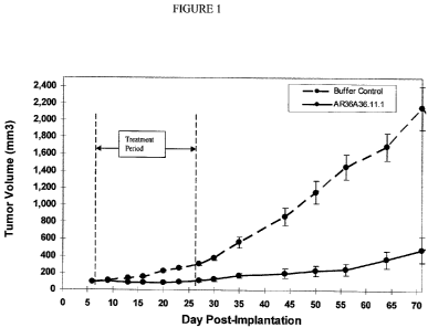
Figure 1 demonstrates the effect of AR36A36.1 1.1 on tumor growth in an
established human PC-3 prostate cancer model. The vertical dashed lines
indicate the period
during which the antibody was intraperitoneally administered. Data points
represent the mean
+/- SEM.
Figure 2 demonstrates the effect of AR36A36.11.1 on mouse body weight in
an established PC-3 prostate cancer model. Data points represent the mean +/-
SEM.
Figure 3 demonstrates the effect of AR36A36.11.1 on tumor growth in an
established human breast MDA-MB-468 cancer model. The vertical dashed lines
indicate the
period during which the antibody was intraperitoneally administered. Data
points represent
the mean +/- SEM.
Figure 4 demonstrates the effect of AR36A36.11.1 on mouse body weight in
an established MDA-MB-468 breast cancer model. Data points represent the mean
+/- SEM.
Figure 5 demonstrates the effect of AR36A36.11.1 in a dose-response manner
on tumor growth in an established human breast (MDA-MB-23 1) cancer model. The
vertical
dashed lines indicate the period during which the antibody was
intraperitoneally administered.
Data points represent the mean +/- SEM.
Figure 6 demonstrates the effect of AR36A36.11.1 on mouse body weight in
an established MDA-MB-231 breast cancer model. Data points represent the mean
+/- SEM.
Figure 7 demonstrates the effect of AR36A36.11.1 on tumor growth in a
prophylactic NCI-H520 human lung squamous cell carcinoma model. The vertical
dashed
lines indicate the period during which the antibody was intraperitoneally
administered. Data
points represent the mean +/- SEM.
23
CA 02687575 2009-11-18
WO 2008/144889 PCT/CA2008/000977
Figure 8 demonstrates effect of AR36A36.11.1 on mouse survival in a
prophylactic NCI-14520 human lung squamous cell carcinoma model. Data points
represent
the survival percentage.
Figure 9 demonstrates the effect of AR36A36.11.1 on mouse body weight in a
prophylactic NCI-H520 human lung squamous cell carcinoma model. Data points
represent
the mean +/- SEM.
Figure 10. Western blot of a total membrane preparation of MDA-MB-231
breast cancer cells probed with different primary antibody solutions. Lanes 3
to 7 were
probed with biotinylated AR36A36.1 1.1 mixed with 0.5 micrograms/mL, 5
micrograms/mL,
50 micrograms/mL, 500 micrograms/mL and 1000 micrograms/mL of non-biotinylated
AR36A36.11.1 respectively. Lanes 9-13 were probed with biotinylated
AR36A36.11.1 mixed
with 0.5 micrograms/mL, 5 micrograms/mL, 50 micrograms/mL, 500 micrograms/mL
and
1000 micrograms/mL of non-biotinylated 10A304.7 respectively. Lanes 15-19 were
probed
with biotinylated AR36A36.1 1.1 mixed with 0.5 micrograms/mL, 5 micrograms/mL,
50
micrograms/mL, 500 micrograms/mL and 1000 micrograms/mL of non-biotinylated
8B1B:1
respectively. Lanes 8 and 14 were incubated with negative control solution and
lane 8 was not
incubated in secondary solution. Lanes 1, 2 and 20 were incubated with TBST
only.
Figure 11. Western blot of a total membrane preparation of MDA-MB-231
breast cancer cells probed with different primary antibody solutions. Lanes 3
to 7 were
probed with biotinylated 10A304.7 mixed with 0.5 micrograms/mL, 5
micrograms/mL, 50
micrograms/mL, 500 micrograms/mL and 1000 micrograms/mL of non-biotinylated
10A304.7 respectively. Lanes 9 to 13 were probed with biotinylated 10A304.7
mixed with 0.5
micrograms/mL, 5 micrograms/mL, 50 micrograms/mL, 500 micrograms/mL and 1000
micrograms/mL of non-biotinylated AR36A36.11.1 respectively. Lanes 15 to 19
were probed
with biotinylated 10A304.7 mixed with 0.5 micrograms/mL, 5 micrograms/mL, 50
micrograms/mL, 500 micrograms/mL and 1000 micrograms/mL of non-biotinylated
8A3B.6
respectively. Lanes 8 and 14 were incubated with negative control solution and
lane 8 was
not incubated in secondary solution. Lanes 1, 2 and 20 were incubated with
TBST only.
Figure 12. Binding of 10A304.7 to CLIPS peptides (SEQ ID NOS: 15-17, 17,
16, 18-22, 17, 23, 19, 24-27, 16 and 28-29, respectively, in order of
appearance) synthesized
based on CD59 amino acid sequence.
24
CA 02687575 2009-11-18
WO 2008/144889 PCT/CA2008/000977
Figure 13. Binding of AR36A36.11.1 to CLIPS peptides (SEQ ID NOS: 17,
30-33, 15, 34-37, 17, 38, 18, 39-40, 19, 41-42, 37 and 16, respectively, in
order of
appearance) synthesized based on CD59 amino acid sequence.
Figure 14. Amino acid sequence of CD59 (SEQ ID NO: 43). The
discontinuous epitope recognized by both 10A304.7 and AR36A36.1 1.1 is
contained within
the underlined sequences.
Figure 15. Primers used in the PCR amplification of light chain (SEQ ID NOS:
44-62, respectively, in order of appearance).
Figure 16. Primers used in the PCR amplification of heavy chain (SEQ ID
NOS: 63-78, respectively, in order of appearance).
Figure 17. Mouse AR36A36.11.1 VH Sequence (SEQ ID NO: 79). CDRs are
underlined.
Figure 18. Mouse AR36A36.1 1.1 VL Sequence (SEQ ID NO: 80). CDRs are
underlined.
Figure 19. Oligonucleotides (SEQ ID NOS: 81-91, respectively, in order of
appearance) used for the generation of chimeric and variant humanized
AR36A36.11.1 VH
sequences.
Figure 20. Oligonucleotides (SEQ ID NOS: 92-108, respectively, in order of
appearance) used for the generation of chimeric and variant humanized
AR36A36.11.1 VL
sequences.
Figure 21. Light chain and heavy chain expression vectors.
Figure 22A and Figure 22B. Humanized AR36A36.1 1.1 VH variants. CDRs
are underlined. Heavy Chain HV3 disclosed as SEQ ID NO: 7, Heavy Chain HV2
disclosed
as SEQ ID NO: 9, and Heavy Chain HV 1 disclosed as SEQ ID NO: 109.
Figure 23A and Figure 23B. Humanized AR36A36.1 1.1 VL variants. CDRs
are underlined. Light Chain KV3 disclosed as SEQ ID NO: 8, Light Chain KV2
disclosed as
SEQ ID NO: 110, Light Chain KV 1 disclosed as SEQ ID NO: 111 and Light chain
KV4
disclosed as SEQ ID NO: 10.
Figure 24. Activities of humanized AR36A36.11.1 VH and VL variants.
Figure 25 demonstrates the binding of humanized variants, chimeric and
murine AR36A36.11.1 to the human breast cancer cell line MDA-MB-23 1.
CA 02687575 2009-11-18
WO 2008/144889 PCT/CA2008/000977
Figure 26 demonstrates the in vitro CDC activity of murine and humanized
variants of AR36A36.11.1 on the human breast cancer cell line MDA-MB-23 1.
DETAILED DESCRIPTION OF THE INVENTION
In general, the following words or phrases have the indicated definition when
used in the summary, description, examples, and claims.
The term "antibody" is used in the broadest sense and specifically covers, for
example, single monoclonal antibodies (including agonist, antagonist, and
neutralizing
antibodies, de-immunized, murine, chimeric or humanized antibodies), antibody
compositions
with polyepitopic specificity, single-chain antibodies, diabodies, triabodies,
immunoconjugates and antibody fragments (see below).
The term "monoclonal antibody" as used herein refers to an antibody obtained
from a population of substantially homogeneous antibodies, i.e., the
individual antibodies
comprising the population are identical except for possible naturally
occurring mutations that
may be present in minor amounts. Monoclonal antibodies are highly specific,
being directed
against a single antigenic site. Furthermore, in contrast to polyclonal
antibody preparations
which include different antibodies directed against different determinants
(epitopes), each
monoclonal antibody is directed against a single determinant on the antigen.
In addition to
their specificity, the monoclonal antibodies are advantageous in that they may
be synthesized
uncontaminated by other antibodies. The modifier "monoclonal" indicates the
character of the
antibody as being obtained from a substantially homogeneous population of
antibodies, and is
not to be construed as requiring production of the antibody by any particular
method. For
example, the monoclonal antibodies to be used in accordance with the present
invention may
be made by the hybridoma (murine or human) method first described by Kohler et
al., Nature,
256:495 (1975), or may be made by recombinant DNA methods (see, e.g., U.S.
Pat.
No.4,816,567). The "monoclonal antibodies" may also be isolated from phage
antibody
libraries using the techniques described in Clackson et al., Nature, 3 52:624-
628 (1991) and
Marks et al., J. Mol. Biol., 222:581-597 (1991), for example.
"Antibody fragments" comprise a portion of an intact antibody, preferably
comprising the antigen-binding or variable region thereof. Examples of
antibody fragments
include less than full length antibodies, Fab, Fab', F(ab')2, and Fv
fragments; diabodies; linear
26
CA 02687575 2009-11-18
WO 2008/144889 PCT/CA2008/000977
antibodies; single-chain antibody molecules; single-chain antibodies, single
domain antibody
molecules, fusion proteins, recombinant proteins and multispecific antibodies
formed from
antibody fragment(s).
An "intact" antibody is one which comprises an antigen-binding variable
region as well as a light chain constant domain (CL) and heavy chain constant
domains, CH1,
CH2 and CH3. The constant domains may be native sequence constant domains
(e.g. human
native sequence constant domains) or amino acid sequence variant thereof.
Preferably, the
intact antibody has one or more effector functions.
Depending on the amino acid sequence of the constant domain of their heavy
chains, intact antibodies can be assigned to different "classes". There are
five-major classes of
intact antibodies: IgA, IgD, IgE, IgG, and IgM, and several of these may be
further divided
into "subclasses" (isotypes), e.g., IgGl, IgG2, IgG3, IgG4, IgA, and IgA2. The
heavy-chain
constant domains that correspond to the different classes of antibodies are
called a, fi, s, y, and
, respectively. The subunit structures and three-dimensional configurations of
different
classes of immunoglobulins are well known.
Antibody "effector functions" refer to those biological activities
attributable to
the Fc region (a native sequence Fc region or amino acid sequence variant Fc
region) of an
antibody. Examples of antibody effector functions include C 1 q binding;
complement
dependent cytotoxicity; Fc receptor binding; antibody-dependent cell-mediated
cytotoxicity
(ADCC); phagocytosis; down regulation of cell surface receptors (e.g. B cell
receptor; BCR),
etc.
"Antibody-dependent cell-mediated cytotoxicity" and "ADCC" refer to a cell-
mediated reaction in which nonspecific cytotoxic cells that express Fc
receptors (FcRs) (e.g.
Natural Killer (NK) cells, neutrophils, and macrophages) recognize bound
antibody on a
target cell and subsequently cause lysis of the target cell. The primary cells
for mediating
ADCC, NK cells, express FcyRI1I only, whereas monocytes express FcyRI, FcyRII
and
FcyRI1I. FcR expression on hematopoietic cells is summarized in Table 3 on
page 464 of
Ravetch and Kinet, Annu. Rev. Immunol 9:457-92 (1991). To assess ADCC activity
of a
molecule of interest, an in vitro ADCC assay, such as that described in U.S.
Pat. No.
5,500,362 or 5,821,337 may be performed. Useful effector cells for such assays
include
peripheral blood mononuclear cells (PBMC) and Natural Killer (NK) cells.
Alternatively, or
27
CA 02687575 2009-11-18
WO 2008/144889 PCT/CA2008/000977
additionally, ADCC activity of the molecule of interest may be assessed in
vivo, e.g., in a
animal model such as that disclosed in Clynes et al. PNAS (USA) 95:652-656
(1998).
"Effector cells" are leukocytes which express one or more FcRs and perform
effector functions. Preferably, the cells express at least FcyRIII and perform
ADCC effector
function. Examples of human leukocytes which mediate ADCC include peripheral
blood
mononuclear cells (PBMC), natural killer (NK) cells, monocytes, cytotoxic T
cells and
neutrophils; with PBMCs and NK cells being preferred. The effector cells may
be isolated
from a native source thereof, e.g. from blood or PBMCs as described herein.
The terms "Fc receptor" or "FcR" are used to describe a receptor that binds to
the Fc region of an antibody. The preferred FcR is a native sequence human
FcR. Moreover, a
preferred FcR is one which binds an IgG antibody (a gamma receptor) and
includes receptors
of the FcyRI, FcyR11, and Fcy RIII subclasses, including allelic variants and
alternatively
spliced forms of these receptors. FcyRII receptors include FcyRIIA (an
"activating receptor")
and FcyRIIB (an "inhibiting receptor"), which have similar amino acid
sequences that differ
primarily in the cytoplasmic domains thereof. Activating receptor FcyRIIA
contains an
immunoreceptor tyrosine-based activation motif (ITAM) in its cytoplasmic
domain. Inhibiting
receptor FcyRIIB contains an immunoreceptor tyrosine-based inhibition motif
(ITIM) in its
cytoplasmic domain. (see review M. in Daeron, Annu. Rev. Immunol. 15:203-234
(1997)).
FcRs are reviewed in Ravetch and Kinet, Annu. Rev. Immunol. 9:457-92 (1991);
Capel et al.,
Immunomethods 4:25-34 (1994); and de Haas et al., J. Lab. Clin. Med. 126:330-
41 (1995).
Other FcRs, including those to be identified in the future, are encompassed by
the term "FcR"
herein. The term also includes the neonatal receptor, FcRn, which is
responsible for the
transfer of maternal IgGs to the fetus (Guyer et al., J Immunol. 117:587
(1976) and Kim et
al., Eur. J. Immunol. 24:2429 (1994)).
"Complement dependent cytotoxicity" or "CDC" refers to the ability of a
molecule to lyse a target in the presence of complement. The complement
activation pathway
is initiated by the binding of the first component of the complement system (C
1 q) to a
molecule (e.g. an antibody) complexed with a cognate antigen. To assess
complement
activation, a CDC assay, e.g. as described in Gazzano-Santoro et al., J.
Immunol. Methods
202:163 (1996), may be performed.
28
CA 02687575 2009-11-18
WO 2008/144889 PCT/CA2008/000977
The term "variable" refers to the fact that certain portions of the variable
domains differ extensively in sequence among antibodies and are used in the
binding and
specificity of each particular antibody for its particular antigen. However,
the variability is not
evenly distributed throughout the variable domains of antibodies. It is
concentrated in three
segments called hypervariable regions both in the light chain and the heavy
chain variable
domains. The more highly conserved portions of variable domains are called the
framework
regions (FRs). The variable domains of native heavy and light chains each
comprise four FRs,
largely adopting a(3-sheet configuration, connected by three hypervariable
regions, which
form loops connecting, and in some cases forming part of, the 0-sheet
structure. The
hypervariable regions in each chain are held together in close proximity by
the FRs and, with
the hypervariable regions from the other chain, contribute to the formation of
the antigen-
binding site of antibodies (see Kabat et al., Sequences of Proteins of
Immunological Interest,
5th Ed. Public Health Service, National Institutes of Health, Bethesda, Md.
(1991)). The
constant domains are not involved directly in binding an antibody to an
antigen, but exhibit
various effector functions, such as participation of the antibody in antibody
dependent cellular
cytotoxicity (ADCC).
The term "hypervariable region" when used herein refers to the amino acid
residues of an antibody which are responsible for antigen-binding. The
hypervariable region
generally comprises amino acid residues from a "complementarity determining
region" or
"CDR" (e.g. residues 24-34 (L1), 50-56 (L2) and 89-97 (L3) in the light chain
variable
domain and 31-35 (H1), 50-65 (H2) and 95-102 (143) in the heavy chain variable
domain;
Kabat et al., Sequences of Proteins of Immunological Interest, 5th Ed. Public
Health Service,
National Institutes of Health, Bethesda, Md. (1991)) and/or those residues
from a
"hypervariable loop" (e.g. residues 2632 (L1), 50-52 (L2) and 91-96 (L3) in
the light chain
variable domain and 26-32 (H1), 53-55 (1-12) and 96-101 (H3) in the heavy
chain variable
domain; Chothia and Lesk J. Mol. Biol. 196:901-917 (1987)). "Framework Region"
or "FR"
residues are those variable domain residues other than the hypervariable
region residues as
herein defined. Papain digestion of antibodies produces two identical antigen-
binding
fragments, called "Fab" fragments, each with a single antigen-binding site,
and a residual
"Fe" fragment, whose name reflects its ability to crystallize readily. Pepsin
treatment yields
29
CA 02687575 2009-11-18
WO 2008/144889 PCT/CA2008/000977
an F(ab')2 fragment that has two antigen-binding sites and is still capable of
cross-linking
antigen.
"Fv" is the minimum antibody fragment which contains a complete antigen-
recognition and antigen-binding site. This region consists of a dimer of one
heavy chain and
one light chain variable domain in tight, non-covalent association. It is in
this configuration
that the three hypervariable regions of each variable domain interact to
define an antigen-
binding site on the surface of the VH-VL dimer. Collectively, the six
hypervariable regions
confer antigen-binding specificity to the antibody. However, even a single
variable domain
(or half of an Fv comprising only three hypervariable regions specific for an
antigen) has the
ability to recognize and bind antigen, although at a lower affinity than the
entire binding site.
The Fab fragment also contains the constant domain of the light chain and the
first constant
domain (CH I) of the heavy chain. Fab' fragments differ from Fab fragments by
the addition
of a few residues at the carboxy terminus of the heavy chain CH1 domain
including one or
more cysteines from the antibody hinge region. Fab'-SH is the designation
herein for Fab' in
which the cysteine residue(s) of the constant domains bear at least one free
thiol group.
F(ab')2 antibody fragments originally were produced as pairs of Fab' fragments
which have
hinge cysteines between them. Other chemical couplings of antibody fragments
are also
known.
The "light chains" of antibodies from any vertebrate species can be assigned
to
one of two clearly distinct types, called kappa (x) and lambda (X), based on
the amino acid
sequences of their constant domains.
"Single-chain Fv" or "scFv" antibody fragments comprise the VH and VL
domains of antibody, wherein these domains are present in a single polypeptide
chain.
Preferably, the Fv polypeptide further comprises a polypeptide linker between
the VH and VL
domains which enables the scFv to form the desired structure for antigen
binding. For a
review of scFv see Pluckthun in The Pharmacology af Monoclonal Antibodies,
vol. 113,
Rosenburg and Moore eds., Springer-Verlag, New York, pp. 269-315 (1994).
The term "diabodies" refers to small antibody fragments with two antigen-
binding sites, which fragments comprise a variable heavy domain (VH) connected
to a
variable light domain (VL) in the same polypeptide chain (VH-VL). By using a
linker that is
too short to allow pairing between the two domains on the same chain, the
domains are forced
CA 02687575 2009-11-18
WO 2008/144889 PCT/CA2008/000977
to pair with the complementary domains of another chain and create two antigen-
binding
sites. Diabodies are described more fully in, for example, EP 404,097; WO
93/11161; and
Hollinger et al., Proc. Natl. Acad. Sci. USA, 90:6444-6448 (1993).
The term "triabodies" or "trivalent trimers" refers to the combination of
three
single chain antibodies. Triabodies are constructed with the amino acid
terminus of a VL or
VH domain, i.e., without any linker sequence. A triabody has three Fv heads
with the
polypeptides arranged in a cyclic, head-to-tail fashion. A possible
conformation of the
triabody is planar with the three binding sites located in a plane at an angle
of 120 degrees
from one another. Triabodies can be monospecific, bispecific or trispecific.
An "isolated" antibody is one which has been identified and separated and/or
recovered from a component of its natural environment. Contaminant components
of its
natural environment are materials which would interfere with diagnostic or
therapeutic uses
for the antibody, and may include enzymes, hormones, and other proteinaceous
or
nonproteinaceous solutes. Isolated antibody includes the antibody in situ
within recombinant
cells since at least one component of the antibody's natural environment will
not be present.
Ordinarily, however, isolated antibody will be prepared by at least one
purification step.
An antibody "which binds" an antigen of interest, e.g. CD59 antigen, is one
capable of binding that antigen with sufficient affinity such that the
antibody is useful as a
therapeutic or diagnostic agent in targeting a cell expressing the antigen.
Where the antibody
is one which binds CD59, it will usually preferentially bind CD59 as opposed
to other
receptors, and does not incl'ude incidental binding such as non-specific Fc
contact, or binding
to post-translational modifications common to other antigens and may be one
which does not
significantly cross-react with other proteins. Methods, for the detection of
an antibody that
binds an antigen of interest, are well known in the art and can include but
are not limited to
assays such as FACS, cell ELISA and Western blot.
As used herein, the expressions "cell", "cell line", and "cell culture" are
used
interchangeably, and all such designations include progeny. It is also
understood that all
progeny may not be precisely identical in DNA content, due to deliberate or
inadvertent
mutations. Mutant progeny that have the same function or biological activity
as screened for
in the originally transformed cell are included. It will be clear from the
context where distinct
designations are intended.
31
CA 02687575 2009-11-18
WO 2008/144889 PCT/CA2008/000977
"Treatment or treating" refers to both therapeutic treatment and prophylactic
or
preventative measures, wherein the object is to prevent or slow down (lessen)
the targeted
pathologic condition or disorder. Those in need of treatment include those
already with the
disorder as well as those prone to have the disorder or those in whom the
disorder is to be
prevented. Hence, the mammal to be treated herein may have been diagnosed as
having the
disorder or may be predisposed or susceptible to the disorder.
The terms "cancer" and "cancerous" refer to or describe the physiological
condition in mammals that is typically characterized by unregulated cell
growth or death.
Examples of cancer include, but are not limited to, carcinoma, lymphoma,
blastoma, sarcoma,
and leukemia or lymphoid malignancies. More particular examples of such
cancers include
squamous cell cancer (e.g. epithelial squamous cell cancer), lung cancer
including small-cell
lung cancer, non-small cell lung cancer, adenocarcinoma of the lung and
squamous carcinoma
of the lung, cancer of the peritoneum, hepatocellular cancer, gastric or
stomach cancer
including gastrointestinal cancer, pancreatic cancer, glioblastoma, cervical
cancer, ovarian
cancer, liver cancer, bladder cancer, hepatoma, breast cancer, colon cancer,
rectal cancer,
colorectal cancer, endometrial or uterine carcinoma, salivary gland carcinoma,
kidney or renal
cancer, prostate cancer, vulval cancer, thyroid cancer, hepatic carcinoma,
anal carcinoma,
penile carcinoma, as well as head and neck cancer.
A "chemotherapeutic agent" is a chemical compound useful in the treatment of
cancer. Examples of chemotherapeutic agents include alkylating agents such as
thiotepa and
cyclosphosphamide (CYTOXANTM); alkyl sulfonates such as busulfan, improsulfan
and
piposulfan; aziridines such as benzodopa, carboquone, meturedopa, and uredopa;
ethylenimines and methylamelamines including altretamine, triethylenemelamine,
triethylenephosphoramide, triethylenethiophosphoramide and
trimethylolomelamine; nitrogen
mustards such as chlorambucil, chlornaphazine, cholophosphamide, estramustine,
ifosfamide,
mechlorethamine, mechlorethamine oxide hydrochloride, melphalan, novembichin,
phenesterine, prednimustine, trofosfamide, uracil mustard; nitrosureas such as
carmustine,
chlorozotocin, fotemustine, lomustine, nimustine, ranimustine; antibiotics
such as
aclacinomysins, actinomycin, authramycin, azaserine, bleomycins, cactinomycin,
calicheamicin, carabicin, camomycin, carzinophilin, chromomycins,
dactinomycin,
daunorubicin, detorubicin, 6-diazo-5-oxo-L-norleucine, doxorubicin,
epirubicin, esorubicin,
32
CA 02687575 2009-11-18
WO 2008/144889 PCT/CA2008/000977
idarubicin, marcellomycin, mitomycins, mycophenolic acid, nogalamycin,
olivomycins,
peplomycin, potfiromycin, puromycin, quelamycin, rodorubicin, streptonigrin,
streptozocin,
tubercidin, ubenimex, zinostatin, zorubicin; anti-metabolites such as
methotrexate and 5-
fluorouracil (5-FU); folic acid analogues such as denopterin, methotrexate,
pteropterin,
trimetrexate; purine analogs such as fludarabine, 6-mercaptopurine,
thiamiprine, thioguanine;
pyrimidine analogs such as ancitabine, azacitidine, 6-azauridine, carmofur,
cytarabine,
dideoxyuridine, doxifluridine, enocitabine, floxuridine, 5-FU; androgens such
as calusterone,
dromostanolone propionate, epitiostanol, mepitiostane, testolactone; anti-
adrenals such as
aminoglutethimide, mitotane, trilostane; folic acid replenisher such as
frolinic acid;
aceglatone; aldophosphamide glycoside; aminolevulinic acid; amsacrine;
bestrabucil;
bisantrene; edatraxate; defofamine; demecolcine; diaziquone; elformithine;
elliptinium
acetate; etoglucid; gallium nitrate; hydroxyurea; lentinan; lonidamine;
mitoguazone;
mitoxantrone; mopidamol; nitracrine; pentostatin; phenamet; pirarubicin;
podophyllinic acid;
2-ethylhydrazide; procarbazine; PSK ; razoxane; sizofiran; spirogermanium;
tenuazonic
acid; triaziquone; 2,2',2"-trichlorotriethylamine; urethan; vindesine;
dacarbazine;
mannomustine; mitobronitol; mitolactol; pipobroman; gacytosine; arabinoside
("Ara-C");
cyclophosphamide; thiotepa; taxanes, e.g. paclitaxel (TAXOL , Bristol-Myers
Squibb
Oncology, Princeton, N.J.) and docetaxel (TAXOTERE , Aventis, Rhone-Poulenc
Rorer,
Antony, France); chlorambucil; gemcitabine; 6-thioguanine; mercaptopurine;
methotrexate;
platinum analogs such as cisplatin and carboplatin; vinblastine; platinum;
etoposide (VP-16);
ifosfamide; mitomycin C; mitoxantrone; vincristine; vinorelbine; navelbine;
novantrone;
teniposide; daunomycin; aminopterin; xeloda; ibandronate; CPT- 11;
topoisomerase inhibitor
RFS 2000; difluoromethylornithine (DMFO); retinoic acid; esperamicins;
capecitabine; and
pharmaceutically acceptable salts, acids or derivatives of any of the above.
Also included in
this definition are anti-hormonal agents that act to regulate or inhibit
hormone action on
tumors such as anti-estrogens including for example tamoxifen, raloxifene,
aromatase
inhibiting 4(5)-imidazoles, 4-hydroxytamoxifen, trioxifene, keoxifene,
LY117018,
onapristone, and toremifene (Fareston); and anti-androgens such as flutamide,
nilutamide,
bicalutamide, leuprolide, and goserelin; and pharmaceutically acceptable
salts, acids or
derivatives of any of the above.
33
CA 02687575 2009-11-18
WO 2008/144889 PCT/CA2008/000977
"Mammal" for purposes of treatment refers to any animal classified as a
mammal, including humans, mice, SCID or nude mice or strains of mice, domestic
and farm
animals, and zoo, sports, or pet animals, such as sheep, dogs, horses, cats,
cows, etc.
Preferably, the mammal herein is human.
"Oligonucleotides" are short-length, single- or double-stranded
polydeoxynucleotides that are chemically synthesized by known methods (such as
phosphotriester, phosphite, or phosphoramidite chemistry, using solid phase
techniques such
as described in EP 266,032, published 4 May 1988, or via deoxynucleoside H-
phosphonate
intermediates as described by Froehler et al., Nucl. Acids Res., 14:5399-5407,
1986. They are
then purified on polyacrylamide gels.
In accordance with the present invention, "humanized" and/or "chimeric"
forms of non-human (e.g. murine) immunoglobulins refer to antibodies which
contain specific
chimeric immunoglobulins, immunoglobulin chains or fragments thereof (such as
Fv, Fab,
Fab', F(ab')2 or other antigen-binding subsequences of antibodies) which
results in the
decrease of a human anti-mouse antibody (HAMA), human anti-chimeric antibody
(HACA)
or a human anti-human antibody (HAHA) response, compared to the original
antibody, and
contain the requisite portions (e.g. CDR(s), antigen binding region(s),
variable domain(s) and
so on) derived from said non-human immunoglobulin, necessary to reproduce the
desired
effect, while simultaneously retaining binding characteristics which are
comparable to said
non-human immunoglobulin. For the most part, humanized antibodies are human
immunoglobulins (recipient antibody) in which residues from the
complementarity
determining regions (CDRs) of the recipient antibody are replaced by residues
from the CDRs
of a non-human species (donor antibody) such as mouse, rat or rabbit having
the desired
specificity, affinity and capacity. In some instances, Fv framework region
(FR) residues of the
human immunoglobulin are replaced by corresponding non-human FR residues.
Furthermore,
the humanized antibody may comprise residues which are found neither in the
recipient
antibody nor in the imported CDR or FR sequences. These modifications are made
to further
refine and optimize antibody performance. In general, the humanized antibody
will comprise
substantially all of at least one, and typically two, variable domains, in
which all or
substantially all of the CDR regions correspond to those of a non-human
immunoglobulin and
all or substantially all of the FR residues are those of a human
immunoglobulin consensus
34
CA 02687575 2009-11-18
WO 2008/144889 PCT/CA2008/000977
sequence. The humanized antibody optimally also will comprise at least a
portion of an
immunoglobulin constant region (Fc), typically that of a human immunoglobulin.
"De-immunized" antibodies are immunoglobulins that are non-immunogenic,
or less immunogenic, to a given species. De- immunization can be achieved
through structural
alterations to the antibody. Any de- immunization technique known to those
skilled in the art
can be employed. One suitable technique for de- immunizing antibodies is
described, for
example, in WO 00/34317 published June 15, 2000.
An antibody which induces "apoptosis" is one which induces programmed cell
death by any means, illustrated by but not limited to binding of annexin V,
caspase activity,
fragmentation of DNA, cell shrinkage, dilation of endoplasmic reticulum, cell
fragmentation,
and/or formation of membrane vesicles (called apoptotic bodies).
As used herein "antibody induced cytotoxicity" is understood to mean the
cytotoxic effect derived from the hybridoma supernatant or antibody produced
by the
hybridoma deposited with the IDAC as accession number 280104-02, a humanized
antibody
of the isolated monoclonal antibody produced by the hybridoma deposited with
the IDAC as
accession number 280104-02, a chimeric antibody of the isolated monoclonal
antibody
produced by the hybridoma deposited with the IDAC as accession number 280104-
02, antigen
binding fragments, or antibody ligands thereof, which effect is not
necessarily related to the
degree of binding.
Throughout the instant specification, hybridoma cell lines, as well as the
isolated monoclonal antibodies which are produced therefrom, are alternatively
referred to by
their internal designation, AR36A36.11.1 (murine), (ch)AR36A36.11.1
(chimeric),
(hu)AR36A36.1 1.1 (humanized) or Depository Designation, IDAC 280104-02.
As used herein "antibody-ligand" includes a moiety which exhibits binding
specificity for at least one epitope of the target antigen, and which may be
an intact antibody
molecule, antibody fragments, and any molecule having at least an antigen-
binding region or
portion thereof (i.e., the variable portion of an antibody molecule), e.g., an
Fv molecule, Fab
molecule, Fab' molecule, F(ab')2 molecule, a bispecific antibody, a
fusion protein, or any
genetically engineered molecule which specifically recognizes and binds at
least one epitope
of the antigen bound by the isolated monoclonal antibody produced by the
hybridoma cell line
designated as IDAC 280104-02 (the IDAC 280104-02 antigen), a humanized
antibody of the
CA 02687575 2009-11-18
WO 2008/144889 PCT/CA2008/000977
isolated monoclonal antibody produeced by the hybridoma deposited with the
IDAC as
accession number 280104-02, a chimeric antibody of the isolated monoclonal
antibody
produced by the hybridoma deposited with the IDAC as accession number 280104-
02 and
antigen binding fragments.
As used herein "cancerous disease modifying antibodies" (CDMAB) refers to
monoclonal antibodies which modify the cancerous disease process in a manner
which is
beneficial to the patient, for example by reducing tumor burden or prolonging
survival of
tumor bearing individuals, and antibody-ligands thereo
A "CDMAB related binding agent", in its broadest sense, is understood to
include, but is not limited to, any form of human or non-human antibodies,
antibody
fragments, antibody ligands, or the like, which competitively bind to at least
one CDMAB
target epitope.
A "competitive binder" is understood to include any form of human or non-
human antibodies, antibody fragments, antibody ligands, or the like which has
binding affinity
for at least one CDMAB target epitope.
Tumors to be treated include primary tumors and metastatic tumors, as well as
refractory tumors. Refractory tumors include tumors that fail to respond or
are resistant to
treatment with chemotherapeutic agents alone, antibodies alone, radiation
alone or
combinations thereof. Refractory tumors also encompass tumors that appear to
be inhibited
by treatment with such agents but recur up to five years, sometimes up to ten
years or longer
after treatment is discontinued.
Tumors that can be treated include tumors that are not vascularized, or not
yet
substantially vascularized, as well as vascularized tumors. Examples of solid
tumors, which
can be accordingly treated, include breast carcinoma, lung carcinoma,
colorectal carcinoma,
pancreatic carcinoma, glioma and lymphoma. Some examples of such tumors
include
epidermoid tumors, squamous tumors, such as head and neck tumors, colorectal
tumors,
prostate tumors, breast tumors, lung tumors, including small cell and non-
small cell lung
tumors, pancreatic tumors, thyroid tumors, ovarian tumors, and liver tumors.
Other examples
include Kaposi's sarcoma, CNS neoplasms, neuroblastomas, capillary
hemangioblastomas,
meningiomas and cerebral metastases, melanoma, gastrointestinal and renal
carcinomas and
36
CA 02687575 2009-11-18
WO 2008/144889 PCT/CA2008/000977
sarcomas, rhabdomyosarcoma, glioblastoma, preferably glioblastoma multiforme,
and
leiomyosarcoma.
As used herein "antigen-binding region" means a portion of the molecule
which recognizes the target antigen.
As used herein "competitively inhibits" means being able to recognize and
bind a determinant site to which the monoclonal antibody produced by the
hybridoma cell line
designated as IDAC 280104-02, (the IDAC 280104-02 antibody), a humanized
antibody of
the isolated monoclonal antibody produeced by the hybridoma deposited with the
IDAC as
accession number 280104-02, a chimeric antibody of the isolated monoclonal
antibody
produced by the hybridoma deposited with the IDAC as accession number 280104-
02, antigen
binding fragments, or antibody ligands thereof, is directed using conventional
reciprocal
antibody competition assays. (Belanger L., Sylvestre C. and Dufour D. (1973),
Enzyme linked
immunoassay for alpha fetoprotein by competitive and sandwich procedures.
Clinica Chimica
Acta 48, 15).
As used herein "target antigen" is the IDAC 280104-02 antigen or portions
thereof.
As used herein, an "immunoconjugate" means any molecule or CDMAB such
as an antibody chemically or biologically linked to cytotoxins, radioactive
agents, cytokines,
interferons, target or reporter moieties, enzymes, toxins, anti-tumor drugs or
therapeutic
agents. The antibody or CDMAB may be linked to the cytotoxin, radioactive
agent, cytokine,
interferon, target or reporter moiety, enzyme, toxin, anti-tumor drug or
therapeutic agent at
any location along the molecule so long as it is able to bind its target.
Examples of
immunoconjugates include antibody toxin chemical conjugates and antibody-toxin
fusion
proteins.
Radioactive agents suitable for use as anti-tumor agents are known to those
skilled in the art. For example, 1311 or 211 At is used. These isotopes are
attached to the
antibody using conventional techniques (e.g. Pedley et al., Br. J. Cancer 68,
69-73 (1993)).
Alternatively, the anti-tumor agent which is attached to the antibody is an
enzyme which
activates a prodrug. A prodrug may be administered which will remain in its
inactive form
until it reaches the tumor site where it is converted to its cytotoxin form
once the antibody
complex is administered. In practice, the antibody-enzyme conjugate is
administered to the
37
CA 02687575 2009-11-18
WO 2008/144889 PCT/CA2008/000977
patient and allowed to localize in the region of the tissue to be treated. The
prodrug is then
administered to the patient so that conversion to the cytotoxic drug occurs in
the region of the
tissue to be treated. Alternatively, the anti-tumor agent conjugated to the
antibody is a
cytokine such as interleukin-2 (IL-2), interleukin-4 (IL-4) or tumor necrosis
factor alpha
(TNF-a). The antibody targets the cytokine to the tumor so that the cytokine
mediates damage
to or destruction of the tumor without affecting other tissues. The cytokine
is fused to the
antibody at the DNA level using conventional recombinant DNA techniques.
Interferons may
also be used.
As used herein, a "fusion protein" means any chimeric protein wherein an
antigen binding region is connected to a biologically active molecule, e.g.,
toxin, enzyme,
fluorescent proteins, luminescent marker, polypeptide tag, cytokine,
interferon, target or
reporter moiety or protein drug.
The invention further contemplates CDMAB of the present invention to which
target or reporter moieties are linked. Target moieties are first members of
binding pairs.
Anti-tumor agents, for example, are conjugated to second members of such pairs
and are
thereby directed to the site where the antigen-binding protein is bound. A
common example
of such a binding pair is avidin and biotin. In a preferred embodiment, biotin
is conjugated to
the target antigen of the CDMAB of the present invention, and thereby provides
a target for
an anti-tumor agent or other moiety which is conjugated to avidin or
streptavidin.
Alternatively, biotin or another such moiety is linked to the target antigen
of the CDMAB of
the present invention and used as a reporter, for example in a diagnostic
system where a
detectable signal-producing agent is conjugated to avidin or streptavidin.
Detectable signal-producing agents are useful in vivo and in vitro for
diagnostic purposes. The signal producing agent produces a measurable signal
which is
detectable by external means, usually the measurement of electromagnetic
radiation. For the
most part, the signal producing agent is an enzyme or chromophore, or emits
light by
fluorescence, phosphorescence or chemiluminescence. Chromophores include dyes
which
absorb light in the ultraviolet or visible region, and can be substrates or
degradation products
of enzyme catalyzed reactions.
Moreover, included within the scope of the present invention is use of the
present CDMAB in vivo and in vitro for investigative or diagnostic methods,
which are well
38
CA 02687575 2009-11-18
WO 2008/144889 PCT/CA2008/000977
known in the art. In order to carry out the diagnostic methods as contemplated
herein, the
instant invention may further include kits, which contain CDMAB of the present
invention.
Such kits will be useful for identification of individuals at risk for certain
type of cancers by
detecting over-expression of the CDMAB's target antigen on cells of such
individuals.
Diagnostic Assay Kits
It is contemplated to utilize the CDMAB of the present invention in the form
of a diagnostic assay kit for determining the presence of a tumor. The tumor
will generally be
detected in a patient based on the presence of one or more tumor-specific
antigens, e.g.
proteins and/or polynucleotides which encode such proteins in a biological
sample, such as
blood, sera, urine and/or tumor biopsies, which samples will have been
obtained from the
patient.
The proteins function as markers which indicate the presence or absence of a
particular tumor, for example a colon, breast, lung or prostate tumor. It is
further
contemplated that the antigen will have utility for the detection of other
cancerous tumors.
Inclusion in the diagnostic assay kits of binding agents comprised of CDMABs
of the present
invention, or CDMAB related binding agents, enables detection of the level of
antigen that
binds to the agent in the biological sample. Polynucleotide primers and probes
may be used to
detect the level of mRNA encoding a tumor protein, which is also indicative of
the presence
or absence of a cancer. In order for the binding assay to be diagnostic, data
will have been
generated which correlates statistically significant levels of antigen, in
relation to that present
in normal tissue, so as to render the recognition of binding definitively
diagnostic for the
presence of a cancerous tumor. It is contemplated that a plurality of formats
will be useful for
the diagnostic assay of the present invention, as are known to those of
ordinary skill in the art,
for using a binding agent to detect polypeptide markers in a sample. For
example, as
illustrated in Harlow and Lane, Antibodies: A Laboratory Manual, Cold Spring
Harbor
Laboratory, 1988. Further contemplated are any and all combinations,
permutations or
modifications of the afore-described diagnostic assay formats.
The presence or absence of a cancer in a patient will typically be determined
by (a) contacting a biological sample obtained from a patient with a binding
agent; (b)
detecting in the sample a level of polypeptide that binds to the binding
agent; and (c)
comparing the level of polypeptide with a predetermined cut-off value.
39
CA 02687575 2009-11-18
WO 2008/144889 PCT/CA2008/000977
In an illustrative embodiment, it is contemplated that the assay will involve
the
use of a CDMAB based binding agent immobilized on a solid support to bind to
and remove
the polypeptide from the remainder of the sample. The bound polypeptide may
then be
detected using a detection reagent that contains a reporter group and
specifically binds to the
binding agent/polypeptide complex. Illustrative detection reagents may include
a CDMAB
based binding agent that specifically binds to the polypeptide or an antibody
or other agent
that specifically binds to the binding agent, such as an anti-immunoglobulin,
protein G,
protein A or a lectin. In an alternative embodiment, it is contemplated that a
competitive assay
may be utilized, in which a polypeptide is labeled with a reporter group and
allowed to bind to
the immobilized binding agent after incubation of the binding agent with the
sample.
Indicative of the reactivity of the sample with the immobilized binding agent,
is the extent to
which components of the sample inhibit the binding of the labeled polypeptide
to the binding
agent. Suitable polypeptides for use within such assays include full length
tumor-specific
proteins and/or portions thereof, to which the binding agent has binding
affinity.
The diagnostic kit will be provided with a solid support which may be in the
form of any material known to those of ordinary skill in the art to which the
protein may be
attached. Suitable examples may include a test well in a microtiter plate or a
nitrocellulose or
other suitable membrane. Alternatively, the support may be a bead or disc,
such as glass,
fiberglass, latex or a plastic material such as polystyrene or
polyvinylchloride. The support
may also be a magnetic particle or a fiber optic sensor, such as those
disclosed, for example,
in U.S. Pat. No. 5,359,681.
It is contemplated that the binding agent will be immobilized on the solid
support using a variety of techniques known to those of skill in the art,
which are amply
described in the patent and scientific literature. The term "immobilization"
refers to both
noncovalent association, such as adsorption, and covalent attachment, which,
in the context of
the present invention, may be a direct linkage between the agent and
functional groups on the
support, or may be a linkage by way of a cross-linking agent. In a preferred,
albeit non-
limiting embodiment, immobilization by adsorption to a well in a microtiter
plate or to a
membrane is preferable. Adsorption may be achieved by contacting the binding
agent, in a
suitable buffer, with the solid support for a suitable amount of time. The
contact time may
CA 02687575 2009-11-18
WO 2008/144889 PCT/CA2008/000977
vary with temperature, and will generally be within a range of between about 1
hour and
about 1 day.
Covalent attachment of binding agent to a solid support would ordinarily be
accomplished by first reacting the support with a bifunctional reagent that
will react with both
the support and a functional group, such as a hydroxyl or amino group, on the
binding agent.
For example, the binding agent may be covalently attached to supports having
an appropriate
polymer coating using benzoquinone or by condensation of an aldehyde group on
the support
with an amine and an active hydrogen on the binding partner (see, e.g., Pierce
Immunotechnology Catalog and Handbook, 1991, at A12 A13).
It is further contemplated that the diagnostic assay kit will take the form of
a
two-antibody sandwich assay. This assay may be performed by first contacting
an antibody,
e.g. the instantly disclosed CDMAB that has been immobilized on a solid
support, commonly
the well of a microtiter plate, with the sample, such that polypeptides within
the sample are
allowed to bind to the immobilized antibody. Unbound sample is then removed
from the
immobilized polypeptide-antibody complexes and a detection reagent (preferably
a second
antibody capable of binding to a different site on the polypeptide) containing
a reporter group
is added. The amount of detection reagent that remains bound to the solid
support is then
determined using a method appropriate for the specific reporter group.
In a specific embodiment, it is contemplated that once the antibody is
immobilized on the support as described above, the remaining protein binding
sites on the
support will be blocked, via the use of any suitable blocking agent known to
those of ordinary
skill in the art, such as bovine serum albumin or Tween 20TM (Sigma Chemical
Co., St. Louis,
Mo.). The immobilized antibody would then be incubated with the sample, and
polypeptide
would be allowed to bind to the antibody. The sample could be diluted with a
suitable diluent,
such as phosphate-buffered saline (PBS) prior to incubation. In general, an
appropriate
contact time (i.e., incubation time) would be selected to correspond to a
period of time
sufficient to detect the presence of polypeptide within a sample obtained from
an individual
with the specifically selected tumor. Preferably, the contact time is
sufficient to achieve a
level of binding that is at least about 95 percent of that achieved at
equilibrium between
bound and unbound polypeptide. Those of ordinary skill in the art will
recognize that the time
41
CA 02687575 2009-11-18
WO 2008/144889 PCT/CA2008/000977
necessary to achieve equilibrium may be readily determined by assaying the
level of binding
that occurs over a period of time.
It is further contemplated that unbound sample would then be removed by
washing the solid support with an appropriate buffer. The second antibody,
which contains a
reporter group, would then be added to the solid support. Incubation of the
detection reagent
with the immobilized antibody-polypeptide complex would then be carried out
for an amount
of time sufficient to detect the bound polypeptide. Subsequently, unbound
detection reagent
would then be removed and bound detection reagent would be detected using the
reporter
group. The method employed for detecting the reporter group is necessarily
specific to the
type of reporter group selected, for example for radioactive groups,
scintillation counting or
autoradiographic methods are generally appropriate. Spectroscopic methods may
be used to
detect dyes, luminescent groups and fluorescent groups. Biotin may be detected
using avidin,
coupled to a different reporter group (commonly a radioactive or fluorescent
group or an
enzyme). Enzyme reporter groups may generally be detected by the addition of
substrate
(generally for a specific period of time), followed by spectroscopic or other
analysis of the
reaction products.
In order to utilize the diagnostic assay kit of the present invention to
determine
the presence or absence of a cancer, such as prostate cancer, the signal
detected from the
reporter group that remains bound to the solid support would generally be
compared to a
signal that corresponds to a predetermined cut-off value. For example, an
illustrative cut-off
value for the detection of a cancer may be the average mean signal obtained
when the
immobilized antibody is incubated with samples from patients without the
cancer. In general,
a sample generating a signal that is about three standard deviations above the
predetermined
cut-off value would be considered positive for the cancer. In an alternate
embodiment, the
cut-off value might be determined by using a Receiver Operator Curve,
according to the
method of Sackett et al., Clinical Epidemiology. A Basic Science for Clinical
Medicine, Little
Brown and Co., 1985, p. 106-7. In such an embodiment, the cut-off value could
be
determined from a plot of pairs of true positive rates (i.e., sensitivity) and
false positive rates
(100 percent-specificity) that correspond to each possible cut-off value for
the diagnostic test
result. The cut-off value on the plot that is the closest to the upper left-
hand corner (i.e., the
value that encloses the largest area) is the most accurate cut-off value, and
a sample
42
CA 02687575 2009-11-18
WO 2008/144889 PCT/CA2008/000977
generating a signal that is higher than the cut-off value determined by this
method may be
considered positive. Alternatively, the cut-off value may be shifted to the
left along the plot,
to minimize the false positive rate, or to the right, to minimize the false
negative rate. In
general, a sample generating a signal that is higher than the cut-off value
determined by this
method is considered positive for a cancer.
It is contemplated that the diagnostic assay enabled by the kit will be
performed in either a flow-through or strip test format, wherein the binding
agent is
immobilized on a membrane, such as nitrocellulose. In the flow-through test,
polypeptides
within the sample bind to the immobilized binding agent as the sample passes
through the
membrane. A second, labeled binding agent then binds to the binding agent-
polypeptide
complex as a solution containing the second binding agent flows through the
membrane. The
detection of bound second binding agent may then be performed as described
above. In the
strip test format, one end of the membrane to which binding agent is bound
will be immersed
in a solution containing the sample. The sample migrates along the membrane
through a
region containing second binding agent and to the area of immobilized binding
agent.
Concentration of the second binding agent at the area of immobilized antibody
indicates the
presence of a cancer. Generation of a pattern, such as a line, at the binding
site, which can be
read visually, will be indicative of a positive test. The absence of such a
pattern indicates a
negative result. In general, the amount of binding agent immobilized on the
membrane is
selected to generate a visually discernible pattern when the biological sample
contains a level
of polypeptide that would be sufficient to generate a positive signal in the
two-antibody
sandwich assay, in the format discussed above. Preferred binding agents for
use in the instant
diagnostic assay are the instantly disclosed antibodies, antigen-binding
fragments thereof, and
any CDMAB related binding agents as herein described. The amount of antibody
immobilized on the membrane will be any amount effective to produce a
diagnostic assay,
and may range from about 25 nanograms to about 1 microgram. Typically such
tests may be
performed with a very small amount of biological sample.
Additionally, the CDMAB of the present invention may be used in the
laboratory for research due to its ability to identify its target antigen.
In order that the invention herein described may be more fully understood, the
following description is set forth.
43
CA 02687575 2009-11-18
WO 2008/144889 PCT/CA2008/000977
The present invention provides CDMAB (i.e., IDAC 280104-02 CDMAB, a
humanized antibody of the isolated monoclonal antibody produced by the
hybridoma
deposited with the IDAC as accession number 280104-02, a chimeric antibody of
the isolated
monoclonal antibody produced by the hybridoma deposited with the IDAC as
accession
number 280104-02, antigen binding fragments, or antibody ligands thereof)
which
specifically recognize and bind the IDAC 280104-02 antigen.
The CDMAB of the isolated monoclonal antibody produced by the hybridoma
deposited with the IDAC as accession number 280104-02 may be in any form as
long as it has
an antigen-binding region which competitively inhibits the immunospecific
binding of the
isolated monoclonal antibody produced by hybridoma IDAC 280104-02 to its
target antigen.
Thus, any recombinant proteins (e.g., fusion proteins wherein the antibody is
combined with a
second protein such as a lymphokine or a tumor inhibitory growth factor)
having the same
binding specificity as the IDAC 280104-02 antibody fall within the scope of
this invention.
In one embodiment of the invention, the CDMAB is the IDAC 280104-02
antibody.
In other embodiments, the CDMAB is an antigen binding fragment which may
be a Fv molecule (such as a single-chain Fv molecule), a Fab molecule, a Fab'
molecule, a
F(ab')2 molecule, a fusion protein, a bispecific antibody, a heteroantibody or
any recombinant
molecule having the antigen-binding region of the IDAC 280104-02 antibody. The
CDMAB
of the invention is directed to the epitope to which the IDAC 280104-02
monoclonal antibody
is directed.
The CDMAB of the invention may be modified, i.e., by amino acid
modifications within the molecule, so as to produce derivative molecules.
Chemical
modification may also be possible. Modification by direct mutation, methods of
affinity
maturation, phage display or chain shuffling may also be possible.
Affinity and specificity can be modified or improved by mutating CDR and/or
phenylalanine tryptophan (FW) residues and screening for antigen binding sites
having the
desired characteristics (e.g., Yang et al., J. Mol. Biol., (1995) 254: 392-
403). One way is to
randomize individual residues or combinations of residues so that in a
population of otherwise
identical antigen binding sites, subsets of from two to twenty amino acids are
found at
particular positions. Alternatively, mutations can be induced over a range of
residues by error
44
CA 02687575 2009-11-18
WO 2008/144889 PCT/CA2008/000977
prone PCR methods (e.g., Hawkins et al., J. Mol. Biol., (1992) 226: 889-96).
In another
example, phage display vectors containing heavy and light chain variable
region genes can be
propagated in mutator strains of E. coli (e.g., Low et al., J. Mol. Biol.,
(1996) 250: 359-68).
These methods of mutagenesis are illustrative of the many methods known to one
of skill in
the art.
Another manner for increasing affinity of the antibodies of the present
invention is to carry out chain shuffling, where the heavy or light chain are
randomly paired
with other heavy or light chains to prepare an antibody with higher affinity.
The various
CDRs of the antibodies may also be shuffled with the corresponding CDRs in
other
antibodies.
Derivative molecules would retain the functional property of the polypeptide,
namely, the molecule having such substitutions will still permit the binding
of the polypeptide
to the IDAC 280104-02 antigen or portions thereof.
These amino acid substitutions include, but are not necessarily limited to,
amino acid substitutions known in the art as "conservative".
For example, it is a well-established principle of protein chemistry that
certain
amino acid substitutions, entitled "conservative amino acid substitutions,"
can frequently be
made in a protein without altering either the conformation or the function of
the protein.
Such changes include substituting any of isoleucine (I), valine (V), and
leucine
(L) for any other of these hydrophobic amino acids; aspartic acid (D) for
glutamic acid (E)
and vice versa; glutamine (Q) for asparagine (N) and vice versa; and serine
(S) for threonine
(T) and vice versa. Other substitutions can also be considered conservative,
depending on the
environment of the particular amino acid and its role in the three-dimensional
structure of the
protein. For example, glycine (G) and alanine (A) can frequently be
interchangeable, as can
alanine and valine (V). Methionine (M), which is relatively hydrophobic, can
frequently be
interchanged with leucine and isoleucine, and sometimes with valine. Lysine
(K) and arginine
(R) are frequently interchangeable in locations in which the significant
feature of the amino
acid residue is its charge and the differing pK's of these two amino acid
residues are not
significant. Still other changes can be considered "conservative" in
particular environments.
CA 02687575 2009-11-18
WO 2008/144889 PCT/CA2008/000977
EXAMPLE I
In vivo Tumor Experiment with human PC-3 Cancer Cells
AR36A36.11.1 has previously demonstrated (as disclosed in S.N. 11/067,366)
efficacy in a preventative in vivo model of prostate cancer. To extend this
finding
AR36A36.11.1 was tested in an established PC-3 prostate cancer xenograft
model. With
reference to Figures 1 and 2, 8 to 10 week old male athymic nude mice were
implanted with 5
million human prostate cancer cells (PC-3) in 100 microliters PBS solution
injected
subcutaneously in the right flank of each mouse. The mice were randomly
divided into 2
treatment groups of 10. On day 6 after implantation, when the average mouse
tumor volume
reached approximately 95 mm3, 20 mg/kg of AR36A36.11.1 test antibody or buffer
control
was administered intraperitoneally to each cohort in a volume of 300
microliters after dilution
from the stock concentration with a diluent that contained 2.7 mM KCI, 1 mM
KH2P04, 137
mM NaCI and 20 mM Na2HPO4. The antibody and control samples were then
administered
three times per week for around 3 weeks. Tumor growth was measured every 4-10
days with
calipers. The treatment was completed after 10 doses of antibody. Body weights
of the
animals were recorded at the same time as tumor measurement. All animals were
euthanized
according to CCAC guidelines at the end of the study once they had reached
endpoint.
AR36A36.11.1 significantly inhibited tumor growth in the PC-3 in vivo
established model of human prostate cancer. Treatment with ARIUS antibody
AR36A36.11.1
reduced the growth of PC-3 tumors by 81.1 percent (p=0.0004084, t-test),
compared to the
buffer-treated group, as determined on day 71, 44 days after the last dose of
antibody (Figure
1). Tumor growth inhibition was calculated after subtracting the initial tumor
volume for both
the control and treatment groups.
There were no obvious clinical signs of toxicity throughout the study. Body
weight measured at weekly intervals was a surrogate for well-being and failure
to thrive. The
mean body weight increased in all groups over the duration of the study
(Figure 2). The mean
weight gain between day 6 and day 71 was 3.47 g (14.3 percent) in the control
group and 4.93
g (19.8 percent) in the AR36A36.11.1-treated group. There was no significant
difference
between the groups during the treatment period.
In summary, AR36A36.11.1 was well-tolerated and significantly inhibited the
tumor growth in this established xenograft model of human prostate cancer.
46
CA 02687575 2009-11-18
WO 2008/144889 PCT/CA2008/000977
EXAMPLE 2
In vivo Tumor Experiment with human MDA-MB-468 Breast Cancer Cells
AR36A36.11.1 has previously demonstrated (as disclosed in S.N. 11/067,366)
efficacy in a MDA-MB-231 human breast cancer xenograft model. To extend this
finding to
another human breast cancer model, AR36A36.1 1.1 was tested in an established
MDA-MB-
468 human breast cancer xenograft model. With reference to Figures 3 and 4, 8
to 10 week
old female athymic nude mice were implanted with 5 million human breast cancer
cells
(MDA-MB-468) in 100 microliters PBS solution injected subcutaneously in the
right flank of
each mouse. The mice were randomly divided into 2 treatment groups of 10. On
day 35 after
implantation when the average mouse tumor volume reached approximately 83 mm3,
20
mg/kg of AR36A36.11.1 test antibody or buffer control was administered
intraperitoneally to
each cohort in a volume of 300 microliters after dilution from the stock
concentration with a
diluent that contained 2.7 mM KCI, 1 mM KH2PO4, 137 mM NaCI and 20 mM Na2HPO4.
The antibody and control samples were then administered three times per week
for around 3
weeks. Tumor growth was measured once per week with calipers. The treatment
was
completed after 10 doses of antibody. Body weights of the animals were
recorded at the same
time as tumor measurement. All animals were euthanized according to CCAC
guidelines at
the end of the study once they had reached endpoint.
AR36A36.1 1.1 significantly inhibited tumor growth in the MDA-MB-468 in
vivo established model of human breast cancer. Treatment with ARIUS antibody
AR36A36.1 1.1 reduced the growth of MDA-MB-468 tumors by 98.6 percent
(p=0.000147, t-
test), compared to the buffer-treated group, as determined on day 79, 26 days
after the last
dose of antibody (Figure 3). Tumor growth inhibition was calculated after
subtracting the
initial tumor volume for both the control and treatment groups.
There were no obvious clinical signs of toxicity throughout the study. Body
weight measured at weekly intervals was a surrogate for well-being and failure
to thrive. The
mean body weight increased in all groups over the duration of the study
(Figure 4). The mean
weight gain between day 35 and day 79 was 1.82 g (7.2 percent) in the control
group and 1.59
g(6.7 percent) in the AR36A36.1 1. 1 -treated group. There was no significant
difference
between the groups during the treatment period.
47
CA 02687575 2009-11-18
WO 2008/144889 PCT/CA2008/000977
In summary, AR36A36.11.1 was well-tolerated and significantly inhibited the
tumor growth in another human breast cancer xenograft model.
EXAMPLE 3
In vivo Tumor Experiment with human MDA-MB-231 Breast Cancer Cells
AR36A36.1 1.1 has previously demonstrated (as disclosed in S.N. 11/067,366)
efficacy in an established MDA-MB-231 human breast cancer xenograft model. To
determine effective dose levels, AR36A36.11.1 was tested at various doses in
an established
MDA-MB-31 human breast cancer xenograft model. With reference to Figures 5 and
6, 8 to
week old female SCID mice were implanted with 5 million human breast cancer
cells
10 (MDA-MB-231) in 100 microliters PBS solution injected subcutaneously in the
right flank of
each mouse. The mice were randomly divided into 5 treatment groups of 10 when
the
average mouse tumor volume reached approximately 100 mm3. On day 11 after
implantation,
20, 10, 2 or 0.2 mg/kg of AR36A36.1 1.1 test antibody or buffer control was
administered
intraperitoneally to each cohort in a volume of 300 microliters after dilution
from the stock
concentration with a diluent that contained 2.7 mM KCI, 1 mM KH2PO4, 137 mM
NaCI and
mM Na2HPO4. The antibody and control samples were then administered three
times per
week for around 3 weeks. Tumor growth was measured once every 4-7 day with
calipers.
The treatment was completed after 10 doses of antibody. Body weights of the
animals were
recorded at the same time as tumor measurements. All animals were euthanized
according to
20 CCAC guidelines at the end of the study once they had reached endpoint.
AR36A36.11.1 demonstrated dose-dependent tumor growth inhibition and
regression in the MDA-MB-231 in vivo established model of human breast cancer
at the
lowest dose of 0.2 mg/kg during the treatment period. Tumor growth regression
was also
maintained, with the lowest dose, after treatment. Treatment with ARIUS
antibody
AR36A36.11.1 at doses of 20, 10 and 2 mg/kg completely eradicated the growth
of MDA-
MB-231 tumors by 100 percent (p<0.00001, t-test), and treatment at dose 0.2
mg/kg by 98
percent (p<0.0001), compared to the buffer-treated group, as determined on day
48, 16 days
after last dose of antibody (Figure 5).
There were no obvious clinical signs of toxicity throughout the study. Body
weight measured at 4-7 day intervals was a surrogate for well being and
failure to thrive. The
mean body weight increased in all groups over the duration of the study
(Figure 6). The mean
48
CA 02687575 2009-11-18
WO 2008/144889 PCT/CA2008/000977
weight gain between day 11 and day 48 was 2.5 g (13.4 percent) in the control
group and 1.6
g (8.4 percent), 2.7 g(14.1 percent), 2.6 g (13.6 percent), and 2.9 g (15.3
percent) in the
AR36A36.11.1-treated group at doses of 20, 10, 2 and 0.2 mg/kg, respectively.
There was no
significant difference between the groups during the treatment period.
In summary, AR36A36.11.1 was well-tolerated and demonstrated dose-
dependent significant tumor growth inhibition and regression in this human
breast cancer
xenograft model with significant efficacy still being demonstrated at the
lowest dose of 0.2
mg/kg.
EXAMPLE 4
In vivo Tumor Experiment with human NCI-H520 Lung Cancer Cells
AR36A36.1 1.1 has previously demonstrated (as disclosed in S.N. 11/067,366)
efficacy in human breast, prostate and colon cancer xenograft models. To
demonstrate
efficacy in a lung cancer model, AR36A36.11.1 was tested in a NCI-H520 human
lung
squamous cell carcinoma xenograft model. With reference to Figures 7, 8 and 9,
8 to 10 week
old female SCID mice were implanted with 5 million human squamous cell lung
carcinoma
cells (NCI-H520) in 100 microliters PBS solution injected subcutaneously in
the right flank of
each mouse. The mice were randomly divided into 2 treatment groups of 10. One
day after
implantation, 20 mg/kg of AR36A36.1 1.1 test antibody or buffer control was
administered
intraperitoneally to each cohort in a volume of 300 microliters after dilution
from the stock
concentration with a diluent that contained 2.7 mM KCI, 1 mM KH2PO4, 137 mM
NaC1 and
20 mM NaZHPO4. The antibody and control samples were then administered once
per week
for 7 weeks. Tumor growth was measured once per week with calipers. The
treatment was
completed after 8 doses of antibody. Body weights of the animals were recorded
at the same
time as tumor measurement. All animals were euthanized according to CCAC
guidelines at
the end of the study once they had reached endpoint.
AR36A36.11.1 significantly inhibited tumor growth in the NCI-H520 in vivo
prophylactic model of human lung squamous cell carcinoma. Treatment with ARIUS
antibody AR36A36.1 1.1 reduced the growth of NCI-H520 tumors by 58.9 percent
(p=0.03113, t-test), compared to the buffer-treated group, as determined on
day 55, 5 days
after the last dose of antibody (Figure 7). The study was continued and
survival was
monitored until day 100, 50 days after the last dose, when 90 percent (9/10)
of the mice in the
49
CA 02687575 2009-11-18
WO 2008/144889 PCT/CA2008/000977
control group had been removed from the study due to reaching endpoint.
However, 50
percent (5/10) of the mice in the AR36A36.11.1-treated group were still alive
(Figure 8) at
that time point.
There were no obvious clinical signs of toxicity throughout the study. Body
weight measured at weekly intervals was a surrogate for well-being and failure
to thrive. The
mean body weight increased in all groups over the duration of the study
(Figure 9). The mean
weight gain between day 0 and day 55 was 3.7 g (18.9 percent) in the control
group and 2.6 g
(12.9 percent) in the AR36A36.1 1. 1 -treated group. There was no significant
difference
between the groups during the treatment period.
In summary, AR36A36.1 1.1 was well-tolerated and significantly inhibited
tumor growth and increased survival in this human lung squamous cell carcinoma
xenograft
model. AR36A36.11.1 has demonstrated efficacy against four different human
cancer
indications: lung squamous cell, prostate, breast and colon. Treatment
benefits were observed
in several well-recognized models of human cancer disease suggesting
pharmacologic and
pharmaceutical benefits of this antibody for therapy in other mammals,
including man. In
toto, this data demonstrates that the AR36A36.11.1 antigen is a cancer
associated antigen and
is expressed on human cancer cells, and is a pathologically relevant cancer
target.
EXAMPLE 5
Cross Competition Experiments
In order to further characterize the binding properties of AR36A36.11.1,
antibody competition experiments were carried out with 10A304.7 (another
previously
disclosed anti-CD59 antibody; S.N. 10/413,755 now U.S. patent 6,794,494, S.N.
10/944,664
and S.N. 11/361,153). Western blots were done to determine if 10A304.7 and
AR36A36.11.1
recognize similar or distinct epitopes of CD59. Five hundred micrograms of an
MDA-MB-
231 total membrane preparation was subjected to SDS-PAGE under non-reducing
conditions
using preparative well combs that spanned the entire length of each of two 10
percent
polyacrylamide gels. The proteins from the gels were transferred to PVDF
membranes at
150V for 2 hours at 4 C. The membranes were blocked with 5 percent skim milk
in TBST
for approximately 17 hours at 4 C on a rotating platform. The membranes were
washed twice
with approximately 20 mL of TBST and were placed in a Western multiscreen
apparatus
CA 02687575 2009-11-18
WO 2008/144889 PCT/CA2008/000977
creating twenty separate channels in which different probing solutions were
applied.
Previously, biotinylated 10A304.7 and AR36A36.11.1 had been prepared using EZ-
Link
NHS-PEO Solid Phase Biotinylation Kit (Pierce, Rockford, IL). Primary antibody
solutions
were prepared by mixing biotinylated 10A304.7 or biotinylated AR36A36.1 1.1
with varying
concentrations of non-biotinylated antibodies. Specifically, solutions were
prepared
containing 0.05 micrograms/mL of biotinylated AR36A36.11.1 in 3 percent skim
milk in
TBST plus 0.5 micrograms/mL, 5 micrograms/mL, 50 micrograms/mL, 500
micrograms/mL
or 1000 micrograms/mL of non-biotinylated antibody. The non-biotinylated
antibodies that
were used were AR36A36.11.1, 10A304.7 and control antibody 8B1B.1 (anti-
bluetongue
virus; IgG2b, kappa, purified in-house). Solutions containing 0.05
micrograms/mL of
biotinylated 10A304.7 were prepared with the same concentrations listed above
of the non-
biotinylated antibodies 10A304.7, AR36A36.1 1.1 and control antibody 8A3B.6
(anti-
bluetongue virus; IgG2a, kappa, purified in-house). A negative control
solution consisting of
three percent skim milk in TBST was added to two channels on each membrane.
The primary antibody solutions were incubated in separate channels on the
membranes for 2 hours at room temperature on a rocking platform. Each channel
was washed
3 times with TBST for 10 minutes on a rocking platform. Secondary solution
consisting of
0.01 microgram/mL peroxidase conjugated streptavidin (Jackson Immunoresearch,
West
Grove, PA) in 3 percent skim milk in TBST was applied to each channel on the
membrane,
except for one channel on each membrane to which 3 percent skim milk in TBST
alone was
applied as a negative control. The membranes were incubated in secondary
solution for 1
hour at room temperature on a rocking platform. Each channel was washed 3
times with
TBST for 10 minutes on a rocking platform. The membranes were removed from the
multiscreen apparatus and incubated with an enhanced chemiluminescence
detection solution
(GE Healthcare, Life Sciences formerly Amersham Biosciences, Piscataway, NJ)
according to
manufacturer's directions. The membranes were then exposed to film and
developed.
Figures 10 and 11 show the results of the antibody competition experiments.
Binding of biotinylated AR36A36.1 1.1 was completely inhibited when mixed with
non-
biotinylated AR36A36.1 1.1 at concentrations of 5 micrograms/mL and greater
(100X excess;
Figure 101anes 3-7) while the binding of biotinylated 10A304.7 was completely
inhibited
when mixed with non-biotinylated 10A304.7 at concentrations of 50
micrograms/mL and
51
CA 02687575 2009-11-18
WO 2008/144889 PCT/CA2008/000977
greater (1000X excess; Figure I 1 lanes 3-7). The binding of biotinylated
AR36A36.1 1.1 was
not inhibited in any of the samples containing IgG2b isotype control antibody
(Figure 10
lanes 15-19) and the binding of biotinylated 10A304.7 was not inhibited in any
of the samples
containing IgG2a isotype control antibody (Figure 11 lanes 15-19). This
indicates that the
inhibition of binding observed with the biotinylated antibodies mixed with the
same non-
biotinylated antibody was due to the occupation of antigen binding sites by
the non-
biotinylated antibody, not by non-specific interactions of excess antibody
alone. The binding
of biotinylated AR36A36.11.1 was completely inhibited when mixed with non-
biotinylated
10A304.7 at concentrations of 500 micrograms/mL and higher (10000X excess;
Figure 10
lanes 9-13), and the binding of biotinylated 10A304.7 was completely inhibited
when mixed
with non-biotinylated AR36A36.1 1.1 at all concentrations tested (Figure 11
lanes 9-13).
These results indicate that the binding of AR36A36.11.1 prevents the binding
of 10A304.7
and vice versa. Overall, the results of the competition Western blots suggest
that the epitopes
of the CD59 molecule that are recognized by AR36A36.1 1.1 and 10A304.7 are
similar to
each other.
EXAMPLE 6
Epitope Mapping
Epitope mapping experiments were carried out in order to determine the
region(s) of the CD59 molecule that were recognized by 10A304.7 (another
previously
disclosed anti-CD59 antibody; S.N. 11/361,153) and AR36A36.1 1. 1. Overlapping
15-mer
peptides were synthesized based on the amino acid sequence of CD59 using
standard Fmoc-
chemistry and deprotected using trifluoric acid with scavengers. Additionally,
up to 30-mer
double-looped, triple-looped and sheet-like peptides were synthesized on
chemical scaffolds
in order to reconstruct discontinuous epitopes of the CD59 molecule, using
Chemically
Linked Peptides on Scaffolds (CLIPS) technology. The looped peptides were
synthesized
containing a dicysteine, which was cyclized by treating with alpha, alpha'-
dibromoxylene and
the size of the loop was varied by introducing cysteine residues at variable
spacing. If other
cysteines besides the newly introduced cysteines were present, they were
replaced by an
alanine. The side-chains of the multiple cysteines in the peptides were
coupled to CLIPS
templates by reacting onto credit-card format polypropylene PEPSCAN cards (455
peptide
formats/card) with a 0.5 mM solution of CLIPS template such as 1,3-bis
(bromomethyl)
52
CA 02687575 2009-11-18
WO 2008/144889 PCT/CA2008/000977
benzene in ammonium bicarbonate (20 mM, pH 7.9)/acetonitrile (1:1(v/v)). The
cards were
gently shaken in the solution for 30 to 60 minutes while completely covered in
solution.
Finally, the cards were washed extensively with excess of H20 and sonicated in
disrupt-buffer
containing 1 percent SDS/0.1 percent beta-mercaptoethanol in PBS (pH 7.2) at
70 C for 30
minutes, followed by sonication in H20 for another 45 minutes. In total, 3811
different
peptides were synthesized. The binding of antibody to each peptide was tested
in a
PEPSCAN- based ELISA. The 455-well credit card format polypropylene cards
containing
the covalently linked peptides were incubated with primary antibody solution
consisting of 10
micrograms/mL of either 10A304.7 or AR36A36.11.1 diluted in blocking solution
(5 percent
ovalbumin (w/v) in PBS) overnight. After washing, the peptides were incubated
with a
1/1000 dilution of rabbit anti-mouse antibody peroxidase for one hour at 25 C.
After
washing, the peroxidase substrate 2,2'-azino-di-3-ethylbenzthiazoline
sulfonate (ABTS) and 2
microlitres of 3 percent H202 were added. After one hour, the color
development was
measured. The color development was quantified with a charge coupled device
(CCD) -
camera and an image processing system.
The twenty peptides (out of 3811) to which 10A304.7 and AR36A36.1 1.1
bound most strongly are listed in Figures 12 and 13, respectively. Three amino
acid hotspot
regions (VYNKCW (SEQ ID NO: 11), NFNDVT (SEQ ID NO: 12) and LTYY (SEQ ID NO:
13)) were identified for both 10A304.7 and AR36A36.11.1 by analyzing the
composition of
the peptides to which both a3ntibodies bound. Various combinations of the
sequences
VYNKCW (SEQ ID NO: 11), NFNDVT (SEQ ID NO: 12) and LTYY (SEQ ID NO: 13) are
present in 17 of the top 20 highest binding peptides for 10A304.7 and 16 of
the top 20 highest
binding peptides for AR36A36.11.1. The position of these amino acid sequences
within the
entire CD59 molecule amino acid sequence is presented in Figure 14. Overall,
these results
indicate that 10A304.7 and AR36A36.11.1 recognize a similar discontinuous
epitope of three
parts contained within the sequence VYNKCWKFEHCNFNDVTTRLRENELTYY (SEQ ID
NO: 14) of CD59.
53
CA 02687575 2009-11-18
WO 2008/144889 PCT/CA2008/000977
EXAMPLE 7
Humanization of AR36A36.1 1.1
Recombinant DNA techniques were performed using methods well known in
the art and, as appropriate, supplier instructions for use of enzymes used in
these methods.
Detailed laboratory methods are also described below.
mRNA was extracted from the hybridoma AR36A36.11.1 cells using a Poly A
Tract System 1000 mRNA extraction kit: (Promega Corp., Madison, WI) according
to
manufacturer's instructions. mRNA was reverse transcribed as follows: For the
kappa light
chain, 5.0 microliters of mRNA was mixed with 1.0 microliter of 20
pmol/microliter
MuIgGxVL-3' primer OL040 (Figure 15) and 5.5 microliters nuclease free water
(Promega
Corp., Madison, WI). For the lambda light chain, 5.0 microliters of mRNA was
mixed with
1.0 microliter of 20 pmol/microliter MuIgGWL-3' primer OL042 (Figure 15) and
5.5
microliters nuclease free water (Promega Corp., Madison, WI). For the gamma
heavy chain,
5 microliters of mRNA was mixed with 1.0 microliter of 20 pmol/microliter
MuIgGVH-3'
primer OL023 (Table 1) and 5.5 microliters nuclease free water (Promega Corp.,
Madison,
WI). All three reaction mixes were placed in the pre-heated block of the
thermal cycler set at
70 C for 5 minutes. These were chilled on ice for 5 minutes before adding to
each 4.0
microliters ImPromlI 5x reaction buffer (Promega Corp,. Madison, WI), 0.5
microliters
RNasin ribonuclease inhibitor (Promega Corp,. Madison, WI), 2.0 microliters
25mM MgC12
(Promega Corp,. Madison, WI), 1.0 microliter l OmM dNTP mix (Invitrogen,
Paisley, UK)
and 1.0 microliter Improm II reverse transcriptase (Promega Corp., Madison,
WI). The
reaction mixes were incubated at room temperature for 5 minutes before being
transferred to a
pre-heated PCR block set at 42 C for 1 hour. After this time the reverse
transcriptase was heat
inactivated by incubating at 70 C in a PCR block for fifteen minutes.
Heavy and light chain sequences were amplified from cDNA as follows: A
PCR master mix was prepared by adding 37.5 microliters l Ox Hi-Fi Expand PCR
buffer:
(Roche, Mannheim, Germany), 7.5 microliters 10mM dNTP mix (Invitrogen,
Paisley, UK)
and 3.75 microliters Hi-Fi Expand DNA polymerase (Roche, Mannheim, Germany) to
273.75
microliters nuclease free water. This master mix was dispensed in 21.5
microliters aliquots
into 15 thin walled PCR reaction tubes on ice. Into six of these tubes was
added 2.5
microliters of MuIgVH-3' reverse transcription reaction mix and 1.0
microliters of heavy
54
CA 02687575 2009-11-18
WO 2008/144889 PCT/CA2008/000977
chain 5' primer pools HA to HF (see Figure 16 for primer sequences and primer
pool
constituents). To another seven tubes was added 2.5 microliters of MuIgKVL-3'
reverse
transcripton reaction and 1.0 microliter of light chain 5' primer pools LA to
LG (Figure 15).
Into the final tube was added 2.5 microliters of MuIgKVL-3' reverse
transcripton reaction and
1.0 microliter of lambda light chain primer MuIga,VL5'-LI. Reactions were
placed in the
block of the thermal cycler and heated to 95 C for 2 minutes. The polymerase
chain reaction
(PCR) reaction was performed for 40 cycles of 94 C for 30 seconds, 55 C for 1
minute and
72 C for 30 seconds. Finally the PCR products were heated at 72 C for 5
minutes, and then
held at 4 C.
Amplification products were cloned into the pGEM-T easy vector using the
pGEM-T easy Vector System I (Promega Corp., Madison, WI) kit and sequenced.
The
resultant VH and VL sequences are shown in Figures 17 and 18 respectively.
For generation of a chimeric antibody, VH region genes were amplified by
PCR using the primers OL330 and OL331 (Figure 19); these were designed to
engineer in a
5' MluI and a 3' HindIII restriction enzyme site using plasmid DNA from one of
the cDNA
clones as template. Into a 0.5 mL PCR tube was added 5 microliters l Ox Hi-Fi
Expand PCR
buffer (Roche, Mannheim, Germany), 1.0 microliter 10mM dNTP mix (Invitrogen,
Paisley,
UK), 0.5 microliters of Primer OL330, 0.5 microliters of primer OL331, 1.0
microliter
template DNA and 0.5 microliters Hi-Fi Expand DNA polymerase (Roche, Mannheim,
Germany) to 41.5 microliters nuclease free water.
VL regions were amplified in a similar method using the oligonucleotides
OL332 and OL333 (Figure 20) to engineer in BssHII and BamHl restriction enzyme
sites.
Reactions were placed in the block of the thermal cycler and heated to 95 C
for 2 minutes.
The polymerase chain reaction (PCR) reaction was performed for 30 cycles of 94
C for 30
seconds, 55 C for 1 minute and 72 C for 30 seconds. Finally the PCR products
were heated at
72 C for 5 minutes, and then held at 4 C. VH and VL region PCR products were
then cloned
into the vectors pANT15 and pANT13 respectively (Figure 21) at the
MluI/HindIll and
BssHII/BamHI sites respectively. Both pANT15 and pANT13 are pAT153-based
plasmids
containing a human Ig expression cassette. The heavy chain cassette in pANT 15
consists of a
human genomic IgGl constant region gene driven by hCMVie promoter, with a
downstream
human IgG polyA region. pANT15 also contains a hamster dhfr gene driven by the
SV40
CA 02687575 2009-11-18
WO 2008/144889 PCT/CA2008/000977
promoter with a downstream SV40 polyA region. The light chain cassette of
pANT13 is
comprised of the genomic human kappa constant region driven by the hCMVie
promoter with
a downstream light chain polyA region. Cloning sites between a human Ig leader
sequence
and the constant regions allows for the insertion of the variable region
genes.
NSO cells (ECACC 85110503, Porton, UK) were co-transfected with these two
plasmids via electroporation and selected in DMEM (Invitrogen, Paisley, UK)
plus 5 percent
FBS (Ultra low IgG Cat No. 16250-078 Invitrogen, Paisley, UK) plus
Penicillin/Streptomycin
(Invitrogen, Paisley, UK) plus 100 nM Methotrexate (Sigma, Poole, UK).
Methotrexate
resistant colonies were isolated and antibody was purified by Protein A
affinity
chromatography using a 1 mL HiTrap MabSelect SuRe column (GE Healthcare,
Amersham,
UK) following the manufacturers recommended conditions.
The chimeric antibody was tested in an ELISA-based competition assay using
AR36A36.11.1 mouse antibody that was biotinylated using Biotintag micro
biotinylation kit
(Sigma, Poole, UK). Biotinylated mouse AR36A36.11.1 was used to bind to MDA-MB-
231
cells in the presence of varying concentrations of competing antibody. MDA-MB-
231 cells
were cultured to near confluence in tissue culture treated, flat bottomed, 96
well plates and
then fixed. Biotinylated mouse AR36A36.11.1 antibody was diluted to 1
microgram/mL and
mixed with equal volumes of competing antibody at concentrations ranging from
0 to 5
micrograms/mL. 100 microliters of the antibody mixes were transferred into the
wells of the
MDA-MB-231 coated plate and this was incubated at room temperature for 1 hour.
The plate
was washed and bound biotinylated mouse AR36A36.11.1 was detected by adding a
strepavidin-HRP conjugate (Sigma, Poole, UK) (diluted at 1:500) and OPD
substrate (Sigma,
Poole, UK). The assay was developed in the dark for 5 minutes before being
stopped by the
addition of 3 M HCI. The assay plate was then read in a MRX TCII plate reader
(Dynex
Technologies, Worthing, UK) at absorbance 490nm. The chimeric antibody
((ch)AR36A36.1 1. 1) was shown to be equivalent to the mouse AR36A36.11.1
antibody in
competing with biotinylated AR36A36.11.1 antibody for binding to MDA-MB-231
cells.
Humanized VH and VL sequences were designed by comparison of mouse
AR36A36.11.1 sequences and homologous human VH and VL sequences. Sequences of
the
VH variants are given in Figures 22A and 22B and of the VL variants in Figures
23A and
23B. Humanized V region genes were constructed using the mouse AR36A36.11.1 VH
and
56
CA 02687575 2009-11-18
WO 2008/144889 PCT/CA2008/000977
VL templates for PCR using long overlapping oligonucleotides to introduce
amino acids from
homologous human VH and VL sequences. Oligonucleotides used for generation of
variant
humanized VH and VL sequences are shown in Figures 19 and 20 respectively.
Variant
genes were cloned directly into the expression vectors pSVgpt and pSVhyg as
detailed in
US2004260069 (Hellendoorn, Carr and Baker).
All combinations of variant humanized heavy and light chains (including the
chimeric constructs) were transiently transfected into CHO-K1 cells (ECACC
85051005,
Porton, UK) and supematants harvested after 48 hours. The supematants were
quantified for
antibody expression in IgG Fc/Kappa ELISA using purified human IgGl/Kappa
(Sigma,
Poole, UK) as standards. Immunosorb 96 well plates (Nalge nunc, Hereford, UK)
were coated
with mouse anti-human IgG Fc-specific antibody (16260 Sigma, Poole, UK)
diluted at 1:1500
in 1X PBS (pH 7.4) at 37 C for 1 hour. Plates were washed three times in PBS +
0.05 percent
Tween 20 before adding samples and standards, diluted in 2 percent BSA/PBS.
Plates were
incubated at room temperature for 1 hour before washing three times in
PBS/Tween and
adding 100 microliters/well of detecting antibody goat anti-human kappa light
chain
peroxidase conjugate (A7164 Sigma, Poole, UK) diluted 1:1000 in 2 percent
BSA/PBS. Plates
were incubated at room temperature for 1 hour before washing five times with
PBS/Tween
and bound antibody detected using OPD substrate (Sigma, Poole, UK). The assay
was
developed in the dark for 5 minutes before being stopped by the addition of 3
M HCI. The
assay plate was then read in a MRX TCII plate reader (Dynex Technologies,
Worthing, UK)
at 490 nm.
Binding of humanized variants was assayed in the competition binding ELISA
described above. A standard curve was generated with varying concentrations
(156.25 ng/mL
to 5 micrograms/mL) of purified chimeric antibody ((ch)AR36A36.11.1) competing
for
binding with mouse AR36A36.11.1 to fixed MDA-MB-231 cells on a 96-well
microtitre
plate. Binding of mouse AR36A36.11.1 to MDA-MB-231 cells was detected with
goat anti-
mouse IgG:HRP conjugate (A2179 Sigma, Poole, UK) and developed using TMB
substrate
(Sigma, Poole, UK). Using the chimeric standard curve, the percentage
inhibition expected at
the concentrations tested was calculated for each variant and compared to that
actually
observed. The results were then normalized by dividing the observed inhibition
of the test
sample by the expected inhibition for each of the various heavy/light chain
combinations.
57
CA 02687575 2009-11-18
WO 2008/144889 PCT/CA2008/000977
Thus a sample with an observed/expected ratio = 1.0 has the same binding
affinity as the
chimeric antibody whereas a value >1.0 has reduced binding to CD59 and a
sample with a
ratio <1.0 has improved binding to CD59. The results are shown in Figure 24.
Combinations of VH and VL genes were cloned into the dual vector pANT18
(pANT 18 vector is based on the plasmid pANT 15 described previously, with the
light chain
cassette from pANT 13 cloned into the SpeUPcil restriction enzyme sites) and
transfected into
CHO/dhfr- cells (ECACC, 94060607) by electroporation and selected in media
(high glucose
DMEM with L-glutamine and Na pyruvate (Invitrogen, Paisley, UK) plus 5 percent
dialysed
FBS (Cat No. 26400-044 Invitrogen, Paisley, UK), Proline (Sigma, Poole, UK)
and
Penicllin/Streptomycin (Invitrogen, Paisley, UK)) depleted of Hypoxanthine and
Thymidine.
Antibodies were purified by Protein A affinity chromatography as above. The
purified
antibodies were filter sterilized before storing (in PBS pH 7.4) at +4 C. The
concentrations of
the antibodies were calculated by a human IgGl/kappa capture ELISA as above.
Three of the purified antibody samples were tested for binding to MDA-MB-
231 cells expressing human CD59 via the competition ELISA as above. Varying
concentrations of each antibody (156 ng/mL to 5 micrograms/mL) were mixed with
purified
mouse AR36A36.11.1 and added to microtiter plates coated with fixed MDA-MB-231
cells.
Binding of mouse AR36A36.11.1 was detected with goat anti-mouse IgG (Fc):HRP
conjugate
as above. Absorbance 450nm was measured on a plate reader and this was plotted
against the
test antibody concentration. The concentration of selected variants required
to inhibit mouse
AR36.A36.11.1 binding to MDA-MB-231 cells by 50 percent (IC50) was calculated
and
compared to the chimeric antibody.
The IC50 for lead variant humanized antibodies and the chimeric were as
follows;
(ch)AR36A316.11.1 = 26.27 micrograms/mL
(hu)AR36A36.11.1 variant HV3/KV3 = 11.71 micrograms/mL
(hu)AR36A36.11.1 variant HV2/KV3 = 11.68 micrograms/mL
(hu)AR36A36.1 1.1 variant HV2/KV4 = 13.30 micrograms/mL
58
CA 02687575 2009-11-18
WO 2008/144889 PCT/CA2008/000977
EXAMPLE 8
Cell ELISA of murine AR36A36.11.1, (ch)AR36A36.11.1 and Humanized Variants,
(hu)AR36A36.1 1.1
The three lead humanized variants, chimeric and murine AR36A36.11.1 along
with isotype control were tested for binding to MDA-MB-231 cells expressing
human CD59
via cell ELISA. The MDA-MB-231 cells were plated and fixed prior to use. The
plates were
washed thrice with PBS containing MgC12 and CaC12 at room temperature. 100
microliters of
2 percent paraformaldehyde diluted in PBS was added to each well for 10
minutes at room
temperature and then discarded. The plates were again washed with PBS
containing MgCl2
and CaC12 three times at room temperature. Blocking was done with 100
microliters/well of 5
percent milk in wash buffer (PBS plus 0.05 percent Tween) for 1 hour at room
temperature.
The plates were washed thrice with wash buffer and varying concentrations of
each antibody
(0.3 ng/mL to 10 micrograms/mL) were added in 100 microliters/well of 1
percent milk in
wash buffer (PBS plus 0.05 percent Tween) for 1 hour at room temperature. The
plates were
washed 3 times with wash buffer and 100 microliters/well of 1/10,000 dilution
of goat anti-
mouse IgG or goat anti-human IgG antibody conjugated to horseradish peroxidase
(diluted in
PBS containing 5 percent milk) was added. After 1 hour incubation at room
temperature the
plates were washed 3 times with wash buffer and 100 microliters/well of TMB
substrate was
incubated for 1-3 minutes at room temperature. The reaction was terminated
with 100
microliters/we112 M H2SO4 and the plate was read with Spectramax M5 (Molecular
Devices)
using Softmax Pro software at 450 nm with subtraction of absorbance at 595
nm.. The
antibody binding to MDA-MB-231 cells by 50 percent (EC50) was calculated
(Figure 25). The
EC50 for the three variant humanized antibodies, chimeric and murine
AR36A36.11.1 is as
follows:
Murine AR36A36.1 1.1 = 0.091 micrograms/mL
(ch)AR36A36.11.1 = 0.561 micrograms/mL
(hu)AR36A36.11.1 variant HV3/KV3 = 0.096 micrograms/mL
(hu)AR36A36.11.1 variant HV2/KV3 = 0.092 micrograms/mL
(hu)AR36A36.1 1.1 variant HV2/KV4 = 0.055 micrograms/mL
59
CA 02687575 2009-11-18
WO 2008/144889 PCT/CA2008/000977
EXAMPLE 9
Demonstration of in vitro Complement-Dependent Cytotoxicity (CDC) activity of
the murine
and humanized variants of antibody AR36A36.11.1
Therapeutic efficacy of murine AR36A36.11.1 has previously been
demonstrated in xenograft tumor models of human breast cancer (as disclosed in
S.N.
11/067,366 and in Examples 2 and 3 above). In order to elucidate possible
mechanisms of
action and to demonstrate in vitro efficacy of humanized clones of
AR36A36.11.1, CDC
activity was evaluated on the human breast cancer cell line MDA-MB-23 1.
Established
monolayers of MDA-MB-231 cells; two days post plating; were treated with both
murine (20
micrograms/mL) and humanized (2, 0.2 and 0.02 micrograms/mL) antibody and
allowed to
bind for one hour (37 C; 5 percent C02). Rabbit complement was added to yield
a final
concentration of 10 percent (v/v) and was allowed to incubate for an
additional 3 hours at
37 C, 5 percent COZ. CDC activity was evaluated by measuring the residual
lactate
dehydrogenase present in uncompromised cells using the Cytotox 96TM kit
(Promega
Corporation, Madison, WI, USA). Each test antibody was evaluated in triplicate
and the
results were expressed as percent cytotoxicity, as compared to rabbit
complement only treated
wells, using the following equation: percent Cytotoxicity = 100-[Test
Antibody(492õ~õ~-
Background(492n,,,)]/Complement On1y(492õm) - Background(492am)] * 100.
The results from this experiment (Figure 26) demonstrate that the humanized
variant clones of AR36A36.11.1 are capable of recruiting rabbit complement in
a dose-
dependent manner in MDA-MB-231 target cells. CDC activity was not observed in
the breast
cancer cells with isotype-matched control at the highest concentration (20
micrograms/mL).
This data demonstrates that the complement dependent activity of murine
AR36A36.11.1 is
conserved during the humanization process.
EXAMPLE 10
Isolation of Competitive Binders
Given an antibody, an individual ordinarily skilled in the art can generate a
competitively inhibiting CDMAB, for example a competing antibody, which is one
that
recognizes the same epitope (Belanger L et al. Clinica Chimica Acta 48:15-18
'(1973)). One
method entails immunizing with an immunogen that expresses the antigen
recognized by the
antibody. The sample may include but is not limited to tissues, isolated
protein(s) or cell
CA 02687575 2009-11-18
WO 2008/144889 PCT/CA2008/000977
line(s). Resulting hybridomas could be screened using a competition assay,
which is one that
identifies antibodies that inhibit the binding of the test antibody, such as
ELISA, FACS or
Western blotting. Another method could make use of phage display antibody
libraries and
panning for antibodies that recognize at least one epitope of said antigen
(Rubinstein JL et al.
Anal Biochem 314:294-300 (2003)). In either case, antibodies are selected
based on their
ability to displace the binding of the original labeled antibody to at least
one epitope of its
target antigen. Such antibodies would therefore possess the characteristic of
recognizing at
least one epitope of the antigen as the original antibody.
EXAMPLE 11
Cloning of the Variable Regions of the AR36A36.11.1 Monoclonal Antibody
The sequences of the variable regions from the heavy (VH) and light (VL)
chains of monoclonal antibody produced by the AR36A36.11.1 hybridoma cell line
were
determined (as disclosed in Example 7 above). To generate chimeric and
humanized IgG, the
variable light and variable heavy domains can be subcloned into an appropriate
vector for
expression (as disclosed in Example 7 above).
In another embodiment, AR36A36.11.1 or its de-immunized, chimeric or
humanized version is produced by expressing a nucleic acid encoding the
antibody in a
transgenic animal, such that the antibody is expressed and can be recovered.
For example, the
antibody can be expressed in a tissue specific manner that facilitates
recovery and
purification. In one such embodiment, an antibody of the invention is
expressed in the
mammary gland for secretion during lactation. Transgenic animals include but
are not limited
to mice, goat and rabbit.
(i) Monoclonal Antibody
DNA encoding the monoclonal antibody (as disclosed Example 7 above) is
readily isolated and sequenced using conventional procedures (e.g., by using
oligonucleotide
probes that are capable of binding specifically to genes encoding the heavy
and light chains of
the monoclonal antibodies). The hybridoma cell serves as a preferred source of
such DNA.
Once isolated, the DNA may be placed into expression vectors, which are then
transfected
into host cells such as E. coli cells, simian COS cells, Chinese hamster ovary
(CHO) cells, or
myeloma cells that do not otherwise produce immunoglobulin protein, to obtain
the synthesis
61
CA 02687575 2009-11-18
WO 2008/144889 PCT/CA2008/000977
of monoclonal antibodies in the recombinant host cells. The DNA also may be
modified, for
example, by substituting the coding sequence for human heavy and light chain
constant
domains in place of the homologous murine sequences. Chimeric or hybrid
antibodies also
may be prepared in vitro using known methods in synthetic protein chemistry,
including those
involving crosslinking agents. For example, immunotoxins may be constructed
using a
disulfide exchange reaction or by forming a thioether bond. Examples of
suitable reagents for
this purpose include iminothiolate and methyl-4-mercaptobutyrimidate.
(ii) Humanized Antibody
A humanized antibody has one or more amino acid residues introduced into it
from a non-human source. These non-human amino acid residues are often
referred to as
"import" residues, which are typically taken from an "import" variable domain.
Humanization
can be performed using the method of Winter and co-workers by substituting
rodent CDRs or
CDR sequences for the corresponding sequences of a human antibody (Jones et
al., Nature
321:522-525 (1986); Riechmann et al., Nature 332:323-327 (1988); Verhoeyen et
al., Science
239:1534-1536 (1988); reviewed in Clark, Immunol. Today 21:397-402 (2000)).
A humanized antibody can be prepared by a process of analysis of the parental
sequences and various conceptual humanized products using three-dimensional
models of the
parental and humanized sequences. Three dimensional immunoglobulin models are
commonly available and are familiar to those skilled in the art. Computer
programs are
available which illustrate and display probable three-dimensional
conformational structures of
selected candidate immunoglobulin sequences. Inspection of these displays
permits analysis
of the likely role of the residues in the functioning of the candidate
immunoglobulin sequence,
i.e. the analysis of residues that influence the ability of the candidate
immunoglobulin to bind
its antigen. In this way, FR residues can be selected and combined from the
consensus and
import sequence so that the desired antibody characteristic, such as increased
affinity for the
target antigen(s), is achieved. In general, the CDR residues are directly and
most substantially
involved in influencing antigen binding.
(iii) Antibody Fragments
Various techniques have been developed for the production of antibody
fragments. These fragments can be produced by recombinant host cells (reviewed
in Hudson,
Curr. Opin. Immunol. 11:548-557 (1999); Little et al., Immunol. Today 21:364-
370 (2000)).
62
CA 02687575 2009-11-18
WO 2008/144889 PCT/CA2008/000977
For example, Fab'-SH fragments can be directly recovered from E. coli and
chemically
coupled to form F(ab')2 fragments (Carter et al., Biotechnology 10:163-167
(1992)). In
another embodiment, the F(ab')2 is formed using the leucine zipper GCN4 to
promote
assembly of the F(ab')2 molecule. According to another approach, Fv, Fab or
F(ab') 2
fragments can be isolated directly from recombinant host cell culture.
EXAMPLE 12
A Composition Comprising the Antibody of the Present Invention
The antibody of the present invention can be used as a composition for
preventing/treating cancer. The composition for preventing/treating cancer,
which comprises
the antibody of the present invention, can be administered as they are in the
form of liquid
preparations, or as pharmaceutical compositions of suitable preparations to
human or
mammals (e.g., rats, rabbits, sheep, swine, bovine, feline, canine, simian,
etc.) orally or
parenterally (e.g., intravascularly, intraperitoneally, subcutaneously, etc.).
The antibody of
the present invention may be administered in itself, or may be administered as
an appropriate
composition. The composition used for the administration may contain a
pharmacologically
acceptable carrier with the antibody of the present invention or its salt, a
diluent or excipient.
Such a composition is provided in the form of pharmaceutical preparations
suitable for oral or
parenteral administration.
Examples of the composition for parenteral administration are injectable
preparations, suppositories, etc. The injectable preparations may include
dosage forms such as
intravenous, subcutaneous, intracutaneous and intramuscular injections, drip
infusions,
intraarticular injections, etc. These injectable preparations may be prepared
by methods
publicly known. For example, the injectable preparations may be prepared by
dissolving,
suspending or emulsifying the antibody of the present invention or its salt in
a sterile aqueous
medium or an oily medium conventionally used for injections. As the aqueous
medium for
injections, there are, for example, physiological saline, an isotonic solution
containing glucose
and other auxiliary agents, etc., which may be used in combination with an
appropriate
solubilizing agent such as an alcohol (e.g., ethanol), a polyalcohol (e.g.,
propylene glycol,
polyethylene glycol), a nonionic surfactant (e.g., polysorbate 80, HCO-50
(polyoxyethylene
(50 mols) adduct of hydrogenated castor oil)), etc. As the oily medium, there
are employed,
e.g., sesame oil, soybean oil, etc., which may be used in combination with a
solubilizing agent
63 1
CA 02687575 2009-11-18
WO 2008/144889 PCT/CA2008/000977
such as benzyl benzoate, benzyl alcohol, etc. The injection thus prepared is
usually filled in an
appropriate ampoule. The suppository used for rectal administration may be
prepared by
blending the antibody of the present invention or its salt with conventional
bases for
suppositories. The composition for oral administration includes solid or
liquid preparations,
specifically, tablets (including dragees and film-coated tablets), pills,
granules, powdery
preparations, capsules (including soft capsules), syrup, emulsions,
suspensions, etc. Such a
composition is manufactured by publicly known methods and may contain a
vehicle, a diluent
or excipient conventionally used in the field of pharmaceutical preparations.
Examples of the
vehicle or excipient for tablets are lactose, starch, sucrose, magnesium
stearate, etc.
Advantageously, the compositions for oral or parenteral use described above
are prepared into pharmaceutical preparations with a unit dose suited to fit a
dose of the active
ingredients. Such unit dose preparations include, for example, tablets, pills,
capsules,
injections (ampoules), suppositories, etc. The amount of the aforesaid
compound contained is
generally 5 to 500 mg per dosage unit form; it is preferred that the antibody
described above
is contained in about 5 to about 100 mg especially in the form of injection,
and in 10 to 250
mg for the other forms.
The dose of the aforesaid prophylactic/therapeutic agent or regulator
comprising the antibody of the present invention may vary depending upon
subject to be
administered, target disease, conditions, route of administration, etc. For
example, when used
for the purpose of treating/preventing, e.g., breast cancer in an adult, it is
advantageous to
administer the antibody of the present invention intravenously in a dose of
about 0.01 to about
20 mg/kg body weight, preferably about 0.1 to about 10 mg/kg body weight and
more
preferably about 0.1 to about 5 mg/kg body weight, about 1 to 5 times/day,
preferably about 1
to 3 times/day. In other parenteral and oral administration, the agent can be
administered in a
dose corresponding to the dose given above. When the condition is especially
severe, the
dose may be increased according to the condition.
The antibody of the present invention may be administered as it stands or in
the form of an appropriate composition. The composition used for the
administration may
contain a pharmacologically acceptable carrier with the aforesaid antibody or
its salts, a
diluent or excipient. Such a composition is provided in the form of
pharmaceutical
preparations suitable for oral or parenteral administration (e.g.,
intravascular injection,
64
CA 02687575 2009-11-18
WO 2008/144889 PCT/CA2008/000977
subcutaneous injection, etc.). Each composition described above may further
contain other
active ingredients. Furthermore, the antibody of the present invention may be
used in
combination with other drugs, for example, alkylating agents (e.g.,
cyclophosphamide,
ifosfamide, etc.), metabolic antagonists (e.g., methotrexate, 5-fluorouracil,
etc.), anti-tumor
antibiotics (e.g., mitomycin, adriamycin, etc.), plant-derived anti-tumor
agents (e.g.,
vincristine, vindesine, Taxol, etc.), cisplatin, carboplatin, etoposide,
irinotecan, etc. The
antibody of the present invention and the drugs described above may be
administered
simultaneously or at staggered times to the patient.
The method of treatment described herein, particularly for cancers, may also
be
carried out with administration of other antibodies or chemotherapeutic
agents. For example,
an antibody against EGFR, such as ERBITUX (cetuximab), may also be
administered,
particularly when treating colon cancer. ERBITUX has also been shown to be
effective for
treatment of psoriasis. Other antibodies for combination use include Herceptin
(trastuzumab) particularly when treating breast cancer, AVASTIN particularly
when treating
colon cancer and SGN- 15 particularly when treating non-small cell lung
cancer. The
administration of the antibody of the present invention with other
antibodies/chemotherapeutic agents may occur simultaneously, or separately,
via the same or
different route.
The chemotherapeutic agent/other antibody regimens utilized include any
regimen believed to be optimally suitable for the treatment of the patient's
condition.
Different malignancies can require use of specific anti-tumor antibodies and
specific
chemotherapeutic agents, which will be determined on a patient to patient
basis. In a preferred
embodiment of the invention, chemotherapy is administered concurrently with
or, more
preferably, subsequent to antibody therapy. It should be emphasized, however,
that the
present invention is not limited to any particular method or route of
administration.
The preponderance of evidence shows that AR36A36.1 1.1 mediates anti-
cancer effects and prolongs survival through ligation of epitopes present on
CD59. It has
previously been shown, as disclosed in S.N. 11/361,153, that the AR36A36.11.1
antibody can
be used to immunoprecipitate the cognate antigen from expressing cells such as
MDA-MB-
231 cells. Further it could be shown that AR36A36.11.1, chimeric AR36A36.11.1
or
humanized variants, (hu)AR36A36.11.1 can be used in the detection of cells
and/or tissues
CA 02687575 2009-11-18
WO 2008/144889 PCT/CA2008/000977
which express a CD59 antigenic moiety which specifically binds thereto,
utilizing techniques
illustrated by, but not limited to FACS, cell ELISA or IHC.
As with the AR36A36.1 1.1 antibody, other anti-CD59 antibodies could be used
to
immunoprecipitate and isolate other forms of the CD59 antigen, and the antigen
can also be
used to inhibit the binding of those antibodies to the cells or tissues that
express the antigen
using the same types of assays.
66
CA 02687575 2009-11-18
WO 2008/144889 PCT/CA2008/000977
SEQ ID Sequence
Heavy CDRl 1 SYDMS
Heavy CDR2 2 YISSGGGSTHYPDTVKG
Heavy CDR3 3 DGYYAEYYVMDY
Light CDRI 4 RASENIYSYLA
Light CDR2 5 NAKTLAE
Light CDR3 6 QHHYGSPLT
HV3 7 EVQLLESGGGLVQPGGSLRLSCAASGFAFSSYDMSWV
RQAPGKGLE W V SYIS SGGGSTHYPDTV KGRFTISRDNS
KNTLYLQMNSLRAEDTAVYYCARDGYYAEYYVMDY
WGQGTLVTVSS
KV3 8 DIQMTQSPSSLSASVGDRVTITCRASENIYSYLAWYQQ
KPGKAPKLLV YNAKTLAEGVPSRFSGSGSGTDFTLTIS S
LQPEDFATYYCQHHYGSPLTFGQGTKLEIK
HV2 9 EVQLLESGGGLVQPGGSLRLSC
A A S G F A F S S Y D M S W V R Q A P G K
G L E W V S Y I S S G G G S T H Y P D T V K
G R F T I S R D N S K N T L Y L Q M N S L R
AEDTAVYYCARDGYYAEYYVM
D Y W G Q G T S V T V S S
KV4 10 D I Q M T Q S P S S L S A S V G D R V T I T
C R A S E N I Y S Y L A W Y Q Q K P G K A
P K L L I Y N A K T L A E G V P S R F S G S
G S G T D F T L T I S S L Q P E D F A T Y Y
C Q H H Y G S P L T F G Q G T K L E I K
SEQ IDs
67
CA 02687575 2009-11-18
WO 2008/144889 PCT/CA2008/000977
All patents and publications mentioned in this specification are indicative of
the levels of those skilled in the art to which the invention pertains. All
patents and
publications are herein incorporated by reference to the same extent as if
each individual
publication was specifically and individually indicated to be incorporated by
reference.
It is to be understood that while a certain form of the invention is
illustrated, it
is not to be limited to the specific form or arrangement of parts herein
described and shown.
It will be apparent to those skilled in the art that various changes may be
made without
departing from the scope of the invention and the invention is not to be
considered limited to
what is shown and described in the specification. One skilled in the art will
readily appreciate
that the present invention is well adapted to carry out the objects and obtain
the ends and
advantages mentioned, as well as those inherent therein. Any oligonucleotides,
peptides,
polypeptides, biologically related compounds, methods, procedures and
techniques described
herein are presently representative of the preferred embodiments, are intended
to be
exemplary and are not intended as limitations on the scope. Changes therein
and other uses
will occur to those skilled in the art which are encompassed within the spirit
of the invention
and are defined by the scope of the appended claims. Although the invention
has been
described in connection with specific preferred embodiments, it should be
understood that the
invention as claimed should not be unduly limited to such specific
embodiments. Indeed,
various modifications of the described modes for carrying out the invention
which are obvious
to those skilled in the art are intended to be within the scope of the
following claims.
68
CA 02687575 2009-11-18
WO 2008/144889 PCT/CA2008/000977
Internationat Depositary Authority of Canada Tel: (204) 789-2070
National Microbiology Laboratory, Health Canada Fax:(204) 789-2097
1015 Arlington Street
Winnipeg, Manitoba Canada R3E 3R2
International Form IDAC/BP/4
RECEIPT IN THE CASE OF AN ORIGINAL DEPOSIT
(issued pursuant to Rule 7.1 of the Budapest Treaty Regulations)
ATTACH COPIES OF THE ORIGINAL DEPOSIT CONTRACT AND VIABILITY STATEMENT
This International Depository Authority accepts the deposit of the
microorganism
specified below, which was received by it on JaOuarv 28. 2Q04
To (Name of Depositor): Valerie Harris ARIUS Research Inc.
Address: 55 York St. 16`h floor Toronto ON M5J I R7
Identification of Deposit
Reference assigned by deposftor AR36A36.11.1
Accession Number assigned by this lDA: 280104-02
The deposit identified above was accompanied by:
^ a scientific description (specify):
^ a proposed taxonomic designation (specify)=
Signature of person(s)authorized to represent IDAC:
~--} ~
Date: January 28, 2004
Receipt in the Case of an Original Deposit 1/1 File 053 (03)
69
CA 02687575 2009-11-18
WO 2008/144889 PCT/CA2008/000977
lnternational Depoeiry Authority of Canada , Tel: (204) 789-2070
National Microbiology LG.joratory, Health Canada Fax:(204) 789-2097
1015 Arlington Street
Winnipeg, Manitoba Canada R3E 3R2
lnternationai Form (DAC/BP/9
STATEMENT OF VIABILITY
(issued pursuant to Rule 10.2 of the Budapest Treaty Regulations)
Party to Whom the Viability Statement is Issued
Name: Mr. Fprrea Lander
Address'2$5,GA BouiPvard,palm B_ach rardens, Florida, l1SA 33410
Depositor
Name: V I~rIe Harris. ARIUS Research Inc. Address- 55 York St., Toronto, ON
M,5.1 1 R7
Identification of the Deposit
Accession Number given by the International Depository AuthorityL280104-02
Date of the original deposit (or most recent relevant date): lanua 28, 2004
Viability Test
Viability of the deposit identified above was tested on (most recent
date):Feb. 23. 2004
On the date indicated above, the culture was:
,Ed viable
^ no longer viable
Conditions under which the Viability Test were performed (to be filled in if
the
information has been requested and the results of the test were negative):
Si p son(s) authorized to represent IDAC
Statement of Viability i/1 File 053 (03)