Note: Descriptions are shown in the official language in which they were submitted.
DEMANDES OU BREVETS VOLUMINEUX
LA PRESENTE PARTIE I)E CETTE DEMANDE OU CE BREVETS
COMPREND PLUS D'UN TOME.
CECI EST LE TOME DE _2
NOTE: Pour les tomes additionels, veillez contacter le Bureau Canadien des
Brevets.
JUMBO APPLICATIONS / PATENTS
THIS SECTION OF THE APPLICATION / PATENT CONTAINS MORE
THAN ONE VOLUME.
THIS IS VOLUME 1 OF 2
NOTE: For additional volumes please contact the Canadian Patent Office.
CA 02687787 2009-11-19
WO 2008/153743 PCT/US2008/006583
COMPOSITIONS AND METHODS FOR CANCER GENE DISCOVERY
CROSS REFERENCE TO RELATED APPLICATIONS
[0001] This application claims the benefit of and priority to U.S.
Provisional Application No. 60/931,294, filed on May 21, 2007, the contents of
which is hereby incorporated by reference in its entirety.
GOVERNMENT SUPPORT
[0002] The work described herein was funded, in whole or in part, by
Grant Number CA84628 (ROl ) and CA84313 (UO 1). The United States
government may have certain rights in the invention.
FIELD OF THE INVENTION
[0003] The present invention relates generally to the use of a genome
unstable animal cancer model for cancer gene discovery.
BACKGROUND INFORMATION
[0004] Cancer is a genetic disease driven by the stochastic acquisition of
mutations and shaped by natural selection. Genomic instability, a hallmark of
many
human cancers, propagates these mutations, allowing cells to overcome critical
barriers to unregulated growth, and may therefore herald a defining event in
malignant transformation. Genomic instability is manifested by chromosomal
aberrations, such as translocations and amplifications. How and when during
the
course of tumor progression significant genomic instability arises, and
whether a
cancer can be cured or even contained after that point, represent pivotal and
largely
unanswered questions.
[0005] Animal models for human carcinomas are valuable tools for the
investigation and development of cancer therapies. Murine models having
oncogenes incorporated into its genome, or tumor suppressor genes suppressed
have
been widely used for human cancer research. However, an impediment towards
maximal utilization of murine models for guiding human cancer gene discovery
efforts is the relatively benign cytogenetic profiles of most standard
genetically
CA 02687787 2009-11-19
WO 2008/153743 PCT/US2008/006583
engineered mouse models of cancer (see, e.g., N. Bardeesy, et al., Proc Natl
Acad
Sci U S A 103 (15), 5947 (2006); M. Kim, et al., Cell 125 (7), 1269 (2006); L.
Zender, et al., Cell 125 (7), 1253 (2006); A. Sweet-Cordero, et al., Genes
Chromosomes Cancer 45 (4), 338 (2006)). These models do not reflect the global
chromosomal aberrations associated with many types of human cancers.
[0006] Several cancer-prone murine models have recently been developed
that more closely simulate the rampant chromosomal instability of human
cancers.
For example, Artandi et al. describe the development of epithelial cancers in
a
telomerase-definition p53-mutant mouse model (Nature 406 (6796), 641 (2000));
Zhu et. al describe oncogene translocation and amplification in a mouse model
that
is deficient in both p53 and nonhomologous end-joining (NHEJ) (Cell 109 (7),
811
(2002)); Olive et. al describe a Li-Fraumeni Syndrome mouse model having
dominant p53 mutant alleles (Cell 119 (6), 847 (2004)); Lang et. al describe a
Li-
Fraumeni Syndrome mouse model having p53 missense mutations (Cell 119 (6), 861
(2004)); and Hingorani et. al describe a mouse model of pancreatic ductal
adenocarcinoma, expressing mutant forms of TP53 and KRAS2 (Cancer Cell 7 (5),
469 (2005)). However, the frequency of chromosomal aberrations in these mouse
models are relatively low, and the transgenic mice do not necessarily develop
malignant cancer. To facilitate oncogenomic anlayses, there is a need to
create new
mammal models that are genetically modified to develop cancer, having
chromosomal aberrations at a frequency that is comparable to human cancers.
SUMMARY OF THE INVENTION
[0007] Highly rearranged and mutated cancer genomes present major
challenges in the identification of pathogenetic events driving the cancer
process.
Here, we engineered lymphoma-prone mice with chromosomal instability to assess
the utility of animall models in cancer gene discovery and the extent of cross-
species
overlap in cancer-associated copy number alterations. Integrating with
targeted re-
sequencing, our comparative oncogenomic studies identified FBXW7 and PTEN as
commonly deleted or mutated tumor suppressors in human T-cell acute
lymphoblastic leukemia/lymphoma (T-ALL). More generally, the murine cancers
acquire widespread recurrent clonal amplifications and deletions targeting
loci
-2-
CA 02687787 2009-11-19
WO 2008/153743 PCT/US2008/006583
syntenic to alterations present in not only human T-ALL but also diverse
tumors of
hematopoietic, mesenchymal and epithelial types. These results thus support
the
view that murine and human tumors experience common biological processes
driven
by orthologous genetic events as they evolve towards a malignant phenotype.
The
highly concordant nature of genomic events encourages the use of genome
unstable
animal cancer models in the discovery of biologically relevant driver events
in
human cancer.
[0008] In one aspect, the invention provides a non-human transgenic
mammal that is genetically modified to develop cancer, such that the genome of
a
cancer cell from the mammal comprises chromosomal structural aberrations at a
frequency that is at least 5-fold higher than the frequency of chromosomal
structural
aberrations in such mammal without the genetic modification. In certain
embodiments, the mammal is a rodent. In certain embodiments, the mammal is a
mouse.
[0009] In certain embodiments, the mammal comprises engineered
inactivation of: at least one allele of one or more genes encoding a protein
involved
in DNA repair function (such as a protein involved in non-homologous end
joining
(NHEJ), a protein involved in homologous recombination, or a DNA repair
helicase), and at least one allele of one or more genes encoding a component
that
synthesizes and maintains telomere length. Alternatively, the mammal may
comprise engineered inactivation of: at least one allele of one or more genes
encoding a protein involved in DNA repair function and at least one allele of
one or
more genes encoding a DNA damage checkpoint protein. Alternatively, the
mammal may comprise engineered inactivation of: at least one allele of one or
more
genes encoding a DNA damage checkpoint protein and at least one allele of one
or
more genes encoding a component that synthesizes and maintains telomere
length.
[0010] In certain embodiments, the genome of the mammal further
comprises at least one additional cancer-promoting modification, such as an
activated oncogene, an inactivated tumor suppressor gene, or both.
[0011] In another aspect, the invention provides a method of identifying a
chromosomal region of interest for the identification of a gene or genetic
element
that is potentially related to human cancer, comprising the step of:
identifying a
-3-
CA 02687787 2009-11-19
WO 2008/153743 PCT/US2008/006583
DNA copy number alteration in a population of cancer cells from a non-human
mammal that is engineered to produce chromosomal instability. The chromosomal
region of the DNA copy number alteration is a chromosomal region of interest
for
identifying a gene or genetic element that is potentially related to human
cancer.
[0012] In certain embodiments, the DNA copy number alteration is
recurrent in two or more cancer cells from the non-human mammal. The DNA copy
number alteration can be a DNA gain or a DNA loss.
[0013] In another aspect, the invention provides a method of identifying a
chromosomal region of interest for the identification of a gene or genetic
element
that is potentially related to human cancer, comprising the step of:
identifying a
chromosomal structural aberration in a population of cancer cells from a non-
human
mammal that is engineered to produce genome instability. A chromosomal region
containing the chromosomal structural aberration is a chromosomal region of
interest for identifying a gene or genetic element that is potentially related
to human
cancer.
[0014] In certain embodiments, the method further comprises the steps of:
(1) identifying a DNA copy number alteration in the population of cancer cells
from
the non-human mammal, and (2) identifying a chromosomal region in the genome
of
the cancer cell of the non-human mammal that contains a chromosomal structural
aberration and a DNA copy number alteration. The chromosomal region containing
a chromosomal structural aberration and a DNA copy number alteration is a
chromosomal region of interest for identifying a gene and genetic element that
is
potentially related to human cancer. In certain embodiments, the method
further
comprises the step of determining the uniform copy number segment boundary of
the DNA copy number alteration.
[0015] In another aspect, the invention provides a method for identifying a
potential human cancer-related gene, comprising the steps of: (a) identifying
a
chromosomal region of interest (e.g., comprising a gene or genetic element
that is
potentially related to human cancer); (b) identifying a gene or genetic
element
within the chromosomal region of interest in the non-human mammal, and (c)
identifying a human gene or genetic element that corresponds to the gene or
genetic
element identified in step (b). The human gene or genetic element is a
potential
-4-
CA 02687787 2009-11-19
WO 2008/153743 PCT/US2008/006583
human cancer-related gene or genetic element. In certain embodiments, the
human
gene is orthologous, paralogous, or homologous to the gene or genetic element
identified in step (b). In certain embodiments, the method further comprises
the step
of detecting a mutation in the non-human mammalian gene or genetic element
identified in step (b), the human gene or genetic element identified in step
(c), or
both.
[0016] In another aspect, the invention provides a method of identifying a
potential human cancer-related gene or genetic element, comprising the steps
of: (a)
detecting a DNA copy number alteration in a population of cancer cells from a
non-
human mammal that is engineered to produce genome instability, (b) identifying
a
gene or genetic element located within the boundaries of the DNA copy number
alteration detected in step (a), and (c) identifying a human gene or genetic
element
that corresponds to the gene or genetic element identified in step (b) and
that is
located within the boundaries of a DNA copy number alteration or of a
chromosomal structural aberration in a human cancer cell. The human gene or
genetic element identified in step (c) is a gene or genetic element
potentially related
to human cancer.
[0017] In another aspect, the invention provides a method of identifying a
potential human cancer-related gene or genetic element, comprising the steps
of (a)
detecting a chromosomal structural aberration in a population of cancer cells
from a
non-human mammal that is engineered to produce genome instability, (b)
identifying a gene or genetic element located at the site of the chromosomal
structural aberration detected in step (a), and (c) identifying a human gene
or genetic
element that corresponds to the gene or genetic element identified in step (b)
and
that is located within the boundaries of a DNA copy number alteration or at
the site
of a chromosomal structural aberration in a human cancer cell. The human gene
or
genetic element identified in step (c) is a gene or genetic element
potentially related
to human cancer. In certain embodiments, the method further comprises the step
of
detecting a mutation in the non-human mammalian gene or genetic element
identified in step (b), the human gene or genetic element identified in step
(c), or
both.
-s-
CA 02687787 2009-11-19
WO 2008/153743 PCT/US2008/006583
[0018] In certain embodiments, the method further comprises the step of
defining the minimum common region (MCR) of a recurrent gene copy number
alteration. In certain embodiments, the MCR is defined by boundaries of
overlap
between two or more samples. In certain embodiments, the MCR is defined by the
boundaries of a single tumor against a background of larger alteration in at
least one
other tumor.
[0019] In another aspect, the invention provides a method for identifying
subjects with T-cell acute lymphoblastic leukemia (T-ALL) who may have a
decreased response to y-secretase inhibitor therapy, comprising detecting the
expression or activity of FBXW7 in a tumor cell from the subject. A decreased
expression or activity of FBXW7, as compared to a control, is indicative that
the
subject may have a decreased response to y-secretase inhibitor therapy.
[0020] In certain embodiments, the method further comprises detecting the
expression or activity of NOTCHI in a tumor cell from the subject. An
increased
expression or activity of NOTCH 1, as compared to a control, is indicative
that the
subject may have a decreased response to y-secretase inhibitor therapy.
[0021] In another aspect, the invention provides a method for identifying
subjects with T-ALL that may benefit from treatment with a P13K pathway
inhibitor,
comprising detecting the expression or activity of PTEN in a tumor cell from
the
subject. A decreased expression or activity of PTEN, as compared to a control,
is
indicative that the subject may benefit from a treatment with a P13K
inhibitor. In
certain embodiments, the method further comprises treating the subject with a
P13K
inhibitor.
[0022] In another aspect, the invention provides a method of assessing
whether a subject is afflicted with cancer or at risk for developing cancer,
comprising: determining the expression or activity level of at least one
cancer gene
or candidate cancer gene located in an amplified MCR in Table I in a
biological
sample from the subject. An increase in the expression or activity the gene,
as
compared to a control, indicates that the subject is afflicted with cancer or
at risk for
developing cancer. Alternatively, if there is a decrease in the expression or
activity
of a cancer gene or candidate cancer gene located in a deleted MCR in Table 1,
as
-6-
CA 02687787 2009-11-19
WO 2008/153743 PCT/US2008/006583
compared to a control, the decreased expression or activity level also
indicates that
the subject is afflicted with cancer or at risk for developing cancer.
[0023] In another aspect, the invention provides a method of assessing
whether a subject is afflicted with cancer or at risk for developing cancer,
the
method comprising: determining the copy number of at least one amplified
minimal
common region (MCR) listed in Table 1 in a biological sample from the subject.
An
increased copy number of the MCR in the sample, as compared to the normal copy
number of the MCR, indicates that the subject is afflicted with cancer or at
risk for
developing cancer. Alternatively, a decreased copy number of a deleted MCR
(also
listed in Table 1) in the sample, as compared to the normal copy number of the
MCR, also indicates that the subject is afflicted with cancer or at risk for
developing
cancer. The normal copy number of an MCR is typically one per chromosome.
[0024] In another aspect, the invention provides a method f6r monitoring
the progression of cancer in a subject, the method comprising: a) determining
in a
biological sample from the subject at a first point in time, the expression or
activity
level of a cancer gene or a candidate cancer gene listed in Table 1; b)
repeating step
a) at a subsequent point in time; and c) comparing the expression or activity
of the
gene in steps a) and b), and therefrom monitoring the progression of cancer in
the
subject.
[0025] In another aspect, the invention provides a method of assessing the
efficacy of a test agent for treating a cancer in a subject, comprising: a)
determining
the expression or activity level of at least one cancer gene or a candidate
cancer gene
located in an amplified MCR in Table 1 in a biological sample from the subject
in
the presence of the test agent; and b) determining the expression or activity
level of
the gene in a biological sample from the subject in the absence of the test
agent. A
decreased expression or activity of the gene in step (a), as compared to that
of (b), is
indicative of the test agent's potential efficacy for treating the cancer in
the subject.
Alternatively, if the test agent increases the expression or activity of at
least one
cancer gene or a candidate cancer gene located in a deleted MCR in Table 1,
the test
agent is also potentially effective for treating the cancer in a subject.
[0026] In another aspect, the invention provides a method of assessing the
efficacy of a therapy for treating cancer in a subject, the method comprising:
a)
-7-
CA 02687787 2009-11-19
WO 2008/153743 PCT/US2008/006583
determining the expression or activity level of at least one cancer gene or a
can(lidate cancer gene located in an amplified MCR in Table 1 in a biological
sample from the subject prior to providing at least a portion of the therapy
to the
subject; and b) determining the expression or activity level of the gene in a
biological sample from the subject following provision of the portion of the
therapy.
A decreased expression or activity of the gene in step (a), as compared to
that of (b),
is indicative of the therapy's efficacy for treating the cancer in the
subject.
Alternatively, if the therapy increases the expression or activity of at least
one
cancer gene or a candidate cancer gene located in a deleted MCR in Table 1,
the
therapy is also potentially effective for treating the cancer in a subject.
[0027] In another aspect, the invention provides a method of treating a
subject afflicted with cancer comprising administering to the subject an agent
that
decreases the expression or activity level of at least one cancer gene or
candidate
cancer gene located in am amplified MCR in Table 1. Alternatively, the
invention
provides a method of treating a subject afflicted with cancer comprising
administering to the subject an agent that increases the expression or
activity level of
at least one cancer gene or candidate cancer gene located in a deleted MCR in
Table
l.
[0028] In certain embodiments, the agent is an antibody, or its antigen-
binding fragment thereof, that specifically binds to a cancer gene or
candidate cancer
gene listed in Table 1.
[0029] In another aspect, the invention provides a method of assessing
whether a subject is afflicted with cancer or at risk for developing cancer,
the
method comprising: determining the copy number of at least one minimal common
region (MCR) listed in Table 5 in a biological sample from the subject. A
change of
copy number of the MCR in the sample, as compared to the normal copy number of
the MCR, indicates that the subject is afflicted with cancer or at risk for
developing
cancer. The normal copy number of an MCR is typically one per chromosome.
[0030] In certain embodiments, the cancer is lymphoma. In certain
embodiments, the lymphoma is T-ALL.
[00311 In another aspect, the invention provides a method of assessing
whether a subject is afflicted with cancer or at risk for developing cancer,
by
-8-
CA 02687787 2009-11-19
WO 2008/153743 PCT/US2008/006583
comparing the copy number of an MCR, identified using a genome-unstable non-
human mammal model (including a genome-unstable mouse model of the
invention), with the normal copy number of the MCR. The normal copy number of
an MCR is typically one per chromosome.
BRIEF DESCRIPTION OF THE DRAWINGS
100321 Figure 1: Spectral Karyotype (SKY) profiles of TKO tumors. G-
band and SKY images of representative metaphases for selected TKO tumors with
and without telomere dysfunction. Figure 1 A represents GO (mTerc +/+ or +/-)
and
Figure 1 B represents G 1-G4 (mTerc-/-) TKO tumors. The pictures show an
overall
increase in frequency of chromosome structural aberrations in TKO tumors with
telomere dysfunction. Nonreciprocal translocations and chromosomal fragments
are
marked by arrows. Figure 1 C shows representative array-CGH Log2 ratio plots
of
syntenic murine TKO (left; A689) and human (right; HPB-ALL) TCRB deletions. Y
axis, log2 ratio of copy number (normal set at log2=0); amplifications are
above and
deletions are below this axis; X axis, chromosome position.
100331 Figure 2. Characterization of the TKO model. Figure 2A is a
graph showing Kaplan-Meier curve of thymic lymphoma-free survival for G3-G4
TKO mice on p53 wildtype, heterozygous and null background. Figure 2B shows
the loss of heterozygosity for p53 using PCR; N, normal; T, tumor. Figure 2C
is a
representative FACS profile of TKO tumor, using antibodies against cell
surface
markers CD4 and CD8. Figure 2D is a representative SKY images from metaphase
spreads from GO (top) and G1-G4 (bottom) thymic lymphomas. Of equal number of
metaphase spreads (90), 410 aberrations per 4533 chromosomes (9%) were found
among GO versus 1257 per 3659 (34%) among G1-G4 TKO tumors. No significant
differences in ploidy level were observed. Figure 2E is a plot showing
quantification of total number of cytogenetic aberrations detected by SKY in
GO
(blue) and Gl-G4 (red) thymic lymphomas. Darker color indicates proportion of
events representing non-reciprocal translocations and lighter color indicates
proportion representing dicentric/Robertsonian-like rearrangements. Figure 2F
is a
recurrence plot of CNAs defined by array-CGH for 35 TKO lymphomas. X axis
represents physical location of each chromosomes, and Y axis represents % of
-9-
CA 02687787 2009-11-19
WO 2008/153743 PCT/US2008/006583
tumors exhibiting copy number alterations. The percentage of tumors harboring
gains, amplifications, losses and deletions for each locus is depicted
according to the
following scheme: dark red (gains with a log2 ratio =>0.3) and green (loss
with a
log2 ratio <=-0.3) are plotted along with bright red (Amplifications with a
log2 ratio
=> 0.6) and bright green (deletions with log2 ratio <= -0.6). Location of
physiologically-relevant CNAs at Tcr,8, Tcra/8, and Tcryis indicated with
arrows,
and other loci discussed in the text (Notchl, Pten) are indicated by
asterisks.
[0034] Figures 3: Notchl array-CGH and SKY. Figure 3A shows a
representative array-CGH Log2 ratio plot from murine TKO lymphoma A1052
showing focal amplification targeting the 3'-end of Notchl and its location
relative
to other genes in the region (http://genome.ucsc.edu/), NBCI mouse build 34. Y
axis,
log2 ratio of copy number (nonmal set at log2=0); amplifications are above and
deletions are below this axis; Xaxis, chromosome position. Figure 3B are SKY
analyses of murine TKO tumors A1052 and A895 cells that harbor chromosome 2
amplifications which target the 3' end of Notchl. Upper panels: metaphase
spreads
from the indicated tumors showing non-reciprocal translocations involving
murine
chromosome 2, marked by arrows; the asterisk indicates an abnormal band
chr2A3.
Lower panels: representative SKY images of individual rearranged chromosomes
involving chromosome 2 and other chromosomes, as indicated. Each panel is a
composite of raw spectral image (left), DAPI image (middle), and computer-
interpreted spectral image (right) for the indicated rearranged chromosome.
Figure
3C shows breakpoint separating two contiguous BAC probes overlapping at
Notchl,
using FISH. Red signal, BAC probe RP24-369L23; green signal, BAC probe RP23-
412013.
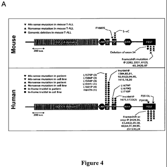
[0035] Figure 4. NOTCHI alterations in both murine and human T-
ALLs. Figure 4A is a graphic illustration of Location of sequence alterations
affecting Notch] in murine TKO and human T-ALL tumors. Each marker is
indicative of an individual cell line/patient. Figure 4B shows Western
blotting
analysis of murine full-length Notchl (FL; top), cleaved active Notchl (V1744;
middle), and tubulin loading control (bottom). High levels of activated Notchl
protein were expressed in many TKO tumors, including those harboring 3'
translocations (in blue: A577, A1052, A 1252) and truncating deletion
mutations (in
-10-
CA 02687787 2009-11-19
WO 2008/153743 PCT/US2008/006583
red: A494, A1040), in which faster migrating V1744 forms are apparent. Human
ALL-SIL (left) and normal mouse thymus (right) samples were loaded for
controls.
Figure 4C shows that high levels of Notch] mRNA correlate with high mRNA
levels of known downstream targets of Notchl protein, as assessed by
expression
profiling of TKO tumors. Each bar represents an individual probe set. Samples
in
blue lettering harbor 3' translocations near Notch]; samples in red lettering
harbor
truncating deletion mutations, as indicated for Figure 4B.
[0036] Figure 5. FBXW7 alterations are common in human T-ALL and
conserved in the murine TKO tumors. Figure 5A are a group of Log2 ratio array-
CGH plots showing conservation of CNAs resulting in deletion of FBXW7 in both
mouse TKO and human T-ALL cell lines; the genomic location of Fbxw7 is
indicated in green. Y axis, log2 ratio of copy number (normal set at log2=0);
amplifications are above and deletions are below this axis; X axis, chromosome
position. Figure 5B shows relative expression level of mouse Fbxw7 mRNA, as
assessed by real-time qPCR in the indicated murine TKO tumors. Figure 5C is a
graphic illustration of location of mutations in human FBXW7 identified in a
panel
of human T-ALL patients and cell lines. Each marker reprensents an individual
cell
line/patient.
[0037] Figure 6: Focal deletion of Pten in TKO tumors. Figure 6A is a
representative array-CGH Log2 ratio plot from a TKO lymphoma showing focal
deletion encompassing Pten, and its location relative to other genes in the
region
(http://genome.ucsc.edu/, NBCI mouse build 34). Y axis, log2 ratio of copy
number
(normal set at log2=0); amplifications are above and deletions are below this
axis; X
axis, chromosome position. Figure 6B summarizes the result of real-time qPCR
(showing deletion in several tumors), with a graphic illustration of real-time
qPCR
with primer sets to the indicated regions (arrows) and the location of array-
CGH 60-
mer oligo probes (Agilent 44K array). A494 is shown as a control without
evidence
of deletion.
[0038] Figure 7. Conservation of PTEN genetic alterations in human
and mouse T-ALLs. Figure 7A are a group of Log2 ratio array-CGH plots
demonstrating conservation of CNAs resulting in deletion of PTEN in both mouse
TKO and human T-ALL cell lines; the genomic location of Pten is indicated in
-ii-
CA 02687787 2009-11-19
WO 2008/153743 PCT/US2008/006583
green. Y axis, log2 ratio of copy number (normal set at log2=0);
amplifications are
above and deletions are below this axis; Xaxis, chromosome position. Figure 7B
is
a Western blotting analysis, showing the expression level of PTEN, phospho-
Akt,
and Akt in a panel of murine TKO and human T-ALL cell lines. BE13 and PEER
are synonymous lines. Tubulin was probed simultaneously as a loading control.
Samples in red harbor confirmed sequence mutations; samples in blue harbor
aCGH-detected deletions. Figure 7C are a group of Log2 ratio array-CGH plots
showing the effects of CNAs on other members of the Pten-Akt axis in murine
TKO
tumors. The location of each gene (Aktl, Tscl) is shown in green.
[00391 Figure 8: TKO cells with Pten mutation/deletion are sensitive to
inhibition of phospho-Akt by the drug triciribine. Cells were plated in
triplicate
and exposed to the indicated doses of triciribine or vehicle alone for 48
hours and
then quantified by MTS assay for viable cells. The fraction of surviving cells
is
plotted relative to survival in vehicle alone (set at 1). Tumor A1040 retains
wildtype
Pten expression and A1005 harbors a point mutation in one copy of Pten,
whereas
cell lines A577, A1240, A1252, and A494 are deficient for Pten expression.
[0040] Figure 9. Substantial overlap between genomic alterations of
murine TKO lymphomas and human tumors of diverse origins. Figure 9A
summarizes the result of statistical analysis of the cross-species overlap. We
obtained Human array-CGH profiles from the indicated tumor types. We further
defined MCRs as described in the Examples section (in particular, Example 4).
Characteristics of each set are listed on the left portion of the panel. The
number of
TKO MCRs (amp, amplifications; del, deletions) with syntenic overlap with
corresponding human CGH dataset is indicated on the right side of the panel,
with p
value for each based on 10,000 permutations. Figure 9B are a group of Pie-
chart
representation of numbers of TKO MCRs (indicated within each segment) with
syntenic overlap identified in one or multiple human tumor types (indicated by
different colors of the segments); left, amplifications; right, deletions. For
example,
21 of the 61 syntenic amplifications in Figure 9A were observed in 2 different
human tumor CGH datasets. Figure 9C are a group of Venn diagram representation
of the degree of overlap between murine TKO MCRs and MCRs from human
-12-
CA 02687787 2009-11-19
WO 2008/153743 PCT/US2008/006583
cancers of T-ALL, multiple myeloma, or solid tumors (encompassing
glioblastoma,
melanoma, and pancreatic, lung, and colon adenocarcinoma).
DETAILED DESCRIPTION OF THE INVENTION
[0041] In vivo cancer models used for the discovery of cancer-related genes
and therapeutic cancer targets typically produce cancer cells with benign
chromosomal profiles, i.e, nearly normal chromosomal stability. In contrast,
in
naturally occurring human cancer, cancer cell genomes display widespread
instability as evidenced by chromosomal structural aberrations. Accordingly,
the
present invention provides an in vivo cancer model with a destabilized genome
("genome unstable ").
[0042] The genomes of cancer cells from the genome unstable model of the
invention simulate the chromosomal instability displayed by human cancer cell
genomes The genome unstable cancer model of the invention, thus, provides
significant advantages for the discovery of genes and genetic elements
involved in
human cancer initiation, maintenance and progression. The chromosomal
aberrations in cancer cells from the model, particularly recurrent
aberrations, permit
investigation of chromosomal events in cancer that is not possible in cancer
models
with "benign" chromosomal profiles. Such chromosomal aberrations also focus
attention on particular regions of the genome more likely to harbor cancer-
related
elements. The validation herein of a genome unstable mouse cancer model that
generates chromosomal and genetic events that mirror those in multiple types
of
human cancers provides an important new tool for the discovery of cancer-
related
genes and therapeutic targets of relevance to human cancer. Although useful by
itself to discover genes and genetic elements relevant to human cancer, the
genome
unstable model of the invention also can be used as a background for
establishing
other cancer models, including known cancer models. Layering genetic
modifications in known oncogenes and/or tumor suppressors onto the genome
unstable model of the invention provides improved models that more closely
replicate naturally occurring cancer. Even more importantly, the genome
unstable
model of the invention permits cross-species comparison with human cancer
genomes to identify shared chromosomal and genetic events. Such shared events
-13-
CA 02687787 2009-11-19
WO 2008/153743 PCT/US2008/006583
provide a powerful guide for the discovery of cancer-related genes and
therapeutic
targets.
1. Definitions.
[0043] Throughout this specification and embodiments, the word "comprise" or
variations such as "comprises" or "comprising" will be understood to imply the
inclusion of a stated integer or group of integers but not the exclusion of
any other
integer or group of integers.
[0044] Unless otherwise defined herein, scientific and technical terms used in
connection with the present invention shall have the meanings that are
commonly
understood by those of ordinary skill in the art. Further, unless otherwise
required
by context, singular terms shall include pluralities and plural terms shall
include the
singular. Generally, nomenclatures used in connection with, and techniques of,
cell
and tissue culture, molecular biology, cell and cancer biology, virology,
immunology, microbiology, genetics and protein and nucleic acid chemistry
described herein are those well known and commonly used in the art.
2. Animal Models
[0045] Most standard genetically engineered mouse models of cancer have
relatively benign cytogenetic profiles. These genomically stable models do not
reflect the widespread chromosomal instability that is typical of human
genomes in
cancer. It has been reported that in most "genome-stable" murine tumor models,
about 20 to 40 chromosomal aberrations were detected per genome, or, less than
0.1
chromosomal rearrangements per chromosome.
[0046] Accordingly, in one aspect, the invention provides a non-human animal
that is genetically modified to develop cancer, wherein the genomes of cancer
cells
from the animal display enhanced chromosomal instability as evidenced by a
frequency of chromosomal structural aberration that approaches or matches that
seen
in human cancer cells. In various embodiments, the frequency of chromosomal
structural aberrations in a population of cancer cells from the non-human
animal
model is at least 1.5-fold, 2-fold, 3-fold, 4-fold, 5-fold or 10-fold higher
than the
frequency of chromosomal structural aberrations in such mammal without the
genetic modification, whether defined on a per-genome or per-chromosome basis.
-14-
CA 02687787 2009-11-19
WO 2008/153743 PCT/US2008/006583
[0047] The frequency of chromosomal abnormalities can be based on the average
number of such abnormalities per genome or per chromosome, or the average
number of a particular type of chromosomal abnormality per genome, or the
average
number of aberrations in a particular chromosome. Methods of measuring
chromosomal alterations are known in the art (see, e.g., R. C. O'Hagan, et
al.,
Cancer Res 63 (17), 5352 (2003); N. Bardeesy, et al., Proc Natl Acad Sci U S A
103
(15), 5947 (2006); M. Kim, et al., Cell 125 (7), 1269 (2006); L. Zender, et
al., Cell
125 (7), 1253 (2006)), and are further disclosed below. Cancer cells from the
genome unstable non-human animal model of the invention will have an enhanced
frequency of chromosomal aberrations compared to cells derived from comparable
non-human animal models lacking the genome destabilizing mechanisms described
above, by at least one of the aforementioned parameters.
[0048] A chromosomal structural aberration may be any chromosomal
abnormality resulting from DNA gains or losses, DNA amplification, DNA
deletion,
and DNA translocation. Exemplary chromosomal structural aberrations include,
for
example, sister chromatid exchanges, multi-centric chromosomes, inversions,
gains,
losses, reciprocal and non-reciprocal translocations (NRTs), p-p robertsonian-
like
translocations of homologous and/or non-homologous chromosomes, p-q
chromosome arm fusions, and q-q chromosome arm fusions.
[0049] The genetic modifications in the genome unstable animal model of the
invention can be in any gene or genetic element that renders the animal cancer-
prone
and affects genome structure or genome stability, so that the modifications
destabilize the genome, as evidenced by an increased frequency of chromosomal
structural aberrations in the genomes and/or chromosomes of cancer that
develops in
the animal compared to genomes and/or chromosomes in comparable animal models
lacking such genome destabilizing mechanisms. Genetic elements include [DNA
that is not translated to produce a protein product such as micro RNA,
expression
control sequences including DNA transcription factor binding sites, RNA
transcription initiation sites, promoters, enhancers, response elements and
the like.
In some embodiments the genetic modifications inactivate a gene or genetic
element
involved in chromosomal structural stability or integrity. Inactivation may be
by
directly inactivating the gene or genetic element, by suppressing the
expression, or
-15-
CA 02687787 2009-11-19
WO 2008/153743 PCT/US2008/006583
by inactivating or inhibiting the activity of a gene product, which can be a
nucleic
acid product including RNA or a protein gene product
[0050] In some embodiments, the genetic modifications comprise inactivation of
at least one allele of one or more genes or genetic elements involved in DNA
repair
and inactivation of at least one allele of one or more genes or genetic
elements
involved in a DNA damage checkpoint. In some embodiments, the genetic
modifications further comprise inactivation of at least one allele of a gene
or genetic
element involved in telomere maintenance. In any of the foregoing embodiments,
both alleles of the DNA repair related, DNA damage checkpoint related and/or
telomere maintenance related genes or genetic elements may be inactivated.
[0051] Any gene or genetic element involved in DNA repair or in a DNA damage
checkpoint can be inactivated in the genome unstable model of the invention.
Many
such genes and genetic elements in humans an other mammals will be known to
those of skill in the art. See, for example, R.D. Wood et al., Human DNA
Repair
Genes, Science, 291: 1284-1289 (February 2001); R A Bulman, S D Bouffler,
R Cox and T A Dragani, Locations of DNA Damage Response and Repair Genes in
the Mouse and Correlation with Cancer Risk Modifiers, National Radiological
Protection Board Report, October 2004 (ISBN 0-85951-544-3). The mouse DNA
repair gene database is available at the UK Health Protection Agency website.
[0052] They include, for example, genes encoding base excision repair (BER)
proteins such as ung, smug], mbd4, tdg, off], myh, nthl, mpg, ape], ape2,
lig3,
xrccl, adprt, adprtl2 and adprtl3 or species homologs thereof; mismatch
excision
repair proteins such as msh2, msh3, msh4, msh5, msh6, pms], pms3, mlhl, mlh3,
pms213 and pms214 or species homologs thereof; nucleotide excision repair
(NER)
proteins, non-homologous end joining (NHEJ) proteins, homologous recombination
proteins, DNA polymerases, editing and processing nucleases and DNA repair
helicases, among others. Wood et al., supra.
[0053] Exemplary NHEJ proteins include Ligase4, XRCC4, H2AX, DNAPKcs,
Ku70, Ku80, Artemis, Cernunnos/XLF, MRE11, NBS1, and RAD50. Exemplary
homologous recombination proteins include RAD51, RAD52, RAD54, XRCC3,
RAD51C, BRCA1, BRCA2 (FANCD1), FANCA, FANCB, FANCC, FANCD2;
-16-
CA 02687787 2009-11-19
WO 2008/153743 PCT/US2008/006583
FANCE, FANCF, FANCG, FANCJ (BRIP 1/BACH 1), FANCL, and FANCM.
Exemplary DNA repair helicases include BLM and WRN.
[0054] Any gene or genetic element involved in a DNA damage ckeckpoint can be
used in the genome unstable model of the invention. Information about many
such
genes and genetic elements is readily available and will be well-known those
of skill
in the art. Exemplary DNA checkpoint proteins include sensor proteins such as
RAD 1, RAD9, RAD 17, HUS 1, MRE 11, Rad50, and NB S 1; mediators such as
ATRIP; phosphoinositide 3-kinase related kinase (PIKK) family proteins such as
ATM, ATR, SMG-1 and DNA-PK; checkpoint kinases such as Chkl and Chk2; and
effector proteins such as p53, p63, p73, CDC25A, B and C, p21 and 14-3-
3P,y,~,a,E,rl,i APC; BRCA1, MDM2, MDM4, NBS1, RAD24, RAD 25, RAD50,
MDC 1, SMC 1, and claspin.
[0055] In one embodiment of the genome unstable model of the invention, the
non-human transgenic animal further comprises engineered inaction of at least
one
allele of one or more genes or genetic elements involved in synthesizing or
maintaining telomere length. In some embodiments, the non-human transgenic
mammal is engineered for decreased telomerase activity, for example by
inactivation
of telomerase reverse transcriptase, Tert, or telomerase RNA (Terc). In some
embodiments the genetic modification decreases the activity of a protein
affecting
telomere structure such as capping function. Exemplary proteins that affect
telomere structure include TRF1, TRF2, POT1a, POT1b, RAP1, TIN2, and TPP1.
[0056] The non-human genome unstable model of the invention may be any
animal, including, fish, birds, mammals, reptiles, amphibians. Preferably, the
animal
is a mammal, including rodents, primates, cats, dogs, goats, horses, sheep,
pigs,
cows. In preferred embodiments, the mammal is a mouse.
[0057] The genome unstable animal models of the invention include animals in
which all or only some portion of cells comprise the genetic modifications
that
create genome instability. In some embodiments, the germ cells of the animal
comprise the genetic modifications.
[0058] In some embodiments, the genome unstable model comprises inactivation
of one or both alleles of atm, terc or p53 or any combination of those genes.
In a
particular embodiment, one or both alleles of all three genes are inactivated.
In
-17-
CA 02687787 2009-11-19
WO 2008/153743 PCT/US2008/006583
some embodiments both alleles of atm are inactivated. In a particular
embodiment,
both alleles of all three genes are inactivated.
[0059] Also within the invention are tissues and cells from the genome
unstable
model of the invention, including somatic cells, germ cells, stem cells
including
embryonic stem cells, differentiated cells and undifferentiated cells. The
cells may
be cancer cells, non-cancer cells, or pre-cancer cells.
[0060] Inactivation of a gene or a genetic element in the genome unstable
animal
model of the invention can be achieved by any means, many of which are well-
known to those of skill in the art. Such means include deletion of all or part
of the
gene or genetic element or introducing an inactivating mutation (lesion) in
the gene
or genetic element. Deletion of all or a portion of a gene or genetic element
may be
by knock-out such as by homologous recombination or techniques using Cre
recombinase (e.g., a Cre-Lox system). Deletions including knock-outs can be
conditional knock-outs, where alteration of a nucleic acid sequences can occur
upon,
for example, exposure of the animal to a substance that promotes gene
alteration,
introduction of an enzyme that promotes recombination at the gene site (e.g.,
Cre in
the Cre-lox system), or other method for directing the gene alteration.
Conditional
or constitutive knock-outs can be tissue-specific, temporally-specific (e.g.,
occurring
during a particular developmental stage) or both.
[0061] Inactivating mutations may be introduced using any means, many of which
are well known. Such methods include site directed mutagenesis for example
using
homologous recombination or PCR. Such mutations may be introduced in the 5'
untranslated region (UTR) of a gene, including in an expression control
region, in a
coding region (intron or exon) or in the 3' UTR.
[0062] The expression or activity of a gene or genetic element also may be
accomplished by any means including but not limited to RNA interference,
antisense
including triple helix formation and ribozymes including RNaseP, leadzymes,
hairpin ribozymes and hammerhead ribozymes.
[0063] In some embodiments, the genome unstable animal model of the invention
further comprises one or more additional cancer-promoting genetic
modifications
including but not limited to the introduction of one or more activated
oncogenes,
modifications to increase the expression of one or more oncogenes, targeted
-18-
CA 02687787 2009-11-19
WO 2008/153743 PCT/US2008/006583
inactivation of one or more tumor-suppressors, or combinations of the
foregoing.
Such additional cancer-promoting modifications may be inducible, tissue
specific,
temporally specific or any combination of the three. For example, an oncogene
can
be introduced into the genome using an expression cassette that includes in
the 5'-3'
direction of transcription, a transcriptional and translational initiation
region that is
associated with gene expression in a specific tissue type, an oncogene, and a
transcriptional and translational termination region functional in the host
animal.
One or more introns may also be present. In addition to the oncogene of
interest, a
detectable marker, such as GFP (and its variants), luciferase, and lacZ may be
optionally operably linked to the oncogene and co-expressed. Similarly, a
tumor-
suppressor-gene may be inactivated using, for example, gene targeting
technology.
[0064] Introducing additional cancer-promoting modifications into a genome-
unstable animal model described herein creates a powerful tool for cancer gene
discovery. For example, Kras activation and p53 mutation in pancreas are known
to
cause pancreas cancer in human. A genome-unstable model having pancreas-
specific Kras activation, p53 inactivation (and optionally, a decreased
telomere
function) would greatly facilitate the discovery of pancreas cancer gene in
human.
[0065] The cancer in the genome unstable model any type of cancer, including
carcinoma, sarcoma, myeloma, leukemia, lymphoma or mixed cancer types. The
cancer can arise from any tissue type including epithelial tissue, mesenchymal
tissue, nervous tissue and hematopoietic tissue and be located in any organ or
tissue
of the body. The frequency of chromosomal aberrations can be determined in
cells
from any of the aforementioned cancers and can be from a primary tumor, a
secondary tumor, a metastatic tumor,a tumor recurrence perhaps normal cells
derived from said genomically unstable model that were genetically manipulated
in
vitro, through additional oncogene activation and tumor suppressor gene
inactivation
iintroduced by those knowledgeable in the art, to become cancerous
[0066] The genome unstable mouse model of the invention may develop any
cancer including but not limited to acral lentiginous melanoma, actinic
keratoses,
adenocarcinoma, adenoid cycstic carcinoma, adenomas, adenosarcoma,
adenosquamous carcinoma, adrenocortical carcinoma, AIDS-related lymphoma, anal
cancer, anaplastic glioma, astrocytic tumors, astrocytomas, bartholin gland
-19-
CA 02687787 2009-11-19
WO 2008/153743 PCT/US2008/006583
carcinoma, basal cell carcinoma, biliary tract cancer, bone cancer, bile duct
cancer,
bladder cancer, brain stem glioma, brain tumors, breast cancer, bronchial
gland
carcinomas, capillary carcinoma, carcinoids, carcinoma, carcinosarcoma,
cavernous,
central nervous system lymphoma, cerebral astrocytoma, cervical cancer,
connective
tissue cancer, cholangiocarcinoma, chondosarcoma, choriod plexus
papilloma/carcinoma, clear cell carcinoma, colon cancer, colorectal cancer,
cutaneous T-cell lymphoma, cystadenoma, endodermal sinus tumor, endometrial
hyperplasia, endometrial stromal sarcoma, endometrioid adenocarcinoma,
ependymal, ependymoma, epitheloid, esophageal cancer, Ewing's sarcoma,
extragonadal germ cell tumor, eye cancer, fibrolamellar, focal nodular
hyperplasia,
gallbladder cancer, gangliogliomas , gastric cancer, gastrinoma, germ cell
tumors,
gestational trophoblastic tumor, glioblastoma multiforme, glioma, glucagonoma,
head and neck cancer, hemangiblastomas, hemangioendothelioma, hemangiomas,
hepatic adenoma, hepatic adenomatosis, hepatocellular carcinoma, Hodgkin's
lymphoma, hypopharyngeal cancer, hypothalamic and visual pathway glioma,
childhood, insulinoma, intaepithelial neoplasia, interepithelial squamous cell
neoplasia, intraocular melanoma, intra-epithelial neoplasm, invasive squamous
cell
carcinoma, large cell carcinoma, islet cell carcinoma, Kaposi's sarcoma,
kidney
cancer, laryngeal cancer, leiomyosarcoma, lentigo maligna melanomas, leukemia-
related disorders, lip and oral cavity cancer, liver cancer, lung cancer,
lymphoma,
malignant mesothelial tumors, malignant thymoma, medulloblastoma,
medulloepithelioma, melanoma, meningeal, merkel cell carcinoma, mesothelial,
metastatic carcinoma, mucoepidermoid carcinoma, multiple myeloma/plasma cell
neoplasm, mycosis fungoides, myelodysplastic syndrome, myeloproliferative
disorders, nasal cavity and paranasal sinus cancer, nasopharyngeal cancer,
neuroblastoma, neurofibromatosis, neuroepithelial adenocarcinoma nodular
melanoma, non-Hodgkin's lymphoma, non-small cell lung cancer, oat cell
carcinoma, oligodendroglial, oligoastrocytomas, oral cancer, oropharyngeal
cancer,
osteosarcoma, pancreatic polypeptide, ovarian cancer, ovarian germ cell tumor,
pancreatic cancer, papillary serous adenocarcinoma, pineal cell, pituitary
tumors,
plasmacytoma, pseudosarcoma, pulmonary blastoma, parathyroid cancer, penile
cancer, pheochromocytoma, pineal and supratentorial primitive neuroectodermal
-20-
CA 02687787 2009-11-19
WO 2008/153743 PCT/US2008/006583
tumors, pituitary tumor, plasma cell neoplasm, pleuropulmonary blastoma,
prostate
cancer, rectal cancer, renal cell carcinoma, cancer of the respiratory system,
retinoblastoma, rhabdomyosarcoma, sarcoma, serous carcinoma, skin cancer,
small
cell carcinoma, small intestine cancer, soft tissue carcinomas, somatostatin-
secreting
tumor, squamous carcinoma, squamous cell carcinoma, stomach cancer, stromal
tumors, submesothelial, superficial spreading melanoma, supratentorial
primitive
neuroectodermal tumors, testicular cancer, thyroid cancer, undifferentiatied
carcinoma, urethral cancer, uterine sarcoma, uveal melanoma, verrucous
carcinoma,
vaginal cancer, vipoma, vulvar cancer, Waldenstrom's macroglobulinemia, well
differentiated carcinoma, and Wilm's tumor.
[0067] The animal models described herein are typically obtained using
transgenic
technologies. Transgenic technologies are well known in the art. For example,
transgenic mouse can be prepared in a number of ways. A exemplary method for
making the subject transgenic animals is by zygote injection. This method is
described, for example in U.S. Pat. No. 4,736,866. The method involves
injecting
DNA into a fertilized egg, or zygote, and then allowing the egg to develop in
a
pseudo-pregnant mother. The zygote can be obtained using male and female
animals
of the same strain or from male and female animals of different strains. The
transgenic animal that is born is called a founder, and it is bred to produce
more
animals with the same DNA insertion. In this method of making transgenic
animals,
the exogenous DNA typically randomly integrates into the genome by a non-
homologous recombination event. One to many thousands of copies of the DNA
may integrate at one site in the genome.
3. Methods of Identifying Cancer-related genes
[0068] In another aspect, the invention provides methods for identifying
genes and genetic elements involved in cancer initiation, maintenance and/or
progression in humans utilizing the genome unstable model of the invention.
The
gene discovery and identification methods are based on the surprising
discovery
described herein that chromosomal structural aberrations, copy number
alterations
and mutations in cancer cells in a genome unstable mouse model have syntenic
-2i-
CA 02687787 2009-11-19
WO 2008/153743 PCT/US2008/006583
counterparts (i.e., occurring in evolutionarily related chromosomal regions)
in
human cancer cells.
[0069] Accordingly, in one embodiment, the invention provides a method
of identifying a chromosomal region of interest for the identification of a
gene that is
potentially related to human cancer, comprising the step of identifying a DNA
copy
number alteration in a population of cancer cells from a non-human, genome-
unstable mammal described above. The chromosomal region where the DNA copy
number alteration occurred is a chromosomal region of interest for the
identification
of a gene or genetic element (such as microRNAs) that is potentially related
to
human cancer.
[0070] A DNA copy number alteration may be a DNA gain (such as
amplification of a genomic region) or a DNA loss (such as deletion of a
genomic
region). Methods of evaluating the copy number of a particular genomic region
are
well known in the art, and include, hybridization and amplification based
assays.
According to the methods of the invention, DNA copy number alterations may be
identified using copy number profiling, such as comparative genomic
hybridization
(CGH) (including both dual channel hybridization profiling and single channel
hybridization profiling (e.g. SNP-CGH)). Other suitable methods including
fluorescent in situ hybridization (FISH), PCR, nucleic acid sequencing, and
loss of
heterozygosity (LOH) analysis may be used in accordance with the invention.
[0071] In one embodiment of the invention, the DNA copy number
alterations in a genome are determined by copy number profiling.
[0072] In some embodiments of the invention, the DNA copy number
alterations are identified using CGH. In comparative genomic hybridization
methods, a "test" collection of nucleic acids (e.g. from a tumor or cancerous
cells) is
labeled with a first label, while a second collection (e.g. from a normal cell
or tissue)
is labeled with a second label. The ratio of hybridization of the nucleic
acids is
determined by the ratio of the first and second labels binding to each fiber
in an
array. Differences in the ratio of the signals from the two labels, for
example, due to
gene amplification in the test collection, is detected and the ratio provides
a measure
of the gene copy number, corresponding to the specific probe used. A
cytogenetic
representation of DNA copy-number variation can be generated by CGH, which
-22-
CA 02687787 2009-11-19
WO 2008/153743 PCT/US2008/006583
provides fluorescence ratios along the length of chromosomes from
differentially
labeled test and reference genomic DNAs.
[0073] In some embodiments of the present invention, the DNA copy
number alterations are analyzed by microarray-based CGH (array-CGH).
Microarray technology offers high resolution. For example, the traditional CGH
generally has a 20 Mb limited mapping resolution; whereas in microarray-based
CGH, the fluorescence ratios of the differentially labeled test and reference
genomic
DNAs provide a locus-by-locus measure of DNA copy-number variation, thereby
achieving increased mapping resolution. Details of various microarray methods
can
be found in the literature. See, for example, U.S. Pat. No. 6,232,068; Pollack
et al.,
Nat. Genet., 23 (1):41-6, (1999), Pastinen (1997) Genome Res. 7: 606-614;
Jackson
(1996) Nature Biotechnology 14:1685; Chee (1995) Science 274: 610; WO
96/17958, Pinkel et al. (1998) Nature Genetics 20: 207-211 and others.
[0074] The DNA used to prepare the CGH arrays is not critical. For
example, the arrays can include genomic DNA, e.g. overlapping clones that
provide
a high resolution scan of a portion of the genome containing the desired gene
or of
the gene itself. Genomic nucleic acids can be obtained from, e.g., HACs, MACs,
YACs, BACs, PACs, PIs, cosmids, plasmids, inter-Alu PCR products of genomic
clones, restriction digests of genomic clones, cDNA clones, amplification
(e.g.,
PCR) products, and the like. Arrays can also be obtained using oligonucleotide
synthesis technology. For example, see, e.g., light-directed combinatorial
synthesis
of high density oligonucleotide arrays U.S. Pat. No. 5,143,854 and PCT Patent
Publication Nos. WO 90/15070 and WO 92/10092.
[0075] The sensitivity of the hybridization assays may be enhanced through
use of a nucleic acid amplification system that multiplies the target nucleic
acid
being detected. Examples of such systems include the polymerase chain reaction
(PCR) system and the ligase chain reaction (LCR) system. Other suitable
methods
include are the nucleic acid sequence based amplification (NASBAO, Cangene,
Mississauga, Ontario) and Q Beta Replicase systems.
[0076] In one embodiment of the invention, the DNA copy number
alterations in a genome are determined by single channel profiling, such as
single
nucleotide polymorphism (SNP)-CGH. Traditional CGH data consists of two
~
-23-
CA 02687787 2009-11-19
WO 2008/153743 PCT/US2008/006583
channel intensity data corresponding to the two alleles. The comparison of
normalized intensities between a reference and subject sample is the
foundation of
traditional array-CGH. Single channel profiling (such as SNP-CGH) is different
in
that a combination of two genotyping parameters are analyzed: normalized
intensity
measurement and allelic ratio. Collectively, these parameters provide a more
sensitive and precise profile of chromosomal aberrations. SNP-CGH also
provides
genetic information (haplotypes) of the locus undergoing aberration.
Importantly,
SNP-CGH has the capability of identifying copy-neutral LOH events, such as
gene
conversion, which cannot be detected with array-CGH.
[0077] In another embodiment, FISH is used to determine the DNA copy
number alterations in a genome. Fluorescence in situ hybridization (FISH) is
known
to those of skill in the art (see Angerer, 1987 Meth. Enzymol., 152: 649).
Generally,
in situ hybridization comprises the following major steps: (1) fixation of
tissue or
biological structure to be analyzed; (2) prehybridization treatment of the
biological
structure to increase accessibility of target DNA, and to reduce nonspecific
binding;
(3) hybridization of the mixture of nucleic acids to the nucleic acid in the
biological
structure or tissue; (4) post-hybridization washes to remove nucleic acid
fragments
not bound in the hybridization, and (5) detection of the hybridized nucleic
acid
fragments.
[0078] In a typical in situ hybridization assay, cells or tissue sections are
fixed to a solid support, typically a glass slide. If a nucleic acid is to be
probed, the
cells are typically denatured with heat or alkali. The cells are then
contacted with a
hybridization solution at a moderate temperature to permit annealing of
labeled
probes specific to the nucleic acid sequence encoding the protein. The targets
(e.g.,
cells) are then typically washed at a predetermined stringency or at an
increasing
stringency until an appropriate signal to noise ratio is obtained.
[0079] The probes used in such applications are typically labeled, for
example, with radioisotopes or fluorescent reporters. Preferred probes are
sufficiently long, for example, from about 50, 100, or 200 nucleotides to
about 1000
or more nucleotides, to enable specific hybridization with the target nucleic
acid(s)
under stringent conditions.
[0080] In some applications it is necessary to block the hybridization
-24-
CA 02687787 2009-11-19
WO 2008/153743 PCT/US2008/006583
capacity of repetitive sequences. Thus, in some embodiments, tRNA, human
genomic DNA, or Cot-1 DNA is used to block non-specific hybridization.
[0081] In another embodiment, Southern blotting is used to determine the
DNA copy number alterations in a genome. Methods for doing Southern blotting
are
known to those of skill in the art (see Current Protocols in Molecular
Biology,
Chapter 19, Ausubel, et al., Eds., Greene Publishing and Wiley-Interscience,
New
York, 1995, or Sambrook et al., Molecular Cloning: A Laboratory Manual, 2d Ed.
vol. 1-3, Cold Spring Harbor Press, NY, 1989). In such an assay, the genomic
DNA
(typically fragmented and separated on an electrophoretic gel) is hybridized
to a
probe specific for the target region. Comparison of the intensity of the
hybridization
signal from the probe for the target region with control probe signal from
analysis of
normal genomic DNA (e.g., genomic DNA from the same or related cell, tissue,
organ, etc.) provides an estimate of the relative copy number of the target
nucleic
acid.
[0082] In one embodiment, amplification-based assays, such as PCR, are
used to determine the DNA copy number alterations in a genome. In such
amplification-based assays, the genomic region where a copy number alteration
occurred serves as a template in an amplification reaction. In a quantitative
amplification, the amount of amplification product will be proportional to the
amount of template in the original sample. Comparison to appropriate controls
provides a measure of the copy number of the genomic region.
[0083] Methods of "quantitative" amplification are well known to those of
skill in the art. For example, quantitative PCR involves simultaneously co-
amplifying a known quantity of a control sequence using the same primers. This
provides an internal standard that may be used to calibrate the PCR reaction.
Detailed protocols for quantitative PCR are provided, for example, in Innis et
al.
(1990) PCR Protocols, A Guide to Methods and Applications, Academic Press,
Inc.
N.Y.
[0084] Real time PCR can be used in the methods of the invention to
determine DNA copy number alterations. (See, e.g., Gibson et al., Genome
Research
6:995-1001, 1996; Heid et al., Genome Research 6:986-994, 1996). Real-time PCR
evaluates the level of PCR product accumulation during amplification. To
measure
-25-
CA 02687787 2009-11-19
WO 2008/153743 PCT/US2008/006583
DNA copy number, total genomic DNA is isolated from a sample. Real-time PCR
can be performed, for example, using a Perkin Elmer/Applied Biosystems (Foster
City, Calif) 7700 Prism instrument. Matching primers and fluorescent probes
can be
designed for genes of interest using, for example, the primer express program
provided by Perkin Elmer/Applied Biosystems (Foster City, Calif.). Optimal
concentrations of primers and probes can be initially determined by those of
ordinary skill in the art, and control (for example, beta-actin) primers and
probes
may be obtained commercially from, for example, Perkin Elmer/Applied
Biosystems
(Foster City, Calif.). To quantitate the amount of the specific nucleic acid
of interest
in a sample, a standard curve is generated using a control. Standard curves
may be
generated using the Ct values determined in the real-time PCR, which are
related to
the initial concentration of the nucleic acid of interest used in the assay.
Standard
dilutions ranging from 10-106 copies of the gene of interest are generally
sufficient.
In addition, a standard curve is generated for the control sequence. This
permits
standardization of initial content of the nucleic acid of interest in a tissue
sample to
the amount of control for comparison purposes.
[0085] Methods of real-time quantitative PCR using TaqMan probes are well
known in the art. Detailed protocols for real-time quantitative PCR are
provided, for
example, for RNA in: Gibson et al., 1996, A novel method for real time
quantitative
RT-PCR. Genome Res., 10:995-1001; and for DNA in: Heid et al., 1996, Real time
quantitative PCR. Genome Res., 10:986-994.
[0086] A TaqMan-based assay also can be used to quantify a particular
genomic region for DNA copy number alterations. TaqMan based assays use a
fluorogenic oligonucleotide probe that contains a 5' fluorescent dye and a 3'
quenching agent. The probe hybridizes to a PCR product, but cannot itself be
extended due to a blocking agent at the 3' end. When the PCR product is
amplified
in subsequent cycles, the 5' nuclease activity of the polymerase, for example,
AmpliTaq, results in the cleavage of the TaqMan probe. This cleavage separates
the
5' fluorescent dye and the 3' quenching agent, thereby resulting in an
increase in
fluorescence as a function of amplification (see, for example,
http://www2.perkin-
elmer.com).
[0087] Other suitable amplification methods include, but are not limited to
-26-
CA 02687787 2009-11-19
WO 2008/153743 PCT/US2008/006583
ligase chain reaction (LCR) (see Wu and Wallace (1989) Genomics 4:560,
Landegren et al. (1988) Science 241:1077, and Barringer et al. (1990) Gene
89:117),
transcription amplification (Kwoh et al. (1989) Proc. Natl. Acad. Sci. USA
86:1173), self-sustained sequence replication (Guatelli et al. (1990) Proc.
Nat. Acad.
Sci. USA 87:1874), dot PCR, and linker adapter PCR, etc.
[0088] In one embodiment, DNA sequencing is used to determine the DNA
copy number alterations in a genome. Methods for DNA sequencing are known to
those of skill in the art.
[0089] In one embodiment, karyotyping (such as spectral karyotyping, SKY)
is used to determine the chromosomal- structural aberrations in a genome.
Methods
for karyotyping are known to those of skill in the art. For example, for SKY,
a
collection of DNA probes, each complementary to a unique region of one
chromosome, may be prepared and labeled with a fluorescent color that is
designated for a specific chromosome. DNA amplification, deletion,
translocations
or other structural abnormalities may be determined based on fluorescence
emission
of the probes.
[0090] In certain embodiments, tumor samples from two or more genome-
unstable animal models of the invention are analyzed for DNA copy number
alterations, and the common genomic regions where the copy number alterations
occurred in at least two of the samples are identified. Such recurrent DNA
copy
number alterations are of particular interest.
100911 A minimum common region (MCR) of the recurrent DNA copy
number alteration may be defined when copy number alterations of two or more
samples are compared. In one embodiment, the MCR is defined by the boundaries
of overlap between two samples, or by boundaries of a single tumor against a
background of larger alterations in at least one other tumor.
[0092] Methods for determining MCRs is known in the art (see, e.g., D. R.
Carrasco, et al., Cancer Cell 9 (4), 313 (2006); A. J. Aguirre, et al., Proc
Natl Acad
Sci U S A 101 (24), 9067 (2004)). Briefly, a "segmented" dataset was generated
by
determining uniform copy number segment boundaries and then replacing raw log
2
ratio for each probe by the mean log 2 ratio of the segment containing the
probe. A
threshold representing minimal copy number alterations (CNAs) is then chosen
to
-27-
CA 02687787 2009-11-19
WO 2008/153743 PCT/US2008/006583
filter out noise. For example, the median log2 ratio of a two-fold change for
the
platform may be chosen as a threshold. In an exemplary embodiment, the
thresholds
representing CNAs are +/-0.6 (Agilent 22K a-CGH platform) and +/-0.8 (Agilent
44K/244K a-CGH platform), and the width of MCR is less than 10 Mb.
[0093] The boundaries of MCRs can be mapped by any method that is
known in the art, such as southern blotting, or PCR.
[0094] Genes and genetic elements located within an MCR are potentially
related to human cancer and such genes and genetic elements can be subject to
additional analyses to further characterize them. For example, a gene that is
initially
identified by array-CGH may be quantitatively amplified. Quantitative
amplification of either the identified genomic DNA or the corresponding RNA
can
confirm DNA gain or loss. Alternatively, if the sequence encodes a protein,
the
mRNA level, protein level, or activity level of the encoded protein may be
measured. An increase in RNA/protein/acitivity level, as compared to a
control,
confirms DNA amplification; a decrease in RNA/protein/acitivity level, as
compared to a control, confirms DNA deletion.
[0095] The gene or genetic element identified through initial screening may
also be re-sequenced to confirm amplification or deletion. Further, DNA
sequencing
and protein expression profiling may also be used to identify genetic
mutations that
may be associated with tumorigenesis.
[0096] In another aspect, the invention provides a method of identifying a
chromosomal region of interest for the identification of a gene or genetic
element
that is potentially related to human cancer, comprising the step of
identifying a
chromosomal structural aberration in a population of cancer cells from a
genome-
unstable animal models of the invention. A chromosomal region containing the
chromosomal structural aberration is a chromosomal region of interest for the
identification of a gene or genetic element that is potentially related to
human
cancer.
100971 In some embodiments, the chromosomal structural aberration is
detected using karyotyping, such as SKY. In some embodiments, the method
further comprises determining the DNA copy number alteration, as described
above.
-28-
CA 02687787 2009-11-19
WO 2008/153743 PCT/US2008/006583
A chromosomal region containing the both chromosomal structural aberration and
a
DNA copy number alteration is a chromosomal region of interest for the
identification of a gene or genetic element that is potentially related to
human
cancer.
[0098] In another aspect, the invention provides a method of identifying a
potential human cancer-related gene or genetic element, comprising the steps
of (a)
identifying a chromosomal region of interest as described herein; (b)
identifying a
gene or a genetic element within the chromosomal region of interest in the non-
human animal, and (c) identifying a human gene or genetic element that
corresponds
to the gene or genetic element identified in step (b).
[0099] Additionally, many public and private databases provide cancer gene
information (for example, Sanger's Cancer Gene Census, at
http://www.sanger.ac.uk/genetics/CGP/Census), and the information may be used
to
map known cancer genes to a particular chromosomal region.
[0100] If a gene or a genetic element is found to be potentially relevant to
human cancer, the corresponding human gene may be identified by homolog
mapping, ortholog mapping, paralog mapping, among other methods. As used
herein, a homolog is a gene related to a second gene by descent from a common
ancestral DNA sequence, an ortholog is a gene in a different species that
evolved
from a common ancestral gene by speciation, and a paralogs is a gene related
by
duplication within a genome.
[0101] In one embodiment, human homologs are identified by using, for
exmaple, the NCBI homologene website,
http://www.ncbl.nlm.nih.gov/entrez/query.fcgi?db=homologene.
[0102] In some embodiments, the method further comprises detecting a
mutation in the identified non-human gene or genetic element. In another
embodiment, a mutation in the corresponding human gene or genetic element is
identified. In another embodiment, mutations in the both the non-human gene or
genetic element and the human gene or genetic element are identified, and the
mutations are compared.
[0103] In another aspect, the invention provides a method of identifying a
potential human cancer-related gene or genetic element, comprising the steps
of (a)
-29-
CA 02687787 2009-11-19
WO 2008/153743 PCT/US2008/006583
detecting a DNA copy number alteration in a population of cancer cells from a
non-
human mammal, wherein the genome of the non-human mammal is engineered to
produce genome instability, (b) identifying a gene or genetic element located
within
the boundaries of the copy number alteration detected in step (a), (c)
identifying a
human gene or genetic element that corresponds to the gene or genetic element
identified in step (b) and that is located within the boundaries of a copy
number
alteration or of a chromosomal structural aberration in a human cancer cell.
The
human gene or genetic element identified in step (c) is a gene potentially
related to
human cancer.
[01041 Methods for detecting a copy number alteration or a chromosomal
structural aberration have been described above in detail. Methods for
identifying a
gene or genetic element located within the boundaries of the copy number
alteration
are also described above in detail.
[0105] In one embodiment, a copy number alteration or a chromosomal
structure aberration in the non-human animal model of the invention is
compared
with a copy number alteration or a chromosomal structural aberration in human
cancer cell. A potentially relevant human cancer related gene or genetic
element is
identified based on synteny. Synteny describes the preserved order and
orientation
of genes between related species. Comparisons of non-human animal model and
human cancer syntenic chromosomal regions may reveal the conserved nature of
certain genetic modification in tumorgeneis.
[0106] The cross-species comparison based on synteny has several
advantages. First is the ability to narrow the chromosomal regions of interest
-
certain genomic modification is more focal in one species than the other, and
a
cross-species comparison may eliminate such species-specific event. Second, a
minimal common region (MCR) typically contains a number of genes; a cross-
species comparison of syntenic regions allows an efficient way to reduce the
gene
numbers because the syntenic regions of the genome between non-human mammals
(in particular, mice) and humans may be in relatively small portions. Genes
located
within syntenic MCRs may be highly relevant to human cancers.
101071 In another aspect, the invention provides a method of identifying a
potential human cancer-related gene or genetic element, comprising the steps
of (a)
-30-
CA 02687787 2009-11-19
WO 2008/153743 PCT/US2008/006583
detecting a chromosomal structural aberration in a population of cancer cells
from a
non-human mammal, wherein the genome of the non-human mammal is engineered
to produce genome instability, (b) identifying a gene or genetic element
located
within the boundaries of the copy number alteration detected in step (a), (c)
identifying a human gene or genetic element that corresponds to the gene or
genetic
element identified in step (b) and that is located within the boundaries of a
copy
number alteration or of a chromosomal structural aberration in a human cancer
cell.
The human gene or genetic element identified in step (c) is a gene potentially
related
to human cancer.
4. Diagnosis and Methods of Treatment.
[0108] In one aspect, the present invention provides a method for identifying
subjects with T-cell acute lymphoblastic leukemia (T-ALL) who may have a
decreased or increased response to y-secretase inhibitor therapy, based on the
discovery that inactivation of FBXW7 is associated with human T-cell
malignancy.
[0109] In one embodiment, the method for identifying subjects with T-ALL
who may have a decreased response to a y-secretase inhibitor therapy
comprises:
detecting in a cancer cell from the subject the expression level or activity
level of
FBXW7; a decreased expression/activity of FBXW7, as compared to a control,
indicates that the subject may have a decreased response to a y-secretase
inhibitor
therapy. The expression or activity level of NOTCH 1 in the cancer cell may
also be
determined simultaneously; an increased expression/activity of NOTCHI, as
compared to a control, further indicates that the subject may have a decreased
response to a y-secretase inhibitor therapy. Conversely, an increased
expression/activity of FBXW7 (together with a decreased expression/activity of
NOTCH 1, optionally), as compared to a control, indicates that the subject may
be
sensitive to a y-secretase inhibitor therapy.
[0110] y-Secretase is a complex composed of at least four proteins, namely
presenilins (presenilin 1 or -2), nicastrin, PEN-2, and APH-1. Several
proteins have
been identified as substrates for y-secretase cleavage, include Notch and the
Notch
ligands Deltal and Jagged2, ErbB4, CD44, and E-cadherin (Wong, G.T. et. al, J.
-31-
CA 02687787 2009-11-19
WO 2008/153743 PCT/US2008/006583
Biol. Chem., Vol. 279, Issue 13, 12876-12882, March 26, 2004). The cleavage of
Notch by y-secretase has been studied most extensively. Notch plays an
evolutionarily conserved role in regulating cell growth and lineage
specification
particularly during embryonic development. Notch is activated by several
ligands
(Delta, Jagged, and Serrate) and is then proteolytically processed by a series
of
ligand-dependent and -independent cleavages. y-Secretase catalyzes the
terminal
cleavage event (S3 cleavage), which releases a fragment known as the Notch
intracellular domain (NICD). The NICD fragment then translocates to the
nucleus
where it acts as a nuclear transcription factor. As expected from its role in
Notch S3
cleavage, y-secretase inhibitors have been shown to block NICD production in
vitro.
In vivo, Notch function appears to be critical for the proper differentiation
of T and B
lymphocytes, and y-secretase inhibitors reduce the thymocyte number and block
thymocyte differentiation at an early stage in fetal thymic organ cultures.
[0111] The FBXW7 gene (also called hCDC4) encodes a key component of
the E3 ubiquitin ligase that is implicated in the control of chromosome
stability
(Mao J. et. al, Nature 432, 775-779 (2004)). FBXW7 is responsible for binding
the
PEST domain of intracellular NOTCHI, leading to ubiquitination and degradation
by the proteasome. Because there exists a statistically significant anti-
correlation
between PEST domain mutations in NOTCHl and FBXW7 mutation in human T-
ALL, T-ALL cells having a reduced expression/activity of FBXW7 will less
likely
to respond to y-secretase inhibitors.
[0112] One of the recurring problems of cancer therapy is that a patient in
remission (after the initial treatment by surgery, chemotherapy, radiotherapy,
or
combination thereof) may experience relapse. The recurring cancer in those
patients
is frequently resistant to the apparently successful initial treatment. In
fact, certain
cancers in patients initially diagnosed with the disease may be already
resistant to
conventional cancer therapy even without first being exposed to such
treatment. y-
secretase inhibitor therapy can be physically exhausting for the patient. Side
effects
of secretase inhibitors include weight loss, changes in gastrointestinal tract
architecture, accumulation of necrotic cell debris, dilation of crypts and
infiltration
of inflammatory cells, nausea, vomiting, weakness, diarrhea elevation in white
blood
cell count, and esophageal failure (Siemers E. et al, 2005 May-Jun;28(3):126-
32;
-32-
CA 02687787 2009-11-19
WO 2008/153743 PCT/US2008/006583
Wong, GT. et al, J Biol Chem. 2004 Mar 26;279 (13):12876-82). Thus there is a
need to determine whether a cancer patient may benefit from a chemotherapeutic
treatment prior to the commencement of the treatment.
[0113] In one embodiment, a cancer patient is screened based on the
expression level of FBXW7 and optionally, NOTCHI, in a cancer cell sample.
[0114] The expression level of FBXW7 or NOTCHI may be measured by
DNA level, mRNA level, protein level, activity level, or other quantity
reflected in
or derivable from the gene or protein expression data. For example, a genetic
alteration may result in a decreased expression of FBXW7. Common genetic
alterations include deletion of at lease one FBXW7 gene from the genome, or a
mutation in at least one allele of an FBXW7 gene. The mutation may be a mis-
sense
mutation; a non-sense mutation; an insertion, deletion, or substitution of one
or more
nucleotides; a truncation from the 5' terminal (either untranslated region or
coding
region), 3' terminal (either untranslated region or coding region), or both; a
substitution of one or more nucleotides in the 5' untranslated region, 3'
untranslated
region, coding region (which results in an amino acid change), or combinations
of
the three. Exemplary genetic alterations include a mutation in the third WD40
domain or the fourth WD40 domain of the FBXW7, G423V, R465C, R465H,
R479L. R479Q, R505C and D527G mutations. A genetic alteration may also result
in an increased expression of NOTCHI, such as translocation or copy number
amplification of NOTCH 1 gene.
[0115] The mRNA level of FBXW7 or NOTCHI may be measured using
any art-known method, such as PCR, northern blotting, RNase Protection Assay,
or
microarray hybridization. For example, Real-time polymerase chain reaction,
also
called quantitative real time PCR (QRT-PCR) or kinetic polymerase chain
reaction,
is widely used in the art to measure mRNA level of a target gene. The QRT-PCR
procedure follows the general pattern of polymerase chain reaction, but the
DNA is
quantified after each round of amplification. Two common methods of
quantification are the use of fluorescent dyes that intercalate with double-
strand
DNA, and modified DNA oligonucleotide probes that fluoresce when hybridized
with a complementary DNA. QRT-PCR can be combined with reverse transcription
polymerase chain reaction to quantify low abundance messenger RNA (mRNA),
-33-
CA 02687787 2009-11-19
WO 2008/153743 PCT/US2008/006583
SFCE-125-WO1
enabling one to quantify relative gene expression at a particular time, or in
a
particular cell or tissue type.
[0116] The expression level of FBXW7 or NOTCHI may also be measured
by protein level using any art-known method. Traditional methodologies for
protein
quantification include 2-D gel electrophoresis, mass spectrometry and antibody
binding. Frequently used methods for assaying target protein levels in a
biological
sample include antibody-based techniques, such as immunoblotting (western
blotting), immunohistological assay, enzyme linked immunosorbent assay
(ELISA),
radioimmunoassay (RIA), or protein chips. Gel electrophoresis,
immunoprecipitation and mass spectrometry may be carried out using standard
techniques. Additionally, NOTCH 1 expression may be measured by detection of
cleaved, intranuclear (ICN) form of NOTCH 1 protein in cells.
[0117] The expression level of FBXW7 or NOTCH 1 may also be measured
by the activity level of the gene product using any art-known method, such as
transcriptional activity of NOTCH 1 or ligase activity of FBXW7. For example,
NOTCHI activity may be measured by a increased binding of ICN of NOTCH1.
Alternatively, the expression level of a transcriptional downstream target of
NOTCH 1 may be measured as an indicator of NOTCH 1 activity, , such as c-Myc,
PTCRA, Hes l , etc.
[0118] In certain embodiments, it is useful to compare the
expression/activity level of FBXW7 or NOTCHI to a control. The control may be
a measure of the expression level of FBXW7 or NOTCH I in a quantitative form
(e.g., a number, ratio, percentage, graph, etc.) or a qualitative form (e.g.,
band
intensity on a gel or blot, etc.). A variety of controls may be used. Levels
of
FBXW7 or NOTCH 1 expression from a non-cancer cell of the same cell type from
the subject may be used as a control. Levels of FBXW7 or NOTCH 1 expression
from the same cell type from a healthy individual may also be used as a
control.
Alternatively, the control may be expression levels of FBXW7 or NOTCH I from
the
individual being treated at a time prior to treatment or at a time period
earlier during
the course of treatment. Still other controls may include expression levels
present
in a database (e.g., a table, electronic database, spreadsheet, etc.) or a pre-
determined
threshold.
-34-
CA 02687787 2009-11-19
WO 2008/153743 PCT/US2008/006583
SFCE-125-WO1
[0119] The present invention further discloses methods of treating a T-ALL
subject who will likely be sensitive a treatment with y-secretase inhibitors
(identified
using the methods described above), comprising administering to the patients a
y-
secretase inhibitor. y-secretase inhibitors are known in the art, exemplary y-
secretase
inhibitors include LY450139 Dihydrate and LY411575.
[0120] The present invention further discloses methods of treating a T-ALL
subject who will has a decreased expression/activity of FBXW7 (identified
using the
methods described above) with an agent that increases the expression/activity
of
FBXW7. The agent may be a recombinant FBXW7 protein or a functionally active
fragment or derivative thereof, a nuclei acid that encodes FBXW7 protein or a
functionally active fragment or derivative thereof, or an agent that activates
FBXW7. A "functionally active" PBXW7 fragment or derivative exhibits one or
more functional activities associated with a full-length, wild-type FBXW7
protein,
such as antigenic or immunogenic activity, ability to bind natural cellular
substrates,
etc. The functional activity of FBXW7 proteins, derivatives and fragments can
be
assayed by various methods known to one skilled in the art (Current Protocols
in
Protein Science, Coligan et al., eds., John Wiley & Sons, Inc., Somerset, N.J.
(1998)).
[0121] In another aspect, the present invention provides a method for
identifying subject with T-ALL who may benefit from treatment with a
phosphatidylinositol 3-kinase (P13K) pathway inhibitor, based on the discovery
that
PTEN inactivation is associated with human T-cell malignancy.
[0122] PTEN has been characterized as a tumor suppressor gene that
regulates cell cycle. PTEN functions as a phosphodiesterase and an inhibitor
of the
PI3K/AKT pathway, by removing the 3' phosphate group of phosphatidylinositol
(3,4,5)-trisphosphate (PIP3). When PTEN is inactivated, increased production
of
PIP3 activates AKT (protein kinase B). The AKT pathway promotes tumor
progression by enhancing cell proliferation, growth, survival, and motility,
and by
suppressing apoptosis. AKT is activated by two phosphorylation events
catalyzed
by the phosphoinositide dependent kinase PDKI, an enzyme that is activated by
P13K.
-35-
CA 02687787 2009-11-19
WO 2008/153743 PCT/US2008/006583
~ri r.-ic~-wU1
[0123] In one embodiment, the method for identifying subject with T-ALL
who may benefit from treatment with a P13K pathway inhibitor comprises:
detecting
in a tumor cell from the subject the expression level or activity level of
PTEN. A
decreased expression/activity of FBXW7, as compared to a control, indicates
that
the subject may benefit from a P13K inhibitor therapy.
[0124] The phospho-AKT level in the cancer cell from the subject may also
be determined simultaneously; an increased phospho-AKT level, as compared to a
control, further indicates that the subject may benefit from a P13K inhibitor
therapy.
[0125] The expression level of PTEN may be measured by DNA level,
mRNA level, protein level, activity level, or other quantity reflected in or
derivable
from the gene or protein expression data. For example, a genetic alteration
may
result in a decreased expression of PTEN. Common genetic alterations include
deletion of at lease one PTEN gene from the genome, or a mutation in at least
one
allele of a PTEN gene. The mutation may be a mis-sense mutation; a non-sense
mutation; an insertion, deletion, or substitution of one or more nucleotides;
a
truncation from the 5' terminal (either untranslated region or coding region),
3'
terminal (either untranslated region or coding region), or both; a
substitution of one
or more nucleotides in the 5' untranslated region, 3' untranslated region,
coding
region (which results in an amino acid change), or combinations of the three.
[0126] The expression level of PTEN may also be measured by mRNA level
using any method known in the art, such as PCR, Northern blotting, RNase
Protection Assay, and microarray hybridization.
101271 The expression level of PTEN may also be measured by protein level
using any method known in the art, such as 2-D gel electrophoresis, mass
spectrometry and antibody binding
[0128] The expression level of PTEN may also be measured by the activity
level of PTEN using any art-known method, such as measuring the phosphatase
activity. Additionally, the expression or activity of other proteins involved
in the
PI3K/AKT pathway may also be measured as a proxy for PTEN activity. For
example, the phospho-AKT level in a cell generally reflects the PTEN activity,
therefore may be measured as a marker for PTEN activity.
-36-
CA 02687787 2009-11-19
WO 2008/153743 PCT/US2008/006583
5r%_r.-11.Z3-vvU1
[0129] In certain embodiments, a control may be used to compare the
expression/activity level of PTEN. As described in detail above, a control may
be
derived from a non-cancer cell of the same type from the subject, same cell
type
from a healthy individual, a predetermined value, etc.
[0130] The present invention further discloses methods of treating a T-ALL
subject who may benefit from a treatment with P13K inhibitors (identified
using the
methods described above), comprising administering to the patients a P13K
inhibitor. P13K inhibitors are well know in the art (e.g., Pinna, LA and
Cohen, PTW
(eds.) Inhibitors of Protein Kinases and Protein Phosphates, Springer (2004)
and
Abelson, JN, Simon, MI, Hunter, T, Sefton, BM (eds.) Methods in Enzymology,
Volume 201: Protein Phosphorylation, Part B: Analysis of Protein
Phosphorylation,
Protein Kinase Inhibitors, and Protein Academic Press (2007)).
[0131] The present invention further discloses methods of treating a T-ALL
subject who will has a decreased expression/activity of PTEN (identified using
the
methods described above) with an agent that increases the expression/activity
of
PTEN. The agent may be a recombinant PTEN protein or a functionally active
fragment or derivative thereof, a nuclei acid that encodes PTEN protein or a
functionally active fragment or derivative thereof, or an agent that activates
PTEN.
[0132] In another aspect, the invention provides a method of assessing
whether a subject is afflicted with cancer or at risk for developing cancer,
comprising: determining the expression or activity level of at least one
cancer gene
or candidate cancer gene located in an amplified MCR in Table 1 in a
biological
sample from the subject. An increase in the expression or activity the gene,
as
compared to a control, indicates that the subject is afflicted with cancer or
at risk for
developing cancer. Alternatively, if there is a decrease in the expression or
activity
of a cancer gene or candidate cancer gene located in a deleted MCR in Table 1,
as
compared to a control, the decreased expression or activity level also
indicates that
the subject is afflicted with cancer or at risk for developing cancer.
101331 In another aspect, the invention provides a method of assessing
whether a subject is afflicted with cancer or at risk for developing cancer,
the
method comprising: determining the copy number of at least one amplified
minimal
common region (MCR) listed in Table I in a biological sample from the subject.
An
-37-
CA 02687787 2009-11-19
WO 2008/153743 PCT/US2008/006583
5r%.r,-iz.z-vvO1
increased copy number of the MCR in the sample, as compared to the normal copy
number of the MCR, indicates that the subject is afflicted with cancer or at
risk for
developing cancer. Alternatively, a decreased copy number of a deleted MCR
(also
listed in Table 1) in the sample, as compared to the normal copy number of the
MCR, also indicates that the subject is afflicted with cancer or at risk for
developing
cancer. The normal copy number of an MCR is typically one per chromosome.
[0134] In another aspect, the invention provides a method for monitoring
the progression of cancer in a subject, the method comprising: a) determining
in a
biological sample from the subject at a first point in time, the expression or
activity
level of a cancer gene or a candidate cancer gene listed in Table 1; b)
repeating step
a) at a subsequent point in time; and c) comparing the expression or activity
of the
gene in steps a) and b), and therefrom monitoring the progression of cancer in
the
subj ect.
[0135] In another aspect, the invention provides a method of assessing the
efficacy of a test agent for treating a cancer in a subject, comprising: a)
determining
the expression or activity level of at least one cancer gene or a candidate
cancer gene
located in an amplified MCR in Table 1 in a biological sample from the subject
in
the presence of the test agent; and b) determining the expression or activity
level of
the gene in a biological sample from the subject in the absence of the test
agent. A
decreased expression or activity of the gene in step (a), as compared to that
of (b), is
indicative of the test agent's potential efficacy for treating the cancer in
the subject.
Alternatively, if the test agent increases the expression or activity of at
least one
cancer gene or a candidate cancer gene located in a deleted MCR in Table 1,
the test
agent is also potentially effective for treating the cancer in a subject.
101361 In another aspect, the invention provides a method of assessing the
efficacy of a therapy for treating cancer in a subject, the method comprising:
a)
determining the expression or activity level of at least one cancer gene or a
candidate cancer gene located in an amplified MCR in Table I in a biological
sample from the subject prior to providing at least a portion of the therapy
to the
subject; and b) determining the expression or activity level of the gene in a
biological sample from the subject following provision of the portion of the
therapy.
A decreased expression or activity of the gene in step (a), as compared to
that of (b),
-38-
CA 02687787 2009-11-19
WO 2008/153743 PCT/US2008/006583
ar ~.~-ia~- vv ~l
is indicative of the therapy's efficacy for treating the cancer in the
subject.
Alternatively, if the therapy increases the expression or activity of at least
one
cancer gene or a candidate cancer gene located in a deleted MCR in Table 1,
the
therapy is also potentially effective for treating the cancer in a subject.
[0137] In another aspect, the invention provides a method of treating a
subject afflicted with cancer comprising administering to the subject an agent
that
decreases the expression or activity level of at least one cancer gene or
candidate
cancer gene located in am amplified MCR in Table 1. Alternatively, the
invention
provides a method of treating a subject afflicted with cancer comprising
administering to the subject an agent that increases the expression or
activity level of
at least one cancer gene or candidate cancer gene located in a deleted MCR in
Table
1.
[0138] In certain embodiments, the agent is an antibody, or its antigen-
binding fragment thereof, that specifically binds to a cancer gene or
candidate cancer
gene listed in Table 1. Optionally, the antibody may be conjugated to a toxin,
or a
chemotherapeutic agent.
[0139] Alternatively, the agent may be an RNA interfering molecule (such
as an shRNA or siRNA moleucle) that inhibits expression of a cancer gene or
candidate cancer gene in an amplified MCR in Table 1, or an antisense RNA
molecule complementary to a cancer gene or candidate cancer gene in an
amplified
MCR in Table 1.
[0140] Alternatively, the agent may be a peptide or peptidomimetic, a small
organic molecule, or an aptamer.
[0141] Preferrably, the agent is administered in a pharmaceutically
acceptable formulation.
101421 In another aspect, the invention provides a method of assessing
whether a subject is afflicted with cancer or at risk for developing cancer,
the
method comprising: determining the copy number of at least one minimal common
region (MCR) listed in Table 5 in a biological sample from the subject. A
change of
copy number of the MCR in the sample, as compared to the normal copy number of
the MCR, indicates that the subject is afflicted with cancer or at risk for
developing
cancer. The normal copy number of an MCR is typically one per chromosome.
-39-
CA 02687787 2009-11-19
WO 2008/153743 PCT/US2008/006583
src;k;-izs-wOl
[0143] In certain embodiments, the cancer is lymphoma. In certain
embodiments, the lymphoma is T-ALL.
[0144] In another aspect, the invention provides a method of assessing
whether a subject is afflicted with cancer or at risk for developing cancer,
by
comparing the copy number of an MCR, identified using a genome-unstable non-
human mammal model (including a genome-unstable mouse model of the
invention), with the normal copy number of the MCR. The normal copy number of
an MCR is typically one per chromosome.
EXAMPLES
_Example 1: Generation and Characterization of Murine T Cell Lymphomas
With Highly Complex Genomes
[0145] In this example, we created a murine lymphoma model system that
combines the genome-destabilizing impact of Atm deficiency and telomere
dysfunction to effect T lymphomagenesis in a p53-dependent manner.
[0146] We interbred mTerc Atm p53 heterozygous mice and maintained them in
pathogen-free conditions. We intercrossed the null alleles of mTerc, Atm and
p53 to
generate various genotypic combinations from this "triple"-mutant colony (for
simplicity, hereafter designated as "TKO" for all genotypes from this colony).
[0147] We monitored animals for signs of ill-health every other day. Moribund
animals were euthanized and subjected to complete autopsy; mice found dead
were
subject to necropsy specifically for signs of lymphoma. We performed all
animal
uses and manipulations according to approved IACUC protocol. Tumors were
harvested from TKO mice and partitioned in the following manner. One section
was
snap-frozen for DNA and RNA extraction, a second portion was processed for
histology, and the remaining portion was disaggregated for in vitro culture.
Suspensions of tumor cells were maintained in RPMI supplemented with 50 M
beta-mercaptoethanol, 10% Cosmic Calf serum (HyClone), 0.5 ng/ml recombinant
IL-2, and 4 ng/ml recombinant IL-7 (both from Peprotech). Tumor cells were
immunostained with antibodies against CD4, CD8, CD3, and B220/CD45R
(eBioscience) and subjected to FACS analysis.
-40-
CA 02687787 2009-11-19
WO 2008/153743 PCT/US2008/006583
or %,r -ic0- vVt)1
[0148] We prepared DNA frozen tumors with the PureGene kit according to
manufacturer's instructions (Gentra Systems). We prepared RNA by an initial
extraction with Trizol (Invitrogen) according to the manufacturer's
instructions.
Pelleted total RNA was then digested with RQ1 DNase (Promega) and subsequently
purified through RNA purification columns (Gentra). Proteins were obtained
either
from cell lines or tumor pieces by dis-aggregation in lysis buffer (according
to Cell
Signaling Technology) followed by sonication in a bath sonicator for 30 s.
Lysates
were clarified by centrifugation prior to quantification according to
manufacturer's
instructions (BioRad Protein Assay) and separation on 4-12% NuPage gels
(Invitrogen).
[0149] We found that TKO mice which are p53+/- or p53-/- succumbed to lethal
lymphoma with shorter latency and higher penetrance relative to TKO animals
wildtype for p53 (Figure 2A). Moreover, lymphomas from TKO mice heterozygous
for p53 showed reduction to homozygosity in 14 specimens (out of 15 specimens
examined) (Figure 2B), indicating strong genetic pressure to inactivate p53
during
lymphomagenesis in this context. Phenotypically, these TKO tumors resembled
lymphomas in the conventional Atm-/- mouse model with effacement of thymic
architecture by CD4+/CD8+ (less commonly CD4-/CD8- or mixed single/double
positive) lymphoma cells (Figure 2C). Taken together, the genetic and
molecular
observations strongly suggest that an Atm-independent p53-dependent telomere
checkpoint is operative to constrain lymphoma development.
[0150] To quantify chromosomal rearrangements, we used Spectral Karyotype
(SKY) analyses according to the following protocol. Metaphase preparations
were
typically obtained within 48 hours of establishment, although in a few
instances
establishment of the cell line was required to obtain good quality metaphases.
Harvested cells were incubated in 105 mM KCl hypotonic buffer for 15 min prior
to
fixation in 3:1 methanol-acetic acid. Spectral karyotyping was done using the
SkyPaint Kit and SkyView analytical software (Applied Spectral Imaging,
Carlsbad,
CA) according to manufacturer's protocols. Chromosome aberrations were defined
using the rules from the Committee on Standard Genetic Nomenclature for Mice.
T-
test comparison between GO and G 1-G4 cytogenetics is based on 90 SKY profiles
each set (ten metaphase spreads for each of TKO lymphomas).
-41-
CA 02687787 2009-11-19
WO 2008/153743~1 LaVC~ ~w. l ll 111vwvvl aw A~~V,.. -.....PCT/US2008/006583_
SFCE-125-WO1
[0151] Figure 1, Figure 2D, and Table 3 summarize the SKY analyses of
chromosomal rearrangement in 9 telomere deficient (Gl-G4 mTerc') TKO
lymphomas and 9 telomere intact (GO mTerc+l+ or mTerc+i-) TKO lymphomas.
Relative to GO tumors, G1-G4 TKO lymphomas displayed an overall greater
frequency of chromosome structural aberrations of various types (0.34 versus
0.09
per chromosome, respectively, p<0.0001, t test) including a multitude of multi-
centric chromosomes, non-reciprocal translocations (NRTs), p-p robertsonian-
like
translocations of homologous and/or non-homologous chromosomes, p-q fusions,
and q-q fusions. When examined on a chromosome-by-chromosome basis, several
chromosomes (specifically, 2, 6, 8, 14, 15, 16, 17, and 19) were involved in
significantly more dicentric and robertsonian-like rearrangement events in G1-
G4
relative to GO TKO tumors (p<0.05; t test; Figure 2E). Without being bound by
a
particular theory, the recurrent non-random nature of these chromosomal
rearrangements in the TKO model may provide adaptive mechanisms to tolerate
telomere dysfunction and/or play causal roles in lymphoma development (e.g.,
chromosome 2, see below).
Example 2: TKO Lymphomas Harbor Genomic Alterations Syntenic To Those
In Human T Cell Malignancy
[0152] To assess the degree of syntenic overlap in the murine lymphoma-prone
TKO instability model and in human T-ALL and other cancers, we applied and
integrated multiple genome analysis technologies to survey cancer-associated
alterations for comparison with T-ALL and a diverse set of major human
cancers.
[0153] Synteny describes the preserved order and orientation of genes between
species. Disruption of synteny, caused by chromosome rearrangement, is an
indication of divergent evolution. Comparisons of TKO mouse model and human
T-ALL syntenic chromosomal regions may reveal the conserved nature of certain
genetic modification in tumorigeneis.
101541 Because TKO lymphomas harbored a large number of complex
nonreciprocal translocations (NRTs), we sought to determine whether these
genome-
unstable tumors possess increased numbers of recurrent amplifications and
deletions. To this end, we compiled high-resolution genome-wide array-CGH
-42-
CA 02687787 2009-11-19
WO 2008/153743 PCT/US2008/006583
brUE-il~-wO1
profiles for 35 TKO tumors (Table 3) and 26 human T-ALL cell lines and tumors
(Tables 4A and 4B) for comparison.
[0155] T-ALL cell lines used in this example, and in Examples 3-7 are listed
in
Table 4A. A subset was subjected to both array-CGH (described in detail below)
and re-sequencing, as indicated.
[01561 We used two cohorts of clinical human T-ALL samples in this example. A
cohort of 8 samples (Table 4B) comprised of cryopreserved lymphoblasts or
lymphoblast cell lysates, obtained with informed consent and IRB approval at
the
time of diagnosis from pediatric patients with T-ALL treated on Dana-Farber
Cancer
Institute study 00-001. We subjected these samples to genome-wide array-CGH
profiling.
[0157] For genome-wide array-CGH profiling, we used the following protocol.
Genomic DNA processing, labeling and hybridization to Agilent CGH arrays were
performed as per manufacturer's protocol
(http://www.home.agilent.com/agilent/home.jspx). Murine tumors were profiled
against individual matched normal DNA (e.g., non-tumor cell of the same cell
type
from the same individual) or, when not available, pooled DNA of matching
strain
background. Labeled DNAs were hybridized onto 44K or 244K microarrays for
mouse, and 22K or 44K microarrays for human. The Mouse 44K array contained
42,404 60-mer elements for which unique map positions were defined (National
Center for Biotechnology Information, Mouse Build 34). The median interval
between mapped elements was 21.8 kb, 97.1 % of intervals of <0.3 megabases
(Mb),
and 99.3% are <1 Mb. The 244K array contained 224,641 elements for which
unique map positions were defined based on the same mouse genome build. The
Human 22K array contained 22,500 elements designed for expression profiling
for
which 16,097 unique map positions were defined with a median interval between
mapped elements of 54.8 kb. The Human 44K microarray contained 42,494 60-mer
oligonucleotide probes for which unique map positions were defined (National
Center for Biotechnology Information, Human Build 35). The 244K array
contained
226,932 60-mer oligonucleotide probes for which unique map positions were
defined based on the same human genome build.
-43-
CA 02687787 2009-11-19
WO 2008/153743 PCT/US2008/006583
arUk'-iz2~-w01
[0158] Profiles generated on 244K density arrays were extracted for the same
42K
probes on the 44K microarrays to allow combination of profiles generated on
the
two different platforms. Fluorescence ratios of scanned images were normalized
and calculated as the average of two paired (dye swap), and copy number
profile
was generated based on Circular Binary Segmentation, an algorithm that uses
permutation to determine the significance of change points in the raw data (A.
B.
Olshen, et al., Biostatistics 5 (4), 557 (2004)).
[0159] TKO profiles revealed marked genome complexity with all chromosomes
exhibiting recurrent CNAs - both regional and focal in nature (Figure 2F).
Many
CNAs were highly recurrent, observed in more than 40% of samples (e.g.,
amplicons targeting distinct regions on mouse chromosomes 1, 2, 3, 4, 5, 9,
10, 12,
14, 15, 16, and 17; and deletions on 6, 11, 12, 13, 14, 16 and 19). These
patterns of
genomic alteration corresponded well with the SKY analyses showing predominant
involvement of these chromosomes in rearrangement events. Attesting to the
robustness and resolution of this platform, highly recurrent physiological
deletions
of the T cell receptor (Tcr) loci were readily detected (Figure 2F, arrows) as
expected for clonal CD4/CD8-positive T-cells, e.g., chromosome 6 Tcr/3locus
sustained focal deletion in 28/35 tumors, as well as focal deletions of
chromosome
14 Tcrcr/TcrBlocus and chromosome 13 TcrSlocus (Figure 1C; Figure 2F).
[0160] The pathogenetic relevance of these recurrent genomic events, and of
this
instability model, is supported by integrated array-CGH and SKY analyses of a
high
amplitude genomic event on chromosome 2 in several independent TKO tumors.
These CNAs shared a common boundary defined by array-CGH and contained a
recurrent NRT involving the A3 band of chromosome 2 with different partner
chromosomes by SKY (Figure 3).
Example 3. Frequent NOTCH1 Rearrangement in TKO Mouse Model
[0161] For further comparison of genomic events in the TKO model and in human
T-All, we used a separate series of 38 human clinical specimens (Table 4C) for
re-
sequencing of NOTCH 1, FBXW7 and PTEN (see Examples 5-6). These T-ALL
samples were collected from 8 children and adolescents diagnosed at the Royal
Free
Hospital, London, and 30 adult patients enrolled in the MRC UKALL-XII trial.
-44-
CA 02687787 2009-11-19
WO 2008/153743 PCT/US2008/006583
or~ ~-i~rvvV1
Appropriate informed consent was obtained from the patients (if over 18 years
of
age) or their guardians (if under 18 years), and the study had Ethics
Committee
approval.
[0162] 1. HPLC and Sequencing. Gene mutation status was established by
denaturing high-performance liquid chromatography (see, e.g., M. R. Mansour,
et
al., Leukemia 20 (3), 537 (2006)), and by bidirectional sequencing. Briefly,
genomic DNA was extracted using the Qiagen (Hilden, Germany) genomic
purification kit. PCR primers were designed to amplify exons and flanking
intronic
sequences. PCR amplification and direct sequencing were done according to art-
known methods (for details, see H. Davies, et al., Cancer Res 65 (17), 7591
(2005) ).
Sequence traces were analysed using a combination of manual analysis and
software-based analyses, where deviation from normal is indicated by the
presence
of two overlapping sequencing traces (indicating the presence of one normal
allelic
and one mutant allelic DNA sequence), or the presence of a single sequence
trace
that deviates from normal (indicating the presence of only a mutant DNA
allele).
All variants were confirmed by bidirectional sequencing of a second
independently
amplified PCR product.
[0163] 2. Expression profiling. Biotinylated target cRNA was generated from
total sample RNA from a TKO model and hybridized to mouse oligonucleotide
probe arrays against normal control murine thymus RNA (Mouse Development
Oligo Microarray, Agilent, Palo Alto, CA) according to manufacturer's
protocols.
Expression values for each gene were mapped to genomic positions based on
National Center for Biotechnology Information Build 34 of the mouse genome.
[0164] 3. Real-Time PCR. To confirm genetic loci, Real-time PCR was
performed with a Quantitect SYBR green kit (Qiagen USA, Valencia, CA) using 2
ng DNA from each tumor run in triplicate, on Applied Biosystems or Stratagene
MX3000 realtime thermocyclers. Each triplicate run was performed twice;
quantification was performed using the standard curve method and the average
fold
change for the combined run was calculated. Primer sequences are listed in
Table 8.
[0165] 4. Western Blotting. Western blots were performed on clarified tumor
lysates on PVDF membranes using the following antibodies: PTEN (9552), Akt
(9272), phospho-Akt (9271), Notchl, activated Notchl Va11744 (2421) (Cell
-45-
CA 02687787 2009-11-19
WO 2008/153743 PCT/US2008/006583
SFCE-125-WO1
Signaling Technology, Ipswich, MA), and tubulin (Sigma Chemical, St. Loius,
MO), according to the manufacturer's instructions and developed with HRP-
labeled
secondary antibodies (Pierce; Rockford, IL) and enhanced chemiluminescent
substrate.
[0166] 5. Common Boundary Analysis of NOTCHI. Detailed structural
analysis of the common boundary of CNAs revealed Notch] locus alterations with
rearrangement close to the 3' region of the Notch] gene in four TKO tumors,
and
focal amplifications encompassing Notch] in two additional tumors (Figure 3;
data
not shown). Notch] activation by C-terminal structural alteration and point
mutations is a signature event of human T-ALL (see, A. P. Weng, et al.,
Science 306
(5694), 269 (2004), F. Radtke, et al., Nat Immunol 5 (3), 247 (2004), L. W.
Ellisen,
et al., Cell 66 (4), 649 (1991)). Although the structure of the rearrangements
in the
TKO samples did not precisely mirror NOTCH] translocations in human T-ALL (L.
W. Ellisen, et al., Cell 66 (4), 649 (1991)), their common shared boundary
involving
Notch] suggested potential relevance of the TKO tumors. Accordingly, we
performed Notchl re-sequencing in several TKO lymphomas without evidence of
genomic rearrangement at this locus and uncovered truncating
insertion/deletion
mutations and non-conservative amino acid substitutions in the Notchl PEST and
heterodimerization (HD) domains, as well as one case of an intragenic 379 bp
deletion within exon 34 encoding the PEST domain (sample A1040) (Figure 4A;
Table 3). This mutation spectrum is similar to that observed in human T-ALL,
as
the PEST and HD domains are two hot spots of NOTCH] mutation (Figure 4A, see
below) (A. P. Weng, et al., Science 306 (5694), 269 (2004). Biochemically,
various
types of genomic rearrangements, intragenic deletions and mutations promoted
activation of Notchl, as evidenced by Western blot assays designed to detect
full-
length protein and the active cleaved form (V 1744) of Notchl proteins (Figure
4B)
as well as by transcriptional profiles showing up-regulation of several Notch
1
transcriptional targets including Ptcra, Hesl, Dtxl, and Cd3e that correlated
well
with mRNA levels of Notchl (F. Radtke, et al., Nat Immunol 5 (3), 247 (2004))
(Figure 4C).
-46-
CA 02687787 2009-11-19
WO 2008/153743 PCT/US2008/006583
or %.r.-iZ.a- VVV1
Example 4: Determining Synteny Across Species By Ortholog Mapping Of
Genes Within The Minimal Common Regions Of Copy number
alterations
[0167] In this Example, We further assessed the CNAs in the TKO mouse model
by defining and characterization the minimal common regions of CNAs.
[0168] Synteny describes the preserved order and orientation of genes
between species. Disruption of synteny, caused by chromosome rearrangement, is
an indication of divergent evolution. Comparisons of TKO mouse model and
human T-ALL syntenic chromosomal regions may reveal the conserved nature of
certain genetic modification in tumorigeneis.
[0169] The observation of physiological deletion of TCR loci and human-like
pattern of Notch] genomic and mutational events prompted us to assess the
extent to
which the highly unstable genome of the TKO model engendered CNAs targeting
loci syntenic to CNAs in human T-ALL using ortholog mapping of genes resident
within the minimal common regions (MCRs) of copy number alterations.
[0170] 1. Defmition of MCRs. To facilitate this comparison, we first defined
the
MCRs in TKO genome by an established algorithm (see, e.g., D. R. Carrasco, et
al.,
Cancer Cell 9 (4), 313 (2006); A. J. Aguirre, et al., Proc Natl Acad Sci U S A
101
(24), 9067 (2004)) with criteria of CNA width <=10Mb and amplitude > 0.75
(log2
scale). Briefly, a "segmented" dataset was generated by determining uniform
copy
number segment boundaries according to the method of Olshen (A. B. Olshen, et
al.,
Biostatistics 5 (4), 557 (2004) and then replacing raw log 2 ratio for each
probe by
the mean log 2 ratio of the segment containing the probe. For 22K and 44K
profiles,
thresholds representing minimal CNA were chosen at 0.15 and 0.3,
respectively.
Thresholds representing CNAs were chosen at 0.4 and 0.6, respectively.
Higher
thresholds were used for 44K profiles comparing to 22K profiles to adjust for
signal-
to-noise detection difference in platform performance. For examples 3-6, w
selected
minimal common region (MCR) by requiring at least one sample to show an
extreme
CNA event, defined by a log2 ratio of 0.60 and 0.75 for 22K and 44K
profiles,
respectively, and the width of MCR is less than 10 Mb.
[0171] 2. Homolog Mapping. We identified human homologs of genes identifies
in regions of chromosomal structural alteration of CNAs within mouse TKO MCRs
using NCBI HOMOLOGENE database. In parallel, we identified CNAs in seven
-47-
CA 02687787 2009-11-19
WO 2008/153743 PCT/US2008/006583
human tumor datasets ( pancreatic, glioblastoma, melanoma, lung, colorectal
and
multiple myeloma). The human homolog gene list was then used to merge with
genes within CNAs of each of the seven human tumor datasets.
[0172] 3. Cancer Gene Mapping. For cancer gene mapping, the mouse homologs
were obtained based on Sanger's Cancer Gene Census 55
(http://www.sanger.ac.uk/genetics/CGP/Census). The mouse cancer genes were
then
mapped to TKO's MCRs.
[0173] We obtained a list of 160 MCRs with average sizes of 2.12 Mb (0.15-9.82
Mb) and 2.33 Mb (0.77-9.6 Mb) for amplifications and deletions, respectively
(Table 5). This frequency of genomic alterations is comparable to that of most
human cancer genomes (e.g. Figure 9A) and significantly above the typical 20
to 40
events detected in most genetically engineered `genome-stable' murine tumor
models (e.g., R. C. O'Hagan, et al., Cancer Res 63 (17), 5352 (2003); N.
Bardeesy,
et al., Proc Natl Acad Sci U S A 103 (15), 5947 (2006); M. Kim, et al., Cell
125 (7),
1269 (2006); L. Zender, et al., Cell 125 (7), 1253 (2006)). When compared to
similarly defined MCR list in human T-ALL, 18 of the 160 MCRs (11 %)
overlapped
with defined genomic events present in the human counterpart (Table 1).
[0174] In Table 1, each murine TKO MCR with syntenic overlap with an MCR in
the human T-ALL dataset is listed, separated by amplification and deletion,
along
with its chromosomal location (Cytoband/Chr) and base number (Start and End,
in
Mb). The minimal size of each MCR is indicated in bp. Peak ratio refers to the
maximal log2 array-CGH ratio for each MCR. Rec refers to the number of tumors
in which the MCR was defined. Cancer genes and candidate cancer genes located
in
the amplified MCRs and deleted MCRs are also listed. The NCBI accession
numbers and identification numbers for these cancer genes and candidate cancer
genes are listed in Table 9.
101751 To calculate the statistic significance of MCR overlap between mouse
TKO
and each of the human cancers of different histological types, we implemented
a
permutation test to determine the expected frequency of achieving the same
degree
of overlap between two genomes by chance alone. Specifically, we randomly
generated simulated mouse genome containing the same number and sizes of
amplification MCRs in the corresponding chromosomes as the actual TKO genome
-48-
CA 02687787 2009-11-19
WO 2008/153743 PCT/US2008/006583
sr'c;E-12s-wO1
a similar set was created for each of the human cancer genomes. The number of
overlapping amplifications between mouse and each human genome was calculated
and stored. This simulation process was repeated 10,000 times. The p value for
significance of amplification overlap was then calculated by dividing the
frequency
of randomly achieving the same or greater degree of overlap as actually
observed
during the 10,000 permutations by 10,000. p values for deletion overlap were
calculated in a similar fashion.
[0176] We concluded that this degree of overlap was not by chance. First,
statistic
significance (p=0.001 and 0.004 for deletions and amplifications,
respectively)
supports this conclusion, as demonstrated by the rigorous permutation testing
to
validate the significance of the cross-species overlap. Second, we identified
several
genes already known or implicated in T-ALL biology, such as Crebbp, Ikaros,
and
Abl, present within these identified syntenic MCRs. Together, these data
support the
relevance of this engineered murine model to a related uman cancer and its
usefulness.
Example 5: Frequent Fbxw7 inactivation in T-ALL.
[0177] In this example, We identified Fbxw7 gene as a target of frequent
inactivation or deletion in the TKO mouse model.
[0178] We observed that a few TKO tumors with minimal Notch] expression
exhibited elevated Notch4 or Jagged] (Notch ligand) mRNA levels (data not
shown). To investigate this observation, we conducted a more detailed
examination
of the genomic and expression status of known components in the Notch pathway
The four core elements of the Notch signaling system include the Notch
receptor,
DSL (Delta, Serrate, Lag-2) ligands, CSL (CBF1, Suppressor of hairless, Lag-1)
transcriptional cofactors, and target genes. Upon binding ligand the Notch
signaling
converts CSL from a transcriptional repressor to a transcriptional activator.
TKO
sample A577 was one of the two tumors harboring a syntenic MCR encompassing
the Fbxw7 gene (MCR#18, Table 1). In human T-ALL, focal FBXW7 deletions
including one case with a single-probe event were detected (Figure 5A, right
panel).
Although extremely focal, the syntenic overlap across species made it unlikely
that
such deletion events represented copy number polymorphism. Indeed, FBXW7 re-
-49-
CA 02687787 2009-11-19
WO 2008/153743 PCT/US2008/006583
sr'c:h-u5-wO1
sequencing in a cohort of human T-ALL clinical specimens (n=38) and cell lines
(n=23) (Tables 4A, 4C, 6) revealed that FBXW7 was mutated or deleted in 11/23
of
the human cell lines (48%) and 11/38 of the clinical samples (29%), marking
this
gene as one of those most commonly mutated in human T-ALL (Table 2).
Consistent with reduced expression of Fbxw7 relative to non-neoplastic thymus
in
19 of the 24 TKO lymphomas (Figure 5B), these FBXW7 mutations in human T-
ALL were predominantly mis-sense mutations, and particularly clustered in
evolutionarily conserved residues of the third and fourth WD40 domains of the
protein (Figure 5C). Furthermore, re-sequencing of FBXW7 in matched normal
bone marrows from several patients in complete remission showed that the two
most
frequently mutated positions (R465, R479) were acquired somatically (data not
shown); along the same line, none of the identified mutations were found in
public
SNP databases, attesting to the likelihood that these mutations were somatic
in
nature. Finally, 19 of the 21 mutations were heterozygous, consistent with
previous
reports that Fbxw7 may act as a haplo-insufficient tumour suppressor gene. -
[0179] FBXW7 is a key component of the E3 ubiquitin ligase responsible for
binding the PEST domain of intracellular NOTCH1, leading to ubiquitination and
degradation by the proteasome (N. Gupta-Rossi, et al., JBiol Chem 276 (37),
34371
(2001); C. Oberg, et al., JBiol Chem 276 (38), 35847 (2001); G. Wu, et al.,
Mol Cell
Biol 21 (21), 7403 (2001)). PEST domain mutations in human T-ALL are thought
to prolong the half-life of intracellular NOTCH 1, raising the possibility
that loss of
FBXW7 function may cause similar effects on this pathway. To address this, we
additionally characterized the human cell lines and clinical samples for
NOTCH]
mutations (Table 2; Tables 4A, 4C, 6). Interestingly, there was no association
between known functional mutations of NOTCH] (HD-N, HD-C and PEST
domains) and FBXW7 mutations (p=0.16). However, among samples with NOTCH]
mutations, FBXW7 mutations were found less frequently in samples with a
mutated
PEST domain (4/19; 21%) than samples with mutations of only the HD-N or HD-C
domain (13/20; 65%; p=0.009 by Fisher exact test). One explanation of this
observation is that mutations of FBXW7 and the PEST domain of NOTCH] target
the same degradation pathway, and little selective advantage accrues to the
majority
of leukaemias from mutating both components. At the same time, the lack of
-50-
CA 02687787 2009-11-19
WO 2008/153743 PCT/US2008/006583
or %.r,-icr VV1J1
NOTCH] and FBXW7 mutual exclusivity may suggest non-overlapping activities by
FBXW7 on pathways other than NOTCH signaling.
Example 6: Pten Inactivation is a Common Event In Mouse And Human T-Cell
Malignancy
[0180] In this example, We identified Pten gene as a target of frequent
inactivation
or deletion in the TKO mouse model.
[0181] Focal deletion on chromosome 19, centering on the Pten gene, was among
the most common genomic event in TKO lymphomas (Table 1, Figure 2F). Using
array-CGH, coupled with real-time PCR verification, we documented homozygous
deletions of Pten in 15/35 (43%) TKO lymphomas (Figure 6, Figure 7A). PTEN is
a
well-known tumor suppressor and its inactivation in the murine thymus is known
to
generate T cell tumors (A. Suzuki, et al., Curr Biol 8 (21), 1169 (1998)).
Correspondingly, array-CGH confinned that 4 of the 26 human T-ALL samples (2
cell lines and 2 primary tumors) had sustained PTEN locus rearrangements.
Additionally, re-sequencing of the 61 T-ALL cell lines and clinical specimens
(Table 4) uncovered inactivating PTEN mutations in 9 cases (none of which were
found in public SNP databases), but with no clear correlation with status of
NOTCH] mutations (Table 2-, Table 6). In addition, we observed that PTEN
mutations occurred more frequently in cell lines (7/23; 30.4%) than in
clinical
specimens (2/38; 5.2%) (Table 6). As these clinical specimens were derived
from
newly diagnosed cases whilst the cell lines were established primarily from
relapses,
without being bound by a particular theory, this difference in mutation
frequency
may suggest that PTEN inactivation is a later event associated with
progression,
among other possibilities.
[0182] In addition to these genomic and genetic alterations, Northern and
Western
blot analyses and transcriptome profiling of the TKO and human T-ALL samples
revealed a broader collection of tumors with low to undetectable PTEN
expression
(Figure 7B, data not shown) with elevated phosphor-AKT. In addition to low
PTEN
expression, there appears to be additional mechanisms driving AKT activation
as
evidenced by the presence of focal Aktl amplification and Tscl loss in two TKO
samples (Figure 7C; data not shown). Lastly, the biological significance of
Pten
-51-
CA 02687787 2009-11-19
WO 2008/153743 PCT/US2008/006583
~rC~-iz5-wO1
status in TKO lymphoma is supported by their sensitivity to Akt inhibition in
a Pten
dependent manner (Figure 8) in response to triciribine, a drug known to block
Akt
phosphorylation and shown to inhibit cells dependent on the Akt pathway.
Briefly,
twenty thousand cells were plated in triplicate in 96-well format and were
incubated
in standard media with varying doses of triciribine (BioMol, Plymouth Meeting,
PA)
or an equivalent concentration of vehicle (DMSO; Sigma Chemical, St. Louis,
MO)
for 2 days at 37 C, 5% C02. At the end of the incubation period, cell growth
was
quantified with MTS assay (AqueousOne Cell Titer System; Promega, Madison,
WI) and absorbance read at OD490. Relative cell growth was plotted against
growth
of the cell line in the equivalent amount DMSO alone. Experiments were
repeated 3-
5 times for each cell line and dose. As shown in Figure 8, TKO cells with Pten
mutations or deletions were sensitive to tricibine.
Example 7: Broad Comparison Of TKO Genome With Diverse Human
Cancers
[0183] In examples 3-6, Applicant identified and characterized Fbxw7 and Pten
using the TKO mouse model. Both Fbxw7 and Pten have been previously identified
as tumor suppressor genes. Thus their identification as mutated in human T-ALL
provided proof of principle for the Applicants' approach and demonstrated that
the
mouse model described herein provides a powerful tool to cancer. gene
discovery. In
this example, Applicants extended the cross-species genomic analyses to other
human cancers.
[0184] While above cross-species comparison showed numerous concordant
lesions in cancers of T cell origin, the fact that this instability model is
driven by
mechanisms of fundamental relevance (e.g., telomere dysfunction and p53
mutation)
to many cancer types, including non-hematopoietic malignancies, suggested
potentially broader relevance to other human cancers. A case in point is the
Pten
example above, in that PTEN is a bona.fide tumor suppressor for multiple
cancer
types 49,50To assess this, we extended the cross-species comparative genomic
analyses to 6 other human cancer types (n=421) of hematopoietic, mesenchymal
and
epithelial origins, including multiple myeloma (n=67) 53, glioblastoma (n=38)
-52-
CA 02687787 2009-11-19
WO 2008/153743 PCT/US2008/006583
Nruk;-li~O-wO1
(unpublished) and melanoma (n=123) (unpublished), as well as adenocarcinomas
of
the pancreas (n=30) (unpublished), lung (n=63) 54 and colon (n=74)
(unpublished).
[0185] Compared against similarly defined MCR lists (i.e. MCR width <=10 Mb;
see Example 4 and Figure 5A) of each of these cancer types, Applicants found
that
102 (61 amplifications and 41 deletions) of the 160 MCRs (64%) in the TKO
genomes matched with at least one MCR in one human array-CGH dataset (Figure
5A), with strong statistical significance attesting to non-randomness of this
degree of
overlap. Confidence in the genetic relevance of these syntenic events was
further
bolstered by the observation that more than half of these syntenic MCRs (38 of
61
amplifications or 62%; 22 of 41 deletions or 53%) overlapped with MCRs
recurrent
in two or more human tumor types (Figure 5B). Moreover, a significant
proportion
of the TKO MCRs are evolutionarily conserved in human tumors of non-
hematopoietic origin (Figure 5C). Among the 61 amplifications with syntenic
hits,
58 of them (95%) were observed in solid tumors, while the remaining 3 were
uniquely found in myeloma (Figure 5C). Similarly, 33 of the 41 (80%) syntenic
deletions were present in solid tumors (Figure 5C). In particular, Applicants
found
that p53 was present in a deletion MCR in 5 of 7 human cancer types, while Myc
was the target of an amplification that overlapped with 6 human cancers. This
substantial overlap with diverse human cancers was unexpected.
[0186] Next, Applicants determined whether these syntenic MCRs targeted known
cancer genes to provide an additional level of validation for these TKO
genomic
events. Among the 363 genes listed on the Cancer Gene Census 55, 237 genes
have
a mouse homolog based on NCBI homologene (see Example 4). Of these, 24 known
cancer genes were found to be resident within one of the 104 syntenic MCRs
(Table
7). These included 17 oncogenes in amplifications and 7 tumor suppressor genes
in
deletions. The majority 6f these syntenic MCRs do not contain known cancer
genes,
raising the strong possibility that re-sequencing focused on resident genes of
syntenic MCRs may provide a high-yield strategy to identify somatic mutations
in
human cancers, a thesis supported by the FBXW7 and PTEN examples.
101871 The practice of the various aspects of the present invention may
employ, unless otherwise indicated, conventional techniques of cell biology,
cell
culture, molecular biology, transgenic biology, microbiology, recombinant DNA,
-53-
CA 02687787 2009-11-19
WO 2008/153743 PCT/US2008/006583
3ra,r,-i1.Z)-vvV1
and immunology, which are within the skill of the art. Such techniques are
explained fully in the literature. See, for example, Molecular Cloning A
Laboratory Manual, 2nd Ed., ed. by Sambrook, Fritsch and Maniatis (Cold Spring
Harbor Laboratory Press: 1989); DNA Cloning, Volumes I and II (D. N. Glover
ed., 1985); Current Protocols in Molecular Biology, by Ausubel et al., Greene
Publishing Associates (1992, and Supplements to 2003); Oligonucleotide
Synthesis
(M. J. Gait ed., 1984); Mullis et al. U.S. Patent No: 4,683,195; Nucleic Acid
Hybridization (B. D. Hames & S. J. Higgins eds. 1984); Transcription And
Translation (B. D. Hames & S. J. Higgins eds. 1984); Culture OfAnimal Cells
(R. I. Freshney, Alan R. Liss, Inc., 1987); Immobilized Cells And Enzymes (IRL
Press, 1986); B. Perbal, A Practical Guide To Molecular Cloning (1984); the
treatise, Methods In Enzymology (Academic Press, Inc., N.Y.); Gene Transfer
Vectors For Mammalian Cells (J. H. Miller and M. P. Calos eds., 1987, Cold
Spring Harbor Laboratory); Methods In Enzymology, Vols. 154 and 155 (Wu et al.
eds.), Immunochemical Methods In Cell And Molecular Biology (Mayer and Walker,
eds., Academic Press, London, 1987); Handbook Of Experimental Immunology,
Volumes I-IV (D. M. Weir and C. C. Blackwell, eds., 1986); Manipulating the
Mouse Embryo, (Cold Spring Harbor Laboratory Press, Cold Spring Harbor, N.Y.,
1986); Coffin et al., Retroviruses, Cold Spring Harbor Laboratory Press; Cold
Spring Harbor, N.Y. (1997); Bast et al., Cancer Medicine, 5th ed., Frei, Emil,
editors, BC Decker Inc., Hamilton, Canada (2000); Lodish et al., Molecular
Cell
Biology, 4th ed., W. H. Freeman & Co., New York (2000); Griffiths et al.,
Introduction to Genetic Analysis, 7th ed., W. H. Freeman & Co., New York
(1999);
Gilbert et al., Developmental Biology, 6th ed., Sinauer Associates, Inc.,
Sunderland,
MA (2000); and Cooper, The Cell - A Molecular Approach, 2nd ed., Sinauer
Associates, Inc., Sunderland, MA (2000). All patents, patent applications and
references cited herein are incorporated in their entirety by reference.
-54-
CA 02687787 2009-11-19
WO 2008/153743 PCT/US2008/006583
ar~ r-ia~-v~U1
References
R. C. O'Hagan, C. W. Brennan, A. Strahs et al., Cancer Res 63 (17), 5352
(2003).
N. Bardeesy, A. J. Aguirre, G. C. Chu et al., Proc Natl Acad Sci U S A 103
(15), 5947 (2006).
M. Kim, J. D. Gans, C. Nogueira et al., Cell 125 (7), 1269 (2006).
L. Zender, M. S. Spector, W. Xue et al., Cell 125 (7), 1253 (2006).
A. Sweet-Cordero, G. C. Tseng, H. You et al., Genes Chromosomes Cancer
45(4),338 (200S. E. Artandi, S. Chang, S. L. Lee et al., Nature 406 (6796),
641
(2000).
C. Zhu, K. D. Mills, D. O. Ferguson et al., Cell 109 (7), 811 (2002).
G. A. Lang, T. Iwakuma, Y. A. Suh et al., Cell 119 (6), 861 (2004).
K. P. Olive, D. A. Tuveson, Z. C. Ruhe et al., Cell 119 (6), 847 (2004).
S. R. Hingorani, L. Wang, A. S. Multani et al., Cancer Cell 7 (5), 469
(2005).
A. P. Weng, A. A. Ferrando, W. Lee et al., Science 306 (5694), 269 (2004).
F. Radtke, A. Wilson, S. J. Mancini et al., Nat Immunol 5 (3), 247 (2004).
L. W. Ellisen, J. Bird, D. C. West et al., Cell 66 (4), 649 (1991).
J. H. Mao, J. Perez-Losada, D. Wu et al., Nature 432 (7018), 775 (2004).
N. Gupta-Rossi, O. Le Bail, H. Gonen et al., JBiol Chem 276 (37), 34371
(2001).
C. Oberg, J. Li, A. Pauley et al., JBiol Chem 276 (38), 35847 (2001).
G. Wu, S. Lyapina, I. Das et al., Mol Cell Biol 21 (21), 7403 (2001).
A. Suzuki, J. L. de la Pompa, V. Stambolic et al., Curr Biol 8 (21), 1169
(1998).
L. Yang, H. C. Dan, M. Sun et al., Cancer Res 64 (13), 4394 (2004).
D. R. Carrasco, G. Tonon, Y. Huang et al., Cancer Cell 9 (4), 313 (2006).
A. B. Olshen, E. S. Venkatraman, R. Lucito et al., Biostatistics 5 (4), 557
(2004).
A. J. Aguirre, C. Brennan, G. Bailey et al., Proc Natl Acad Sci U S A 101
(24), 9067 (2004).
M. R. Mansour, D. C. Linch, L. Foroni et al., Leukemia 20 (3), 537 (2006).
-55-
CA 02687787 2009-11-19
WO 2008/153743 PCT/US2008/006583
H. Davies, C. Hunter, R. Smith et al., Cancer Res 65 (17), 7591 (2005).
Wong, G.T. et. al, J. Biol. Chem., Vol. 279, Issue 13, 12876-12882, March
26, 2004
-56-
CA 02687787 2009-11-19
WO 2008/153743 PCT/US2008/006583
Y2 n ~ O N co CD N co 0) 0 tn lA
0 u O' O O) O c0 O n O> O) o O> OO n OO
U) a
0 O O c.i O O o o O o O
V ~ co co <O U') n O ~ V C_D N ln N _M
W N 00 N 7 O) ~ M (D ~ n
Y O M O V V CD M O n 0o n CO M n
n O n 00 CO O M M LA OO V O R O) OO
LA V N N n N 00 LO n M co co
N C') N V N ~ M C') C) CD O
3 O N OD N
Un Ln Ln n tn Ln Un Uh!U
O v CO R CO lf> CO O ~ N V COD O ti
N
c~a y
14)
u) ln l() lf~ l~ U~ lI') ln ln OD ln U) ln IP)
0) co ln N N O co M 1f) O O n Q) U.)
M V n O M N 00 CD ~ N (O M N
~ 0) co LO ~ n Q) V O 00 n O) Q) 0)
(O n O O 'Ir Q) 0 N m 0) W)
¾' J O co n M O c0 O ~ n O0 M N
N CO 0) c0 N. 'r 0) M CO Q) LO
c0 c~~) V O) ~[) M O n O ~ R ~f)
CC
~ N C
l0
a~ ~o E
Cd 2
a¾i w V (0 n ao rn n Oo tO n n
73
~
N N
~
O ~ N ~ a N N N
~ ~T C j4 Q =.~'- ,Q ~9 CV
vOi N W N m N V a
.~. bA V ~ (~ .Q Q = ' ty a z ^ ~ Q
O ~ C 0 Y ~ e= ~ L ~ ,-
p c
0 cj p 3 o m~ Q (1) m `' x m= m Q
y a ~ OQ Q~W0 cnQ F- 2 m UZE 2UZ~ Q.
+a ¾ C3
M N a0 N O
J 0 Y O O ap n O dp M ~ M n n M M M
Q =~ f0 ' 00 O) K) OO O n O) n Q) 6) n OO n V
(D m p p O O O O O O M
C ~ +r
N C. LO R O C_O C+0 CO K) n N
ln M M CO 0) GO 00 O O O
3 U. Q n ~_ CD n ln - M n 00 V N OO O
~. ~ V_ n V n CO N n OO O Q) O) n Lo
y 00 ~ 00 co n OD 00 V ~ O) N C.
N M O) N O) (O N M CO O
(O 00 LC) GO
~ N M CD N n N M CM n n O M M M
C -= r- a
Y `Z 6.
~ C1. YC M co U') 00 n V n M n ln M
~ tn M M 00 M ~ O O V
y.~.~ = n ln 00 M_ f0 N O tn n N N
C y 'fl c n O O cD rn v O co n _ n n n
l+ Y W (O 0) N C) ~ co N
=~ 04 ~ cG ~ N r ~ M O CO N
O
N O M n V tn
C N L" .~
TCC O r M ^ N 000 0 n N n M N O
~'~" N M n a0 C_D U') O V O N N OO 00 n
~ 4-0 U m M tn N O M V ~ (O n V (O
c~ c`') ~ N CO CD ~ M N O 00
Q ~. ln ~ M ~ N O U) ~,.~ M Lf) c0
~a ~=
~ U U ~ ti
0 O cCC N N N C V N
o~~ ~Y a v~ w ¾ U Q m m= m w 04 LL
C v t
7i tC j ~~ X () 'T CO N M (O -,r
tC U.s' 'U V;k ~ N M 7 tf) (O ~ n 00 O O N M
Q 0
120
CA 02687787 2009-11-19
WO 2008/153743 PCT/US2008/006583
o ~ r~i n
ri o ai
N CO ~ r
O') +) 0
(
O O ln M
cn (+) O
O) V V O
N M ~ m
lC) lf~ U~ V~
00 N V LO
R O N V
OrO (_D f'M ~
)
O
O Q) O O>
V f- 0 0) ~2
U~ U~ LIQ U~
(O N Q) 00
V 60) I- O
ao r- Q) r
r I- O) (=)
f- 00 t!7
m
(D x
'IT c') N
v n v rn
ui o o
C) O M Lo
(D N N f~
00 V_ Lf) CO
M ln ~
V f~
K) Q) 00 O)
O O 00
00 c"~
( CO O
lf) N f-
M 00
'It
O OO V C)
CO M M O
O O n m
N ~ N
hW
r)
O W
M
u~ cD r ao
121
CA 02687787 2009-11-19
WO 2008/153743 PCT/US2008/006583
Table 2. Summary of mutations in human T-ALL cell lines and primary
samples
Each case has been characterized for mutations in NOTCHI, FBXW7 and PTEN. The
table shows the breakdown of cell lines and primary T-ALL samples by two
pairwise
comparisons NOTCHI x FBXW7 and NOTCHI x PTEN. Thus each case appears twice in
the table, once in the FBXW7 column and once in the PTEN column.
Cell lines FBXW7 PTEN
Wildtype Mut'd/DeI'd* Wildtype Mutated
NOTCHI Wildtype 5 3 7 1
HD only 1 6 4 3
PEST only 3 1 3 1
HD+PEST 3 1 2 2
Primary Samples FBXW7 PTEN
Wildtype Mut'd/Del'd* Wildtype Mutated
NOTCHI Wildtype 12 2 12 2
HD only 6 7 13 0
PEST only 2 1 3 0
HD+PEST 7 1 8 0
* mutated or deleted
-122-
CA 02687787 2009-11-19
WO 2008/153743 PCT/US2008/006583
LL
y
U) U w
a~+ c: U 0
m Q Q U 0
,C co c U O ~~ U o ULn
C ~. .o c0 0~ ~.. (0
0 0 a> ~ ~ Q ~ ti_ U) ti
N Z U ~ U-) (D co ~ ~ ~C` N ~
L U ~ CD Q Ur OO CO V Cfl C d @
G~ (+)
~ ~ x (B E N ~ N N (0 N (0 N E '5
N (B (0 "0 ~ -o "a (Q "a (o -o (6 o
R
t
N N N N N N N N N o N o o o o o o o o o o o N o N
~
N N N N N N N N N N N N N N N N fA fA N N N N N N N N N
()
l0
~ 4 +
U U
= c , c
s o+o + + + + + + + + , + + + ~
G ~ o+o + 0 CC) ~~ p ~ 000~0~ ~ ~+
y c paoU aUU~U -aU-0 -a-a-0UUUUUU~-0 -cU~~co
U+U ~+ ,, o c + c, c c c +
+ ~, ~++ c c c+ c ~
o o ~~~vv U
3 ~ V p (~j U U V U UU~UU U p
~ v U U
U) ~ ~ ~
U) E
C"~
~ c'' c' ~ L L L L L L L L~ L~ L L C L
Q L L L >
~
0
E ~ -- ------ ------------------
Y+ 7 7 7 n 7D 7 7 7 7 7 7 7 7 7 7 7 7~~~ 7~~~~ 7
~==7j C Q C C C C C C C C C C C C C C C C C C C C C C C C C C C
O C'
~
~ L
000000 00000 - NN~C)M ,tC)
=~ E0 c9 cD o cD o o o o o o 0 cD 0 0 0 cD (D 0 0 cD 0 cD 0
M N
0 ~~~NMLn o00LO 0- pp
O ~ I- N - 0) C~) ~ ti ~MO00lnOIzi-Mf~I1-oO
O M M OMONMM MNNNM CV (Mp~N0000i- Cfl~l~Of~
F- QYo QQQQUU UUUUUQQQQQQQQaQQaQ
H
123
CA 02687787 2009-11-19
WO 2008/153743 PCT/US2008/006583
(0
o ~; ~; o0
-~IL c c
M 2 M" U U
00
Cfl
_ Q n. Q
m(a E E EE
~ f6 f0 f0 f6 (0
cn (n cn cn (A O O O
~, ~ ~ ~ C c C
4) 4) O O O O N N
+
0
U
~
c: + ~~
ao+~ ~~
-c0 co U -a -oUU-o
c U C) + c ++ c
+ U p v 'IT
U U00
U
U
-o
a)
x_
=3 D D D ~ ~
c c c c c~~~
Mce) ~It C')O00
00 000000
0) O ~ ~ O Q Q ~
QQ aQQUUY
124
CA 02687787 2009-11-19
WO 2008/153743 PCT/US2008/006583
Table 4A: T-ALL cell lines Table 4C: Clinical
specimens Sequenced*
Sample Type Age Sex Sequenced* Array- Sample Type Age Sex
CGH*
BE-13 cell line 4 F yes yes PD2716a clinical 17 F
CCRF- cell line 4 F yes yes PD2717a clinical 19 M
CEM
CML-T1 cell line 36 F yes no PD2718a clinical 16 M
CTV-1 cell line 40 F yes no PD2719a clinical 14 M
DND41 cell line 13 M yes yes PD2720a clinical 9 M
DU528 cell line 16 M yes yes PD2721 a clinical 33 M
HBP-ALL cell line 14 M yes yes PD2722a clinical 26 F
J-RT3-T3- cell line 14 M yes no PD2724a clinical 55 M
KARPAS- cell line 2 M yes no PD2725a clinical 46 M
KE-37 cell line 27 M yes no PD2726a clinical 25 M
KopTK1 cell line pediatric yes yes PD2727a clinical 39 M
LOUCY cell line 38 F yes yes PD2728a clinical 24 M
ML-2 cell line 26 M yes no PD2729a clinical 42 M
MOLT-13 cell line 2 F yes yes PD2730a clinical 26 F
MOLT-16 cell line 5 F yes yes PD2731a clinical 19 M
MOLT-4 cell line 19 M yes yes PD2732a clinical 46 F
P12- cell line 7 M yes no PD2733a clinical 21 M
ICHIKAWA
PF-382 cell line 6 F yes yes PD2734a clinical 37 F
RPMI- cell line 16 F yes yes PD2735a clinical 27 M
8402
SupT11 cell line 74 M yes yes PD2736a clinical 16 M
SupT13 cell line pediatric yes yes PD2737a clinical 36 M
SupT7 cell line pediatric yes yes PD2738a clinical 8 M
TALL-1 cell line 28 M yes yes PD2739a clinical 31 M
Jurkat cell line 14 M no yes PD2740a clinical 35 M
ALL-SIL cell line 17 M no yes PD2741a clinical 37 M
= indicates whether samples were used PD2742a clinical 44 M
for either aCGH and/or re-squencing
efforts
PD2743a clinical 2 M
PD2744a clinical 25 M
Table 4B: T-ALL tumors profiled PD2745a clinical 39 F
by array-CGH*
Sample Type Age Sex PD2746a clinical 32 M
XCO18-PB clinical 10 M PD2747a clinical 32 M
TL037 clinical 11 M PD2748a clinical 7 M
MD108 clinical 15 F PD2749a clinical 19 M
C0155 clinical 15 F PD2750a clinical 44 M
RS128 clinical 4 F PD2751a clinical 17 M
MP496 clinical 13 F PD2752a clinical 30 M
JB238-PB clinical 4 M PD2753a clinical 15 M
BN066- normal PD2754a clinical 17 M
D28 remission
* Clinical samples profiled by aCGH; * Clinical specimens used for re-
samples not subjected to re-sequencing sequencing;samples not profiled by
aCGH
-125-
CA 02687787 2009-11-19
WO 2008/153743 PCT/US2008/006583
Table 5. List of 160 MCRs defined in TKO genomes
niid ~ Po,itionI~ ~ Cytobonds Penk Recvrrenco Wiiith (Gp) of"Ge+ies%
tart end_ a i. 5tart cnd
141 1 1.05E+08 1.06E+08 lqE2.1 lqE2.1 1.044 9 1,110,166 5
68 1.28E+08 1.28E+08 lqE3 lqE3 0.945 10 362,010 5
67 1.28E+08 1.28E+08 lqE3 lqE3 2.099 13 142,785 4
70 1.31E+08 1.36E+08 lqE4 lqE4 0.888 10 5,086,790 100
69 1.36E+08 1.39E+08 lqE4 lqE4 0.888 11 2,430,212 14
149 1.5E+08 1.5E+08 lqGl lqGl 1.041 13 31,937 2
86 2 18256403 19011398 2qA3 2qA3 1.552 11 754,995 7
85 2 26220146 26426743 2qA3 2qA3 2.521 13 206,597 10
87 2 29076116 29113534 2qB 2qB 0.946 7 37,418 1
88 2 29315580 31992174 2qB 2qB 1.782 7 2,676,594 60
89 2 32141443 33152477 2qB 2qB 1.258 6 1,011,034 35
2 86526803 87088323 2qD 2qD 0.937 5 561,520 33
105 2 1.29E+08 1.31E+08 2qFl 2qF1 1.191 6 2,182,234 49
73 2 1.49E+08 1.57E+08 2qG3 2qHl 0.907 7 8,124,884 176
72 2 1.57E+08 1.58E+08 2qHl 2qHl 0.898 8 89,827 2
42 2 1.78E+08 1.78E+08 2qH4 2qH4 1.043 5 56,696 4
45 4 5601642 13568807 4qAl 4qA1 1.001 11 7,967,165 50
48 4 43960797 44207047 4qB1 4qB1 0.855 14 246,250 2
49 4 46581252 48074866 4qBl 4qBl 0.966 15 1,493,614 12
46 4 59204015 59696580 4qB3 4qB3 1.312 15 492,565 6
47 4 61574346 61615586 4qB3 4qB3 1.759 16 41,240 4
50 4 67845996 69605630 4qC1 4qC2 0.962 15 1,759,634 6
107 4 73573051 82835399 4qC3 4qC3 0.844 15 9,262,348 24
8 4 1.06E+08 1.06E+08 4qC7 4qC7 0.928 16 121,051 4
6 4 1.47E+08 1.51E+08 4qE2 4qE2 0.821 15 4,128,560 67
7 4 1.53E+08 1.55E+08 4qE2 4qE2 0.881 13 1,314,752 53
118 5 29600288 31438940 5qBl 5qBl 0.882 11 1,838,652 30
75 5 44135455 44256743 5qB3 5qB3 1.188 12 121,288 2
9 5 85392518 85451062 5qEl 5qEl 0.882 11 58,544 2
14 5 1.02E+08 1.02E+08 5qE5 5qE5 0.841 9 185,602 3
12 5 1.05E+08 1.08E+08 5qE5 5qF 1.956 10 2,704,253 33
5 1.13E+08 1.15E+08 5qF 5qF 0.839 12 2,276,889 54
11 5 1.35E+08 1.36E+08 5qG2 5qG2 1.472 13 905,844 15
13 5 1.36E+08 1.38E+08 5qG2 5qG2 0.867 14 2,284,734 75
10 5 1.48E+08 1.5E+08 5qG3 5qG3 0.958 15 1,707,628 22
120 6 98525054 1.03E+08 6qD3 6qD3 1.417 1 4,114,423 14
121 8 30677625 34627880 8qA3 8qA4 0.752 6 3,950,255 31
111 8 74189294 74204190 8qCl 8qCl 0.895 5 14,896 2
17 9 29333867 32712352 9qA4 9qA4 1.776 12 3,378,485 21
9 44813433 45348832 9qA5.2 9qA5.2 0.850 7 535,399 15
16 9 46329619 47484838 9qA5.3 9qA5.3 1.555 15 1,155,219 5
123 9 53345703 54059125 9qA5.3 9qA5.3 0.752 4 713,422 14
124 9 56482435 56638553 9qB 9qB 0.887 5 156,118 2
125 9 59310802 59590013 9qB 9qB 0.752 5 279,211 3
76 10 18124375 22105516 lOqA3 lOqA3 1.914 11 3,981,141 37
77 10 39797713 39991041 lOqBl lOqBl 0.933 10 193,328 4
114 10 75079313 75286215 lOqCl lOqCl 0.918 5 206,902 5
127 10 93180073 99904446 lOqC2 lOqDl 0.854 5 6,724,373 56
104 10 1.27E+08 1.27E+08 lOqD3 lOqD3 0.854 11 299,603 18
143 11 3094931 4168597 11 qA 1 11 qA 1 0.757 2 1,073,666 33
100 11 32195496 36843135 llqA4 llqA5 0.872 7 4,647,639 29
101 11 40488257 44855717 llqA5 11qB1.1 0.898 6 4,367,460 23
102 11 45787203 48749988 11qB1.1 11qB1.2 0.932 7 2,962,785 32
128 11 1.17E+08 1.18E+08 llqE2 llqE2 0.755 7 822,168 21
129 11 1.18E+08 1.19E+08 llqE2 llqE2 0.808 8 726,438 14
78 12 38086004 46238385 l2qBl 12qB3 0.981 11 8,152,381 20
79 12 47390537 52540991 12qB3 12qC1 1.466 10 5,150,454 44
80 12 55790095 55837560 12qC1 12qC1 0.942 11 47,465 5
51 12 75416967 76481214 12qC3 12qC3 0.828 11 1,064,247 17
53 13 3825590 10409879 l3qAl 13qA1 1.243 3 6,584,289 34
54 13 23330778 24380522 13qA3.1 13qA3.1 1.039 1 1,049,744 17
56 13 46322053 47532316 13qA5 13qA5 0.976 1 1,210,263 10
13 99644459 1.01 E+08 13q D 1 13q D 1 1.195 2 1,193, 251 13
-126-
CA 02687787 2009-11-19
WO 2008/153743 PCT/US2008/006583
26 13 1.03E+08 1.1E+08 13qD2.1 13qD2.2 1.811 2 6,946,446 47
57 14 40458276 41162221 14qB 14qB 2.846 25 703,945 9
58 14 41747861 44316485 14qC1 14qC1 2.997 24 2,568,624 30
59 14 46887800 48318364 14qC1 14qC1 1.980 22 1,430,564 63
62 14 61322898 67876948 14qD1 14qD2 0.957 15 6,554,050 72
60 14 73311656 73991889 14qD3 14qD3 1.042 14 680,233 11
61 14 81055230 81965738 14qE1 14qE1 2.163 14 910,508 2
64 14 90605302 91070049 14qE2.1 14qE2.1 2.038 14 464,747 1
65 14 92428111 93598116 14qE2.1 14qE2.1 1.919 14 1,170,005 5
66 14 94810852 97523812 14qE2.2 14qE2.3 1.526 14 2,712,960 10
63 14 1.16E+08 1.17E+08 14qE5 14qE5 0.982 16 966,790 12
28 15 4902782 6271853 15qA1 15qA1 1.578 17 1,369,071 9
30 15 23144859 32967402 15qA2 15qB3.1 1.233 18 9,822,543 41
29 15 54425386 63790043 15qD1 15qD1 1.498 20 9,364,657 68
27 15 95452330 1.03E+08 15qF1 15qF3 1.028 20 7,131,911 192
33 16 42899450 43217357 16qB4 16qB4 0.988 12 317,907 5
31 16 48142711 55198270 16qB5 16qC1.1 0.989 13 7,055,559 27
32 16 55961953 56077653 16qC1.1 16qC1.1 0.913 13 115,700 4
34 16 74969013 76202427 16qC3.1 16qC3.1 1.030 16 1,233,414 4
83 16 83801341 84228153 16qC3.3 16qC3.3 1.293 18 426,812 7
82 16 86584797 87663238 16qC3.3 16qC3.3 1.178 18 1,078,441 11
81 16 91250715 97408345 16qC4 16qC4 1.378 21 6,157,630 53
36 17 11029895 11172149 17qA1 17qA1 0.997 5 142,254 2
35 17 12996985 13092851 17qA1 17qA1 1.423 9 95,866 6
37 17 28187374 28772915 17qA3.3 17qA3.3 1.272 14 585,541 4
40 17 31307004 32045121 17qB1 17qB1 0.920 6 738,117 46
39 17 33888591 33972790 17qB1 17qB1 1.647 6 84,199 2
41 17 48468702 54249820 17qC 17qC 0.834 4 5,781,118 65
84 18 44249076 44496478 18qB3 18qB3 0.907 3 247,402 6
92 19 3307019 4813998 19qA 19qA 1.091 3 1,506,979 64
93 19 8172318 9587961 19qA 19qA 1.242 4 1,415,643 23
94 19 9746944 12276560 19qA 19qA 1.449 4 2,529,616 107
103 19 38219064 38791620 19qC3 19qC3 0.763 3 572,556 7
95 19 43353084 43585182 19qC3 19qC3 0.961 2 232,098 5
96 19 44700687 44972460 19qC3 19qC3 1.023 2 271,773 3
97 19 45365601 46170449 19qC3 19qC3 0.876 2 804,848 20
140 19 54723418 54846569 19qD2 19qD2 0.898 2 123,151 5
98 19 59483972 60620320 19qD3 19qD3 1.339 3 1,136,348 13
221 1 29038485 29089894 lqA5 1qA5 -1.092 1 51,409 2
193 2 26426743 30018849 2qA3 2qB -0.884 1 3,592,106 70
209 2 33052450 33773524 2qB 2qB -0.948 3 721,074 9
177 2 1.67E+08 1.68E+08 2qH3 2qH3 -1.072 2 694,349 12
194 2 1.69E+08 1.7E+08 2qH3 2qH3 -0.871 2 548,165 3
195 2 1.72E+08 1.72E+08 2qH3 2qH3 -0.786 3 64,794 2
196 3 53093840 57750461 3qC 3qD -1.000 3 4,656,621 39
237 3 72799409 73392410 3qE3 3qE3 -0.841 3 593,001 2
191 3 78211040 78797254 3qE3 3qE3 -0.841 5 586,214 4
197 3 79297034 87003791 3qE3 3qFl -0.932 2 7,706,757 56
186 3 1.55E+08 1.59E+08 3qH4 3qH4 -0.752 3 3,387,316 13
198 4 1.11E+08 1.12E+08 4qDl 4qDl -0.921 2 654,234 8
212 4 1.37E+08 1.37E+08 4qD3 4qD3 -1.153 3 217,944 2
224 4 1.51E+08 1.55E+08 4qE2 4qE2 -0.834 2 3,899,207 78
150 5 21196088 21737788 5qA3 5qA3 -1.044 2 541,700 1
151 6 41191601 41690238 6qBl 6q51 -5.480 28 498,637 21
235 6 73593839 80776018 6qCl 6qC3 -0.787 3 7,182,179 20
229 7 1.26E+08 1.26E+08 7qF3 7qF3 -1.048 2 106,584 3
225 7 1.37E+08 1.4E+08 7qF5 7qF5 -0.895 3 2,633,930 38
213 8 76735909 76808515 8qCl 8qCl -0.881 4 72,606 2
201 10 3207257 9357502 lOqAl lOqAl -0.976 1 6,150,245 38
183 11 8844892 12372703 11qA1 llqAl -3.730 14 3,527,811 18
184 11 16565410 17157549 llqA2 llqA2 -0.947 7 592,139 11
230 11 25513879 33407529 11qA3.2 llqA4 -0.916 5 7,893,650 61
226 11 44209892 44304867 11qB1.1 11qB1.1 -0.935 5 94,975 2
189 11 68759068 72041187 llqB3 llqB4 -0.932 4 3,282,119 125
218 11 92848956 93404029 llqD 11qD -0.927 3 555,073 2
227 12 93606364 93916807 12qE 12qE -0.870 3 310,443 3
154 12 96250531 96496843 12qE 12qE -0.895 5 246,312 4
153 12 98783592 1.04E+08 12qE 12qF1 -1.602 15 5,234,816 66
155 12 1.12E+08 1.15E+08 12qF2 12qF2 -1.427 9 3,605,092 25
179 13 18627216 18826113 13qA2 13qA2 -3.237 12 198,897 1
-127-
CA 02687787 2009-11-19
WO 2008/153743 PCT/US2008/006583
180 13 37254725 37524185 13qA3.3 13qA3.3 -0.986 9 269,460 3
181 13 48176346 50100290 13qA5 13qA5 -1.190 9 1,923,944 31
156 13 97118503 98856406 13qD1 13qD1 -0.875 8 1,737,903 2
203 13 1.14E+08 1.15E+08 13qD2.3 13qD2.3 -0.913 8 405,653 1
157 14 24250524 24460588 14qA3 14qA3 -1.187 6 210,064 6
240 14 44277623 45455380 14qC1 14qC1 -0.833 4 1,177,757 22
214 14 46642257 46906069 14qC1 14qC1 -2.581 7 263,812 7
215 14 46983329 47000386 14qC1 14qC1 -0.874 3 17,057 3
158 14 47563191 48727495 14qC1 14qC1 -4.918 20 1,164,304 41
204 14 63792812 64013139 14qD1 14qD1 -1.202 8 220,327 4
234 14 1.1E+08 1.19E+08 14qE4 14qE5 -0.990 3 8,712,984 54
205 15 3059822 10112117 15qA1 15qA1 -0.999 2 7,052,295 52
206 15 33212025 41060793 15qB3.1 15qB3.1 -0.935 2 7,848,768 59
228 15 91904361 93343014 15qE3 15qE3 -0.997 2 1,438,653 9
159 16 3264231 10275117 16qA1 16qA1 -0.971 21 7,010,886 74
160 16 15680940 16190296 16qA2 16qA2 -0.779 10 509,356 16
161 16 17292404 18721258 16qA3 16qA3 -0.958 11 1,428,854 35
162 16 19589196 21020820 16qA3 16qB1 -0.892 9 1,431,624 20
208 18 11094974 11165506 18qA1 18qA1 -0.791 3 70,532 2
239 19 11295986 15610191 19qA 19qA -0.773 4 4,314,205 106
164 19 26046566 28527676 19qC1 19qC1 -0.851 7 2,481,110 21
165 19 28881381 29036087 19qC1 19qC1 -0.851 5 154,706 4
163 19 31573449 32118682 19qC1 19qC1 -4.479 13 545,233 8
166 19 33295876 35125747 19qC1 19qC2 -3.887 6 1,829,871 22
187 19 36783412 41421335 19qC2 19qC3 -0.951 6 4,637,923 62
220 19 46457272 56116765 19qC3 19qD2 -0.768 8 9,659,493 65
185 19 59063578 59662870 19qD3 19qD3 -0.768 9 599,292 3
-128-
CA 02687787 2009-11-19
WO 2008/153743 PCT/US2008/006583
M
M ~
M
N
N
Q.
0
a c.. ri.
o C7 0) a-- U M
. ~ =U w
'~N p U
~ ~ Y~ CU`)~V pUQ
z O a ri c~ V N
h
a n A U ai a + H U o
rn o = p `n n(o 0
Ol o n 00 C 0 C n
~ rn ~ X ~ o ~I
CO D U U W U I CO
N O O O O d n O
= 2 2 = U 2
~
U
U` w
0
~
U v 0 0
N F- ~ ~ 0) n
N
tA
U 'n U a N
V ¾U' cv H U ~ ~ >
p Q U Q a ~ a
LO rn U in
0 U U U U U Q N 00 N Q N
U U 7T) U
0 -o < p n U ci Qo- U' O
p 0 0 ~ U C~ U H
~ Q `A n LC) ~ c(7 c C A
U c
=U
= rn v~ U o U Q iv ci
n 0 co n
U U U
Z U U U w ~.- Q Q A
LO Lo V ~ 0)
UJ CV NW 2 U LLS = 2
~r_ d ~ a o_d anp~ ~ Q a d a~ 0 d
c Uc~i~ ~~i _~c _o U~ Upa ~_iC~ p coo ~~ cno u~=>
O. U Z ~ p= J J C'1 J ~ J U J J~ (, J J d J J
a a Q a ci a Ln a LO UU) o- U (~ a a a o. a
E A U~ Q n U U f- U ~ U U 2 U U C~ U U F- Q U
~ F- N J A f- A A A A In A n N A U ~ A A A A A
N CW U_ H H U F- h I C Q H C C H H U h H_
T O'7 d 00 V 6) Co N V(O VLo Q C Op N n(`') (0
00 00 A n n N N n O V M 7(O A NU) n O ~U') cl) 00
n~ LO V n M 00 LO O LO N n n n 00 n N C) n
E o v rn n 6 v ~n n v nu-) n~n N v v v v v nUn
2 2 ti c~ ci ci ti o ci _ o o ci o ci c~ ci o
a o a) E a) a) N N N U) ~ d > N N N U) N N N U)
2 S a = 2 S =__ _= 2= n 2 2 = 2 2 2=_
ia
a
a~
=y c u 0 ~ ~ n ~ ~ ~ O ~
U O C~p CO O (0 N O O <O ~ O (O
OIt LO VLOLO LO LO
d d d a d H < d a
E C1 H Q F- Q C~ H H < A A
F- h A c ~ c c~ c A ~A¾ c c~ c~ ~ v c c
~ X 0 O) O) ~ 0) 00 M M
Lo O) ~I) M M 0)
~ ~ ~ ~ LO ~
O m T ~ ~~
L lL 0 U U
C ~~ U U U U U U U E E U U
N N
=- O 4) O 4) N U) N N N 0 0
N
C 2 S 2 2 = 2 2 2 I 2 2 2 I
O
I
N U9 IT Q (V
'a U J M N ~(O f0 (D f9 f6 (0 f9 (D
~ ~ CM n ~ CO n QO D) N N N
~~ LL ~ v c~ d n
E ~ N~ Q~ a a ONO n
r ~ n n
J; 0 a~ a. M ~ N J J J N= M a Q Q J N N N N N N N
m W U ~ F- Z~ m~ Q W o p~ 0 0 0 W d >>> Q 0 0 O 0 U ~ 0
H C ll U U U C) Ct 2 ~ Y Y Y J d d x t n U) U ) F- L L d ( L d d E L
CA 02687787 2009-11-19
WO 2008/153743 PCT/US2008/006583
M
(+)
')
N Lo
Q. w
Q f+cli
)
U cli
U n
0
0
N Q
C 0
00 0
~~ ~
A
Q) O)
U U
d N
~
M
C\l
~
a (9
QCo)
0 N
~ U
N Q a~ ~ M
a a U) i v r,
04 d
i ~~, o a c)
~ d
(7 J H 0) c.Mj d
LO ~ f-
af O LL U f- A
~ ~ ~ U a v ~ ~ = U ~ ao
M
N ^ (D LC)
O) CO d A n ~ ~ V (7 h C U
LL C~ N> V =~ C Lf) O LC)
Q N~
~ ~ } d h U d ~ C^~ V ~^=
~
h- U Q U CD V'IT N
L2 Ei (.) U = C ~ LL I = = t[~ ~ Q
a n U U(Uj a a `r () _ d fl~
rn~ U U Q U o ao v C¾7 FU- rn cn n_ U
U ~ ~ `o `n c_ (D C)
~ < o> A U a a U U v Qo. a~ U Q
U H F- C~ U U U ~ Q C~ U Q CD U~
A tn N f/) ~A A A OO V A A c'M A cA JJ N
O V N N f~ (0 CO (V OLC) Oa) CV
00 00 L[) (O OLf) t~ 1- ln 6) N N wU- U") f- f~ 1-
f~ f~ 00 r OO 00 f~ I- R 00 h o h V CO 1~ I- M
v v v v v~n v v r- v vU') v co v v v n
U U U U U U U U U U U U U U N U U U U
d N N N N N N N N N N N N N Q N N O O
_ _ _ _ _ _ _ _ _ = a = _ _ _
C1
N
Q> > l) tU LO U') Cl n CO
~ co
V
w Of U~
a a ri a a a A Q.
n n n Q cD n n U n U + c
C9 U U n
c_
l) (D O) ~ O) ~ ~ c O
~ cn m 0 O
X N
U U U U U U U W U
N O N N N O N N N
m m m m m m m m m m m m m m m m m m m m m m m m m m m m m m
V~f) (O 1- 00 41 O ~ N C`M V~(O h OO O) O ~ N m 'IT t!') (D f- OO D1 O ~ N K)
V
N N N N N N M M K) c") f`') C) m M(") CM V 7 V V V V V V V l!) U) lC) l0 ln
r~ I~ h r~ I~ r~ r~ I~ r~ r~ n 1~ r~ n i~ I~ I~ n r~ r~ 1~ 1~ I~ r~ r~ h I~ r~
I~ r~ r~
N N N N N N N N N N N N N N N N N N N N N N N N N N N N N N
0 O 000 00 0 000 0 0 000 0 O ~ 00 0000 000O 00
a~ a a a a a a a a a a. a a na a a a a aa. a a a a a a a a a a
130
CA 02687787 2009-11-19
WO 2008/153743 PCT/US2008/006583
Q1 mi ~~'tMO) 0) O>MI~OCOM
Z O) 0000CO001n CY)O-I-
:$ N N 00 CO 00 00 00 0 ~~Y CO N~ ln o0 CO M Cfl ~
- - - - N
61
R%
t
Q
0
N
O
C)
O)
N O
I I o VN p Q~ p O o O E
O o ~ a) o~~c~ ~ 0 a~ o m >.~
0 C(6 O1 w L. L N ~~ O c-
~c~. ~ocu~cL~O~ m ~
0~ O o c~ o QO c~ o Q ~?~ Q ~ p~~ QN cn
V a~ N~~mC_ ~ Qs O~ m O p Q c cB c O p~
p O O~~ N V p Q~ O~~ m=0 ~~ V Q O
o
=~ pc oo~~co~< o 0 o ~o~E~
4; >. N Z -~ o Y v ~ Q E U N
cv o c F- c E Z
E Z 0i ~ o p
Q p L c a-p cn ~ o 0
O co
ca N aN
a~ ? Li
E ~
Q -o
Q c
m cu
E o
N ~
d U.
C
d
a)
~
V
~
V
~
0
~ N
Y W 0
rn c0
O ~ cM
4.0 E. 0~ ce) Q N M >, >, N
t~ U
n.N~~ Q ~ C XE C
m
tn cA c Q m p~ a LL a> nF- (n C) LL ZUo ZC~ ~Z N~-
~ ~
Z
131
CA 02687787 2009-11-19
WO 2008/153743 PCT/US2008/006583
%D
oZS
Ln
c
0
x
a)
w
F- z
~ a~ c a
0 0
c X
LU N x ~ w
V)
z 3 z
a Q w w
0U < Q a m a
p N
tp N N
~ ^ I~
z z z z~ ~ a 1-4 ~ z~
0 Q U w ~ z CY ~I- (D ~0 1-4
a ~ w w Fu- u a a
,- -Lf) - v
Qu ~ a Q
a ~ a U
a
~~ ~ Q ~ Q p
QUU C~U U u UU
F- <
QUF- QU U U U~
QUQ (.9 L!) V U Q Q
UQ~ Q~ U U C7
u U~C7 QC9..U.-.~ U
Q ~U"~"Q ~QQ
6 Q C7 QOUO QiC7O CU.7~
u~i f-UQ CD UZC7Z QnF- z F- Q
~
a
00 ~o
o
U a `^0 0
a L(L) 0 n^i
U
io c Q a
m
d
L
L
C
W.
~
~
H
3
N
L
~
~ Q. >
M M w
LL OC
O .--i N 00 N M
i ,-i .--i ,--4 ,--i .--i N N
N
+--
rn rn
m =~ ~ Y 2: 2:
0~ 0~ ,-I .-4
132
CA 02687787 2009-11-19
WO 2008/153743 PCT/US2008/006583
Table 9: NCBI accession and reference numbers for cancer genes or
candidate cancer genes listed in Table 1
Gene Name Murine mRNA NM Murine Entrez Human Gene
designation Gene ID ID
Mm Dvll NM_010091 13542 1855
ccn12 NM_207678 56036 81669
aurkai 1 NM_025338 66077 54998
myb NM_010848 17863 4602
ahil NM026203 52906 54806
runxl NM_009821; 12394 861
NM_001111021;
NM_001111022;
N M_001111023
ets2 NM_011809 23872 2114
tmprss2 NM_015775 50528 7113
ripk4 NM_023663 72388 54101
erg NM_133659 13876 2078
gnb2 NM_010312 14693 2783
er 1 NM_031408 57330 64599
tox NM_145711 252838 9760
set NM023871 56086 6418
fnbpl NM_001038700; 14269 23048
NM_019406
abll NM_001112703; 11350 25
NM_009594
nu 214 NM_172268 227720 8021
trp53 NM_011640.3 22059 7157
bc16 NM_009744 12053 604
negrl NM_001039094; 320840 257194
NM_177274
baalc NM_080640 118452 79870
fzd6 NM_008056 14368 8323
crebbp NM_001025432 12914 1387
c2ta NM_007575 12265 4261
mxil NM_010847; 17859 4601
NM_001008542;
NM 001008543
hes3 NM_008237 15207 390992
r 122 NM_009079 19934 6146
chd5 NM_001081376 269610 26038
ikaros NM_009578 22778 10320
ptprn2 NM_011215 19276 5799
tcrb 21577 6957
gnag NM_008139 14682 2776
pten NM_008960 19211 5728
fbxw7 NM 080428 50754 55294
-133-
DEMANDES OU BREVETS VOLUMINEUX
LA PRESENTE PARTIE DE CETTE DEMANDE OU CE BREVETS
COMPREND PLUS D'UN TOME.
CECI EST LE TOME 1 DE 2
NOTE: Pour les tomes additionels, veillez contacter le Bureau Canadien des
Brevets.
JUMBO APPLICATIONS / PATENTS
THIS SECTION OF THE APPLICATION / PATENT CONTAINS MORE
THAN ONE VOLUME.
THIS IS VOLUME 1 OF 2
NOTE: For additional volumes please contact the Canadian Patent Office.