Note: Descriptions are shown in the official language in which they were submitted.
CA 02688047 2009-12-09
1
COMPOSITION OF BIOCOMPATIBLE MICROPARTICLES OF ALGINIC ACID FOR
THE CONTROLLED RELEASE OF ACTIVE INGREDIENTS BY INTRAVENOUS
ADMINISTRATION
DESCRIPTION
The present invention relates to a biocompatible composition which comprises
microparticles of alginic acid or its salts and an active ingredient. More
particularly,
the present invention relates to microparticles for the encapsulation of an
active
ingredient to be administered intravenously to a patient who needs it. These
microparticles show a combination of size adequate to increase the half-life
or
survival of the active ingredient in blood, with a low uptake in the liver and
a fast cell
clearance when administered intravenously. The active ingredient in the
composition
of the present invention can be a peptide, protein or hormone, of human or
animal
origin, of natural, recombinant or transgenic nature. Included in examples of
active
ingredients in the composition of the present invention are blood clotting
factors, such
as factor VIII, factor IX or factor VIIa.
Background of the invention
The increase in the half-life in the blood of a therapeutic active ingredient
has
advantages, including fewer administrations being necessary to gain the
desired
therapeutic effect. This reduction in the number of administrations is of
special
importance in drugs for parenteral administration, most especially in those
for
intravenous use and of special relevance to long-term medications such as, for
example, those for the treatment of chronic disorders.
The current tendency is, as far as possible, to administer active ingredients
by routes
which do not need intravenous access, because of complexity and inconvenience
for
the patient when this method is used. However, there is a series of active
ingredients
for which there is at present no alternative to intravenous administration.
Included in
these are active ingredients of great size and complexity, such as biological
or
biotechnological products, which include proteins and hormones.
One example of a chronic therapeutic condition where the repeated intravenous
administration of complex active ingredients is necessary is haemophilia.
CA 02688047 2009-12-09
2
Haemophilia is a hereditary disease featuring the appearance of internal and
external
bleeding due to the total or partial deficiency of proteins related to the
clotting of
blood. Haemophilia A features a deficiency of clotting Factor VIII, which
impedes the
normal generation of thrombin, making it difficult for the blood to clot
normally as a
result. In the case of haemophilia B, the deficiency of Factor IX causes a
similar
clinical state.
For the treatment of haemophilia, the first therapeutic option consists of
replacing the
absent protein (FVIII or FIX) by the administration of a therapeutic
concentrate
containing this factor. Another therapeutic option to obtain correct
haemostasis in
haemophilia is the administration of FVIIa, which has the ability to generate
thrombin
in the absence of FVIII or FIX. However, this type of treatment is usually
limited to
cases where treatment with FVIII or FIX is problematic or has proved
ineffective, such
as for example in patients who have had an inhibitory immunological response
to
these active ingredients. To date, none of these products has been
successfully
administered by any method of administration except intravenous, given its
structural
complexity and low epithelial permeability.
Therefore, patients affected by haemophilia require intravenous
administrations
repeated with a frequency determined by its half-life in the plasma. In the
case of
FVIII the half-life is about twelve hours. This implies, according to the
monograph of
the World Federation of Haemophilia (Casper, CK, Hereditary Plasma Clotting
Factor
Disorders and Their Management 5th ed. WFH, Sam Schulman Ed., 2008), that for
a
primary prophylaxis regime, i.e., for the prevention of bleeding in children
without
articular damage a dose of about 20 U/kg every 48 hours is used, sufficient to
maintain a level of plasma FVIII of more than 1 % of the normal value.
Essentially,
this treatment changes a person with severe haemophilia into one with slight
or
moderate haemophilia. In the case of FIX, the half life is about 26 hours, so
that for
primary prophylaxis doses of about 40 U/kg twice a week can be administered in
order to maintain a minimum level of 1 %.
It has to be taken into account that prophylaxis from an early age (about age
one year
or at the start of crawling) is the standard of care required in order to
avoid articular
damage in cases of severe haemophilia.
CA 02688047 2009-12-09
3
Consequently, haemophilia is a clear example where an increase in the half-
life of the
active ingredient would provide a substantial improvement in the patient's
quality of
life, as it would reduce the number of intravenous administrations, especially
difficult
in children of a young age.
Other examples of long-term treatments with intravenous administration
products are
for example, the use of immunoglobulins (IgG) in primary immunodeficiencies
and the
use of antithrombin III (AT) and alpha-1 antitrypsin (AAT) in congenital
deficiencies.
There are numerous technological approaches aiming at extending the plasma
half-
life of these types of active ingredients. One of the most studied has been
the
derivatisation of proteins with compatible polymers, as is the case of
polyethylene
glycol (PEG). This technology consists of the practice of carrying out a
chemical
reaction to join PEG chains covalently to protein amino acids. This technique
has
proved useful in the case of hormones and peptide chains of small size, such
as
interferon, since for compounds of this type the principal mechanism of
elimination is
renal clearance, easily controllable by a simple increase in size (Bailon
Pascal et at,
Bioconjugate Chem. 2001, 12, 195-202). However, it is still to be decided
whether it
can be used in more complex active ingredients, as they are based on the
external
modification of the protein structure to be treated. In addition, covalent
bonds of this
type with the protein considerably reduce the biological activity of the
treated
hormone or protein.
Another alternative to modify the half-life has been the addition or
modification of the
sugar residues naturally present in proteins or hormones (Perlman Signe et al,
The
Journal of Clinical Endocrinology & Metabolism 88 (7): 3227-3235, 2003). This
procedure claims to alter the protein, by modifying its recognition by the
receptors
involved in its degradation. However, the inherent risks of this alteration
are obvious,
given the high immunogenic potential of the glycosylations present in the
proteins.
A third line of action has been to obtain chimeric proteins where the active
sequence
of a protein of interest is expressed, bonded to sequences of plasma proteins
which
have a considerable half-life, as is the case of albumin or fragments of
immunoglobulins (Dennis, Marks S. et al, The Journal of Biological Chemistry
vol.
277, No. 38, Issue of September 20, pp. 35035-35043, 2002). However, this
CA 02688047 2009-12-09
4
technology has as its principal disadvantage, in addition to the expected
immunogenicity associated with exposing patients to proteins not present in
nature,
loss of efficacy of the protein upon the modification of its structure in such
a dramatic
way.
Another possibility investigated to extend the half-life of complex active
ingredients
has been the co-administration of the product with a liposome stabilised with
PEG.
This technique is based on the affinity of the active ingredient for PEG,
which allows a
reversible association between the protein and the liposome. This transitory
association must provide an increase in the half-life of the active protein
ingredient,
since liposomes stabilised with PEG stay in circulation for a long time.
However, it
has not been possible to corroborate this hypothesis in practice, as this
system has
proved to be ineffective in extending the half-life of FVIII in haemophilia
patients
(Powell J.S et al, Journal of Thrombosis and Haemostasis, 6: 277-283, 2007).
To date, no system amongst those previously described has been able to
significantly
modify the half-life, with the exceptions described where the introduction of
structural
modifications and alterations make their application unviable or very complex
for the
treatment of human pathologies.
The controlled release of therapeutic agents encapsulated in biodegradable
polymeric
microspheres has been extensively studied. The microencapsulation of the
active
ingredient in biodegradable polymers allows the release of the drug to be
controlled.
This approach has recently been applied in controlled release formulations for
subcutaneous use based on derivatives of lactic and glycolic acids. These
formulations have been used successfully in the encapsulation of a wide
variety of
active ingredients, including cytostatics, anti-inflammatories, peptides and
hormones,
inter alia (Tamilvanan S. et al, PDA Journal of Pharmaceutical Science and
Technology, vol. 62, No. 2, March-April 2008 pp. 125-154).
Pankaj (United States Patent Number 5,417,982) describes the use of lactic and
glycolic acid microspheres for the controlled release of hormones by oral
administration. Although Pankaj describes the possibility of obtaining an
injectable
product, it is very unlikely that this invention can be administered
intravenously, given
CA 02688047 2009-12-09
the requirements of this method of administration, and in any case, this
invention
does not anticipate the use of alginates for this purpose.
Sivadas (Sivadas Neeraj et al, International Journal of Pharmaceutics 358
(2008) pp.
5 159-167) describes the use of different polymers, including hydroxypropyl
cellulose,
chitosan, hyaluronic acid, gelatine, ovalbumin and glycolic polylactic acid,
as vehicles
for the encapsulation of proteins for their administration by inhalation.
One disadvantage of the use of lactic and glycolic acid derivatives is the
need to
make the preparations in the presence of organic solvents, some of them of
known
toxicity, such as polyvinyl alcohol, which exhibit incompatibilities with the
conservation
of the biological activity of complex active ingredients such as proteins and
hormones.
The use of these polymers also results in highly hydrophobic particles, which,
as is
discussed below, are rapidly eliminated from the circulation by cellular
uptake
mechanisms. An additional disadvantage is the creation of a locally very acid
environment around the particle at the time of its dissolution and, therefore,
at the
time when the active ingredient is released. This is due to the fact that the
polymer
decomposes in lactic acid and glycolic acid, which creates an extremely acidic
environment around the particle in dissolution. It is this acid environment
which can
damage sensitive active ingredients and particularly those which have complex
amino
acid structures with labile biological activity.
Alginates have many applications in the food and pharmaceutical industries and
in
the chemical industry in general. This wide variety of applications is defined
by their
hydrocolloid property, i.e., their ability to hydrate themselves in water so
as to form
viscous solutions, dispersions or gels. This feature gives alginates unique
properties
as thickening agents, stabilising agents, gelling agents and film formers.
One area where the properties of alginates have been widely exploited has been
in
the encapsulation of active ingredients in particular in order to improve
their solubility,
or to assist the administration of drugs (Tonnesen, Hanne Hjorth et al, Drug
Development and Industrial Pharmacy, 28(6), 621-630 (2002)) by various routes.
Amongst these is the use of oral administration given the mucoadhesive
properties of
alginate. The subcutaneous method has also been examined. However there is no
CA 02688047 2009-12-09
6
history of intravenous use due to the strict requirements of this route of
administration.
For example, Benchabane (Benchabane, Samir et al, Journal of
Microencapsulation,
September 2007; 24(6): pp. 565-576) describes the use of alginates in the
production
of albumin microcapsules by "spray-drying" for oral administration. In a
similar
antecedent, Coppi (Coppi, Gilberto et al, 2001, Drug Development and
Industrial
Pharmacy, 27(5), pp. 393 - 400) demonstrates the formation of microspheres
crosslinked with calcium and chitosan for the oral administration of proteins.
In both
cases, alginate acts as a protector of protein against the proteolytic
degradation
which occurs naturally during gastric digestion.
Further, Mladenovska (Mladenovska, K., International Journal of Pharmaceutics
342
(2007) pp. 124-136) describes obtaining microparticles of alginate/chitosan
for
colonic administration.
Sivadas (Sivadas Neeraj et al, International Journal of Pharmaceutics 358
(2008) pp.
159-167) also mentions the use of alginates as a vehicle for the encapsulation
of
proteins for administration by inhalation.
Apart from the direct administration of active ingredients, alginates have
also been
suggested as vehicles for the administration of complex therapeutic forms. For
example, in patent WO 2006/028996 A2 the use of alginate and Emulsan for the
transport of detoxifying agents of bacterial toxins is described.
Another example is the use of alginate in the encapsulation of multivesicular
liposomes (Dai, Chuanyun, et al, Colloids and Surfaces B: Biointerfaces 47
(2006) pp.
205-210) or live cells (European Patent, publication number: 2 159 523). In
this case,
the administration of live cells has as its objective their application in
regenerative
medicine or gene therapy (WO 2007/046719 A2; Peirone, Michael et al, J.
Biomed.
Mater. Res. 42, pp. 587-596, 1998; Garcia-Martin, Carmen et al, The Journal of
Gene
Medicine, J Gene Med 2002; 4: pp. 215-223). Curiously, Garcia-Martin (Garcia-
Martin, Carmen et al, The Journal of Gene Medicine, J Gene Med 2002; 4: pp.
215-
223) describes the possible application of the administration of genetically
modified
. live cells for the treatment of haemophilia A, exemplifying the medical
relevance of
CA 02688047 2009-12-09
7
the problem. In this case, alginate microcapsules which contain live cells are
implanted intraperitoneally by the introduction of a catheter. In this case,
both the
objective of the treatment and the method of administration - non-intravenous -
are
radically far from the present invention.
In spite of this wide experience in the use of polymers for the encapsulation
of
complex active ingredients, such as proteins, there are no references which
can
resolve the problems associated with the intravenous administration of these
products. As Wong et al describe (Wong, Joseph et al, Advanced Drug Delivery
Reviews 60 (2008) pp. 939-954) there are only three approved products which
use
particle suspensions for their intravenous administration. None of them
include the
use of alginates in their composition. In all cases, an increase in half-life
is not sought,
but an improvement in the solubility of the product.
The difficulty of effectively administering microparticles intravenously can
be
expressed in (a) the basic aspects of design of the product, such as the size
of the
particle and distribution, absence of organic solvents, and also the
homogeneity,
viscosity and "syringeability" of the suspension - understanding as
"syringeability" the
ease of suction and injection of the product; (b) the technical aspects of
production
and preparation on an industrial scale, such as the uniformity of the dose,
the
unwanted crystallisation of salts in the case of products obtained by solvent
precipitation, the sterility and apyrogenicity of the product; and (c)
biological aspects,
such as the non-deliberate alteration of the pharmacokinetic and
pharmacodynamic
profile, alteration of the biodistribution, the bioaccumulation of the
polymer,
phagocytic activation, toxicity and effects of embolisation or activation of
the
complement.
In this connection, one of the most significant problems in the development of
these
products is its fast clearance by the mononuclear phagocyte system (MPS),
previously called reticuloendothelial system (RES), which includes all the
cells
derived from the monocytic precursors of the bone marrow, the monocytes of the
peripheral blood and the macrophages or histiocytes of the various organs and
tissues. Amongst the latter must be mentioned, because of their importance in
the
clearance of microparticles in plasma, the Kupfer cells of the liver and the
CA 02688047 2009-12-09
8
macrophages distributed in the spleen and the bone marrow (Passirane,
Catherine et
al, Pharmaceutical Research, Vol. 15, No. 7, 1998 pp. 1046-1050).
It has been widely described that after the intravenous administration of nano-
or
micro-particles they are rapidly opsonised by the proteins of the plasma.
These
proteins absorbed in their surface induce recognition and uptake by the MPS
cells
(Passirane, Catherine et al, Pharmaceutical Research, Vol. 15, No. 7, 1998 pp.
1046-
1050).
A similar effect has been observed in liposomes (Ishida, Tatsuhiro et al,
Journal of
Controlled Release 126 (2008) pp. 162-165), where a phenomenon known as
Accelerated Blood Clearance (ABC) has been described. Both in the case of
polymeric microparticles and in that of liposomes, the opsonisation phenomena
are
also directly related to the activation of the complementary system (Ishida,
Tatsuhiro
et al, Journal of Controlled Release 126 (2008) pp. 162-165; Koide, Hiroyuki
et al,
International Journal of Pharmaceutics 362 (2008) pp. 197-200).
In practice, this phagocytosis phenomenon prevents the development of drugs
with
an extended half-life based on microparticles administered intravenously,
since the
increase in size associated with encapsulation does not just increase but on
occasions causes accelerated degradation. Obviously, this phenomenon is only
observed by means of in vivo experimentation, which involves studies of
pharmacokinetics in animals.
The relationship between this clearance via phagocytosis and the size of the
particle
has been widely documented. Champion (Champion, JA, Pharm Res. 2008 Aug;
25(8): 1815-21. Epub 2008 Mar 29) specifically describes the relationship
between
the phagocytosis experienced by polymeric microparticles and their size,
observing a
maximum effect between 2-3 pm. Other features which define the uptake of
microparticles by the MPS in vivo are the hydrophobicity of the particles and
their
Zeta Potential (Z Potential) (Szycher, Michael, High Performance Biomaterials:
A
Comprehensive Guide to Medical and Pharmaceutical Applications, published by
CRC Press, 1991 ISB 0877627754, 97808776277 53, 812 pages).
CA 02688047 2009-12-09
9
Z Potential is a property of the particles. Specifically, disperse particles
tend to
become electrically charged by the adsorption of ions from the external phase,
or by
ionisation of functional groups on their own surface. One consequence of this
is that a
layer of counterions called the Stern layer will appear back to back with the
particle in
the environment of a negatively charged dispersed particle. A diffused layer
appears
on said stern layer featuring the presence of mobile charges (of both signs)
which will
counteract the charge of the particle, as a function of the distance to the
same. Z
Potential is what we call the difference in potential between the layer of
counterions
and the point of electrokinetic neutrality.
Z Potential values are crucial for the stability of the majority of dispersed
systems,
since the latter will regulate the degree of repulsion between dispersed
particles of
similar charge, preventing said particles from coming too close to one another
and the
forces of inter-particle attraction, caused by the coalescence phenomena, from
becoming predominant. As regards the Z potential, it has been disclosed
(Szycher,
Michael, High Performance Biomaterials: A Comprehensive Guide to Medical and
Pharmaceutical Applications, published by CRC Press, 1991 ISB 0877627754,
9780877627753, 812 pages) that partially negative Z potentials close to 0
reduce
phagocytosis.
Moreover, hydrophobicity also assists the opsonisation and uptake of the
particles.
This is of particular interest, since particles derived from polylactic and
glycolic acids
are, for example, highly hydrophobic.
One approach achieved to extend the half-life in plasma of microparticles and
liposomes was the introduction, onto the surface thereof, of charged polymers
which
are able to modify their charge and generate a hydrophilic surface layer to
protect
them from opsonisation and phagocytosis. Amongst them is the use of
polyethylene
glycol (PEG) (Ishida, Tatsuhiro et al, Journal of Controlled Release 126
(2008) 162-
165; Owens III, Donald E et al, International Journal of Pharmaceutics, volume
307,
Issue 1, 3 January 2006, Pages 93-102) or heparin (Passirane, Catherine et al,
Pharmaceutical Research, Vol. 15, No. 7, 1998 pp. 1046-1050).
This approach complicates and makes difficult the development of a
pharmaceutical
product because of the increase in the complexity of the system. In addition,
as has
CA 02688047 2012-04-10
been previously discussed, the use of PEG-liposomes has proved to be
ineffective in
extending the half-life of a complex protein such as FVIII (Powell J.S et al
2007, Journal of
Thrombosis and Haemostasis, 6: pp. 277-283).
5 In the case of microparticles, in order to obtain a viable product for
intravenous
administration it would be necessary to have hydrophilic particles with a
suitable
combination of size and Z potential.
Terrence (European Patent, Publication Number: 2 286 040, European Application
10 Number: 00973477.3) describes the use of polymers as a system of
administration
capable of increasing the half-life of the active encapsulated ingredients.
For this purpose,
this invention requires the use of (1) a first water-soluble polymer, (2) at
least one anionic
polysaccharide as first complexing agent and (3) a divalent cation as a second
complexing
agent. As has been observed, the invention mentioned is technically complex
and difficult
to use in practice. In contrast, in the present invention the controlled
release of the active
ingredient is achieved with far simpler microparticles, which involve the use
of a single
polymer that possesses all the properties necessary for its application.
Furthermore,
Terrence's invention does not demonstrate the compatibility of its preparation
for
intravenous use by size, or explain or illustrate how to avoid cellular
phagocytosis.
Alginate, unlike other polymers with PLA or PLGA, is hydrophilic. Particles
generated in
the present invention have been shown to have partially negative Z potentials
sufficient to
prevent the aggregation of particles, but neutral enough to provide a low
opsonisation
profile.
The maximum sizes of particle acceptable for intravenous administration are
around 5 pm.
This is demonstrated by the existence of registered drugs which use albumin
marked for
diagnosis by ultrasounds (Optison*, data sheet 28) with an average size of 3.0
- 4.5 pm.
Alginate is biocompatible, and has been used extensively for oral
administration in
* trademark
CA 02688047 2012-04-10
11
humans, given its wide use in the food industry. When injected intravenously
as a non-
particulate polymer, it is eliminated in a biphasic form with half-lives of 4
and 22 hours
(Hagen, A. et at, European Journal of Pharmaceutical Sciences, Volume 4,
Supplement 1,
September 1996, pp. 100-100 (1)) without adverse effects being observed.
Alginate is
eliminated via urine.
In addition, the fact that it is a water-soluble polymer assists its
compatibility with complex
proteins, as these latter are its natural solvent.
The present invention relates to a composition comprising microparticles of
alginic acid or
its pharmaceutically acceptable salts by which a controlled release is
achieved, and
achieves an increase in the half-life of the active ingredients administered
intravenously,
and results in a lower frequency of application and achieves more stable
levels of active
ingredient in the blood, thus potentially reducing the peaks and troughs
typical in the
concentration of the active ingredient, which occur as a result of the
periodical infusion of
the same.
The present invention describes hydrophilic microparticles of alginate with a
combination
of size suitable for intravenous infusion and physio-chemical characteristics
suitable for
preventing the rapid phagocytosis of the same, allowing a controlled release
of complex
active ingredients.
The present invention more particularly concerns a biocompatible composition
for
intravenous administration comprising a blood clotting factor and
microparticles of alginic
acid or salts thereof for the controlled release of the blood clotting factor,
characterised in
that the microparticles are less than or equal to 5 pm in size and have a
negative Z
potential.
Description of the invention
Alginic acid and its salts (ammonium alginate, calcium alginate, potassium
alginate,
sodium alginate and propylene glycol alginate) are among the polymers most
used and
CA 02688047 2012-04-10
11a
studied in the encapsulation of active ingredients due to their
physicochemical and
biochemical properties. They are polysaccharides of natural origin,
commercially produced
from algae or bacteria.
Alginates are alginic acid salts, a linear polysaccharide made up of two
monomer units, R-
(1-4)-D-mannuronic (M) acid and a-(1-4)-L-guluronic (G) acid. These are
grouped in
blocks forming a wide variety of sequences, the most common being G, M and MG.
In the presence of multivalent cations like calcium (Ca++), strong bonds are
made between
contiguous G blocks forming an extended network of alginates. Calcium ions are
situated
as bridges between the groups with a negative charge of guluronic acid.
CA 02688047 2009-12-09
12
In some formulations they are often accompanied by other polysaccharides such
as
chitosan. Chitosan is a linear polysaccharide composed of randomly distributed
chains of R-(1-4) D-glucosamine (deacetylated units) and N-acetyl-D-
glucosamine
(acetylated unit).
In some alginate formulations albumin can be used as the substance of charge,
preferably sterile and pyrogen-free human albumin, which can also act as a
protector
of the active ingredient in the process of manufacture or as a stabiliser
during the
long-term conservation of the product.
The active ingredient which release in plasma is intended to be modified can
be a
complex and labile active ingredient. More specifically, the active ingredient
features
exhibits biological activity. This biological activity can be developed
through
enzymatic activity, transport, molecular interaction or binding with a ligand.
In both
cases, it would be a question of active ingredients labile or sensitive to
energetic
conditions of manufacture in temperature, pressure and/or nonpolar
environments
amongst others, since small structural changes can lead to an irreversible
loss of
biological activity.
As examples of active ingredients with biological activity, human peptide
hormones
such as melatonin, serotonin, thyroxin, epinephrine, norepinephrine, dopamine,
adrenocorticotropic hormone, angiotensinogen and angiotensin, vasopressin,
atriopeptin, calcitonin, erythropoietin, follicle stimulating hormone,
glucagon, human
chorionic gonadotropin, human placental lactogen, growth hormone, inhibin,
insulin,
insulin-type growth factor (or somatomedin), luteinising hormone, melanocyte-
stimulating hormone, oxytocin, prolactin, thrombopoietin, neuropeptide Y,
histamine,
together with their derivatives can be mentioned.
Other examples can be biologically active proteins such as albumin, alpha 1-
antitrypsin, alpha-acid glycoprotein, alpha-2-macroglobulin, antithrombin,
haptoglobin,
ceruloplasmin, lipoproteins, transferrin, plasminogen, fibrinogen,
complementary
proteins, clotting factors, and immunoglobulins, amongst others.
CA 02688047 2009-12-09
13
The fact that these active ingredients are biologically active makes them
especially
vulnerable to a possible loss of functionality as a result of minor structural
damage.
This structural damage can be associated with temperature, pressure, polarity
of the
medium, osmolality, presence of oxygen, agitation, etc.
In this connection, clotting factor VIII stands out amongst these active
ingredients
because of its extreme lability. Due to its structural complexity, it is very
difficult to
adequately stabilise the biological activity of FVIII, especially in its
purified form. For
example, Parti R et al (Haemophilia 2000; 6: 513-522) explain how even in its
lyophilised form, the biological activity of FVIII begins to be compromised at
temperatures of above 40 C. This instability is most evident when FVIII is in
solution,
where even at 25 C signs of instability are observed. In the case of Factor
IX and of
Factor Vila sensitivity to external factors such as temperature is also known.
In this regard it must be noted that the manufacturing process applied allows
therapeutic preparations with biological activity of FVIII to be obtained.
This means
that the method is applicable to active ingredients exhibiting biological
activities which
are difficult to stabilise, and, therefore, that the present invention is
applicable to
ingredients which are as labile as FVIII. By extension, the present invention
is
applicable to ingredients that are more stable than FVIII. As a result,
clotting factors
are a clear example of an active ingredient which can benefit from the
application of
the formulation as described in the present invention.
In the present invention the active ingredient included in the polymer
microsphere can
thus be a peptide, a protein or a hormone exhibiting biological activity.
Preferably, the
active ingredient is a clotting factor and more preferably, the active
ingredient is the
Vill factor, the von Willebrand factor, the complex formed by the VIII factor
and the
von Willebrand factor, the IX factor or the Vila factor.
These ingredients can be of human, animal, recombinant or transgenic origin.
In the
latter cases, the synthesised molecule can be a reproduction of the natural
molecule
or be deliberately modified.
CA 02688047 2009-12-09
14
Obtaining the composition
Microencapsulation is a process of coating molecules, solid particles or
liquid
globules, with materials of a different nature, in order to create particles
of
micrometric size. The products resulting from this technological process are
named
microparticles, microcapsules or microspheres.
There are several microencapsulation techniques:
- Microencapsulation by chemical methods:
= Interfacial polymerisation
- Microencapsulation by physicochemical methods:
= Evaporation of solvent
= Coacervation
= Gellification
= Chelation
= Formation of vesicles
- Microencapsulation by mechanical methods:
= Extrusion
= Co-extrusion
= Spray drying
= Spray chilling
The chosen technique for the manufacture of microparticles described in the
present
invention is spray drying, as described in Erdinc B.I. [Erdinc B.I. (2007)
Micro/nanoencapsulation of proteins within alginate/chitosan matrix by spray
drying,
Degree Thesis, Queen's University, Kingston, Canada]. This manufacturing
technique
features a single stage and microparticles are obtained as the final product.
The manufacturing process of a biocompatible composition for intravenous
administration which includes microparticles of alginic acid or its salts for
the
controlled release of an active ingredient of the present invention is
characterized by
the stages of:
CA 02688047 2009-12-09
- spraying, in which the solution/suspension/emulsion containing the active
ingredient
and the polymer is pumped through a nozzle and is dispersed in the form of
drops,
5 - drying in the drying chamber, where the hot air assists the evaporation of
the solvent
from the drops, and
- collection of the encapsulated product
10 this procedure being performed at a temperature of between 140 and 180 C
with a
supply flow rate between 35 and 40 m3/h, an injection flow rate between 3.5
and 5
ml/min and a pressure between 4 and 6 psi.
Under these conditions it is possible to obtain particles with a size of less
than or
15 equal to 5 pm, preferably between 1 and 4.5 pm and maintain the activity of
the active
ingredient. In addition, the average size of the particles can be improved in
an
optional process of homogenisation of the emulsion before the spray stage.
This
additional homogenisation process is carried out by means of pressure, for
example
between 1500 and 2000 psi.
The encapsulation of active ingredients by means of spray drying is a
continuous
process in which a solution or emulsion is dehydrated, recovering a solid
formed by
microparticles at the end of the process.
To this end, the fluid containing the active ingredient is driven mechanically
at a
predetermined injection flow rate towards a nozzle or rotating disk in which
it is
sprayed in millions of very small drops. The size of the drops is determined
in large
measure by the pressure of the gas that causes the spray of the fluid. This
process
takes place in a closed chamber where a stream of controlled gas, which is
usually
air, circulates continuously at a predetermined speed of intake and at a
controlled
temperature.
As a result of the spraying, the fluid greatly increases its contact surface
area with the
air, so that when faced with the current of drying air there is a rapid
evaporation of the
fluid solvent, usually water. This rapid evaporation causes the internal
cooling of each
CA 02688047 2012-04-10
16
small drop due to the heat needed for the change in state. In this way it is
possible to carry
out fast drying whilst minimising the thermal shock to the active ingredient.
Upon
completion of the process, the product is collected in solid form.
Description of the composition
The microparticles obtained are distinguished by determining their particle
average size,
their Z potential and biological activity. The size of particle is determined
with a Beckman
Coulter* LS1 3320 device by a diffraction laser.
As it is a question of intravenous administration, it is necessary for the
particle size to be
less than or equal to 5 pm, preferably between 1 and 4.5 pm, because higher
particle
sizes could cause the formation of thrombi.
The Z potential, which is determined with a Malvern Zetasizer* device, is one
of the
fundamental parameters controlling the interaction of the particles in
suspension. It is
determined by the nature of the particle surface and the dispersion medium. In
this case
the optimal values are those above -30mV since this ensures repulsion between
particles
and absence of aggregates. It has been shown that microparticles with Z
potentials close
to 0, preferably between -30 mV and 0, have low liver uptake and cell
clearance levels.
(Szycher, Michael, High Performance Biomaterials: A Comprehensive Guide to
Medical
and Pharmaceutical Applications, published by CRC Press, 1991 ISB 0877627754,
9780877627753, 812 pages).
Use of the composition
The pharmaceutical forms of modified or controlled release are those designed
in such a
way as to change the speed and/or the place of release of the active substance
or
substances in relation to the pharmaceutical form of conventional release,
administered in
the same way.
In the present invention it has been observed how the encapsulation of active
ingredients
exhibiting biological activity, such as proteins, and more specifically,
clotting factors,
* trademarks
CA 02688047 2012-04-10
16a
allows a controlled release in an in vitro release model. Factor VIII is
notable for its
extreme sensitivity to external factors given its structural complexity. In
fact, even freezing
FVIII in human plasma itself, its natural matrix, causes a partial
CA 02688047 2009-12-09
17
loss of biological activity (Bravo, M.I. et al, Pharmeuropa Scientific Notes,
2006-1
pp.1-5).
So when the microparticles containing human FVIII described in the present
invention
are placed in a continuous flow cell in a similar environment to human plasma,
a
delay has been observed, compared with the unencapsulated product, in the
release
of FVIII in the medium.
Similarly, intravenous administration of FVIII-containing microparticles of
the present
invention in rabbits, results in consistent and significant extension of the
half-life of
FVIII in plasma, as compared to the conventional product. Furthermore, no
adverse
effects were observed in animals that might indicate a toxic effect associated
with the
formulation described.
The in vivo pharmacokinetics data are very significant because they prove
without
doubt that the effect of opsonisation and accelerated uptake for the MPS has
been
dealt with properly for the formulation of the invention.
The present invention can be used in the treatment of various pathologies that
require
the intravenous administration of complex ingredients, which can include for
example,
bleeding disorders and clotting disturbances, hormonal disorders, etc. In
these cases,
a significant extension of half-life would be achieved, which for example in
the case of
FVIII, could include reducing the number of administrations for maintaining a
primary
prophylaxis regime, for example, weekly administration.
A possible drawback associated with the use of hydrophilic polymers may be the
partial dissolution of the microparticle during the period of time between
suspension
of the product in an aqueous vehicle of administration, for example, water for
non-
pyrogenic and sterile injection and the time of the intravenous infusion. This
type of
disadvantage can be overcome for example with the use of partially apolar
biocompatible solutions, such as ethanol, propylene glycol, polyethylene
glycol,
dimethylsulphoxide, N-methyl-2-pyrrolidone, glycofurol, isopropylidene-
glycerol,
glycerol formal or acetone (Mottu F et al. Journal of Pharmaceutical Science &
Technology 2000 Vol. 54, No. 6, 456-469), amongst others, as vehicles of
CA 02688047 2009-12-09
18
resuspension and administration of the microparticles described in the present
invention.
The invention can be produced, for example, in the form of a dehydrated or
freeze-
dried product packed in a vacuum or inert atmosphere, allowing long-term
stability in
varying temperature conditions, for example, between 2 C and 40 C. The
product
thus preserved can be administered intravenously after reconstitution with a
solvent
which can be water for injection, or a saline solution, or a mixture or an
aqueous
saline solution with a variable content, for example between 0.5 % and 50 % of
biocompatible solvents such as for example ethanol, propylene glycol,
polyethylene
glycol, dimethylsulphoxide, N-methyl-2-pyrrolidone, glycofurol, isopropylidene-
glycerol, glycerol formal or acetone, amongst others.
Advantages over the prior art
The present invention describes the production of hydrophilic microparticles
of
alginate with a combination of a size suitable for intravenous infusion and
physicochemical features suitable for preventing their rapid phagocytosis,
allowing an
extension of the half-life of complex active ingredients.
Alginate is biocompatible and is eliminated via urine, and has no association
with any
known effect of toxicity. Due to its features, the present invention is
compatible with
the administration of proteins and complex active ingredients.
This invention can overcome all the disadvantages that have made a controlled
administration intravenous system impractical, thus decreasing the number of
administrations necessary for treatment with unchanged active ingredients for
intravenous use. In this regard it should be noted that the present invention
does
require any modification of an active ingredient, in the amino acid sequence,
glycosylations or introduction of synthetic derivatives.
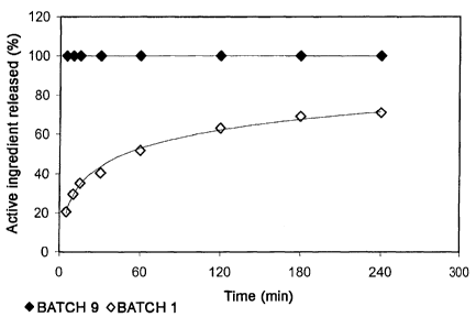
BRIEF DESCRIPTION OF THE DRAWINGS
Fig. 1 shows a comparative graph of the results of the in vitro release tests
of BATCH
9 and BATCH 1.
CA 02688047 2012-04-10
19
Fig. 2 shows the pharmacokinetics of human FVIII:C in rabbit plasma after the
administration of unencapsulated FVIII and after the application of the
composition.
Fig. 3 shows the pharmacokinetics of human VWF:Ag in rabbit plasma after the
administration of unencapsulated FVIII and after the application of the
composition.
Example 1
Preparation of the microparticles
The spray drying process has been used for the production of alginate
microparticles as
described in Erdinc B.I. [Erdinc B.I. (2007) Micro/nanoencapsulation of
proteins within
alginate/chitosan matrix by spray drying, Degree Thesis, Queen's University,
Kingston,
Canada]. Basically, microparticles were prepared by producing an emulsion with
the
polymer and the active ingredient chosen.
A BO chi Mini Spray Dryer* B-290 device was used for spraying the samples
under the
following conditions: spray temperature: 140 C-180 C, intake rate: 35-40 m3/h,
injection
flow rate: 3.5-5 ml/min and pressure 4-6 psi.
Example 2
Description of the microparticles
Tables 1, 2 and 3 describe the materials used in the manufacture of
microparticles and
their features, including size, Z potential and yield. The manufacturing
process and the
conditions used were as described in Example 1.
* trademark
CA 02688047 2012-04-10
19a
Table 1: Description of FVIII microparticles (plasmatic FVIII)
Mean particle size
Batch Polymer Z Potential (mV)
(pm)
BATCH 1
FVIII Sodium Alginate 3.6 -32
BATCH 2
FVIII Sodium Alginate 4.5 -32
BATCH 3
FVIII Sodium Alginate 4.7 -31
CA 02688047 2009-12-09
The FVIII activity/FVIII antigen ratio gives an idea of the proportion of
active protein in
a given sample. In this way, if we compare the activity/antigen ratio in the
initial
sample with that obtained in the encapsulated sample, we can calculate the
5 proportion of active ingredient which remains functional after
microencapsulation. In
the example, we found that the activity yields during the process of
encapsulation,
expressed as a percentage compared to the initial activity yield, are 57.6 %,
33.9 %
and 35.7 % for batches 1, 2 and 3 respectively.
10 Table 2: Description of FIX microparticles (plasmatic FIX)
Mean particle size
Batch Polymer Z Potential (mV)
(pm)
BATCH 4
FIX Sodium Alginate 4.9 -63
BATCH 5
FIX Sodium Alginate 4.5 -18
BATCH 6
FIX Sodium Alginate 4.9 -10
In this case, we found that the activity yields during the process of
encapsulation in
batches 4, 5 and 6, are 100 % in all said batches.
Table 3: Description of rFVIII microparticles (recombinant FVIII) and rFVIIa
(recombinant
FVIIa)
Mean particle
Batch Polymer size Z Potential (mV)
(pm)
BATCH 7
rFVIII Sodium Alginate 4.7 -70
BATCH 8
rFVll a Sodium Alginate 4.9 -64
CA 02688047 2012-04-10
21
In the case of proteins of recombinant origin, the activity yields determined
during the
process of encapsulation were of 25% and of 71 % for batches 7 and 8
respectively.
In all batches, the size of particle was determined with the Beckman Coulter*
LS13320
device through a diffraction laser and the Z Potential was measured with the
Malvern
Zetasizer* device.
The biological activity of FVIII was determined by deficient plasma clotting
assay or by
evaluating the generation of FXa by chromogenesis. In the case of FVIla and
FIX, the
biological activity was determined by evaluating the clotting time (partial
activated
thromboplastin time) of plasmas without FVII and FIX, respectively. The
protein
concentration was determined by the immunological detection method of enzyme-
linked
immunosorbent assay (ELISA) using specific antibodies against FVIII:Ag, FIX:Ag
or
FVII:Ag respectively.
The activity/antigen ratios, indicative of the proportion of active protein in
a given sample
were calculated by obtaining the quotient between the activity and antigen
units for the
specific active ingredient in the sample. The calculation of the
activity/antigen yield is
carried out by estimating the percentage of variation between the
activity/antigen ratios of
the starting sample and of the final encapsulated product.
As can be seen in all cases, the average particle size is less than or equal
to 5 Nm and the
Z Potential is negative. Also the results of activity/Ag yield indicate that
the biological
activity during the process is being maintained.
The various tables show that the controlled release system is suitable for
different active
ingredients.
Example 3
In Vitro Release Test
A controlled release test with a continuous flow cell is performed in a Sotax*
CE1 device in
closed circuit in order to evaluate the release of active ingredient.
* trademarks
CA 02688047 2009-12-09
22
The test was conducted at a temperature of 37 C with a flow rate of 7-25
ml/min
using as a dissolving medium an imidazole pH 7.3 buffer containing 1 % human
albumin. A representative sample was extracted for analysis at different times
(5
minutes, 10 minutes, 15 minutes, 30 minutes, 60 minutes, 120 minutes, 180
minutes
and 240 minutes). The volume of extracted sample was replaced with the same
volume of fresh medium in order to correct the loss of volume.
The biological activity of FVIII was determined by a deficient plasma clotting
assay or
by evaluating the generation of FXa by chromogenesis. In the case of FVIIa and
FIX,
the biological activity was determined by evaluating the clotting time
(partial activated
thromboplastin time) of plasma without FV1I and FIX, respectively. The protein
concentration was determined by the immunological detection method of enzyme-
linked immunosorbent assay (ELISA) using specific antibodies against FVIII:Ag,
FIX:Ag or FVII:Ag respectively.
After completion of the test the following results were obtained:
Table 4. In vitro release test of unencapsulated lyophilised FVIII (BATCH 9)
BATCH 9 (unencapsulated)
Time (min) FVIII:C released (%)
5 100
Table 5. In vitro release test of FVIII nanoparticles (BATCH 1)
BATCH I (encapsulated)
Time (min) FVIII:C released (%)
5 20.7
10 - 29.6
15 35.2
30 40.5
60 51.7
120 63.0
180 69.0
240 71.0
CA 02688047 2009-12-09
23
We can see that the composition of the microparticle applied to the active
ingredient
modifies the release kinetics of the product compared to unencapsulated
product.
Example 4
Pharmaco kinetics of Factor VIII in animals
In order to evaluate the effect of the composition on the release of active
ingredient in
vivo, a pharmacokinetics test was carried out on rabbits. For this, a dose of
501U/kg of
human FVIII from Batch 9 (not encapsulated) was administered intravenously to
three
female New Zealand White rabbits. Similarly, a dose of 501U/kg of encapsulated
FVIII
from Batch 1 as manufactured as described in example 1 and described according
to
example 2 was administered intravenously to a further three female New Zealand
White rabbits. At various times, plasma samples were obtained which were
analysed
to detect the presence of human FVIII:C, as described in Table 6. The
detection of
human FVIII was performed by chromogenesis after selective immunological
capture
of the human FVIII molecules. This allows the activity of infused human FVIII
to be
distinguished from that of rabbit FVII I.
Table 6. Pharmacokinetics of human FVIII:C in rabbit plasma after the
administration
2 0 of unencapsulated FVIII and after the application of the composition
FVIII FVIII microparticles
Time (unencapsulated) (encaps ulated)
(hours) BATCH 9 BATCH I
hFVIII:C (U/mi) hFVIII:C (U/ml)
0 0.018 0.024 0.046 0.012
0.5 0.931 0.069 0.459 0.186
2 0.678 0.236 0.534 0.158
6 0.238 0.165 0.346 0.076
12 0.054 0.062 0.243 0.005
24 0.023 0.027 0.090 0.008
36 0.022 0.024 0.073 0.009
49 0.021 0.026 0.033 0.011
CA 02688047 2012-04-10
24
We can see from the results that the composition delays the release of the
active
ingredient in plasma. In addition, these results demonstrate that there is no
cell
mechanism (liver, spleen, or macrophages) which rapidly removes the
microparticles from
the circulation, in spite of their size.
The analysis of this data using appropriate software for this purpose
(WinNonlin* 5.2)
allowed us to calculate the pharmacokinetic constants detailed in table 7.
Table 7. Pharmacokinetic parameter of human FVIII:C in rabbit plasma after the
administration of unencapsulated FVIII and after the application of the
composition
FBI FVIII
microparticles
(unencapsulated)
(encapsulated)
BATCH 9
BATCH 1
Half-life (h) 3.0 1.6 12.7 2.7
FVIII:C
Average residence
5.1 f 1.1 17.4 # 3.8
time (h)
Example 5
Pharmacokinetics of the von Willebrand factor in animals
Both in the case of the BATCH 9 preparation (unencapsulated FVIII) and in the
preparation of Batch 1 (encapsulated FVIII), the FVIII was of plasma origin
with a
significant content of von Willebrand factor (VWF). This means that the
encapsulation of
the VWF occurs at the same time as the encapsulation of FVIII. For this, their
behaviour
can be studied independently. For this we proceeded to independently analyse
the VWF
pharmacokinetics, by assessing the presence of the human VWF antigen (VWF:Ag)
in the
rabbit plasma. The results are shown in Table 8.
* trademark
CA 02688047 2012-04-10
Table 8. Pharmacokinetics of human VWF: Ag in rabbit plasma after the
administration of
the unencapsulated VWF and after the application of the composition
FVIII/VWF FVIIINWF microparticles
Time (hours) (unencapsulated) (encapsulated)
BATCH 9 BATCH 1
VWF:Ag (Ul/mi) VWF:Ag (Ul/mi)
0 0.000 0.000 0.000 0.000
0.5 0.859 0.193 1.053 0.048
2 0.552 0.247 0.862 0.055
6 0.150 0.080 0.384 0.106
12 0.033 0.022 0.207 0.031
24 0.005 0.002 0.040 0.005
36 0.001 0.000 0.019 0.008
49 0.001 0.000 0.009 0.005
We can see from the results that the composition delays the release of the
active
5 ingredient in plasma. In addition, these results demonstrate that there is
no cell
mechanism (liver, spleen, or macrophages) which rapidly removes the
microparticles from
the circulation, in spite of their size.
The analysis of this data using appropriate software for this purpose
(WinNonlin* 5.2)
10 allowed us to calculate the pharmacokinetic constants detailed in Table 9.
Table 9. Pharmacokinetic parameter of human VWF: Ag in rabbit plasma after the
administration of unencapsulated FVIII/VWF and after the application of the
composition
* trademark
CA 02688047 2009-12-09
26
FVIII Microparticles of
(unencapsulated) FVIII (encapsulated)
BATCH 9 BATCH I
Half-life
5.7 0.3 11.1 2.8
(h)
VWF:Ag Average
residence time 3.6 0.5 11.9 3.7
(h)
As can be observed, the encapsulation of the active ingredient, VWF in this
case,
significantly extends its half-life.
While the invention has been described for examples of preferred embodiments,
these should not be considered limitative of the invention which will be
defined by the
broader interpretation of the following claims.