Note: Descriptions are shown in the official language in which they were submitted.
CA 02688778 2014-12-30
SYSTEM AND METHOD FOR ULTRASONIC HARMONIC IMAGING
COPYRIGHT NOTICE
[0001] Copyright Laws. Verathone Incorporated. All Rights Reserved. A
portion of
the disclosure of this patent document contains material which is subject to
copyright protection.
The copyright owner has no objection to the facsimile reproduction by anyone
of the patent
document or the patent disclosure, as it appears in the Patent and Trademark
Office patent file or
records, but otherwise reserves all copyrights whatsoever.
CROSS REFERENCE TO RELATED APPLICATIONS
[0002] This application is related to the following U.S. patent
application publication
numbers:
[0003] U.S. 2009/0062644 Al;
[0004] U.S. 2008/0139938 Al;
[0005] U.S. 2008/0146932 Al;
100061 U.S. 2008/0249414 Al;
[0007] U.S. 2009/0112089 Al;
[0008] U.S. 2008/0139934 Al; and
[0009] U.S. 2008/0242985 Al.
FIELD OF THE INVENTION
[0010] Embodiments of the invention pertain to the image processing of
targeted regions
of interest scanned by ultrasound transceivers.
BACKGROUND OF THE INVENTION
[0011] Ultrasound imaging depending on Fast Fourier Transforms (FFT) may
lack the
CA 02688778 2014-12-30
2
needed spectral information to generate diagnostically useful images under
certain
circumstances. The deficiency inherent in some FFT procedures can be overcome
by using other
approaches.
SUMMARY OF THE PARTICULAR EMBODIMENTS
100121 Systems and methods utilizing artificial intelligence via a
harmonics analysis
kernel (HAK) algorithm using returning first and second echo wavelength
harmonics that arise
from differential and non-linear wavelength distortion and attenuation
experienced by transiting
ultrasound energy returning from a targeted region-of-interest (ROI). The HAK
algorithm is non-
parametric and is substantially less susceptible to modeling errors. Using the
harmonic ratios
with a sub-aperture algorithm provides diagnostically
useful images.
[0013] The sub-aperture algorithms are substantially fast enough to be
implemented in
real time within the time constraints enforced by ultrasound scanning
protocols to acquire organ
size information using scanning modalities besides the original ultrasound B-
mode images. The
harmonic information is collected using a long interrogating pulse with a
single fundamental
frequency. The received signal is collected, analyzed for its spectrum
information about the first
and second harmonics. The ratio of these two harmonics provides the
quantitative information on
how much harmonics have been generated and attenuated along its propagation.
The sub-
aperture algorithm may be executed in non-parametric mode to minimize data
modeling errors.
[0014] In an illustrative embodiment, a system for evaluating an organ of
a patient
includes an ultrasound transceiver configured to transmit to the organ at
least one ultrasound
pulse having a fundamental frequency, and receive at least one echo signal
corresponding to the
pulse. The system further includes a processing device coupled to the
transceiver, the processing
device including a computer-readable medium including instructions that, when
executed by the
processing device, enable the processing device to perform a method including
determining a
plurality of data segments associated with the at least one echo signal, and
independently
calculating, for each data segment of the plurality of data segments, a first
average of a first
harmonic of the fundamental frequency and a second average of a second
harmonic of the
fundamental frequency. The method further includes calculating a respective
ratio of the first
CA 02688778 2014-12-30
3
average to the second average for each data segment of the plurality of data
segments, and
generating an image of the organ based on the calculated ratios.
[0014a] In another illustrative embodiment, in a system configured to
transmit to an organ
of a patient at least one ultrasound pulse having a fundamental frequency and
receive at least one
echo signal corresponding to the pulse, a non-transitory computer-readable
medium includes
instructions that, when executed by a processing device, enable the processing
device to perform
a method of evaluating the organ, The method includes determining a plurality
of data segments
associated with the at least one echo signal, and independently calculating,
for each data segment
of the plurality of data segments, a first average of a first harmonic of the
fundamental frequency
and a second average of a second harmonic of the fundamental frequency. The
method further
includes calculating a respective ratio of the first average to the second
average for each data
segment of the plurality of data segments, and generating an image of the
organ based on the
calculated ratios.
BRIEF DESCRIPTION OF THE DRAWINGS
[0015] The file of this patent contains at least one drawing executed in
color. Copies of
the corresponding U.S. or PCT international application with color drawing(s)
will be provided
by the United States Patent and Trademark Office upon request and payment of
the necessary
fee. Embodiments for the system and method to develop, present, and use
clarity enhanced
ultrasound images are described below.
[0016] FIGURES 1A-D depict a partial schematic and a partial isometric
view of a
transceiver, a scan cone comprising a rotational array of scan planes, and a
scan plane of the
array of an ultrasound harmonic imaging system;
CA 02688778 2009-11-16
WO 2008/144452
PCT/US2008/063803
4
[0017] FIGURE 2A depicts a partial schematic and partial isometric and
side view of a transceiver, and a scan cone array comprised of 3D-distributed
scan
lines in alternate embodiment of an ultrasound harmonic imaging system;
[0018] FIGURE 2B depicts a partial isometric view of an ultrasound
harmonic bladder scanner system utilizing a transceiver probe and console
combination; The object 77B is shown on display 16. A different set of arrows
is
shown on 77A but the format is a little different.
[0019] FIGURE 3 is a schematic illustration of a server-accessed local
area network in communication with a plurality of ultrasound harmonic imaging
systems;
[0020] FIGURE 4 is a schematic illustration of the Internet in
communication with a plurality of ultrasound harmonic imaging systems;
[0021] FIGURE 5 schematically illustrates sub-algorithm processing
algorithm to obtain harmonic profile smoothing;
[0022] FIGURE 6A is a raw data image of a bladder region of a patient;
100231 FIGURE 6B is the ratio of the magnitude of the second harmonic
to the magnitude of the first harmonic using an FFT of the raw data image of
the
bladder image of FIGURE 6A;
[0024] FIGURE 6C is the ratio of the magnitude of the second harmonic
to the magnitude of the first harmonic frequency using the HAK of FIGURE 5
applied to the raw data image of the bladder image of FIGURE 6A;
[0025] FIGURE 7 is a panel of raw data images of a bladder region
obtained from a patient;
[0026] FIGURE 8 are profiles of the ratio of the magnitude of the
second harmonic to the first harmonic frequency based on the FFT of the
respective raw data images of the patient's bladder regions of FIGURE 7;
CA 02688778 2009-11-16
WO 2008/144452
PCT/US2008/063803
[0027] FIGURE 9 are profiles of the ratio of the magnitude of the
second harmonic to the first harmonic frequency based on the HAK of the raw
data images presented in the B-mode scan of FIGURE 7;
[0028] FIGURE 10A depicts a panel of four second harmonic profiles of
scan planes having theta angular values of 0, 15, 30, 45, 60, 75, 90, 105,
120, 135,
150, and 165 degrees;
[0029] FIGURE 10B depicts the twelve-second harmonic profiles of
FIGURE 10A arranged or aligned in three-dimensional space;
[0030] FIGURE 10C depicts a simulated C-mode top view of the
twelve-second harmonic parabolas projecting above the threshold plane;
100311 FIGURES 11A-15A illustrates a series of 2D plot of the FFT
processed second harmonics;
[0032] FIGURES 11B-15B illustrates the respective HAK companions
of FIGURES 11A-15A in which the series of 2D plots are HAK processed second
harmonics;
100331 FIGURES 16A-B are regression analysis of harmonic ratio vs.
bladder size plots of bladders not voided of urine;
[0034] FIGURES 17A-B are regression analysis of harmonic ratio vs.
bladder size plots in of bladders after voiding of urine; and
[0035] FIGURES 18A-C are regression analysis of measured urine
volume vs. HAK algorithm predicted volumes of a clinical group comprising 8
males and ten females.
DETAILED DESCRIPTION OF THE PARTICULAR EMBODIMENTS
[0036] Particular embodiments described include a system and method
to improve image clarity in ultrasound images that utilize an ultrasound
transceiver receiving ultrasound energy returning from a targeted region of
interest and producing a plurality of echoic signals. The region-of-interest
may
include an organ, an organ cavity, for example a bladder, or a portion of an
organ
CA 02688778 2009-11-16
WO 2008/144452
PCT/US2008/063803
6
or organ cavity. The echoic signals then receive signal processing via an
executable algorithm configured to image the targeted region-of-interest from
the
echoic signals using at least one of a first harmonic, a second harmonic, and
a
fundamental frequency of the ultrasound energy. The algorithm generates a
harmonic value that may then be plotted on a grid or render a map presentable
on
a computer display or other visual means. Alternate embodiments provide that
the
executable algorithm may be non-parametric and include a Harmonic Analysis
Kemal (HAK). The HAK includes a window process, a Fast Fourier Transform
process, an average process, a normalization of intensity process, a
compensation-
by-depth process, and a harmonic smoothing process to generate the harmonic
values. A map of the harmonic values then may be coded, for example, by color-
coding according to the magnitude of the harmonic value, to present an image
of
the region-of-interest.
100371 Other systems, methods, and devices are configured for
determining transducer functionality by using simulated body fluids, simulated
body tissue, and combination simulated body fluids and body tissues for
transducers having the characteristics of a 13 mm, 2.949 Mhz transducer in an
ultrasound transceiver, for example the 9400 device developed by Verathon ,
Inc.
The transducer tests compare drive signal to produced signal in various
environments (attenuating, non-attenuating, and a combination) and can also
help
us compare the produced signal to the received signal in the same various
environments. These transducer tests help quantify the functionality of the
13mm
transducers maximize the determination of transducer precision and accuracy.
[0038] The ultrasound transceivers or DCD devices developed by
Verathon , Inc. (formerly Diagnostic Ultrasound Inc.) are capable of
collecting in
vivo three-dimensional (3-D) cone-shaped ultrasound images of a patient. Based
on these 3-D ultrasound images, various applications have been developed such
as
bladder volume and mass estimation. The clarity of images from the DCD
CA 02688778 2009-11-16
WO 2008/144452
PCT/US2008/063803
7
ultrasound transceivers depends significantly upon the functionality,
precision,
and performance accuracy of the transducers used in the DCD ultrasound
transceivers.
[0039] During the data collection process initiated by DCD, a pulsed
ultrasound field is transmitted into the body, and the back-scattered "echoes"
are
detected as a one-dimensional (1-D) voltage trace, which is also referred to
as a
RF line. After envelope detection, a set of 1-D data samples is interpolated
to form
a two-dimensional (2-D) or 3-D ultrasound image.
100401 FIGURES 1A-D depicts a partial schematic and a partial
isometric view of a transceiver, a scan cone comprising a rotational array of
scan
planes, and a scan plane of the array of various ultrasound harmonic imaging
systems 60A-D capable of employing fundamental and/or harmonic ultrasound
frequencies further illustrated in FIGURES 3 and 4 below.
[0041] FIGURE 1A is a side elevation view of an ultrasound transceiver
10A that includes an inertial reference unit, according to an embodiment of
the
invention. The transceiver 10A includes a transceiver housing 18 having an
outwardly extending handle 12 suitably configured to allow a user to
manipulate
the transceiver 10A relative to a patient. Ultrasound transducers operating
within
the transceiver 10A may be equipped to collect and ready signals for
ultrasound
fundamental and/or harmonic frequency analysis.
[0042] The handle 12 includes a trigger 14 that allows the user to initiate
an ultrasound scan of a selected anatomical portion, and a cavity selector
(not
shown). The transceiver 10A also includes a transceiver dome 20 that contacts
a
surface portion of the patient when the selected anatomical portion is
scanned.
The dome 20 generally provides an appropriate acoustical impedance match to
the
anatomical portion and/or permits ultrasound energy to be properly focused as
it is
projected into the anatomical portion. The transceiver 10A further includes
one, or
preferably an array of separately excitable ultrasound transducer elements
(not
CA 02688778 2009-11-16
WO 2008/144452
PCT/US2008/063803
8
shown in FIGURE 1A) positioned within or otherwise adjacent with the housing
18. The transducer elements may be suitably positioned within the housing 18
or
otherwise to project ultrasound energy outwardly from the dome 20, and to
permit
reception of acoustic reflections generated by internal structures within the
anatomical portion. The one or more array of ultrasound elements may include a
one-dimensional, or a two-dimensional array of piezoelectric elements that may
be moved within the housing 18 by a motor. Alternately, the array may be
stationary with respect to the housing 18 so that the selected anatomical
region
may be scanned by selectively energizing the elements in the array.
[00431 A directional indicator panel 22 includes a plurality of arrows
that may be illuminated for initial targeting and guiding a user to access the
targeting of an organ or structure within an ROI. In particular embodiments if
the
organ or structure is centered from placement of the transceiver 10A
acoustically
placed against the dermal surface at a first location of the subject, the
directional
arrows may be not illuminated. If the organ is off-center, an arrow or set of
arrows may be illuminated to direct the user to reposition the transceiver 10A
at a
second or subsequent dermal location of the subject. The acrostic coupling may
be achieved by liquid sonic gel applied to the skin of the patient or by sonic
gel
pads to which the transceiver dome 20 is placed against. The directional
indicator
panel 22 may be presented on the display 54 of computer 52 in harmonic imaging
subsystems described in FIGURES 3 and 4 below, or alternatively, presented on
the transceiver display 16.
[00441 Transceiver 10A may include an inertial reference unit that
includes an accelerometer 22 and/or gyroscope 23 positioned preferably within
or
adjacent to housing 18. The accelerometer 22 may be operable to sense an
acceleration of the transceiver 10A, preferably relative to a coordinate
system,
while the gyroscope 23 may be operable to sense an angular velocity of the
transceiver 10A relative to the same or another coordinate system.
Accordingly,
CA 02688778 2014-12-30
9
the gyroscope 23 may be of conventional configuration that employs dynamic
elements, or it
may be an optoelectronie device, such as the known optical ring gyroscope. In
one embodiment,
the accelerometer 22 and the gyroscope 23 may include a commonly packaged
and/or solid-state
device. One suitable commonly packaged device may be the MT6 miniature
inertial
measurement unit, available from Omni Instruments, Incorporated, although
other suitable
alternatives exist. In other embodiments, the accelerometer 22 and/or the
gyroscope 23 may
include commonly packaged micro-electromechanical system (MEMS) devices, which
are
commercially available from MEMSense, Incorporated. As described in greater
detail below, the
accelerometer 22 and the gyroscope 23 cooperatively permit the determination
of positional
and/or angular changes relative to a known position that is proximate to an
anatomical region of
interest in the patient. Other configurations related to the accelerometer 22
and gyroscope 23
concerning transceivers 10AB equipped with inertial reference units and the
operations thereto
may be obtained from U.S. Patent Application Publication No. US 2007/0276247
Al.
[0045]
The transceiver 10A includes (or if capable at being in signal communication
with) a display 16 operable to view processed results from an ultrasound scan,
and/or to allow an
operational interaction between the user and the transceiver 10A. For example,
the display 24
may be configured to display alphanumeric data that indicates a proper and/or
an optimal
position of the transceiver 10A relative to the selected anatomical portion.
Display 16 may be
used to view two- or three-dimensional images of the selected anatomical
region. Accordingly,
the display 16 may be a liquid crystal display (LCD), a light emitting diode
(LED) display, a
cathode ray tube (CRT) display, or other suitable display devices operable to
present
alphanumeric data and/or graphical images to a user.
CA 02688778 2009-11-16
WO 2008/144452
PCT/US2008/063803
[0046] Still referring to FIGURE 1A, the cavity selector (not shown)
includes a pressable button similar to the trigger 14 may be operable to
adjustably
adapt thc transmission and reception of ultrasound signals to the anatomy of a
selected patient. In particular, the cavity selector adapts the transceiver
10A to
accommodate various anatomical details of male and female patients. For
example, when the cavity selector is adjusted to accommodate a male patient,
the
transceiver 10A may be suitably configured to locate a single cavity, such as
a
urinary bladder in the male patient. In contrast, when the cavity selector is
adjusted to accommodate a female patient, the transceiver 10A may be
configured
to image an anatomical portion having multiple cavities, such as a bodily
region
that includes a bladder and a uterus. Alternate embodiments of the transceiver
10A may include a cavity selector configured to select a single cavity
scanning
mode, or a multiple cavity-scanning mode that may be used with male and/or
female patients. The cavity selector may thus permit a single cavity region to
be
imaged, or a multiple cavity region, such as a region that includes a lung and
a
heart to be imaged.
[00471 To scan a selected anatomical portion of a patient, the transceiver
dome 20 of the transceiver 10A may be positioned against a surface portion of
a
patient that is proximate to the anatomical portion to be scanned. The user
actuates
the transceiver 10A by depressing the trigger 14. In response, the transceiver
10
transmits ultrasound signals into the body, and receives corresponding return
echo
signals that may be at least partially processed by the transceiver 10A to
generate
an ultrasound image of the selected anatomical portion. In a particular
embodiment, the transceiver 10A transmits ultrasound signals in a range that
extends from approximately about two megahertz (MHz) to approximately about
ten MHz. Ultrasound energies beyond 10 MHz may be utilized.
[0048] In one embodiment, the transceiver 10A may be operably
coupled to an ultrasound system that may be configured to generate ultrasound
CA 02688778 2009-11-16
WO 2008/144452
PCT/US2008/063803
11
energy at a predetermined frequency and/or pulse repetition rate and to
transfer
the ultrasound energy to the transceiver 10A. The system also includes a
processor that may be configured to process reflected ultrasound energy that
is
received by the transceiver 10A to produce an image of the scanned anatomical
region. Accordingly, the system generally includes a viewing device, such as a
cathode ray tube (CRT), a liquid crystal display (LCD), a plasma display
device,
or other similar display devices, that may be used to view the generated
image.
The system may also include one or more peripheral devices that cooperatively
assist the processor to control the operation of the transceiver 10A, such a
keyboard, a pointing device, or other similar devices. In still another
particular
embodiment, the transceiver 10A may be a self-contained device that includes a
microprocessor positioned within the housing 18 and software associated with
the
microprocessor to operably control the transceiver 10A, and to process the
reflected ultrasound energy to generate the ultrasound image. Accordingly, the
display 16 may be used to display the generated image and/or to view other
information associated with the operation of the transceiver 10A. For example,
the information may include alphanumeric data that indicates a preferred
position
of the transceiver 10A prior to performing a series of scans. In yet another
particular embodiment, the transceiver 10A may be operably coupled to a
general-
purpose computer, such as a laptop or a desktop computer that includes
software
that at least partially controls the operation of the transceiver 10A, and
also
includes software to process information transferred from the transceiver 10A,
so
that an image of the scanned anatomical region may be generated. The
transceiver
10A may also be optionally equipped with electrical contacts to make
communication with receiving cradles 50 as illustrated in FIGURES 3 and 4
below. Although transceiver 10A of FIGURE IA may be used in any of the
foregoing embodiments, other transceivers may also be used. For example, the
transceiver may lack one or more features of the transceiver 10A. For example,
a
CA 02688778 2009-11-16
WO 2008/144452
PCT/US2008/063803
12
suitable transceiver need not be a manually portable device, and/or need not
have
a top-mounted display, and/or may selectively lack other features or exhibit
further differences.
[0049] FIGURE 1B is a graphical representation of a plurality of scan
planes that form a three-dimensional (3D) array having a substantially conical
shape. An ultrasound scan cone 40 formed by a rotational array of two-
dimensional scan planes 42 projects outwardly from the dome 20 of the
transceivers 10A. Other transceiver embodiments of transceiver 10A may also be
configured to develop a scan cone 40 formed by a rotational array of two-
dimensional scan planes 42. The pluralities of scan planes 40 may be oriented
about an axis 11 extending through the transceivers 10A-B. One or more, or
preferably each of the scan planes 42 may be positioned about the axis 11,
preferably, but not necessarily at a predetermined angular position 0 . The
scan
planes 42 may be mutually spaced apart by angles 01 and 02. Correspondingly,
the scan lines within each of the scan planes 42 may be spaced apart by angles
0 1
and 02. Although the angles 01 and 02 are depicted as approximately equal, it
is
understood that the angles 0 1 and 0 2 may have different values. Similarly,
although the angles 0 1 and 02 are shown as approximately equal, the angles 0
1
and 02 may also have different angles. Other scan cone configurations are
possible. For example, a wedge-shaped scan cone, or other similar shapes may
be
generated by the transceiver 10A.
[0050] FIGURE IC is a graphical representation of a scan plane 42. The
scan plane 42 includes the peripheral scan lines 44 and 46, and an internal
scan
line 48 having a length r that extends outwardly from the transceivers 10A.
Thus,
a selected point along the peripheral scan lines 44 and 46 and the internal
scan line
48 may be defined with reference to the distance r and angular coordinate
values
0 and 9. The length r preferably extends to approximately 18 to 20 centimeters
(cm), although any length is possible. Particular
embodiments include
CA 02688778 2009-11-16
WO 2008/144452
PCT/US2008/063803
13
approximately seventy-seven scan lines 48 that extend outwardly from the dome
20, although any number of scan lines is possible.
[0051] As described above, the angular movement of the transducer
may be mechanically effected and/or it may be electronically or otherwise
generated. In either case, the number of lines 48 and the length of the lines
may
vary, so that the tilt angle sweeps through angles approximately between -60
and +60 for a total arc of approximately 120 . In one particular embodiment,
the
transceiver 10 may be configured to generate approximately about seventy-seven
scan lines between the first limiting scan line 44 and a second limiting scan
line
46. In another particular embodiment, each of the scan lines has a length of
approximately 18 to 20 centimeters (cm). The angular separation between
adjacent scan lines 48 (FIGURE 1B) may be uniform or non-uniform. For
example, and in another particular embodiment, the angular separation 0 1 and
02 (as shown in FIGURE 1C) may be about 1.5 . Alternately, and in another
particular embodiment, the angular separation 0 1 and 02 may be a sequence
wherein adjacent angles may be ordered to include angles of 1.50, 6.8 , 15.5 ,
7.2 ,
and so on, where a 1.50 separation is between a first scan line and a second
scan
line, a 6.8 separation is between the second scan line and a third scan line,
a 15.5
separation is between the third scan line and a fourth scan line, a 7.2
separation is
between the fourth scan line and a fifth scan line, and so on. The angular
separation between adjacent scan lines may also be a combination of uniform
and
non-uniform angular spacings, for example, a sequence of angles may be ordered
to include 1.50, 1.5 , 1.5 , 7.2 , 14.3', 20.2', 8.0 , 8.0 , 8.0 , 4.3 , 7.8 ,
and so on.
[0052] FIGURE ID a graphical representation of a plurality of scan lines
emanating from a hand-held ultrasound transceiver forming a single scan plane
42
extending through a cross-section of an internal bodily organ. The number and
location of the internal scan lines emanating from the transceivers 10A within
a
given scan plane 42 may thus be distributed at different positional
coordinates
CA 02688778 2009-11-16
WO 2008/144452
PCT/US2008/063803
14
about the axis line 11 as may be required to sufficiently visualize structures
or
images within the scan plane 42. As shown, four portions of an off-centered
region-of-interest (ROI) arc exhibited as irregular regions 49. Three portions
may
be viewable within the scan plane 42 in totality, and one may be truncated by
the
peripheral scan line 44.
10053] FIGURE 2A depicts a partial schematic and partial isometric and
side view of a transceiver, and a scan cone array comprised of 3D-distributed
scan
lines in alternate embodiment of an ultrasound harmonic imaging system. A
plurality of three-dimensional (3D) distributed scan lines emanating from a
transceiver that cooperatively forms a scan cone 30. Each of the scan lines
have a
length r that projects outwardly from the transceivers 10A-10E of FIGURES 1A-
1D. As illustrated the transceiver 10A emits 3D-distributed scan lines within
the
scan cone 30 that may be one-dimensional ultrasound A-lines. The other
transceiver embodiments 10B-10E may also be configured to emit 3D-distributed
scan lines. Taken as an aggregate, these 3D-distributed A-lines define the
conical
shape of the scan cone 30. The ultrasound scan cone 30 extends outwardly from
the dome 20 of the transceiver 10A, 10B and IOC centered about an axis line
11.
The 3D-distributed scan lines of the scan cone 30 include a plurality of
internal
and peripheral scan lines that may be distributed within a volume defined by a
perimeter of the scan cone 30. Accordingly, the peripheral scan lines 31A-31F
define an outer surface of the scan cone 30, while the internal scan lines 34A-
34C
may be distributed between the respective peripheral scan lines 31A-31F. Scan
line 34B may be generally collinear with the axis 11, and the scan cone 30 may
be
generally and coaxially centered on the axis line 11.
100541 The locations of the internal and peripheral scan lines may be
further defined by an angular spacing from the center scan line 34B and
between
internal and peripheral scan lines. The angular spacing between scan line 34B
and
peripheral or internal scan lines may be designated by angle 1:0 and angular
CA 02688778 2009-11-16
WO 2008/144452
PCMIS2008/063803
spacings between internal or peripheral scan lines may be designated by angle
0.
The angles 01, (1302, and 4:1)3 respectively define the angular spacings from
scan
line 34B to scan lines 34A, 34C, and 31D. Similarly, angles 01, 02, and 03
respectively define the angular spacings between scan line 31B and 31C, 31C
and
34A, and 31D and 31E.
[0055] With continued reference to FIGURE 2A, the plurality of
peripheral scan lines 31A-E and the plurality of internal scan lines 34A-D may
be
three dimensionally distributed A-lines (scan lines) that are not necessarily
confined within a scan plane, but instead may sweep throughout the internal
regions and along the periphery of the scan cone 30. Thus, a given point
within
the scan cone 30 may be identified by the coordinates r,(1), and 0 whose
values
generally vary. The number and location of the internal scan lines emanating
from the transceivers 10A-10E may thus be distributed within the scan cone 30
at
different positional coordinates as may be required to sufficiently visualize
structures or images within a region of interest (ROT) in a patient. The
angular
movement of the ultrasound transducer within the transceiver 10A-10E may be
mechanically effected, and/or it may be electronically generated. In any case,
the
number of lines and the length of the lines may be uniform or otherwise vary,
so
that angle 41) sweeps through angles approximately between -60 between scan
line 34B and 31A, and +60 between scan line 34B and 31B. Thus angle (I) in
this
example presents a total arc of approximately 120 . In one embodiment, the
transceiver 10A, 10B and 10C may be configured to generate a plurality of 3D-
distributed scan lines within the scan cone 30 having a length r of
approximately
18 to 20 centimeters (cm).
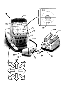
100561 FIGURE 2B illustrates a partial isometric and schematic view of
an ultrasound harmonic bladder scanner system 70 utilizing a transceiver probe
10C and console 74 combination 74. The harmonic bladder scanner system 70 is
battery powered and portable and may also be referred to as the BVI9400
CA 02688778 2009-11-16
WO 2008/144452
PCT/US2008/063803
16
BladderScan system. Other embodiments may include line power. The
ultrasound transceiver 10C is configured to detect and provide ultrasound
harmonic echo signals to which the console 74 may image process using neural
harmonic algorithms.
[0057] The transceiver 10C presents a similar transceiver display 16,
housing 18 and dome 20 design as transceivers 10A and 10B, and is in signal
communication to console 74 via signal cable 17. The console 74 may be pivoted
from console base 72. The console 74 includes a display 76, detection and
operation function panel 78, and select panel 80. The detection and operation
function provide for targeting the bladder, allow user voice annotation
recording,
retrieval and playback of previously recorded voice annotation files, and
current
and previously stored 3D and 2D scans. In the display 76 is screenshot 76
having
a targeting icon 79A with cross hairs centered in a cross sectional depiction
of a
bladder region. Other screen shots may appear in the display 76 depending on
which function key is pressed in the function panel 78. A targeting icon
screenshot 77B with a plurality of directional arrows may appear and flash to
guide the user to move the transceiver 10C to center the bladder and can
appear on
either display 76 or display 16. The targeting icon screenshot 77B similar
guides
the user to place the transceiver 10C to center the bladder or other organ of
interest as the directional indicator panel 22 depicted in FIGURE IA above. An
initial bladder view screenshot 77C may appear in which target icon 79A shows
a
central bladder region appearing within the cross hairs above the oval shaped
pubic bone. In wireless communication via wireless signal 82, the output from
the
transceiver 10C may be outputted to a wireless hub 84 via wireless signal port
86.
The wireless hub 84 also serves to charge batteries 88 for loading into the
battery
compartment (not shown) of console 74. All the calculations may be performed
in
the imaging console 74. The 9400 embodiment system 70 does not require a
computer or network to complete the harmonic imaging processing. In other
CA 02688778 2009-11-16
WO 2008/144452
PCT/US2008/063803
17
embodiments, the system 70 may utilize the wireless hub 84 as a gateway to
transmit transceiver 10C acquired harmonic imaging information in local and
Internet systems similar to those described in FIGURES 3 and 4 below.
[00581 FIGURE 3 is a schematic illustration of a server-accessed local
area network in communication with a plurality of ultrasound harmonic imaging
systems. An ultrasound harmonic imaging system 100 includes one or more
personal computer devices 52 that may be coupled to a server 56 by a
communications system 55. The devices 52 may be, in turn, coupled to one or
more ultrasound transceivers 10A and/or 10B, for examples the ultrasound
harmonic sub-systems 60A-60D. Ultrasound based images of organs or other
regions of interest derived from either the signals of echoes from fundamental
frequency ultrasound and/or harmonics thereof, may be shown within scan cone
30 or 40 presented on display 54. The server 56 may be operable to provide
additional processing of ultrasound information, or it may be coupled to still
other
servers (not shown in FIGURE 3) and devices. Transceivers 10A or 10B may be
in wireless communication with computer 52 in sub-system 60A, in wired signal
communication in sub-system 10B, in wireless communication with computer 52
via receiving cradle 50 in sub-system 10C, or in wired communication with
computer 52 via receiving cradle 50 in sub-system 10D.
[0059] FIGURE 4 is a schematic illustration of the Internet in
communication with a plurality of ultrasound harmonic imaging systems. An
Internet system 110 may be coupled or otherwise in communication with the
ultrasound harmonic sub-systems 60A-60D.
100601 The information obtainable from the scanning transceivers 10A
or 10B used in sub-systems 60A-60D are derived from ultrasound echoes that are
converted to signals received from structures in the body. These ultrasound
echo
signals carry not only the frequencies of the original transmit pulse, but
also
CA 02688778 2009-11-16
WO 2008/144452
PCT/US2008/063803
18
include multiples, or harmonics of these frequencies. These linear components
are
used in conventional, fundamental B-mode imaging.
[0061] In contrast, non-linear effects cause the harmonic echo
frequencies during the propagation of ultrasound through various mediums. For
example, THI (tissue harmonic imaging) is based on the phenomenon wherein
ultrasound signals are distorted while propagating through tissue with varying
acoustic properties. However, THI is merely an imaging method that does not
solve the bladder detection problem.
100621 Harmonic information is hidden in the frequency domain and it is
an effective indicator for harmonic build-up on each scan line at different
depth,
based on which bladder lines and tissue lines can be separated. For example,
inside a bladder region, there is not enough reflection, so the attenuations
of the
first and second harmonics are low. Deep behind the bladder wall, both the
first
and the second harmonics can be attenuated, while the second harmonic can be
attenuated much faster than the first one. As a result, harmonic information
can be
higher for a scan line which passes through a bladder, compared to a scan line
that
penetrates tissue only.
[0063] One way to use the harmonic information is to use relative
change of the harmonic information around the 2nd harmonic frequency compared
with response at fundamental frequency. The ratio (Goldberg Number) of the
peak
value around the 2nd harmonic and the peak value around the fundamental
frequency is a suitable indicator for such change.
[0064] From the clinical data collected from an ultrasound device, it can
be observed that its spectrum is very noisy. This holds true even when there
is
little or no noise presented within the data. The convolution theory indicates
that
it is hard to use conventional FFT method to get good spectral estimation, not
to
mention that the stationary assumption does not hold for this data. A robust
CA 02688778 2009-11-16
WO 2008/144452
PCT/US2008/063803
19
harmonic processing algorithm enables such a device to have good harmonic
estimation results.
[0065] FIGURE 5 depicts a method flow chart of a Harmonic Analysis
Kernel (HAK) 100 signal processing algorithm used to extract out information
of
a scanned region of interest of a subject. In general, such an approach may be
based on sub-aperture processing technology, and it can be approximately
regarded as a deconvolution process. The HAK 100 begins with process block 102
where a region-of-interest with the organ, or the whole organ, for example the
bladder, is targeted with ultrasound transceivers 10A-B and ultrasound echoes
are
retrieved and converted to signals. The signals are subdivided along scan
lines or
a series of data segments from 1 to N. Thereafter, at block 104, the Data
segments
1-N are processed. Each data segment 1 to N is independently processed by a
Window block 106, a Fast Fourier Transform block (FFT) block 108, and an
averaging processing block 110. After averaging, the pixel intensity is then
normalized by intensity at process block 112, compensated and averaged across
the depth of the scan line at process block 114, then outputted as a smoothed,
harmonic profile at process block 116 to complete the HAK algorithm 100.
[0066] The window-processing block 106 utilizes a signal processing
technique that applies a series of numerical weight to the echo signals
resulting in
a sub-signal set. The sub-signal set utilizes the Tailor window, a processing
algorithm that allows computational adjustments between the mainlobe width and
sidelobc levels common with signals exhibiting edge discontinuities.
Thereafter,
the FFT is applied at block 108, and the results thereof normalized with
regard to
the first harmonic spectrum intensity by taking the first harmonic spectrum
intensity average and dividing it by the second harmonic spectrum intensity
average. These calculations are then compensated with the expectation that a
predicted attenuation of 2.5 dB/cm occurs to the imaging and/or echoic
ultrasound
energies.
CA 02688778 2009-11-16
WO 2008/144452
PCT/US2008/063803
[0067] The resulting data segments from the deconvolution process can
be either overlapping or non-overlapping. For each data segment (on a single
radio frequency (RF) pulse ultrasound data line), a Taylor window is applied
to
reduce its sidelobes from the Fast Fourier Transform FFT 108. After the FFT
108,
an average of the spectrum around the first and the second harmonic
frequencies
is obtained at process block 110. Next, the normalization or compensation
process
is applied at block 112 to obtain an average the harmonic ratios based on the
following sub-al gori thm:
100681 Ratio Sum =0;
[0069] Counter = 0;
[0070] For each data segment(i)
[0071] If (first harmonic>threshold)
[0072] Ratio_SA(i) = 20*loglO(first
harmonic/second harmonic);
[0073] Ratio_SA(i) = Ratio_SA(i) + i*Att_Comp;
[0074] Ratio Sum = Ratio_Sum + Ratio_SA(i);
[0075] Counter = Counter +1;
[0076] End if
[0077] End for
[0078] If (Counter > 0)
[0079] Ratio = Ratio Sum/Counter;
[0080] Else
[0081] Ratio = Ratio low
[0082] End if
100831 In the above sub-algorithm, `Att_Comp' is an attenuation
compensation parameter (a value of 2.5dB/cm can be used as and estimate from
the clinical data). The 'threshold' is a parameter used to reject the data
when they
are too small. Ratio_low = -35dB. In summary, the 'normalization' step can
CA 02688778 2009-11-16
WO 2008/144452
PCT/US2008/063803
21
remove the data segments that are too weak, the compensation step can
compensate the harmonic ratio loss in tissue, and the averaging step can
provide a
more robust ratio estimator. The final step may be a spatial smoothing of the
harmonic ratios across the scan lines within a scan plane.
[0084] Clinical results obtained from a properly targeted scan in which
the bladder is substantially centered are described in FIGURES 6A-C and
FIGURES 7-9. A comparison is made of FFT and HAK processing of the scan
results.
100851 FIGURE 6A is a raw data image of a bladder region of a patient.
[0086] FIGURE 6B is the ratio of the magnitude of the second harmonic
to the magnitude of the first harmonic using an FFT of the raw data image of
the
bladder image of FIGURE 6A
[0087] FIGURE 6C depicts a histogram of the ratio of the magnitude of
the second harmonic to the magnitude of the first harmonic frequency using the
HAK of FIGURE 5 applied to the raw data image of the bladder image of
FIGURE 6A. Clearly the HAK's ratio is strongly correlated with the bladder,
while the FFT has poor ratio estimates for the entire scan plane. According to
this
harmonic ratio model, the harmonic ratio estimated from each scan line can be
related to the bladder size interrogated by this scan line.
[0088] FIGURE 7 is a panel of raw data images of a bladder region
obtained from a patient.
[0089] FIGURE 8 illustrate profiles of the ratio of the magnitude of the
second harmonic to the first harmonic frequency based on the FFT of the
respective raw data images of the patient's bladder regions of FIGURE 7.
100901 FIGURE 9 illustrates profiles of the ratio of the magnitude of the
second harmonic to the first harmonic frequency based on the HAK of the raw
data images presented in the B-mode scan of FIGURE 7. As with the HAK
profiles of FIGURE 6C, these profiles obtained by the HAK algorithm of
CA 02688778 2009-11-16
WO 2008/144452
PCT/US2008/063803
22
FIGURE 5 provide stronger correlations with the bladder depth, while the
respective FFT of FIGURE 8 demonstrates poor ratio estimates for each scan
plane.
[0091] The second harmonics from the FFT and the HAK profiles may
be plotted in 2-D presentations to enhance the visual delineation of a bladder
region within a given scan plane via a color-coding process. The color-coding
process is illustrated in FIGURES 10A-C.
100921 FIGURE 10A depicts a panel of four second harmonic profiles
of scan planes having theta angular values of 0, 15, 30, 45, 60, 75, 90, 105,
120,
135, 150, and 165 degrees. The second harmonic profiles present substantially
parabolic and normally distributed patterns having varying distribution widths
and
peak maxima locations.
[00931 FIGURE 10B depicts the twelve-second harmonic profiles of
FIGURE 10A arranged or aligned in three-dimensional space. The parabolic
profiles of each scan plane may be arranged in a scan cone plane array similar
to
scan cone 40 of FIGURE 1B. The second harmonic parabolas rise above the
threshold value represented as a gray plane.
[0094] FIGURE 10C depicts a simulated C-mode top view of the
twelve-second harmonic parabolas projecting above the threshold plane. The
threshold is represented in the gray plane.
[00951 FIGURES 11A-15A illustrates a series of 2D plot of the FFT
processed second harmonics and illustrates a diffusely appearing bladder which
is
hard to delineate and irregular and diffuse shapes presents greater
difficulties to
estimate bladder volume. The color legend in this figure set denotes red to be
the
highest harmonic, blue to be the lowest harmonic, and yellow to have a
harmonic
value of intermediate magnitude. The yellow regions encircling the red regions
correspond to bladders. In these FFT processed harmonics, blue like regions
CA 02688778 2009-11-16
WO 2008/144452
PCT/US2008/063803
23
appear within the red bladder regions, presenting gaps or other
discontinuities
within the expected bladder regions.
[0096] FIGURES 11B-15B illustrates the respective HAK companions
of FIGURES 11A-15A in which the series of 2D plots are HAK processed second
harmonics and illustrates a more delineated and compact appearing bladder
regions that lends itself to regular shapes that can allow more accurately
determined bladder volumes. The color legend in this figure set denotes red to
be
the highest harmonic, blue to be the lowest harmonic, and yellow regions
encircling red regions to correspond to bladders. In FIGURE 11B, a relatively
thicker or wider yellow band is seen circumscribing around a centrally located
red
region. This more pronounced yellow pathway region or band envelops the more
central and continuously located red region to denote with more certainty the
location of the bladder. In general the HAK processing shows significant
improvement in harmonic ratio estimations over that harmonics that are just
FFT
processed. There are virtually no blue like gaps in the HAK detected and
centrally located red regions where the expected bladder regions exhibit a
contiguous presence.
100971 FIGURES 16A-B illustrate regression analyses of harmonic ratio
vs. bladder size plots of bladders not voided of urine. Filled bladders
exhibit a
minimum of data outliers.
[0098] FIGURES 17A-B illustrate regression analyses of harmonic ratio
vs. bladder size plots in of bladders after voiding of urine. Empty bladders
exhibit
more data outliers than the filled bladders of FIGURES 16A-B.
[0099] FIGURES 18A-C illustrate regression analysis of measured urine
volume vs. HAK algorithm predicted volumes of a clinical group comprising 8
males and ten females. The slopes vary between 0.81 and 0.96 with strong
correlation coefficients R2 varying between 0.85 and approximately 0.94.
CA 02688778 2014-12-30
24
[00100]
While the preferred embodiments of the invention have been illustrated and
described, many changes can be made without departing from the scope of the
invention as
defined by the claims. For example, gelatinous masses may be used to modify
synthetic tissue
and combination fluid and tissue to further define and optimize the sub-
aperture neural network
algorithm. Thus, the described embodiments should be viewed as illustrative
only, and not as
limiting the invention as defined by the accompanying claims.