Note: Descriptions are shown in the official language in which they were submitted.
CA 02688972 2009-12-22
1
CATHETER WITH MULTIPLE ELECTRODE ASSEMBLIES
FOR USE AT OR NEAR TUBULAR REGIONS OF THE HEART
FIELD OF INVENTION
[0001] The present invention relates to an improved
electrophysiologic catheter that is
particularly useful for ablation and sensing electrical activity at or near a
tubular region of the heart.
BACKGROUND OF INVENTION
[0002] Cardiac an-ythmias, and atrial fibrillation in particular,
persist as common and
dangerous medical ailments, especially in the aging population. In patients
with normal sinus
rhythm, the heart, which is comprised of atrial, ventricular, and excitatory
conduction tissue, is
electrically excited to beat in a synchronous, patterned fashion. In patients
with cardiac arrythmias,
abnormal regions of cardiac tissue do not follow the synchronous beating cycle
associated with
normally conductive tissue as in patients with normal sinus rhythm. Instead,
the abnormal regions
of cardiac tissue aberrantly conduct to adjacent tissue, thereby disrupting
the cardiac cycle into an
asynchronous cardiac rhythm. Such abnormal conduction has been previously
known to occur at
various regions of the heart, such as, for example, in the region of the sino-
atrial (SA) node, along
the conduction pathways of the atrioyentricular (AV) node and the Bundle of
His, or in the cardiac
muscle tissue forming the walls of the ventricular and atrial cardiac
chambers.
[0003] Cardiac arrhythmias, including atrial arrhythmias, may be of a
multiwavelet reentrant
type, characterized by multiple asynchronous loops of electrical impulses that
are scattered about
the atrial chamber and are often self propagating. Alternatively, or in
addition to the multiwavelet
reenetrant type, cardiac arrhythmias may also have a focal origin, such as
when an isolated region
of tissue in an atrium fires autonomously in a rapid, repetitive fashion.
-1-
CA 02688972 2016-07-21
=
[0004] A host of clinical conditions may result from the irregular
cardiac function and resulting
hemodynamic abnormalities associated with atrial fibrillation, including
stroke, heart failure, and
other thromboembolic events. In fact, atrial fibrillation is believed to be a
significant cause of
cerebral stroke, wherein the abnormal hemodynamics in the left atrium caused
by the fibrillatory
wall motion precipitate the formation of thrombus within the atrial chamber. A
thromboembolism
is ultimately dislodged into the left ventricle, which thereafter pumps the
embolism into the
cerebral circulation where a stroke results. Accordingly, numerous procedures
for treating atrial
arrhythmias have been developed, including pharmacological, surgical, and
catheter ablation
procedures.
[0005] It has been found that by mapping the electrical properties of
the endocardium and the
heart volume, and selectively ablating cardiac tissue by application of
energy, it is sometimes
possible to cease or modify the propagation of unwanted electrical signals
from one portion of the
heart to another. The ablation process destroys the unwanted electrical
pathways by formation of
non-conducting lesions. Examples of catheter-based devices and treatment
methods have generally
targeted atrial segmentation with ablation catheter devices and methods
adapted to form linear or
curvilinear lesions in the wall tissue which defines the atrial chambers, such
as those disclosed in
U.S. Patent No 5,617,854 to Munsif, U.S. Patent No. 4,898,591 to Jang, et al.,
U.S. Patent
No. 5,487,385 to Avitall, and U.S. Patent No. 5,582,609 to Swanson. In
addition, various energy
delivery modalities have been disclosed for forming such atrial wall lesions,
and include use of
microwave, laser and more commonly, radiofrequency energies to create
conduction blocks along
the cardiac tissue wall, as disclosed in WO 93/20767 to Stem, et al., U.S.
Patent No. 5,104,393 to
Isner, et al. and U.S. Patent No. 5,575,766 to Swartz, et al., respectively.
-2-
CA 02688972 2016-07-21
[0006] In this two-step procedure--mapping followed by ablation--
electrical activity at points in
the heart is typically sensed and measured by advancing a catheter containing
one or more
electrical sensors into the heart, and acquiring data at a multiplicity of
points. These data are then
utilized to select the target areas at which ablation is to be performed.
[0007] Mapping and ablation in regions of or near the pulmonary veins
poses special
challenges due to the configuration of the ostia and surrounding tubular
tissue. Catheters have been
developed that are particularly useful for mapping and ablating the pulmonary
veins and other
tubular regions of or near the heart, including the ostium. U.S. Pat. Nos.
6,090,084 and 6,251,109
to Hassett et al., U.S. Pat. No. 6,117,101 to Diederich et al., U.S. Pat. No.
5,938,660 to Swartz et
al., U.S. Pat. Nos. 6,245,064 and 6,024,740 to Lesh et al., U.S. Pat. Nos.
5,971,983, 6,012,457
and 6,164,283 to Lesh, U.S. Pat. No. 6,004,269 to Crowley et al., and U.S.
Pat. No. 6,064,902 to
Haissaguerre et al. describe apparatus for tissue ablation to treat atrial
arrhythmia, primarily tissue
located within the pulmonary veins or on the ostia of the pulmonary veins.
Catheters having lasso,
open-spine or closed-spine (basket) assemblies are also known. Such catheters
are disclosed in, for
example, U.S. Patent Nos. 6,728,455, 6,973,339, 7,003,342, 7,142,903, and
7,412,273.
[0008] "Lasso" catheters are particularly useful during
circumferential ablations around the
ostium of the pulmonary veins. One technique utilizes one catheter for mapping
and finding
abnormal potentials and a second catheter for ablating the ostium. However,
during a procedure it
is desirable to have continuous feedback of the potential recordings or
electrograms (ECGs) inside
the pulmonary vein (PV) as a circumferential ablation is performed around the
vein's ostium.
Having feedback of the ECGs inside a pulmonary vein during PV ostium ablation
allows a user to
know whether the undesired potentials have been successfully blocked by the
circumferential
ablation. Currently, if the user desires real time ECG feedback from inside
the pulmonary vein
-3-
CA 02688972 2009-12-22
1
during a circumferential ablation, a third catheter is used. Accordingly, it
is desired that a single
catheter be adapted to both ablate and detect potentials, and in particular,
that a single catheter have
both a proximal electrode assembly for ablating an ostium and a distal
electrode assembly for
detecting potentials in the tubular region of the ablated ostium so that it is
possible to obtain ECG
signals inside a pulmonary vein when ablating around the ostium.
SUMMARY OF THE INVENTION
[0009] The present invention is directed to a catheter with ablation and
potential sensing
capabilities that is adapted for outer circumferential contact with an opening
of a tubular region and
inner circumferential contact within the tubular region. In one embodiment,
the present invention
provides a single catheter having both a proximal electrode assembly and a
distal electrode
assembly for ablation of an ostium and potential sensing inside the pulmonary
vein so that it is
possible to obtain ECG signals inside a pulmonary vein when ablating around
the ostium.
[0010] In a more detailed embodiment, the catheter has an elongated
catheter body and a
control handle at its proximal end. At its distal end is an electrode
structure comprising a distal
electrode assembly and a proximal electrode assembly. The distal electrode
assembly has an
elongated member defining a longitudinal axis and a plurality of spines
surrounding the member
and converging at their proximal and distal ends, where each spine has at
least one electrode and a
curvature so that the spine bows radially outwardly from the member. The
proximal electrode
assembly has a proximal electrode assembly has an elongated member configured
with a generally
radial portion and a generally circular portion generally transverse to the
catheter axis, where the
generally circular portion comprising a plurality of electrodes. The control
handle advantageously
allows a user to manipulate a tensile member for changing the curvature of the
spine. The catheter
may also have a deflectable section between the catheter body and the
electrode structure where the
-4-
CA 02688972 2009-12-22
1
control handle allows a user to manipulate a second tensile member for
deflecting the deflectable
section.
[0011] In a more detailed embodiment, the catheter may have electrodes on
the distal electrode
assembly that are adapted for sensing electrical activity in the heart while
having electrodes on the
proximal electrode assembly that are adapted for ablation. Moreover, the
electrode assemblies may
have shape-memory elements to help the assemblies retain their shape.
[0012] In another embodiment, the catheter has a control handle has
control members that
allow separate and independent control of tensile members to deflect the
intermediate section, to
expand a basket electrode assembly, and/or to contract a lasso electrode
assembly. In a detailed
embodiment, the control handle has a thumb control and a rotatable grip to
draw different puller,
deflection or contraction wires.
[0013] In a more detailed embodiment, the control handle has a handle
body, a core and a
piston that is longitudinally moveable relative to the core and handle body.
There are also a first
anchor fixedly mounted to the core, a cam receiver mounted within the handle
body, a second
anchor fixedly mounted to the cam receiver, and a cylindrical cam mounted
distal to the cam
receiver in surrounding relation to the piston, wherein rotation of the cam
relative to the piston
causes longitudinal movement of the cam receiver and second anchor.
BRIEF DESCRIPTION OF THE DRAWINGS
[0014] These and other features and advantages of the present
invention will be better
understood by reference to the following detailed description when considered
in conjunction with
the accompanying drawings wherein:
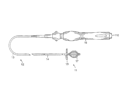
[0015] FIG. la is a top view of an embodiment of the catheter of the
present invention.
[0016] FIG. lb is a perspective view of an embodiment of an electrode
structure of the present
invention, including a proximal electrode assembly and a distal electrode
assembly, wherein the
-5-
CA 02688972 2009-12-22
1
distal electrode assembly is shown in a noi __ mai configuration (broken
lines) and in an expanded
configuration (solid lines).
[0017] FIG. 2 is side elevational view of an embodiment of an electrode
structure positioned
with a distal electrode assembly positioned in a tubular region of the heart
and a proximal electrode
assembly on an ostium of the tubular region.
[0018] FIG. 3a is a side cross-sectional view of an embodiment of a
catheter of the present
invention, including a junction between a catheter body and an intermediate
section along one
diameter.
[0019] FIG. 3b is a side cross-sectional view of an embodiment of a
catheter of the present
invention, including a junction between a catheter body and an intermediate
section along another
diameter.
[0020] FIG. 4 is an end cross-sectional view of an embodiment of an
intermediate section of
the catheter of the present invention.
[0021] FIG. 5a is an end view of an embodiment of an electrode
structure, including a proximal
electrode assembly and a distal electrode assembly.
[0022] FIG. 5b is a detailed view of an alternative embodiment of a
portion of an electrode
structure, including a ring electrode, thermocouple wires and a lead wire.
[0023] FIG. 6a is a side cross-sectional view of an embodiment of a
catheter of the present
invention, including a junction of an intermediate section and a connector
tubing, taken along one
diameter.
[0024] FIG. 6b is a side cross-sectional view of an embodiment of a
catheter of the present
invention, including a junction of an intermediate section and a connector
tubing, taken along
another diameter.
[0025] FIG. 6c is an end cross-sectional view of a connector tubing of
FIGs. 6a and 6b, taken
along line c--c.
-6-
CA 02688972 2009-12-22
1
[0026] FIG. 7a is a side cross sectional view of an embodiment of a
catheter of the present
invention, including a proximal end of a distal electrode assembly, taken
along one diameter.
[0027] FIG. 7b is a side cross sectional view of an embodiment of a
catheter of the present
invention, including a proximal end of a distal electrode assembly, taken
along another diameter.
[0028] FIG. 7c is an end cross-sectional view of a proximal end of a
distal electrode assembly
of FIGs 7a and 7b taken along line c--c.
[0029] FIG. 8a is a side cross sectional view of an embodiment a
catheter of the present
invention, including a distal end of a distal electrode assembly, taken along
one diameter.
[0030] FIG. 8b is a side cross sectional view of an embodiment of a
catheter of the present
invention, including a distal end of a distal electrode assembly, taken along
another diameter.
[0031] FIG. 8c is an end cross-sectional view of a distal end of a
distal electrode assembly of
FIGs. 8a and 8b, taken along line c--c.
[0032] FIG. 8d is an end cross-sectional view of a distal dome tip of FIGs.
8a and 8b, taken
along line d--d.
[0033] FIG. 9 is a side cross sectional view of an embodiment of a
control handle of the present
invention.
[0034] FIG. 10 is an exploded perspective view of interior components
of the control handle
shown in FIG. 9.
[0035] FIG. 11 is an enlarged side cross-sectional view of the
control handle of FIG. 9 showing
a deflection wire adjuster and a contraction wire adjuster.
DETAILED DESCRIPTION OF THE INVENTION
[0036] In a disclosed embodiment of the invention, there is provided a
catheter 10 having an
electrode structure 11 at its distal end. As shown in FIGs. la and lb, the
catheter comprises an
elongated catheter body 12 having proximal and distal ends, an intermediate
deflectable section 14
-7-
CA 02688972 2009-12-22
at the distal end of the catheter body, and a control handle 16 at the
proximal end of the catheter
body. The electrode structure 11 extending from the intermediate section 14
has a proximal
electrode assembly 15 and a distal electrode assembly 17. In the illustrated
embodiment with
reference to FIG. 2, the proximal electrode assembly 15 is lasso-shaped to sit
on an opening 19 of a
tubular region 21 of the heart, for example, an ostium of a pulmonary vein,
for circumferential
tissue contact at the opening. The distal electrode assembly 17 is basket-
shaped to extend past the
opening 19 and into the tubular region for circumferential tissue contact with
an inner surface 23 of
the tubular region. In that regard, the distal electrode assembly 17 is
expandable to a greater
diameter to ensure contact with the inner surface 23.
100371 With reference to FIGs. 3a and 3b, the catheter body 12
comprises an elongated tubular
construction having a single, axial or central lumen 18. The catheter body 12
is flexible, i.e.,
bendable, but substantially non-compressible along its length. The catheter
body 12 can be of any
suitable construction and made of any suitable material. A presently preferred
construction
comprises an outer wall 20 made of polyurethane or PEBAX. The outer wall 20
comprises an
embedded braided mesh of stainless steel or the like to increase torsional
stiffness of the catheter
body 12 so that, when the control handle 16 is rotated, the intermediate
section 14 of the
catheter 10 will rotate in a corresponding manner.
[0038] The outer diameter of the catheter body 12 is not critical, but is
preferably no more than
about 8 french, more preferably 7 french. Likewise the thickness of the outer
wall 20 is not critical,
but is thin enough so that the central lumen 18 can accommodate puller wires,
lead wires, and any
other desired wires, cables or tubings. If desired, the inner surface of the
outer wall 20 is lined with
a stiffening tube 22 to provide improved torsional stability. A disclosed
embodiment, the catheter
has an outer wall 20 with an outer diameter of from about 0.090 inch to about
0.94 inch and an
inner diameter of from about 0.061 inch to about 0.065 inch.
-8-
CA 02688972 2009-12-22
[0039] With additional reference to FIG. 4a, the intermediate section
14 comprises a short
section of tubing 13 having multiple lumens, for example three to five lumens.
In the disclosed
embodiment, there are lumens 24, 25, 26 and 27. The first lumen 24 carries
lead wires 30 for ring
electrodes of the lasso electrode assembly 15, lead wires 32 for ring
electrodes of the basket
electrode assembly 17, and thermocouple wires 41 and 45 for measuring
temperature, for example,
at the ring electrode(s), where the catheter is constructed for bipolar
ablation. The second lumen 25
carries a first tensile member or deflection wire 34 for deflecting the
intermediate section 14. The
third lumen 26 carries a cable 36 for an electromagnetic location sensor 33
located at or near the
electrode structure 11. The fourth lumen 27 carries a tubing 40 having a lumen
67 suitable for a
guidewire to pass, and through which a second tensile member or puller wire 47
extends for
expanding the basket electrode assembly 17.
[0040] The tubing 13 of the intermediate section 14 is made of a
suitable non-toxic material
that is preferably more flexible than the catheter body 12. A suitable
material for the tubing 13 is
braided PEBAX or polyurethane, i.e., polyurethane with an embedded mesh of
braided stainless
steel or the like. The size of each lumen is not critical, but is sufficient
to house the respective
components extending therethrough.
[0041] The useful length of the catheter, i.e., that portion that can
be inserted into the body
excluding the assemblies 15 and 17, can vary as desired. In one embodiment,
the useful length
ranges from about 110 cm to about 120 cm. The length of the intermediate
section 14 is a relatively
small portion of the useful length, and preferably ranges from about 3.5 cm to
about 10 cm, more
preferably about 4 cm to about 8 cm, and still more preferably about 6.5 cm.
[0042] A means for attaching the catheter body 12 to the intermediate
section 14 is illustrated
in FIGs. 3a and 3b. The proximal end of the intermediate section 14 comprises
an outer
circumferential notch 31 that receives the inner surface of the outer wall 20
of the catheter body 12.
The intermediate section 14 and catheter body 12 are attached by glue or the
like.
-9-
CA 02688972 2016-07-21
[0043] If desired, a spacer (not shown) can be located within the
catheter body between the
distal end of the stiffening tube (if provided) and the proximal end of the
intermediate section. The
spacer provides a transition in flexibility at the junction of the catheter
body and intermediate
section, which allows this junction to bend smoothly without folding or
kinking. A catheter having
such a spacer is described in U.S. Pat. No. 5,964,757.
[0044] At the distal end of the intermediate section 14 is the
electrode structure 11 having a
proximal assembly 15 adapted to sit on an opening of a tubular region, and a
distal assembly 17
adapted to enter the tubular region and contact the inner surface of the
tubular region (FIG. 2). The
assemblies 15 and 17 are generally concentric about the axis of the
deflectable intermediate
section 14. With reference to FIGs. lb and 5a, the proximal assembly 15
comprises a connecting
segment 38 and a generally circular main segment 39. The segment 38 is
generally straight and
extending radially from the distal end of the intermediate section 14. The
length of the segment 38
is about equal to the radius of the generally circular main segment 39 such
that the generally
circular main segment is generally concentric with the distal end of the
intermediate section 14.
The proximal assembly 15 has an exposed length, e.g., not contained within the
intermediate
section 14, ranging between about 20 mm and about 70 mm, more preferably about
25 mm and
about 50 mm, still more preferably about 42 mm, but can vary as desired.
[0045] The generally circular main segment 39 is generally traverse to the
catheter body 12 and
is preferably generally perpendicular to the catheter body 12. The generally
circular main segment
39 need not form a flat circle, but can be very slightly helical. The main
segment 39 has an exposed
length ranging between about 40mm and 100mm, more preferably about 50mm and
90mm, and
still more preferably about 60mm, and an outer diameter preferably ranging to
about 10 mm to
about 35 mm, more preferably about 15 mm to about 30 mm, still more preferably
about 25 mm.
-10-
CA 02688972 2009-12-22
1
The main segment 39 can curve in a clockwise direction, as shown in FIG. 6 or
a counterclockwise
direction, as shown in FIG. lb.
[0046] The proximal assembly 15 comprises a non-conductive covering or
tubing 50 (shown
partially broken away in FIG. 5a) that spans the length of the segments 38 and
39. The covering or
tubing 50 can be made of any suitable material that is flexible and
biocompatible and preferably
plastic, such as polyurethane or PEBAX. The tubing 50 (as with all tubes or
tubing herein) may
have any cross-sectional shape and may have a single lumen or multiple lumens.
The illustrated
embodiment, the tubing 50 has a single lumen that is occupied by lead wires 30
or other electrical
connections for ring electrodes 52 or any other electrical or electromagnetic
elements that may be
mounted on the proximal assembly 15. Moreover, the lumen is occupied by a
support element 53
that can have shape memory or be preformed with the radial and generally
circular shape. A shape
memory element can be straightened or bent out of its original shape upon
exertion of a force and is
capable of substantially returning to its original shape upon removal of the
force. A suitable
material for the shape memory element is a nickel/titanium alloy. Such alloys
typically comprise
about 55% nickel and 45% titanium, but may comprise from about 54% to about
57% nickel with
the balance being titanium. A preferred nickel/titanium alloy is nitinol,
which has excellent shape
memory, together with ductility, strength, corrosion resistance, electrical
resistivity and
temperature stability.
[0047] A means for attaching the tubing 50 of the proximal electrode
assembly 15 to the
catheter is illustrated in FIGs 4b and 6a. A nonconductive connector tubing 57
constructed of a
biocompatible materials, e.g., PEEK, with a single lumen, extends from the
distal end of the
tubing 13 of the intermediate section 14. An opening 58 is cut or otherwise
formed in the wall of
the tubing 57 to receive a proximal end of the tubing 50 which can extend
proximally into the
lumen 24 of the tubing 13 and be affixed by glue 60 which also seals the
opening 58. The lead
wires 30 for the proximal assembly 15 extend from the lumen 24 of the tubing
13 of the
-11-
CA 02688972 2009-12-22
intermediate section 14, and into the tubing 50 where they pass through the
radial segment 38 and
the generally circular main segment 39 of the proximal assembly 15. On the
generally circular
main segment 39 are mounted multiple ring electrodes 52, each connected to a
respective lead
wire 30 as shown in FIGs. 5a and 5b. The support member 53 also extends
through the length of
the tubing 50 to give shape and support to the segments 38 and 39 of the
proximal assembly 15. A
proximal end of the member 53 is anchored in the lumen 24 of the intermediate
section 14
(Fig. 6a).
100481 As shown in FIG. 5a, the distal end of the proximal assembly 15 is
sealed with a
dome 54 of polyurethane glue or the like. A short ring 56, made of metal or
plastic, and preferably
polyamidc, is mounted within the distal end of the non-conductive cover 50.
The short ring 56
prevents the distal end of the non-conductive cover 50 from collapsing, there
by maintaining the
diameter of the non-conductive cover at its distal end.
10049] As shown in FIGs. 6a-6c, the electromagnetic position sensor 33 is
housed in the
nonconductive connector tubing 57 as other components pass distally through
the tubing 57,
including the tubing 40 containing the puller wire 47 for the distal electrode
assembly 17 (both
from the lumen 27 of the tubing 13), the lead wires 32 (from the lumen 24) for
ring electrodes 64
mounted on the distal assembly 17. The cable 36 for the sensor 33 passes
through the lumen 26 of
the intermediate section 14.
100501 Distal the proximal electrode assembly 15 is the distal
electrode assembly 17. As
shown in FIGs lb, 7a-7c, the basket-shaped electrode assembly 17 extends
between two fasteners,
for example, nitinol rings 65 and 66, that define the proximal and distal ends
of the assembly 17.
The distal assembly 17 comprises a plurality of spines or arms 70 mounted,
preferably generally
evenly-spaced, around the tubing 40 which defines the longitudinal axis of the
distal assembly 17.
The spines have a convex curvature where each spine bows radially outwardly
from the tubing 40,
such that the spines converge at their distal and proximal ends at the rings
65 and 66.
-1%
CA 02688972 2009-12-22
1
100511 With reference to FIG. 5a, each spine 70 of the basket assembly
17 comprises a flexible
wire 72 (with or without shape memory) with a non-conductive covering or
tubing 71 on which one
or more ring electrodes 64 are mounted. In a preferred embodiment, the
flexible wires 72 each
comprise a flat Nitinol wire and the non-conductive tubing 71 each comprise a
biocompatible
plastic, such as polyurethane or PEBAX. The length of the tubings 71 is
shorter than the length of
the wires so that there are exposed proximal and distal ends of the wires not
covered by the tubings.
Alternatively, the spines 70 can be designed without the internal flexible
wire if a sufficiently rigid
non-conductive material is used for the non-conductive covering to permit
expansion of the
electrode assembly, so long as the spine has an outer surface that is non-
conductive over at least a
part of its surface for mounting of the ring electrodes 64. As will be
recognized by one skilled in
the art, the number of spines 70 can vary as desired depending on the
particular application, so that
the assembly has at least two spines, preferably at least three spines, and as
many as eight or more
spines. The term "basket-shaped" as used herein in describing the electrode
assembly 17 is not
limited to the depicted configuration, but can include other designs, such as
spherical or egg-shaped
designs, that include a plurality of expandable arms connected, directly or
indirectly, at their
proximal and distal ends.
[0052] An embodiment of the distal end of the electrode assembly 17 is
depicted in
FIGs. 8a 8c. The distal end of a distal ring 66 is sealed by a biocompatible
material, such as
polyurethane, which is formed into an atraumatic dome 95. The exposed distal
ends of the support
members 72 of the spines 70 extending pass the coverings 71 are affixed, e.g.,
by soldering 99,
preferably evenly-spaced, to an inner surface 97 of the ring 66. This junction
between the
spines 70, the tubing 40 and the proximal end of the ring 66 is sealed by a
biocompatible
material 93, such as polyurethane.
[0053] An embodiment of the proximal end of the electrode assembly 17
has a similar
construction, as shown in FIGs. 7a-7c, where the exposed proximal ends of the
support
-1:3-
CA 02688972 2009-12-22
1
members 72 are affixed to an inner surface 97 of the ring 65, e.g., by
soldering 99 or glue, and the
junction between the spines 70, the tubing 40 and the distal end of the ring
65 is sealed by a
biocompatible material 93. The rings 65 and 66 can be made of metal or
plastic, so long as it is
sufficient rigid to achieve the above-stated function. It is understood that
the spines can be formed
from a unitary structure, such as a cylinder or tube that is laser cut with
longitudinal cuts extending
between its two opposing ends to form the spines. As would be recognized by
one skilled in the
art, other arrangements for attaching and arranging the spines and tubing 40
could also be used in
accordance with the invention.
[0054] The tubing 40 is generally coaxial with the intermediate
section 14. The tubing 40 has a
distal end distal the distal ring 66 and a proximal end that is in the control
handle 16 such that its
lumen 67 provides a pathway for the second puller wire 47 between the control
handle 16 and the
distal assembly 17, as well as a pathway for a guidewire to extend through the
entire length of the
catheter for introduction of the catheter into a patient's body. Accordingly,
the tubing 40 extends
proximally through the rings 66 and 65, the connector tubing 57, the lumen 27
of the intermediate
section 14, the central lumen of the catheter body 12, and the control handle
16.
[0055] The puller wire 47 for expanding the distal basket assembly 17
can made of any suitable
metal, such as stainless steel or Nitinol, and is preferably coated with
Teflon® or the like. The
coating imparts lubricity to the puller wire. The puller wire preferably has a
diameter ranging from
about 0.006 to about 0.010 inch. The puller wire 47 is anchored at its
proximal end in the control
handle 16 and extends distally through the central lumen 18 of the catheter
shaft 12 and the fourth
lumen 27 of the intermediate section 14.
[0056] The distal end of the puller wire 47 is anchored in a distal
tip 80 at the distal ring 66 by
means of a T-shaped anchor 81 with a short stainless steel tubing crimped onto
the puller wire 47,
and a welded cross-piece 82 that is distal of the distal ring 66 and extends
the width of the ring 66.
So anchored against the ring 66, the puller wire 47 can be manipulated via the
control handle 16 as
-14-
CA 02688972 2009-12-22
1
described further below, thereby changing the curvature of the spines 70. In
particular, as the
puller wire is drawn proximally, the tubing 40 between the rings 65 and 65 is
compressed thereby
decreasing the separation between the rings 65 and 66, thus expanding
(widening) the basket
assembly 17 as the spines 70 bow further outwardly under the compression force
applied by the
puller wire 47. As shown in FIG. lb, the basket-shaped assembly 17 can be
varied between (and to
adopt either of) a more elongated or resting configuration with a smaller
diameter (broken lines)
and an expanded configuration with a greater diameter (solid lines). The
largest diameter at a mid-
section of the basket assembly 17 can range between about 10 mm and 30 mm, and
preferably
between about 15 mm and 25 mm.
[0057] Each of the ring electrodes 52 and 64 of the electrode
assemblies 15 and 17 is
electrically connected to an appropriate mapping or monitoring system and/or
source of ablation
energy by means of respective electrode lead wires 30 and 32. Each electrode
lead wire has its
proximal end terminating in a connector 111 (FIG. 1) at the proximal end of
the control handle 16.
Distally, the electrode lead wires extend through the control handle 16, the
central lumen 18 in the
catheter body 12, and through the lumen 24 of the intermediate section 14. The
portion of the lead
wires 30 and 32 extending through the central lumen 18 of the catheter body
12, control handle 16
and proximal end of the lumen 24 are enclosed within a protective sheath (not
shown) , which can
be made of any suitable material, preferably polyimide. The protective sheath
can be anchored at its
distal end to the proximal end of the intermediate section 14 by gluing it in
the lumen 24 with
polyurethane glue or the like.
[00581 Near the distal end of the intermediate section 14, the lead
wires 30 for the lasso
electrode assembly 15 and the lead wires 32 for the basket electrode assembly
17 diverge with the
lead wires 30 entering the tubing 50 of the electrode assembly 15. The lead
wires 32 for the basket
electrode assembly 17 however extend out of the lumen 24, through the
connector tubing 57,
-15-
CA 02688972 2009-12-22
1
through the proximal ring 65 and through their respective covering 71 of the
spines 71 of the
assembly 17. Each lead wire is attached to its corresponding ring electrode by
any suitable method.
[0059] A preferred method for attaching a lead wire to a ring electrode
involves first making a
small hole through the wall of the non-conductive covering. Such a hole can be
created, for
example, by inserting a needle through the non-conductive covering and heating
the needle
sufficiently to form a permanent hole. The lead wire is then drawn through the
hole by using a
microhook or the like. The end of the lead wire is then stripped of any
coating and welded to the
underside of the ring electrode, which is then slid into position over the
hole and fixed in place with
polyurethane glue 91 or the like (FIG. 5b). Alternatively, each ring electrode
is formed by
wrapping a lead wire around the non-conductive covering a number of times and
stripping the lead
wire of its own insulated coating on its outwardly facing surfaces.
[00601 The ring electrodes can be made of any suitable solid
conductive material, such as
platinum or gold, preferably a combination of platinum and iridium, and
mounted onto the tubing
with glue or the like. Alternatively, the ring electrodes can be formed by
coating the tubing with an
electrically conducting material, like platinum, gold and/or iridium. The
coating can be applied
using sputtering, ion beam deposition or an equivalent technique. While
unipolar ring electrodes
are illustrated herein, it is understood that bi-polar ring electrodes may be
used.
[0061] The number of the ring electrodes on the assemblies can vary as
desired. Preferably, the
number of ring electrodes on the lasso assembly 15 ranges from about six to
about twenty,
preferably from about eight to about twelve, evenly spaced from each other.
For the basket
assembly 17, the number of ring electrodes on each spine ranges from about one
to about four,
preferably about three that are more concentrated in the outermost region of
each spine. In a
disclosed embodiment, a distance of approximately 5 mm is provided between
each ring electrodes
on the lasso assembly 15 and a distance of approximately 2 mm is provided
between each ring
electrode on each spine of the basket assembly.
-16-
CA 02688972 2009-12-22
1
[0062] Where any of the ring electrodes of the assemblies 15 and 17
are adapted for ablation, a
pair of thermocouple wires can be provided to detect temperature of a
respective ring electrode. In
the disclosed embodiment, one pair of' thermocouple wires 41 and 45 are
provided, for example, for
one of the ring electrodes of the proximal electrode assembly 15. The
thermocouple wires 41
and 45 extend through the central lumen 18 of the catheter body 12 (FIG. 3A),
through the
lumen 26 of the tubing 13 of the intermediate section 14 (FIG. 4a), and
through the tubing 50 of the
proximal electrode assembly 15, where their distal ends are positioned near
the ring electrode to
sense temperature (FIG. 6a).
[0063] The deflection wire 34 for deflection of the intermediate
shaft 14 has many similarities
to the basket assembly puller wire 47 as described above. Some of the
differences are described
below.
[0064] The deflection wire 34 is anchored at its proximal end in the
control handle 16 and
extends distally through the central lumen 18of the catheter shaft 12 and the
second lumen 25 of
the intermediate section 14 (FIG. 4a) where its distal end is anchored to the
distal end of the
intermediate section 14, as shown in FIG. 6b. Specifically, a T-shaped anchor
is formed, which
comprises a short piece of tubular stainless steel 43 , e.g., hypodermic
stock, which is fitted over
the distal end of the deflection wire crimped to fixedly secure it to the
puller wire. The distal end of
the tubular stainless steel 43 is fixedly attached, e.g., by welding, to a
cross-piece 44 formed of
stainless steel ribbon or the like. The cross-piece 44 extends through a hole
46 formed in the
tubing 13 and because the cross-piece 44 is larger than the hole 46 and,
therefore, cannot be pulled
through the hole, the cross-piece 44 anchors the distal end of the deflection
wire 34 to the distal end
of the intermediate section 14.
[0065] A compression coil 35 is situated within the catheter body 12 in
surrounding relation to
the deflection wire 34. In the disclosed embodiment, the compression coil 35
extends from the
proximal end of the catheter body 12 to the proximal end of the intermediate
section 14 (see
-17-
CA 02688972 2016-07-21
=
Fig. 3b). The compression coil 35 is made of any suitable metal, preferably
stainless steel, and is
tightly wound on itself to provide flexibility, i.e., bending, but to resist
compression. The inner
diameter of the compression coil is preferably slightly larger than the
diameter of the deflection
wire 34. The Teflon® coating on the deflection wire 34 allows it to slide
freely within the
compression coil. Within the catheter body 12, the outer surface of the
compression coil 35 is also
covered by a flexible, non-conductive sheath 68, e.g., made of polyimide
tubing. The compression
coil is anchored at its proximal end to the outer wall 20 of the catheter body
12 by a proximal glue
joint and to the intermediate shaft 14 by a distal glue joint. Within the
lumen 25 of the intermediate
shaft 14, the deflection wire 34 extends through a plastic, preferably
Teflon®, puller wire
sheath 37, which prevents the deflection wire 34 from cutting into the wall of
the tubing 13 when
the intermediate section 14 is deflected.
[0066] A compression coil 103 is also provided for the puller wire 47
extending through the
tubing 40. In the disclosed embodiment, the distal end of the coil 103 is in
the connector tubing 57,
a few millimeters distal of the location of the opening 58. The proximal end
of the compression
coil 103 is at or near the proximal end of the catheter body 12. A tubing 101
surrounds the puller
wire 47 within the compression coil 103. The tubing 101 may be a tight fitting
tubing of TEFLON.
[0067] Separate and independent longitudinal movement of the
deflection wire 34 and the
puller wire 47 relative to the catheter body 12, which results in,
respectively, deflection of the
intermediate section 14 and expansion of the distal electrode assembly 17, is
accomplished by
suitable manipulation of the control handle 16. A suitable control handle is
disclosed in U.S.
Patent No. 6987995 to Drysen entitled Multifunctional Catheter Handle. As
shown in FIGs. 1 and
9, the control handle 16 has a thumb control knob 184, and a cam 120 rotatable
by means of a
flexible grip 128 that can be independently manipulated by a user.
-18-
CA 02688972 2009-12-22
1
100681
In the embodiment of FIGS. 9 to 11, the control handle 16 includes a
handle body 174
in which a core 176 is fixedly mounted. Although in the depicted embodiment,
the core 176 is
separate from the handle body 174, the core could instead be formed as a
single unitary piece with
the handle body. The core has a generally cylindrical distal region 175 and a
generally cylindrical
proximal region 177 having a larger diameter than the distal region. For
longitudinal movement of
the deflection wire 34, a piston 182 is slidably mounted over the distal
region 177 of the core 176.
The proximal end of the piston 182 is maintained within the handle body 174,
and the distal end of
the piston extends outside the handle body. The thumb knob 184 is mounted in
surrounding relation
to a portion of the distal end of the piston 182 so that the user can more
easily move the piston
longitudinally relative to the core 176 and handle body 174. The proximal end
of the catheter
body 12 is fixedly mounted to the distal end of the piston 182 through a tip
portion 178 that is
mounted on the distal end of the piston. The proximal end of the catheter body
12 is inserted into
an axial passage 180 in the tip portion and optionally glued in place. The
piston includes an axial
passage 186 in communication with the axial passage 180 of the tip portion
178, and the core 176
includes an axial passage 188 in communication with the axial passage in the
piston.
100691
The lead wires 30 and 32 (not shown for better clarity of other
components in the
control handle), the puller wire 47 and deflection wire 34 that extend through
the catheter body 12
extend out the proximal end of the catheter body and through the axial
passages in the tip
portion 178, piston 182 and core 176. The lead wires can extend out the
proximal end of the
control handle 16 or can be connected to a connector (not shown) that is
incorporated into the
control handle, as is generally known in the art.
100701
The proximal end of the deflection wire 34 is anchored to the core
176. As best seen in
FIG. 11, the portion of the axial passage 188 extending through the proximal
region 177 of the
core 176 has a larger diameter than the portion of the axial passage extending
through the distal
region 175 of the core 176. A deflection wire adjuster 190 is adjustably
mounted, as described
-19-
CA 02688972 2009-12-22
further below, in a portion of the axial passage 188 near the distal end of
the proximal region 177
of the core 176. The deflection wire adjuster 190 has an opening 192 extending
therethrough in a
direction generally transverse, and preferably generally perpendicular, to the
axial passage 188 of
the core 176. The deflection wire 34 extends through the opening 192 in the
deflection wire
adjuster 190 such that the deflection wire changes directions.
[0071] The distal region 177 of the core 176 includes a generally
rectangular opening 194 that
extends generally parallel to the axial passage 188 of the core. A channel 196
connects the
proximal end of the generally rectangular opening 194 to the distal end of the
portion of the axial
passage 188 in the proximal region 175 of the core 176. The proximal end of
the deflection
wire 164 extends through the channel 196 and into the generally rectangular
opening 194. A
deflection wire anchor 198, which can comprise a short piece of hypodermic
stock, is fixedly
attached, for example, by crimping, to a portion of the proximal end of the
deflection wire 164
within the generally rectangular opening 194. The deflection wire anchor 198
has a diameter
greater than the width of the channel 196 and thus prevents the proximal end
of the deflection
wire 34 from being pulled through the channel, thereby anchoring the
deflection wire to the
core 176. Thus, the deflection wire anchor 198 is fixedly mounted to the core
176 even though the
deflection wire anchor still has a small amount of free play within the
opening 194.
[0072] In use, the piston 182 is moved distally relative to the handle body
74 and core 176 by
means of the thumb knob 184, thereby pulling the catheter body 12 distally
relative to the
deflection wire 34, which is anchored to the core. As a result, the deflection
wire 34 pulls on the
side of the intermediate shaft 14 to which it is anchored, thereby deflecting
the distal shaft in that
direction. To straighten the intermediate shaft 14, the piston 182 is moved
proximally back to its
original position relative to the handle body 174 and core 176.
[0073] Manipulation of the deflection wire adjuster 190 adjusts the
amount of free play in the
deflection wire 34. As noted above, the deflection wire adjuster 190 is
adjustably mounted in a
-20-
CA 02688972 2009-12-22
1
portion of the axial passage 188 near the distal end of the proximal region
177 of the core 176. The
portion of the axial passage 88 in which the deflection wire adjuster 190 is
mounted includes a
series of ridges 100 extending along the surface of the core 176, with the
ridges being generally
perpendicular to the axis of the core. The deflection wire adjuster 190
carries an outwardly
extending tab 102 that fits in the spaces between the ridges 100. The
deflection wire adjuster 190
can be moved along the length of the core 176 and snapped into place by
placing the tab 102
between two ridges 100. As the deflection wire adjuster 190 is moved
proximally (away from
catheter body 12) less free play is provided for the deflection wire 34. The
precise mechanism for
adjusting the amount of free play of the deflection wire 34 is not critical,
and alternative
mechanisms can be provided. Alternatively, the deflection wire 34 can be
anchored directly to the
core 176 so that it is not adjustable.
[0074] The control handle 16 is also used for longitudinal movement
of the puller wire 47 for
expanding the basket assembly 17 by means of the flexible grip 128. The puller
wire 47 extends
from the catheter body 12, through the axial passage 186 in the piston 182 and
through the axial
passage 188 within the distal region 175 of the core 176. The proximal end of
the puller wire 47 is
anchored to a contraction wire adjuster 104 that is slidably mounted in the
core 176.
[0075] The puller wire adjuster 104 is generally rectangular having a
bottom region 108 that
extends downward through a slot 110 in the proximal region 177 of the core
176, the slot being in
communication with the axial passage 188 of the core. The proximal end of the
puller wire 47,
which, as noted above, extends through the axial passage 188, is anchored in
the puller wire
adjuster 104 in a manner very similar to the manner in which the deflection
wire 164 is anchored to
the core 176, as described above. Specifically, a puller wire anchor 108,
which can comprise a
short piece of hypodermic stock, is fixedly attached, for example, by
crimping, to a portion of the
proximal end of the puller wire 47 within an opening 110 in the puller wire
adjuster 104. A
channel 112 connects the opening 110 to the axial passage 88 in the core. The
puller wire anchor
-21-
CA 02688972 2009-12-22
1
98 has a diameter greater than the width of the channel 112 and thus prevents
the proximal end of
the puller wire 47 from being pulled through the channel, thereby anchoring
the puller wire to the
puller wire adjuster 104. The distal end of the puller wire adjuster 104 is
adjustably attached to a
cam receiver 106. The cam receiver 106 is generally tubular, having a short
slot 114 extending
from its proximal end sized to receive the distal end of the puller wire
adjuster 104. The cam
receiver 106 is slidably mounted over the piston 182 and the distal region 175
of the core 176 with
the bottom portion of the puller wire adjuster 104 positioned in the slot 114
in the core and a
corresponding slot 115 in the piston. Thus, the puller wire anchor 98 is
fixedly mounted to the cam
receiver 106 through the puller wire adjuster 104, even though the puller wire
anchor has some free
play within the opening 110 in the puller wire adjuster.
[0076] As shown in FIG. 10, the top of the distal end of the puller
wire adjuster 104 includes a
series of outwardly extending teeth 116 that mate with a plurality of notches
118 within the
slot 114 of the cam receiver 106 so that the puller wire adjuster can be
snapped into the cam
receiver. The position of the puller wire adjuster 104 relative to the cam
receiver 106 can be
longitudinally adjusted by repositioning the teeth 116 relative to the notches
118, to thereby adjust
the tension on the puller wire 47. Alternatively, the puller wire 40 is not
adjustable, in which case
the puller wire anchor 98 is mounted within an opening (not shown) within the
cam receiver 106.
[0077] Longitudinal movement of the cam receiver 106 and puller wire
adjuster 104 relative to
the core 76, to which the catheter body 12 is indirectly mounted, results in
longitudinal movement
of the puller wire 47 relative to the catheter body. Longitudinal movement of
the cam receiver 106
is accomplished through a cam 120 mounted in the control handle 16 in
surrounding relation to the
piston 182 and distal region 175 of the core 176. A retaining ring 121
maintains the longitudinal
position of the cam 120 relative to the handle body 74.
[0078] The cam 120 includes a ramped proximal surface 122. The cam
receiver 106 includes a
ramped distal surface 123 and an outwardly extending tab 124 at the most
distal point of the
-22-
CA 02688972 2009-12-22
1
ramped distal surface. The tab 124 contacts the ramped proximal surface 122 of
the cam 120. When
the cam 120 is rotated counterclockwise, the ramped proximal surface 112
correspondingly rotates
and pushes the cam receiver 104 proximally relative to the core 176 and
catheter body 12. As the
cam receiver 104 and the attached puller wire adjuster 104 are moved
proximally relative to the
core 176 and catheter body 12, the puller wire 47 is pulled proximally to
thereby expand the basket
assembly 17.
[0079] The ramped proximal surface 122 of the cam 120 includes an
outwardly extending
tab 126 at its most proximal point. As the cam 120 is rotated
counterclockwise, the tab 124 on the
cam receiver 104 contacts the tab 126 on the ramped proximal surface 122,
thereby prohibiting
further rotation of the cam relative to the cam receiver. As the cam 120 is
rotated clockwise, the
tab 126 on the ramped proximal surface 122 pushes the tab 124 on the cam
receiver 104 such that
the cam receiver moves distally, thereby releasing the tension on the puller
wire 47 so that the
basket assembly 17 returns to its original configuration. As would be
recognized by one skilled in
the art, the direction of the ramped proximal surface 122 can be changed so
that clockwise rotation
of the cam 120 causes expansion of the basket assembly and counterclockwise
rotation causes it to
return to its original configuration. The flexible grip 128 is provided over
the cam 120 for the user
to more easily and comfortably rotate the cam 120.
[0080] In use, a suitable guiding sheath is inserted into the patient with
its distal end positioned
at a desired tubular region of the heart such as a pulmonary vein. An example
of a suitable guiding
sheath for use in connection with the present invention is the Preface.TM.
Braiding Guiding
Sheath, commercially available from 13iosense Webster, Inc. (Diamond Bar,
Calif.). The distal end
of the sheath is guided toward the ostium of the pulmonary vein and a catheter
of the present
invention is fed through the guiding sheath until its distal and proximal
electrode assemblies 15
and 17 both extend out of the distal end of the guiding sheath. As the
catheter is fed through the
guiding sheath, the spines of the basket assembly 17 are pressed inwardly
toward the tubing 40 so
-23-
CA 02688972 2009-12-22
1
that the assembly 17 adopts a more elongated profile, and the lasso assembly
15 is straightened
with the distal dome end 54 leading through the sheath. Once the distal tip 80
of the catheter is
positioned at the desired treatment location, the guiding sheath is pulled
proximally, exposing the
deflectable intermediate section 14 and the assemblies 15 and 17 to extend
outside the sheath,
whereupon the assemblies return to their original shapes due to the shape-
memory of the support
members 53 and 72. The user can manipulate the thumb control 184 of the
control handle 16 to
deflect the intermediate section 14 for positioning the assemblies 15 and 17
as appropriate. With
proper manipulation, the basket assembly 17 is inserted into a pulmonary vein
or other tubular
region (such as the coronary sinus, superior vena cava, or inferior vena cava)
so that the lasso
assembly 15 comes into contact and sits on the ostium and the electrodes 52
are positioned
circumferentially about the ostium. Manipulation of the flexible grip 128 of
the control handle 16
expands the basket assembly 17 within the tubular region so that the
electrodes 64 come into
contact with a circumferential inner surface of the tubular region. The user
may then apply energy
(e.g., RF, laser, or microwave) to the electrodes 52 of the lasso assembly 15
to form a generally
circumferential lesion ring around the ostium, especially by rotating the
catheter handle 16 and the
catheter body 12 which rotation translates along the length of the catheter to
the electrode
assemblies 15 and 17. The electrodes 64 of the basket assembly 17 are in
contact with a
circumference inside the tubular region. Preferably at least about 50%, more
preferably at least
about 70%, and still more preferably at least about 80% of the circumference
of the generally
circular main region is in contact with a circumference inside the tubular
region. The circular
arrangement of the electrodes 64 of the distal basket assembly 15 permits
measurement of the
electrical activity at that circumference of the tubular structure so that the
catheter can provide real-
time and continuous feedback of the potential recordings or electrograms
(ECGs) inside the tubular
region as a circumferential ablation is performed around the vein's ostium by
the proximal lasso
assembly 15.
-24-
CA 02688972 2009-12-22
1
[00811
In an alternative embodiment, the deflection wire 34 is replaced by or
adapted to
function as a contraction wire to contract the generally circular main region
39 to thereby reduce its
diameter. The foregoing description of the deflection wire as to its
configuration in the control
handle 16, the catheter shaft 12 and the intermediate section 14 applies to
this alternative
embodiment, except for differences that include the extension of the wire
through the tubing 50 of
the lasso assembly 15 and its distal end being anchored in the distal tip 54.
Contraction of the lasso
assembly could still be accomplished by manipulation of the thumb control knob
184 as described
above.
[0082]
As understood by one of ordinary skill in the art, the tubing 50 may
be adapted, such as
a plastic tube of multiple layering, including an inner layer of polyimide
over which a braided layer
is formed, the braided layer comprising a braided stainless steel mesh or the
like, as is generally
known in the art, for reducing the tendency for contraction wire to straighten
the preformed curve
of the lasso assembly 15. A thin plastic layer of polytetrafluoroethylene is
provided over the
braided layer to protect the braided layer from getting tangled with the lead
wires within the non-
conductive cover. The plastic tube has a proximal end anchored to the distal
end of the intermediate
section 14. The support member 53 extends through the plastic tube with the
contraction wire. The
distal end of the support member 53 and the contraction wire are soldered or
otherwise attached to
a small stainless steel tube 44. With this arrangement, the relative positions
of the contraction wire
and the support member 53 can be controlled so that the contraction wire can
be positioned on the
side of the generally circular region closer to the center of the generally
circular region, as
described above. The contraction wire on the inside of the curve pulls the
support member 53 to the
inside of the curve, enhancing contraction of the generally circular region
39. Further, when the
plastic tube 42 includes a braided layer, it keeps the contraction wire from
tearing through the non-
conductive cover.
-25-
.
CA 02688972 2009-12-22
*1
[0083] It is understood by one of ordinary skill in the art that the
catheter of the present
invention can be readily adapted so that either thumb control or the flexible
grip of the control
handle 16 can deflect the intermediate section 14, contract the lasso assembly
15 or expand the
basket assembly 17 by means of a tensile member, such as a deflection wire, a
contraction wire, or
puller wire. It is further understood that the electrode assemblies 15 and 17
can each be adapted
with sensing ring electrodes, ablation ring electrodes or combinations thereof
as desired or
appropriate.
(0084] The preceding description has been presented with reference to
certain exemplary
embodiments of the invention. Workers skilled in the art and technology to
which this invention
pertains will appreciate that alterations and changes to the described
structure may be practiced
without meaningfully departing from the principal, spirit and scope of this
invention. Accordingly,
the foregoing description should not be read as pertaining only to the precise
structures described
and illustrated in the accompanying drawings. Rather, it should be read as
consistent with and as
support for the following claims which are to have their fullest and fairest
scope.
25
-26-