Note: Descriptions are shown in the official language in which they were submitted.
CA 02689484 2009-12-04
WO 2008/148218 PCT/CA2008/001104
SKIN-DERIVED PRECURSOR CELLS AND USES THEREOF
Background of the Invention
The invention relates to skin-derived precursor (SKP) cells, and method
of using such cells.
While adult mammalian stem cells were previously thought only to
differentiate into cells of their tissue of origin, a number of recent reports
have
identified cultured adult stem cells that show a surprisingly diverse
differentiation repertoire. Although at least some reports of multipotency are
due to unanticipated cellular fusion events that occurred in vivo, compelling
evidence still exists for the multipotency of a number of cultured adult stem
cell populations. Perhaps the most striking examples of this multipotency
derive from blastocyst injection studies, where both multipotent adult
progenitor cells were isolated following long-term culture of bone marrow
cells
and neural stem cells from the central nervous system contributed to many
different developing tissues.
We have previously identified one such multipotent precursor cell
population from adult mammalian dermis. These cells, termed SKPs for skin-
derived precursors, can be isolated and expanded from rodent and human skin,
and differentiate into both neural and mesodermal progeny, including into cell
types that are never found in skin, such as neurons.
Summary of the Invention
In a first aspect, the invention features a method for inducing hair
follicle formation in a mammal. The method includes introducing a
composition including skin derived precursors (SKPs) and keratinocytes into
the skin of the mammal to induce hair follicle formation. In some
embodiments, at least 5%, 10%, 20%, 30%, 40%, 50%, 60%, 70%, 80%, 90%,
95%, 99%, or even 100% of the cells in the composition are SKPs and
keratinocytes. The ratio of SKPs to keratinocytes in the composition may be at
CA 02689484 2009-12-04
WO 2008/148218 PCT/CA2008/001104
least 1:1,000, 1:100, 1:50, 1:20, 1:10, 1:5, 1:4, 1:3, 1:2, 1:1, 2:1, 3:1,
4:1, 5:1,
10:1, 20:1, 50:1, 100:1, or 1,000:1. The method may further include isolating
SKPs from the new hair follicles produced by introducing the composition; and
re-introducing the newly isolated SKPs and keratinocytes into the skin of the
mammal.
In a related aspect, the invention features another method for inducing
hair follicle formation in a mammal. This method includes the steps of (a)
providing a first cellular composition where at least 5% (e.g., at least 10%,
20%, 30%, 40%, 50%, 60%, 70%, 80%, 90%, 95%, 99%, or even 100%) of the
cells are SKPs; (b) providing a second cellular composition where at least 5%
(e.g., at least 10%, 20%, 30%, 40%, 50%, 60%, 70%, 80%, 90%, 95%, 99%, or
even 100%) of the cells are keratinocytes; and (c) co-transplanting the first
and
second compositions into the skin of the mammal, thereby inducing hair
follicle formation. The method may further include the steps of: (d) isolating
SKPs from the hair follicles produced by step (c); and (e) co-transplanting
the
isolated SKPs of step (d) and keratinocytes into the skin of the mammal.
In either of the above two aspects, the mammal may be a human. The
method may be performed in conjunction with treating a skin wound (e.g., a
burn, an ulcer, an infection, or a physical injury). In some embodiments, the
mammal may be suffering from alopecia (e.g., due to cancer therapy such as
chemotherapy or radiation therapy), male pattern baldness, or female pattern
baldness.
The invention also features a method for inducing hair follicle formation
in a mammal (e.g., a human) including the steps of (a) isolating SKPs from the
mammal; (b) providing keratinocytes; (c) optionally culturing the SKPs; and
(c) co-transplanting the SKPs and the keratinocytes into the mammal, thereby
inducing hair follicle formation. The method may be performed in conjunction
with treating a skin wound (e.g., a burn, an ulcer, an infection, or a
physical
injury). The mammal may be suffering from alopecia (e.g., due to a cancer
2
CA 02689484 2009-12-04
WO 2008/148218 PCT/CA2008/001104
therapy such as chemotherapy or radiation therapy), male pattern baldness, or
female pattern baldness.
The invention also features a composition that includes SKPs and
keratinocytes. In some embodiments the SKPs and the keratinocytes include at
least 5% (e.g., at least 10%, 20%, 30%, 40%, 50%, 60%, 70%, 80%, 90%,
95%, 99%, or even 100%) of the cells of the composition and the ratio of SKPs
to keratinocytes is between 1:1,000, 1:100, 1:50, 1:20, 1:10, 1:5, 1:4, 1:3,
1:2,
1:1, 2:1, 3:1, 4:1, 5:1, 10:1, 20:1, 50:1, 100:1, or 1,000:1. The composition
may further include additional cell types (e.g., stromal cells, adipocytes) or
may include a pharmaceutically acceptable carrier (e.g., suitable for
intradermal administration). The invention also features kits including a
composition comprising SKPs and keratinocytes and instructions for use (e.g.,
for any of the indications disclosed herein).
The invention also features kits that include a first composition
containing SKPs; a second composition containing keratinocytes, and
instructions for use. Each composition may include 10, 100, 1,000, 10,000,
100,000, 1,000,000 SKPs or keratinocytes, respectively. The cells of each
composition may be at least (e.g., at least 10%, 20%, 30%, 40%, 50%, 60%,
70%, 80%, 90%, 95%, 99%, or even 100%) SKPs or keratinocytes,
respectively.
The invention also features a method of generating a dermal sheet by
culturing SKPs (e.g., human SKPs) under conditions which permit formation of
a dermal sheet. The culture may include a surface capable of adhering to the
SKPs (e.g., a surface coated with poly-d-lysine and laminin). The method may
further include overlaying a sheet of epidermal cells onto the dermal sheet.
The dermal sheets of the present invention may be administered (e.g.,
applied to the skin) to a mammal to regenerate skin. The mammal may have a
burn or an ulcer, may have or previously had an infection resulting in skin
loss,
may have undergone a surgical procedure requiring skin regeneration, or may
3
CA 02689484 2009-12-04
WO 2008/148218 PCT/CA2008/001104
have an injury resulting in skin loss. The mammal, alternatively or in
addition
to these conditions, may be receiving the dermal sheet for cosmetic purposes.
The invention also features a dermal sheet produced by a method
described herein. The dermal sheet may include human cells and may be
capable of being grafted onto a mammal. The dermal sheet may further include
a scaffold or a matrix (e.g., any material described herein). The scaffold or
matrix may be bioabsorable, biodegradable, or non-bioabsorbable.
By "skin derived precursors" or "SKPs" is meant a multipotent stem cell
with at least some of the following characteristics. SKPs can generate
floating
spherical colonies when grown in the presence of FGF2 (fibroblast growth
factor) and EGF (epidermal growth factor). The SKP spheres express specific
markers including Sox2, fibronectin, nestin, vimentin, and versican and may
also express the p75 receptor and platelet derived growth factor receptor
alpha.
SKPs can be derived from the dermal components of the skin and hair follicles
(e.g., the dermal papilla of hair follicles) from neonatal, infant, and adult
mammals. SKPs also include cultured stem cells whose ancestors were derived
from multipotent stem cells naturally found in the skin or hair follicles.
These
cells are described in detail, for example, in U.S. Patent Application
Publication Nos. 2002/0016002, 2002/0123143, and 2003/0003574, hereby
incorporated by reference. SKPs are typically capable of differentiating into
both neural and mesodermal cell types, including neurons, catecholaminergic
neurons, Schwann cells, glia, smooth muscle cells, and adipocytes.
By a "population of cells" is meant a collection of at least ten cells. In
some embodiments, the population consists of at least twenty cells, at least
one
hundred cells, at least one thousand, or even one million cells. Because the
SKPs of the present invention exhibit a capacity for self-renewal, they can be
expanded in culture to produce populations of even billions of cells.
A "mammal" may be either a human or a non-human (e.g., rat, mouse,
pig, and dog) mammal.
4
CA 02689484 2009-12-04
WO 2008/148218 PCT/CA2008/001104
By "scaffold" or "matrix" is meant a structural element. A scaffold or
matrix may includestructural proteins (e.g., collagen and gelatin),
carbohydrates or polysaccharides (e.g., cellulose, dextran, alginate, and
chitosan), polymers (e.g., polyamide, polyester, polystyrene, polypropylene,
polyacrylate, polyvinyl, polycarbonate, polytetrafluorethylene, and dextran),
fibers (e.g., cotton), foams, or nitrocellulose compounds. Other exemplary
scaffold and matrix materials useful in the invention are described herein.
By "bioabsorbable" is meant a material that is capable of being degraded
by the body.
Other features and advantages of the invention will be apparent from the
following Detailed Description, the drawings, and the claims.
Brief Description of the Drawings
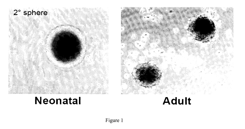
Figure 1 is a set of photographs showing skin-derived precursors
(SKPs) generated from rodent and human. Representative images of neonatal
rodent and human SKPs following 3 weeks expansion in vitro are shown.
Figures 2A and 2B are images showing cultured (GFP-labeled) rat
SKPs 14 days after transplantation into adult mouse backskin. Figure 2A
shows SKPs survive and migrate throughout the interfollicular dermis, as well
as into dermis-derived components of the hair follicle (i.e., dermal papillae
and
dermal sheath). Transplanted SKPs never generate or migrate into the
epidermis or the epidermal derivatives of hair follicle. Figure 2B is a higher
magnification image of SKPs within the dermal papillae of a hair follicle
(arrow). Within the dermis, SKPs appear to have differentiated into dermal
fibroblasts and adipocytes (arrowhead) within the lower dermis/hypodermis.
Figures 3A-3D are images showing that when injected into normal skin
or following either wounding or depilation, transplanted SKPs will migrate
into
the hair follicle. Figure 3A shows that three weeks following transplant, SKPs
have migrated and seemingly completely repopulated the dermal papillae of a
hair follicle, possibly inducing formation of a new follicle. Figure 3B shows
CA 02689484 2009-12-04
WO 2008/148218 PCT/CA2008/001104
SKPs within the papillae express versican, a marker specific to the follicular
dermal papillae cells. Figure 3C shows that transplanted SKPs can also be
observed within the dermal sheath, a specialized layer of cells surrounding
the
hair bulb that are thought to be in continuous cellular exchange with the
dermal
papillae. Figure 3D shows that cells within the sheath also undergo cell
division (ki67-positive) suggesting they are able to respond to endogenous
cues
within the niche.
Figure 4 is an image showing that SKPs integrate into dermal papillae,
but not into matrix cells or melanocytes within the hair follicle. SKPs do not
co-localize with Pax3, a marker of melanocytic cell lineage within the hair
follicle.
Figures 5A-5E are images showing that SKPs contribute to formation
of new hair follicles surrounding a wound. Figure 5A is a diagram illustrating
phenotypic stages of hair follicle formation. Figure 5B is a low magnification
image showing wound a putative immature follicle at the perimeter of the
wound three weeks post-lesion and transplant. Figure 5C is a high
magnification image of boxed region in Figure 5B, illustrating the immature
follicular phenotype and the integration of transplanted SKPs surrounding the
follicle, as well as within the dermal papillae. Figure 5D depicts another
example of a confocal optical section depicting colocalization of transplanted
SKPs with versican-positive dermal papillae cells of the immature follicle.
Pan-cytokeratin staining (red) of epidermal keratinocyte is not expressed by
transplanted SKPs, further demonstrating their restriction to dermis-derived
structures within the follicle. Figure 5E depicts another example of an
immature follicle containing transplanted SKPs expressing p75NTR, which is
enriched within anagen phase follicular dermal papillae. The occurrence of
these `immature follicles' was typically only observed within the regions
containing transplanted SKPs.
Figures 6A-6D are images showing that SKPs integrating into the
dermal papillae of existing hair follicles are functional. Figures 6A and 6B
6
CA 02689484 2009-12-04
WO 2008/148218 PCT/CA2008/001104
show that rat SKPs have integrated into dermal papillae of existing hair
follicles or induced formation of new hair follicles within normal mouse
backskin. Figure 6C and 6D show that, after six weeks, follicles containing
SKP-derived dermal papillae generate longer and thicker hair (arrow),
suggesting that SKPs retain inductive capacity, and retain regulatory
properties
specific to the donor (rat).
Figure 7A is a schematic diagram showing the "patch assay" of hair
follicle formation. In order to determine if SKPs could actively
induce/participate in de novo hair follicle formation, SKPs (generated from
E17 or adult yellowgreen fluorescent protein-expressing backskin) were
cultured for 14 days. Newborn backskin from C57/B16 mice was dissociated to
single cells and then combined with dissociated SKPs at a ratio of 1:2 and
suspended in Hanks Balanced Salt Solution. Adult Nu1Nu (hairless) mice were
given three injections of cells (15 l in each injection) along the length of
the
back. Three weeks later, patches of black hairs could be seen growing
underneath the skin.
Figure 7B is a schematic diagram showing that reisolated SKPs from
transplanted hair follicles are capable of serial induction and reconstitution
of
new hair follicles.
Figures 8A-8D are images showing that SKPs induce de novo hair
follicle formation. Figure 8A shows a brightfield image of hair new hair
follicles within the backskin of a hairless mouse. Figure 8B shows that three
weeks after combination with dissociated newborn skin cells, E17 SKPs can be
seen comprising the entire dermal papillae and as well as in the dermal sheath
of new hair follicles. Figure 8C and 8D are high magnification images of SKP-
derived hair follicles.
Figure 8E is a graph showing the number of new hair follicles
generated upon transplantation of SKPs and keratinocytes.
Figures 9A-9G are images showing that adult SKPs participate in new
hair formation. Figure 9A shows that, following the same assay as described
7
CA 02689484 2009-12-04
WO 2008/148218 PCT/CA2008/001104
above, adult (8 weeks old) SKPs combined with newborn skin cells were found
comprising entire dermal papillae and dermal sheath of the majority of new
hair follicles that had been generated within the graft. Figure 9B shows that
GFP fluorescence indicating location of GFP-labeled SKPs within the follicle.
Figures 9C and 9D show another example of a newly generated hair follicle
showing SKPs within the dermal papillae. Mesenchymal stem cells in vitro
(Figure 9E) expressing GFP, do not participate in hair follicle formation when
grafted in the same hair formation assay (Figures 9F and 9G) suggesting that
the inductive properties are unique to SKPs.
Figures 10A-10D show that clonally-derived adult SKP are capable of
generating new hair follicles. Secondary clonal SKPs spheres were generated
at a density of 1000 cells/ml. A single clonal sphere was isolated,
dissociated
and expanded to generate large numbers of tertiary clones. Each skin graft
containing a single clonal population of adult SKPs (combined with newborn
epidermal cells) gave rise to clusters of new hair follicles (Figure 10A)
which
contained GFP-expressing SKPs clones within the dermal papillae and dermal
sheath. Figure lOB shows YFP labeling. Figures 10C and 10D are higher
magnification images of new clonal SKP-derived hair follicles.
Figures 11A-11F are images showing transplanted SKPs within the
follicular dermal papillae niche, retain self renewal and multipotency.
Dissociation of new hair follicles containing GFP-labeled SKP-derived dermal
papillae, retain their ability to self renew, generating clonal spherical
colonies
(Figure 11 A) after 7-14 days following exposure to fibroblast growth factor
and epidermal growth factor. Figure 11 B shows that same sphere showing
expression of GFP to confirm that the sphere originated from a cell which had
been transplanted into the skin and generated a new hair follicle 4 weeks
prior.
Figure 11C and 11D show that these re-cultured spheres also retain
multipotency such that they still retain the ability to stimulate formation of
hair
follicles in vivo, as well as to generate neurons in vitro which express
nestin
8
CA 02689484 2009-12-04
WO 2008/148218 PCT/CA2008/001104
(red, arrows; Figure 11E) and beta III tubulin (red; arrows; Figure 11F) a
marker specific to neurons.
Figures 12A-12C depict transplanted adult SKPs forming new hair
follicles express markers specific to the dermal papillae. Engrafted SKPs
within the follicular dermal papillae express versican (Figure 12A), neural
cell
adhesion molecule (NCAM) and p75 neurotrophin receptor (Figure 12B), and
cells within the dermal sheath immunostain with alpha-smooth muscle actin
(red) (Figure 12C).
Figures 13A-13C show that SKPs migrate into and contribute to wound
healing. Adult NODSCID mice received a 3mm full thickness skin wound.
Immediately following, YFP labeled SKPs were transplanted (intra-dermal)
into the surrounding regions of intact skin. Three weeks following, SKPs can
be observed filling the wound cavity (Figures 13A and 13B), comprising what
would be the scar, suggesting that SKPs respond to migratory cues and actively
contribute to wound healing. Figure 13C shows that, within the wound, SKPs
differentiate into putative dermal fibroblasts immunostaining for fibronectin
(arrows) and myofibroblasts staining with alpha-smooth muscle actin
(arrowheads).
Figures 14A and 14B show that transplanted SKPs support formation
of epidermal appendages after 1 week. Depicted is a dorsal view of a dermal
sheet comprised of SKPs which have been combined with epidermal
keratinocytes. SKPs surround structures immunostaining for p63 (Figure 14A)
and e-cadherin (Figure 14B), which are specific to epidermal cell types.
Figure 15 shows that human SKPs generate dermal sheets in vitro.
Human SKPs (grown adherent or as spheres) are capable of generating dermal
sheets. Sheets generated by SKPs are significantly thicker than normal human
fibroblasts.
Figures 16A-16M is a set of images showing that SKPs regenerate the
dermis and home back to a hair follicle niche upon transplantation. Figure 16A
shows back skin transplanted with dissociated YFP-tagged neonatal mouse
9
CA 02689484 2009-12-04
WO 2008/148218 PCT/CA2008/001104
SKPs two weeks earlier. Transplanted cells (lighter color) are in the
interfollicular dermis (arrows) and the dermal papilla (DP) and dermal sheath
DS (arrowhead) of hair follicles. Figures 16B and 16C show dermis
transplanted with SKPs as in Figure 16A, and immunostained for GFP (left
panel) and two dermal fibroblast markers, PDGFRa (Figure 16B, center panel)
and collagen type 1(Figure 16C, center panel). Right panels of Figures 16B
and 16C are merges, with the arrows indicate double-labeled cells. Figures
16D-16G show hair follicles containing YFP-positive SKPs 2-4 weeks post-
transplantation, as in Figure 16A. Figure 16D shows a hair follicle with the
DP
(arrow) comprised entirely of YFP-labeled cells. Figure 16E shows a follicle
triple-labeled for YFP (left panel), versican (a marker of DP; center-left
panel)
and pax3 (a melanocyte/melanoblast marker; center-right panel). The right
panel is a merge, and arrow indicates the DP. Figure 16F shows a follicle
cross
section which shows transplanted cells in the DS (arrows) expressing a-sma
but not e-cadherin (an epidermal marker). Figure 16G shows transplanted cells
within the DS (arrowhead) but not DP (arrow) expressed the proliferation
marker Ki67. Figures 16H-16J show quantification of the number of YFP-
positive cells associated with follicles (Figure 161) or present within the DP
of
individual follicles (Figure 16J; hatched lines in Figure 16H) following
transplantation into depilated versus shaved skin. (p<0.05 for Figures 161 and
16J). Figures 16K-16M show skin four weeks following transplantation of
neonatal mouse SKPs adjacent to a punch wound. Transplanted cells
repopulated the wound, and express fibroblast-specific antigen (Figures 16K
and 16L, arrows), fibronectin (Figure 16M), and a-SMA (Figure 16M,
arrowhead). Scale bars = 200 m (16A), 16 m (16B, 16C), 50 m (16D, 16H,
16L), 25 m (16E, 16F, 16G, 16M), 100 m (16K). epi = epidermis, hypo =
hypodermis. Some sections were counterstained with Hoechst 33258 or
fluorescent Nissl to show tissue morphology, as indicated.
Figures 17A-17K are a set of images and graphs showing that SKPs can
reconstitute their niche and instruct epidermal cells to generate hair
follicles.
CA 02689484 2009-12-04
WO 2008/148218 PCT/CA2008/001104
Figures 17A and 17B show skin four weeks after transplantation of neonatal
mouse YFP-tagged SKPs adjacent to a punch wound. Transplanted cells
(green) are present in "peg-like" hair follicles (Figure 17A, arrowheads), in
DP
and DS (Figure 17B, arrow and arrowheads). Those in the DP express versican
(Figure 17B, top right panel). The bottom panel in 17B is a merge. Figures
17C-17E show patches formed by mixing GFP-tagged dissociated adult rat
SKPs with newborn C57/B16 epidermal aggregates, showing that the DP and
DS (Figures 17C and 17D; arrow and arrowheads) were comprised of SKPs.
Quantification of follicles with GFP-positive DP (Figure 17E) revealed that
rat
SKPs were enriched in follicle inductive ability relative to newborn dermal
cells (105 cells n=3; 106 cells n=2, *p=0.001). Figure 17F and 17G show adult
mouse skin transplanted 8 weeks earlier with GFP-tagged adult rat SKPs.
Transplanted cells contributed extensively to the dermis, and the DP of hair
follicles (Figure 17F, arrows), many of which were in anagen (Figure 17G,
arrows). Figures 17H and 171 show that chimeric rat/mouse hairs were thicker
(Figure 17H) and longer (Figure 171) than endogenous pelage hairs. Figure 17J
shows patch assays with murine dermal cells versus rat SKPs. Figure 17K is
graph showing that hairs induced by rat SKPs had larger bulbs (n=2
experiments, *p=0.0074). Tissue was counterstained with Hoechst 33258
(17A), fluorescent Nissl stain (17F), or propidium iodide (17G) to show tissue
morphology. Scale bars = 100 m (17A, 17F, 17J), 50 m (17B, 17D, 17G) and
500 m (17C). epi = epidermis, hypo = hypodermis.
Figures 18A-18F are images showing that clonally-derived SKPs
reconstitute the dermis and induce hair follicle formation. Figures 18A and
18B show one adult rat GFP-positive SKPs clone (clone 3) that was expanded
for 12 weeks and was used in follicle patch assays. Figure 18D-18F show
clone 3 transplanted into the adult mouse dermis. Figure 18A-18C show that
clonal SKPs comprised the DP (arrowheads) of newly-formed hair follicles
after 2-4 months (18A and 18B) or 11 (18C) months in culture. Figures 18D-
18F show that transplanted cells (green) homed to hair follicle DP (18D,
arrow)
11
CA 02689484 2009-12-04
WO 2008/148218 PCT/CA2008/001104
and integrated into interfollicular dermis (18D, arrowheads), where they
expressed fibronectin (18D, center panel), vimentin (18E), and a-sma (18F) 3
weeks post-transplant.
Figures 18G-18K show that SKPs isolated from their hair follicle niche
self-renew and serially reconstitute hair follicles. Figure 18G is a schematic
showing the serial reconstitution assay of hair growth. Figure 18H shows a
single hair follicle containing adult rat GFP-labeled cells within the DP and
DS
dissected from a patch assay graft. Figure 181 shows that cells, isolated from
follicles as in Figure 18H, generated GFP-positive SKP spheres after 12 days
of culture (arrows) as seen by phase (top panel) and fluorescence (bottom
panel) illumination. Figure 18J shows that cells from these spheres generated
secondary hair follicles in the patch assay (arrows). Figure 18K shows that,
in
tertiary follicle reconstitutions, GFP-labeled SKPs were surrounded by black
melanocytes (arrow), but did not induce hair follicle formation. Scale bars =
100 m (18A-18D, 18J), 25 m (18E, 18F), 50 m (18H), 200 m (18J), 250 m
(18K).
Figures 19A-19L are a set of images and a graph showing that SKPs
isolated from the hair follicle niche remain multipotent. Figures 19A-19C
show that skin transplanted with GFP-positive follicle-derived SKPs for 4
weeks. In Figure 19A, transplanted cells (green) are seen to home back to the
DS and DP of hair follicles (arrows) and reconstituted the dermis
(arrowheads).
Figures 19B and 19C show that they expressed the dermal fibroblast markers
PDGFRa and collagen type 1. Right panels are the merges, and arrows
indicate double-labelled cells. Figures 19D and 19E show that, when
differentiated in culture under mesodermal conditions, follicle-derived SKPs
generated adipocytes, as indicated by the lipophilic dye oil red 0 (19D,
arrows), and a-sma-positive cells, potentially myofibroblasts (19E, arrow).
Figures 19F and 19G show that, when differentiated under neurogenic
conditions, they generated nestin-positive cells after 5 days (19F, arrows),
and
morphologically-complex, 0111-tubulin positive cells after 14 days (19G,
12
CA 02689484 2009-12-04
WO 2008/148218 PCT/CA2008/001104
arrow). Figure 19H and 191 show sciatic nerve sections 6 weeks following
crush and transplantation, showing that follicle-derived SKPs generated cells
positive for the Schwann cell markers p75NTR (19H) and P0 (19I), as did the
endogenous Schwann cells. Figures 19J-19L show transplantation of follicle-
derived SKPs into the chick neural crest migratory stream (stage 18). After 8
days in ovo, some of the transplanted cells that had migrated to the dermis
(Figures 19J and 19K, green) were versican-positive (Figure 19K, arrows).
Quantification after 3 days in ovo (Figure 19L) demonstrated follicle-derived
(6 transplants) and clonal (8 transplants) SKPs behaved like total SKPs (9
transplants), migrating to the nerve or DRG and to the skin, with some
remaining close to the neural tube. Samples were counterstained with Hoechst
33258, as indicated. epi = epidermis. Scale bars = 200 m (19A), 25 m (19B,
19C, 19H, 191), 50 m (19D, 19E, 19G, 19J, 19M), 100 m (19F).
Figures 20A-20F are photomicrographs showing that transplanted
SKPs, but not NSCs or MSCs, home to a dermal papilla niche and generate
dermal fibroblasts. Figure 20A shows GFP-expressing adult rat SKPs
transplanted into depilated adult NOD/SCID mouse dermis 21 days earlier, and
immunostained for GFP (left), vimentin (center-left) and fibronectin (center
right). The right panel is the merged image. The arrow denotes a transplanted
cell expressing both vimentin and fibronectin. Figure 20B shows analysis of
transplants performed as in Figure 20A, and immunostained for GFP (left) and
a-sma (center). The right panel is the merged image. Arrows denote cells
positive for both markers. Figure 20C shows adult GFP-expressing rat SKPs
transplanted into dermis as in Figure 20A, immunostained for GFP to mark
transplanted cells, and for the melanoblast/melanocyte marker tyrosinase.
Arrow indicates transplanted cells that have homed to the DP, but that they do
not express melanocyte markers. Figures 20D and 20E show backskin of
NOD/SCID mice 21 days post-grafting, indicating that transplanted YFP-
tagged neonatal mouse NSCs (Figure 20D, arrowheads) display poor survival
and are never observed associating with hair follicles, while GFP-tagged adult
13
CA 02689484 2009-12-04
WO 2008/148218 PCT/CA2008/001104
rat MSCs (Figure 20E, arrowheads) were found within the interfollicular
dermis but were never recruited into the DP of hair follicles. Figure 20F is a
photomicrograph of a hair follicle in a section similar to Figure 20E,
immunostained for GFP to identify the transplanted MSCs (left),
PDGFRa (center left), and the MSC marker cd73 (center right), showing that
MSCs are never found in the DP (denoted by dashed lines). Nuclei are stained
with Hoechst 33258 in Figures 20B-20E. epi = epidermis, hypo = hypodermis.
Scale bars are 16 m (20A, 20B), 25 m (20C), 200 m (20D, 20E), 40 m
(20F).
Figures 21A-21D are a photomicrographs showing that SKPs
participate in dermal wound healing. Figure 21A shows that, three weeks after
transplantation of neonatal YFP-tagged murine SKPs into the cavity and within
the intact tissue surrounding a backskin punch wound (arrows denote the
location of the transplant), transplanted cells are found within the
regenerated
tissue filling the wound cavity and scar (denoted by dashed lines). Figure 21B-
21D are photomicrographs of skin sections transplanted with YFP neonatal
mouse SKPs into the intact tissue surrounding a wound immunostained for
GFP, the DP marker versican (Figure 21B), and the dermal fibroblast markers
vimentin (Figure 21C), or collagen type 1 (Figure 21D). Transplanted
interfollicular cells express dermal fibroblast markers (Figures 21C-21D,
arrows), but do not express versican (Figure 21B, arrowheads), although
transplanted cells within the DP do express this marker (for example, see
Figure 16E). Nuclei are stained with Hoechst 33258 (blue) in Figures 21B-
21D. epi = epidermis. Scale bars are 200gm (21A), 50 m (21B-21D).
Figures 22A-22M are a set of photomicrographs and a graph showing
that SKPs, but not NSCs or MSCs, instruct epidermal cells to generate hair
follicles. Figures 22A-221 are photomicrographs of patch assays at 12 days.
Newborn murine epidermal aggregates alone do not generate hair follicles
(Figure 22A), and neither GFP-tagged rat MSCs (Figures 22B and 22C) nor
YFP-tagged neonatal mouse NSCs (Figures 22D and 22E) induced follicle
14
CA 02689484 2009-12-04
WO 2008/148218 PCT/CA2008/001104
formation when mixed with epidermal cells, as shown in phase (Figures 22B
and 22D) and fluorescence (Figures 22C and 22E) images of the patches. In
contrast, 106 dissociated GFP-expressing neonatal rat dermal cells induced
hair
follicle formation when combined with epithelial aggregates as seen by phase
(Figure 22F) and fluorescence (Figure 22G) illumination, as did 106 adult GFP-
tagged SKPs (Figures 22H and 221) (arrowheads in Figures 22F and 22H show
hair follicles, while those in Figure 221 show GFP-positive DPs). Note that in
Figure 22G several GFP follicles are entirely green due to contaminating GFP-
expressing epidermal cells in the dermal preparation. Figures 22J and 22K
show quantification of total hair follicle numbers in patches similar to those
shown in Figures 22A-221, demonstrating that adult rat SKPs were enriched for
follicle inductive ability relative to total neonatal dermal cells and to
other stem
cell populations such as MSCs and NSCs. In Figure 22J, 106 dissociated cells
were mixed with 10,000 epidermal aggregates and all follicles were counted. *
p<0.001 relative to epi only, **p<0.001 relative to epi only and dermis.
Figures 22K and 22L are photomicrographs of hair follicles in patch assays
where 106 adult GFP-tagged rat SKPs were mixed with 106 dissociated total
skin cells from newborn C57/B16 skin (epidermis and dermis), as shown with
combined phase with coincident fluorescence illumination. In Figure 22K,
arrowheads indicate follicles with DP generated from GFP-positive SKPs.
Figure 22L shows higher magnification of the boxed area, and the DP and DS
are denoted by an arrow and an arrowhead, respectively. In these experiments,
more than 80% of hair follicles contained GFP-positive DP (arrowheads)
suggesting that the rat SKPs had a competitive advantage over the endogenous
murine inductive cells. Figure 22M shows GFP-tagged adult rat SKPs were
transplanted into adult NOD/SCID mouse skin, and analyzed 8 weeks later.
Transplanted GFP-positive cells (green) comprised the DP of many hair
follicles, including some in telogen (arrows). Scale bars are lmm (22A, 22F-
221), 500 m (22B-22E), 250 m (22K), 100 m (22L, 22M).
CA 02689484 2009-12-04
WO 2008/148218 PCT/CA2008/001104
Figures 23A and 23B are photomicrographs showing that clonal SKPs
can both induce hair follicle formation and contribute dermal fibroblasts to
the
interfollicular dermis. Figure 23A shows hair follicles in a patch assay where
SKP clone 2 (generated from adult, GFP-tagged rat skin) was expanded 8
weeks in culture and then mixed with epidermal cells. The DP and DS of these
hair follicles are comprised of SKP-derived cells. Figure 23B shows high
magnification photomicrograph of a skin section transplanted with GFP-tagged
rat SKP clone 3 and immunostained for GFP (green) and fibronectin (blue).
Scale bars are 200 m (23A), 25 m (23B).
Figure 24 shows that follicle-derived SKPs reconstitute the
interfollicular dermis. High magnification images of the same field showing
transplanted cells (left) immunostained for the dermal fibroblast marker S
1000
(center). Right panel is the merge, and arrows indicate double-labeled cells.
Scale bar is 25 m.
Figure 25 is a set of images showing that, although cells are retained
within the DP and DS of grafted hair follicles, the dermal papillae/dermal
sheath is a reservoir of dermal stem cells that continuously contribute cells
to
the dermis.
Figure 26 is a set of images showing that the are involved in dermal
wound healing. SKPs within the DP/DS of transplanted hair follicles are
observed to migrate to wound sites and contribute to wound healing.
Figure 27 shows that Sox2GFP+ cells are found in the skin, and are
localized to the hair follicle.
Figure 28 shows that SKPs from Sox2GFP mice, but not dermal
fibroblasts from non-hairy skin, home to hair follicles. Staining with keratin
15, GFP, and hoecsht shows that GFP expressing SKPs are found in hair
follicles, (top panels), whereas fibroblasts from non-hairy dermis do not
incorporate into follicles (bottom panels).
Figures 29A-29H are a set of photomicrographs and graphs showing
that Sox2+ cells are found in the DP and DS of anagen hair follicles and Sox2+
16
CA 02689484 2009-12-04
WO 2008/148218 PCT/CA2008/001104
cells from skin can form spherical colonies, can induce hair follicle
formation,
and can generate nestin-postive neural precursors. Figures 29A and 29B show
Sox2 expressing cells from P2 Sox2GFP mice in backskin. Keratin 5 and
hoecsht staining are also shown. Figure 29B shows expression of Sox2 (top
left), keratin 5 (top right), and versican (bottom left) in P2 backskin from
Sox2GFP mice. A merge is also shown (bottom right). Figure 29C shows
expression of Sox2 (left) and keratin 5 (center) in whisker pad skin. A merge
including Sox2, keratin5, and hoecsht is also shown (right). Figure 29D shows
that dissociated neonatal skin cells from the backskin (top left) and facial
skin
(bottom left) of Sox2GFP mice generate spherical colonies when grown in
proliferation medium. Many of these colonies are Sox2GFP, as shown in the
right-most images. Figure 29E is a histogram showing that fractionated
Sox2GFP+ facial skin cells show a 5-fold enrichment, and backskin cells a two-
fold enrichment, for sphere formation relative to total cells. Figure 29F
shows
that Sox2GFP+ cells are enriched in hair follicle formation, as compared to
epidermal cells. Figure 29G is a histogram showing that SoxGFP2+ cells
exhibit a 10-fold greater capacity for follicle formation relative to ungated
cells
or Sox2GFP- fraction. Figure 29H shows that fractionated Sox2GFP+ cells are
multipotent, generating nestin-positive neural precursors (left and left
center
panels), which are not observed in the Sox2GFP- fraction (right center panel).
Unfractionated cells also exhibit nestin-positive cell formation (right
panel).
Detailed Description
The present invention provides methods for generating de novo hair
follicles in a mammal, compositions of SKPs and keratinocytes, dermal sheets
grown in vitro, and methods of making and using such sheets to regenerate skin
(e.g., in a mammal having a burn or an ulcer, having or previously having had
an infection resulting in skin loss, having undergone a surgical procedure
requiring skin regeneration, or having an injury resulting in skin loss). The
methods of generating hair follicles can be used to treat conditions such as
17
CA 02689484 2009-12-04
WO 2008/148218 PCT/CA2008/001104
alopecia, male pattern baldness, or female pattern baldness. All methods can
be used for cosmetic purposes, either in conjunction with or in addition to
the
conditions noted above.
SKP cells and culture conditions
SKP cells have been described previously in PCT Publication Nos.
WO 01/53461 and WO 03/010243, and WO 2005/071063, each of which is
incorporated by reference. Rodent SKPs can be obtained, for example, from
skin of mouse embryos (E 15-E 19), mouse, or rat neonates (postnatal day 2
(P2) to P6). In one method, the skin is cut into 2-3 mm2 pieces. Tissue is
digested with 0.1 % trypsin or 1 mg/ml collagenase for 10-45 min at 37 C,
mechanically dissociated, and filtered through a 40 m cell strainer (Falcon,
Franklin Lakes, NJ). Cells are plated at a density of 1-2.5 x 10⁴ cells/ml in
DMEM/F-12 at 3:1 (Invitrogen, Carlsbad, Calif.), with 20 ng/ml epidermal
growth factor (EGF) and 40 ng/ml FGF2 (both from Collaborative Research,
Bedford, Mass.), hereafter referred to as proliferation medium. SKPs are then
passaged by mechanically dissociating spheres and splitting one to three with
75% new medium and 25% conditioned medium. Clonal spheres are prepared
as described previously (Fernandes et al. (2004) Nat. Cell Biol. 6:1082-93)
and
were differentiated similarly with the addition of 1% serum for the first
three
days (Figure 1).
In the experiments described herein, human SKPs were isolated and
cultured as follows. Pieces of human foreskin of 1-2 cm2 deriving from
voluntary circumcisions of children aged 4 weeks to 12 years of age were
washed with Hanks' balanced salt solution (Invitrogen Corporation), cut into 4-
to 6-mm pieces, washed again, and incubated in Liberase Blendzyme 1 (0.62
Wunsch U/ml; Roche Molecular Biochemicals, Laval, Quebec, Canada)
overnight at 4 C. The epidermis was manually removed from each tissue
piece, and the dermis was cut into 1-mm3 pieces and incubated in Liberase
Blendzyme 1 for 30-40 minutes at 37 C. DNase I was added for 1 minute, and
18
CA 02689484 2009-12-04
WO 2008/148218 PCT/CA2008/001104
10% fetal bovine serum (FBS) (Cambrex, Walkersville, Md.) was added to
inhibit the enzymes. The supernatant was removed, and tissue pieces were
resuspended in medium (Dulbecco's modified Eagle's medium (DMEM)/F12,
3:1 (Invitrogen) containing 1% penicillin/streptomycin unless otherwise
indicated) and manually dissociated by pipetting into a 2-ml pipette, a
process
that was repeated until the tissue could be broken down no further. The cell
suspension was then centrifuged at 1,000 rpm for 5 minutes and the supernatant
removed, leaving the pellet and 3 ml of medium behind. The pellet was
resuspended in the remaining medium using a fire-polished Pasteur pipette, and
the suspension passed through a 70- m cell strainer (BD Biosciences,
Mississauga, Ontario, Canada). The strained cell suspension was then
centrifuged, the medium removed, the pellet resuspended in 10 ml proliferation
medium (DMEM-F12, 3:1 and 40 ng/ml FGF2, 20 ng/ml EGF (both from BD
Biosciences), B27 (Invitrogen), and 1 g/ml fungizone (Invitrogen)) and then
transferred to a 25-cm2 tissue culture flask (BD Biosciences).
For subculturing, medium containing SKPs growing in suspension was
centrifuged at 1,000 rpm for 5 minutes and the supernatant was removed,
leaving 6 ml of medium and the pellet behind. The pellet was resuspended in
the remaining medium with a fire-polished Pasteur pipette, proliferation
medium was added to a total of 20 ml, and the cell suspension was then split
into two 25-cm2 flasks. The cells were grown at 37 C for an additional 2-3
weeks and then split again as above.
For immunocytochemical analysis of SKP spheres, 100 l of medium
containing suspended spheres was removed from a flask and spun down onto
coated slides using a ThermoShandon Cytospin 4 apparatus (Thermo Shandon
Inc., Pittsburgh, Penn.). The slides were then air-dried for 5 minutes and
analyzed. For quantitation of the size of SKP spheres grown in different
growth factors, the diameter of spheres was measured along both the x and y
axes, because spheres were not uniformly spherical. The average of these two
measurements was then used as the diameter of the sphere. Within a given
19
CA 02689484 2009-12-04
WO 2008/148218 PCT/CA2008/001104
experiment, multiple spheres were measured in each well, the mean diameter
and SD of all measured spheres in each individual well were determined, and
then four wells per experimental manipulation were considered to obtain a
statistical comparison between growth factor treatments.
Human epidermal cells
Human epidermal cells (keratinocytes) can be obtained using any means
known in the art. Specimens of split-thickness skin can be collected from
donors (e.g., either live or cadavers). Alternatively, human keratinocyte
cells
are commercially available from vendors including ScienCell (Carlsbad, Calif.)
and PromoCell (Heidelberg, Germany). Autologous keratinocytes can also be
used.
Should it be necessary to culture keratinocytes, any culture technique
known in the art may be used. One exemplary technique, the method described
by Staiano-Coico et al. (1986) J. Clin. Invest. 77:396-404), is as follows.
Cells
are stored at 4 C, washed three times in MEM with antibiotics, then incubated
in a solution of 0.5% trypsin (Difco laboratories, Detroit, Mich., 1:250) in
Ca2+
and Mg2+ free phosphate-buffered saline (PBS; Gibco) for 90 mm at 37 C.
Single-cell suspensions of epidermal cells are prepared by vigorous stirring
in a
solution of 0.25% DNase I; Sigma Chemical Co., St. Louis, Mo.) and 1% fetal
bovine serum in PBS and filtered through sterile gauze; FBS was added to the
cell suspensions to neutralize trypsin activity. After centrifugation and
resuspension in complete culture medium (MEM, 20% fetal bovine serum, 2
mM L-glutamine, hydrocortisone (0.5 pg/ml), penicillin (100 U/ml),
streptomycin (0.1 mg/ml, and fungizone (0.25 pg/ml)), the viability of
epidermal cells prepared in this manner was determined to be 90-95% by
trypan blue dye exclusion. Plastic tissue culture flasks containing 2 x 10⁵
epidermal cells/cm2 were incubated at 37 C in a humid 95% air/5% CO2
environment; the medium was changed every third day.
CA 02689484 2009-12-04
WO 2008/148218 PCT/CA2008/001104
De novo generation of hair follicles
We have discovered that de novo hair follicle formation is induced when
a combination of SKPs and epidermal keratinocytes are introduced into the
skin of a mammal. Based on this discovery, the present invention provides
methods of growing hair in by administration of a combination of SKPs and
keratinocytes and pharmaceutical compositions comprising SKPs and
keratinocytes (e.g., in a pharmaceutically acceptable carrier).
SKPs are capable of surviving after transplantation (Figures 2A and 2B)
and migrate to the appropriate regions of existing hair follicle (see Figures
3A-
3D, 4, and 25). SKPs also contribute to hair follicle formatian in region
adjacent to a skin wound (see Figures 5A-5E). In addition, transplanted SKPs
retain the inductive capacity and regulatory properties specific to the donor
cells; rat SKPs transplanted into mice form "rat" hair (Figures 6A-6D).
We also determined that SKPs retain hair follicle-inductive properties by
using YFP-labeled SKPs co-transplanted with newborn mouse epidermal
keratinocytes into the back skin of adult nude mice using the "patch assay"
(Figure 7A), described for freshly isolated dermal papillae cells (Zheng et
al.,
(2005) J. Invest. Dermatol. 124:867-876). Dorsal backskin keratinocytes were
isolated from newborn C57B1/6 mice by floating skin on 0.25% trypsin
overnight at 4 C and then carefully peeling off the overlying epidermis.
Epidermal sheets were then minced and incubated in trypsin--EDTA for 30
minutes at 37 degrees and then gently triturated in 10% FBS to stop the
reaction. Similar methods for isolating epidermal keratinocytes have been
previously described (Lichti et al., (1993) J. Invest. Dermatol. 101:124S-
129S).
GFP-tagged SKPs were then suspended in HBSS with various concentrations
of keratinocytes (typically 2:1, meaning that approximately 106 SKPs
combined with 5 x 10⁵ keratinocytes) in 20-30 1 of HBSS. Alternatively,
intact SKP spheres were also transplanted with fresh keratinocytes.
Importantly, grafting of intact SKP spheres, rather than dissociated SKP
cells,
yielded greater efficiency of de novo follicle formation. In addition, these
21
CA 02689484 2009-12-04
WO 2008/148218 PCT/CA2008/001104
experiments confirm two important points. First, the dermal papillae or dermal
sheath, as these two structures may actually be one and the same, is an
endogenous niche for SKPs. Second, SKPs are capable of inducing formation
of new hair follicles. (Figures 8A-8D). As few as 50 SKPs spheres could be
transplanted with 5 x 10⁵ keratinocytes resulting in typically '25-35 new hair
follicles. Cell suspensions were injected into the dermis/hypodermis of dorsal
backskin of athymic nude mice (Charles River Laboratories) using a 27-gauge
Hamilton syringe. Two weeks later, hair follicles could be observed in a
protruding from the skin as well as coursing throughout the graft beneath the
skin. Control transplants consisted of fresh or cultured dermal cells combined
with keratinocytes, or keratinocytes alone. Similar results were observed
using
SKP cells from adult rodents (Figures 9A-9G).
We were also able to reconstitute follicular dermal papillae serially
(Figure 7B). As described above, de novo hair follicles were generated by
combining neonatal or adult SKPs (passaged between 1-5 tinies) with either
dissociated newborn skin cells, or epidermal keratinocytes. Two weeks later,
grafts of SKP-derived hair follicles were excised, minced and digested in
collagenase (Type XI) at 37 C for 1 hour. Alternatively, single graft-derived
hair follicles containing SKP-derived dermal papillae (GFP-tagged) were
isolated and the follicle bulbs were dissected, minced and digested with
collagenase as above. Tissues were dissociated to single cells by gentle
trituration and then grown at 5,000 to 20,000 cells/ml in proliferation media
consisting of DMEM:F12 (3:1; Invitrogen) supplemented with 2% B27
(Invitrogen) and containing basic fibroblast growth factor (40 ng/ml) and
epidermal growth factor (40 ng/ml) as described above. After 10 to 14 days,
floating GFP-labeled spherical colonies were observed. 2 x l05 to 1 x 10⁶
GFP-labeled follicle-derived spheres were then recombined with newborn
keratinocytes or whole skin in 30 1 of HBSS and injected into the dermis
where they formed new hair follicles comprised of GFP positive dermal sheath
and dermal papillae. Three successive isolations and expansion of dermal stem
22
CA 02689484 2009-12-04
WO 2008/148218 PCT/CA2008/001104
cells (SKPs) with subsequent reconstitution of hair follicles were done
(Figures
l0A-lOD). These experiments were done twice using two different adult (8
weeks old) backskin samples.
Consistent with these results, transplanted SKPs retairi their capacity for
self-renewal and multipotency (Figures 11 A-11 F) and express appropriate
dermal papilla markets within newly formed hair follicles (Fiigure 12A-12C).
To determine multipotentiality, S KPs were differentiated in vitro under
defined
conditions to promote generation of neural, and mesodermal cell types.
Schwann cell medium consisted of DMEM-F12 3:1 with 1% N2 supplement,
ng/ml neuregulin-1(3 (heregulin-(31; R&D Systems) and 4 M forskolin,
referred to as Schwann cell differentiation medium. Neuronal medium
contained DMEM-F12 3:1 with 1% N2 supplement, 1% B27 supplement, 10%
fetal bovine serum (FBS), 50ng/ml NGF and 50ng/ml of BDNF. Medium was
changed every 3-4 days.
Generation of hair follicles is useful in disorders including conditions
characterized by loss or lack of hair, including for example, alopecia, male
pattern baldness, female pattern baldness, accidental injury, damage to hair
follicles, surgical trauma, bum wound, radiation or chemotherapy treatment
site, incisional wound, donor site wound from skin transplant, and ulceration
of
the skin. In some embodiments, hair growth is induced in an area or areas
where hair was previously present but has been lost. Alternatively or in
addition to the conditions noted above, the induced hair grovvth may be for
cosmetic purposes.
Compositions of SKPs and keratinocytes
Based on the discovery that transplanting a combination of SKPs and
keratinocytes can induce de novo hair follicle formation in nlammals, the
present invention provides compositions including a combination of SKPs and
keratinocytes. Such compositions may include cells isolatecl from any source
and may include any amounts, any ratio, or any purity of SK:Ps and
23
CA 02689484 2009-12-04
WO 2008/148218 PCT/CA2008/001104
keratinocytes. Such compositions may include at least 10, 100, 1,000, 10,000,
or 100,000, 500,000, or 1,000,000 cells. The ratio of SKPs to keratinocytes in
the composition may be at least 1:1,000, 1:100, 1:50, 1:20, 1:10, 1:5, 1:4,
1:3,
1:2, 1:1, 2:1, 3:1, 4:1, 5:1, 10:1, 20:1, 50:1, 100:1, or 1,000:1. The cells
may be
enriched such that the combination of SKPs and keratinocytes make up at least
10%, 20%, 30%, 40%, 50%, 60%, 70%, 80%, 90%, 95%, 99%0, or even 100%
of the total cells in a composition of the invention (e.g., free from
macrophages
or lymphocytes).
Compositions of the invention may further include a pharmaceutically
acceptable carrier (e.g., suitable for epidermal, intradermal, subdermal, or
subcutaneous administration) and may further contain non-toxic
pharmaceutically acceptable adjuvants. The formulation and preparation of
such compositions are well known to those skilled in the art of pharmaceutical
formulation.
Compositions for parenteral (e.g., epidermal, intradermal, subdermal,
and subcutaneous) use may be provided in unit dosage forms (e.g., in single-
dose ampoules), or in vials containing several doses and in which a suitable
preservative may be added (see below). The composition may be in form of a
solution, a suspension, an emulsion, an infusion device, or a delivery device
for
implantation. Apart from the cells, the composition may include suitable
parenterally acceptable carriers and/or excipients. The cells mtay be
incorporated into microspheres, microcapsules, nanoparticles, liposomes, or
the
like for controlled release. Furthermore, the composition may include
suspending, solubilizing, stabilizing, pH-adjusting agents, tonicity adjusting
agents, and/or dispersing agents.
As indicated above, the pharmaceutical compositions according to the
invention may be in a form suitable for sterile injection. To prepare such a
composition, the cells are suspended in a parenterally acceptable liquid
vehicle.
Among acceptable vehicles and solvents that may be employed are water,
water adjusted to a suitable pH by addition of an appropriate amount of
24
CA 02689484 2009-12-04
WO 2008/148218 PCT/CA2008/001104
hydrochloric acid, sodium hydroxide or a suitable buffer, 1,3.-butanediol,
Ringer's solution, dextrose solution, and isotonic sodium chloride solution.
The aqueous formulation may also contain one or more preservatives (e.g.,
methyl, ethyl or n-propyl p-hydroxybenzoate).
Kits containing SKPs
The invention further provide kits containing SKPs cells. Exemplary
kits include SKPs cells, keratinocytes, and instructions for use (e.g.,
instructions for introduction into the skin of a mammal). The SKP cells may be
in a composition with keratinocytes. In other embodiments, ihe kit includes
two compositions, one composition including SKPs and one composition
including keratinocytes. The kits may further include any of the reagents
described herein (e.g., cell culture apparatus, dermal sheets containing
either
SKPs or SKPs and keratinocytes).
Wound healing
Transplanted SKPs migrate from areas surround a wound into the
wound itself, and integrate into structures associated with the hair follicle
(see
WO 2005/071063). Here, we show that SKPs cells both migrate into the
wound and contribute to wound healing, both upon transplantation (Figures
13A-13C) and from adjacent hair follicles (Figure 26).
Dermal sheets
Using SKPs, we have generated dermal sheets in vitro. The invention
thus features methods of making dermal sheets from SKPs, sheets produced by
these methods, and methods of treating skin injuries using the dermal sheets.
We have also shown that sheets of dermis produced by SKP cells are capable
of supporting growth of epidermal cells (Figures 14A and 14B). Such sheets
can be useful in all applications in which skin grafts are used, for example,
in
the treatment of burns, mechanical injury, and ulcers (e.g., resulting from
CA 02689484 2009-12-04
WO 2008/148218 PCT/CA2008/001104
diabetes), or as part of a surgical procedure requiring skin replacement. The
dermal sheets may additionally be combined with matrix or scaffolding
elements (e.g., collagen, alginate, and polymers) to provide structure to the
dermal sheet, as detailed below. The dermal sheet may contain cells solely
differentiated from SKPs. In other embodiments, the sheets contain two or
more layers of cells (e.g., a layer of dermal cells and a layer of epidermal
cells).
Generation of dermal sheets
In one example, SKPs from human or rodent were generated as
described above. SKP spheres (human or rat) were dissociated to single cells
and grown adherently in 10 cm plastic tissue culture dishes coated with poly-D-
lysine and laminin. Culture medium consisting of DMEM supplemented with
10% FBS and ascorbic acid was used for 4 weeks. SKP-derived dermal sheets
were compared to dermal sheets derived from normal skin fibroblasts, and
found to be significantly thicker (Figure 15).
Epidermal sheets were generated using similar techniques.
Keratinocytes were isolated by floating skin on 0.25% trypsin overnight at 4 C
and then carefully peeling off the overlying epidermis. Epidermal sheets were
then minced and incubated in trypsin-EDTA for 30 minutes at 37 C and then
gently triturated in 10% FBS to stop the reaction. Similar methods have
previously been described by Lichti et al. ((1993) J. Invest. Dermatol.
101:124S-129S). Isolated keratinocytes were cultured in DMEM containing
low calcium and 5% serum. Epidermal sheets were then ove:rlayed onto dermal
sheets and dermal thickness was assessed two weeks later.
The dermal sheets can further be applied to or generated on a scaffold or
matrix structure to provide support or to generate a particular shape. Any
scaffolding or matrix materials known in the art may be used in the present
invention. Exemplary materials for such a matrix include ch:itosan, alginate,
and collagen (see, e.g., U.S. Patent No. 6,699,287). Foams useful as matrices
are described, for example, in U.S. Patent Application Publication No.
26
CA 02689484 2009-12-04
WO 2008/148218 PCT/CA2008/001104
2003/0105525. Alginate-based matrices are described, for example, in U.S.
Patent No. 6,642,363. Such materials may be bioabsorbable or biodegradable,
such as cotton, polyglycolic acid, cellulose, gelatin, and dextran.
Nonbioabsorble or materials include polyamide, polyester, polystyrene,
polypropylene, polyacrylate, polyvinyl, polycarbonate, polytetrafluorethylene,
and nitrocellulose compounds. See, e.g., U.S. Patent No. 5,512,475.
The dermal sheets of the invention may include additional cell types as
well. For example, stromal cells (e.g., fibroblasts, endothelial cells,
macrophage, monocytes, leukocytes, and adipocytes) may be added to the
dermal sheets or co-cultured with the SKPs.
Treatment using dermal sheets
The dermal sheets of the invention may be used in any application
where skin grafts are typically used, including wounds resulting from bums,
mechanical damage to the skin (e.g., damage resulting from a bone fracture),
infection, ulcers (e.g., resulting from diabetes) as well as post-surgically
or for
cosmetic reasons. The sheet can be applied to the site requiring the sheet
(e.g.,
the site of injury or infection) using any attachment method including
stitches,
sutures, and adhesives (e.g., fibrin glue) known in the art.
Further characterization of SKP cells
We have performed additional studies defining the biological role of
SKPs in vivo, and provide evidence that they represent an adult dermal stem
cell. In particular, they can reconstitute the adult dermis, contribute to
dermal
wound-healing, and home to a hair follicle niche, and instruct epidermal cells
to make hair follicles. In addition, hair follicle-derived SKF's will self-
renew,
maintain their multipotency, and can serially reconstitute hair follicles.
To determine whether SKPs represented dermal sterr.i cells, SKPs were
generated from back skin of neonatal YFP-expressing mice, passaged once, and
transplanted into back skin of adult NOD/SCID mice. Two to three weeks
27
CA 02689484 2009-12-04
WO 2008/148218 PCT/CA2008/001104
later, YFP-positive SKPs were observed throughout the dermis, with a
morphology and location similar to interfollicular dermal fibroblasts (Figure
16A). Many SKPs were also present in the dermal papilla (L)P) and dermal
sheath (DS) of hair follicles (Figures 16A and 16D). Immunocytochemistry
revealed the phenotype of these transplanted cells. Within interfollicular
dermis, most YFP-positive cells expressed the dermal fibroblast markers
collagen type I, fibronectin, vimentin, and PDGFRa and sorrrne expressed a-
smooth muscle actin (a-sma), characteristic of dermal myofibroblasts (Figures
16B, 16C, 20A, and 20B). By contrast, YFP-positive cells within the DP
expressed DP markers such as versican (Figure 16E), while those in the DS
were a-sma-positive, as were resident DS cells (Figure 16F)., Moreover, a
small subpopulation of YFP-positive DS, but not DP, cells expressed the
proliferation marker Ki67 (Figure 16G). YFP-positive cells were never
observed within epidermis or epidermal components of hair follicles, and did
not express markers for melanocytes such as Pax3 or tyrosinase (Figures 16E
and 20C). Thus, SKPs transplanted into adult dermis differentiate into dermal
cell types, with some homing back to a hair follicle niche.
Three lines of evidence indicated that recruitment of SKPs to a follicle
niche was an active process. First, two other adult stem cells, bone marrow
mesenchymal stem cells (MSCs) and forebrain neural stem cells (NSCs), did
not associate with hair follicles when transplanted in the sarne way (Figures
20D-20F). Second, recruitment of SKPs into the follicle niche increased 3-fold
when follicles were induced to enter the anagen growth phase by hair
depilation prior to transplant. Two to three weeks post-transplant, SKPs were
recruited to the DS and many had entered the DP (Figures 16H and 161), with
each DP containing approximately 6-fold more transplantecl cells (Figure 16J).
The third line of evidence came from experiments where SKPs were
transplanted adjacent to or within punch wounds on back skin of NOD/SCID
mice. Two weeks post-transplant, YFP-positive cells recoristituted a large
part
of the scar, where most expressed fibroblast-specific antigen, collagen type
1,
28
CA 02689484 2009-12-04
WO 2008/148218 PCT/CA2008/001104
and fibronectin, and some expressed a-sma (Figures 16K-16M; Figures 21A-
D). By three weeks, YFP-positive, versican-positive cells were also present
within the DP of hair follicles with an immature appearance t:ypical of newly-
forming follicles (Figures 17A and 17B). Thus, SKPs may contribute the
inductive mesenchymal cells necessary for new follicle formation in wounded
skin.
Thus, SKPs are actively recruited into a follicle niche, SKPs re-entering
this niche may further retain the ability to induce hair follicle formation.
To
test this directly, we used the "patch assay" of hair follicle formation
(Zheng et
al. (2005) J Invest Dermatol 124:867-76); SKPs were generated from either
YFP-expressing mice or GFP-expressing rats, were mixed with neonatal
epidermal cells from C57/B16 mice, and were transplanted beneath the dermis
of adult nude mice. Epidermal cells generated no or very feiv hair follicles
when transplanted alone or with MSCs or NSCs (Figures 22.A-22E). By
contrast, epidermal cells mixed with neonatal or adult SKPs generated hair
follicles where the entire DS and DP were comprised of genetically-tagged
cells (Figures 17C, 17D, 22H, and 221). By direct comparison, dissociated rat
SKPs were 5-fold more efficient at inducing hair follicle formation than were
neonatal rat dermal cells (Figure 17E and 22F-22J). As a consequence, SKPs
reconstituted the dermal components of hair follicles even when mixed with
total neonatal skin cells (Figures 22K and 22L).
SKPs can thus instruct neonatal epidermal cells to generate hair follicles.
To determine if they could do so in vivo, GFP-positive SKPs from adult rats
were transplanted into adult NOD/SCID mouse back skin. 'Chese transplanted
rat SKPs appeared to have a competitive advantage, as 8 weeks post-transplant,
they comprised the majority of dermal cells in the transplanted region (Figure
17F). Moreover, the DP and DS of many correctly-oriented hair follicles were
entirely comprised of GFP-positive cells (Figures 17F and l 7G). Remarkably,
relative to the endogenous murine hairs, hairs induced by the rat SKPs were
longer (10.41 mm 0.23 versus 7.96 mm 0.11; p < 0.0001) and had
29
CA 02689484 2009-12-04
WO 2008/148218 PCT/CA2008/001104
increased follicle bulb diameter (107.236 m 4.99 versus 82.27 m 2.51;p
<0.01) and hair fiber width (49.26 rn 0.871 versus 44.6 pm 0.83; p <
0.001) (Figures 17G-171). Although many follicles containing SKP-derived
cells were in anagen (Figure 17G), some cycled in synchron.y with endogenous
follicles and had progressed to catagen/telogen phase (Figure 22M), indicating
that follicle-associated SKPs respond to local signals goveniing the hair
cycle.
To ask whether rat SKPs intrinsically induced these larger follicles, we
performed patch assays, mixing mouse epidermal cells with. dissociated rat
SKPs. Quantification indicated that rat SKPs instructed mouse epidermal cells
to generate larger, more rat-like hair follicles than did muriiie dermal cells
(Figures 17J and 17K).
Thus, SKPs have the capacity to both generate dermal cells and to
induce hair follicle morphogenesis. To determine if indiviclual SKP cells were
multipotent with regard to these two activities, we analyzecl clones of adult
rat
SKPs. Of seven clonally-derived lines that were passaged a minimum of six
times (approximately 8-12 weeks in culture), five induced de novo follicle
formation in the patch assay (Figures 18A, 18B, and 23A). Indeed, when 50
clonal spheres were mixed with 5 x 10⁵ total neonatal skin cells, 30 2 hair
follicles had DP entirely comprised of GFP-positive SKPs. This activity was
persistent; one clone induced follicle formation after 11 months in culture,
albeit relatively inefficiently (Figure 18C). Transplantation of two clones
into
adult NOD/SCID mouse skin demonstrated that they both reconstituted the DP
and DS of hair follicles in vivo (Figure 18D), and generated fibronectin- and
vimentin-positive interfollicular dermal fibroblasts and SMA-positive
myofibroblasts (Figures 18D-18F and 23B). Thus, single SKP clones were
multipotent with regard to both dermal activities in vivo.
These data are consistent with the idea that SKPs represent an
endogenous dermal stem cell. Two cardinal properties of stem cells are self-
renewal and multipotentiality, and one of the most striking assays of in vivo
stem cell functionality is the ability of isolated hematopoetic stem cells
(HSCs)
CA 02689484 2009-12-04
WO 2008/148218 PCT/CA2008/001104
to serially repopulate the blood system. We therefore asked whether
genetically-tagged SKPs that had reconstituted their hair follicle niche could
be
reisolated, expanded, and subsequently reconstitute secondary, de novo hair
follicles. To do this, the patch assay was used to generate hair follicles
where
the entire DP and DS were comprised of genetically-tagged cells (Figures 18G
and 18H). Cells were dissociated from these follicles and cultured in SKPs
proliferation medium. Ten to fourteen days later, genetically-tagged spheres
were observed that could be passaged (Figure 181). When these secondary
spheres (after one passage) were mixed with epidermal cells in the patch
assay,
they induced de novo hair follicle formation (Figure 18J). IJsing this
approach,
we could serially repopulate hair follicles with SKPs up to three times.
However, the SKPs generated from tertiary follicle reconstitutions lost their
inductive ability (Figure 18K), similar to what is seen with serial HSC blood
reconstitution.
Four lines of evidence indicate that SKPs generated from these
reconstituted hair follicles maintain their multipotency. First, when follicle-
derived SKPs were transplanted into adult NOD/SCID mouse skin, they
generated interfollicular dermal fibroblasts, and homed back and integrated
into
the DS and DP of follicles, where they expressed appropria.te markers (Figures
19A-19C and 24). Second, when differentiated under conditions defined for
neonatal SKPs, follicle-derived SKPs generated adipocytes, nestin- and (3III-
tubulin-positive cells with the morphology of neural precursors and neurons,
and SMA-positive myofibroblasts/smooth muscle cells (Fi;gures 19D-19G).
They also generated cells with characteristics of osteocytes and chondrocytes.
Third, when transplanted into the injured sciatic nerve of NOD/SCID mice, a
subpopulation of follicle-derived SKPs progeny aligned with axons, and
expressed P0 and p75NTR (Figure 19H and 191), markers of Schwann cells.
Finally, when follicle-derived SKPs were transplanted inta the embryonic chick
neural crest migratory stream, the majority migrated out of the neural tube
and
into neural crest targets such as the spinal nerve and DRGs, in a manner
31
CA 02689484 2009-12-04
WO 2008/148218 PCT/CA2008/001104
analogous to that seen with total SKPs (Femandes et al. (2004) Nature Cell
Biol 6:1082-1093) (Figure 19L). Intriguingly, a subpopulation of both total
and follicle-derived SKPs migrated to the presumptive dermis, and at late
timepoints, some of these expressed the DP marker versican (Figures 19J-19L).
Thus, follicle-derived SKPs reconstitute the dermis, induce hair follicles,
self-
renew, maintain their multipotency, and home to a dermal ndche within the
embryonic chick.
We have also shown that SKPs, but not dermal fibroblast cells from
non-hairy skin home to hair follicles (Figure 28).
We have further shown that Sox2, a marker of SKPs both in vivo and in
isolated cells, is expressed exclusively within the dermal papillae and dermal
sheath cells of anagen hair follicles taken from mice expressing GFP under the
control of the Sox2 promoter (Sox2GFP mice) (Figure 27). In particular, this
is
observed in P2 backskin (Figures 29A and 29B) and in whisker pad skin
(Figure 29C). Skin cells dissociated from neonatal Sox2GFP mice form
spherical colonies form when the cells are grown in proliferation medium.
Many of the colonies are Sox2GFP+ (Figure 29D). When such cells are
fractionated based on GFP expression, facial skin cells show a 5-fold
enrichment, and backskin cells show a 2-fold enrichment, ifor sphere formation
relative to total cells (Figure 29E). Sox2GFP+ cells are also enriched 10-fold
for hair follicle formation related to ungated cells or the Sox2GFP- fraction
(Figures 29F and 29G). Sox2GFP+ cells are also multipotent, and capable of
generating nestin-positive neural precursors, which are not observed in the
Sox2GFP- fraction (Figure 29H).
These experiments provide evidence for a dermal stem cell that resides
within hair follicles, and that can both contribute dermal cells to the intact
or
injured dermis and induce de novo hair follicle morphogenesis. We propose
that these two activities are essential for ongoing dermal maintenance and for
the normal cycle of adult follicle morphogenesis. Moreover, we provide
evidence that these cells can be actively recruited to their hair follicle
niche,
32
CA 02689484 2009-12-04
WO 2008/148218 PCT/CA2008/001104
and that they are maintained within this niche as undifferentiated multipotent
precursors that are capable of self-renewal. The identification of SKPs as an
adult dermal stem cell provides a biological rationale for the presence of a
multipotent precursor in adult dermis, and suggests an autologous source of
precursors for a variety of therapeutic purposes.
Methods
The following methods were used in the experiments described above.
Tagged SKPs were generated from dorsal backskin of developing (embryonic
day 17 or postnatal day 1-3) YFP-expressing transgenic rr.iice (Hadjantonakis
et
al. (1998) Mech Dev 76:79-90) or neonatal (P0-P3) and adult (5-10 week old)
GFP-expressing transgenic Sprague Dawley rats (SLC, Japan). Cells were
cultured at densities of 20,000 cells/mi or less, as previously published
(Fernandes et al. (2004) Nature Cell Bio16:1082-1093; Toma et al. (2001)
Nature Cell Bio13:778-523). Spheres were passaged at 7-14 days and replated
at densities of 20,000 cells/ml or less. Secondary spheres (or greater, as
indicated in text) were used for all transplant experiments. SKPs were
differentiated and clones generated as described (Fernandes et al. (2004)
Nature Cell Biol 6:1082-1093; Toma et al. (2001) Nature Cell Bio13:778-523;
Fernandes et al. (2006) Exp Neuro1201:32-48)
For skin transplantation experiments, 2x 10⁵ to 106 dissociated YFP-
tagged murine (n=8) or GFP-tagged rat (n=12) SKPs were transplanted into the
dorsal backskin dermis of 42-48 day old (telogen) NOD/SCID mice.
Immediately prior, backskin was either shaved or depilated and animals were
examined 2 to 4 weeks later. Alternatively, SKPs were transplanted adjacent to
or into a 3 mm wide full-thickness punch wound.
For hair follicle induction, SKPs (n=6 adult, n=4 neonatal) were
analyzed in patch assays as published (Zheng et al. (2005) J Invest Dermatol
124:867-76). Backskin epithelial aggregates were isolated from newborn
C57B1/6 mice as described (Weinberg et al. (1993) J Invest Dermatol 100:229-
33
CA 02689484 2009-12-04
WO 2008/148218 PCT/CA2008/001104
36), and approximately 10,000 epidermal aggregates (or approximately 5 x 10⁵
single cells) were mixed with varying concentrations of SKPs. Controls were
newborn (n=2) or adult rat dermal cells (n=3), bone marrow-derived MSCs
(n=3) or neonatal forebrain neurospheres (n=3).
For serial reconstitution of hair follicles, genetical'ly-tagged SKP-
derived hair follicles were isolated from patch assays, and digested in
collagenase (Type XI) at 37 C for 30 minutes. In some experiments, follicles
were digested in 0.25% trypsin-EDTA for 20 minutes. Digested tissue was
triturated to single cells, and cultured at 2,000 to 10,000 cells/ml in SKPs
proliferation medium. After 10 to 14 days, the genetically-tagged spheres were
dissociated and 2x10⁵ to 1x10⁶ cells were used in patch assays. Reconstitution
experiments were performed four times, twice with neonatal (P1-P3) and twice
with adult (8 weeks old) SKPs from four different skin samples.
Additional methods are described below.
Tissue culture. For skin and hair reconstitution assays, dorsal back skin
was removed from embryonic (E17/18) YFP-expressing transgenic mice
(Hadjantonakis et al. (1998) Mech Dev 76:79-90) (Jackson Laboratory) or
postnatal (P0-P3) or adult (5-10 week old) GFP-expressiiig transgenic Sprague
Dawley rats (SLC, Japan) and cultured according to procedures previously
described ((Fernandes et al. (2004) Nature Cell Biol 6:1082-1093; Toma et al.
(2001) Nature Cell Bio13:778-523). Briefly, skin was digested in collagenase
type XI (1 mg/ml; Sigma), dissociated to single cells, filtered and grown at
densities between 1,000 to 20,000 cells/ml. SKPs proliferation medium
consisted of DMEM:F 12 (3:1; Invitrogen) supplemented with 2% B27
(Invitrogen) and 40 ng/ml each of FGF2 and EGF (BD Biosciences). Primary
SKPs spheres generated after 7-21 days of culture were passaged by
collagenase digestion and resuspended as single cells at clensities ranging
from
1,000 to 20,000 cells/ml. Secondary (or greater) passage spheres were used for
transplant experiments.
34
CA 02689484 2009-12-04
WO 2008/148218 PCT/CA2008/001104
To generate clonal SKP colonies, secondary spheres were dissociated to
single cells and grown at a density of 1,000 cells/ml, a density where little
or
no mixing of spheres occurs. Individual single clonal spheres were isolated,
dissociated to single cells and replated in proliferation medium. Clonal
cultures were fed every three days and expanded for a minimum of 5 weeks.
MSCs were isolated from bone marrow of adult GFP-expressing rats
(generously provided by Dr. Fabio Rossi, U.B.C.). MSC's were plated on
uncoated culture dishes at a density of 50,000 cells/ml and grown in Mesencult
human MSC medium containing 10% fetal bovine serum. (FBS; both from
Stem Cell Technologies). YFP-labeled neurospheres were generated from P1
forebrain lateral ventricles as described (Reynolds et al. (1992) Science
255:1707-10; Reynolds et al. (1992) J Neurosci 12, 4565-74; Morshead et al.
(1994) Neuron 13:1071-82).
Skin transplantation. Passaged SKPs were injected into dorsal backskin
of six-week old adult NOD/SCID mice (Charles River laboratories) that was
depilated (n=1 1) or shaved (n=10) immediately prior to tr'ansplantation.
Alternatively, a 3 mm wide biopsy punch was used to make a full thickness
wound in the dorsal backskin, and GFP-labelled SKPs (approximately 5 x 10⁵
to 106 cells) were injected intradermally into intact tissue adjacent to the
wound. Control transplants were performed with MSCs (n=4) or NSCs (n=4).
Skin was analyzed 2 to 8 weeks later. To assess recruitment to the follicle
niche, equal numbers of genetically-tagged SKPs were injected intradermally
following shaving (telogen) or depilation. The number of follicles containing
GFP-positive cells within the DS and DP were counted. To assess hair growth
in these experiments, transplanted regions were identified and individual
follicles were plucked. 30-50 hairs were analyzed from each transplant and
compared to hairs from adjacent non-transplanted regions. Length and width
were measured using a Leica stereoscope at 0.7x or 12x magnifications,
respectively. For width measurements, awl-type hairs were used for hair width
comparison.
CA 02689484 2009-12-04
WO 2008/148218 PCT/CA2008/001104
Cell sorting. Skin from neonatal (P0-P3; n=3) and adult (n=2)
Sox2EGFP mice were enzymatically digested and dissociated to a single cells
suspension as described above. Viable cells were identified with propidium
iodide and then GFP, GFP- and ungated populations were collected and
fractionated cells were subsequently grown in proliferation medium at a
density
of 10,000 cells/ml. In addition, 300,000 cells from each population were
infected with GFP retrovirus (kind gift of Drs. Akitsu Hotta and James Ellis,
Hospital for Sick Children, Toronto, ON) in the presence of 4 g/ml polybrene.
Sorted cells were immediately incubated in virus-containing medium for 18
hours, washed extensively in fresh medium and then injected into the backskin
of adult NOD SCID mice, adjacent to a full thickness skiin wound.
Nerve and in ovo chicken embryo transplantation.. Genetically-tagged
clonal SKPs or follicle-derived SKPs were transplanted into the crushed
sciatic
nerve of adult NOD/SCID mice distal to the injury, as described (McKenzie et
al. (2006) J Neurosci 26:6651-60). In ovo transplants were performed as
described (Toma et al. (2001) Nature Cell Biol 3:778-523). Fertile white
leghorn chicken eggs were incubated at 37 C until Ham:ilton/Hamburger stage
18. The lumbar region was identified and a single GFP-labeled SKP sphere
was injected into the dorsal-most region of the neural-crest migratory stream
of
the developing embryo. Eggs were subsequently sealed and incubated for a
further 1 to 9 days (Stage 30 to 35).
Hair follicle induction assay. For hair follicle patch assays, genetically-
tagged SKPs, neonatal or adult dermis, NSCs or MSCs were mixed with
newborn epidermal aggregates, the latter isolated as desciribed (Weinberg et
al.
(1993) J Invest Dermatol 100:229-36), and injected into the back skin of adult
athymic nude mice (nu/nu; Charles River) as described (Zheng et al. (2005) J
Invest Dermatol 124:867-76). Epidermal aggregates were grafted alone as an
additional control in each experiment (n=9) and did not generate hair follicle
formation. 106 precursor cells were combined with 5 x 11)5 to 2 x 10⁶
epidermal cells and suspended in 30 l of DMEM mediuin. Using a 27 gauge
36
CA 02689484 2009-12-04
WO 2008/148218 PCT/CA2008/001104
Hamilton syringe, the cell suspension was injected intradermally into the
dorsal
backskin forming a`bleb' . After 10-12 days, hair follic les were observed
within the graft beneath the skin. For all patch assays, SKPs, MSCs, and NSCs
were passaged at least once and no more than 5 times. Inductive ability was
quantified by counting the total number of hair follicles generated within
each
graft and the percentage of those containing only GFP-positive cells within
the
DP. To assess follicle bulb size, grafts containing muriiie dermis-derived
hair
follicles or rat SKP-derived follicles were dissected and individual bulb
diameters (50 follicles/graft; n=2 grafts for each cell type) measured using
Volocity acquisition software and a Leica MZ16F stereomicroscope.
Serial reconstitution offollicular dernial papillae. Subcutaneous grafts
containing de novo SKP-derived hair follicles were excised, minced, and
digested in collagenase (Type XI) at 37 C for 1 hour. Alternatively, in three
experiments, graft-derived hair follicles with GFP-positive DP (n=40
hairs/experiment) were individually dissected from the graft, minced and
digested with 0.25% trypsin-EDTA as above. Similar results were obtained
with both approaches. Tissues were dissociated to singl.e cells by gentle
trituration and grown at 5,000 to 20,000 cells/ml in proliferation medium.
After 14 days, floating genetically-tagged spheres were isolated and 2 x 10⁵
to
1 x 10⁶ cells were combined with newborn epidermal cells in 30 l of DMEM
medium and injected into the dermis. Three successive isolations and
expansion of genetically-tagged follicle-derived cells with subsequent
follicle
reconstitution were performed. Reconstitution experiments were repeated four
times with different backskin SKP samples, two adult (8 week old) and two
neonatal (Pl). Similar results were obtained with all samples.
In vitro differentiation. SKPs were differentiateci in vitro under
previously-defined conditions for neurons, Schwann cells, and SMA-positive
cells (McKenzie et al. (2006) J Neurosci 26:6651-60; Biernaskieet et al.
(2006)
Nat Protocols 1:2803-2812). Adipocytes were differeni[iated in DMEM-F12
containing 1% penicillin streptomycin, 10% FBS, dexamethasone (1 M,
37
CA 02689484 2009-12-04
WO 2008/148218 PCT/CA2008/001104
Sigma), isobutylmethylxanthine (1 mM, Sigma), and insulin (20 g/mL,
Gibco/Invitrogen). Medium was changed every 3 days.
Immunocytochemistry and histology. Primary and secondary antibodies
are described below. Immunocytochemistry was performed as described
(Fernandes et al. (2004) Nature Cell Biol 6:1082-1093; McKenzie et al. (2006)
J Neurosci 26:6651-60; Fernandes et al. (2006) Exp Neurol 201:32-48), and
immunofluorescence was visualized using a Zeiss Axioplan microscope fitted
with deconvolution software (Northern Eclipse, Empix, Mississauga, Canada).
Co-localization was confirmed by adjacent 0.2 m to 1 m optical slices using
a Hamamatsu spinning disk confocal microscope fitted to a Zeiss Axioplan 200
inverted microscope. Cell nuclei and tissue morphology were visualized using
Hoechst 33258 (Sigma), red fluorescent Nissl stain (Invitrogen), and propidium
iodide (Sigma).
Antibodies. Primary antibodies used were those raised against versican
(1:250; a gift from R. LeBaron), PDGFRa (1:500, Santa Cruz), tyrosinase
(1:500, Santa Cruz), mouse fibroblast antigen pan reticular (1:500, Serotec),
a-
smooth muscle actin (1:500, Sigma), fibronectin (1:500, Sigma), S100(3 (1:500,
Sigma), Pax3 (1:400, Developmental Studies Hybridonla Bank), MBP (1:100,
Serotec), Ki67 (1:200, BD Biosciences Pharmingen), nestin (1:500, BD
Biosciences Pharmingen), Po (1:1000, Aves Labs), p75NTR (1:500, Promega),
01II-tubulin (1:500, Covance), e-cadherin (1:500, Santa Cruz), cd73 (BD
Biosciences), collagen type I(1:400), vimentin (1:500), chicken green
fluorescent protein (1:1000, all from Chemicon/Millipore) were used as
previously described (Fernandes et al. (2004) Nature Cell Biol 6:1082-1093;
McKenzie et al. (2006) J Neurosci 26:6651-60). Secoridary antibodies used
were Alexa488-conjugated goat anti-mouse, -rabbit, or -chicken, A1exa555
goat anti-mouse, -rabbit or -chicken and Alexa647 goa-t anti-rabbit, -mouse or
-
rat (1:1000; all from Invitrogen).
Fate mapping of hair follicle dermal papilla and dermal sheath cells.
Hair follicles were generated in the patch assay by combining adult GFP-
38
CA 02689484 2009-12-04
WO 2008/148218 PCT/CA2008/001104
tagged SKPs combined with neonatal epidermal aggregates. After 12 days,
grafts were dissected and fully formed hair follicles containing GFP-positive
DP and DS were carefully dissected and whole follicles were transplanted into
the backskin of immunocompromised NOD SCID mice. Skin incisions were
allowed to heal for 3-4 weeks (at which time mature tufts of hair had emerged
through the skin), and then harvested for histological assessment.
Alternatively, full thickness wounds were made adjacent to the grafted hair
follicles in order to determine whether the GFP-tagged DP or DS cells would
migrate to the wound. Skin was allowed to heal and harvested after 3-4 weeks
after wounding (See Figures 24-26).
Statistics. All data are represented as mean SEM. Data were analyzed
using two-tailed t-tests or one-way ANOVA where appropriate. A p-value of
0.05 was considered significant. All experiments were done at least in
triplicate, unless otherwise noted.
Other Embodiments
All patents, publications, and patent applications, including U.S.
Provisional Patent Application Nos. 60/933,302, filed June 6, 2007, and
60/934,419, filed June 13, 2007, cited in this specification are hereby
incorporated by reference as if each individual publication or patent
application
were specifically and individually indicated to be incorporated by reference.
Although the foregoing invention has been described in some detail by way of
illustration and example for purposes of clarity of understanding, it will be
readily apparent to those of ordinary skill in the art in light of the
teachings of
this invention that certain changes and modifications may be made thereto
without departing from the spirit or scope of the appended claims.
What is claimed is:
39