Note: Descriptions are shown in the official language in which they were submitted.
CA 02690633 2013-07-25
WO 2008/156669 PCT/US2008/007399
METHOD AND SYSTEM FOR
STANDARDIZING MICROSCOPE INSTRUMENTS
FIELD
[0002] The present invention relates generally to the field of microscopy and
more
particularly to standardizing quantitative analytical results obtained from
the same or
different microscope systems to allow comparisons therebetween.
BACKGROUND
[0003] As microscopy platforms become quantitative, a simple method and
hardware combination to allow standardization between these platforms needs to
be
developed. For example, in fluorescent microscopy applications, there are
products
currently available to measure fluorescence intensity for a system (i.e.,
fluorescent
microspheres, fluorescent targets), but they do not provide an overall
efficiency factor
that is related to variations in the overall construction of a microscope
platform, or
variations in the light source independent of sample variations.
SUMMARY
[0004] The systems and processes described herein provide normalization
factors
for a given optical microscopy system that can be used to standardize and
scale
quantitative measurement results. Standardization allows for comparison of
quantitative results obtained from different instruments, the results from
each
instrument having undergone the same standardization. Also, as an extension of
this,
the same hardware that contributes to the instrument efficiency normalization
factors
-1-
CA 02690633 2009-12-14
WO 2008/156669 PCT/US2008/007399
can be used to measure variations in light source intensity during an
experiment in
which several exposures are taken on a single platform over time.
[0005] In one aspect, the invention relates to a process for standardizing a
quantitative measurement of target sample data imaged by an optical system.
The
optical system has an excitation light source, an optics portion, an image
capture
portion, and a data storage portion. The various portions of the optical
system are
cooperatively arranged for obtaining an image of the target sample. A light
source
correction factor is obtained for the excitation light source. The light
source
correction factor is applied to the target sample data, thereby obtaining a
target sample
data standardized with regard to light intensity variability. A quantitative
measure of
the standardized target sample data is determined.
[0006] In another aspect, the invention relates to a calibration instrument
for
sampling illumination of an excitation light source of a microscopy system.
The
system includes a calibration surface positioned along an optical path. The
calibration
surface substantially uniformly scatters illumination from the excitation
light source
toward a detection portion of the microscopy system. In some embodiments, the
system includes a dichromatic mirror positioned to reflect illumination from
the
excitation light source along an optical path through an objective toward a
target
sample and to transmit at least a portion of illumination from the target
sample toward
a detection portion of the microscopy system. In such embodiments, the
calibration
surface is positioned to temporarily block the optical path between the
dichromatic
mirror and the objective during calibration and scatter a substantial portion
of the
reflected excitation light through the dichromatic mirror toward the detector.
[0007] In another aspect, the invention relates to a process for obtaining a
quantitative standardized target sample data measurement from an optical
system.
The optical system has an excitation light source, an optics portion, a
detection
portion, and a data storage portion, cooperatively arranged for obtaining
target sample
data. An optical system intrinsic factor is obtained for the optical system.
The optical
system intrinsic factor is applied to the target sample data, thereby
obtaining a target
sample data measurement standardized with regards to intrinsic optical
factors.
-2-
CA 02690633 2009-12-14
WO 2008/156669 PCT/US2008/007399
[0008] In another aspect, the invention relates to a process for obtaining a
standardized measurement from a microscope system having an excitation light
source, an optics portion, and a detection portion cooperatively arranged for
obtaining
an image of a target sample. The process includes illuminating with the
excitation
light source a calibration sample configured to produce a standard response to
the
illumination. A calibration sample image of the illuminated calibration sample
obtained through the optics portion is captured with the detection portion. A
calibration instrument configured to direct a sample portion of illumination
from the
excitation light source toward the detector is illuminated with excitation
light source.
An excitation light source sample image of the directed sample portion is
captured
with the detection portion, and a machine intrinsic factor for correcting
variations
along the optical path is determined from the calibration sample image and the
excitation light source sample image. The machine intrinsic factor is usable
to
compensate a target sample image for intrinsic variations of the microscope
system.
[0009] In another aspect, the invention relates to a computer-usable medium
having
computer readable instructions stored thereon for execution by a processor
performing
one or more of the processes described herein.
[0010] In another aspect, the invention relates to electromagnetic signal
carrying
computer-readable instructions for obtaining a standardized measurement from a
microscope system having an excitation light source, an optics portion, and a
detection portion cooperatively arranged for obtaining an image of a target
sample, in
which the instructions perform the process described above.
[0011] In another aspect, the invention relates to a microscope system
providing a
standardized measurement, including means for illuminating with the excitation
light
source a calibration sample configured to produce a standard response to the
illumination, means for capturing with the detection portion a calibration
sample
image of the illuminated calibration sample obtained through the optics
portion,
means for illuminating with excitation light source a calibration instrument
configured
to direct a sample portion of illumination from the excitation light source
toward the
detector, and means for capturing with the detection portion an excitation
light source
sample image of the directed sample portion. The system also includes means
for
-3-
CA 02690633 2009-12-14
WO 2008/156669 PCT/US2008/007399
determining from the calibration sample image and the excitation light source
sample
image a machine intrinsic factor for correcting variations along the optical
path, the
machine intrinsic factor usable to compensate a target sample image for
intrinsic
variations of the microscope system.
[0012] In another aspect, the invention relates to a system for compensating
for
intensity measurements of a target sample in a microscope system. The system
includes a stage for supporting the target sample, an excitation light source
for
illuminating the stage supported target sample, a detection portion for
detecting an
image of the illuminated target sample, and a calibration instrument
configured for
temporary insertion along an optical axis between the excitation light source
and the
detection portion to redirect a sample portion of the excitation light source
to the
detection portion during calibration. Beneficially, the calibration instrument
allows
for redirection of the excitation light source without disturbing the staged
target
sample. The system also includes an analyzer in communication with the
detection
portion for determining an intensity correction factor determined from the
redirected
sample portion. The intensity correction factor is usable to adjust detected
images of
the illuminated target sample to compensate for excitation light source
variations.
[0013] In yet another aspect, the invention relates to a process for
correcting
intensity fluctuations in a fluorescence microscope system having an
excitation light
source, an optics portion, and a detection portion cooperatively arranged for
obtaining
an image of a target sample. The process includes inserting a calibration
element in
an optical path between an objective and the detection portion. The
calibration
element includes a dichromatic mirror and a calibration surface. The mirror is
adapted to reflect light from the excitation light source toward the
calibration surface
and to transmit a sample of excitation light returned from the calibration
surface
toward the detector. An intensity variation of the excitation light source is
determined
from the sample of excitation light returned from the calibration surface. The
calibration element is replaced with a filter set adapted to reflect a
selected spectrum
of the excitation light source toward the target sample. A selected spectrum
of
illumination is transmitted from the target sample toward the detector
portion.
Selected emission light spectrum is detected from the target sample and the
detected
-4-
CA 02690633 2014-06-25
,
emission light spectrum from the target sample is corrected using the
determined intensity
variation of the excitation light source.
In one aspect, the disclosure provides a method for standardizing a
quantitative measurement of
target sample data obtained from an intensity measurement, imaged by an
optical system having
an excitation light source, an optics portion, an image capture portion, and a
data storage portion,
cooperatively arranged for obtaining an image of the target sample,
comprising: a. collecting a
relative light intensity measurement substantially coincidentally with the
target sample image,
and obtaining for the excitation light source, a light source correction
factor; b. applying the light
source correction factor to the target sample data, thereby obtaining a target
sample data
standardized with regard to light intensity variability; and c. determining a
quantitative measure
of the standardized target sample data.
In another aspect, the disclosure provides a method for obtaining a
quantitative standardized
target sample data measurement from a from an optical system having an
excitation light source,
an optics portion, a detection portion, a plurality of channels and a data
storage portion,
cooperatively arranged for obtaining target sample data, comprising: a.
obtaining an optical
system intrinsic factor for each individual channel; b. applying the optical
system intrinsic factor
for a particular channel to the target sample data from the same channel,
thereby obtaining a
target sample data measurement standardized with regards to intrinsic optical
factors; and c.
determining a quantitative measure of the standardized target sample data.
In a further aspect, the disclosure provides a computer-usable medium having
computer readable
instructions stored thereon for execution by a processor to perform a method
for standardizing a
quantitative measurement of target sample data obtained from an intensity
measurement, imaged
by an optical system having an excitation light source, an optics portion, an
image capture
portion, and a data storage portion, cooperatively arranged for obtaining an
image of the target
sample, where the instructions comprise the steps of: illuminating with the
excitation light source
a calibration sample configured to produce a standard response to the
illumination; a. collecting a
relative light intensity measurement substantially coincidentally with the
target sample image,
and obtaining for the excitation light source, a light source correction
factor; b. applying the light
source correction factor to the target sample data, thereby obtaining a target
sample data
- 5 -
CA 02690633 2014-06-25
standardized with regard to light intensity variability; and c. determining a
quantitative measure
of the standardized target sample data.
In a further aspect, the disclosure provides a microscope system for obtaining
a standardized
quantitative measurement, of target sample data obtained from an intensity
measurement, imaged
by an optical system having an excitation light source, an optics portion, an
image capture
portion, and a data storage portion, cooperatively arranged for obtaining an
image of the target
sample, comprising: means for collecting a relative light intensity
measurement substantially
coincidentally with the target sample image, and obtaining for the excitation
light source, a light
source correction factor; means for applying the light source correction
factor to the target
sample data, thereby obtaining a target sample data standardized with regard
to light intensity
variability; and means for determining a quantitative measure of the
standardized target sample
data.
BRIEF DESCRIPTION OF THE DRAWINGS
[0014] The foregoing and other objects, features and advantages of the
invention will be
apparent from the following more particular description of preferred
embodiments of the
invention, as illustrated in the accompanying drawings in which like reference
characters refer to
the same parts throughout the different views. The drawings are not
necessarily to scale,
emphasis instead being placed upon illustrating the principles of the
invention.
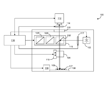
[0015] FIG. 1 shows a block diagram of an exemplary microscope system.
[0016] FIG. 2 shows a schematic diagram of a calibration instrument
constructed in accordance
with principles of the present invention.
[0017] FIG. 3 shows front, back, left side, right side, top, and bottom views
of an exemplary
calibration instrument constructed in accordance with principles of the
present invention.
[0018] FIG. 4 shows an exploded top perspective view of the calibration
instrument of FIG. 3.
[0019] FIG. 5 shows temporal variation of excitation lamp intensity obtained
for different
calibration instruments in accordance with principles of the present
invention.
- 5a-
CA 02690633 2014-06-25
[0020] FIG. 6 shows temporal variation of qualitative results for multiple
channels of a first
calibration slide.
[0021] FIG. 7 shows temporal variation of qualitative results for multiple
channels of a second
sample slide.
[0022] FIG. 8 shows a flow diagram of a process for standardizing qualitative
analysis results in
accordance with principles of the present invention.
[0023] FIG. 9 shows a flow diagram of a process for obtaining correction
factors used in the
standardization of qualitative analysis results of FIG. 8.
- 5b -
CA 02690633 2009-12-14
WO 2008/156669 PCT/US2008/007399
[0024] FIG. 10 shows comparative results obtained using different microscope
systems without and with correction accordance with principles of the present
invention.
[0025] FIG. 11A and 11B show comparison of uncorrected and corrected sample
data obtained in accordance with principles of the present invention.
[0026] FIG. 12A shows exemplary qualitative analysis results obtained from the
same sample using different microscope systems without standardization.
[0027] FIG. 12B shows correction of the qualitative results of FIG. 12A in
accordance with principles of the present invention.
DETAILED DESCRIPTION
[0028] Systems and processes are described herein for obtaining standardized
quantitative analytical results from a system including optical components and
an
illumination source. In particular, the systems and processes related to
standardizing
quantitative, microscopic analysis of target samples, such as biological
samples.
Exemplary biological samples include a cell, a group of cells, a tissue
section, an
array of tissue sections (e.g., a micro tissue array) and combinations of one
or more of
any of these. Biological samples can be treated with one or more stains. In
some
instances, the stains are immunohistochemical stains. In some instances, the
immunohistochemical stains are fluorescent stains.
[0029] Generally, a microscope system includes an illumination source
configured to
illuminate a target sample, optics configured to produce a magnified image of
the
illuminated target sample, and a detector, such as a digital camera,
configured to
capture a digital image of the magnified image. Quantitative results can be
obtained
through manipulation of the captured digital images. Such image manipulation
can
include image processing techniques known to those skilled in the art. In at
least
some embodiments, one or more of such image capture and image manipulation is
accomplished with the aid of a processor. The processor can include a computer
implementing pre-programmed instructions.
[0030] The system also includes a calibration device configured to redirect a
standardized sample of the illumination source to the detector. In at least
some
-6-
CA 02690633 2009-12-14
WO 2008/156669 PCT/US2008/007399
embodiments a system processor is configured to determine a correction factor
for a
given microscope. The correction factor can be determined from a measurement
of
the standardized sample of the illumination source obtained using the
calibration
device. The correction factor can be used (e.g., by the processor) to correct
for any
variations in intensity of a detected image of the target sample. In some
embodiments, a system processor is configured with instructions (e.g.,
software) for
obtaining the calibration factor. Alternatively or in addition, the system
processor is
configured with instructions for using the correction factor to correct
detected images.
Such calibration is useful to remove from any quantitative results,
variability in
intensity of the illumination source within the same microscope system, as may
occur
over time, and between quantitative results obtained using different
microscope
systems and/or different illumination sources.
[0031] In some embodiments, the calibration device includes a scattering
surface
positionable along an optical path between the illumination source and the
detector so
as to direct a scattered portion of light from the illumination source toward
the
detector. Variability in the detected scattered illumination can be used to
develop
such a correction factor.
[0032] In other embodiments a system processor is configured to determine or
access
a correction factor for the optical component of a given microscope. The
correction
factor can be determined from a measurement of the standardized sample using
the
optical component of the microscope. The correction factor can be used (e.g.,
by the
processor) to correct for any variations in optical features of a given
microscope
impacting the intensity of a detected image of the target sample.
[0033] Microscope System
[0034] Systems and processes described herein are general applicable to any
microscopy system incorporating an illumination source. Examples of at least
some
microscope systems in which the systems and processes can be used included
optical
microscopy, fluorescent microscopy, and confocal laser scanning microscopy. An
exemplary microscopy system is the PM-2000Tm instrument commercially available
from HistoRx, Inc., of New Haven, CT. The systems and processes are
particularly
useful for systems geared towards providing a semi-quantitative or
quantitative result.
-7-
CA 02690633 2009-12-14
WO 2008/156669 PCT/US2008/007399
Exemplary applications include the use of immunohistochemistry (IHC) as used
within the field of pathology (See, for example, Immunohistochemistry and
Quantitative Analysis of Protein Expression, by M. Cregger et al., Arch.
Pathol. Lab.
Med., Vol. 130, July 2006 at pgs. 1026-1030). Typically, these results are
based on
the intensity of staining of a sample examined using the microscopy system.
Samples
can be biological specimens. Stains can be general histological stains,
special stains,
IHC, FISH, chromogenic, fluorescent, etc.
[0035] According to Cregger et al., a diagnostic pathologist typically
interprets IHC
according to a subjective approach by using a binary positive-negative end
point or a
3- to 4- point scale. With the assistance of a computer, an automated analysis
can be
obtained for target samples using a computer program to help eliminate the
inherent
variability of pathologist-based scoring. In immuno-fluorescence, a
fluorescent
product is deposited at the site of an antigen, allowing for visual
localization of the
antigen in the sample. After photographic capture, the reaction product may be
quantified by image-analysis software. Furthermore the antigen may be located
in a
specific cellular (e.g., nuclear, organellular, cytoplasmic, membranous) or
extra-
cellular location (See, for example Camp et al, Nature Medicine 8(11) 1323-
1327,
2002) Numerous computer-based programs have been designed for analysis of IHC,
such as BLISS and IHCscore available from Bacus Laboratories, Inc. of Lombard,
IL,
ACTS of Clarient, Inc of San Juan Capistrano, CA, and AQUAS analysis of
HistoRx,
Inc. of New Haven, CT.
100361 Generally, for fluorescent IHC, multiple digital images (e.g., TIFF,
JPEG,
GIFõ bitmap, PNG) are obtained from the same target tissue sample stained with
protein biomarker-specific antibodies and secondary fluorescent detection
reagents.
When optimized, the fluorescent stains provide a broader dynamic range than
available by absorbance-based chromogenic stains. Each of the digital images
can be
obtained using a different optical filter configured to pass a respective one
of the
secondary fluorescent signals. Thus, at least one respective digital image is
obtained
for each of the secondary fluorescent signals. For quantitative analysis, the
captured
digital images are manipulated (e.g., using image processing software) to
obtain a
respective score of the tissue sample.
-8-
CA 02690633 2009-12-14
WO 2008/156669 PCT/US2008/007399
[0037] More specifically, the systems and processes are described in reference
to an
exemplary system illustrated in FIG. 1.
[0038] Referring to FIG. 1, an exemplary reflected-light fluorescent
microscope
system 100 includes a an excitation source 102, an objective lens 104, a
sample
supporting stage 106, a filter block 108', and an observation head 110. The
sample
supporting stage 106 is configured to support a sample under test along an
optical axis
and within a focal plane of the objective lens 104. The filter block 108' is
also
located along the optical axis between the objective lens 104 and the
observation head
110. The filter block 108' is a three port device with two opposite ports
disposed
along the optical axis and a third port disposed off-axis. As illustrated, the
third port
can be orthogonal to a line joining the two opposite ports.
[0039] Illumination from the excitation source 102 is directed toward the
orthogonal
port of the filter block 108'. The filter block 108' redirects a portion of
the
illumination from the excitation source 102 toward the objective lens 104. The
objective lens 104 preferably includes a relatively high numerical aperture
thereby
allowing it to capture a substantial portion of excitation light. The
objective lens 104
functions as a condenser directing excitation light toward a sample under test
placed
upon the stage. In some embodiments, multiple objective lenses 104 (e.g., 4X,
10X,
20X, 40X, 60X) are included within a single nosepiece (not shown). The
nosepiece
can be manipulated to selectively bring different ones of the multiple
objective lenses
104 into alignment with the optical axis to adjust magnification of the sample
under
test.
[0040] Illumination (emission) from the sample under test travels along the
optical
path through the objective lens 104 and into a first one of the opposite ports
of the
filter block 108'. At least a portion of the sample illumination continues
along the
optical path, exiting a second one of the opposite ports of the filter block
108' towards
the observation head 110. As described in more detail below, the filter block
108'
selectively filters illumination passed therethrough. In fluorescence
microscopy,
filtration can be used to selectively view emissions from different
fluorophores (e.g.,
red, green, blue). As illustrated, the microscope system 100 can include
multiple
filter blocks 108', 108", 108" (generally 108), each filter block 108 being
tuned to
-9-
CA 02690633 2009-12-14
WO 2008/156669 PCT/US2008/007399
pass a selected emission wavelength toward the observation head 110. The
different
filter blocks 108 can be stored within a carousel or turret 115, allowing for
rapid
selection of a different filter block 108 without disturbing the sample under
test. In
some embodiments, the different filter blocks 108 are radially disposed within
the
turret 115 about an axis of rotation. The turret 115 is positioned with its
axis of
rotation parallel and to a side of the optical axis, such that one of the
filter blocks 108'
is aligned with the optical axis. Rotation of the turret 115 selectively moves
one filter
block 108' out of alignment and brings another one of the filter blocks 108",
108"
into alignment with the optical axis.
100411 The observation head 110 directs at least a portion of light from the
filter
block 108 toward an image collection device, such as a charge coupled device
(CCD)
camera 112. In some embodiments, the observation head 110 additionally
includes
one or more eyepieces (not shown) allowing for manual observation of the
sample
under test. Such an eyepiece can be used to adjust placement of a sample 107
upon
the stage 106 and to coordinate positioning of the stage 106 before and during
test. In
some embodiments, a first shutter 117 is provided to control exposure time of
the
sample 107 to the excitation source 102. A second shutter 114 is provided to
control
exposure time of an imaging device, such as the CCD camera 112. As shown, the
shutter 114 can be an independent component located along the optical path
between
the sample under test and the observation head 110. Alternatively or in
addition to an
independent shutter 114, the shutter can be integrated into the CCD camera
112.
100421 The microscope system 100 also includes a controller 116. The
controller
116 can be used for controlling the overall image acquisition process.
Preferably, the
controller 116 is in communication with one or more sub elements of the
microscope
system 110 to allow automated control of the system 100. In the exemplary
embodiment, the controller 116 is in communication with one or more of the
excitation source 102, an axial translator 119 (focus adjust) of the objective
lens 104,
the CCD camera 112, the shutter 114, the turret 115, and a stage positional
controller
118. The controller 116 can include at least one microprocessor or computer
116
operating under the control of program code.
-10-
CA 02690633 2009-12-14
WO 2008/156669 PCT/US2008/007399
[0043] In operation, the controller 116 may send a signal to the stage
positional
controller 118 to position the stage 106, such that a selected region or spot
109 of the
sample under test is brought into alignment with the optical axis. The
controller 116
may also send a signal to the axial translator 119 configured to position and
reposition
the objective lens 104 along the optical axis with respect to the stage 106.
For
embodiments including a motorized nosepiece, the controller 116 may send a
second
signal to the nosepiece causing it to rotate a selected one of multiple
objective lenses
104 into alignment with the optical axis prior to focusing. The controller 116
may
also send a signal to the turret 115 causing a controlled rotation of the
turret to select
one of the multiple filter blocks 118. In response, the turret 118 rotates,
bringing the
selected one of the filter blocks 118 into alignment with the optical axis.
The
controller 116 next sends a signal to the excitation source 102 turning the
source 102
on, at least momentarily, to illuminate the sample under test. The shutter 114
is
normally closed blocking the optical path between the sample under test and
the CCD
camera 112. For some microscopes the light source 102 is turned on during
initialization of the instrument. With fluorescent microscopes, the high-
intensity
lamps require a warm-up period to allow intensity of the source 102 to
stabilize before
any test samples are measured.
[0044] For such fluorescent systems, the light source 102 may remain on during
operation. In such applications, a first shutter 117 provided between light
source 102
and test sample is used to block illumination of the sample until ready to
view the
sample and acquire an image of the sample. Such limited exposure of the test
sample
to illumination may avoid bleaching of the sample. Optionally, a second
shutter 114
is provided within the CCD camera 112. Upon receiving a trigger signal from
the
controller 116, the first shutter 117 opens for a predetermined exposure
period before
closing again. A second trigger signal from the controller is sent to the
second shutter
114 associated with the CCD camera 112. This signal controls exposure allows a
controlled sample of emission from the sample under test to reach the CCD
camera
112. Thus, the first shutter 117 is open for at least the entire duration of
an exposure
controlled by the second shutter 114. In some embodiments, operation of the
two
shutters 114, 117 can be controlled by a common signal, or otherwise
configured to
-11-
CA 02690633 2009-12-14
WO 2008/156669 PCT/US2008/007399
operate in synchronization. Under control of the controller 116, the CCD
camera 112
captures an electronic image of illumination from the sample under test. The
image
can be forwarded to the controller 116 or to an external system for analysis.
[0045] With optional independent control of the two shutters 114, 117, timing
of
each shutter can be varied to produce different effects. For example, in some
embodiments, the first shutter 117 is opened to expose test sample for a
predetermined period of time and then closed. This can be performed to expose
a
luminescent test sample to illumination from the source 102. The second
shutter 114
could be operated after closure of the first shutter 117 to sample
luminescence of the
sample, without interference from source illumination.
100461 In one particular embodiment, the a fluorescent microscope system is
part of
an integrated quantitative IHC analysis system, such as the AQUAS analysis PM-
2000Tm system, commercially available from HistoRx, Inc. of New Haven, CT.
AQUA is a registered trademark of HistoRx, Inc. The IHC analysis system
consists
of the following components assembled in a light-tight enclosure: a
fluorescent
microscope, such as the Olympus BX51 epi-fluorescence microscope, commercially
available from Olympus America, Inc. of Center Valley, PA; the microscope is
equipped with a motorized nosepiece to control selection among different
objective
lenses (e.g., 4X, 10X, 20X, 40X, 60X), and a motorized filter turret to
control
selection among different filter cube selection (e.g., in DAPI, Cy2, Cy3, Cy5
and Cy7
or equivalent wavelengths). The system also includes a motorized stage, such
as the
Prior Scientific part no. H101A. The PCI card that drives the stage is Prior
Scientific
part no. H252 motorized stage commercially available from Prior Scientific,
Inc. of
Rockland, MA. The control card occupies a PCE expansion slot within a computer
controller. Focus control is facilitated by integrated software. The system
also
includes a light source, such as the X-CITE 120 system, commercially available
from
EXFO Life Sciences & Industrial Division of Ontario, Canada, which is equipped
with a mercury/metal halide lamp; a monochromatic digital camera for images
capture, such as the QUANTIFIRE camera, commercially available from
OPTRONICS of Goleta, California; and a computer controller. In the exemplary
-12-
CA 02690633 2009-12-14
WO 2008/156669 PCT/US2008/007399
embodiment, the computer is a personal computer running WINDOWS XP or higher
operating system environment.
[0047] Instrument Standardization
[0048] In order to standardize quantitative results obtained using a
particular
system, a system intrinsic factor can be determined to account for intensity
variability
of the excitation source and device variability i.e., along the optical path.
In order to
achieve this, a measurement of the intensity of the excitation light source
may also be
obtained for example by using an inline lamp intensity measuring tool. Also a
measurement of a standard or a calibration sample i.e., a calibration
microscope slide
may be obtained using the particular system to define one or more optical path
factors. Use of such a calibration slide is particularly useful for
fluorescence-based
IHC applications, in which sample fluorescent regions of the calibration slide
emit
radiation within respective bandwidths. The fluoresced emissions allow for
characterization of an optical path at each of the one or more respective
wavelengths.
These measurement can be obtained simultaneously or separately.
[0049] Light Source Intensity Measurement
[0050] Generally, a process or instrument to provide for direct measurement of
the
light source intensity is most conveniently incorporated into the system. A
light
source sampling instrument provides for capturing a sample of the light source
intensity. In some embodiments, a sampled portion of the light source
intensity is
directed to a detector (e.g., a camera). The light source intensity
measurement can be
accomplished independent of the optical portion of the system.
[0051] More generally, the sampling process or instrument accesses a sample of
the
light source at intensity levels below alight source detector saturation
threshold and
above a noise level. For example, the light source intensity can be sampled by
an
electronic sensor within an exposure period (e.g., 10 milliseconds).
Alternatively or
in addition, the light source can be attenuated to ensure that the obtained
sample falls
within the sensitivity range of a given detection device.
[0052] The sampled light source intensity can be accomplished using an in line
radiometer, resulting in a measurable voltage representative of the light
source
intensity. Such measured voltages can be processed automatically by the
system. For
-13-
CA 02690633 2009-12-14
WO 2008/156669 PCT/US2008/007399
example, the voltages can be sent to a processor for further processing. In
some
embodiments, the voltage levels are converted into a digital representation of
the
voltages. Such conversion can be accomplished using analog-to-digital
conversion,
allowing for digital processing of the sampled voltage. The digital processing
can be
accomplished by one or more of software running on the processor and hardware
adapted for digital signal processing.
[0053] The sampled light source intensity can be obtained directly or
indirectly
from the light source. In some embodiments, the sampled light source intensity
is
obtained independent of at least some other parts of the system, such as the
optics
(e.g., an objective lens), that may independently impact the sampled result.
Alternatively or in addition, the sampled light source intensity is measured
at light
source itself, thereby avoiding any effects of the microscope.
[0054] Calibration Cube
[0055] In some embodiments, a special calibration instrument can be used for
the
purpose of obtaining a sample of the light in order to measure the intensity
of a light
source. Preferably, the calibration instrument allows a relative light
intensity
measurement to be obtained substantially simultaneously with the target sample
image. In at least some embodiments, this is accomplished by switching a
special
calibration instrument into the optical path to obtain the relative light
intensity
measurement, and then out of the optical path to obtain the sample image. For
example, if the microscopy system is a fluorescent system using multiple
filter cubes
pre-loaded in a rotatable turret 115, the calibration instrument can be
included as one
of the filter cubes (i.e., a calibration cube) within the turret 115. This
will allow for
the calibration cube to be interchanged with the other filter cubes
automatically during
the course of measurements.
[0056] Generally a filter cube has openings on the top, bottom, and front
faces of a
cube-shaped frame. The front opening or port allows light from the
illumination
source to enter the cube, after which the light is reflected off an internal
reflective
surface generally positioned at 45 degrees to the axis of the entering light.
The angled
reflective surface (e.g., mirror) redirects a reflected portion of sampled
light toward
the bottom opening or port of the cube. In operation the redirected light may
be used
-14-
CA 02690633 2009-12-14
WO 2008/156669 PCT/US2008/007399
to illuminate a target (a tissue sample, etc.). The redirected light travels
along an
optical path that may include objective optics as provided in microscope
systems. At
least some portion of the illuminating light may be reflected from the sample.
For at
least some applications, stimulated light may also be emitted from the sample,
as
through fluorescence. In either instance, at last a portion of light from the
sample
(reflected and/or emitted) travels back along the same optical path, entering
the cube
from the bottom port. At least a portion of the light entering the calibration
cube
travels through the angled reflective surface of the angled mirror along the
optical
path and exits through the top opening or port of the cube and to an imaging
device.
[0057] In some embodiments, the calibration cube is a modified filter cube in
which
a light scattering surface is affixed to block the bottom opening. In
operation, light
entering the cube is reflected off the internal angled reflective surface and
directed
toward the light scattering surface. The reflected light illuminates the light
scattering
surface. At least a portion of scattered light from the light scattering
surface is
directed back up through the internal reflective surface, exiting at the top
opening of
the cube toward the imaging device. The same calibration cube having the same
light
scattering surface can be used to sample light from the same illumination
source at
different times and/or different illumination sources. In this manner the
calibration
cube provides a means for sampling light intensity scattered off of a
standardized
surface (instead of the typical sample) to be captured by the imaging device,
and
usable to determine a standardized light intensity measurement. Beneficially,
such
sampling can be accomplished without repositioning one or more of the target
slide
and the objective lens.
[0058] The calibration cube serves as an in-line access tool for measuring
intensity
of the lamp. In cooperation with a processor 116 (FIG. 1), the intensity
measuring
tool not only allows for tracking lamp intensity deviations, but also enables
a
straightforward means of normalizing quantitative results. Accounting for such
lamp
intensity deviations promotes precision measurements of biomarker expression
in a
tissue sample. For example, data from captured images obtained by several
microscopy systems equipped with identically constructed, standardized,
calibration
cubes, can be corrected to effectively eliminate any contributions that would
-15-
CA 02690633 2009-12-14
WO 2008/156669 PCT/US2008/007399
otherwise have been attributable to lamp intensity variations. Thus,
quantitative
analysis results, such as AQUA scores obtained from corrected images may be
compared for a reliable indication of target sample differences, not system
differences. Light source calibration data obtained using the calibration cube
can be
collected, stored and accessed from various system software applications, such
as
system initialization and setup programs, image acquisition programs allowing
for
minimal user interaction and negligible time and cost.
[0059] In one embodiment, referring to FIG. 2, a calibration instrument, or
cube 130
includes a housing 132 including a first port 134a and a second port 134b
opposite the
first and aligned therewith along a common optical axis. The housing 132 also
includes a side port 134c that is not aligned with the optical axis. As
illustrated, the
side port 134c is orthogonal to the optical path. The housing 132 also
includes an
internal reflective surface 136 forming a nonzero angle 0 with the optical
axis.
Illumination is received from an excitation source 102 through the side port
134c.
The reflective surface 136 is angled to redirect a portion of the received
excitation
light along the optical axis, through the second port 134b. The calibration
cube 130
also includes a light scattering surface 137 positioned relative to the second
port 134b
to scatter, or return excitation light in an opposite direction along the same
optical
axis. At least a portion of the scattered excitation light passes through the
reflective
surface and exits the housing 132 through the first port 134a. This scattered
light can
be detected by a CCD camera 112 aligned with the first port 134a.
[0060] In alternative embodiments, a calibration instrument can be formed
without a
mirrored surface. For example, considering the same general structure of the
cube
130 illustrated in FIG. 2, the internal reflective surface 136 can be replaced
by a light
scattering surface. The light scattering surface can be angled within the cube
to
promote redirection of scattered light from the illumination source 102
through the
side port 134c.
[0061] The light scattering surface 137 is generally uniform, having a surface
that is
a minimally reflective surface (e.g., a matte surface) and provides uniform
reflectance/fluorescence across the field of view which provides for also
measuring a
uniformity of sampled light from the light source. In at least some
embodiments, the
-16-
CA 02690633 2009-12-14
WO 2008/156669 PCT/US2008/007399
light scattering surface 137 scatters light substantially uniformly. Material
forming
the light scattering surface 137 should be able to withstand high temperatures
and
light intensity without degradation or variation. The material is preferably
rigid or at
least semi-rigid and not susceptible to yellowing, degradation, or photo
bleaching.
Furthermore the material should be reproducible, and relatively inexpensive.
In some
embodiments, certain metallic, ceramic or plastic materials meeting these
conditions
are acceptable. Ceramic materials, such as gold and white ceramic targets are
commercially available from Avian Technologies, Inc. of Wilmington, OH. Such
materials can be used for the calibration cube filter 148 (FIG. 3).
Alternatively or in
addition, such materials can also be used for standard calibration slides. In
an
alternative embodiment a piece of flat filter paper may be used.
[0062] In fluorescent microscope applications, emission light detected by the
CCD
camera 112 is substantially lower in intensity than the excitation light. In
order to
avoid saturation of the CCD camera 112 when detecting the excitation source
itself,
one or more filters are included between the excitation source and the camera
112 to
attenuate the light to a sufficiently low level. In some embodiments, one or
more
neutral density, or gray filters are provided along an optical path between
the
excitation source 102 and the CCD camera 112. For example, a first neutral
density
filter 138a is provided at the side port 134c attenuating excitation light
entering the
housing 132. A second neutral density filter 138b is provided at the first
port 134a
attenuating scattered light returned to the CCD camera 112. The attenuation
values of
each filter 138a, 138b can be the same or different, as long as their combined
effect
ensures that the CCD camera 112 will not be saturated by scattered light from
the
excitation source 102.
[0063] EXAMPLE: In an exemplary embodiment of the calibration cube 130
shown in FIG. 3, the cube 130 consists of a regular OLYMPUS filter housing or
holder 140 (OLYMPUS Part no. U-M610, U-MF2 BX filter holder cube) equipped
with neutral density filters 142a, 142b at the emission and excitation
openings 144a,
144b and a 50/50 dichroic mirror 146. In some embodiments, the filters 142a,
142b
are retained in proximity to the openings 144a, 144b using respective filter
frames
145a, 145b (FIG. 4).
-17-
CA 02690633 2009-12-14
WO 2008/156669 PCT/US2008/007399
[0064] The bottom of the housing 140 has a light scattering surface 137 (FIG.
2);
148 (FIGS. 3, 4) mounted over the port 144c (FIG. 3) positioned to completely
block
the sample opening 144c. A top perspective exploded view of the exemplary
calibration cube is shown in FIG. 4.
[0065] EXAMPLE: In an exemplary embodiment, the calibration cube 130
includes two neutral density filters, 25 mm, such as Chroma cat. no. 2200a,
commercially available from Chroma Technology Corp. of Rockingham, VT. The
cube 130 also includes a dichroic mirror, 50/50 beam splitter, such as Chroma
cat. no.
21000, housed within a filter holder (cube) 140, such as Chroma cat. no.
91018. The
scattering surface can include filter paper 148, such as VWR cat # 28306-153,
commercially available from VWR of West Chester, PA. Which particular brand of
filter paper used is not important, but preferably the same filter is used
among all
calibration cubes 130 of different microscope systems to ensure uniformity of
results.
As will be described below, even this is not critical, as relative
measurements can be
made for calibration cubes 130 using different filter paper 148 compared to a
common, or standardized calibration cube. Such a comparison can be used to
determine an offset to be accounted for in the correction process.
[0066] In other embodiments, the calibration cube 130 includes a microscope
filter
holder, such as OLYMPUS Part no. U-M610, U-MF2 BX filter holder cube,
commercially available from Chroma Technology Corp of Rockingham, VT, cat #
91018. Other commercially available filter cubes compatible with the
microscope
system may be used. In a particular example, the neutral density filters are
ND 1.0
Part no. UVND1.0, ND 1.0 neutral density filter, 10% transmittance, 25 mm, and
ND
2.0 Part no. UVND2.0, ND 2.0 neutral density filter, 1% transmittance, 25 mm,
commercially available from Chroma Technology Corp. The 50/50 beam
splitter/dichroic is Chroma Part no. 21000, 50/50 beam splitter, 38x26 mm.
WHATMAN Filter Paper, Grade 1, Cat No. 1001-125, VWR of West Chester, PA is
affixed to the bottom of the cube. Beneficially, the filter material scatters
an
appropriate amount of light back towards the CCD camera 112, such that an
image
can be acquired by the camera 112 in a reasonable exposure period. For
example, the
exposure period can be chosen between approximately 3 and 200 milliseconds.
Other
-18-
CA 02690633 2009-12-14
WO 2008/156669 PCT/US2008/007399
exposure periods can be selected outside of this range, provided they are
appropriate
for the applicable camera capabilities.
[0067] Scattered light received at the CCD camera 112 is preferably below the
level
of camera saturation for the exposure time selected with consideration given
to
variations in other cube assemblies which may be brighter or dimmer. Less
desirable,
but acceptable, is a scattering material that provides usable signal (below
the limit of
camera saturation) for exposure periods greater than about 200 milliseconds.
[0068] For use in standardization of systems, the specially designed
calibration filter
cube 130 can be installed within the turret of the microscope system 100 (FIG.
1).
This calibration cube 130 serves as an inline lamp intensity measuring tool
that
provides a means for measuring excitation light intensity, by sampling
scattered light
that is directly proportional to the incident excitation light source
intensity. Thus, the
sampled scattered light can be used to track variation in light intensity of
the
excitation source during use. Such variations in intensity might occur from
long-term
effects of the excitation source such as aging, in which intensity of the
source may be
diminished slowly during the normal process of aging. Variations may also
result
from short term effects that may result from other effects, such as ambient
temperature variations, device temperature variations from device heating, and
excitation voltage and current among others.
[0069] During image acquisition in which a sample of the illumination source
light
intensity is obtained, the light traveling through the excitation neutral
density filter
142a is attenuated, passed through the beam splitter 146 and reflected off of
the
calibration material 148 (i.e., white paper target). Reflected (or scattered)
light is then
further attenuated at the emission neutral density filter 142b and then
captured by the
camera 112 (FIG. 4). The neutral density filters 142a, 142b are arranged such
that the
excitation light is highly attenuated to reduce the intensity impinging on the
calibration material 148. The emission filter 142a allows more light through
and thus
helps reduce intensity observed by the digital camera 112.
[0070] Calibration Cube Standardization (CC)
[0071] Individual calibration cubes may have intrinsic variations due to
material
differences that are preferably accounted for in order to normalize
quantitative results
-19-
CA 02690633 2009-12-14
WO 2008/156669 PCT/US2008/007399
obtained across instruments. In manufacturing a universal standard cube may be
identified. Thereafter all manufactured cubes are compared to the universal
standard
cube by sampling of a consistent light source with each new cube and any
inherent
differences are accounted for, i.e., by applying a cube correction or cube
calibration
factor (CC). The cube calibration factor is preferably determined initially
for every
new calibration cube. In some instances, the cube calibration factor can be
determined periodically thereafter for system maintenance, and when material
properties of a cube may have changed (i.e., due to aging).
[0072] EXAMPLE: In order to characterize a number of similar calibration
cubes,
results were obtained for each of a batch of five cubes (J145) using the same
excitation source and camera configuration. A specific one of the cubes (i.e.,
J5) was
designated as a reference cube for a group. This cube could be referred to as
a
universal standard cube. The reference cube J5, along with the other cubes to
be
tested, were installed simultaneously into the turret 115 of the microscope
system 100
(FIG. 1). Images of the sampled illumination from the illumination source
obtained
through the calibration cube 130 were acquired by the digital camera 112 for
each
cube J145. Light intensity measurements so obtained were compared between the
different calibration cubes being tested. A ratio of intensity measured for
each test
cube J1-J4 to the intensity measured using the reference cube J5 was
determined
representing a cube calibration factor (CC). In an exemplary experiment,
sixteen
measurements were collected for each of the different cubes J1-J5. A ratio of
the
intensity obtained for each cube J1-J5 to intensity of the reference cube J5
was
determined. Table 1 shows the CC values determined for the five cubes as
compared
to the reference cube (J5). Since the construction of the cubes J1-J5 was
similar, the
ratios of the intensities are all close to 1.
-20-
CA 02690633 2009-12-14
WO 2008/156669 PCT/US2008/007399
[0073] Table 1: Cube Calibration Factor (CC Values)
Calibration Cube No. CC
CC2 0.894
J1 0.929
J2 0.989
J3 0.907
J4 0.883
J5 (reference cube) 1.000
[0074] To determine temporal effects, light intensity measurements were
measured
for each cube using the same instrument over a two-day period. A minimum of
ten
measurements were taken through each cube on each day (sum of pixel
intensities in
the captured image). The results are shown graphically in FIG. 5 as an average
total
intensity on a scale of 0 to 120,000. The results show that light source
intensity
fluctuations were recognized in the measurements using all cubes J1-J5.
Preferably,
correlation coefficient R2 values between all cubes are relatively high (e.g.,
> 0.95).
The test results suggest that during actual specimen image acquisition, the
intensity of
the light source can fluctuate. These fluctuations can occur over a short
duration each
time the lamp is ignited and as slow variations occurring over time while a
lamp
remains ignited.
[0075] Light Source Standardization (LS)
In an exemplary procedure for determining a light source standardization LS,
pixel intensities of a captured image of the illuminated calibration surface
are
combined in a sum. Different ranges can be identified depending upon the
particular
intensity scale values used for the pixels, as well as the number of pixels in
the image.
Exemplary LS values obtained using the AQUAS system range from about of 20,000
to 120,000. A ratio can be formed from the LS factor and a chosen intensity
value.
Such a ratio can then be used to compensate target sample data to essentially
remove
light source variation. In an exemplary embodiment, a ratio is formed using a
chosen
value of 100,000 and an LS factor falling within the AQUAS system range.
[0076] Device Optical Path (OP) Measurement
-21-
CA 02690633 2009-12-14
WO 2008/156669 PCT/US2008/007399
Generally an optical path correction procedure uses a calibration sample
(e.g.,
a calibration slide) providing a known reflectivity and/or fluorescence that
is usable in
direct measurement of the specific device or system's optical path
performance. A
calibration sample can be used to obtain a correction factor for the intrinsic
optical
path performance of a given microscopy system. When different microscopy
systems
are similarly corrected, target sample results obtained from the different
systems may
be compared reliably across the different microscopy systems.
[0077] Most generally the calibration sample has the following
characteristics:
= displays some characteristics of the samples typically analyzed on the
system (i.e., for fluorescent systems, these characteristics can include
wavelength of excitement/emission);
= constructed using a uniform material, with optical properties (e.g.,
reflectivity) that are reproducible, available, and inexpensive;
= for at least the fluorescent applications, the uniform material can be
opaque (i.e., a ceramic, etc);
= for bright field transparent applications, the uniform material
attenuates light source, if necessary, to an acceptable level for the
detector; and
= provides minimal bleaching (for fluorescent systems).
[0078] Variations along an optical path of a given microscopy system will not
likely
vary to any significant degree over time for the same system. Thus, there is
no
apparent need to re-perform the optical path correction procedure during
normal
operations. In at least some embodiments, the optical path correction factor
is
determined at the time of manufacture. The optical path correction factor can
be re-
determined after servicing (e.g., cleaning) of the microscopy system.
[0079] Control or Calibration Slide
[0080] A standardized instrument calibration sample (control slide or
calibration
slide) is used for acquiring data in a particular system to be calibrated to
approximate
the light throughput efficiency of a specific microscope system and optical
configuration.
-22-
CA 02690633 2009-12-14
WO 2008/156669 PCT/US2008/007399
For example, the control slide can be a Fluorescent Reference Slide Set XF900,
providing a blue or green fluorescent reference slide commercially available
from
Omega Optical Inc. of Brattleboro, VT. Other uniform sample materials may be
used
so long as sufficient signal in each channel (i.e., wavelength) may be
acquired within
the set exposure time (e.g., between about 3 and 1000 milliseconds, or current
range
of the CCD camera 112) and the material preferably demonstrates minimal
bleaching
over a standard number of runs. The sample material should be reproducible
such
that it can be used for standardizing each instrument and sized to fit on the
microscope stage. The material preferably emits or reflects a light signal
with spectral
components in the appropriate wavelength band(s) to be acquired through each
filter
cube in use on the microscope system, in an environment of low specular
reflection.
Other examples include, but are not limited to, alternative colored plastics,
paper and
ceramic reflective materials, metallic surface or surfaces coated with various
inks and
dyes.
[0081] EXAMPLE: A calibration slide was placed on the stage of a microscope
system 100 previously fitted with a pre-standardized calibration cube 130
(FIG. 2) in
the filter turret 115 (FIG. 1). Two different instrument control slides were
tested:
sample 1 having a spectra approximating FITC/GFP (green excitation) and sample
2
having a spectra approximating DAPI/Indo/Fura (Blue excitation). Calibration
slide 1
was illuminated, and an image obtained of the fluorescence emission of the
sample
through each of three different filter cubes 108 (FIG. 1), one for each
channel and the
calibration cube. Over 900 iterations were performed over a period of about
thirty
hours. Quantitative results, For each channel per iteration an intensity score
("derived
AQUA score") was calculated: mean intensity multiplied by exposure time
multiplied by bit depth (i.e., 0-255) The light intensity through the FITC
channel
when using sample 1 was too bright indicating this material is not ideal for
normalizing light fluctuations when acquiring in this channel, see results
described for
sample 2 below as an alternative. The results were graphed as the AQUA
analysis
score verses iteration for slide 1 (FIG. 6) for calibration slide intensity
scores obtained
in each Cy3, Cy5 channels and separately for the calibration cube. Essentially
an
identical light intensity pattern was obtained using instrument control sample
1 in the
-23-
CA 02690633 2009-12-14
WO 2008/156669 PCT/US2008/007399
Cy3, Cy5 and calibration cube channel indicating the variability is unlikely
due to
bleaching of the instrument control slide material. Rather, variation is
indicative of
true light intensity variability inherent to the system 100. A subtle long-
term decrease
in quantitative measurements is observable over at least the first half of the
samples.
Superimposed on this are relative short-term variations in both directions.
Interestingly, similar trends are observable in the measurements obtained for
each of
the different channels independently, suggesting that the variation is due at
least in
part to fluctuations in the intensity of the excitation source.
[0082] Light source variability was further tracked by acquiring images of the
second calibration slide (spectra approximating DAPI/Indo/Fura, blue
excitation)
through Cy3, Cy5, and FITC channels, and the calibration cube over
approximately
20 hours for approximately 450 iterations. In an exemplary embodiment,
quantitative
results, such as the "derived AQUA scores" discussed above, were determined
for
each iteration. The results were graphed as the AQUA analysis score verses
iteration
for slide 2 (FIG. 7). Essentially an identical light intensity pattern
resulted from using
instrument control sample 2 in the Cy3, Cy5 and calibration cube channel
indicating
the variability is unlikely due to bleaching of the instrument control slide
material.
Rather, variation is indicative of true light intensity variability inherent
to the system
100. The light fluctuation pattern seen through the FITC channel when using
sample
2 was on scale and could be used for standardizing when images are to be
acquired in
this channel. Modest bleaching of the slide material is evident as a negative
slope in
the FITC channel over many iterations. Such bleaching is unlikely to adversely
impact images acquired under normal operating conditions. Ideally the
instrument
control slide is replaced after about 10% bleaching has occurred. For example
after
231 runs for sample 1 and approximately 20 runs with sample 2.
[0083] Instrument optical path standardization (OP)
[0084] The measurement of signal at the digital camera 112 results from light
that
has traveled from the excitation source 102 (FIG. 1) through the microscope
system
100 and has been modified by the intrinsic properties of that system 100 and
the
sample 107 being measured. The optical path of an instrument 100 may comprise
the
light pipe which travels from the excitation lamp to the microscope, the
filter cubes
-24-
CA 02690633 2009-12-14
WO 2008/156669 PCT/US2008/007399
108 and associated optics for each fluorescent channel and the objective lens
104
being used. A machine intrinsic factor (OP) that corrects for variations along
this
optical path can be established for each device 100 in order to standardize
results
obtained across devices 100.
[0085] To calculate the machine intrinsic factor for a specific system,
multiple
images (e.g., 16 images) were acquired of a standard instrument control slide
107.
For each system being standardized, the instrument control slide 107 was of
the same
material in the same configuration ¨ presumably to yield the same results, but
for
effects of the system 100. Immediately after camera acquisition of a single
field of
view using a specified light filter cube 108 (e.g., FITC, Cy3 or Cy5 filter
cubes), the
filter turret 115 was turned so as to align the calibration cube 130 (FIG. 2).
The
calibration cube 130 was separately imaged, without disturbing either the
objective
lens or the sample (i.e., control slide 107) under test. Exposure times for
each channel
were fixed. A ratio of the calibration cube intensity to the observed signal
intensity in
each channel provided a machine intrinsic factor for the microscope system
optical
path efficiency for each filter. This value is applicable for that system in
that specific
configuration. The configuration is determined by such features as
magnification,
light filter, and optics.
[0086] The machine intrinsic factor of the system can also be scaled by
referencing
it to a specific value. The machine intrinsic factor was determined using data
obtained using a non-bleached instrument control slide, at exposure times
chosen to
avoid saturation, from multiple runs (e.g., five runs) and in independent
experiments
on different days. The intrinsic value was calculated for each run. The %CV
between
run intrinsic values was extremely low such that one run was effective for
calculating
intrinsic values.
[0087] EXAMPLE: Table 2 shows the resulting machine intrinsic factors for five
instruments across filters for three channels: FITC, Cy3 and Cy5.
-25-
CA 02690633 2009-12-14
WO 2008/156669 PCT/US2008/007399
[0088] Table 2: Machine Intrinsic Factors (OP Values)
Instrument FITC-OP Cy3-0P Cy5-0P
1 1.15 1.14 1.09
2 1.61 1.49 1.86
3 1.46 1.38 1.18
4 1.00 1.00 1.00
1.38 1.07 1.32
[0089] Machine intrinsic factors were further scaled as the intrinsic
value/empirical
value, where the empirical value was the lowest average value recorded on the
particular system. Intrinsic values were determined using two different blue
instrument control slides and were found to be reproducible regardless of
which slide
was used. Average values and percent coefficient of variation (% CV) values
were
calculated. Preferably, the %CV is less than about 20%, more preferably the %
CV is
less than about 5%.
[0090] Standardization
[0091] The standardization factors described above (CC, LS, OP) were used to
transform quantitative data collected on each individual microscope system to
that of
an idealized system. When applied to more than one system, data obtained
therefrom
are normalized, such that any influence of the respective microscope and light
source
fluctuations to the results were mitigated.
[0092] Referring to FIG. 8, a flow diagram of a process for standardizing
qualitative
analysis results includes a preliminary initialization procedure 300 followed
by a test
sample procedure 200. As part of the initialization procedure 300, the
instrument is
setup at step 210. Instrument setup includes configuring the microscope system
100
(FIG. 1) with the appropriate excitation source 102, filter cubes 108 (FIG. 1)
and
calibration cube 130 (FIG. 2), ensuring that the controller 116 (FIG. 1)
includes the
proper program control, and performing any initialization routine that may be
required
for the microscope system 100 and CCD camera 112. Once instrument setup is
complete, the CCD camera 112 is capable of obtaining images of a sample under
test
using the microscope system 100 under control of the controller 116. Once
setup, a
-26-
CA 02690633 2009-12-14
WO 2008/156669 PCT/US2008/007399
correction factor (CC) for the calibration cube 130 and a machine intrinsic
factor (OP)
for the microscope system 100 are obtained at step 220. As described above,
the
machine intrinsic factor (OP) may be determined at the time of manufacture,
and/or at
the time of servicing/repair of the microscopy system and stored for later
use. Thus,
obtaining the machine intrinsic factor OP may included looking up a pre-stored
value.
One or more of these factors (CC, OP) can be stored by the controller 116, or
image
analyzer for later use in analyzing images of test samples.
[0093] As part of the test procedure 200, a sample under test is imaged by the
system 100 at step 230. A light source correction factor (LS) is obtained
during this
step. In more detail, an actual test sample 107 is placed on the microscope
stage 106
and positioned such that a target spot 109 is aligned with an optical axis
including the
objective lens 104 (FIG. 1). This initial alignment can be performed manually
through the observation head 110 (FIG. 1), automatically using the controller
116, or
through a combination of a course manual adjustment followed by a fine
controller
116 adjustments. For test samples including a regular array of target spots
109, the
test sample 107 is preferably aligned once (e.g., for one target spot 109) and
then re-
positioned to test additional target spots 109 of the sample 107 using
preprogrammed
offset adjustments of the stage 106.
[0094] Once the target spot 109 is aligned with the optical axis, the system
100
acquires a sample image of the target spot 109 using the CCD camera 112 (FIG.
1).
For the exemplary fluorescence IHC system, the sample image is obtained for a
chosen wavelength band or channel of interest using a respective one of the
filter
blocks 108 (FIG. 1) corresponding to the channel. The calibration cube 130
(FIG. 2)
is selected through rotation of the turret 115 (FIG. 1). A reference image of
the
excitation source is also obtained using the calibration cube 130 for a
determination of
the excitation source intensity. Additional filter cubes 108 can be used to
obtain
additional sample images through different channels, as required. The
particular
order in which the channels and excitation source samples are obtained can be
varied,
provided that the one or more channel images are related to the excitation
source
reference image (e.g., taken at approximately the same time). Such a
relationship can
-27-
CA 02690633 2009-12-14
WO 2008/156669 PCT/US2008/007399
be accomplished by forming a composite image of the multiple images, or
otherwise
labeling the images to reflect their relationship.
[0095] The sample and reference images can be sent from the camera 112 to the
controller 116 or separate image analyzer for later analysis and correction at
step 240.
Image analysis can include calculating AQUA scores for each of the different
channels. One or more of the correction factors (CC, OP, LS) are applied at
step 250
to obtain corrected or standardized results. The standardized output data for
the
particular target spot 109 of the test sample 107 is provided at step 260. The
test
sample procedure 200 can be repeated for additional target spots 109 of the
same test
sample 107. The test sample procedure 200 can also be repeated for one or more
additional test samples 107 using the same correction factors obtained at step
220.
[0096] In more detail, an exemplary flow diagram of an initialization
procedure 300
for obtaining correction factors used in the standardization of qualitative
analysis
results is shown in FIG. 9. The camera 112 (FIG. 1) and microscope are
respectively
initialized at steps 310 and 320. These steps may be conducted sequentially or
in
parallel. Providers of the camera 112 and microscope system 110 typically
define
these initialization steps 310, 320 in the form of an initialization
procedure. One or
more of the initialization procedures may be automated and occur as part of a
power
on cycle.
[0097] Next, the microscope stage 106 (FIG. 1) is initialized at step 330 to
allow for
proper alignment of test samples during test. In some embodiments, a
calibration
slide including a target sample is placed onto the microscope stage 106 at
step 350.
The calibration slide is selected to provide a known response during
characterization
of the optical path, as may be performed at the time of manufacture, or
servicing of
the microscopy system. An image of the calibration slide is obtained at step
360 and
an optical path correction factor, or machine intrinsic factor (OP) is
determined at step
370. The machine intrinsic factor can be stored for later use during normal
operation
to remove optical path variability between different microscopy systems. This
step of
determining the machined intrinsic factor includes obtaining a sample image of
the
excitation source using the calibration cube 130 (FIG. 2) to determine
intensity of the
calibration source. Preferably, the excitation source sample is obtained
immediately
-28-
CA 02690633 2009-12-14
WO 2008/156669 PCT/US2008/007399
adjacent to the step of obtaining an image of the calibration slide to
minimize the
likelihood of intensity variation between samples.
100981 Next, the calibration cube correction factor (CC) is accessed at step
380.
This value can be stored into the system or manually entered during the
preliminary
initialization process 300. This value can be obtained by comparing results
obtained
from the calibration cube 130 with results obtained using a universally
standard
calibration cube and formulating a ratio of the results. Similar to the
optical path
correction factor, determination of the calibration cube correction factor
need not be
repeated during normal use. As described above, the calibration cube is
preferably
constructed to reduce or eliminate any variability in its performance over
time. Thus,
an initial determination of the calibration cube correction factor can be
obtained at the
time of its manufacture (the factory holds a "gold" standard calibration cube
used in
comparison to manufactured calibration cubes to obtain the correction factor).
The
resulting correction factor can be marked on the calibration cube itself
and/or
provided to the processor for later use during correction of sampled images.
Once
obtained, the machine intrinsic factor (OP) and calibration cube correction
factor (CC)
are output to image analysis software at step 390 (e.g., read from memory
locations
containing pre-stored values). This can include forwarding the factors (OP,
CC) to
the controller 116 or separate image analyzer for storage and later use to
standardize
images of actual test samples. Steps 360 and 380 can be repeated for different
channels of the same calibration slide and output separately to the image
analysis
software at step 390. Thus, the machine intrinsic factor (OP) is determined
and
retained on a per-channel basis and stored separately for later analysis of
test samples
on a per-channel basis using the appropriate machine intrinsic factor. In some
embodiments, steps 360, 380, and 390 can be repeated for the same channel to
allow
for statistical determination of the machine intrinsic factor. Thus, multiple
results for
each channel can be obtained and used to formulate an average result that is
stored for
later use.
[0099] An equation provided below (EQ. 1) was used to standardize results
obtained
for each system 100. The calibration cube correction factor (CC) and machine
intrinsic factors (OP) were determined for each system and stored for later
use in
-29-
CA 02690633 2009-12-14
WO 2008/156669 PCT/US2008/007399
manipulating test results. Next, standard quantitative results (specimen
quantitative
score raw) were obtained for a particular specimen, or sample under test.
Through a
correction process, the raw quantitative score is multiplied by the various
correction
factors, to yield a normalized quantitative result suitable for comparison
among
different systems.
Specimen Quantitative Score normalized =
(specimen quantitative score )*(CC)*(0P) *(100,000/LS)
(Eq. 1)
[0100] The above equation provided a direct multiplicative standardization
scheme
using two constants which were intrinsic to the calibration cube and
microscopy
system (CC and OP). Both of these factors were scaled as described above, such
that
data were standardized to an ideal system. Thus, a calibration cube
approximating the
"gold standard" would result in a CC approaching 1Ø Similarly, an optical
path
approaching a standard reference optical path would also approach 1Ø In the
case of
the light source fluctuation factor (LS), an empirically derived value of
100,000 was
used to define an ideal intensity value for this factor. This value of 100,000
was then
divided by the particular LS value obtained during each measurement. Table 3
shows
the resulting standardization correction factor obtained for five different
instruments.
[0101] Table 3: Standardization Factor for Five Instruments
Instrument Correction: (CC)*(0P)*(100,000)
1 97446
2 116702
3 172794
4 88300
132000
[0102] Using the correction factors described herein (CC, OP, LS), the
quantitative
data obtained using the five different instruments were all correlated to
instrument 1.
The correlation results are illustrated graphically in FIG. 10. As can be seen
from the
-30-
CA 02690633 2009-12-14
WO 2008/156669 PCT/US2008/007399
figure, before correction, the correlations of each instrument to instrument 1
result in
non-overlapping curves varying substantially from instrument 1. After
application of
the correction factors, however, the corrected correlation curves for all five
instruments to instrument 1 overlap substantially, demonstrating a high degree
of
correlation to instrument 1 and to each other.
[0103] EXAMPLE: In use, a test sample can include a tissue microarray (TMA)
including a matrix of tissue samples on a single microscope slide. For
example, a 36
spot tissue microarray including breast cancer tissue samples, BT474, MCF7,
T47D
cell line control samples was stained. The staining protocol involved
deparafinization
in xylene, rehydration through a series of decreasing amounts of ethanol to
pure
water, and antigen retrieval in Tris EDTA. After endogenous peroxidase
blocking
and blocking with background sniper, HER 2, (CB 11) and cytokeratin (Rabbit,
Dako)
primary antibodies were applied and rinsed off after 1 hour. Dako Envision
anti-
mouse and Invitrogen alexa 555 GAR were then applied. After extensive washing,
cy
tyramide was applied. The slides were then washed in TBS/Tween 20. Finally, a
mounting media with Dapi was applied and the slides were dried.
[0104] The fluorescent intensity of staining for HER2 and resulting AQUA score
were collected for each tissue spot of the tissue micro array using
instruments 1 and 2
included in Table 3.
[0105] Scores were standardized using Eq. 2 and Eq. 3 below.
Quantitative Scorenonnalized=
(Quantitative Score raw Instrument 1 /LS Instrumenti
)*97,446 (Eq.
2).
Quantitative Scorenormaltzed=
(Quantitative Score raw Instrument 2 /LS Instrument 2)* 116,702. (Eq. 3)
[0106] AQUA scores for each sample in the 36 spot tissue micro array were
acquired using two different instruments. The raw AQUA scores shown in FIG.
11A
and the normalized AQUA scores shown in FIG. 11B from the 36 spot tissue micro
array, acquired on two instruments were graphed in scatter plot format. The
results
-31-
CA 02690633 2009-12-14
WO 2008/156669 PCT/US2008/007399
show a slope of the regression line of the correlation coefficient of 2.6:1
between the
two instruments using the raw scores. Using the standardization methods of the
invention, a slope of the regression line of the correlation coefficients of
0.98:1
between the two instruments was achieved showing that the distribution of
scores are
not effected by standardization.
[0107] Non-parametric Spearman's rho statistical analysis was used to describe
the
ranking relationship before and after standardization. The analysis was
performed
using the AQUA score data set obtained from two instruments. Rank-orders are
assigned from smallest original value (= rank 1) to highest original value for
each
AQUA score. The correlation coefficient and the P-Value were calculated for
the
standardized and the non-standardized (raw) datasets. The rank order of the
data was
unaffected by standardization (Table 4).
[0108] Table 4. Spearman Rho analysis
Normalized Data Raw Data
Rho: 0.72 0.721
P-Value: <0.0001 <0.0001
[0109] EXAMPLE: The same 36 spot stained tissue micro array described in the
above example was acquired and scored on five different instruments.
[0110] The results indicated that the percent coefficient of variation (%CV)
of the
raw AQUA scores and the standardized AQUA scores for each tissue micro array
tissue spot acquired on the five different instruments shows significantly
better %CV
is achieved by standardization by the methods of the invention.
[0111] Tables 5 and 6 are compilations of the slope of the regression line of
the
correlation coefficient generated with a single slide run on the five
instruments.
Correlations based on raw AQUA scores are shown in Table 5 and those based on
standardized AQUA scores are shown in Table 6. This comparison was done with
numbers generated from validated images.
-32-
CA 02690633 2009-12-14
WO 2008/156669 PCT/US2008/007399
[0112] Table 5: Raw data
Raw Data Instrument 1 Instrument 2 Instrument 3 Instrument 4 Instrument
Instrument N/A 0.44x 2.55x 1.68x
2.51x
1
Instrument 0.44x N/A 0.17x 3.85x
5.76x
2
Instrument 2.55x 0.17x N/A 0.66c
0.98x
3
Instrument 1.68x 3.85x 0.66x N/A
1.50x
4
Instrument 2.51x 5.76x 0.98x 1.50x
N/A
[0113] Table 6. Standardized data
Normalized
Data Instrument 1 Instrument 2 Instrument 3 Instrument 4
Instrument
Instrument N/A 0.88x 0.95x 1.09x
0.89x
1
Instrument 0.88x N/A 0.92x 1.24x
1.00x
2
Instrument 0.95x 0.92x N/A 1.14x
0.93x
3
Instrument 1.09x 1.24x 1.14x N/A
1.23x
4
Instrument 0.89x 1.00x 0.93x 1.23x
N/A
5
[0114] The percent coefficient of variation (%CV) of the raw AQUA scores and
the standardized AQUA scores for each tissue micro array tissue spot acquired
on five
instruments shows significantly better %CV is achieved by standardization by
the
methods of the invention.
[0115] The mean AQUA scores for HER2 staining of 26 validated tissue micro
array spots of the 36 are shown in FIG. 12A and FIG. 12B. While trends or
comparisons across the 36 samples are similar on each instrument, score
variance for
each individual sample have an average CV of approximately 60% from instrument
to
instrument. In comparison the mean scores obtained on each of the same 5
instruments, once standardized FIG. 12B are more consistent, with an average
CV of
approximately 20%.
-33-
CA 02690633 2009-12-14
WO 2008/156669 PCT/US2008/007399
[0116] An analysis of variance (ANOVA) test for significant differences
between
means before and after standardization using the null hypothesis that the
instruments
are identical produces a significant p value <0.05 thus rejecting the null
hypothesis
indicating the marginal mean scores collected on multiple instruments are
different.
After standardization the p value of >0.05 indicates that marginal mean scores
collected on multiple instruments is now not significantly different.
[0117] Although the exemplary embodiments relate primarily to fluorescent
microcopy, the invention is broadly applicable to optical microscopes in
general. The
techniques of various embodiments apply to correcting for variations in
intensity of
any light source using a calibration instrument, correcting for calibration
instrument
variations using offsets to a universally standard calibration instrument,
and/or
normalizing effects of the optical path through a particular optical
microscope system.
Thus, various embodiments of the invention can be applied generally with a
variety of
optical microscopes using incoherent illumination sources, polarized
illumination
sources, and coherent illumination sources, such as confocal laser scanning
microscope systems.
[0118] System requirements for an exemplary fluorescent microscopy system are
included in Table 7.
-34-
CA 02690633 2009-12-14
WO 2008/156669 PCT/US2008/007399
[01191 Table 7: Exemplary Fluorescent Microscopy System Requirements
Component Target Specifications Example
Epi-fluorescence microscope Olympus BX51
Stage automation to facilitate image HistoRx PM2000Tm system
acquisition (optional) (Prior Stage) with
associated
AQUAsition TM software
Mercury (Hg) arc fluorescence light Exfo X-cite with
adjustable iris
source. It is strongly recommended
that light sources possess an adjustable
Microscope
iris or safety shutter if light
measurements are being made
Fluorescence filter/channels to No example given
accommodate DAPI (UV)/
Cy3/Alexa555, and Cy5/Alexa 647
Objectives based on camera resolution Olympus UPLSAPO series
described below objectives
Standard slide to provide reference for Omega optical
fluorescence
microscope standardization. [Optional] reference slide (blue; XF900);
Monitoring/Calibration Ability to measure incoming light [Optional] Exfo
NIST traceable
intensity to microscope (in Watts) radiometer (part no.
P010-
00200)
CCD monochromatic capability, 8 or Optronics QuantiFire XI
CCD
12 bit resolution camera (2048x2048
pixels,
Camera 7.4 M/pixel) coupled
with
Olympus UPLSAPO 20X
objective
Pixel size objective magnification Calculation for example
combination which provides field-of- hardware: (7.4 p.M) *
(2048) /
view size between 671 i.LM and 888 (20) = 7581..LM field
of view
Field of view size
(Camera/Objective Field of view size calculation (for
combination) cameras with rectangular CCDs, values
must be calculated for both
dimensions):
(CCD pixel size) * (number of CCD
pixels) / (objective magnification)
Images must be acquired at optimal
exposure settings such that image
Acquisition exposure
pixels are not saturated, yet intensity
dynamic range is maximized
Windows XP Professional (SPS)
Computer, monitor,
equipped with a DVD-ROM drive. 20"
keyboard, mouse
monitor for image visualization.
[0120] It will be realized by one skilled in the art, that one or more of the
steps of
obtaining the various correction factors: CC, LS, OP can be obtained
automatically or
at least semi-automatically with the assistance of a processor, such as
computer.
Alternatively or in addition, one or more of the steps used in determining
standardized
target image data and/or a quantitative measure therefrom can be automated,
for
-35-
CA 02690633 2009-12-14
WO 2008/156669 PCT/US2008/007399
example, with the assistance of a processor. For example, such a processor can
be
implemented by a computer executing pre-programmed instructions. Such
automation facilitates elimination of the inherent variability of pathologist-
based
scoring.
101211 While this invention has been particularly shown and described with
references to preferred embodiments thereof, it should be apparent that unique
operational features have been described. Although particular embodiments have
been disclosed herein in detail, this has been done by way of example for
purposes of
illustration only, and is not intended to be limiting with respect to the
scope of the
appended claims which follow. In particular, it is contemplated by the
inventors that
various substitutions, alterations, and modifications may be made to the
invention
without departing from the spirit and scope of the invention encompassed in
the
appended claims. For instance, the choice of materials for the filter, the
ordering of
measurement and analysis steps, and the configuration of the filters, stage,
and
excitation source employed is believed to be matter of routine for a person of
ordinary
skill in the art with knowledge of the embodiments described herein.
-36-