Note: Descriptions are shown in the official language in which they were submitted.
CA 02690825 2015-06-01
FUSION MOLECULES AND IL-15 VARIANTS
10
GOVERNMENT SUPPORT
Research supporting this application was carried out by the United States of
America as represented by the Secretary, Department of Health and Human
Services.
BACKGROUND OF THE INVENTION
T Cell Receptors (TCR) are primary effectors of the immune system that have
unique advantages as a platform for developing therapeutics. While antibody
therapeutics
are limited to recognition of pathogens in the blood and extracellular spaces
or to protein
targets on the cell surface, T cell receptors can recognize antigens displayed
with MHC
molecules on the surfaces of cells (including antigens derived from
intracellular proteins).
Depending on the subtype of T cells that recognize displayed antigen and
become
activated, T cell receptors and T cells harboring T cell receptors can
participate in
controlling various immune responses. For instance, T cells are involved in
regulation of
the humoral immune response through induction of differentiation of B cells
into
antibody producing cells. In addition, activated T cells act to initiate cell-
mediated
immune responses. Thus, T cell receptors can recognize additional targets not
available
to antibodies.
T-cells are a subgroup of cells which together with other immune cell types
(polymorphonuclear, eosinophils, basophils, mast cells, B-, NK cells)
constitute the
cellular component of the immune system. Under physiological conditions T-
cells
function in immune surveillance and in the elimination of foreign antigen.
However,
under pathological conditions there is compelling evidence that T-cells play a
major role
-1-
CA 02690825 2009-11-10
WO 2008/143794
PCT/US2008/005918
in the causation and propagation of disease. In these disorders, breakdown of
T-cell
immunological tolerance, either central or peripheral is a fundamental process
in the
causation of autoimmune disease.
The TCR is believed to play an important role in the development and function
of
the immune system. For example, the TCR has been reported to mediate cell
killing,
increase B cell proliferation, and impact the development and severity of
various
disorders including cancer, allergies, viral infections and autoimmune
disorders.
It thus would be desirable to provide novel targeting agents based on T cell
receptors, as well as methods for producing and using such agents for
therapeutic and
diagnostic settings. Accordingly, it would be particularly desirable to
provide such
molecules that would have certain advantages in comparison to prior art
complexes based
on antibody targeting.
Moreover, it is desirable to use the TCR to target various effector molecules
to the
disease site where they can provide therapeutic benefit without the side
effects associated
with system non-targeted activity. One such is 1L-15, a member of the four
alpha-helix
bundle family of lymphokines. IL-15 plays a multifaceted role in development
and
control of the immune system. More specifically, IL-15 influences the
function,
development, survival, and proliferation of CD8+ T cells, NK cells, killer T
cells, B cells,
intestinal intraepithelial lymphocytes (IEL) and antigen-presenting cells
(APC). It has
been demonstrated that both IL-1 5-1-, and IL-15Ra-/- transgenic mice lack
peripheral NK
and killer T cell populations, certain IEL subsets, and most memory phenotype
CD8+ T
cells, suggesting IL-15 plays role in the development, proliferation or/and
survival of
these cell types. The IL-15 receptor (R) consists of three polypeptides, the
type-specific
IL-15R alpha ("IL-15Ra" or "IL-15Ra"), the IL-2/IL-15Rbeta ("IL-2R13" or "IL-
15Rb"),
and the common gamma chain ("yC," or "gC" which is shared by multiple cytokine
receptors).
IL-I 5 signaling can occur through the heterotrimeric complex of IL-15Ra, IL-
2Rf3 and yC; through the heterodimeric complex of IL-214i and yC. A novel
mechanism
of IL-15 action is that of transpresentation in which IL-15 and 1L-15Ra are
coordinately
expressed by antigen-presenting cells (monocytes and dendritic cells), and IL-
15 bound to
IL-15Ra is presented in trans to neighboring NK or CD8 T cells expressing only
the IL-
-2-
CA 02690825 2009-11-10
WO 2008/143794 PCT/US2008/005918
151tRyC receptor. As a co-stimulatory event occurring at the immunological
synapse, IL-
15 transpresentation now appears to be a dominant mechanism for IL-15 action
in vivo
and appears to play a major role in tumor immunosurveillance (Waldmann, TA,
2006,
Nature Rev. Immunol. 6:595-601). Soluble IL-2Ra subunits, inducing isoforms
containing a deletion of exon3 and the so-called "sushi" domain at the N
terminus, have
been shown to bear most of the structural elements responsible for cytokine
binding.
Whereas IL-2Ra alone is a low affinity receptor for IL-2 (Kd _10 nM), IL-15Ra.
binds
IL-15 with high affinity (Kd _ 100 pM). Thus soluble IL-2Ra and IL-15 are able
to form
stable heterodimeric complexes in solution and these complexes are capable of
modulating (i.e. either stimulating or blocking) immune responses via the
intermediate or
high affinity IL-15R complex (Mortier et al. 2006. J Bid l Chem 281: 1612-
1619; Stoklasek
et al. 2006. J Immunol 177: 6072-6080; Rubinstein et al. 2006. Proc Natl Acad
Sci U S A
103: 9166-9171).
Given the known effects of IL-15 on the immune system, a number of IL-15-
based approaches have been explored to manipulate the immune system for the
hosts
benefit. While IL-15 administration has been employed to bolster immune
responses or
augment immune system reconstitution, blockade of IL-15 activity can inhibit
autoimmune and other undesirable immune responses (Waldmann, TA, 2006, Nature
Rev. Immunol. 6:595-601). In fact, one of the limitations with systemic IL-15
treatment
is the possible induction of autoimmune disease. Other limitations include the
difficulty
in produce this cytokine in standard mammalian cell expression systems as well
as its
very short half-life in vivo. Therefore, there is a need to generate a
suitable
immunostimulatory therapeutic form of IL-15 that displays a longer in vivo
half-life,
increased activity binding to immune cells, or enhanced bioactivity.
Additionally there is
a need for effective IL-15 antagonists. Ideally it would be desirable that
such molecules
be selectively targeted to the disease site to avoid unwanted systemic
toxicities and
provide a more effective therapeutic benefit.
SUMMARY OF THE INVENTION
-3-
CA 02690825 2009-11-10
WO 2008/143794
PCT/US2008/005918
The instant invention provides a number of IL-15 variants and soluble fusion
complexes that have therapeutic use and methods for making such proteins. The
instant
invention provides methods for killing target cells using the soluble fusion
complexes of
the invention. The IL-15 variants and soluble complexes described herein have
potential
therapeutic utility.
Accordingly, in one aspect, the invention provides a soluble fusion protein
complex comprising at least two soluble fusion proteins, wherein the first
fusion protein
comprises a first biologically active polypeptide covalently linked to
interleulcin-15 (IL-15)
or functional fragment thereof, and the second fusion protein comprises a
second
biologically active polypeptide covalently linked to soluble interleukin-15
receptor alpha
(IL-15Ra) polypeptide or functional fragment thereof, wherein IL-15 domain of
a first
fusion protein binds to the soluble IL-15Ra domain of the second fusion
protein to form a
soluble fusion protein complex.
In one embodiment, one of the first and second biologically active
polypeptides
comprises a first soluble T-celtreceptor (TCR) or functional fragment thereof.
In another
embodiment, an other of the biologically active polypeptides comprises the
first soluble
TCR or functional fragment thereof, thereby creating a multivalent TCR fusion
protein
complex with increased binding activity. In a further embodiment, the other
biologically
active polypeptide comprises a second soluble TCR or functional fragment
thereof,
different than the first soluble TCR.
In another embodiment of the aspect, the TCR is specific for recognition of a
particular antigen.
In a further embodiment of the aspect, the TCR is a heterodimer comprising a
and b
chain TCR.
In still another embodiment of the aspect, the TCR comprises a single chain
TCR
polypeptide. In a further embodiment, the single chain TCR comprises a TCR V-a
chain
covalently linked to a TCR V-f3 chain by a peptide linker sequence. In another
further
embodiment, the single chain TCR further comprises a soluble TCR Cf3 chain
fragment
covalently linked to a TCR V-fl chain.
In another embodiment, the single chain TCR further comprises a soluble TCR Ca
chain fragment covalently linked to a TCR V-a chain.
-4-
CA 02690825 2009-11-10
WO 2008/143794 PCT/US2008/005918
In a further embodiment, one or both of the first and second biologically
active
polypeptides comprises an antibody or functional fragment thereof.
In still another embodiment, the antibody is specific for recognition of a
particular
=
antigen. In a further embodiment, the antibody is a single-chain antibody or
single-chain
Fv. In another particular embodiment, the single-chain antibody comprises an
immunoglobulin light chain variable domain covalently linked to immunoglobulin
heavy
chain variable domain by polypeptide linker sequence.
In one embodiment of the above described aspects, the first biologically
active
polypeptide is covalently linked to IL-15 (or functional fragment thereof) by
polypeptide
linker sequence.
In another embodiment of the above described aspects, the second biologically
active polypeptide is covalently linked to IL-15Ra polypeptide (or functional
fragment
thereof) by polypeptide linker sequence.
In another embodiment, the antigen for the TCR domain comprises peptide
antigen
presented in an MHC or HLA molecule. In a further embodiment, the peptide
antigen is
derived from a tumor associated polypeptide or virus encoded polypeptide.
In another embodiment, the antigen for the antibody domain comprises a cell
surface receptor or ligand.
In a further embodiment, the antigen comprises a CD antigen, cytokine or
chemokine receptor or ligand, growth factor receptor or ligand, tissue factor,
cell adhesion
molecule, MHC/MHC-like molecules, FC receptor, Toll-like receptor, NK
receptor, TCR,
BCR, positive/negative co-stimulatory receptor or ligand, death receptor or
ligand, tumor
associated antigen, or virus encoded antigen.
In another embodiment of the above described aspects, the IL-15Ra polypeptide
comprises the extracellular domain of the IL-15 receptor alpha capable for
binding IL-15.
In another embodiment of the above described aspects, the IL-15Ra polypeptide
comprise either the IL-15a sushi domain (Wei et al. Journal of Immunology,
2001, 167:
277-282) or the IL-15aAE3 domain (Anderson et al. 1995. J. Biol. Chem.
270:29862-
29869, Dubois et al. 1999. J. Biol. Chem. 274:26978-26984).
In another aspect, the invention provides for an IL-15 variant (also referred
to herein
as IL-15 mutant) that has a different amino acid sequence that the native (or
wild type) IL-
-5-
CA 02690825 2009-11-10
WO 2008/143794
PCT/US2008/005918
15 protein and that binds the IL-15Ra polypeptide and functions as an IL-15
agonist or
antagonist. Embodiments of the invention provide an IL-15 variant as a non-
fusion protein
or as a soluble fusion protein comprising a biologically active polypeptide
described above,
wherein the IL-15 variant is used in place of the IL-15 domain.
In one embodiment of the above described aspects, the invention features a
nucleic
acid sequence encoding the first fusion protein of any of the aspects or
embodiments as
described herein.
In one embodiment of the above described aspects, the invention features a
nucleic
acid sequence encoding the second fusion protein of any of the aspects or
embodiments as
described herein.
In one embodiment of the above described aspects, the invention features a
nucleic
acid sequence encoding the IL-15 variant of any of the aspects or embodiments
as
described herein.
In a one embodiment, the nucleic acid sequence further comprises a promoter,
.. translation initiation signal, and leader sequence operably linked to the
sequence encoding
the fusion protein or IL-15 variant. In another embodiment, any of the nucleic
acid
sequences as described above are contained in a DNA vector.
In another aspect, the invention features a method for making a soluble fusion
protein complex of the above-described aspects, the method comprising
introducing into a
first host cell a DNA vector of the above-described aspects and embodiments
that encodes
the first fusion protein, culturing the first host cell in media under
conditions sufficient to
express the first fusion protein in the cell or the media, purifying the first
fusion protein
from the host cells or media, introducing into a second host cell a DNA vector
of the above-
described aspects and embodiments encoding the second fusion protein,
culturing the
.. second host cell in media under conditions sufficient to express the second
fusion protein in
the cell or the media, and purifying the second fusion protein from the host
cells or media,
and mixing the first and second fusion protein under conditions sufficient to
allow binding
between IL-15 domain of a first fusion protein and the soluble IL-15Ra domain
of a second
fusion protein to form the soluble fusion protein complex.
In another aspect, the invention features a method for making a soluble fusion
protein complex of the above-described aspects, the method comprising
introducing into a
-6-
CA 02690825 2009-11-10
WO 2008/143794
PCT/US2008/005918
host cell a DNA vector of the above-described 'aspects and embodiments,
encoding the first
fusion protein and a DNA vector as described in the above-described aspects
and
embodiments, encoding the second fusion protein, culturing the host cell in
media under
conditions sufficient to express the fusion proteins in the cell or the media
and allow
association between IL-15 domain of a first fusion protein and the soluble IL-
15Ra domain
of a second fusion protein to form the soluble fusion protein complex,
purifying the soluble
fusion protein complex from the host cells or media.
In still another aspect, the invention features a method for making a soluble
fusion
protein complex of the above-described aspects, the method comprising
introducing into a
host cell a DNA vector of claim 28 encoding the first and second fusion
proteins, culturing
the host cell in media under conditions sufficient to express the fusion
proteins in the cell or
the media and allow association between IL-15 domain of a first fusion protein
and the
soluble IL-15Ra domain of a second fusion protein to form the soluble fusion
protein
complex, purifying the soluble fusion protein complex from the host cells or
media.
In still other aspects of the above described methods, the DNA vector encoding
the
IL-15 variant is used in place of the DNA vector encoding the first fusion
protein to
generate a host cell capable of expressing the IL-15 variant and the IL-15
variant is allowed
associate with the IL-15Ra domain of a second fusion protein to form a soluble
fusion
protein complex.
In another aspect, the invention features a method for making an IL-15 variant
of
the above-described aspects, the method comprising introducing into a host
cell a DNA
vector of the above-described aspects and embodiments that encodes an IL-15
variant,
culturing the host cell in media under conditions sufficient to express the IL-
15 variant in
the cell or the media, purifying the an IL-15 variant from the host cells or
media.
In another aspect, the invention features a method for killing a target cell,
the
method comprising contacting a plurality of cells with a soluble fusion
protein complex or
IL-15 variant of any of the above-described aspects or embodiments, wherein
the plurality
of cells further comprises immune cells bearing the IL-15R chains recognized
by the IL-15
domain of the above-described aspects and the target cells bearing an antigen
recognized by
at least one of the biologically active polypeptides of the above-described
aspects, forming
a specific binding complex (bridge) between the antigen on the target cells
and the IL-15R
-7-
CA 02690825 2009-11-10
WO 2008/143794
PCT/US2008/005918
chains on the immune cells sufficient to bind and activate the immune cells,
and killing the
target cells by the bound activated immune cells.
In one embodiment of the method, the target cells are tumor cells or virally
infected
cells.
In another embodiment of the method, the biologically active polypeptide
comprises
a TCR.
In still another embodiment of the method, the antigen on the target cells
comprises
a tumor or virally encoded peptide antigen presented in an MHC or HLA molecule
and
recognized by the TCR.
In a further embodiment of the method, the immune cells are T-cells, LAK cells
or
NK cells.
In another aspect, the invention features a method for preventing or treating
disease
in a patient in which the diseased cells express a disease associated antigen,
the method
comprising administering to the patient a soluble fusion protein complex or IL-
15 variant of
any of the above-described aspects or embodiments, comprising a biologically
active
polypeptide recognizing a disease-associated antigen forming a specific
binding complex
(bridge) between antigen-expressing diseased cells and IL-15R-expressing
immune cells
sufficient to localize the immune cells, and damaging or killing the disease
cells sufficient
to prevent or treat the disease in the patient.
In one aspect, the invention features a method for preventing or treating
disease in a
patient in which the diseased cells express a disease associated antigen, the
method
comprising mixing immune cells bearing the IL-15R chains with a soluble fusion
protein
complex of claim 1 - 22 comprising a biologically active polypeptide
recognizing a disease-
associated antigen, administering to the patient the immune cell-fusion
protein complex
mixture, forming a specific binding complex (bridge) between antigen-
expressing diseased
cells and IL-I5R-expressing immune cells sufficient to localize the immune
cells; and
damaging or killing the disease cells sufficient to prevent or treat the
disease in the patient.
In another aspect, the invention features a method for preventing or treating
an
disease in a patient in which the patient's cells express a disease associated
antigen, the
method comprising administering to the patient a soluble fusion protein
complex or IL-15
variant of any of the above-described aspects or embodiments, comprising a
biologically
-8-
CA 02690825 2009-11-10
WO 2008/143794
PCT/US2008/005918
active polypeptide recognizing a disease-associated antigen on the patient's
cells, localizing
soluble fusion protein complex or IL-15 variant on the patient's cells wherein
the IL-15
domain of the soluble fusion protein complex or IL-15 variant binds immune
cells bearing
the IL-15R chains and suppressing the immune response of the immune cells.
In one embodiment of the method, the disease is cancer or viral infection.
In another embodiment of the method, the disease is an immune disorder,
autoimmune disease or inflammatory disorder.
In another embodiment of the method, the disease associated antigen is a
peptide/MHC complex.
In another embodiment, the invention features a method of stimulating immune
responses in a mammal comprising administering to the mammal an effective
amount of
the soluble fusion protein complex or IL-15 variant of any of the above-
described aspects
and embodiments.
In another embodiment, the invention features a method of suppressing immune
responses in a mammal comprising administering to the mammal an effective
amount of
the soluble fusion protein complex or IL-15 variant of any of the above-
described aspects
and embodiments.
DESCRIPTION OF THE DRAWINGS
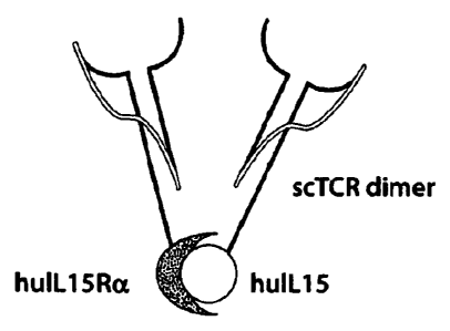
Figure 1(A & B) are schematic drawings. (A) is a schematic depicting an
example
of a fusion protein complex containing single chain TCR polypeptides. (B) is a
schematic
depicting representative fusion protein constructs comprising the fusion
protein complex.
Figure 2 (A ¨ C) consists of three panels. (A) depicts a map of pNEF38-
c264scTCR/hulL15 expression vector. (B) shows the sequence of c264scTCR/huIL15
fusion gene and (C) shows the sequence of c264scTCR/huIL15 fusion protein,
including
the leader sequence.
-9-
CA 02690825 2009-11-10
WO 2008/143794
PCT/US2008/005918
Figure 3 (A ¨ C) consists of three panels. (A) depicts a map of pNEF38-
c264scTCR-hinge-hulL15 expression vector. (B) shows the sequence of c264seTCR-
hinge-huIL15 fusion gene and (C) shows the sequence of c264scTCR-hinge-huIL15
fusion protein, including the leader sequence.
Figures 4 (A ¨ C) consists of three panels. (A) depicts a map of pNEF38-
c264seTCR/huIL15RaDE3 expression vector. (B) shows the sequence of
c264scTCR/huIL15RaAE3 fusion gene and (C) shows the sequence of
e264seTCR/huIL15RaAE3 fusion protein, including the leader sequence.
Figure 5 (A ¨ C) consists of three panels. (A) depicts a map of the pNEF38-
c264seTCR/buIL15RaSushi expression vector. (B) shows the sequence of
c264scTCR/hulL15RaSushi fusion gene and (C) shows the sequence of
c264seTCR/hulL15RaSushi fusion protein, including the leader sequence.
Figure 6 (A ¨ C) consists of three panels. (A) depicts the pNEF38-c264scTCR-
hinge-huILI5RaSushi expression vector. (B) shows the sequence of c264scTCR-
hinge-
huIL15RaSushi fusion gene and (C) shows the sequence of c264scTCR-hinge-
huIL15RaSushi fusion protein, including the leader sequence.
Figure 7 is a map of pSun-c264scTCRIL15/c264scTCRIL15RaSushi expression
vector.
Figure 8 is a map of pSun-c264scTCRIL15/c264scTCRIL15RaDE3 expression
vector.
Figures 9 (A & B) set forth characterization of transfected cells expressing
TCR/IL15Ra fusion protein. (A) is two graphs showing flow cytometric analysis
of
fusion protein expressing cells. (B) is a graph showing TCR-based ELBA results
for
fusion protein production.
-10-
CA 02690825 2009-11-10
WO 2008/143794
PCT/US2008/005918
Figures 10 (A & B) shows analysis of TCR/IL15 and TCR/IL15Ra fusion
proteins by reducing SDS PAGE. (A) shows cell culture supernatants containing
c264scTCR/huIL15 or c264scTCR/huIL15RaSushi. (B) shows cell culture
supernatants
containing c264seTCR/huIL15 or c264scTCR/hu1L15RaAE3.
Figures 11 (A ¨ C) shows analysis of TCR/IL15, TCR/IL15Ra and fusion protein
complexes by size exclusion chromatography. (A) is a graph showing the SEC
chromatography profile of c264scTCR/huIL15. (B) is a graph showing the SEC
chromatography profile of c264seTCR/huIL15RaSushi. (C) is a graph showing the
SEC
chromatography profile of c264seTCR/huIL15 + c264seTCR/huILI5RaSushi fusion
protein complex.
Figures 12 (A & B) is an analysis of TCR/IL15Ra and fusion protein complexes
by size exclusion chromatography. (A) is a graph illustrating the SEC
chromatography
profile of c264scTCR/huIL15RaAE3. (B) is a graph illustrating the SEC
chromatography
profile of c264seTCR/hulL15 + c264scTCR/huIL15RccAE3 fusion protein complex.
Figure 13 is a graph showing the binding of TCR/IL15, TCR/IL15Ra and fusion
protein complexes to peptide/MHC complexes displayed on cells, as determined
by flow
cytometry.
Figure 14 (A -D) consists of four panels. (A) shows the sequence of mature
human ILI5 protein (SEQ ID NO:1) and the blue underlined residues are
substituted in
the IL-15 variants as showed in Table 1. (B) depicts the pNEF38-c264scTCR-
hinge-
huILI5D8A and pNEF38-c264scTCR-hinge-huIL15D8N expression vectors. (C) shows
the sequence of pNEF38-c264scTCR-hinge-hulL15D8A and pNEF38-c264scTCR-hinge-
huIL15D8N genes and (D) shows the sequence of pNEF38-c264scTCR-hinge-
huIL15D8A and pNEF38-c264seTCR-hinge-huIL15D8N fusion protein, including the
CA 02690825 2009-11-10
WO 2008/143794
PCT/US2008/005918
leader sequence. The blue underlined nucleotides were changed to generate the
indicated
IL-15 variants.
Figure 15 is a graph showing flow cytometric analysis of IL-15R-bearing CTLL2
cells stained with fusion proteins and complexes following by TCR-specific
peptide/MHC reagent.
Figure 16 (A - C) are graphs showing the binding of dimeric fusion proteins
complexes of TCR/IL15RaSushi and TCR/IL15, comprising native and variant forms
of
IL15, to cognate peptide/MHC complexes displayed on cells loaded with peptide,
as
determined by flow cytometry. Background binding of the dimeric fusion
proteins
complexes on cells with no loaded peptide is also shown (A) is a graph showing
the
binding of the dimeric complexes of 1CR/IL15RaSushi and TCR/IL15wt (native
form),
or TCR/IL I5D8N or TCR/IL15D8A variants to cognate peptide/MHC complexes
displayed on cells. (B) is a graph showing the slight background binding of
dimeric
complexes of TCR/IL15RaSushi and 1CR/IL15wt (native form) to the cells without
loaded peptide. No background binding of dimeric complexes of TCR/ILI5RaSushi
and
TCR/IL15D8N or TCR/IL15D8A variants was seen to the cells with not loaded. (C)
is
graph(showing flow cytometric analysis of IL-1512.13yC-bearing 32E43 cells
stained with
dimeric complexes of TCR/IL15RaSushi and TCR/IL15wt (native form), or
TCR/IL I 5N72D, TCR/IL15D8N or 1CR/IL15D8A variants. Enhanced IL-15RfiyC
binding of the complex containing 1CR/IL15N72D and decreased IL-15ROyC binding
of
complexes containing TCR/IL I5D8N or TCR/IL I 5D8A was observed.
Figure 17 (A&B) are graphs showing binding activities of wide type,
antagonist,
and agonist TCR/IL15 fusion proteins to cognate peptide/MHC complexes and
IL15Ra
as determined by ELISA. (A) is analysis showing binding activity of fusion
proteins to
cognate peptide/MHC complexes. (B) is analysis showing binding activity of
fusion
proteins to IL15Ra.
-12-
CA 02690825 2009-11-10
WO 2008/143794
PCT/US2008/005918
Figure 18 is a graph showing fusion proteins and fusion protein complexes
support growth of IL15R-bearing cells as determined by cell proliferation
assay.
Figure 19 (A ¨ C) are graphs showing the ability of TCR/IL-15 fusion proteins
comprising IL-15 variants to inhibit or enhance growth of IL15R-bearing cells,
as
determined by cell proliferation assay. (A) is graph showing the activity of
fusion proteins
comprising IL-15 variants to inhibit the proliferation of high affinity IL15R
(apy receptor
complex) bearing CTLL-2 cells. (B) is graph showing the activity of fusion
proteins
comprising IL-15 variants to inhibit or enhance the proliferation of low
affinity IL15R (Py
receptor complex) bearing 32Dri cells. (C) is graph showing the activity of
fusion
proteins comprising IL-15 variants to block TCR/IL15wt-stimulated
proliferation of high
affinity IL15R (aPy receptor complex) bearing CTLL-2 cells.
Figure 20 depicts the effects of in vitro incubation of NK cells with dimeric
fusion
proteins complexes of TCR/IL15RaSushi and TCR/IL15 on the survival of
xenograft
tumor-bearing nude mice. Athymic nude mice were injected with human NSCLC A549-
A2 cells to allow establishment of lung metastases. Purified NK cells isolated
from
spleens of allogenic donor mice were incubated in vitro with rhIL-2, MARTI
seTCR-IL2,
c264scTCR-IL2 or c264scTCR-IL15/c264scTCR-IL15Ra and adoptively transferred
into
the tumor-bearing mice that had been pretreated with cyclophosphamide (CTX),
as
indicated in the figure legend. The percent survival following treatment was
plotted.
Figure 21 sets forth Table I showing the amino acid replacements in the ILA 5
variants and the affects of these changes on 1L-15 activity.
Figures 22A-B set forth the amino acid seugnece of IL-15(SEQ ID NO: I) and the
nucleic acid sequence of IL-15 (SEQ ID NO:2), respectively.
DETAILED DESCRIPTION OF THE INVENTION
-13-
=
CA 02690825 2009-11-10
WO 2008/143794
PCT/US2008/005918
It has been established that the IL-15 stably binds to the extracellular
domain of
the IL-15Ra and that the resulting complex is capable of modulating (i.e.
either
stimulating or blocking) immune responses via the intermediate or high
affinity IL-15R
complex (1-4). In addition, it has been demonstrated that single-chain TCR or
antibody
polypeptides can be fused to cytokines and other immune effector domains and
that such
bispecific fusion molecules retain functional activity of both fusion domains
(5-8).
Further, it has been shown that multivalent forms of the TCR provide enhanced
binding
to their ligands (9).
Definitions
The following definitions are provided for specific terms which are used in
the
following written description.
As used in the specification and claims, the singular form "a", "an" and "the"
include plural references unless the context clearly dictates otherwise. For
example, the
term "a cell" includes a plurality of cells, including mixtures thereof. The
term "a nucleic
acid molecule" includes a plurality of nucleic acid molecules.
As used herein, the term "comprising" is intended to mean that the
compositions
and methods include the recited elements, but do not exclude other elements.
"Consisting
essentially or, when used to define compositions and methods, shall mean
excluding
other elements of any essential significance to the combination. Thus, a
composition
consisting essentially of the elements as defined herein would not exclude
trace
contaminants from the isolation and purification method and pharmaceutically
acceptable
carriers, such as phosphate buffered saline, preservatives, and the like.
"Consisting of'
shall mean excluding more than trace elements of other ingredients and
substantial
method steps for administering the compositions of this invention. Embodiments
defined
by each of these transition terms are within the scope of this invention.
An "antibody" is any immunoglobulin, including antibodies and fragments
thereof, that binds a specific epitope. The term encompasses polyclonal,
monoclonal,
chimeric and single-chain antibodies as well as bispecific antibodies.
-14-
.
CA 02690825 2009-11-10
WO 2008/143794
PCT/US2008/005918
The term "antigen" as used herein is meant any substance that causes the
immune
system to produce antibodies or specific cell-mediated immune responses
against it. A
disease associated antigen is any substance that is associated with any
disease.
The term "biologically active polypeptide" as used herein is meant to refer to
an
amino acid sequence such as a protein, polypeptide or peptide; a sugar or
polysaccharide;
a lipid or a glycolipid, glycoprotein, or lipoprotein that can produce the
desired effects as
discussed herein, including a TCR or antibody fusion protein complex with
antigen
binding activity.
The term "cell" as used herein is meant to include any prokaryotic,
eukaryotic,
primary cell or immortalized cell line, any group of such cells as in, a
tissue or an organ.
Preferably the cells are of mammalian and particularly of human origin, and
can be
infected by one or more pathogens. A "host cell" in accord with the invention
can be a
transfected, transformed, transduced or infected cell of any origin, including
prokaryotic,
eulcaryotic, mammalian, avian, insect, plant or bacteria cells, or it can be a
cell sof any
origin that can be used to propagate a nucleic acid described herein.
The term "conjugate molecule" as it is used herein is meant to refer to a TCR
or
antibody molecule and an effector molecule usually a chemical or synthesized
molecule
covalently linked (i.e. fused) by chemical or other suitable method. If
desired, the
conjugate molecule can be fused at one or several sites through a peptide
linker sequence
or a carrier molecule. Alternatively, the peptide linker or carrier may be
used to assist in
construction of the conjugate molecule. Specifically preferred conjugate
molecules are
conjugate toxins or detectable labels.
The term "effector molecule" as used herein is meant to refer to an amino acid
sequence such as a protein, polypeptide or peptide; a sugar or polysaccharide;
a lipid or a
glycolipid, glycoprotein, lipoprotein or chemical agent that can produce the
desired
effects as discussed herein, including an IL-15 domain, IL-15 variant or IL-15
receptor
such as IL-I 5Ra, IL-210 or TC, or functional fragments thereof.
The terms "fusion molecule" and "fusion protein" are used interchangeably and
are meant to refer to a biologically active polypeptide usually a TCR or
antibody and an
effector molecule usually a protein or peptide sequence covalently linked
(i.e. fused) by
recombinant, chemical or other suitable method. If desired, the fusion
molecule can be
-15-
.
CA 02690825 2009-11-10
WO 2008/143794
PCT/US2008/005918
fused at one or several sites through a peptide linker sequence.
Alternatively, the peptide
linker may be used to assist in construction of the fusion molecule.
Specifically preferred
fusion molecules are fusion proteins. Generally fusion molecule also can be
comprised of
conjugate molecules.
The term "host cell" is meant to refer to any prokaryotic or eukaryotic cell
that
contains either a cloning vector or an expression vector. This term also
includes those
prokaryotic or eukaryotic cells that have been genetically engineered to
contain the
cloned gene(s) in the chromosome or genome of the host cell.
The term "immune response" as used herein is meant to refer to the process
whereby immune cells are stimulated and recruited from the blood to lymphoid
as well as
non-lymphoid tissues via a multifactorial process that involves distinct
adhesive and
activation steps. Activation conditions cause the release of cytokines, growth
factors,
chemokines and other factors, upregulate expression of adhesion and other
activation
molecules on the immune cells, promote adhesion, morphological changes, and/or
extravasation concurrent with chemotaxis through the tissues, increase cell
proliferation
and cytotoxic activity, stimulate antigen presentation and provide other
phenotypic
changes including generation of memory cell types. Immune response if also
meant to
refer to the activity of immune cells to suppress or regulate inflammatory or
cytotoxic
activity of other immune cells.
As used herein, the terms "polynucleotide" and "nucleic acid molecule" are
used
interchangeably to refer to polymeric forms of nucleotides of any length. The
polynucleotides may contain deoxyribonucleotides, ribonucleotides, and/or
their analogs.
Nucleotides may have any three-dimensional structure, and may perform any
function,
known or unknown. The term "polynucleotide" includes, for example, single-,
double-
stranded and triple helical molecules, a gene or gene fragment, exons,
introns, mRNA,
tRNA, rRNA, ribozymes, antisense molecules, cDNA, recombinant polynucleotides,
branched polynucleotides, aptamers, plasmids, vectors, isolated DNA of any
sequence,
isolated RNA of any sequence, nucleic acid probes, and primers. A nucleic acid
molecule
may also comprise modified nucleic acid molecules (e.g., comprising modified
bases,
sugars, and/or intemucleotide linkers).
-16-
.
CA 02690825 2009-11-10
WO 2008/143794
PCT/US2008/005918
The term "polypeptide" is meant to refer to any polymer preferably consisting
essentially of any of the 20 natural amino acids regardless of its size.
Although the term
"protein" is often used in reference to relatively large proteins, and
"peptide" is often used
in reference to small polypeptides, use of these terms in the field often
overlaps. The term
"polypeptide" refers generally to proteins, polypeptides, and peptides unless
otherwise
noted. Peptides useful in accordance with the present invention in general
will be
generally between about 0.1 to 100 KD or greater up to about 1000 KD,
preferably
between about 0.1, 0.2, 0.5, 1, 2,5, 10, 20 ,30 and 50 KD as judged by
standard molecule
sizing techniques such as centrifugation or SDS-polyacrylamide gel
electrophoresis.
The terms "prevent," "preventing," "prevention," "prophylactic treatment" and
the
like are meant to refer to reducing the probability of developing a disorder
or condition in
a subject, who does not have, but is at risk of or susceptible to developing a
disorder or
condition.
The term "single chain antibody" is meant to refer to an antibody based on a
single chain format. Single chain antibodies can consist of the minimal
binding subunit
of antibodies. Single-chain antibodies can combine only those antigen-binding
regions of
antibodies on a single stably-folded polypeptide chain. As such, single-chain
antibodies
are of considerably smaller size than classical immunoglobulins but retain the
antigen-
specific binding properties of antibodies. Single chain antibodies may be
linked to a wide
range of ligands, for example effector molecules or drug conjugates.
The term "soluble" as used herein is meant that the fusion molecule and
particularly a fusion protein that is not readily sedimented under low G-force
centrifugation (e.g. less than about 30,000 revolutions per minute in a
standard
centrifuge) from an aqueous buffer, e.g., cell media. Further, the fusion
molecule is
soluble if it remains in aqueous solution at a temperature greater than about
5-37 C and at
or near neutral pH in the presence of low or no concentration of an anionic or
non-ionic
detergent. Under these conditions, a soluble protein will often have a low
sedimentation
value e.g., less than about 10 to 50 svedberg units.
Aqueous solutions referenced herein typically have a buffering compound to
establish pH, typically within a pH range of about 5-9, and an ionic strength
range
between about 2mM and 500mM. Sometimes a protease inhibitor or mild non-ionic
-17-
CA 02690825 2009-11-10
WO 2008/143794
PCT/US2008/005918
detergent is added. Additionally, a carrier protein may be added if desired
such as bovine
serum albumin (BSA) to a few mg/ml. Exemplary aqueous buffers include standard
phosphate buffered saline, tris-buffered saline, or other well known buffers
and cell media
formulations.
The term "stimulate" or "stimulating" is meant to refer to increase, to
amplify, to
augment, to boost an immune response. Stimulation can be a positive
alteration. An
exemplary increase can be e.g., by 5%, 10%, 25%, 50%, 75%, or even 90-100%.
Other
exemplary increases include 2-fold, 5-fold, 10-fold, 20-fold, 40-fold, or even
100-fold.
The term "suppress" or "suppressing" is meant to refer to decrease, to
attenuate, to
diminish, to arrest, or to stabilize an immune response. Suppression may be a
negative
alteration. An exemplary decrease can be e.g., by 5%, 10%, 25%, 50%, 75%, or
even 90-
100%. Exemplary decreases include 2-fold, 5-fold, 10-fold, 20-fold, 40-fold,
or even
100-fold.
The term "T-cell Receptor" (TCR) is meant to refer to polypeptides of a
complex
of integral membrane proteins that participates in the activation of T cells
in response to
the presentation of antigen. T cells recognize a peptide bound to the MHC
product
through the a13. heterodimeric T cell receptor (TCR). The TCR repertoire has
extensive
diversity created by the same gene rearrangement mechanisms used in antibody
heavy
and light chain genes [Tonegawa, S. (1988) Biosci. Rep. 8:3-26]. Most of the
diversity is
generated at the junctions of variable (V) and joining (J) (or diversity, D)
regions that
encode the complementarity determining region 3 (CDR3) of the a and 13 chains
[Davis
and Bjorkman (1988) Nature 334:395-402]. However, TCRs do not undergo somatic
point mutations as do antibodies and, perhaps not coincidentally. TCRs also do
not
undergo the same extent of affinity maturation as antibodies. TCRs as they
occur in
.. nature appear to have affinities that range from 105 to 10.7 M. whereas
antibodies
typically have affinities that range from l Os to l09 M-1 [Davis et al. (1998)
Arum. Rev.
Immunol. 16:523-544; Eisen et al. (1996) Adv. Protein Chem. 49:1-56]. While
the
absence of somatic mutation in TCRs may be associated with lower affinities,
it has also
been argued that there is not a selective advantage for a TCR to have higher
affinity. In
fact, the serial-triggering [Valitutti et al. (1995) Nature 375:148-151] and
kinetic
proofreading [Rabinowitz et al. (1996) Proc. Natl. Acad. Sci. USA 93:1401-
1405] models
-18-
=
CA 02690825 2009-11-10
WO 2008/143794
PCT/US2008/005918
of T cell activation both suggest that longer off-rates (associated with
higher affinity)
would be detrimental to the signaling process. It is also possible that higher
affinity TCRs
might not maintain the peptide specificity required for T cell responses. For
example,
peptides bound within the MHC groove display limited accessible surface
[Bjorkman, P.
J. (1997) Cell 89:167-1701, which may in turn limit the amount of energy that
can be
generated in the interaction. On the other hand, raising the affinity of a TCR
by directing
the energy toward the MHC helices would presumably lead to thymic deletion
during
negative selection [Bevan, M. J. (1997) Immunity 7:175-178]. The term "TCR"
encompasses polyclonal, monoclonal, chimeric, humanized, heterodimeric and
single-
chain T-cell receptors or functional fragment thereof, including molecule
comprising the
TCR Vu and lip domains. The term "TCR" also encompasses T-cell receptors
disclosed
in for example, US Provisional Application Entitled "T CELL RECEPTOR FUSIONS
AND CONJUGATES AND METHODS OF USE THEREOF", filed March 19, 2008 and
US Patent Publication US 2003 01-44474A1.
The term "vector" is a nucleic acid molecule that is able to replicate
autonomously
in a host cell and can accept foreign DNA. A vector carries its own origin of
replication,
one or more unique recognition sites for restriction endonucleases which can
be used for
the insertion of foreign DNA, and usually selectable markers such as genes
coding for
antibiotic resistance, and often recognition sequences (e.g. promoter) for the
expression of
the inserted DNA. Common vectors include plasmid vectors and phage vectors.
T-Cell Receptors (TCR)
T-cells are a subgroup of cells which together with other immune cell types
(polymorphonuclear, eosinophils, basophils, mast cells, B-, NK cells),
constitute the
cellular component of the immune system. Under physiological conditions T-
cells
function in immune surveillance and in the elimination of foreign antigen.
However,
under pathological conditions there is compelling evidence that T-cells play a
major role
in the causation and propagation of disease. In these disorders, breakdown of
T-cell
immunological tolerance, either central or peripheral is a fundamental process
in the
causation of autoimmune disease.
-19-
CA 02690825 2009-11-10
WO 2008/143794
PCT/US2008/005918
The TCR is composed of at least seven transmembrane proteins. The disulfide-
linked (.alpha..beta.) heterodimer forms the monotypic antigen recognition
unit, while the
invariant chains of CD3, consisting of .epsilon., .gamma., .delta., and .zeta.
and .eta,
chains, are responsible for coupling the ligand binding to signaling pathways
that result in
.. 1-cell activation and the elaboration of the cellular immune responses.
Despite the gene
diversity of the TCR chains, two structural features are common to all known
subunits.
Firstly, they are transmembrane proteins with a single transmembrane spanning
domain--
presumably alpha-helical. Secondly, all the TCR chains have the unusual
feature of
possessing a charged amino acid within the predicted transmembrane domain. The
.. invariant chains have a single negative charge, conserved between the mouse
and human,
and the variant chains possess one (TCR-beta) or two (TCR-alpha) positive
charges. The
transmembrane sequence of TCR-.alpha. is highly conserved in a number of
species and
thus phylogenetically may serve an important functional role. The octapeptide
sequence
containing the hydrophilic amino acids arginine and lysine is identical
between the
species.
A T-cell response is modulated by antigen binding to a TCR. One type of TCR is
a membrane bound heterodimer consisting of an a and 13 chain resembling an
immunoglobin variable (V) and constant (C) region. The TCR a chain includes a
covalently linked V-a. and C-a chain, whereas the 13 chain includes a V-I3
chain
covalently linked to a C-13 chain. The V-a and V-13 chains form a pocket or
cleft that can
bind a superantigen or antigen in the context of a major histacompatibility
complex
(MHC) (known in humans as an HLA complex). See generally Davis Ann. Rev. of
Immunology 3: 537 (1985); Fundamental Immunology 3rd Ed., W. Paul Ed. Rsen
Press
LTD. New York (1993).
Fusions Proteins
The soluble fusion protein and conjugate molecule complexes of the invention
comprise at least two soluble fusion proteins, where the first fusion protein
comprises a first
biologically active polypeptide covalently linked to interleukin-15 (IL-15) or
functional
fragment thereof; and the second fusion protein comprises a second
biologically active
polypeptide covalently linked to soluble interleulcin-15 receptor alpha (IL-
15Ra)
-20-
CA 02690825 2009-11-10
WO 2008/143794
PCT/US2008/005918
polypeptide or functional fragment thereof, and wherein IL-15 domain of a
first fusion
protein binds to the soluble IL-15Ra domain of the second fusion protein to
form a soluble
fusion protein complex.
In certain examples, one of the biologically active polypeptides comprises a
first
soluble TCR or fragment thereof. The other or second biologically active
polypeptide
comprises the first soluble TCR or functional fragment thereof and thus
creates a
multivalent TCR fusion protein complex with increased binding activity for
cognate ligands
compared to the monovalent TCR. Further, the other biologically active
polypeptide
comprises a second soluble TCR or functional fragment thereof, different than
the first
soluble TCR. In certain examples, TCRs are produced that have higher affinity,
or
increased binding affinity for cognate ligands as compared, for example, to
the native TCR.
If the soluble TCR of the invention as described herein has a higher avidity
or affinity for
its ligand, then it is useful as a specific probe for cell-surface bound
antigen. In certain
preferred examples of the invention, the TCR is specific for recognition of a
particular
.. antigen.
In exemplary embodiments, TCR is a heterodimer comprising an a chain (herein
referred to as a, alpha or a chain) and a 13 chain (herein referred to as(3,
beta orb chain). In
other exemplary embodiments, the TCR comprises a single chain TCR polypeptide.
The
single chain TCR may comprise a TCR V-a chain covalently linked to a TCR v-r3
chain by
a peptide linker sequence. The single chain TCR may further comprise a soluble
TCR
chain fragment covalently linked to a TCR v-13 chain. The single chain TCR may
further
comprise a soluble TCR Ca chain fragment covalently linked to a TCR V-a chain.
In a further embodiment, one or both of the first and second biologically
active
polypeptides comprises an antibody or functional fragment thereof.
As used herein, the term "biologically active polypeptide" or "effector
molecule"
is meant an amino acid sequence such as a protein, polypeptide or peptide; a
sugar or
polysaccharide; a lipid or a glycolipid, glycoprotein, or lipoprotein that can
produce the
desired effects as discussed herein. Effector molecules also include chemical
agents.
Also contemplated are effector molecule nucleic acids encoding a biologically
active or
effector protein, polypeptide, or peptide. Thus, suitable molecules include
regulatory
-21-
.
CA 02690825 2009-11-10
WO 2008/143794
PCT/US2008/005918
factors, enzymes, antibodies, or drugs as well as DNA, RNA, and
oligonucleotides. The
biologically active polypeptides or effector molecule can be naturally-
occurring or it can
be synthesized from known components, e.g., by recombinant or chemical
synthesis and
can include heterologous components. A biologically active polypeptides or
effector
molecule is generally between about 0.1 to 100 KD or greater up to about 1000
ICD,
preferably between about 0.1, 0.2, 0.5, 1, 2, 5, 10, 20 ,30 and 50 K.D as
judged by
standard molecule sizing techniques such as centrifugation or SDS-
polyacrylamide gel
electropheresis. Desired effects of the invention include, but are not limited
to, for
example, forming a TCR fusion protein complex with increased binding activity,
killing a
target cell, e.g. either to induce cell proliferation or cell death, initiate
an immune
response, in preventing or treating a disease, or to act as a detection
molecule for
diagnostic purposes. For such detection, an assay could be used, for example
an assay
that includes sequential steps of culturing cells to proliferate same, and
contacting the
cells with a TCR fusion complex of the invention and then evaluating whether
the TCR
fusion complex inhibits further development of the cells.
Covalently linking the effector molecule to the TCR peptide in accordance with
the invention provides a number of significant advantages. TCR fusion
complexes of the
invention can be produced that contain a single effector molecule, including
such a
peptide of known structure. Additionally, a wide variety of effector molecules
can be
produced in similar DNA vectors. That is, a library of different effector
molecules can be
linked to the TCR molecule for presentation of infected or diseased cells.
Further, for
therapeutic applications, rather than administration of an TCR molecule to a
subject, a
DNA expression vector coding for the TCR molecule linked to the effector
peptide can be
administered for in vivo expression of the TCR fusion complex. Such an
approach avoids
costly purification steps typically associated with preparation of recombinant
proteins and
avoids the complexities of antigen uptake and processing associated with
conventional
approaches.
As noted, components of the fusion proteins disclosed herein, e.g., effector
molecule such as cytolcines, chemokines, growth factors, protein toxins,
immunoglobulin
domains or other bioactive molecules and any peptide linkers, can be organized
in nearly
any fashion provided that the fusion protein has the function for which it was
intended.
-22-
CA 02690825 2009-11-10
WO 2008/143794
PCT/US2008/005918
In particular, each component of the fusion protein can be spaced from another
component by at least one suitable peptide linker sequence if desired.
Additionally, the
fusion proteins may include tags, e.g., to facilitate modification,
identification and/or
purification of the fusion protein. More specific fusion proteins are in the
Examples
described below.
Linkers
The fusion complexes of the invention preferably also include a flexible
linker
sequence interposed between the IL-15 or IL-15Ra domains and the biologically
active
polypeptide. The linker sequence should allow effective positioning of the
biologically
active polypeptide with respect to the IL-15 or IL-15Ra domains to allow
functional
activity of both domains. In embodiments where the biologically active
polypeptide is a
TCR, the linker sequence positions the TCR molecule binding groove so that the
T cell
receptor can recognize presenting MHC-peptide complexes and can deliver the
effector
molecule to a desired site. Successful presentation of the effector molecule
can modulate
the activity of a cell either to induce or to inhibit T-cell proliferation, or
to initiate or
inhibit an immune response to a particular site, as determined by the assays
disclosed
below, including the in vitro assays that includes sequential steps of
culturing T cells to
proliferate same, and contacting the T cells with a TCR fusion complex of the
invention
and then evaluating whether the TCR fusion complex inhibits further
development of the
cells.
In certain embodiments, the soluble fusion protein complex has a linker
wherein the
first biologically active polypeptide is covalently linked to IL-15 (or
functional fragment
thereof) by polypeptide linker sequence.
In other certain embodiments, the soluble fusion protein complex as described
herein has a linker wherein the second biologically active polypeptide is
covalently linked
to IL-15Ra polypeptide (or functional fragment thereof) by polypeptide linker
sequence.
The linker sequence is preferably encoded by a nucleotide sequence resulting
in a
peptide that can effectively position the binding groove of the TCR molecule
for
recognition of a presenting antigen. As used herein, the phrase effective
positioning of
the biologically active polypeptide with respect to the IL-15 or IL-15Ra
domains ", or
other similar phrase, is intended to mean the biologically active polypeptide
linked to the
-23-
CA 02690825 2009-11-10
WO 2008/143794
PCT/US2008/005918
IL-15 or IL-15Ra domains is positioned so that the IL-15 or IL-15Ra domains
are
capable of interacting with each other to form a protein complex. In certain
embodiments, the IL-15 or IL-15Ra domains are effectively positioned to allow
interactions with immune cells to initiate or inhibit an immune reaction, or
to inhibit
orstimulate cell development.
Preferably the linker sequence comprises from about 7 to 20 amino acids, more
preferably from about 8 to 16 amino acids. The linker sequence is preferably
flexible so
as not hold the biologically active polypeptide or effector molecule in a
single undesired
conformation. The linker sequence can be used, e.g., to space the recognition
site from
the fused molecule. Specifically, the peptide linker sequence can be
positioned between
the biologically active polypeptide and the effector molecule, e.g., to
chemically cross-
link same and to provide molecular flexibility. The linker is preferably
predominantly
comprises amino acids with small side chains, such as glycine, alanine and
serine, to
provide for flexibility. Preferably about 80 or 90 percent or greater of the
linker sequence
comprises glycine, alanine or serine residues, particularly glycine and serine
residues.
For a fusion protein complex that comprise a heterodimer TCR, the linker
sequence is
suitably linked to the b chain of the TCR molecule, although the linker
sequence also
could be attached to the a chain of the TCR molecule. Alternatively, linker
sequence may
be linked to both a and b chains of the TCR molecule. When such a beta peptide
chain is
expressed along with the a chain, the linked TCR-effector peptide should fold
resulting in
a functional TCR molecule as generally depicted in Figure 1. One suitable
linker
sequence is ASGGGGSGGG (i.e., Ala Ser Gly Gly Gly Gly Ser Gly Gly Gly),
preferably
linked to the first amino acid of the b domain of the TCR. Different linker
sequences
could be used including any of a number of flexible linker designs that .have
been used
successfully to join antibody variable regions together, see Whitlow, M. et
al., (1991)
Methods: A Companion to Methods in Enzymology 2:97-105. In some examples, for
covalently linking an effector molecule to a TCR b chain molecule, the amino
sequence
of the linker should be capable of spanning suitable distance from the C-
terminal residue
of the TCR beta chain to the N-terminal residue of the effector molecule.
Suitable linker
sequences can be readily identified empirically. Additionally, suitable size
and sequences
-24-
CA 02690825 2009-11-10
WO 2008/143794
PCT/US2008/005918
of linker sequences also can be determined by conventional computer modeling
techniques based on the predicted size and shape of the TCR molecule.
In general, preparation of the fusion protein complexes of the invention can
be
accomplished by procedures disclosed herein and by recognized recombinant DNA
techniques involving, e.g., polymerase chain amplification reactions (PCR),
preparation
of plasmid DNA, cleavage of DNA with restriction enzymes, preparation of
oligonucleotides, ligation of DNA, isolation of mRNA, introduction of the DNA
into a
suitable cell, transformation or transfection of a host, culturing of the
host. Additionally,
the fusion molecules can be isolated and purified using chaotropic agents and
well known
electrophoretic, centrifugation and chromatographic methods. See generally,
Sambrook
et al., Molecular Cloning: A Laboratory Manual (2nd ed. (1989); and Ausubel et
al.,
Current Protocols in Molecular Biology, John Wiley & Sons, New York (1989) for
disclosure relating to these methods.
As used herein, biologically active polypeptides or effector molecules of the
invention may include factors such as cytokines, chemokines, growth factors,
protein
toxins, immunoglobulin domains or other bioactive proteins such as enzymes.
Also
biologically active polypeptides may include conjugates to other compounds
such as non-
protein toxins, cytotoxic agents, chemotherapeutic agents, detectable labels,
radioactive
materials and such.
Cytokines of the invention are defined by any factor produced by cells that
affect
other cells and are responsible for any of a number of multiple effects of
cellular
immunity. Examples of cytokines include but are not limited to the IL-2
family,
interferon (IFN), IL-1, IL-17, TGF and TNF cytokine families, and to IL-1
through
IL-35, IFN-a, IFN-y,. TGF-0, TNF-a and TNFP.
In an aspect of the invention, the first fusion protein comprises a first
biologically
active polypeptide covalently linked to interleukin-15 (IL-15) domain or a
functional
fragment thereof. IL-15 is a cytokine which affects T-cell activation and
proliferation. IL-
15 activity in affecting immune cell activation and proliferation is similar
in some respects
to IL2, although fundamental difference have been well characterized
(Waldmann, TA,
2006, Nature Rev. Immunol. 6:595-601).
-25-
CA 02690825 2009-11-10
WO 2008/143794
PCT/US2008/005918
In another aspect of the invention, the first fusion protein comprises an
interleuldn-
15 (IL-15) domain that is an IL-15 variant (also referred to herein as IL-15
mutant). The
IL-15 variant preferably comprises .a different amino acid sequence that the
native (or wild
type) IL-15 protein. The IL-15 variant preferably binds the IL-15Ra
polypeptide and
functions as an IL-15 agonist or antagonist. Preferably IL-15 variants with
agonist activity
have super agonist activity. In some embodiments, the IL-15 variant can
function as an IL-
agonist or antagonist independent of its association with IL-15Ra. IL-15
agonists are
exemplified by comparable or increased biological activity compared to wild
type IL-15.
IL-15 antagonists are exemplified by decreased biological activity compared to
wild type
10 .. IL-15 or by the ability to inhibit IL-15-mediated responses. In some
examples, the IL-15
variant binds with increased or decreased activity to the IL-1511137C
receptors. In some
embodiments, the sequence of the IL-15 variant has at least one amino acid
change, e.g.
substitution or deletion, compared to the native IL-2 sequence, such changes
resulting in
IL-15 agonist or antagonist activity. Preferably the amino acid
substitutions/deletions are in
15 the domains of IL-15 that interact with IL-15R and/or .yC. More
preferably, the amino
acid substitutions/deletions do not affect binding to the IL-15Ra polypeptide
or the ability
to produce the IL-15 variant. Suitable amino acid substitutions/deletions to
generate IL-15
variants can be identified based on putative or known IL-15 structures,
comparisons of IL-
15 with homologous molecules such as IL-2 with known structure, through
rational or
.. random mutagenesis and functional assays, as provided herein, or other
empirical methods.
Additionally suitable amino acid substitutions can be conservative or non-
conservative
changes and insertions of additional amino acids Preferably IL-15 variants of
the invention
contain one or more than one amino acid substitutions/deletions at position 8,
61, 65, 72,
92, 101, 108, or 111 of the mature human IL-15 sequence; particularly, D8N
("D8" refers
to the amino acid and residue position in the native mature human IL-15
sequence and
"N" refers to the substituted amino acid residue at that position in the IL-15
variant),
D8A, D61A, N65A, N72R or Ql 08A substitutions result in IL-15 variants with
antagonist activity and N72D substitutions result in IL-15 variants with
agonist activity.
While in one aspect of the invention the IL-15 variant is a component of a
fusion
protein complex, in other aspects the IL-15 variant is a non-fusion protein.
Preferably the
non-fusion form of the IL-15 variant is a soluble cytokine that functions as
an IL-15 agonist
-26-
CA 02690825 2009-11-10
WO 2008/143794
PCT/US2008/005918
or antagonist. In some embodiments, the non-fusion IL-15 variant forms a
complex with
IL-15Ra whereas in other embodiment it acts independently of IL-15Ra.
Chemokines of the invention, similar to cytokines, are defined as any chemical
factor or molecule which when exposed to other cells are responsible for any
of a number
of multiple effects of cellular immunity. Suitable chemokines may include but
are not
limited to the CXC, CC, C, and CX3C chemokine families and to CCL-1 through
CCL-
28, CXC-1 through CXC-17, XCL-1, XCL-2, CX3CL1, MIP-1b, IL-8, MCP-1, and
Rantes.
Growth factors include any molecules which when exposed to a particular cell
induce proliferation and/or differentiation of the affected cell. Growth
factors include
proteins and chemical molecules, some of which include: GM-CSF, G-CSF, human
growth factor and stem cell growth factor. Additional growth factors may also
be
suitable for uses described herein.
Toxins or cytotoxic agents include any substance which has a lethal effect or
an
inhibitory effect on growth when exposed to cells. More specifically, the
effector
molecule can be a cell toxin of, e.g., plant or bacterial origin such as,
e.g., diphtheria toxin
(DT), shiga toxin, abrin, cholera toxin, ricin, saporin, pseudomonas exotoxin
(PE),
pokeweed antiviral protein, or gelonin. Biologically active fragments of such
toxins are
well known in the art and include, e.g., DT A chain and ricin A chain.
Additionally, the
toxin can be an agent active at the cell surface such as, e.g., phospholipase
enzymes (e.g.,
phospholipase C).
Further, the effector molecule can be a chemotherapeutic drug such as, e.g.,
vindesine, vincristine, vinblastin, methotrexate, adriamycin, bleomycin, or
cisplatin.
Additionally, the effector molecule can be a detectably-labeled molecule
suitable
for diagnostic or imaging studies. Such labels include biotin or
streptavidin/avidin, a
detectable nanoparticles or crystal, an enzyme or catalytically active
fragment thereof, a
fluorescent label such as green fluorescent protein, FITC, phycoerythrin,
cychome, texas
red or quantum dots; a radionuclide e.g., iodine-131, yttrium-90, rhenium-188
or bismuth-
212; a phosphorescent or chemiluminescent molecules or a label detectable by
PET,
ultrasound or MRI such as Gd- or paramagnetic metal ion-based contrast agents.
See
e.g., Moskaug, et al. J. Biol. Chem. 264, 15709 (1989); Pastan, I. et al. Cell
47,641,
-27-
CA 02690825 2009-11-10
WO 2008/143794
PCT/US2008/005918
1986; Pastan et al., Recombinant Toxins as Novel Therapeutic Agents, Ann. Rev.
Biochem. 61, 331, (1992); "Chimeric Toxins" Olsnes and Phil, Pharmac. Ther.,
25, 355
(1982); published PCT application no. WO 94/29350; published PCT application
no. WO
94/04689; published PCT application no. W02005046449 and U.S. Pat. 5,620,939
for
disclosure relating to making and using proteins comprising effectors or tags.
A protein fusion or conjugate complex that includes a covalently linked IL-15
and
IL-15Ra domains has several important uses. For example, the protein fusion or
conjugate complex comprising a TCR can be employed to deliver the IL-15/1L-
15Ra
complex \ to certain cells capable of specifically binding the TCR.
Accordingly, the
protein fusion or conjugate complex provide means of selectively damaging or
killing
cells comprising the ligand. Examples of cells or tissue capable of being
damaged or
killed by the protein fusion or conjugate complexes comprising a TCR include
tumors
and virally or bacterially infected cells expressing one or more ligands
capable of being
specifically bound by the TCR. Cells or tissue susceptible to being damaged or
killed
.. can be readily assayed by the methods disclosed herein.
The IL-15 and IL-15Ra polypeptides of the invention suitably correspond in
amino acid sequence to naturally occurring IL-15 and IL-15Ra molecules, e.g.
IL-15 and
IL-15Ra molecules of a human, mouse or other rodent, or other mammal.
In some settings it can be useful to make the protein fusion or conjugate
complexes of the present invention polyvalent, e.g., to increase the valency
of the sc-
TCR. In particular, interactions between the IL-15 and IL-15Ra domains of the
fusion
protein complex provide a means of generating polyvalent complexes. In
addition, the
polyvalent fusion protein can made by covalently or non-covalently linking
together
between one and four proteins (the same or different) by using e.g., standard
biotin-
.. streptavidin labeling techniques, or by conjugation to suitable solid
supports such as latex
beads. Chemically cross-linked proteins (for example cross-linked to
dendrimers) are
also suitable polyvalent species. For example, the protein can be modified by
including
sequences encoding tag sequences that can be modified such as the
biotinylation BirA tag
or amino acid residues with chemically reactive side chains such as Cys or
His. Such
.. amino acid tags or chemically reactive amino acids may be positioned in a
variety of
positions in the fusion protein, preferably distal to the active site of the
biologically active
-28-
CA 02690825 2009-11-10
WO 2008/143794
PC1/US2008/005918
polypeptide or effector molecule. For example, the C-terminus of a soluble
fusion protein
can be covalently linked to a tag or other fused protein which includes such a
reactive
amino acid(s). Suitable side chains can be included to chemically link two or
more fusion
proteins to a suitable dendrimer or other nanoparticle to give a multivalent
molecule.
Dendrimers are synthetic chemical polymers that can have any one of a number
of
different functional groups of their surface (D. Tomalia, Aldrichimica Acta,
26:91:101
(1993)). Exemplary dendrimers for use in accordance with the present invention
include
e.g. E9 starburst polyamine dendrimer and E9 combust polyamine dendrimer,
which can
link cystine residues.
Nucleic Acids and Vectors
Nucleic Acids
The invention further provides nucleic acid sequences and particularly DNA
sequences that encode the present fusion proteins. Preferably, the DNA
sequence is
carried by a vector suited for extrachromosomal replication such as a phage,
virus,
plasmid, phagemid, cosmid, YAC, or episome. In particular, a DNA vector that
encodes
a desired fusion protein can be used to facilitate preparative methods
described herein and
to obtain significant quantities of the fusion protein. The DNA sequence can
be inserted
into an appropriate expression vector, i.e., a vector which contains the
necessary elements
for the transcription and translation of the inserted protein-coding sequence.
A variety of
host-vector systems may be utilized to express the protein-coding sequence.
These
include mammalian cell systems infected with virus (e.g., vaccinia virus,
adenovirus,
etc.); insect cell systems infected with virus (e.g., baculovirus);
microorganisms such as
yeast containing yeast vectors, or bacteria transformed with bacteriophage
DNA, plasmid
DNA or cosmid DNA. Depending on the host-vector system utilized, any one of a
number of suitable transcription and translation elements may be used. See
generally
Sambrook et at, supra and Ausubel et al. supra.
Included in the invention are methods for making a soluble fusion protein
complex,
the method comprising introducing into a host cell a DNA vector as described
herein
encoding the first and second fusion proteins, culturing the host cell in
media under
conditions sufficient to express the fusion proteins in the cell or the media
and allow
-29-
CA 02690825 2009-11-10
WO 2008/143794
PCMS2008/005918
association between IL-15 domain of a first fusion protein and the soluble IL-
15Ra domain
of a second fusion protein to form the soluble fusion protein complex,
purifying the soluble
fusion protein complex from the host cells or media.
In general, a preferred DNA vector according to the invention comprises a
nucleotide sequence linked by phosphodiester bonds comprising, in a 5' to 3'
direction a
first cloning site for introduction of a first nucleotide sequence encoding a
TCR chain,
operatively linked to a sequence encoding an effector molecule.
The fusion protein components encoded by the DNA vector can be provided in a
cassette format. By the term "cassette" is meant that each component can be
readily
substituted for another component by standard recombinant methods. In
particular, a
DNA vector configured in a cassette format is particularly desirable when the
encoded
fusion complex is to be used against pathogens that may have or have capacity
to develop
serotypes.
To make the vector coding for a TCR fusion complex, the sequence coding for
the
.. TCR molecule is linked to a sequence coding for the effector peptide by use
of suitable
ligases. DNA coding for the presenting peptide can be obtained by isolating
DNA from
natural sources such as from a suitable cell line or by known synthetic
methods, e.g. the
phosphate triester method. See, e.g., Oligonucleotide Synthesis, IRL Press
(Mi. Gait,
ed., 1984). Synthetic oligonucleotides also may be prepared using commercially
available automated oligonucleotide synthesizers. Once isolated, the gene
coding for the
TCR molecule can be amplified by the polymerase chain reaction (PCR) or other
means
known in the art. Suitable PCR primers to amplify the TCR peptide gene may add
restriction sites to the PCR product. The PCR product preferably includes
splice sites for
the effector peptide and leader sequences necessary for proper expression and
secretion of
.. the TCR-effector fusion complex. The PCR product also preferably includes a
sequence
coding for the linker sequence, or a restriction enzyme site for ligation of
such a
sequence.
The fusion proteins described herein are preferably produced by standard
recombinant DNA techniques. For example, once a DNA molecule encoding the TCR
protein is isolated, sequence can be ligated to another DNA molecule encoding
the
effector polypeptide. The nucleotide sequence coding for a TCR molecule may be
-30-
CA 02690825 2009-11-10
WO 2008/143794
PCT/US2008/005918
directly joined to a DNA sequence coding for the effector peptide or, more
typically, a
DNA sequence coding for the linker sequence as discussed herein may be
interposed
between the sequence coding for the TCR molecule and the sequence coding for
the
effector peptide and joined using suitable ligases. The resultant hybrid DNA
molecule
can be expressed in a suitable host cell to produce the TCR fusion complex.
The DNA
molecules are ligated to each other in a 5' to 3' orientation such that, after
ligation, the
translational frame of the encoded polypeptides is not altered (i.e., the DNA
molecules
are ligated to each other in-frame). The resulting DNA molecules encode an in-
frame
fusion protein.
Other nucleotide sequences also can be included in the gene construct. For
example, a promoter sequence, which controls expression of the sequence coding
for the
TCR peptide fused to the effector peptide, or a leader sequence, which directs
the TCR
fusion complex to the cell surface or the culture medium, can be included in
the construct
or present in the expression vector into which the construct is inserted. An
immunoglobulin or CMV promoter is particularly preferred.
In obtaining variant TCR coding sequences, those of ordinary skill in the art
will
recognize that TCR-derived proteins may be modified by certain amino acid
substitutions,
additions, deletions, and post-translational modifications, without loss or
reduction of
biological activity. In particular, it is well-known that conservative amino
acid
substitutions, that is, substitution of one amino acid for another amino acid
of similar size,
charge, polarity and conformation, are unlikely to significantly alter protein
function. The
20 standard amino acids that are the constituents of proteins can be broadly
categorized
into four groups of conservative amino acids as follows: the nonpolar
(hydrophobic)
group includes alanine, isoleucine, leucine, methionine, phenylalanine,
proline,
tryptophan and valine; the polar (uncharged, neutral) group includes
asparagine, cysteine,
glutamine, glycine, serine, tIveonine and tyrosine; the positively charged
(basic) group
contains arginine, histidine and lysine; and the negatively charged (acidic)
group contains
aspartic acid and glutamic acid. Substitution in a protein of one amino acid
for another
within the same group is unlikely to have an adverse effect on the biological
activity of
the protein.
-31-
CA 02690825 2009-11-10
WO 2008/143794
PCT/US2008/005918
Homology between nucleotide sequences can be determined by DNA
hybridization analysis, wherein the stability of the double-stranded DNA
hybrid is
dependent on the extent of base pairing that occurs. Conditions of high
temperature
and/or low salt content reduce the stability of the hybrid, and can be varied
to prevent
annealing of sequences having less than a selected degree of homology. For
instance, for
sequences with about 55% G-C content, hybridization and wash conditions of 40-
50° C., 6×SSC (sodium chloride/sodium citrate buffer) and 0.1%
SDS
(sodium dodecyl sulfate) indicate about 60-70% homology, hybridization and
wash
conditions of 50-65° C., 1×SSC and 0.1% SDS indicate about 82-97%
homology, and hybridization and wash conditions of 52° C.,
0.1×SSC and
0.1% SDS indicate about 99-100% homology. A wide range of computer programs
for
comparing nucleotide and amino acid sequences (and measuring the degree of
homology)
are also available, and a list providing sources of both commercially
available and free
software is found in Ausubel et al. (1999). Readily available sequence
comparison and
multiple sequence alignment algorithms are, respectively, the Basic Local
Alignment .
Search Tool (BLAST) (Altschul et al., 1997) and ClustalW programs. BLAST is
available on the world wide web at ncbi.nlm.nih.gov and a version of ClustalW
is.
available at 2.ebi.ac.uk.
The components of the fusion protein can be organized in nearly any order
provided each is capable of performing its intended function. For example, in
one
embodiment, the TCR is situated at the C or N terminal end of the effector
molecule.
Preferred effector molecules of the invention will have sizes conducive to the
function for which those domains are intended. The effector molecules of the
invention
can be made and fused to the TCR by a variety of methods including well-known
chemical cross-linking methods. See e.g., Means, G.E. and Feeney, R.E. (1974)
in
Chemical Modification of Proteins, Holden-Day. See also, S.S. Wong (1991) in
Chemistry of Protein Conjugation and Cross-Linking, CRC Press. However it is
generally preferred to use recombinant manipulations to make the in-frame
fusion protein.
As noted, a fusion molecule or a conjugate molecule in accord with the
invention
can be organized in several ways. In an exemplary configuration, the C-
terminus of the
TCR is operatively linked to the N-terminus of the effector molecule. That
linkage can be
-32-
CA 02690825 2009-11-10
WO 2008/143794
PCT/US2008/005918
achieved by recombinant methods if desired. However, in another configuration,
the N-
terminus of the TCR is linked to the C-terminus of the effector molecule.
Alternatively, or in addition, one or more additional effector molecules can
be
inserted into the TCR fusion or conjugate complexes as needed.
Vectors and Expression
A number of strategies can be employed to express protein fusion complexes of
the invention. For example, the TCR gene fusion construct described above can
be
incorporatcd into a suitable vector by known means such as by use of
restriction enzymes
to make cuts in the vector for insertion of the construct followed by
ligation. The vector
containing the gene construct is then introduced into a suitable host for
expression of the
TCR fusion peptide. See, generally, Sambrook et al., supra. Selection of
suitable vectors
can be made empirically based on factors relating to the cloning protocol. For
example,
the vector should be compatible with, and have the proper replicon for the
host that is
being employed. Further the vector must be able to accommodate the DNA
sequence
coding for the TCR fusion complex that is to be expressed. Suitable host cells
include
eukaryotic and prokaryotic cells, preferably those cells that can be easily
transformed and
exhibit rapid growth in culture medium. Specifically preferred hosts cells
include
prokaryotes such as E. coil, Bacillus subtillus, etc. and eukaryotes such as
animal cells
and yeast strains, e.g., S. cerevisiae. Mammalian cells are generally
preferred,
particularly J558, NSO, SP2-0 or CHO. Other suitable hosts include, e.g.,
insect cells
such as SD. Conventional culturing conditions are employed. See Sambrook,
supra.
Stable transformed or transfected cell lines can then be selected. Cells
expressing a TCR
fusion complex of the invention can be determined by known procedures. For
example,
expression of a TCR fusion complex linked to an immunoglobulin can be
determined by
an ELISA specific for the linked immunoglobulin and/or by immunoblotting.
Other
methods for detecting expression of fusion proteins comprising TCRs linked to
IL-15 or
1L-15Ra domains are disclosed in the Examples.
As mentioned generally above, a host cell can he used for preparative purposes
to
propagate nucleic acid encoding a desired fusion protein. Thus a host cell can
include a
prokaryotic or eukaryotic cell in which production of the fusion protein is
specifically
intended. Thus host cells specifically include yeast, fly, worm, plant, frog,
mammalian
-33-
CA 02690825 2009-11-10
WO 2008/143794
PCT/US2008/005918
cells and organs that are capable of propagating nucleic acid encoding the
fusion. Non-
limiting examples of mammalian cell lines which can be used include CHO dhfr-
cells
(Urlaub and Chasm, Proc. Natl. Acad. Sci. USA, 77:4216 (1980)), 293 cells
(Graham et
al., J Gen. Virol., 36:59 (1977)) or myeloma cells like SP2 or NSO (Galfre and
Milstein,
Meth. Enzymol., 73(B):3 (1981)).
Host cells capable of propagating nucleic acid encoding a desired fusion
protein
encompass non-mammalian eukaryotic cells as well, including insect (e.g., Sp.
frugiperda), yeast (e.g., S. cerevisiae, S. pombe, P. pastoris., K lactis, H
polymorpha; as
generally reviewed by Fleer, R., Current Opinion in Biotechnology, 3(5):486496
(1992)),
fungal and plant cells. Also contemplated are certain prokaryotes such as E.
coli and
Bacillus.
Nucleic acid encoding a desired fusion protein can be introduced into a host
cell
by standard techniques for transfecting cells. The term "transfecting" or
"transfection" is
intended to encompass all conventional techniques for introducing nucleic acid
into host
cells, including calcium phosphate co-precipitation, DEAE-dextran-mediated
transfection, lipofection, electroporation, microinjection, viral transduction
and/or
integration. Suitable methods for transfecting host cells can be found in
Sambrook et al.
supra, and other laboratory textbooks.
Various promoters (transcriptional initiation regulatory region) may be used
according to the invention. The selection of the appropriate promoter is
dependent upon
the proposed expression host. Promoters from heterologous sources may be used
as long
as they are functional in the chosen host.
Promoter selection is also dependent upon the desired efficiency and level of
peptide or protein production. Inducible promoters such as tac are often
employed in
order to dramatically increase the level of protein expression in E. colt.
Overexpression of
proteins may be harmful to the host cells. Consequently, host cell growth may
be limited.
The use of inducible promoter systems allows the host cells to be cultivated
to acceptable
densities prior to induction of gene expression, thereby facilitating higher
product yields.
Various signal sequences may be used according to the invention. A signal
sequence which is homologous to the TCR coding sequence may be used.
Alternatively, a
signal sequence which has been selected or designed for efficient secretion
and
-34-
CA 02690825 2009-11-10
WO 2008/143794 PCT/US2008/005918
processing in the expression host may also be used. For example, suitable
signal
sequence/host cell pairs include the B. subtilis sacB signal sequence for
secretion in B.
subtilis, and the Saccharomyces cerevisiae .alpha.-mating factor or P.
pastoris acid
phosphatase phol signal sequences for P. pastoris secretion. The signal
sequence maybe
joined directly through the sequence encoding the signal peptidase cleavage
site to the
protein coding sequence, or through a short nucleotide bridge consisting of
usually fewer
than ten codons, where the bridge ensures correct reading frame of the
downstream TCR
sequence.
Elements for enhancing transcription and translation have been identified for
eukaryotic protein expression systems. For example, positioning the
cauliflower mosaic
virus (CaMV) promoter 1000 bp on either side of a heterologous promoter may
elevate
transcriptional levels by 10- to 400-fold in plant cells. The expression
construct should
also include the appropriate translational initiation sequences. Modification
of the
expression construct to include a Kozak consensus sequence for proper
translational
initiation may increase the level of translation by 10 fold.
A selective marker is often employed, which may be part of the expression
construct or separate from it (e.g., carried by the expression vector), so
that the marker
may integrate at a site different from the gene of interest. Examples include
markers that
confer resistance to antibiotics (e.g., bla confers resistance to ampicillin
for E. coli host
cells, nptll confers kanamycin resistance to a wide variety of prokaryotic and
eukaryotic
cells) or that permit the host to grow on minimal medium (e.g., HIS4 enables
P. pastoris
or His- S. cerevisiae to grow in the absence of histidine). The
selectable marker has
its own transcriptional and translational initiation and termination
regulatory regions to
allow for independent expression of the marker. If antibiotic resistance is
employed as a
marker, the concentration of the antibiotic for selection will vary depending
upon the
antibiotic, generally ranging from 10 to 600 µg of the antibiotic/mL of
medium.
The expression construct is assembled by employing known recombinant DNA
techniques (Sambrook et al., 1989; Ausubel et al., 1999). Restriction enzyme
digestion
and ligation are the basic steps employed to join two fragments of DNA. The
ends of the
DNA fragment may require modification prior to ligation, and this may be
accomplished
by filling in overhangs, deleting terminal portions of the fragment(s) with
nucleases (e.g.,
-35-
CA 02690825 2009-11-10
WO 2008/143794
PCT/US2008/005918
ExoIII), site directed mutagenesis, or by adding new base pairs by PCR.
Polylinkers and
adaptors may be employed to facilitate joining of selected fragments. The
expression
construct is typically assembled in stages employing rounds of restriction,
ligation, and
transformation of E. coli. Numerous cloning vectors suitable for construction
of the
expression construct are known in the art (.lambda.ZAP and pBLUESCRIPT SK-1,
Stratagene, LaJolla, Calif, pET, Novagen Inc., Madison, Wis. --cited in
Ausubel et al.,
1999) and the particular choice is not critical to the invention. The
selection of cloning
vector will be influenced by the gene transfer system selected for
introduction of the
expression construct into the host cell. At the end of each stage, the
resulting construct
may be analyzed by restriction, DNA sequence, hybridization and PCR analyses.
The expression construct may be transformed into the host as the cloning
vector
construct, either linear or circular, or may be removed from the cloning
vector and used
as is or introduced onto a delivery vector. The delivery vector facilitates
the introduction
and maintenance of the expression construct in the selected host cell type.
The expression
construct is introduced into the host cells by any of a number of known gene
transfer
systems (e.g., natural competence, chemically mediated transformation,
protoplast
transformation, electroporation, biolistic transformation, transfection, or
conjugation)
(Ausubel et al., 1999; Sambrook et al., 1989). The gene transfer system
selected depends
upon the host cells and vector systems used.
For instance, the expression construct can be introduced into S. cerevisiae
cells by
protoplast transformation or electroporation. Electroporation of S. cerevisiae
is readily
accomplished, and yields transformation efficiencies comparable to spheroplast
transformation.
The present invention further provides a production process for isolating a
fusion
protein of interest. In the process, a host cell (e.g., a yeast, fungus,
insect, bacterial or
animal cell), into which has been introduced a nucleic acid encoding the
protein of the
interest operatively linked to a regulatory sequence, is grown at production
scale in a
culture medium in the presence of the fusion protein to stimulate
transcription of the
nucleotides sequence encoding the fusion protein of interest. Subsequently,
the fusion
protein of interest is isolated from harvested host cells or from the culture
medium.
Standard protein purification techniques can be used to isolate the protein of
interest from
-36-
CA 02690825 2009-11-10
WO 2008/143794
PCT/US2008/005918
the medium or from the harvested cells. In particular, the purification
techniques can be
used to express and purify a desired fusion protein on a large-scale (i.e. in
at least
milligram quantities) from a variety of implementations including roller
bottles, spinner
flasks, tissue culture plates, bioreactor, or a fermentor.
An expressed protein fusion complex can be isolated and purified by known
methods. Typically the culture medium is centrifuged and then the supernatant
is purified
by affinity or immunoaffinity chromatography, e.g. Protein-A or Protein-G
affinity
chromatography or an immunoaffinity protocol comprising use of monoclonal
antibodies
that bind the expressed fusion complex such as a linked TCR or immunoglobulin
region
thereof. The fusion proteins of the present invention can be separated and
purified by
appropriate combination of known techniques. These methods include, for
example,
methods utilizing solubility such as salt precipitation and solvent
precipitation, methods
utilizing the difference in molecular weight such as dialysis, ultra-
filtration, gel-filtration,
and SDS-polyacrylamide gel electrophoresis, methods utilizing a difference in
electrical
charge such as ion-exchange column chromatography, methods utilizing specific
affinity
such as affinity chromatograph, methods utilizing a difference in
hydrophobicity such as
reverse-phase high performance liquid chromatograph and methods utilizing a
difference
in isoelectric point, such as isoelectric focusing electrophoresis, metal
affinity columns
such as Ni-NTA. See generally Sambrook et al. and Ausubel et al. supra for
disclosure
relating to these methods.
It is preferred that the fusion proteins of the present invention be
substantially
pure. That is, the fusion proteins have been isolated from cell substituents
that naturally
accompany it so that the fusion proteins are present preferably in at least
80% or 90% to
95% homogeneity (w/w). Fusion proteins having at least 98 to 99% homogeneity
(w/w)
are most preferred for many pharmaceutical, clinical and research
applications. Once
substantially purified the fusion protein should be substantially free of
contaminants for
therapeutic applications. Once purified partially or to substantial purity,
the soluble fusion
proteins can be used therapeutically, or in performing in vitro or in vivo
assays as
disclosed herein. Substantial purity can be determined by a variety of
standard techniques
such as chromatography and gel electrophoresis.
-37-
CA 02690825 2009-11-10
WO 2008/143794
PCT/US2008/005918
Truncated TCR fusion complexes of the invention contain a TCR molecule that is
sufficiently truncated so the TCR fusion complex can be secreted into culture
medium
after expression. Thus, a truncated TCR fusion complex will not include
regions rich in
hydrophobic residues, typically the transmembrane and cytoplasmic domains of
the TCR
molecule. Thus, for example, for a preferred truncated DR1 TCR molecule of the
invention, preferably from about residues 199 to 237 of the b chain and from
about
residues 193 to 230 of the a chain of the TCR molecule are not included in the
truncated
TCR fusion complex.
The present TCR fusion and conjugate complexes are suitable for in vitro or in
vivo use with a variety of cells that are infected or that may become infected
by one or
more diseases.
Methods
Therapeutic
Included in the invention are methods for preventing or treating disease in a
patient
in which the diseased cells express a disease associated antigen, the method
comprising
administering to the patient a soluble fusion protein complex comprising a
biologically
active polypeptide recognizing a disease-associated antigen, forming a
specific binding
complex (bridge) between antigen-expressing diseased cells and IL-15R-
expressing
immune cells sufficient to localize the immune cells, and damaging or killing
the disease
cells sufficient to prevent or treat the disease in the patient.
Included are methods for preventing or treating disease in a patient in which
the
diseased cells express a disease associated antigen, the method comprising
mixing immune
cells bearing the IL-15R chains with a soluble fusion protein complex
comprising a
biologically active polypeptide recognizing a disease-associated antigen, for
example a
peptide/Ml-IC complex, administering to the patient the immune cell-fusion
protein
complex mixture, forming a specific binding complex (bridge) between antigen-
expressing
diseased cells and IL-1 5R-expressing immune cells sufficient to localize the
immune cells,
and damaging or killing the disease cells sufficient to prevent or treat the
disease in the
patient.
-38-
CA 02690825 2009-11-10
WO 2008/143794
PCT/US200W005918
Also included in the invention are methods for killing a target cell, the
method
comprising contacting a plurality of cells with a soluble fusion protein
complex, where the
plurality of cells further comprises immune cells bearing the IL-15R chains
recognized by
the IL-15 domain of claim 1 and the target cells bearing an antigen recognized
by at least
one of the biologically active polypeptides as described herein, fanning a
specific binding
complex (bridge) between the antigen on the target cells and the IL-15R chains
on the
immune cells sufficient to bind and activate the immune cells; and killing the
target cells by
the bound activated immune cells.
Also included in the inventions are methods to increase in vivo half life of
IL-15
and/or enhance its ability to stability bind immune cells (e.g. increase cell
surface
residency time) through generation of a soluble fusion protein complex. For
example,
evaluation of the pharmacokinetic parameters and cell surface residency time
of the
fusion protein complex are conducted and compared to IL-15, as described
herein.
Fusion protein complexes with an increased serum half life or cell surface
residency time
are preferable as based on their improved therapeutic utility..
Examples of diseases that can be treated include, but are not limited to,
neoplasia,
including cancer, or viral infection. By "neoplasia" is meant any disease that
is caused by
or results in inappropriately high levels of cell division, inappropriately
low levels of
apoptosis, or both. For example, cancer is an example of a neoplasia. Examples
of
cancers include, without limitation, leukemias (e.g., acute leukemia, acute
lymphocytic
leukemia, acute myelocytic leukemia, acute rnyeloblastic leukemia, acute
promyelocytic
leukemia, acute myelomonocytic leukemia, acute monocytic leukemia, acute
erythroleWcemia, chronic leukemia, chronic myelocytic leukemia, chronic
lymphocytic
leukemia), polycythemia vera, lymphoma (Hodgkin's disease, non-Hodgkin's
disease),
Waldenstrom's macroglobulinemia, heavy chain disease, and solid tumors such as
sarcomas and carcinomas (e.g., fibrosarcoma, myxosarcoma, liposarcoma,
chondrosarcoma, osteogenic sarcoma, chordoma, angiosarcoma, endotheliosarcoma,
lymphangiosarcoma, lymphangioendotheliosarcoma, synovioma, mesothelioma,
Ewing's
tumor, leiomyosarcoma, rhabdomyosarcoma, colon carcinoma, pancreatic cancer,
breast
cancer, ovarian cancer, prostate cancer, squamous cell carcinoma, basal cell
carcinoma,
adenocarcinoma, sweat gland carcinoma, sebaceous gland carcinoma, papillary
-39-
CA 02690825 2009-11-10
WO 2008/143794
PCT/US2008/005918
carcinoma, papillary adenocarcinomas, cystadenocarcinoma, medullary carcinoma,
bronchogenic carcinoma, renal cell carcinoma, hepatoma, nile duct carcinoma,
choriocarcinoma, seminoma, embryonal carcinoma, Wilm's tumor, cervical cancer,
uterine cancer, testicular cancer, lung carcinoma, small cell lung carcinoma,
bladder
carcinoma, epithelial carcinoma, glioma, astrocytoma, medulloblastoma,
craniopharyngioma, ependymoma, pinealoma, hemangioblastoma, acoustic neuroma,
oligodenroglioma, schwannoma, meningioma, melanoma, neuroblastoma, and
retinoblastoma). Lymphoproliferative disorders are also considered to be
proliferative
diseases.
Also included are methods of stimulating immune responses in a mammal
comprising administering to the mammal an effective amount of the soluble
fusion protein
complex or IL-15 variant as described herein. Also included are methods of
suppressing
immune responses in a mammal comprising administering to the mammal an
effective
amount of the soluble fusion protein complex or IL-15 variant as described
herein. In the
case of immune suppression, a fusion protein complex or IL-15 variant
comprising IL-15
antagonists or IL-15 domains that lack the ability to bind the IL-1513y,
complex may be
particularly advantageous.
As an illustration of the use of the fusion protein complex therapeutics, a
cultured
cell can be infected by a pathogen of a single serotype. The infected cell is
then contacted
by a specified fusion protein complex in vitro. As discussed previously, the
fusion
protein complex is configured so that the toxic domain is presented to the
infected cell by
the association of the TCR. After providing for introduction of the bioactive
molecule to
the cell (generally less than about 30 minutes), the cells are allowed to
cause a desired
effect for a time period of about up to about 2 to 24 hours, typically about
18 hours. After
this time, the cells are washed in a suitable buffer or cell medium and then
evaluated for
viability. The time allotted for cell killing or injury by the fusion protein
complex will
vary with the particular effector molecule chosen. However viability can often
be
assessed after about 2 to 6 hours up to about 24 hours. As will be explained
in more
detail below, cell viability can be readily measured and quantified by
monitoring uptake
of certain well-known dyes (e.g., trypan blue) or fluors.
-40-
CA 02690825 2009-11-10
WO 2008/143794
PCT/US2008/005918
Cells incubated with the fusion protein complex of the present invention can
be
assayed for viability by standard methods. In one exemplary approach, cell
viability can
be readily assayed by measuring DNA replication following or during
incubation. For
example, a preferred assay involves cell uptake of one or more detectably
labeled
nucleosides such as radiolabelled thymidine. The uptake can be conveniently
measured
by several conventional approaches including trichloroacetic acid (TCA)
precipitation
followed by scintillation counting. Other cell viability methods include well-
known
trypan blue exclusion techniques or WST-1-based proliferation assays.
The TCR molecules of the fusion complexes of the invention suitably correspond
.. in amino acid sequence to naturally occurring TCR molecules, e.g. TCR
molecules of a
human, mouse or other rodent, or other mammal.
Accordingly, one treatment method of the invention for inhibition of an
autoimmune or inflammatory response would include a fusion protein complex
which
comprises a T cell receptor or antibody with binding specificity to a disease
associated
antigen. Preferably, a "truncated" soluble TCR complex is administered, i.e.
the TCR
complex does not contain a transmembrane portion. The fusion protein complex
also
comprises an IL-15 variants that functions as an IL-15 antagonist to suppress
the
unwanted immune response. Follow administration, the TCR or antibody domain
targets
the fusion protein complex to the disease site where the IL-15 antagonist
suppresses the
autoimmune or inflammatory response. Such fusion protein complex may
particularly
useful for treatment of allergies, autoimmune diseases such as multiple
sclerosis, insulin-
dependent diabetes mellitus and rheumatoid arthritis or transplant rejection.
Similar non-
targeted approaches could be carried out using antagonist IL-15 variants as
non-fusion
proteins.
Another treatment method of the invention for induction of an immune response
provides for the administration of an effective amount of a protein fusion
complexes of
the invention in the presence of any costimulatory effector molecule such as a
cytokine to
thereby induce a desired immune response at the location of the presented
antigen which
binds the biologically active polypeptide.
Different therapies of the invention also may be used in combination as well
as
with other known therapeutic agents such as anti-inflammatory drugs to provide
a more
-41-
CA 02690825 2009-11-10
WO 2008/143794
PCT/US2008/005918
effective treatment of a disorder. For example, immunosuppressive protein
fusion
complexes or IL-15 variants can be used in combination with anti-inflammatory
agents
such as corticosteroids and nonsteroidal drugs for the treatment of autoimmune
disorders
and allergies.
Compounds of the invention will be especially useful to a human patient who
has
or is suspected of having a malignant disease, disorder or condition.
Compounds of the
invention will be particularly useful in targeting particular tumor antigens
in human
patients. Specific examples of diseases which may be treated in accordance
with the
invention include cancers, e.g. breast, prostate, etc, viral infections, e.g.
HCV, HIV, etc.
as well as other specific disorders of conditions mentioned herein.
Without wishing to be bound by theory, it is believed the multiple and
distinct
covalently linked compounds of this invention (i.e. at least IL-15 in
combination with at
least one TCR) can significantly enhance efficacy of the IL-15, e.g., by
increasing
targeting of IL-15 to target antigen in subject individuals.
Moreover, by virtue of the covalent linkage, the conjugates of the invention
present the IL-15 and the TCR to the subject cell essentially simultaneously,
an effect that
may not be readily achieved by administering the same compounds in a drug
"cocktail"
formulation without covalently linking the compounds.
It also has been reported that treatment with treatment with one drug can in
turn
sensitize a patient to another drug. Accordingly, the essentially simultaneous
presentation
to the subject cell of IL-15 and TCR via a conjugate of the invention may
enhance drug
activity, e.g., by providing synergistic results and/or by enhancing
production an immune
response.
Diagnostic
High affinity or multivalent TCR proteins specific for a particular pMHC
ligand
are useful in diagnosing animals, including humans believed to be suffering
from a
disease associated with the particular pMHC. The fusion protein complexes of
the present
invention are useful for detecting essentially any antigen, including but not
limited to,
those associated with a neoplastic condition, an abnormal protein, an
autoimmune disease
or an infection or infestation with a bacterium, a fungus, a virus, a
protozoan, a yeast, a
nematode or other parasite.
-42-
CA 02690825 2009-11-10
WO 2008/143794
PCT/US2008/005918
In one such method for detecting a tumor or virally infected cell or tissue in
a
subject, wherein the cell or tissue comprises a tumor or virus-associated
peptide antigen
presented on the cells or tissue in the context of an MHC complex, comprises:
a)
administering to the subject a soluble fusion protein complex of the invention
comprising
a soluble TCR under conditions that form a specific binding complex between
the
presented peptide antigen and the TCR; and b) detecting the specific binding
complex as
being indicative of a tumor or virally infected cell or tissue comprising the
presented
tumor or viral-associated peptide antigen.
The fusion protein complexes can also be used in the diagnosis of certain
genetic
disorders in which there is an abnormal protein produced. Exemplary
applications for
fusion protein complexes are in the diagnosis and treatment of autoimmune
diseases in
which there is a known pMHC. Type I diabetes is relatively well characterized
with
respect to the autoantigens which attract immune destruction. Multiple
sclerosis, celiac
disease, inflammatory bowel disease, Crohn's disease and rheumatoid arthritis
are
additional candidate diseases for such application.
The fusion protein complexes of the present invention comprising IL-15 variant
polypeptides may be particularly useful in these applications. For example,
for a fusion
protein complex comprising TCR molecules, interactions between the IL-15
variant
domain and the IL-15Ra polypeptide generate multivalent TCR molecules with
enhanced
antigen binding activity, as disclosed herein. Moreover, the IL-15 variant
contains amino
acid changes that potentially eliminate binding to cells bearing IL-15111K
receptors,
thereby reducing non-specific or non-targeted binding to immune cells. As a
results,
improved detection of TCR-specific antigens can be achieved with such fusion
protein
complexes. Additionally fusion protein complexes of the invention can be
further
multimerized via peptide tags sequences or conjugation to detectable labels,
as disclosed
herein.
Dosage and Administration
Administration of compounds of the invention may be made by a variety of
suitable routes including oral, topical (including transdermal, buccal or
sublingal), nasal
and parenteral (including intraperitoneal, subcutaneous, intravenous,
intradennal or
-43-
CA 02690825 2009-11-10
WO 2008/143794 PCT/US2008/005918
intramuscular injection) with oral or parenteral being generally preferred. It
also will be
appreciated that the preferred method of administration and dosage amount may
vary
with, for example, the condition and age of the recipient
Compounds of the invention may be used in therapy alone or in conjunction with
other medicaments such those with recognized pharmacological activity to treat
the
desired indications. Exemplary medicaments include recognized therapeutics
such as
surgery, radiation, chemotherapy and other forms of immunotherapy (e.g.
vaccines,
antibody based therapies). The compounds of this invention can be administered
before,
during or after such therapies as needed.
While one or more compounds of the invention may be administered alone, they
also may be present as part of a pharmaceutical composition in mixture with
conventional
excipient, i.e., pharmaceutically acceptable organic or inorganic carrier
substances
suitable for parenteral, oral or other desired administration and which do not
deleteriously
react with the active compounds and are not deleterious to the recipient
thereof.
Pharmaceutical compositions of the invention in general comprise one or more
fusion
protein complex or IL-15 variant of the invention or DNA constructs coding for
such
compounds together with one or more acceptable carriers. The carriers must be
"acceptable" in the sense of being compatible with other ingredients of the
formulation
and not deleterious to the recipient thereof. Suitable pharmaceutically
acceptable carriers
include but are not limited to water, salt solutions, alcohol, vegetable oils,
polyethylene
glycols, gelatin, lactose, amylose, magnesium stearate, talc, silicic acid,
viscous paraffin,
perfume oil, fatty acid monoglycerides and diglycerides, petroethral fatty
acid esters,
hydroxymethyl-cellulose, polyvinylpyrrolidone, etc. The pharmaceutical
preparations
.. can be sterilized and if desired mixed with auxiliary agents, e.g.,
lubricants, preservatives,
stabilizers, wetting agents, emulsifiers, salts for influencing osmotic
pressure, buffers,
colorings, flavorings and/or aromatic substances and the like which do not
deleteriously
react with the active compounds.
For parenteral application, particularly suitable are solutions, preferably
oily or
aqueous solutions as well as suspensions, emulsions, or implants, including
suppositories.
Ampules are convenient unit dosages.
-44-
CA 02690825 2009-11-10
WO 2008/143794 PCT/US2008/005918
For enteral application, particularly suitable are tablets, dragees or
capsules having
talc and/or carbohydrate carrier binder or the like, the carrier preferably
being lactose
and/or corn starch and/or potato starch. A syrup, elixir or the like can be
used wherein a
sweetened vehicle is employed. Sustained release compositions can be
formulated
including those wherein the active component is protected with differentially
degradable
coatings, e.g., by microencapsulation, multiple coatings, etc.
Therapeutic compounds of the invention also may be incorporated into
liposomes.
The incorporation can be carried out according to known liposome preparation
procedures, e.g. sonication and extrusion. Suitable conventional methods of
liposome
=
preparation are also disclosed in e.g. A.D. Bangham et al., J. Mol. Biol.,
23:238-252
(1965); F. Olson et al., Biochim. Biophys. Acta, 557:9-23 (1979); F. Szoka et
al., Proc.
Nat. Acad. Sci., 75:4194-4198 (1978); S. Kim et al., Biochim. Biophys. Acta,
728:339-
348 (1983); and Mayer et al., Biochim. Biophys. Acta, 858:161-168 (1986).
The invention also provides methods for invoking an immune response in a
mammal such as a human, including vaccinating a mammal such as a human against
an
infectious agent or a targeted disorder such as cancer.
These methods comprise administering to a mammal an effective amount of a
DNA sequence that comprises a DNA vector that codes for a fusion protein
complex or
IL-15 variant of the invention. Preparation of expression vectors of fusion
protein
complexes and IL-15 variants is described above and in the Examples which
follow.
Methods for administration of plasmid DNA, uptake of that DNA by cells of the
administered subject and expression of protein has been reported. See Ulmer,
J.B., et al.,
Science (1993) 259: 1745-1749.
DNA vectors that encode fusion protein complexes and IL-15 variantsof the
invention
are suitably administered to a mammal including a human preferably by
intramuscle
injection. Administration of cDNA to skeletal muscle of a mammal with
subsequent
uptake of administered expression vector by the muscle cells and expression of
protein
encoded by the DNA has been described by Ulmer et al. and represents an
exemplary
protocol [Ulmer, J.B., et al., Science 259: 1745-1749]. The optimal dose for a
given
therapeutic application can be determined by conventional means.
-45-
CA 02690825 2009-11-10
WO 2008/143794
PCT/US2008/005918
In addition to treatment of human disorders, fusion protein complexes and IL-
15
variants of the invention and DNA constructs of the invention that encode such
molecules
will have significant use for veterinary applications, e.g., treatment of
disorders of
livestock such as cattle, sheep, etc. and pets such as dog and cats.
It will be appreciated that actual preferred amounts of a given fusion protein
complex and 1L-15 variant of the invention or DNA construct coding for same
used in a
given therapy will vary according to the particular active compound or
compounds being
utilized, the particular compositions formulated, the mode of application, the
particular
site of administration, the patient's weight, general health, sex, etc., the
particular
indication being treated, etc. and other such factors that are recognized by
those skilled in
the art including the attendant physician or veterinarian. Optimal
administration rates for
a given protocol of administration can be readily determined by those skilled
in the art
using conventional dosage determination tests conducted e.g. with regard to
the foregoing
guidelines and the assays disclosed herein.
EXAMPLES
It should be appreciated that the invention should not be construed to be
limited to
the examples that are now described; rather, the invention should be construed
to include
any and all applications provided herein and all equivalent variations within
the skill of
the ordinary artisan.
Example 1 ¨ Design of a fusion protein complex comprising scTCR/huIL15 and
saCR/hulL15Ra fusion proteins.
It has been established that the IL-15 stably binds to the extrac,ellular
domain of
the IL-15Ra and that the resulting complex is capable of modulating (i.e.
either
stimulating or blocking) immune responses via the intermediate or high
affinity IL- I5R
complex (1-4). In addition, it has been demonstrated that single-chain TCR or
antibody
polypeptides can be fused to cytokines and other immune effector domains and
that such
bispecific fusion molecules retain functional activity of both fusion domains
(5-8).
Further, it has been shown that multivalent forms of the TCR provide enhanced
binding
-46-
CA 02690825 2009-11-10
WO 2008/143794
PCT/US2008/005918
to their ligands (9). Therefore a feature of the invention provides for a
fusion protein
complex comprising at least one fusion protein wherein a first TCR polypeptide
is fused
to IL-l5 and at least one fusion wherein a second TCR polypeptide is fused to
the
extracellular domain of IL-15Ra, such that the two fusion proteins form a
complex
through binding interactions between the IL-15 and IL-15Ra domains. In such a
fusion
protein complex, the TCR polypeptides can be the same or different and in
either single-
chain or heterodimeric format.
An example of a fusion protein complex containing single-chain TCR
polypeptides is shown schematically in Figure 1A. In this fusion protein
complex, the
multivalent TCR domains provide increased binding avidity/affinity for their
ligands.
Exemplary ligands include, but are not limited to, peptidc/MHC complexes. The
IL-
15/IL-15Ra domains provide inununomodulatory activity. Representative fusion
protein
constructs comprising the fusion protein complex are schematically shown in
Figure 113.
In these constructs the TCR polypeptide is a single-chain TCR (264scTCR)
comprised of
TCR-Va and ercR-vp-cp domains linked by a peptide linker sequence ((G4S)4).
The
scTCR polypeptide is fused to either the IL-15 or IL-15Ra domains, directly or
via a
peptide linker sequence. Proceeding the scTCR polypeptide is a signal peptide
(or leader
peptide) sequence that permits soluble expression. The signal peptide is
subsequently
cleaved during protein transport to generate the mature fusion protein. In
other examples
of the fusion protein complex, an antibody domain can substitute a TCR domain
depicted
in Figures IA and 113. Such an antibody can be in a single-chain or
heteromultimeric
format. For any of the fusion protein complexes described above, sequences can
be
human or non-human, for example, but not limited to mouse. These sequences can
be
employed for part or all of the fusion protein domains. In addition, the
arrangement of
the domains can vary so long as the fusion proteins remain soluble and
functional.
Example 2 - Construction of the c264seTCR/huIL15 gene fusion in an expression
vector.
Isolation and characterization of TCR genes for the p53 (aa264-272)-specific
TCR
were described previously (5-7). To obtain the human IL15 and IL15Ra genes,
human
-47-
CA 02690825 2009-11-10
WO 2008/143794
PCT/US2008/005918
PBMC were isolated from 200 mL of blood of a donor (Lot#2238789, Community
Blood
Bank, Miami, FL) with HISTOPAGUE-1077 (Sigma). The cells (1.5 x10⁷) were
activated by 30 ng/ml of PMA (Sigma), 200 neml of ionomycin, and 220 ng/ml of
recombinant human IL2 in IMDM containing 10%FBS in a CO2 incubator for 10
days.
The activated cells (1 x 10⁷ per mL) were frozen at ¨70C for the further
applications. To
purify the total RNA from the activated PBMC, RNEASY PLUS MINI (Qiagen) was
used according to the manufacturer's protocol. Human IL15 gene containing the
coding
region and a portion of 5' and 3' flanking regions was amplified from the
total RNA with
the front primer
5 '-CACC'TTGCC ATAGCCAGCTCTTC-3 '
and the back primer
5'-GTCTAAGCAGCAGAGTGATGTTTG-3'
by SUPERSCRIPT III One-Step RT-PCR Platinum Tag HiFi (Invitrogen) according to
the following conditions: for RT; 55C 30 min; 94C, 2 min; for amplifying cDNA;
94C,
30s; 53C, 30s; 68C, lmin; x40 cycles; 68C, 5 min. The 600 bp human IL15 PCR-
cDNA
product was separated by electrophoresis on a 1% agarose gel and isolated. The
cDNA
product was purified from agarose with a Qiaquick Gel Extraction Kit (Qiagen).
The
gene of the mature human IL15 protein was amplified from the 600 bp human IL15
cDNA with the front primer
5'-TGGTTAACAACTGGGTGAATGTAATAAGTG-3'
and the back primer
5'-ACGCGITTATCAAGAAGTGTTGATGAACA1TTGGAC-3'
by PfuUltra (Stratagene) under following PCR conditions: 94C, I min; 63C, 1
min; 72C 1
min; x35 cycles; 72C, 10 min. The mature human IL15 protein gene was gel-
purified and
cloned into the shuttle vector, pcDNA3.1 Directional TOPO Expression Vector
(Invitrogen), with the TOPO reaction according to the manufacture's protocol.
The clone
containing the mature human IL15 protein gene insert was identified based on
the
diagnostic PCR with the front primer
5'-IGGTTAACAACTGGGTGAATGTAATAAGTG-3'
and the back primer
5'-ACGCG1TTATCAAGAAGTGTTGATGAACATTTGGAC-3'
-48-
CA 02690825 2009-11-10
WO 2008/143794
PCT/US2008/005918
by RedTag (Sigma) under the following condition: 94C, 1 mm; 63C, 1 min; 72C, 1
min;
x35 cycles; 72C, 10 min. The sequence of the correct clone was verified by DNA
sequencing with GenomeLab Dye Termination Cycle Sequencing with a QUICK START
KIT (Beckman Coulter) according to the manufacturer's protocol. The mature
human
IL15 protein gene was removed from the shuttle vector by digestion with HpaI
and Mlul
and ligated into an expression vector pNEF38-c264scTCR which had been digested
with
HpaI and MluI. The pNEF38-c264scTCR expression vector contains the gene
fragment
encoding an immunoglobulin light chain leader (or secretory signal) sequence
linked to
the p53 (aa264-272) peptide-specific soluble chimeric single-chain TCR protein
.. (c264scTCR) (5). The vector also contains 5' regulatory/enhancer regions,
transcription
regulatory and promoter regions, translational
regulatory/initiation/termination sequences
including a Kozak consensus sequence and poly-A termination region, and 3'
regulatory
regions with putative matrix attachment regulatory elements. The vector also
contains
DNA sequences allowing selective growth in mammalian cells (SV40 promoter/neoR
gene/poly-A) and bacteria (on/amp gene). Cloning of the DNA fragment encoding
mature human 1L15 protein into the pNEF38-c264scTCR vector resulted in a
c264scTCR/huIL15 fusion gene comprising the following sequence: 3'-
immunoglobulin
light chain leader ¨ 264 TCR V-n ¨ peptide linker - 264 TCR v-p - human TCR C-
13 ¨
human IL-15. The resulting vector (pNEF38-c264scTCR/hulL15), shown in Figure
2A,
was identified based on the diagnostic PCR and reconfirmed by DNA sequencing.
The
sequences of the c264seTCR/huIL15 fusion gene and protein (including the
leader
sequence) are shown at Figure 2B and Figure 2C, respectively.
Example 3 - Construction of the c264scTCR/hu1L15 gene fusion containing a
.. mutated human IgG1 hinge region in an expression vector.
Construction of the pNEF38-c264scTCR/huIL15 vector was described at Example
2. A mutated hinge region from human IgG1 H chain where three cysteine
residues were
substituted with three serine residues was used to link c264seTCR and hulL15.
The
hinge region was mutated and amplified from 264scTCR/IgG1 gene described
previously
(7) with the front primer
-49-
CA 02690825 2009-11-10
WO 2008/143794
PCT/US2008/005918
5'-TGGTGGGTTAACGAGCCCAAATCTTCTG-3'
and the back primer
5'-ATTATTACGCGTTGGAGACGGTGGAGATG -3'
by PfuUltra (Stratagene) under following PCR conditions: 94C, 30 sec; 65C, 30
sec; 70C
1 min; x35 cycles; 72C, 10 min. The 70 bp mutated human IgG1 hinge PCR-cDNA
product was separated by electrophoresis on a 1% agarose gel and isolated. The
cDNA
product was purified from agarose with a Qiaquick Gel Extraction Kit (Qiagen).
The
mutated hinge region gene was digested with Hpar and MluI and ligated into
pNEF38-
c264scTCR which had been digested with Hpal and MluI. The clone containing the
mutated hinge region gene insert was identified based on the diagnostic PCR
with the
front primer
5'-TGAGTGATCGATACCACCATGGAGACAGACAC-3'
and the back primer
5'- ATTATTACGCGTTGGAGACGGTGGAGATG -3'
by RedTag (Sigma) under the following condition: 94C, 30 sec; 64C, 30 sec; 70C
1 mm;
x35 cycles; 72C, 10 min. The huIL15 was amplified from pNEF38-c264scTCR/huIL15
vector described at Example 2 with the front primer
5'-TGGTGGACGCGTAACTGGGTGAATG-3'
and the back primer
5I-TGGTGGTCTAGAATTATCAAGAAGTGTTGATG -3'
by PfuUltra (Stratagene) under following PCR conditions: 94C, 30 sec; 65C, 30
sec; 70C
1 min; x35 cycles; 72C, 10 min. The 380 bp hulL15 PCR-cDNA product was
separated
by electrophoresis on a 1% agarose gel and isolated. The cDNA product was
purified
from agarose with a Qiaquick Gel Extraction Kit (Qiagen). The huIL15 gene was
digested with Midi and XbaI and ligated into pNEF38-c264scTCR containing
mutated
hinge gene which had been digested with Mlul and XbaI. The clone containing
the
hulL15 gene insert was identified based on the diagnostic PCR with the front
primer
5'-TGAGTGATCGATACCACCATGGAGACAGACAC-3'
and the back primer
5'- TGGTGGTCTAGAATTATCAAGAAGTGTTGATG -3'
-50-
CA 02690825 2009-11-10
WO 2008/143794
PCT/US2008/005918
by RedTag (Sigma) under the following condition: 94C, 30 sec; 64C, 2 mm; 70C 2
min;
x35 cycles; 72C, 10 min. The sequence of the correct clone was verified by DNA
sequencing with GenomeLab Dye Termination Cycle Sequencing with a QUICK START
KIT (Beckman Coulter) according to the manufacturer's protocol. The pNEF38-
c264seTCR expression vector is described above at Example 2. Cloning of the
DNA
fragment encoding mutated human IgG1 hinge region and mature human IL15
protein
into the pNEF38-c264scTCR vector resulted in a c264scTCR-hmt-huIL15 fusion
gene
comprising the following sequence: 3'- immunoglobulin light chain leader ¨ 264
TCR V-
a ¨ peptide linker - 264 TCR V-I3¨ human TCR c-i3 ¨mutated human IgG I hinge-
human
IL-15. The resulting vector (pNEF38-c264scTCR-lunt-hulL15), shown in Figure
3A,
was identified based on the diagnostic PCR and reconfirmed by DNA sequencing.
The
sequences of the c264scTCR-hmt-hulL15 fusion gene and protein (including the
leader
sequence) are shown at Figure 3B and Figure 3C, respectively.
Example 4- Construction of the c264scTCR/huIL15RaAE3 gene fusion in an
expression vector.
The total RNA of PBMC was prepared as described above. Human ILI5Ret gene
containing coding region and a portion of 5' and 3' flanking regions was
amplified from
the total RNA of the PBMC with the front primer
5'-AGTCCAGCGGTGTCCTGTGG -3'
and the back primer
5'-TGACGCG1TTAAGTGGTGTCGCTGTGCCCTG-3'
by SUPERSCRIPT III One-Step RT-PCR Platinum Tag HiFi (Invitrogen) according to
the following condition: for RT; 55C, 30 min; 94C, 2 min; for amplifying cDNA;
94C, I
min; 66C, 1 min; 72C, lmin; x35 cycles; 72C, 5 min. The 970 bp human 1115 Ra
PCR
cDNA product was separated by electrophoresis on a I% agarose gel and
isolated. The
cDNA was purified from agarose with a Qiaquick Gel Extraction Kit (Qiagen).
The
human IL15 Ra extracellular domain gene was amplified from the 970 bp human
IL15
Ra cDNA with the front primer
5 '-TGGTTAACATCACGTGCCCTCCCCCCATG-3 '
-51-
CA 02690825 2009-11-10
WO 2008/143794
PCT/US2008/005918
and the back primer
5'-TGACGCGTITAAGTGGTGTCGCTGTGCCCTG-3'
by PfuULTRA (Stratagene) under following PCR conditions: 94C, 1 min; 72C 2
min; x35
cycles, 72C, 10 mm. The human IL15 Ra extracellular domain gene was gel-
purified and
cloned into the shuttle vector, pcDNA3.1 Directional TOPO Expression Vector
(Invitrogen), by TOPO reaction according to the manufacturer's protocol. The
clone
containing the correct human IL15 Ra extracellular domain gene insert was
chosen based
on diagnostic PCR and reconfirmed by DNA sequencing with the GenomeLab Dye
Termination Cycle Sequencing with a Quick Start Kit according to the
manufacturer's
.. protocol. The gene was determined to be human IL15 RaAE3 extracellular
domain gene.
The human IL15 RaM3 extracellular domain gene was removed from the shuttle
vector
by digestion with Hpal and MluI and ligated into pNEF38-c264seTCR which had
been
digested with HpaI and MluI. Cloning of the DNA fragment encoding the human
IL15
RaAE3 extracellular domain into the pNEF38-c264scTCR vector resulted in a
c264seTCR/ImIL15Ra fusion gene comprising the following sequence: 3'-
immunoglobulin light chain leader ¨ 264 TCR V-a ¨ peptide linker - 264 TCR v-
13 ¨
human TCR C-13 IL] 5 RaAE3 extracellular domain. The resulting vector (pNEF38-
c264scTCR/1uILI 5RaDE3), shown in Figure 4A, containing the correct 1L15 RaAE3
extracellular domain gene insert was identified based on the diagnostic PCR
and
reconfirmed by DNA sequencing. The sequences of the c264seTCR/huIL15 RaAE3
gene
and protein are shown at Figure 4B and Figure 4C, respectively.
Example 5 - Construction of c264seTCFt/hulL15RaSushi gene fusion in an
expression vector.
The total RNA of PBMC was prepared as described above. Human IL15RaSushi
gene was amplified from the 970 bp human IL15 Ra cDNA (see Example 3) with the
front primer
51-TCrGTTAACATCACGTGCCCTCCCCCCATG-3'
and the back primer
-52-
CA 02690825 2009-11-10
WO 2008/143794
PCT/US2008/005918
51-TTGTTGACGCG1ITATCTAATGCATTTGAGACTGG-3'
by PfuULTRA (Stratagene) under following PCR conditions: 94C, 1 min; 66C, 1
min;
70C, 1 min; x35 cycles; 72C, 10 min. The PCR product of human 11-15RaSushi
gene
was gel-purified and digested with HpaI and Mlul. Thc gcnc was ligated into
pNEF38-
c264scTCR which had been digested with Hpal and Mlul. Cloning of the DNA
fragment
encoding the human ILI 5RaSushi domain into the pNEF38-c264scTCR vector
resulted
in a e264scTCR/huIL15Ra fusion gene comprising the following sequence: 3%
immunoglobulin light chain leader ¨ 264 TCR V-a ¨ peptide linker - 264 TCR V-
13 ¨
human TCR c-p - human ILI 5RaSushi. The resulting vector, shown in Figure 5A,
containing the correct human IL 1 5RaSushi gene insert was identified based on
the
diagnostic PCR and reconfirmed by DNA sequencing. The sequences of the
c264scTCR/huIL15 RaSushi gene and protein are shown at Figure 5B and Figure
5C,
respectively.
Example 6- Construction of c264seTCR/bulL15RaSushi gene fusion containing a
mutated human IgG1 hinge region in an expression vector.
=
Construction of the pNEF38-c264scTCR/huIL1511aSushi vector was described
above. A mutated hinge region from human IgG1 H chain where three cysteine
residues
were replaced by three serine residues was used to link c264scTCR and
huIL15RaSushi.
The hinge region was mutated, amplified, ligated, and verified as above. The
hulLI5RaSushi was amplified from pNEF38-c264scTCR/hulL15RaSushi vector
described above with the front primer
5'-TAATAAACGCGTATCACGTGCCCTC-3'
and the back primer
5'-TGGTGGTCTAGATTATCATCTAATGCATTTG -3'
by PfuUltra (Stratagene) under following PCR conditions: 94C, 30 sec; 65C, 30
sec; 70C
I min; x35 cycles; 72C, 10 min. The 250 bp huIL15RaSushi PCR-cDNA product was
separated by electrophoresis on a 1% agarose gel and isolated. The cDNA
product was
purified from agarose with a Qiaquick Gel Extraction Kit (Qiagen). The
huIL15RaSushi
-53-
CA 02690825 2009-11-10
WO 2008/143794
PCT/US2008/005918
gene was digested with MluI and Xbal and ligated into pNEF38-c264seTCR
containing
mutated hinge gene which had been digested with MluI and XbaI. The clone
containing
the huILI5 gene insert was identified based on the diagnostic PCR with the
front primer
5'-TGGIGGGTTAACGAGCCCAAATCTTCTG-3'
and the back primer
5'- TGGTGGTCTAGATTATCATCTAATGCATTTG -3'
by RedTag (Sigma) under the following condition: 94C, 30 sec; 65C, 1 min; 70C
1 min;
x35 cycles; 72C, 10 mm. The sequence of the correct clone was verified by DNA
sequencing with GenomeL,ab Dye Termination Cycle Sequencing with a QUICK START
KIT (Beckman Coulter) according to the manufacturer's protocol. The pNEF38-
c264scTCR expression vector is described above. Cloning of the DNA fragment
encoding mutated human IgG1 hinge region and human IL15ROSushi protein into
the
pNEF38-c264seTCR vector resulted in a c264scTCR-hmt-huIL15R0 Sushi fusion gene
comprising the following sequence: 3'- immunoglobulin light chain leader ¨ 264
TCR V-
a ¨ peptide linker - 264 TCR V-f1¨ human TCR c-r3 ¨mutated human IgG1 hinge-
human
IL 15R0Sushi. The resulting vector (pNEF38-c264scTCR-hmt-huILI5R0Sushi), shown
in Figure 6A, was identified based on the diagnostic PCR and reconfirmed by
DNA
= sequencing. The sequences of the c264scTCR-hmt-huILI5R0 Sushi fusion gene
and
protein (including the leader sequence) are shown at Figure 6B and Figure 6C,
respectively.
Example 7- Construction of the c264scTC1R/hulL15RaSushi and
c264scTCR/huIL15 genes in a single expression vector.
To achieved expression of two fusion proteins of the invention in a single
host cell, the
genes encoding c264scTCRJhuIL I 5RaSushi and c264scTCR/huILI 5 were cloned
into a
single expression vector. The c264scTCR/huIL15RaSushi gene was amplified from
the
template described in Example 5 by PfuUltra (Stratagene) with the front primer
5'-
TGAGTGTCCGGAACCACCATGGAGACAGACAC-3' and the back primer 5'-
TTG1TGGCGGCCGCTTATCATCTAATGCATTTGAG-3' under the following
condition: 94C, 1 min; 68C, I min; 72C, 2 min; x35 cycles; 72C, 10 min. The
PCR
-54-
CA 02690825 2009-11-10
WO 2008/143794
PCT/US2008/005918
product of c264seTCR/hulL15RaSushi gene was gel-purified, digested with BspEl
and
Notl and ligatal into the pSUN34RI expression vector which had been digested
with
BspEI and NotI. The pSUN34R1 expression vector contains two sites for cloning
genes-
of-interest as well as 5' regulatory/enhancer regions, transcription
regulatory and
promoter regions, translational regulatory/initiation/termination sequences
including a
Kozak consensus sequence and poly-A termination region, and intron and 3'
regions with
regulatory elements. This vector also contains DNA sequences allowing
selective growth
in mammalian cells (SV40 promoter/neoR gene/poly-A) and bacteria (on/amp
gene).
The vector containing the correct c264scTCR/1L15R 0 Sushi gene insert was
identified
based on the diagnostic PCR and reconfirmed by DNA sequencing. The
c264scTCR/huILI5 gene was amplified from the template described in Example 2
by
PfuUltra (Stratagene) with the front primer
5'-TGAGTGATCGATACCACCATGGAGACAGACAC-3'
and the back primer
5'-TGAGTGTTCGAATTATCAAGAAGTGTTGATGAAC-3'
under the following condition: 94C, 1 mm; 65C, 1 mm; 72C, 2 mm; x35 cycles;
72C, 10
mm. The PCR product of c264seTCR/huIL15 gene was gel-purified, digested with
ClaI
and Csp45I and ligated into pSUN34R1-c264scTCR/hulL15RaSushi expression vector
which had been digested with ClaI and Csp451. The resulting vector (pSun-
c264scTCRIL15/c264scTCRIL15RaSushi), shown in Figure 7, containing the correct
c264seTCR/huILI5 gene insert was identified based on the diagnostic PCR and
reconfirmed by DNA sequencing. This vector contains both
c264scTCR/hulLI5RaSushi
and c264seTCR/huIL15 genes.
Example 8- Construction of c264scTCR/hu1L15RaAF-3 and c264scTCR/huIL15
genes in a single expression vector.
The c264scTCR/hulLI5RaAE3 fusion gene was amplified from the template
described in Example 4 by PfuUltra (Stratagene) with the front primer
5'-TGAGTGTCCGGAACCACCATGGAGACAGACAC-3'
and the back primer
-55-
CA 02690825 2009-11-10
WO 2008/143794
PCT/US2008/005918
5'-TTGTTGGCGGCCGCTTATCAAGTGGTGTCGCTG-3'
under the following condition: 94C, 1 min; 68C, 1 mm; 72C, 2 min; x35 cycles;
72C, 10
min. The PCR product of c264scTCR/hulL15RaAE3 gene was gel-purified, digested
with BspEI and Noll and ligated to the expression vector pSUN34R1 which had
bccn
digested with BspEI and NotI. The vector containing the correct
c264scTCR/hulL15RocaE3 gene insert was identified based on the diagnostic PCR
and
reconfirmed by DNA sequencing. The c264scTCR/hu1L15 gene was amplified and
cloned into the expression vector as described on Example 7. The resulting
vector (pSun-
c264scTCRIL15/ c264scTCRIL15RaDE3), shown in Figure 8, containing the correct
c264scTCR/huIL15 gene insert was identified based on the diagnostic PCR and
reconfirmed by DNA sequencing. This vector contains both c264scTCR/huIL15RaAE3
and c264scTCR/hulL15 genes.
Example 9 - Generation of transfected host cell lines producing fusion
proteins.
The expression vectors can be introduced into a variety of host cell lines by
several different transformation, transfection or transduction methods. In one
such
method, CHO-K1 cells (5 x 10⁴) were seeded in a 6-well plate and cultured
overnight in a
CO2 incubator. The cells were transfected with 5 ps of expression vector
containing the
TCR/IL15 and/or TCR/IL15Ra fusion genes using 10 tiL of Minis TransIT-LT1
reagent
(Minis) according to the manufacturer's protocol. The cells were selected with
4 mg/mL
of G418 (Invitrogen) one day after the transfection. The G418 resistant cells
were
expanded and TCR fusion protein expressing cells were enriched by 3-5 rounds
of MACS
selection as described below. The cells were detached in 10 mM EDTA and washed
once
with IMDM containing 10%FBS. Cells were resuspended (107 cells in 100 L) and
incubated with 5 gig of R-Phycoerythrin (PE) conjugated p53 (aa264-272)/HLA-A2
tetramer reagent for 15 min at 4C. The cells were washed once and incubated
with anti-
PE antibody conjugated magnetic beads (Miltenyi Biotec) for 15 min at 4C. The
cells
were loaded to a magnetic column (in a magnetic field) and the unbound cells
were
removed with wash buffer (PBS containing 0.5% BSA). The column-bound cells
were
eluted with IMDM containing 10% FBS after the column had been removed from the
-56-
CA 02690825 2009-11-10
WO 2008/143794 PCT/US2008/005918
magnetic field. This procedure allows enrichment of fusion protein-expressing
cells
based on the transient display of the soluble fusion protein on the cell
surface during the
production/secretion process. The cell surface association of the fusion
proteins was
monitored after each enrichment. Levels of cell surface-bound fusion proteins
determined by flow cytometry were compared to levels of soluble fusion
proteins present
in the cell culture media as determined by ELISA. An example of the comparison
is
shown in Figure 9A and 9B. In this example, CHO-Kl cells transfected with
pNEF38-
c264scTCR/huIL15RaSushi were enriched by MACS for one to five times and were
then
seeded (1 x 10⁶ cells/well) on a 6-well plate. After 24 hours, cells were then
detached
with 10 mM EDTA, washed once with IMDM + 10%FBS, and stained (at 2 x 10⁵
cells/100 AL of IMDM + 10%FBS) with 0.6 j.tg of PE-conjugated p53 (aa264-
272)/HLA-
A2 tetramer or same amount of control PE-conjugated CMVpp65 (aa495-503)/HLA-A2
tetramer for 30 min at 4C. Cells were washed once and analyzed for levels of
cell surface
associated soluble fusion protein by flow cytometry, as shown in Figure 9A.
The level of
soluble fusion protein secreted into the cell culture medium was also
determined by TCR-
" specific ELISA with a capture antibody, anti-human TCR Cft antibody
(BFI), and a
detection antibody, biotinylated anti-human TCR CO antibody (W4F) described
previously (5), as shown in Figure 9B. The results indicate that the magnetic
bead-based
enrichment process yielded transfectants that produced increased levels of
soluble fusion
protein. The enriched transfected cells were then subcloned three times by the
limiting
dilution and production cell lines were screened based on the level of soluble
fusion
protein secreted into the culture media (determined by ELISA described above).
Production cell lines were expanded and grown in IMDM + 10%FBS or serum-free
media under conditions (i.e. flasks, spinners, fermenters, bags, bottles)
suitable to
generate the soluble fusion protein.
In some cases, host cells were co-transfected with different expression
vectors to
generate transfectants capable of expressing multiple fusion proteins.
Transfectants
expressing one fusion protein could also be re-transfected with a one or more
expression
vectors to generate transfectants expressing multiple fusion proteins. Cells
were also
transfected with an expression vector containing more that one fusion protein
genes, as
-57-
CA 02690825 2009-11-10
WO 2008/143794
PCT/US2008/005918
exemplified in Examples 7 and 8, to generate a transfectant expressing
multiple fusion
proteins. The resulting cells could be used to produce the multi-component
fusion protein
complexes of the invention as soluble molecules in the cell culture medium.
High levels of fusion protein or fusion protein complex production can also be
achieved through cell transfection and selection methods described in U.S.S.N.
09/204,979.
Example 10¨ Purification of the TCR/IL15 and TCRJIL15Ra fusion proteins or
fusion protein complexes.
Soluble fusion proteins or fusion protein complexes of the invention can be
purified from the host cells or cell culture media using a variety of methods,
including by
selective partitioning or solubility in solvents or by separation (i.e. via
chromatography)
based on charge, hydrophobicity, hydrophilicity, size, and/or selective or
semi-selective
binding to a ligand. Soluble fusion proteins or fusion protein complexes can
be generated
from insoluble materials through use of the appropriate protein folding
conditions. In one
example, c264scTCR/IL15 fusion protein was purified from cell culture media by
affinity
chromatography using a antibody (BF 1 ) recognizing the human TCR-C1 domain.
Typically, a column containing BF1-conjugated Sepharose was first equilibrated
with
20mM Tris-HC1 pH 8.0 (loading buffer) and then loaded at 2 ml/min with pH
adjusted
cell culture media containing c264scTCR/IL15 fusion protein. The column was
then
washed with 5 column volumes of the loading buffer to remove unbound proteins,
and the
c264scTCR/IL15 fusion protein was eluted with 4 column volumes of 0.5M Na-
citrate,
pH 4. After collection, the eluate was adjusted to pH 8.0 by 2M Tris-HCl pH
8Ø The
purified protein was buffer exchanged into PBS and filtered using 0.22 pm
filter. The
BFI column was stripped with 50mM Glycine-HC1 pH 3.0, and stored in 20%
ethanol at
4C for further use. The fusion protein could be further purified by ion
exchange and/or
size exclusion chromatography. Cell culture supernatants containing
c264scTCR/IL15,
c264scTCR/IL15RaSushi and c264scTCR/IL15RaAE3 fusion proteins were purified by
the above methods and samples of the purified fusion proteins were analyzed by
-58-
CA 02690825 2009-11-10
WO 2008/143794 PCT/US2008/005918
electorphoresis on SOS polyacrylamide gels under reducing conditions and
followed by
staining with Coomassie brilliant blue. Examples of such gels are shown in
Figure 10.
The major protein bands correspond to the correct molecular weights expected
based on
fusion protein sequences.
Example 11 ¨ Generation of a fusion protein complex of the TCR/IL15 and
TCR/IL15Ra fusion proteins.
IL15 specifically binds-to the extracellular IL15Ra domain with high affinity
(4).
Thus a complex of fusion proteins bearing the IL-15 and IL 1 5Ra domains can
be formed
under a variety of conditions, including within the expression cell or
extracellularly with
unpurified or purified fusion proteins. In one example, equal molar amounts of
purified
fusion proteins can be mixed under the appropriate conditions (i.e. 10 mm at
room
temperature) to form a fusion protein complex. Complex formation can be
monitored in
using a variety of techniques including direct binding assays, competitive
binding assays,
immunoprecipitation, surface plasma resonance, or analyses based on complex
size,
activity or other properties. For example, as shown in Figure 11, size
exclusion
chromatography can monitor the formation of complexes comprising
e264scTCR/huIL15
and c264seTCR/huILI 5RaSushi fusion proteins based on molecular weight. In
this
study, about 100 pg of c264scTCR/huIL15 (0.5 mg/ml) was loaded on a Superdex
200
HR 10/30 column for the analysis. The calculated molecular weight for
c264scTCR/huIL15 is about 57 kD. Based on SEC profile (Figure 11A), the
estimated
molecular weight is about 98 kD, suggesting that this fusion protein is likely
a monomer.
Similarly, about 60 pg of c264scTCR/huIL15RaSushi fusion protein (0.3 mg/m1)
was
loaded on the Superdex column. The calculated molecular weight for
c264scTCR/huIL15RaSushi is about 52 kD. Based on SEC profile (Figure 11B),
estimated molecular weight of the fusion protein is about 81 Id), again
suggesting this
fusion protein is a monomer. Previous SEC analysis of other TCR-based fusion
proteins
showed similar differences between the calculated monomeric molecular weight
and the
estimated molecular weight of the glycosylated fusion protein. When the
c264scTCR/huIL15 and c264scTCR/huIL15RaSushi fusion proteins were mixed in
equal
-59-.
CA 02690825 2009-11-10
WO 2008/143794
PCT/US2008/005918
molar amounts and about 126 g of the mixed proteins (0.63 mg/m1) were loaded
on the
column, the profile shown in Figure 11C was obtained. Molecular weights of two
major
peaks were estimated: one at about 170 kD, which is a heterodimer of the two
fusion
proteins and another one at about 91 ID, which is likely a mix of monomeric
forms of the
.. fusion proteins. Thus, the appearance of the 170 kD species in the mixed
c264scTCR/huIL15+c264scTCR/huIL15RaSushi fusion protein preparation is
evidence
that the fusion protein complex of the invention can be generated.
Analysis of the fusion protein complex comprising c264scTCR/huIL15 and
c264seTCR/hulL15RaAE3 fusion proteins was also conducted. About 100 jig of
c264seTCR/hulL15RaAE3 fusion protein (0.5 mg/ml) was loaded on the Superdex
column. The calculated molecular weight for c264seTCR/huIL15RotAE3 is about 60
kD.
Based on SEC profile (Figure 12A), estimated molecular weight of the protein
is about
173 ICD, suggesting this protein exists as a homodimer. When the
c264scTCR/huIL15
and c264scTCR/huIL15RocAE3 fusion proteins were mixed in equal molar amounts
and
about 118 us of the mixed proteins (0.59 mg/ml) were loaded on the column, the
profile
shown in Figure 12B was obtained. Molecular weights of two major peaks were
estimated: one is >210 kD, which is likely a tetriuner composed of two
heterodimers and
the other is about 93 kD, likely to be c264scTCR/huIL15 monomer. Thus, the
appearance of the 170 kD species in the mixed c264scTCR/hulL15+
c264scTCR/huIL15RwS,E3 fusion protein preparation is evidence that the fusion
protein
complex of the invention can be generated.
Example 12¨ Fusion protein complex of the TCR/1L15 and TCRAL15Ra fusion
.. proteins exhibits enhanced binding for peptide/MHC complexes.
The fusion protein complexes generated as described above were characterized
for
their ability to bind the TCR-specific antigen, p53 (aa264-272)/HLA-A2.1. To
generate
cells presenting this antigen, HLA-A2.1-positive T2 cells were loaded with p53
(aa264-
272) peptide at 26C overnight and then stored at 5 x 10⁶ cells/mL in a liquid
nitrogen. T2
-60-
CA 02690825 2009-11-10
WO 2008/143794
PCT/US2008/005918
cells that were not incubated with peptide serve as controls. The p53 peptide-
loaded or
control T2 cells were thawed and resuspended in I mL of IMDM+10%FBS. The cells
(5
x 105/100 L) were then stained for 30 min at RT with 0.5 1.1g of following
fusion
proteins: c264scTCR/huIL15, c264scTCR/huIL15RaSushi, c264seTCR/huIL15+
c264scTCR/huIL15RaSushi complex. Cells were washed once with washing buffer
(PBS
containing 0.5%BSA and 0.05% sodium azide) and stained with 0.1 ug of
biotinylated
mouse monoclonal anti-human TCR co antibody (BF1) in 100 ILL of washing buffer
for
30 min at RT. Cells were washed once and stained with 0.5 g of R-
Phycoerythrin
conjugated streptavidin in 1004 of washing buffer for 30 min at RT. Cells were
washed and resuspended for analysis by flow cytometry. As shown in Figure 13,
each of
the fusion proteins was capable of specifically staining p53 peptide-loaded
cells. In
addition, the c264seTCR/hulLI5+c264scTCR/huIL15RaSushi fusion protein complex
displayed enhanced specific binding via the multivalent c264scTCR domains to
the p53
(aa264-272)/HLA-A2.1 complexes displayed on the T2 cells. In particular, the
dimeric
fusion protein complex showed better staining of the p53 peptide-loaded T2
cells than the
monomeric c264scTCR/huIL15 or c264seTCRihulL15RaSushi fusion proteins. These
data suggest that the multimeric fusion protein complex will provide better
antigen
recognition properties than monomeric form of the fusion proteins.
Example 13¨ Generation of huIL-15 mutant genes and construction of c264scTCR-
hmt-huIL15 mutant gene expression vectors.
As described above, c264scTCR/huIL15+c264seTCR/huIL15RaSushi
polypeptides are able to form a complex through interactions of the IL-15 and
IL-15Roc
domains and the multivalent fusion protein complex has enhanced binding for
peptide/MHC complexes. Such a fusion protein complex has advantages as an
antigen-
specific or targeted research, diagnostic and therapeutic agent based on the
enhanced
binding activity. The ability of the 1L-15/1L-15Ra domains of the fusion
protein to bind
cells expressing IL-15 receptors is also an desirable feature as indicated
herein. However,
there are applications where it is advantageous to increase or decrease the
ability of the
-61-
CA 02690825 2009-11-10
WO 2008/143794
PCT/US2008/005918
IL-15/IL-15Ra domains to interact with and/or effect the responses of cells
expressing
IL15 receptors. For example; it may be desirable to reduce this interaction in
the
applications (i.e. research and diagnostic uses) where the primary goal is to
use the fusion
protein complex for specific detection of peptide/MHC complexes. In
therapeutic
applications, it may also be desirable to generated fusion protein complexes
that contain
IL-15 domains capable of increasing or decreasing IL-15-mediated responses. To
address
this issue, mutational analysis was carried out to identify residues in IL-15
that effect its
binding to IL-2/15R07c complex without effecting its interactions to IL-15Rot.
The
resulting mutations may create IL-15 variants including antagonists or
agonists. In
addition to use in the fusion proteins of the invention, the resulting IL-15
antagonists and
agonists also may have utility as soluble cytoldnes (i.e., non-fusion
proteins) or as a
complex with IL-15Ra domains, for research, diagnostic or therapeutic
applications. For
example, IL-15 antagonists may be useful in suppressing unwanted immune
responses
whereas IL-15 agonists may be used to stimulate immune responses in
therapeutic
strategies to treat various diseases.
Based on a comparison between the amino acid sequence and structure of IL-15
with IL-2, several amino acids were identified that could potentially effect
interactions
between IL-15 and IL-15Ra, IL-151213 and/or 7C. As showed in Table 1 and
Figure
14A, IL-15 variants were created where the potential binding sites of the
mature human
IL-15 protein to IL-15Rflyc receptors, amino acids at positions 8, 61, 65, 72,
and 108
(numbering based on mature native human IL-15 sequence), were each substituted
or
combined with two or more other substitutions. The aspartic acid at position 8
was
substituted with alanine or asparagine. The aspartic acid at position 61 was
substituted
with alanine. The asparagine at position 65 was substituted with alanine or
aspartic acid.
The asparagine at position 72 was substituted with arginine or aspartic acid.
The
glutamine at position 108 was substituted with alanine. Both Asp at position 8
and Gin at
position 108 were each substituted with an alanine. Both Asp at position 8 and
Mn at
position 65 were each substituted with an asparagine or alanine. Both Asp at
position 8
and Asn at position 65 were each substituted with a serine or arginine. To
generate IL-15
mutants, the overlapping PCR was used.
-62-
CA 02690825 2009-11-10
WO 2008/143794
PCT/ITS2008/005918
For example, to generate Asp at position 8 with the substitution of alanine or
asparagines residues, pNEF38-c264scTCR/hulL15 vector was used as template to
amplify two overlapping c-DNA fragments with the front primer for fragment 1
5'-TGGTGGACGCGTAACTGGGTGAATG-3'
= 5 and with the back primer for fragment 1
5'-AGATCTTCAAT'TTITI-ICAAMICHACTTATTACAT.TCACCCAG -3'
and with the front primer for fragment 2
'-ACTGGGTGAATGTAATAAGTDMKTTGAAAAAAATTGAAGATC-3'
and with the back primer for fragment 2
5 '-TGGTGGTCTAGATTATCAAGAAGTGITGATG -3'
by PfuUltra (Stratagene) under following PCR conditions: 94C, 1 mm; 66C, 1.5
min; 72C
1.5 min; x35 cycles; 72C, 10 min. The fragments 1 and 2 PCR-cDNA products were
separated by electrophoresis on a 1% agarose gel and isolated. The cDNA
product was
purified from agarose with a Qiaquick Gel Extraction Kit (Qiagen). The
fragments 1 and
2 PCR-cDNA products were fused together with PfuUltra (Stratagene) under
following
PCR conditions: 94C, 1 min; 66C, 1.5 min; 72C 1.5 min; x10 cycles. The
overlapping
PCR-cDNA fragment was amplified with the front primer
5'-TGGTGGACGCGTAACTGGGTGAATG-3'
and with the back primer
5'-TGGTGGTCTAGATTATCAAGAAGTGTTGATG -3'
by PfuUltra (Stratagene) under following PCR conditions: 94C, 1 min; 64C, 1.5
min; 69C
1.5 min; x30 cycles; 72C, 10 min. The huIL-15 mutant PCR-cDNA products were
separated by electrophoresis on a 1% agarose gel and isolated. The cDNA
product was
purified from agarose with a Qiaquick Gel Extraction Kit (Qiagen). The huIL15
mutant
gene was digested with MluI and XbaI and ligated into pNEF38-c264scTCR-hmt
which
had been digested with Mlul and XbaL The clone containing the huIL15 gene at
position
8 with a substitution of alanine or asparagine was verified by DNA sequencing
with
GenomeLab Dye Termination Cycle Sequencing with a QUICK START KIT (Beckman
Coulter) according to the manufacturer's protocol. The pNEF38-c264seTCR
expression
vector is described above. Cloning of the DNA fragment encoding mutated human
IL-I 5
-63-
CA 02690825 2009-11-10
WO 2008/143794
PCT/US2008/005918
protein into the pNEF38-c264scTCR vector resulted in a c264scTCR-hmt-huILI5D8A
or
c264seTCR-hmt-huIL15D8N fusion gene comprising the following sequence: 3'-
immunoglobulin light chain leader ¨ 264 TCR V-a ¨ peptide linker - 264 TCR V-
13 ¨
human TCR C-13 ¨mutated human IgG1 hinge- human IL15D8A or ¨human IL15D8N.
The resulting vector (pNEF38-c264seTCR-hmt-hulL15D8A or pNEF38-c264scTCR-
hmt-huILI5D8N), shown in Figure 14B, was confirmed by DNA sequencing. The
sequences of the pNEF38-c264scTCR-hmt-huIL15D8A or pNEF38-c264scTCR-hmt-
huILI5D8N fusion gene and protein (including the leader sequence) are shown at
Figure
14C and Figure 14D, respectively.
Other mutations were introduced in a similar fashion and expression vector
constructed as described above. The expression vectors were introduced into
CHO.K1
cells to generated stable transfectants as described in Example 9. Production
and
purification of the TCR/IL15 fusion proteins and fusion protein complexes
comprising
IL-15 variants was carried out using similar methods as described in Examples
10 and 11.
Generation of IL-15 variants as soluble cytokines can be carried out through a
variety of
methods known in the art, including production in prokaryotic and eukaryotic
expression
systems (see for example, W09527722; Hsu et al. 2005 J. Immunol. 175:7226).
Example 14¨ Functional characterization of the TCR/IL15 and TCR/IL15Ra fusion
proteins and fusion protein complexes
Functional binding of the fusion protein TCR domain was demonstrated based on
the ELISA and cell staining methods using p53 (aa264-272)/HLA-A2.1 reagents
described in Example 9 and the antigen presenting cell staining methods
described in
Example 12. The ability of the fusion protein IL15 and IL15Ra domains to
interact was
demonstrated as described in Example 11. Further the functional activity of
the IL15 and
IL15Ra domains can be assessed through a variety of methods including binding
to IL-
2/15111:17c receptors or modulation of the activity of immune cells bearing IL-
15 receptors.
In one example, CTLL-2 cells, which express the heterotrimeric IL-15R (lake
chains),
were incubated with 0.5 Ag of the individual fusion proteins:
c264scTCR/huIL15,
264seTCR/hulLI5RaSushi, or c264scTCR/huIL15+ c264scTCR/hu1L15RaSushi
-64-
CA 02690825 2009-11-10
WO 2008/143794
PCT/US2008/005918
complex for 30 min at RT. Cells were washed once with washing buffer (PBS
containing
0.5%BSA and 0.05% sodium azide) and stained with 0.5 lig of R-Phycoerythrin
(PE)
conjugated p53 (aa264-272)/HLA-A2 tetramer for 30 mm at RT. Cells were washed
and
resuspended for analysis by flow cytometry. As shown in Figure 15, association
of the
c264scTCR/huIL15 fusion protein and c264scTCR/huIL15+ c264scTCR/hulL I
5RaSushi
complex via their huIL15 domains with the IL-15 receptors on CTLL-2 cells can
be
detected with PE-conjugated p53 (aa264-272)/HLA-A2 tetramer recognizing the
c264scTCR domain of the bound fusion protein. These results indicate that both
the IL-
and TCR domains of the fusion protein/fusion protein complexes are capable of
10 functionally interacting with their cognate ligands.
In addition, CTLL-2 cells are dependent upon cytokines for growth and can
respond to
recombinant human IL-15. A cell-based WST-1 proliferation assay using CTLL-2
cells
was developed to assess the IL-15 biological activity of fusion proteins and
fusion protein
15 complexes. WST-1 (Roche) is a reagent that can be converted into
formazan by
dehydrogenase enzymes found in metabolically active cells. In the WST-1 assay,
the
quantity of formazan in the culture media measured by the amount of 440-450nm
absorbance is directly proportional to the number of living cells in culture.
CTLL-2 cells
(2 x 10⁴/2004) were incubated with the indicated concentrations of fusion
proteins (0-28
ng/mL): c264scTCR/huIL15, c264scTCR/huIL15+ c264seTCR/huILI5RaSushi compleN
or c264seTCR/hulL15+ c264scTCR/hulL15RatiE3 complex for 3 days in 96-well
plates
at 37C in a CO2 incubator. Cells were incubated with 10 pt of WST-1 for 4
hours before
harvesting 100 iAL of culture medium for 440-450 nm absorbance measurement
with a
mierotiter plate reader. As shown in Figure 16, c264seTCR/hulL15 fusion
protein can
support the proliferation of CTLL-2 cells at a concentration as low as 1.8
ng/mL (-31.25
pM), suggesting activation of CTLL-2 cells with c264seTCR/huIL15 fusion
protein via
the high affinity ILI 5 receptor. Interestingly, fusion protein complexes also
supported
CTLL-2 cell proliferation but to a lesser degree suggesting that
c264scTCR/hulL15
stimulatory activity was inhibited following complex formation with
c264scTCR/huIL15RaSushi or c264scTCR/huIL I 5Ra.AE3 (by one fold or four fold,
-65-
CA 02690825 2009-11-10
WO 2008/143794
PCT/US2008/005918
respectively). This suggests the binding of c264scTCR/hulL15 to the high
affinity IL15
receptor is inhibited by c264scTCR/huILI 5RaSushi or c264scTCR/huIL15RatSE3
fusion
proteins. These results provide evidence that the fusion proteins and fusion
protein
complexes can activate or suppress responses of immune cells under different
conditions.
Similar assays were performed with cell lines expressing only the intermediate
affinity
IL-1513),c receptors, such as 32143 cell lines (see below). In some cases, it
is possible that
the biological activity of IL15 in stimulating proliferation of theIL-15R-
bearing cells will
be enhanced when it is in a complex with the IL15Ra domain (1-3). Stimulation
of cell
proliferation by the c264seTCR/huIL15+c264scTCR/huIL15RaSushi or
c264scTCR/hulL15+ c264seTCR/huILI5RotAE3 complexes will be assessed and may
provide additional evidence that the fusion protein complexes can stimulate or
activate
immune responses of immune cells.
Example 15 ¨ Dimeric fusion protein complexes of the TCR/IL15RaSushi and
TCR/1L15 variants exhibit TCR-specific binding to peptide/MHC complex but less
binding to the IL-15R117c receptors.
The fusion protein complexes comprising IL-15 variants as described above were
characterized for their ability to bind the TCR-specific antigen, p53 (aa264-
272)/HLA-
A2.1. To generate cells presenting p53 (aa264-272)/HLA-A2.1, HLA-A2.1-positive
T2
cells (2 x 10⁶/mL) were loaded with 20 phil p53 (aa264-272) peptide at 37C in
the
presence of lx PLE (Altor Bioscience) for 2-3 hrs. T2 cells that were not
incubated with
peptide and 32143 cells expressing IL-2/15RI3yc serve as controls. The p53
peptide-
loaded T2 cells, control T2 cells, or 32D3 cells (2 x 10⁵/100 AL) were then
incubated for
min at 4C with 320 nM of following dimeric fusion protein complexes: 1)
c264scTCR/huIL15+c264scTCR/huIL15RaSushi, 2) c264scTCR/huIL15D8A+
c264scTCRJhuIL15RaSushi, and 3)
c264scTCRAmILI5D8N+c264seTCR/hulL15RaSushi. These complexes were generated
30 by incubating 160 nM of purified c264scTCRhuIL15 fusion protein and 160
nM of
-66-
CA 02690825 2009-11-10
WO 2008/143794
PCT/US2008/005918
purified c264scTCRhuIL15RaSushi fusion protein at 4C for 3 hours: Following
staining,
cells were washed once with washing buffer (PBS containing 0.5%BSA and 0.05%
sodium azide) and stained with 0.5 1.1g of biotinylated mouse monoclonal anti-
human
TCR C13 antibody (BF1) in 100 pl. of washing buffer for 30 min at 4C. Cells
were
washed once and stained with 0.5 jig of R-Phycoerythrin conjugated
streptavidin in 100
1.11., of washing buffer for 30 min at 4C. Cells were washed and resuspended
for analysis
by flow cytometry. As shown in Figure 17A, the
c264scTCR/hu1L15D8A+c264scTCR/huIL15RaSushi complex and
c264scTCR/huIL15D8N+c264scTCR/huIL15RaSushi complex exhibited equivalent
.. activity as the c264scTCR/huIL15+c264seTCR/huIL15RaSushi complex for
specifically
staining p53 peptide-loaded T2 cells. These results indicate that the
multivalent scTCR
domains are fully functional in each of these fusion complexes. However, as
shown in
Figure 17B and Figure 17C, the mutant c264scTCR/huIL15 fusion protein
complexes
showed less background staining on control T2 cells (Figure 17B) and 1L-15RPYc
-
positive 321311 cells (Figure 17C) than the wide type c264scTCRJIL15 fusion
protein
complex. Thus these fusion protein complexes comprising IL-15 variants (D8A
and
D8N) do not show binding activity to the 1L-15R13yc receptors present on the
32143 cells.
Similar studies of IL-15Rfly, receptor binding were carried out with other
fusion proteins
comprising IL-15 variants and are summarized in Table 1. The results indicate
that
fusion proteins and fusion protein complexes of the invention comprising IL-15
variants
retain activity to recognize peptide/MHC complexes and exhibit decreased or
increased
binding activity for IL-15Rflyc receptors.
To confirm the above fusion proteins functional TCR and IL-15 domains,
peptide/MHC and IL-15Ra binding activity was measured by ELISA analysis. The
96-
well microtiter plates were precoated with 20 nM BF1, an anti-TCR CO antibody,
or 20
nM 1CR/IL15RetSushi in carbonated buffer pH 9.1 (sodium bicarbonate 35mM,
Na2CO3,
17.5 mM, NaC1 50 mM) over 3 hours at 4C. Plates were washed with washing
buffer
(Imidazole 40 mM, NaCl 150 mM) for 4 times and blocked with 1%BSA-PBS for 10
minutes. The indicated fusion proteins at a concentration of 0.03-4 nM were
added to the
plates and incubated at RT for 30 minutes. The plates were washed four times.
The BFI
-67-
CA 02690825 2009-11-10
WO 2008/143794
PCT/US2008/005918
captured fusion proteins were incubated with 1 g/mL of HRP-conjugated p53/1-
ILA-
A2.1 tetramer for 45 minutes at RT and the TCR/IL15RaSushi captured fusion
proteins
were incubated with 50 ng/mL of biotinylate mouse anti-human 1L-15 for 30
minutes at
RT. After washing for 4 times, the plate incubated with biotinylate mouse anti-
human IL-
15 was incubated with 0.25 1.1.g/mL of HRP-streptavidin for 15 minutes. The
plates were
washed 4 times and incubated with peroxidase substrate ABT for 1-10 minutes
and
developed for 405 nm absorbance measurement with a microtiter plate reader. As
shown
at Figure 18A and Figure 18B, the fusion proteins shared similar TCR-specific
binding
activity for p53/HLA-A2 tetramer and equivalent IL-15 binding activity for
ILI 5RaSushi. Similar studies of IL-I 5Ra binding were carried out with other
fusion
proteins comprising IL-15 variants and are summarized in Table 1. The results
indicate
that fusion proteins and fusion protein complexes of the invention comprising
IL-15
variants retain activity to recognize peptide/MHC complexes and IL-15R
receptors.
Example 16¨ Functional characterization of the TCRAL15 mutant fusion proteins
and fusion protein complexes
As indicated above, fusion proteins comprising an IL-15 antagonist or agonist
may be a useful as a targeted agents for inhibiting or stimulating IL-15-
mediated
responses (i.e., T cell or NK cell activity) at the disease site. To determine
the IL-15
bioactivity of these fusion proteins to effect immune responses, cell
proliferation studies
were carried out with CTLL-2 cells expressing the high affinity IL-15R (a(3yc
chains) and
with 32D13 cells expressing the intermediate IL-15R (py, chains). The cells(2
x
104/2004) were incubated with 0.4-40 nM of the above described TCR/IL15 fusion
proteins for 3 days in 96-well plates at 37C in a CO2 incubator. Cells were
incubated
with 10 uL of WST-I for 4 hours before harvesting 150 L of culture medium for
440 nm
absorbance measurement with a microtiter plate reader. As shown in Figure 19A
and
Figure 19B, the c264scTCR/huIL15 fusion protein comprising the wild type IL-15
domain can support the proliferation of CTLL-2 and 32Dft cells at a
concentration as low
as 40 pM or 1 nM respectively. Interestingly, the fusion protein comprising an
IL15
-68-
CA 02690825 2009-11-10
WO 2008/143794
PCT/US2008/005918
variant with an asparagine to aspartic acid substitution at position 72 with
an amino acid
(c264scTCR/huIL15N72D) was much more active that the fusion protein comprising
the
wild type IL-15 domain at supporting the proliferation of 32DP cell line,
showing
biological activity at a concentration as low as 80 pM (Figure 19B). In this
respect the
fusion protein comprising IL-15 variant (hulL15N72D) showed super agonist
activity. In
a complex with c264scTCR/IL15RaSushi at one to one ratio, the
c264scTCR/huIL15N72D had similar binding ability as c264scTCR/huILI5wt to
p53/HLA-A2.1 complex on T2 cells (Figure 17A) but exhibited increased binding
ability
to IL-151Wye receptors on 32DP cells (Figure 17C). In contrast, the fusion
proteins
comprising IL-15 variants with substitutions at position 8
(c264scTCR/ImIL15D8N or
c264seTCR/hulL15D8A), position 65 (c264scTCR/huIL15N65A), position 108
(c264scTCR/huILI5Q108A), or a different substitution at position 72
(c264scTCR/huIL15N72R) were less active in supporting proliferation of both
CTLL-2
and 32Dp cells compared to c264seTCR/hu1L15wt fusion protein (Figure 19A and
Figure 19B). Similar studies of IL-15-dependent proliferative activity were
carried out
with other fusion proteins comprising IL-15 variants and are summarized in
Table I. The
data support the hypothesis that mutations at positions 8, 61, 65, 72 and 108
of the IL-15
protein can result in IL-15 antagonists with decreased binding to IL-15R and
little or no
activity to stimulate immune responses. The results with the position 72
substitutions are
unexpected given that one mutant (c264seTCR/hulL15N72R) acted as an IL-15
antagonist whereas a different mutant (c264scTCR/huIL15N72D) showed increased
binding to IL-15 R and enhanced activity at stimulate immune responses.
In a typical circumstance, IL-15 is trans-presented by IL15Ra on a dendritic
cell
surface to IL-1514ST, receptors on memory T, NKT, or NK cell to support cell
survival
and stimulate immune responses. An antagonist should block the trans-
presentation of
IL-15 by binding IL 15Rcc. To evaluate if the antagonist fusion proteins can
compete with
c264scTCR/hulL15wt to block its activity to support CTLL-2 cell growth, 4 x
104 CTLL-
2 cells were incubated with 0.5 nM of c264scTCR/hulL15wt in the presence or
absence
of 50 nM (100-fold molar excess) of various c264scTCR/huIL15 mutant fusion
proteins
at 37C in a CO2 incubator for 24 hours. Cells were incubated with 10 AL of WST-
1 for 4
-69-
CA 02690825 2009-11-10
WO 2008/143794
PCT/US2008/005918
hours before harvesting 150 1.11., of culture medium for 440 run absorbance
measurement
with a microtiter plate reader. As shown in Figure 19C, the ability of
c264seTCR/huIL15wt to support proliferation of CTLL-2 cells was totally
blocked in the
presence of 100-fold more c264scTCR/huILI 5D8N, c264scTCR/huIL15D8A,
.. c264scTCR/huIL15D8A/Q108A, c264seTCR/hulL15Q108A, or
c264scTCR/huIL15D8N/N65A, and was reduced 62% in the presence of
c264seTCR/huIL15N72R fusion protein. It suggested that these fusion proteins
were the
antagonists to c264scTCR/IL15 fusion protein. This data indicates that
c264scTCR/huIL15 mutant fusion proteins were functional antagonist of the 1L-
15
.. activity, as expected based on the ability of these proteins to bind IL-
15Ra but not IL-
15R137c receptors.
Similar studies will be carried out with the other TCR/IL15 fusion proteins
and
IL-15 variants described herein to demonstrate IL-15 antagonist and agonist
activity. As
summarized in Table 1, the substitutions at positions 8, 61, 65, 72, and 108
of IL-15 show
the ability to affect the binding of IL-15 to IL-15R (Ow chains). Other
substitutions at
positions 92, 101, and 111 of IL-15 will also be assessed as potential binding
sites for IL-
15R interaction. In addition, combinations of changes including substitutions
at all or
several of these residues may create the effective antagonists or agonists of
IL-15.
Including the molecules described above, IL-15 variants to be assessed include
those with
.. changes at position 8 to alanine, asparagine, serine, lysine, threonine, or
tyrosine; position
61 to alanine, asparagine, serine, lysine, threonine, or tyrosine; position 65
to alanine,
aspartic acid, or arginine; position 72 to alanine, aspartic acid, or
arginine; and positions
92, 101, 108, or 111 to alanine or serine.
Example 17¨ Cell-cell conjugation and immune cell retargeting by the TCR/IL15
and TCR/IL15Ru fusion proteins and fusion protein complexes.
To demonstrate that the fusion proteins or fusion protein complexes can bridge
IL-
15 receptor-bearing cells with peptide/MHC bearing target cells, T2 cells will
be loaded
.. with either p53 (aa264-272) peptide or control CMVpp65 (aa495-503) peptide
and then
labeled with dihydroethidium. CTLL-2 cells will be labeled with calcein AM and
the two
-70-
CA 02690825 2009-11-10
WO 2008/143794
PCT/US2008/005918
labeled cell populations will be mixed and incubated in the presence or
absence of the
fusion proteins or fusion protein complexes. In the absence of the fusion
protein
complexes or when the T2 cells were loaded with control peptide, the cells are
anticipated
to remain as two distinct populations as assessed by flow cytometry. However,
when the
12 cells are loaded with p53 (aa264-272) and incubated with the CTLL-2 cells
in the
presence of fusion proteins or complexes, the appearance of a double staining
population
of cells would be indicative of conjugation of T2 cells to CTLL-2 cells via
the fusion
proteins or fusion protein complexes.
Similarly, studies can be conducted to demonstrate that the fusion protein
complexes can bridge IL-15 receptor-bearing immune cells with peptide/MHC
bearing
target cells and direct immune cytotoxicity against the target cells. For
example, T2 cells
will be loaded with either p53 (aa264-272) peptide or control CMVpp65 (aa495-
503)
peptide and then labeled with calcein AM. Immune effector cells bearing IL-15
receptors
(i.e. activated NK cells or T cells) will be mixed at different ratios and
incubated under
appropriate conditions (i.e. 37C for 2-4 hours) in the presence or absence of
the fusion
protein complex. Cytotoxicity will be assessed based on release of calcein
from the T2
target cells into the culture media by standard methods. The specific release
of calcein-
AM will be measured or compared to the non-specific control of spontaneous
released
calcein-AM. In the absence of the fusion protein complex or when the 12 cells
were
loaded with control peptide, low levels of target cell cytotoxicity are
expected. However,
when the T2 cells are loaded with p53 (aa264-272) and incubated with the
immune
effector cells in the presence of fusion protein complex, specific lysis of
the T2 cells
would be an indication that the immune effector cells are retargeted against
the p53
peptide-presenting cells via the fusion protein complex. Similar studies will
be conducted
with tumor cell lines presenting p53 (aa264-272)/HLA-A2.1 complexes as target
cells.
Example 18 ¨In vivo demonstration of and-tumor effects of IL-15 variant
agonists,
TCR/1L15 fusion proteins and fusion protein complexes
-71-
CA 02690825 2009-11-10
WO 2008/143794
PCT/US2008/005918
To determine if the fusion protein complexes or IL-15 variant agonists have
anti-
tumor activity in vivo, an experimental xenograft tumor model will be used.
Human
tumor cell lines expressing p53 (aa264-272)/HLA-A2.1 complexes, such as A375
melanoma, MDA-MB-231 mammary adenocarcinoma, PANC1 pancreatic carcinoma,
have been employed in similar animal efficacy studies using other TCR-based
fusion
proteins (5-7). For example, A375 human melanoma cells will be injected
subcutaneously into the flank of nude mice and tumors will be allowed to
establish for
three days. Tumor bearing mice will be injected intravenously with
c264seTCR/hulL15+c264scTCR/huILI5RaSushi complex or an IL-15 variant agonist
(dose range ¨ 0.1 to 2 mg/kg), or the dose volume equivalent of PBS daily for
four or
more days. During the study, tumor size will measured and the tumor volumes
will be
calculated. All mice treated with PBS are expected to develop tumors.
Suppression of
tumor growth or complete tumor regression in some or all the mice treated with
the fusion
protein complex or IL-15 variant agonist would be an indication of an
antitumor effect of
the treatment. Alternative dosing schedules including multi-cycle dosing may
also
demonstrate the antitumor efficacy of the fusion protein or IL-15 variant
agonist. Tumor
cell lines lacking p53 (aa264-272)/HLA-A2.1 complexes (such as HT-29 or AsPC-1
(5,
9)) can be used as controls for antigen specific recognition by the c264scTCR-
domain of
the fusion protein complex. Alternative fusion protein complexes comprising
other TCR
domains (i.e. specific to CMVpp65 (aa495-503) peptide (9) could be used as non-
tumor
targeting controls.
In addition, adoptive cell transfer studies will be carried out in xenograft
tumor bearing
mice. For example, immune cells bearing the IL-15 receptor, such as naïve or
activated
(or memory) splenocytes, NK cell or T cells, will be isolated from mice and
incubated
with c264scTCR/huIL15+c264scTCR/huIL15RaSushi complex or an agonist IL-15
variant under conditions permitting binding to the cells. In some cases the
fusion protein
complex or an agonist IL-15 variant will be used to activate the immune cells.
The IL-15
variant activated cells or fusion protein complex-coated cells will then be
transferred into
nude mice bearing A375 tumors. Controls will include transfer of the untreated
immune
cells, the fusion protein complex alone and PBS. Tumor growth will be
monitored and all
-72-
CA 02690825 2009-11-10
WO 2008/143794
PCT/US2008/005918
mice treated with PBS are expected to develop tumors. Suppression of tumor
growth or
complete tumor regression in some or all the mice treated with the IL-15
variant activated
cells or fusion protein complex-coated cells would be an indication of an
antitumor effect
of the treatment. Alternatively, the IL-15 variant or fusion protein complex
and immune
cells will be administered at the same time, or separately at the same or
different times.
The immune cells may be autologous or allogeneic in relation with the tumor-
bearing
host. The number of cells transferred and dosing schedule will be varied to
assess and
optimize antittunor efficacy. As described above, other tumor lines or fusion
protein
complexes will be employed to determine the role of antigen targeting in any
observed
antitumor activity.
Example 19 - In vitro treatment of immune cells with TCRTIL15:TCR/IL15Ra
fusion protein complexes followed by adoptive cell transfer provide improved
survival in xenograft tumor animal model
To demonstrate the anti-tumor efficacy of enriched allogenic mouse NK cells
preincubated with TCR/IL15:TCR/IL15Ra fusion protein complexes on tumor
growth,
the following study was carried out using human NSCLC A549A2 tumor cells in an
experimental metastasis model in nude mice.
Athymic nude mice (n = 4 per group, female, 5-6 week old) were intravenously
(IV) injected through the lateral tail vein with the human NSCLC tumor cell
line A549-
A2 at 5 x 10⁶ cells/mouse. The A549-A2 cell line represents a transfectant of
the p53-
positive A549 parental line carrying a vector expressing human HLA-A2.1 cDNA.
Spleens from A2 mice (B6 background) were collected and NK cells were isolated
using a NK cell isolation kit from Miltenyi Biotech, Inc. according to the
manufacturer's
instruction. Briefly, a single cell suspension of splenocytes was prepared by
homogenizing the spleens through a metal screen (60 mesh) in HBSS. Red blood
cells
were lysed in ACK red blood lysing buffer. Cells will be incubated with biotin-
antibody
cocktail (10 !IL for 107 cells) for 10 min at 4-8C. The number of leukocytes
was
determined and 30 jL of buffer (PBS pH 7.2, 0.5% BSA and 2 mM EDTA) and 20 pt
of
anti-biotin MicroBeads per 107 cells was added and the mixture was incubated
at 4-8C
-73-
CA 02 690825 2009¨ 11¨ 10
WO 2008/143794
PCT/US2008/005918
for 15 mm. The cells were washed in 2 mL buffer and centrifuge at 300xg for 10
min.
The cells were resuspended in 500111, of buffer for loading to the MACS
column. The
flow through was collected and the purity of the NK cells was determined using
FACScan
analysis.
In order to activate the cells, NK cells (5 x 10⁶) were cultured at 37C
overnight in
the presence or absence of c264scTCR/IL15: c264scTCR/IL15Ra. fusion protein
complex, TCR-1L2 fusion protein or rhIL-2 in T25 flasks in 10 ml RPMI1640
supplemented with 10% FBS. c264scTCRJIL15: c264seTCR/IL15Ra fusion protein
complex and TCR-1L2 fusion protein was added at a concentration of 0.8 pg,/mL
and
rhIL-2 was added at 0.2 pg/mL. After overnight incubation, cells were
harvested and
preincubated in 0.5 mWmL c264scTCR/IL15: c264scTCR/ILI 5Ra fusion protein
complex or TCR-IL2 fusion protein or 0.125 mg/mL rhIL-2 in 100 pL on ice for
30 min.
After wash in PBS (1 mL), cells were resuspended in PBS at 10 x 106/mL for
adoptive
transfer.
On day 1, mice were injected i.v. via the tail vein with A549A2 tumor cells (5
x
106) to establish pulmonary tumors. Fourteen days post tumor cell injection,
mice were
randomized and divided into 5 groups (n=4). Mice were treated with
cyclophosphamide
(CTX) via intraperitoneal injection at a dose of 200 mg,/kg on days 14 and 21.
NK cells
(1 x 10⁶/mouse) preincubated with different fusion proteins or rhIL-2 were
injected i.v. on
days 16 and 22, and mice receiving PBS served as controls. A summary of the
treatment
schedule is a follows:
Group CTX NK cells
Dose Injection (ip) Dose Injection (iv)
(mg/kg) (x 106)
9
CTX 200 Days 14, 21 0 Days16, 22
CTX+NEC/rhIL2 200 Days 14,21 1 Days16, 22
CTX +NIC/MART-IscTCR-IL2 200 Days 14,21 1 Days16, 22
CTX +NK/c264scTCR-1L2 200 Days 14,21 1 Days16, 22
CTX +NK/c264se1CR/ILI 5: c264scTCR/ 200 Days 14,21 I Days16, 20
ILI5Ra fusion protein complex
-74-
CA 02690825 2009-11-10
WO 2008/143794 PCT/US2008/005918
Survival of tumor-bearing mice was monitored every day. Mice that became
moribund were sacrificed and counted as dead. Mice surviving longer than 100
days
=
post-tumor injection were considered as cured.
Median survivals for mice in the CTX, CTX+NK/rhIL-2,
CTX+NIC/MART I scTCR-IL2, CTX+NICJc264scTCR-1L2 and c264scTCR/ILI5:
c264scTCR/IL15Ra fusion protein complex treatment groups are 52, 67.5, 64.5,
85.5, and
80 days, respectively (Figure 20). Thus, adoptive transfer of c264scTCR/IL15:
c264scTCR/IL15Ra -activated NK cells resulted a longer median survival time
than
observed in tumor-bearing animals treated with chemotherapy alone or with NK
cells
activated by the non-targeted MARTscTCR-1L2 or rhIL-2. The results from this
pilot
experiment indicate that activation and targeting mouse NK cells with
c264scTCR/IL15:
c264seTCRAL15Ra may provide enhanced antitumor activity.
Example 20 - Enhanced binding of TCR/IL15:TCR/IL15Ra fusion protein
complexes to IL-15R bearing immune cells as evidenced by an extended cell
surface
residency time
The cell surface residency time of the fusion protein complexes on the IL-15R-
bearing
cells may influence the ability of the fusion protein to target or bridge
effector cells with
the TCR-specific tumor cells. To investigate this, binding of the scTCR/IL-15
fusion
proteins, 1CRAL15:TCR/ILI5Ra fusion protein complexes and recombinant IL-15 to
IL-
15Ra437C receptor-bearing CTLL-2 cells and IL-15Rf3yC receptor-bearing CTLL-2
cells
321:41 cells will be directly compared by flow cytometry. Following incubation
with the
various proteins, cells will be washed and incubated in media at 37 C for up
to 180 min
and the level of proteins remaining on the cell surface was detected with PE-
labeled anti-
IL-15 mAb. Comparisons between the initial time zero staining and subsequent
times
will allow determination of the cell surface residency time for each proteins
binding to
1L-15R. Increased cell surface residency time of the scTCR/IL-15 fusion
proteins or
TCRAL15:TCR/IL15Ra fusion protein complexes compared to IL-15 would be an
indication of enhanced and more stable receptor binding activity.
-75-
CA 026908252009-11-10
WO 2008/143794
PCMS2008/005918
Example 21 ¨ Increased in vivo half life of TCR/IL15 fusion proteins and
TCR/IL15:TCR/IL15Ra fusion protein complexes compared to IL-15 in mice
The phannacokinetic parameters of c264scTCR/IL-15, c264scTCR/1L15:
c264scTCR/IL I5Ra complex, recombinant IL-15 or soluble IL-15:IL-15Ra complex
will
be evaluated in the HLA-A2.1/Kb-transgenic mouse strain. The presence of the
HLA-
A2.1 domain, for which c264scTCR/IL-2 is restricted, may influence the
phannacokinetics of this fusion protein and should give a more relevant
"humanized"
view of the pharmacokinetics than other mouse strains. Mice will be injected
intravenous
with molar equivalent amounts of c264scTCR/IL-15, c264scTCR/IL15:
c264scTCR/IL15Ra complex, recombinant IL-15 or soluble IL-15:IL-15Ra complex
and
blood will be collected at various time points from 5 minutes to two weeks
post injection.
Serum concentrations of the fusion proteins will be evaluated using ELISA
formats,
disclosed above. Concentrations of IL-15 were detected with a standard IL-15-
specific
ELISA.
The in vivo pharmacokinetic parameters of c264scTCR/IL-15, c264seTCR/IL15:
c264scTCR/IL15Ra complex, recombinant IL-15 or soluble IL-15:IL-15Ra complex
will
be determined using curve fitting software (e.g., WinNonlin). Elevated Cmax
values,
increased serum half life or decreased clearance for the c264scTCR/IL-15 or
c264scTCR/IL15: c264scTCR/IL15Ra complex compared to recombinant IL-15 or
soluble IL-15:1L-15Ra complex would be an indication that generation of the
TCR-1L15
fusion or TCR/IL15:TCR/IL-15Ra complex provides more favorable
phannacokinetics
that is observed with IL-15 alone.
Example 22 ¨ In vivo demonstration of immunosuppressive effects of IL-15
variant
antagonists, TCR/IL15 fusion proteins and fusion protein complexes
To determine if the fusion protein complexes or IL-15 variant antagonists have
immuno-suppressive activity in vivo, an experimental autoinunune arthritis
model will be
used, It has been demonstrated that autoimmune arthritis can be induced
following
administration of type 11 collagen (CII) in HLA-DR4-transgenic mice (Rosloniec
et al.
1998. J Immunol. 160:2573-8). Additionally, CH-specific T-cells involved in
the
-76-
CA 02690825 2009-11-10
WO 2008/143794
PCT/US2008/005918
pathology of this disease have been characterized. The TCR genes from these 1-
cells
will be used to construct the appropriate expression vectors and host cell
lines to generate
CIIscTCRJILI5 comprising IL-15 variant antagonists and CIIscTCR/IL15RaSushi
fusion
proteins as described in previous examples. Following induction of arthritis
by CH
administration, the HLA-DR4-transgenic mice will be injected intravenously
with
CIIscTCR/IL15-antagonst+CIIscTCR/IL15RaSushi complex or an IL-15 variant
antagonist (dose range ¨ 0.1 to 2 mg/kg), or the dose volume equivalent of PBS
daily for
four or more days. During the study, the mouse paw joints will be evaluated
and scored
for the degree of inflammation on a scale of 0 to 4. All mice treated with PBS
are
expected to develop arthritis. Suppression of arthritis (e.g. decreased
incidence or clinical
score) in some or all the mice treated with the fusion protein complex or IL-
15 variant
would be an indication of an immunosuppressive effect of the treatment.
Alternative
dosing schedules including multi-cycle dosing may also demonstrate the
immunosuppressive efficacy of the fusion protein or IL-15 variant. Alternative
fusion
protein complexes comprising other TCR domains (i.e. specific to p53 peptide)
could be
used as non-disease targeting controls to demonstrate specificity of the
targeted TCR
fusion proteins ability to direct immunosuppressive activity to the disease
site.
-77-
CA 02690825 2015-06-01
Equivalents
Those skilled in the art will recognize, or be able to ascertain using no more
than
routine experimentation, many equivalents to the specific embodiments of the
invention
described herein. Such equivalents are intended to be encompassed by the
following
claims.
-78-
CA 02690825 2009-11-10
WO 2008/143794
PCT/US2008/005918
References
1. Mortier, E., A. Quemener, P. Vusio, I. Lorenzen, Y. Boublik, J.
Grotzinger, A. Plet,
and Y. Jacques. 2006. Soluble interleulcin-15 receptor alpha (IL-15R alpha)-
sushi as a
selective and potent agonist of IL-I5 action through IL-15R beta/gamma.
Hyperagonist IL-
153.c IL-15R alpha fusion proteins. J Biol Chem 281: 1612-1619.
2. Stoklasek, T. A., K. S. Schluns, and L. Lefrancois. 2006. Combined IL-
15/IL-
15Ralpha immunotherapy maximizes IL-15 activity in vivo. J Immunol 177: 6072-
6080.
3. Rubinstein, M. P., M. Kovar, J. F. Purton, J. H. Cho, 0. Boyman, C. D.
Surh, and J.
Sprent. 2006. Converting IL-15 to a superagonist by binding to soluble IL-15R
{alpha}.
Proc Natl Acad Sci USA 103: 9166-9171.
4. Waldrnann, T. A. 2006. The biology of interleulcin-2 and interleukin-15:
implications for cancer therapy and vaccine design. Nat Rev Immunol 6: 595-
601.
5. Belmont, H. J., S. Price-Schiavi, B. Liu, K. F. Card, H. I. Lee, K. P.
Han, J. Wen, S.
Tang, X. Zhu, J. Merrill, P. A. Chavillaz, J. L. Wong, P. R. Rhode, and H. C.
Wong. 2006.
.. Potent antitumor activity of a tumor-specific soluble TCR/IL-2 fusion
protein. Clin
Immunol 121: 29-39.
6. Card, K. F., S. A. Price-Schiavi, B. Liu, E. Thomson, E. Nieves, H.
Belmont, J.
Builes, J. A. Jiao, J. Hernandez, J. Weidanz, L. Sherman, J. L. Francis, A.
Amirkhosravi,
and H. C. Wong. 2004. A soluble single-chain T-cell receptor IL-2 fusion
protein retains
.. MHC-restricted peptide specificity and IL-2 bioactivity. Cancer Immunol
Imrnunother 53:
345-357.
7. Mosquera, L. A., K. F. Card, S. A. Price-Schiavi, H. J. Belmont, B. Liu,
J. Builes,
X. Zhu, P. A. Chavaillaz, H. I. Lee, J. A. Jiao, J. L. Francis, A.
Amirkhosravi, R. L. Wong,
and H. C. Wong. 2005. In Vitro and In Vivo Characterization of a Novel
Antibody-Like
Single-Chain TCR Human IgG1 Fusion Protein. J Immunol 174: 4381-4388.
S. Penichet, M. L., E. T. Harvill, and S. L. Morrison. 1997. Antibody-IL-
2 fusion
proteins: a novel strategy for immune protection. Hum Antibodies 8: 106-118.
-79-
CA 02690825 2009-11-10
WO 2008/143794
PCT/US2008/005918
9. Zhu, X., H. J. Belmont, S. Price-Schiavi, B. Liu, H. I. Lee, M.
Fernandez, R. L.
Wong, J. Builes, P. R. Rhode, and H. C. Wong. 2006. Visualization of 253264-
272/HLA-
A*0201 Complexes Naturally Presented on Tumor Cell Surface by a Multimeric
Soluble
Single-Chain T Cell Receptor. J Inununol 176: 3223-3232.
=
-80.