Note: Descriptions are shown in the official language in which they were submitted.
CA 02690827 2014-11-25
WO 20091004483
PCTI1B2o08/024S3
Endotracheal intubation device, tester and packaging
The present invention relates to an endotracheal intubation device
adapted for the control of upper aerial ways in reanimation or anesthesia. The
present invention further relates to an assembly comprising an endotracheal
intubation device and to a procedure using it.
in the present application, the distal end of a component or of a
device is to be understood as meaning the end furthest from the practitioner's
hand during proper use and the proximal end is to be understood as meaning
the end closest to the practitioners hand during proper use.
An intubation is a medical act that should be done by one person
under emergency conditions without neither special heavy equipment nor
special room. A tube is inserted Into a patient's trachea in order to ensure
that
the airway is not closed and that air is able to reach the lungs. Although
endotracheal intubation is regarded as the most reliable available method for
protecting a patient's airway, many so called difficult intubaitions happen
leading sometimes to extremely severe consequences Including neurological
disorders due to hypoxia and to patient's death. The majority of difficult
-7" intubations will be predicted by clinical assessment and have been
classified
into four grades proposed by Dr Cormack, according to the view from the
throat The more the airway appears to be hindered, the highest is the grade
and thus the predictable difficulty to intubate. A grade I intubation will
Proceed
straightforward as the trachea is well open and relatively straight whereas a
curved or hindered trachea Shape present in a grade 111 or IV intubation (a
difficult intulmtiort) wilt imply repeated attempts at intubation and
difficulties or
impossibility to introduce the endotracheal tube. These difficulties are due
to.
the low visibility of the airway and are due to the shape the intubation
device
must adopt.
Many solutions were explored and implemented. An intubation
stylet to be inserted into an endotracheal tube is known from EP 1 177 809.
Such a stylet is provided with fiber optics and with manipulation means
cooperating with a spring which helps bend the tube to a desired form. This
kind of stylet can thearetically be used with any kind of endotracheal tube
but is
costly and non disposable, and multiuse. It therefore does not meet
decontamination requirements.
CA 02690827 2009-12-15
WO 2009/004483
PCT/1B2008/002483
2
The patent document WO 02/056951 describes a tracheal tube
comprising a light guide and an aspiration trocar allowing visualization of
the
airway of the patient during intubation and suctioning the debris. The patent
document US 5 285 778 describes an endotracheal tube comprising a pair of
optical fibers to enlighten and to visualize the area around the distal end of
the
endotracheal tube during the intubation. None of the existing solutions permit
to
solve in a satisfying manner the problems encountered during difficult
intubations. In particular a stylet as described in EP 1 177 809 is required,
which represent a source of possible contamination.
Any intubation procedure implies that the cuff of the endotracheal
intubation device must be carefully controlled prior to intubation in order to
ensure the patient's throat will be hermetically closed to potential gastric
reflux.
Moreover the sterility of the device prior to and all through the intubation
must
be preserved which complicate significantly the control procedure.
Prior to a surgery an endotracheal intubation device must be
controlled and prepared to counter possible complications. Opening of the
sterile packaging is generally required to control the cuff and the
endotracheal
intubation device must be thus disposed of, no matter the endotracheal
intubation device has been used during the surgery.
In emergency case, controlling the cuff means a loss of time that
should be reduced-.
There is a need for an endotracheal intubation device that allows
simplified procedures and that avoids all the above mentioned issues. There is
still a need also of an endotracheal intubation device facilitating so called
difficult intubation without any help of an external device like a stylet. The
present invention is designed to overcome at least part of the aforementioned
difficulties or drawbacks and to help conduct the intubation into the trachea
without any external device.
The present invention describes an endotracheal intubation device
including a main tube having a peripheral wall, a proximal end portion, a
central
portion and a distal end portion and comprising, an inflatable cuff being
disposed on the main tube in sealed relation thereto adjacent to the distal
end
portion and connected to an inflation tube and visualization means
characterized in that said visualization means comprise means to enlighten the
area around the distal end portion towards the intubation direction, and in
that
CA 02690827 2009-12-15
WO 2009/004483
PCT/1B2008/002483
3
an image guide extends from the distal end portion to at least the proximal
end
portion.
In an embodiment of the present invention, said means to
enlighten, said image guide, and said inflation tube are associated to a first
three ways connector adapted to connect said endotracheal intubation device
to a control panel. The three ways connector permits to minimize the time lost
during the control procedure or optimize the manner said endotracheal
intubation device is carried out. An endotracheal intubation device according
to
the present invention affords a visualization of the trachea during intubation
and
the controlling means help the practitioner to direct the distal extremity of
the
endotracheal intubation device through the meandering or hindered trachea.
In an embodiment of the present invention, an endotracheal
intubation device further comprises controlling means for directing the distal
end portion, said controlling means comprising proximal prehension means
linked to bending means, and said bending means being integrally mounted in
the peripheral wall in the central portion of said main tube in a way to
permit
bend the main tube. Said controlling means facilitate the introduction of the
andotracheal intubation device without the help of any external device. An
endotracheal intubation device according to the present Invention affords a
visualization of the trachea during intubation and the controlling means help
the
practitioner to direct the distal extremity of the endotracheal intubation
device
through the meandering or hindered trachea.
In an embodiment of the present invention, said bending means
comprise at least one transversal groove made on said peripheral wall and a
plurality of pulling lines, each pulling line being adapted to bring the edges
of a
transversal groove closer. The position where a transversal groove is made on
said peripheral wall defines a bowing zone. Each transversal groove brings
flexibility to said main tube and define an expected bending place.
Preferably,
each transversal groove extends half along the periphery of said peripheral
wall
so as to define a prefered bending direction.
In an embodiment of the present invention, a plurality of transversal
grooves are realized at some distance from each other. Preferably, two
transversal groove are realized on the peripheral wall symetrically in respect
to
the longitudinal axe and at some distance from each other. Such a
configuration lets the practitioner easily obtain an "S"-shaped bending.
CA 02690827 2009-12-15
WO 2009/004483
PCT/1B2008/002483
4
In an embodiment of the present invention, each transversal groove
can be covered with a thin and elastic protection patch.
In an embodiment of the present invention, said prehension means
comprise two manipulation rings. The endotracheal intubation according to the
present invention permit the practionner to bend the main tube using only two
fingers of one hand while the other hand is busy holding a laryngoscope
required by the usual intubation procedure.
In an embodiment of the present invention, each said pulling line
slides inside a longitudinal channel incorporated into the peripheral wall and
extends to at least the distal edge of a transversal groove, and the distal
end of
the pulling line is blocked towards the proximal direction by locking means so
that said pulling line constantly straddles said transversal groove.
An endotracheal intubation device according to the present
invention is cheap and can be made disposable. An endotracheal intubation
device according to the present invention is intended for single-use and allow
sterilization before being packaged.
The present invention further relates to a packaging containing an
endotracheal intubation device provided with said first three ways connector
according to the present invention, characterized in that said packaging is
adapted to seal and maintain said endotracheal intubarori device in sterile
conditions and wherein said first three ways connector extends outside said
packaging. Said packaging allows the endotracheal intubation device included
in the packaging to be verified without impairing its sterility.
In an embodiment of the present invention, a packaging according
to the present invention further comprises a tube having a non expandable
diameter of the order of that of a trachea, said tube being inserted onto the
cuff
of said endotracheal intubation device.
In an embodiment of the present invention, a packaging further has
a test pattern placed inside it so as to face the distal end of said
endotracheal
intubation device placed therein, said test pattern allowing to check
visualizing
means of said endotracheal intubation device.
The present invention relates also to an endotracheal intubation
device tester comprising a case adapted to receive a packaging according to
the present invention, a cover to close said case, a pressure controller, an
optical or video device, a test light source and a second three way connector
connectable to said first three ways connector of the endotracheal intubation
= = CA 02690827 2009-12-15
WO 2009/004483
PCT/1B2008/002483
device, said inflation tube and said pressure controller being connectable
together through a first way of said first and second three ways connectors,
said test light source and said means to enlighten being connectable together
through a second way of said first and second three ways connectors, and said
5 optical or video device and said image guide being connectable together
through a third way of said first and second three ways connectors.
In an embodiment of the present invention, the endotracheal
intubation device tester comprises a printer delivering a sticker. Said
sticker
can collect the test results.
The endotracheal intubation device tester of the present invention
allows to check all technical features of the endotracheal intubation device.
The present invention relates as well to an intubation assembly
comprising an endotracheal intubation device provided with a first three ways
connector according to the present invention and a control panel characterized
in that said control panel comprise a pressure regulator, imaging means, a
light
source, and a third three ways connector, said inflation tube and said
pressure
regulator being connectable together through a first way of said first and
third
three ways connectors, said light source and said means to enlighten being
connectable together through a second way of said first and third three ways"
connectors, said image guide and said imaging means being connectable
together through a third way of said first and third three ways connectors.
Alternatively, an intubation assembly according to the present
invention comprises a packaging according to the present invention and a
control panel, said control panel comprising a pressure regulator, imaging
means, a light source, and a third three ways connector, said inflation tube
and
said pressure regulator being connectable together through.a first way of said
first and third three ways connectors, said light source and said means to
enlighten being connectable together through a second way of said first and
third three ways connectors, said image guide and said imaging means being
connectable together through a third way of said first and third three ways
connectors.
In an embodiment of the present invention, said imaging means
comprise an image sensor adapted to transmit a video signal to a display
device.
Said control panel included in the intubation assembly, once
connected to said endotracheal in tubation device through said first and third
=
= CA 02690827 2009-12-15
WO 2009/004483
PCT/1B2008/002483
6
three ways connectors, controls the pressure into the cuff and permits to the
practitioner to vizualize the patient's trachea all through the intubation.
In an embodiment of the present invention, at least said imaging
means are mounted on a laryngoscope.
In an embodiment of the present invention, an intubation assembly
according to the present invention further comprises an endotracheal
intubation
device tester according to the present invention.
Finally, the present invention relates to an intubation method using
an intubation assembly provided with a three ways connector according to the
present invention and comprising a preliminary step of testing said
endotracheal intubation device with the help of an endotracheal intubation
device tester without impairing the sterility of said packaging.
The present invention will now be further described in reference to
the following description and attached drawings in which:
Figure 1 is a section view of an endotracheal intubation device
according to the invention;
Figure 2 is a transversal cross section view according to A-A shown
on Figure 1;
Figure 3 is a longitudinal cross section view according to B-B
= 20 shown on Figure 2;
Figure 4 is schematic representations of the image guide of the
endotracheal intubation device according to the present inventiOn;_
Figure 5 is a schematic view of an endotracheal intubation device
according to the present invention;
Figure 6 is a large-scaled and longitudinal cross section view of the
detail A of Figure 5;
Figure 7 is a transversal cross section of the endotracheal
intubation device shown on Figure 5 according to the axis A-A of the Figure 6;
Figure 8 is a schematic view of an endotracheal intubation
assembly according to the invention;
Figure 9 is a side view of a control panel included in an intubation
assembly according to the present invention mounted on a laryngoscope;
Figure 10 is a front view of a control panel included in an intubation
assembly according to the present invention mounted on a laryngoscope;
Figure 11 is a perspective view of a endotracheal intubation device
tester according to the present invention;
= CA 02690827 2009-12-15
WO 2009/004483
PCT/1B2008/002483
7
Figure 12 is a bottom view of the endotreacheal intubation device
tester of Figure 11 containing a packaging according to the present invention.
Referring now to the drawings, the present invention will now be
described in detail.
Figure 1 shows a section view of an endotracheal intubation device
1 according to the invention. The endotracheal intubation device 1 comprises a
main tube 2 having a peripheral wall 3, a proximal end portion 4, a central
portion 5 and a distal end portion 6. The proximal end of said main tube 2 is
provided with an adaptor 7. The endotracheal intubation device 1 comprises
also a cuff 8 inflatable and disposed on the main tube 2 in sealed relation
thereto adjacent to the distal end portion 6. The cuff 8 is connected to an
inflation tube 9. The main tube 2 is also provided with controlling means
consisting of two manipulation rings 10 and pulling lines 11 extending from
said
manipulation rings 10. The peripheral wall 3 delineates a main channel 12
adapted for the air passage extending from the proximal end portion 4 to the
distal end portion 6.
As shown on Figure 2 and Figure 3, each pulling line 11 is guided
inside a longitudinal guiding channel 13 incorporated into the peripheral wall
3,
each pulling line 11 extending to different distance from the proximal end of
said main tube 2. A longitudinal second channel 14 extends from at least the
central portion 5 of the main wall 2 to the distal end portion 6 of the main
tube
2. Inside said second chanel 14 are guided the inflation tube 1, means to
enlighten consisting of a light guide 15 and an image guide 16. The inflation
tube 9 exits said second channel 14 into the inflatable cuff 8 whereas said
light
guide 15 and image guide 16 exit the second channel 14 at the distal end of
the
main tube 2. The light guide 15 conducts light from a light source to the
distal
end of said main tube 2 so as to enlighten the area in front of the
endotracheal
intubation device 1 towards the intubation direction.
Said inflation tube 9, said light guide 15 and said image guide 16
exit the second channel 14 in the central portion 6 of said main tube 2 and
are
connected to a first three ways connector 17 on three separated inputs.
The Figure 4 represents three different alternatives to arrange the
image guide 16. As shown on Figure 4, the image guide 16 can consists for
exemple of a flexible fiber bundle image guide 18 provided with a lens 19 at
its
distal end. The lens 19 diameter is roughly equal to the image guide 16
diameter and is tightly integrated to it in order to form a smooth, continuous
=
= CA 02690827 2009-12-15
WO 2009/004483
PCT/1B2008/002483
8
assembly incorporated into the peripheral wall 3. The focal length of the lens
19
and its positioning is chosen so as to create an image of objects in front of
the
distal end of the main tube 2 on the distal end surface of the flexible fiber
bundle 18. In the first alternative shown on figure 4, the flexible fiber
bundle 18
terminates directly into said first three ways connector 17 in a manner that
it is
adapted to be directly coupled with an image sensor 20 mounted into a control
panel 100, 400 as described in Figures 8, 9 or 10.
According to the second alternative shown on Figure 4, the
proximal end of said flexible fiber bundle image guide 18 can be provided with
another lens 21 mounted in the same way the lens 19 is, said lens 21 being
able to focalize an image on said image sensor 20.
The third alternative shown on figure 4 proposes to replace the lens
21 by a fused fiber bundle image guide 22 adapted to said image sensor 20.
Data from the image sensor 20, for exemple CMOS, are gathered
and transformed into a video signal.
The Figure 5 represents a schematic view of an andotracheal
intubation device 1 represented on Figure 1, whereas the detail A encircle a
transversal groove 23 made in the peripheral wall 3 in order to bring
flexibility to
the main tube 2.
The Figure 6 represents a large-scaled and longitudinal cross
section view of the detail A of Figure 5, showing a part of said peripheral
wall 3
and a transversal groove 23. Said guiding channel 13 extends from the central
portion to at least a transversal groove 23 and guides a pulling line 11. Said
pulling line 11 straddles said transversal groove 23 and is blocked towards
the
proximal direction by locking means 24, for exemple an abutment, so that
pulling said pulling line 11 in the proximal direction would bring the edges
of
said transversal groove 23 closer and bend the main tube 2. A thin flexible
patch 25 can cover said transversal groove 23.
Figure 7 represents a cross section according to the Axis A-A of the
Figure 6 and shows that a transversal groove 23 extends along about a half of
the diameter of the main tube 2. Two transversal grooves 23 realized in an
antagonistic manner at some distance from each other permit to the
practitioner
to obtain easily an "S"-shaped bending, when needed, using only two fingers of
one hand.
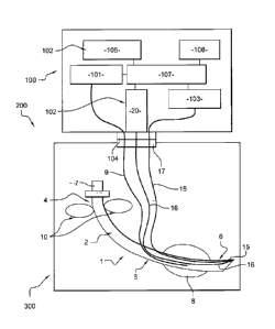
The Figure 8 represents a schematic view of an endotracheal
intubation device 1 connected to a control panel 100 according to the present
= CA 02690827 2009-12-15
=
WO 2009/004483
PCT/1B2008/002483
9
invention and included in an intubation assembly 200 of the present invention.
The control panel 100 comprises a pressure regulator 101, image means 102,
a light source 103 and a third three ways connector 104.
Said third three ways connector 104 is adapted to link said inflation
tube 9 of the endotracheal intubation device 1 to said pressure regulator 101
through a first way so as to allows the pressure regulator 101 to control and
adapt the pressure inside said cuff 8. For exemple, the pressure is regulated
by
use of a sterile compressed air cartridge device incorporated into the
pressure
regulator 101. In order to inflate the cuff 8 to the required pressure, said
pressure regulator is preset to a value corresponding up to a column of water
of
35 cm high. A correct intra-cuff pressure does not restrict tracheal tissue
blood
flow and reduces chance of tissue damage, even during long-term intubation.
This first way incorporates, for exemple, the mechanical self-sealing valve of
a
Luer tip.
A second way of the third three ways connector 104 is dedicated to
connect said light source 103 to light guide 15, thus transmitting light to
the
distal end of said endotracheal intubation device 1 delivering enough light
for
said image means 102 to work prOperly in either the presence or absence of
the classical laryngoscope lighting, i.e before or after the endotracheal
intubation device 1 penetrates the patient's trachea. The area around the
distal
end of the main tube 2 is enlighted by the light that exits the light guide 15
and
feeds the image guide 16 with a view of said area. Typicaly, said image guide
16 comprises a fiber optics.
A third way of the third three ways connector 104 is dedicated to
connect said image means 102 and the image guide 16. Said image means
102 are, for exemple, comprising an image sensor 20 and a display device 105.
The image sensor 20 faces the proximal end of the image guide 16, thus
bringing the image in front of the distal end of the endotracheal intubation
device 1 to the sensing, controlling and displaying part of the visualization
system incorporated in the control panel 100, i.e the image means 102. The
size of the image sensor 20 is a little bigger than the diameter of the image
guide 16 so a direct coupling can be done.
Said control panel 100 is preferably provided with an autonomous
power supply 106 that powers an electronic control units 107 which drives said
pressure regulator 101, said image means 102 and said light source 103.
Thanks to the pressure regulator 101, the practitioner can set up the pressure
= CA 02690827 2009-12-15
WO 2009/004483
PCT/1B2008/002483
inside said cuff 8 and make certain the pressure will not change all through
the
intubation.
An endotracheal intubation device 1 according to the present
invention can be sealed into a thight packaging 300 that keep the sterility of
5 said endotracheal intubation device 1. The first three ways connector 17 is
placed accross the packaging 300 for a proper connection to the control panel
100 without impairing the sterility of the endotracheal intubation device 1.
Optionally, inside the packaging 300, the cuff 8 can be inserted into
a tube 301 having a diameter of the order of that of a trachea, said tube 301
10
being inserted onto the inflatable cuff 8 of said endotracheal intubation
device
.1. Said tube 301 permits to inflate the cuff 8 into the sealed packaging 300
and
then to test its airtighness without opening said packaging 300 or impairing
the
sterility of the endotracheal intubation device 1. Preferably the tube 301 is
provided with lubricant so that removing the endotracheal intubation device 1
from the packaging 300 lubricates the cuff 8.
Optionally, said packaging 300 comprises an identification tag, for
exemple a radio frequency identification tag (RFID tag) or a bar code. The
Identification tag contains for exemple a serial number or other information.
Inside the packaging 300, a test pattern 302 is placed so as to face
the distal end of said endotracheal intubation device 1 placed therein. Said
test
pattern 302 allows to calibrate visualizing means 15, 16 of said endotracheal
intubation device 1.
The Figures 9 and 10 shows a control panel 400 adapted to be
mounted on a classical laryngoscope 26. The control panel comprises a bottom
plate 403 wherein said thrid three ways connector 104 is mounted and which
permits to connect the endotracheal intubation device 1. Said bottom plate 403
is also provided with a screen 402, optionaly foldable, allowing to display
the
image of the trachea captured by the image guide 16 of said endotracheal
intubation device 1 during intubation. The image of the patient's upper aerial
ways can be seen on the screen 402 thus helping the practitioner penetrate
into the patient's trachea. The image dispalyed on said screen 402 plays two
important roles: help in guiding all through the penetration of the trachea
and
also supervising the trachea after the endotracheal intubation device 1 is
installed. For this second function, a color video is preferred.
The figures 11 and 12 represent an endotracheal intubation device
tester 500 according to the present invention. The endotracheal intubation
= CA 02690827 2009-12-15
WO 2009/004483
PC T/IB2008/002483
11
device tester 500 comprises a case 501 adapted to receive a packaging 300 or
an endotracheal intubation device 1, said case 501 being closed by a cover
502. The endotracheal intubation device tester 500 further comprises a
pressure controller 503, an optical or video device 504, a test light source
505
and a second three way connector 506 as represented on Figure 12. Said
second three way connector 506 is connectable to said= first three ways
connector 17 of the endotracheal intubation device 1, said inflation tube 9
and
said pressure controller 503 being connectable together through a first way of
said first and second three ways connectors 17, 506, said test light source
505
and said means to enlighten 15 being connectable together through a second
way of said first and second three ways connectors 17, 506, and said optical
or
video device 504 and said image guide 16 being copnectable together through
a third way of said first and second three ways connectors 17, 506. The
optical
or video device 504 are adapted to display the image captured by the image
guide 16 of the endotracheal intubation device 1.
Once said first three ways connector 17 and said second three
ways connector 504 linked, as shown on Figure 12, the endotracheal intubation
device tester 500 is able to test the vizualisation means 15, 16 and the
inflatable cuff 8.
The light emitted by test light source 505 is transmitted to said
means to enlighten 15 via the first and second three ways connector 17, 506
and this light light up the area around the distal end portion 6 of the
endotracheal intubation device 1. The visualization means 15, 16 are tested
with help of an adequate test pattern 302 placed inside the packaging 300 and
facing the distal end of the endotracheal intubation device 1 so as to be lit
by
the light guide 15 and seen by the image guide 16. The image is then guided to
the control panel 100 by the image guide 16 and seen by the testing person
with the appropriate optical or video device 504 which can be for instance an
eyepiece or a video system similar to that of the control panel 100.
The test of the inflatable cuff 8 is made thanks to the pressure
controler 503 connected to the inflation tube 9 by blowing air to a controled
pressure. In order to keep the cuff 8 and its tube sterile the air used for
testing
must be sterile. Testing air is filtered before entering a reversible micro
air
pump not shown on Figures and designed to inflate and deflate the cuff 8.
Alternatively, a sterile air cartridge can be used. The cuff 8 must be empty
prior
to the intubation.
= = CA 02690827 2009-12-15
WO 2009/004483
PCT/1B2008/002483
12
Inside the packaging 300 the cuff 8 is enveloped with the non
expandable tube 301 having an internal diameter similar to the patients
trachea. When the cuff 8 is inflated its diameter is limited by the tube 301
and
the pressure inside the cuff 8 is then set to a pressure corresponding to the
wight of a column of water of about 25 cm high. If this pressure is achieved
and
kept for a preset time the cuff test is OK.
Once the test is accomplished, a printer 507 prints the results
obtained on a sticker 508 and adds the date/time of test (Figure 11). The
packaging 300, with said sticker 508 stuck on it, is ready to be used for a
given
period of time. Since testing with the endotracheal intubation device tester
500
does not impair the sterility of the endotracheal intubation device 1, it can
be
brought to the operating theatre, kept if not used, and eventually re-tested
later
on. In all other known solutions an endotracheal intubation device entered to
the operating theatre must be opened, tested, and, even if not used, finally
disposed of at the end of the operation.
Optionally, the endotracheal intubation device tester 500 according
to the present invention is provided with identification means to read for
exemple a bar code or a radio frequency identification tag (RFID). The
identification means are adapted to read the identification tags of the
packaging
300.
The method of the present invention will now be detailled:
To practice the intubation method according to the present
invention, it is recommended to dispose of the following material: a packaging
= according to the present invention, a control panel mountable on a
laryngoscope as described above and an endotracheal intubation device tester
according to the present invention. The preset pressure of the inflatable cuff
should be preferably set to a pressure corresponding to a column of water of
35
cm high.
The packaging containing the endotracheal intubation device allows
testing of visualization means and said inflatable cuff. Moreover, the
endotracheal intubation device is lubricated while removed from its packaging.
- a modified laryngoscope or a classical larygoscope should be
adapted with a control panel according to the present invention in order to
present the following features:
= CA 02690827 2009-12-15
WO 2009/004483
PCT/1B2008/002483
13
- a pivoting screen (automatically switched on when opened)
allowing the seeing of the image on the distal end of the endotracheal
intubation device once connected to the control panel;
- sterile, disposable compressed air cartridge for the rapid cuff
inflation;
- Inflation may be started with finger of the hand that holds the
laryngoscope. Integrated connector for the transmission of images and
compressed air for the cuff with automatic disconnection when the cuff
pressure equals the preset value.
The endotracheal intubation device tester according to the present
invention will provide the following advantages:
- allowing testing of vizualisation means of the endotracheal
intubation device and testing of the cuff inflation while
preserving the asepsis of the endotracheal intubation device;
- optical barcode reader for the tube serial number identification;
- a sticker printer to print data to be stick on the tube package
without opening (and to be stuck on the anesthesia document after opening of
the package);
To carry out the method according to the present invention, the
following algorithme is to be followed:
I. Put a packaging containing a sterile endotracheal intubation
device according to the present invention without opening the packaging into
the endotracheal intubation device tester; close the cover of the endotracheal
intubation device tester and starts the auto test:
- read the serial number of the endotracheal intubation device;
- test the vizualisation means;
- test the cuff pressure;
- print the results of the auto test on the sticker (and eventually
store the data in the memory);
In case the test is not successfully passed, the packaging is
disposed of.
In case the test is successfully passed, then step II.
CA 02690827 2009-12-15
WO 2009/004483
PCT/1B2008/002483
14
II. At this step two cases are possible:
A) Intubation is not immediate
open the tester cover and remove the tube package without
opening it then place the sticker on the packaging (possibly replace the
sticker
onto the anesthesia document for intubation).
The packaging according to the invention can be tested and stored.
At any step the sterility of the endotracheal intubation device is not
impaired. In
opposite, a classic endotracheal intubation device must be removed from its
classic sterile packaging in order to be tested and can not thus be stored
afterwards.
B) Intubation is immediate
1) Open the cover together and remove the endotracheal intubation
device form its sterile packaging triggering the cuff lubrication;
2) Then place the sticker on the anesthesia document.
3) Then Connect the endotracheal intubation device to the control
panel mounted on the laryngoscope with the help of the three ways connectors.
4) Then, depending on the visibility of vocal cords:
a) Vocal cords are well visible with the laryngoscope:
_
- Direct (classic) intubation followed by the cuff inflation with sterile
air from the cartridge (integrated to the laryngoscope) and automatic
disconnection of the endotracheal intubation device when the pressure in the
cuff equals the preset value.
- Reconnect the introduced endotracheal intubation device to the
endotracheal intubation device tester in order to check the position of the
probe
and verify or modify the cuff pressure.
b) Impossible to see vocal cords with the laryngoscope:
- open the display of the control panel (automatic switch on);
- guide the endotracheal intubation device using the image from the
distal end transmitted with the vizualisation means and using the guiding
means;
- inflate the cuff with sterile air from the cartridge (integrated to the
laryngoscope) and automatic disconnection of the endotracheal intubation
device when the pressure in the cuff equals the preset value;
= CA 02690827 2009-12-15
WO 2009/004483
PCT/1B2008/002483
- Reconnect the introduced endotracheal intubation device to the
endotracheal intubation device tester in order to check the position of the
probe
and verify or modify the cuff pressure.
5 The
above intubation method permits in particular to keep the
sterility of the endotracheal intubation device while testing it.
During an operation, an endotracheal intubation device must be
available and must have been tested. A classic endotracheal intubation device
is removed from its sterile packaging and tested prior to any surgical
10
intervention. At the end of the surgical intervention, no matter the
endotracheal
intubation device has been used, it must be disposed of.
Though the endotracheal intubation device and the packaging
according to the present invention are more elaborated than a classic
endotracheal intubation device, their use is cost-effective as the packaging
15 does
not need to be open prior to the surgical intervention and can be re-used
after the surgical intervention. The guiding means and the visualization means
of said endotracheal intubation device ensure a safer intubation even in case
of
difficult intubation.
The present ipplication provides an endotracheal intubation
assembly, an endotracheal intubation tester, a packaging and a method that
resolve most if not all the issues occurring during an intubation.