Note: Descriptions are shown in the official language in which they were submitted.
CA 02690896 2009-12-18
WO 2007/147235 PCT/CA2007/001082
JOINT PLACEMENT METHODS AND APPARATUSES
CROSS-REFERENCE TO RELATED APPLICATIONS
This application claims priority from, and the benefit of, the filing date of
United States
Provisional Patent Application 60/814,547 filed 19 June 2006 under title Joint
Placement
Methods and Apparatuses. The contents of the above application are hereby
incorporated by
reference into the Detailed Description hereof.
TECHNICAL FIELD
The invention relates to joint placement generally, more specifically to
placement of prosthetics
for joint repair.
BACKGROUND ART
Joint repair, or arthroplasty, is a surgical intervention for repairing
defects or injuries of a joint.
The goal is to restore as much normal function as possible while at the same
time reducing or
eliminating pain and discomfort. Joint repairs allow patients to regain
mobility, continue
contributing to society and enjoy a greater quality of life.
Arthroplasty attempts to correct how two bones interact with one another. For
example, in a hip
the femoral head 6 fits into the acetabulum 3 of the pelvis 1 creating a ball
and socket type joint.
The movement of the femur 2 in the pelvis 1 is constrained by both the bony-to-
bony interaction
as well as the function of the soft tissue (ligaments and muscle) attached to
both bony anatomies.
Arthroplasty involves resurfacing or replacing the natural surfaces of the
bones where they
interact (the articular surface). For example, referring to FIG. 2, when
performing hip
arthroplasty a surgeon replaces or resurfaces the femoral head 6 and
acetabulum 3 with artificial
implants. To help restore function the soft tissue may also be modified, such
as through a partial
release of its attachment to bone.
Intervention to correct problems of a joint suffers from difficulty in finding
an optimal implant
placement including position and orientation. Problems arising from incorrect
placement may
involve, among other symptoms, post surgical pain, limited range of motion,
dislocation, post
surgical fracture, premature implant failure, and adverse biologic response
consequent to any of
the preceding problems.
The extent of the problem is evidenced in high revision rates. The majority of
arthroplasties
(86%) performed in U.S. are on the hip or knee (American Academy of
Orthopaedic Surgeons,
"Musculoskeletal Conditions in the United States", p. 121, 1999). According
the American
CA 02690896 2009-12-18
WO 2007/147235 PCT/CA2007/001082
Academy of Orthopaedic Surgeons, 54,000 of the total hip and knee procedures
done in the U.S.
each year are revision surgeries (American Academy of Orthopaedic Surgeons
Bulletin,
"Number of arthroplasties to increase dramatically", Vol. 50, No. 1, 2002).
Dislocation following arthroplasty is a major factor in early failure with an
incidence rate, for
example, of 3-5% for hips (for discussion see McCullum, DE, WJ Gray,
"Dislocation After Total
Hip Arthroplasty - Causes and Prevention", Clinical Orthopaedics and Related
Research,
Dec(261), pp. 159-170, 1990). The common reason for dislocation is the
position and
orientation of the artificial implants, particularly of the acetabulum implant
in hip arthroplasty
(for discussion see Lewinnek et al., "Dislocation after Total Hip-Replacement
Arthroplasties",
Journal Bone Joint Surgery, 60-A(2), pp. 217-220, 1978). When the implants are
not correctly
placed, impingements 4 between the implant components and/or anatomy can
occur. These
points of impingement cause a lever effect and potentially result in
dislocation. Besides being
painful and stressful for the patient, it also leads to abductor tissue
damage, which has a
detrimental long-term effect on the stability of the hip joint.
A number of methods and apparatuses for assisting in arthroplasties have been
developed. These
methods and apparatuses include mechanical jigs and computer-assistance (both
image-based
and imageless). A common aspect is the use of an anatomic reference such as
the sagittal and
coronal planes of the patient, or the pelvic plane as defined by the pubic
tubercles 85, 86 and
anterior superior iliac spines 7, 8.
Mechanical guides attempt to address the problem by providing surgical tools
which, when
placed in a specific manner provide a referencing system for determining
implant placement (for
discussion see Eggli et al., "The value of preoperative planning for total hip
arthroplasty",
Journal Bone Joint Surgery, 80-B(4), pp. 382-390, 1998). These solutions can
suffer from one or
more of the following problems.
They are based on standardized placement specifications that may not apply to
an actual patient.
The guides depend on precise, but difficult, placement within anatomy. The
external frame of
reference can change dramatically based on patient anatomy, positioning and
surgical approach.
They typically do not allow for soft tissue effects. The guides provide a
static placement for a
problem that is inherently kinematic in nature. The guides provide limited
flexibility for
anatomical variability.
Image-based computer assisted solutions also exist. These solutions typically
use either CT (for
discussion see DiGioia et al., "HipNav Technical Paper", Centre for Medical
Robotics and
Computer Assisted Surgery) or fluoroscopy (Tannast et al., "Accuracy and
potential pitfalls of
-2-
CA 02690896 2009-12-18
WO 2007/147235 PCT/CA2007/001082
fluoroscopy-guided acetabular cup placement", Computer Aided Surgery, Vol. 10,
Issue 5-6, pp.
329-336) to capture images of the anatomy. Used in conjunction with a spatial
tracking device,
the images are then used to anatomically plan the implant placement, in either
a manual or semi-
automatic manner. These systems then provide the surgeon with guidance to
transfer the
planned placement onto the patient anatomy.
3D solutions (typically based on Computer-Tomography or Magnetic Resonance
Imaging) can
have one or more of the following limitations. They require pre-surgical
scanning of the patient
consuming more time, increasing cost and exposing the patient to additional
radiation. Planning
based on scans and reconstruction of the bony anatomy fail to account for the
important and
significant contribution of soft tissue to the function of the joint. Three-
dimensional scans are
typically used to create a surface model of the anatomy. This adds time and
has potential error
associated with it. Complex, time consuming and potentially error-prone
registration of the
patient to the scan must be performed intra-operatively. A pre-surgical
planning step is often
used with CT-based systems. Such planning is scheduled separately from the
surgery and
consumes more time and thus increases cost. Pre-operative imaging does not
allow for intra-
operative updating.
2D (typically fluoroscopy) solutions can have one or more of the following
limitations. The
prescribed images can be difficult to obtain (e.g., lateral image of the hip).
There is additional
radiation exposure to the patient and operating room staff. Fluoroscopy
requires tracking of the
c-arm (x-ray image intensifier) and image processing to account for image
distortion and to
characterize the c-arm geometry. The hardware necessary to do this increases
cost. The
additional setup can be complicated and time-consuming. The image processing
requires use of
software algorithms that must be written, maintained and presents another
opportunity for
introducing error. Picking 3D anatomical landmarks from 2D images,
particularly on complex
anatomy and/or lower quality images is difficult and error-prone.
Non-image, computer assisted solutions also exist (for discussion see Jansen
et al., "Computer-
Assisted Hip Replacement Surgery, patent US2004/0230199-A1). They make using
of spatial
tracking technology to detect the location of the patient and surgical
instruments. These
solutions attempt to solve the issues involved with image-based solutions by
having the surgeon
palpate specific anatomical points in order to define properties of the
anatomy (e.g., the pelvic
plane) and/or require `painting' of the local bony anatomy in order to morph
standardized
anatomical models to the patient's anatomy. Palpation of anatomy to define
anatomical
properties (axes, planes, etc.) has difficulty in accurately palpating
anatomical features, e.g., the
right 85 and left pubic tubercles 86 and right 7 and left anterior superior
iliac spine (ASIS) 8 for
-3-
CA 02690896 2009-12-18
WO 2007/147235 PCT/CA2007/001082
determining the pelvic plane. It can also have an increased risk of infection
from percutaneous
palpations outside the immediate joint replacement site. Extensive `painting'
of the local
anatomy with a spatially tracked probe has one or more of the following
shortcomings as well.
Inaccessible anatomy for 'painting' (particularly with minimally invasive
approaches). Painting is
time consuming, which increases the procedure time. There is difficulty in
maintaining probe-to-
anatomy contact.
Current image-based and non-imaging computer assisted solutions attempt to
define anatomical
properties (axes, planes, etc.) for the purposes of guiding the user, and face
one or more of the
following problems. Solutions using anatomical properties prescribe implant
placement based
on standardized values derived from a large population. Standardized
orientations are derived by
using a sample population to determine what orientations result in the fewest
complications and
failures. These are defined relative to a standardized frame of reference,
further removing the
solution from the patient specific joint function. Not being patient specific
they are not
necessarily ideal, or even correct, for an individual. Whether point picking
in images or
palpating anatomy, the process for providing the inputs to calculate the
anatomical properties is
often difficult and error-prone. The anatomical properties are based on bony
anatomy and fail to
account for the critical contribution of soft tissue to the behaviour of
joints.
Alternative methods and apparatuses for assisting in implant placement are
desirable to assist in
addressing one or more of the issues with existing methods and apparatuses.
DISCLOSURE OF THE INVENTION
In a first aspect the invention provides a method for determining placement of
prosthetic
components in a joint. The method includes defining a patient-specific frame
of reference for the
joint, determining patient-specific postoperative range of motion of the
joint, evaluating patient-
specific range of motion of the joint, automatically planning placement of the
components
balancing the need for range of motion with prosthesis stability through bony
coverage; and
applying manual adjustments to the automatically planned placement of the
component by giving
greater or lesser weight to the need for range of motion or prosthesis
stability through bony
coverage.
The method may include defining a patient specific frame of reference by
recording passive joint
movements and evaluation of the directions of the movements.
The method may include defining a patient specific frame of reference from
passive movements
by recording movements that are only limited by soft tissue characteristics,
bony to bony
impingement, bony to prosthesis impingement, soft tissue to bony impingement,
soft tissue to
soft tissue impingement, and/or soft tissue to prosthesis impingement.
-4-
CA 02690896 2009-12-18
WO 2007/147235 PCT/CA2007/001082
The method may include evaluating the patient-specific range of motion by
detecting
impingement. The method may include evaluating the patient-specific range of
motion by
calculating and comparing range of motion boundaries based on patient specific
lifestyle
requirements and detection of undesirable laxity or tightness in the soft
tissue.
The method may include determining postoperative outcome of the joint repair
by considering
component stability and postoperative kinematic behaviour. The method may
include
automatically planning placement of the components considering the component
stability and
postoperative kinematics.
Automatically planning placement of the components may consider component
stability and
postoperative kinematics to simulate and evaluate the results of a multitude
of component
placements.
In a second aspect the invention provides a method of determining desired
placement of a cup
within an anatomical joint having a socket and having a stem with a center of
rotation. The cup
has a given range of motion for the stem. The method includes placing the cup
in the socket
such that movement of a center of rotation of the cup is limited, while
rotation of the cup about
the center of rotation is permitted, reducing the stem and cup such that the
center of rotation of
the stem and the center of rotation of the cup are concentric, determining a
range of motion of
the joint by moving the stem, and aligning the range of motion of the cup and
the determined
range of motion of the joint.
The method may include palpating a rim of the socket in order to determine a
plane of the rim,
and, at the same time as aligning the range of motion of the cup and the
determined range of
motion of the joint, aligning the cup within the socket to provide desired
bony coverage to the
cup within the rim.
The method may include modifying the range of motion of the joint to improve
the alignment of
the range of motion of the cup and the range of motion of the socket. The
method may include
repeating steps of the method after modification.
The method may include modifying the joint to change spatial placement of the
cup within the
socket to improve bony coverage to the cup within the rim. The method may
include repeating
steps of the method after modification.
In a third aspect the invention provides an apparatus for defining the center
of a prosthetic
femoral head and axis of a prosthetic femoral neck. The apparatus includes a
primary cylinder
where a first end of the cylinder is adapted for placement over an exposed
neck of a prosthetic
femoral stem such that a longitudinal axis of the primary cylinder is
congruent with an axis of
the prosthetic femoral neck. The apparatus further includes a first alignment
receptacle at an
opposing second end of the cylinder, the first alignment receptacle having a
longitudinal axis
-5-
CA 02690896 2009-12-18
WO 2007/147235 PCT/CA2007/001082
congruent with the longitudinal axis of the primary cylinder and the
receptacle adapted to stop a
spatially tracked probe at a known location relative to the centre of the
prosthetic femoral head.
The apparatus further includes a second alignment receptacle fixed relative to
the primary
cylinder, the receptacle having a longitudinal axis normal to the longitudinal
axis of the primary
cylinder, adapted to receive a spatially tracked probe aligned with the
longitudinal axis of the
alignment receptacle. The apparatus further includes a divot on the exterior
of the primary
cylinder. The divot has a normal parallel to the longitudinal axis of the
second alignment
receptacle and the position of the divot being translated toward an opening of
the first alignment
receptacle on the primary cylinder.
In a fourth aspect the invention provides a method for using the apparatus for
defining the center
of a prosthetic femoral head and axis of a prosthetic femoral neck to
determine the center of the
prosthetic femoral head and the axis of the prosthetic femoral neck. The
method includes
placing the first end of the primary cylinder over an exposed neck of a
prosthetic femoral stem.
The method further includes performing one of the following steps, i) placing
a spatially tracked
probe in the first alignment receptacle, ii) placing the spatially tracked
probe in the second
alignment receptacle and rotating the apparatus about the axis of the
prosthetic femoral neck; and
iii) placing the spatially tracked probe in the second alignment receptacle
and placing the probe
in the divot.
In a fifth aspect the invention provides an apparatus for mounting a spatially
tracked device to an
impactor for impacting a prosthetic cup into a reamed socket. The apparatus
includes a tubular
cylinder adapted to be mounted about a shaft of the impactor such that a
longitudinal axis of the
cylinder is aligned with a longitudinal axis of the impactor and is able to
rotate about the shaft
and move freely along the shaft. The apparatus further includes a mount on the
cylinder adapted
to retain the spatially tracked device in a known orientation relative to the
cylinder.
In a sixth aspect the invention provides a system for assisting in the
placement of prosthetic
components in a joint. The system includes a module for calculation of joint
movements, a
module for calculation of a functional reference system, a module for
recording patient specific
joint range of motion, and a module for evaluation of patient specific range
of motion for
impingement.
The system may include a module for automatically planning a placement for the
prosthetic cup.
The system may include a module for determining bony coverage boundaries such
as the plane
of the socket's rim.
The system may include a module for registration of a prosthetic femoral
stem's femoral head
center and neck axis. The system may include a module for navigating an
impactor to place a
prosthetic cup in a planned orientation. The system may include navigator
technology, at least
-6-
CA 02690896 2009-12-18
WO 2007/147235 PCT/CA2007/001082
one output device, at least one central processing unit, wherein the modules
are associated with
the at least one central processing unit to receive inputs from the navigator
technology and
generate outputs for the at least one output device.
Other aspects of the invention will be evident from the detailed description
and FIGS. provided
herein.
BRIEF DESCRIPTION OF THE DRAWINGS
For a better understanding of the present invention and to show more clearly
how it may be
carried into effect, reference will now be made, by way of example, to the
accompanying
drawings which show the preferred embodiment of the present invention and in
which:
FIGS. la and lb are skeletal representations of a human hip joint.
FIG. 2 is a representation of a total hip arthroplasty.
FIG. 3 is a block diagram illustrating example components of an embodiment of
a system in
accordance with an embodiment of an aspect of the invention.
FIG. 4 is a flow chart illustrating a method in accordance with an embodiment
as applied to a hip
joint placement.
FIG. 5 is a schematic layout of the system of FIG. 3 as employed in a hip
joint placement.
FIG. 6 illustrates an example functional coordinate system for use in the
system of FIG. 3.
FIGS. 7a and 7b illustrate recording an example functional data for
determination of functional
coordinate system for the method of FIG. 6.
FIGS. 8a and 8b illustrate determining an example functional coordinate
system.
FIG. 9 shows freedom of a cup to rotate about the socket center of rotation.
FIG. 10 demonstrates an example definition of a post-operative range of
motion.
FIGS. 1la and 11 b show example factors in performing impingement checking for
range of
motion for the method of FIG. 2.
FIGS. 12a and 12b illustrate example methods for determining patient-specific
impingement free
range of motion for the method of FIG. 4.
FIGS. 13a and 13b illustrate example methods for determining an impingement
free zone for the
prosthetic components of FIG. 2.
FIGS. 14a and 14b illustrate example kinematic evaluations of patient-specific
ROM with
respect to the prosthetic impingement-free ROM.
FIGS. 15a and 15b illustrate example anatomical evaluations of cup placement.
FIGS. 16a and 16b are illustrative of navigating a prosthesis to a planned
placement using
embodiments of aspect of the invention.
FIG. 17 illustrates example of prosthesis femoral stem calibration device.
-7-
CA 02690896 2009-12-18
WO 2007/147235 PCT/CA2007/001082
FIG. 18a illustrates an embodiment of a prosthetic femoral stem calibration
device on a stem and
with a probe in a first location.
FIG. 18b illustrates the embodiment of FIG. 18a with the probe in a second
location.
FIG. 19 illustrates an impactor and an embodiment of a shock-absorbing mount
for a trackable
device about the shaft of the impactor.
FIG. 20 illustrates stable fixation of trackable device to impactor mounting
device.
FIG. 21 identifies system modules and certain inputs and outputs for
calculating joint
movements, functional coordinates and range of motion evaluation.
FIG. 22 identifies system modules and certain inputs and outputs for
calculation of socket rim,
registration of prosthetic stem and recording of patient range of motion.
FIG. 23 identifies system modules and certain inputs and outputs for automatic
planning of
prosthetic cup placement and navigation of impactor to place prosthetic cup.
FIG. 24 illustrates modules of an embodiment of a system for use in total hip
replacement.
MODES FOR CARRYING OUT THE INVENTION
Referring to FIG. 3, a system has a central processing unit(s) 16, one or more
output devices 14,
and navigator technology 17. The navigator technology 17 may be passive
localization or active
positioning technologies. The system also has input devices 15 for example,
keyboard, mouse,
foot pedal, button probes, or virtual keyboards, specialized surgical
instruments 18, and devices
for localizing anatomies 19 using navigator technology 17. The central
processing unit(s) 16 has
the computational power to process localization data, perform complex
mathematical
calculations and, optionally, perform image processing. The central processing
unit(s) 16 also
has associated modules to carry out the various features and functions
described herein,
examples of such modules will be described later herein. The modules may be
software in
memory accessible to the central processing unit(s) 16. There is a means for
communication
between the central processing unit(s) 16, the other apparatus components and,
optionally,
imaging systems and networking with other electronic systems such as, for
example, imaging
archives, inventory databases, and scheduling systems. The navigator
technology 17 has the
ability to determine the spatial pose (position and orientation) of objects in
3-dimensional space.
The output device(s) 14 provides a means for a user of the system to receive
feedback on
planning and navigating placement of an artificial component. The output
device(s) 14 are
typically a display device, such as, for example, a computer monitor but may
employ other forms
of feedback such as, but not limited to, customized LCD, auditory or tactile
devices.
For clarity and by way of example only, a method for determining placement of
and an apparatus
for an artificial component will be described in the context of a system
having modules for
-8-
CA 02690896 2009-12-18
WO 2007/147235 PCT/CA2007/001082
performing a total hip replracement (FIG. 2) as embodied by the system modules
constructed for
this purpose shown in FIG. 24. It is to be recognized that the methods and
apparatuses described
herein can be applied to placement of other types of artificial components
with consequent
modifications, such as, for example, artificial components for knee
replacements and shoulder
replacements. Furthermore, and also for clarity and by way of example, the
apparatus for
employing the method will be limited to a single stand-alone computer for the
central processing
unit(s) 16, a computer monitor for the display device 37 and an optical
spatial tracking system 35
for the navigator technology 17. Other components or combinations thereof
could be used to
provide the functions and features described herein, such as, for example,
alternate spatial
tracking technology based on electromagnetic, visible light, or acoustics
technology.
Referring to FIG. 4, a workflow for applying a method to a hip arthroplasty is
shown.
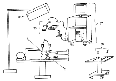
Referring to FIG. 5, when applied to hip procedures, the system is laid out as
illustrated. In this
example the central process unit 16 is a single computer on a cart 37, in
conjunction with an
optical tracking system 35 for real-time spatial localization of anatomy 1, 2
and specialized
instruments 39. Input devices 36 are employed to allow users to interact with
the system in a
manner conducive to, for example, an operating room environment.
A cup 9 is placed in the joint and stem 12 is inserted into the cup 9. The cup
9 may be a trial cup
used for determining placement of a final cup. As is known in the art, a trial
cup may be better
suited for determining placement prior to actual placement of a final cup. For
example, if it is
later determined that another size of cup would better suit the particular
joint, then a more
expensive cup is not wasted in the determination procedure. Devices 52 capable
of being tracked
by the spatial localizer 35 with high accuracy are then affixed rigidly to the
pelvis 1 and femur 2.
The exact placement and orientation of these devices will depend on the manner
in which the
overall joint replacement is being conducted and the placement of the spatial
tracking device 35
in, for example, an operating room.
Referring to FIG. 17, with the trackable devices 52 attached to the patient,
the orientation and
position of the stem component 12 can be determined, for example, by use of a
calibration device
70 that provides for accurate placement of the calibration device relative to
the femoral
component neck 13 and for at least one fixed probe location for locating a
spatially tracked probe
56 relative to the calibration device, such as for example calibration device
70 that facilitates
determining the center of the prosthetic femoral head 11 and the axis of the
prosthetic femoral
neck 13. The probe location and direction is, or a series of probe locations
and directions are,
recorded relative to a reference 52 attached to the femur 2 by a registration
module 64 for
calculating the center of the femoral component head 11 and axis of the
component neck 13.
-9-
CA 02690896 2009-12-18
WO 2007/147235 PCT/CA2007/001082
The femoral calibration device 70 is made from rigid materials that can be
sterilized, such as
stainless steel. The device has a primary cylinder 54, a secondary cylinder 55
and a small divot
71 on the primary cylinder 54 such that the normal of the divot is parallel to
a longitudinal axis
of the secondary cylinder 55 and the position of the divot is outside the
secondary cylinder.
Referring to FIG. 18a, one end of the primary cylinder 54 allows the device to
be fitted to the
exposed end of inserted femoral component. The other end of the primary
cylinder 54 allows a
spatially tracked probe 56 to be inserted into it. This other end of the
primary cylinder 54 is an
example of an alignment receptacle for a spatially tracked probe 56. When
fitted onto the
exposed end of the inserted femoral component 13 the calibration device 70 can
be rotated about
axis of the prosthetic femoral neck 13 without displacing from it. The
secondary cylinder 55 also
allows a spatially tracked probe 56 to be inserted into it. The secondary
cylinder 55 is an
example of an alignment receptacle for a spatially tracked probe 56. The divot
71 on the primary
cylinder 54 allows a spatially tracked probe to be positively seated in it
such that the probe 56 is
aligned to the longitudinal axis of the secondary cylinder 55.
The device 70 allows the system to capture spatial information sufficient for
determination of the
center of the prosthetic femoral head 11 and the axis of the prosthetic
femoral neck 13 using at
least three different methods. A user employs one of the system input
components to indicate the
method being used. In each method, the calibration device 70 is fitted onto
the exposed end of
the inserted prosthetic femoral component 13. In a first method FIG. 18a, the
user places a
spatially tracked probe 56 into the free end of the primary cylinder and
indicates to the system
when the probe is positively seated in the calibration device 70. A
registration module 64
captures the spatial localization information for the probe 56 and the
trackable device 52 attached
to the femur. The axis of the prosthetic femoral neck 13 is defined by the
orientation of the probe
56 and the center of the femoral head 11 is derived from the location of the
probe tip, which,
when positively seated in the calibration device 70, has a known relationship
with the center of
the prosthetic femoral head 11.
Referring to FIG. 18b, in a second method the user places the probe 56 in the
secondary cylinder
55 and indicates to the system to start collecting spatial localization
information for calibration
purposes. With the probe 56 firmly seated in the secondary cylinder 55 the
user then rotates the
calibration device back-and-forth about the axis of the prosthetic femoral
neck 13. The
registration module 64 continues to collect spatial localization information
for the probe 56 and
the trackable device 52 attached to the femur 2 until it has enough data for
calibration purposes.
The registration module 64 uses the series of probe 56 orientations as inputs
to a minimization
algorithm, which will be understood by those skilled in the art. The results
of the minimization
-10-
CA 02690896 2009-12-18
WO 2007/147235 PCT/CA2007/001082
are used to derive the center of the femoral head 11. A line parallel to the
normal of the plane
defined by the movement of the probe 56 and which passes through the derived
center of the
femoral head 11 is used to determine the axis of the prosthetic femoral neck
13. Using a third
method for calibration, the user places a spatially tracked probe 56 into the
secondary cylinder 55
and indicates to the system to capture spatial localization information for
the probe 56 and the
trackable device 52 attached to the femur 2. The user then moves the probe 56
to the divot 71 on
the primary cylinder 54 and indicates to the system to again capture spatial
localization
information for the probe 56 and the trackable device 52 attached to the femur
2. The registration
module 64 uses the two locations of the probe 56 to define a line parallel to
the axis of the
prosthetic femoral neck 13. The axis of the prosthetic femoral neck 13 and
center of the
prosthetic femoral head 11 are then derived by the registration module 64
based on a known
relationship between the calibration device 70 and the prosthetic femoral stem
12.
Regardless of the calibration method used, the system uses captured spatial
localization
information to determine and record the center of the prosthetic femoral head
11 and the axis of
the prosthetic femoral neck 13.
As will be evident to those skilled in the art, other calibration devices
allowing for capture of
such localization information using only one or two of the above three methods
could be
constructed. Each such calibration device may be usable in more limited
circumstances;
however, they can also be useful. A set of calibration devices could be
employed to allow for use
in a variety of circumstances.
Referring to FIGS. 6-8 and 21, the user can define a functional coordinate
system for the joint
42. The functional coordinate system 42 simply acts as a frame of reference
for the display of
visual data later and is defined by first moving the limb in
abduction/adduction 40 to estimate the
sagittal plane 80 and then flexion/extension 41 in order to estimate the
frontal plane 81. The
evaluation of these movements being done by the calculation of joint movements
module 59.
The functional coordinate system module 60 captures spatial data on the
movement of the limb
by virtue of the trackable targets 52 attached to the anatomy. The data
collected during the
abduction/adduction and flexion/extension is also used to define a hip center
of rotation 43. The
functional coordinate system module 60 calculates the functional coordinate
system 42. The hip
center of rotation 43 is used for the origin and the axes are calculated with
respect to the normals
of the defined planes. The functional coordinate system is recorded relative
to the pelvic
reference.
Referring to FIGS. 15 and 22, information on the local anatomy can be obtained
by palpation of
the socket rim 4 with a spatially tracked probe 56. The socket rim module 62
records the
-11-
CA 02690896 2009-12-18
WO 2007/147235 PCT/CA2007/001082
palpations of the rim 4 and determines the plane of the rim 49 using 3 or more
palpated points.
Palpation of local anatomy does not suffer from the problems of accuracy and
infection risk
associated with palpation of global anatomy (such as that which would be
required to define the
pelvic plane). The locally palpated points are used to define a plane 49 that
represents the
boundary of bony coverage for an implanted prosthetic cup 9. The cup placement
module 61
when optimizing cup placement uses it later.
A patient specific ROM (range of motion) is then performed with the trial cup
component 9.
Referring to FIG. 9, as the cup 9 is not fixated, it can change orientation in
the reamed socket 3
without altering its center of rotation 43. Referring to FIG. 10, when
performing the ROM, the
user will move the joint in a manner that is representative of the lifestyle
and daily activities of
the patient. The patient-specific ROM module 63 records these movements using
the navigator
technology 17, the trackable devices 52 attached to the anatomies 1, 2, and
the previously
recorded prosthetic stem registrations. Referring to FIG. 11, in the
impingement free zone of the
ROM, the center of the femoral head 11 coincides with the center of rotation
43. The patient-
specific ROM module 63 determines impingement 44 to have occurred when
movement between
the center of the femoral head 11 and center of rotation 43 exceeds an
acceptable tolerance. This
type of movement will occur for the following types of impingement: bony-to-
bony, bony-to-
stem prosthesis, bony-to-soft tissue and soft tissue-to-soft tissue.
Impingement free orientations represent the patient specific ROM 46. Referring
to FIGS. 13, 21
and 23, an impingement free zone 47 for the prosthetic cup 9 is calculated
from geometric data
of the implant system using the ROM evaluation module 61. The automatic
planning module 65
determines an optimized orientation for the prosthetic cup 9. Referring to
FIG. 14, the optimized
orientation is determined firstly by aligning the impingement free zone 47 of
the prosthetic cup 9
with respect to the patient-specific impingement free ROM 46. Referring to
FIG. 15, the
optimized orientation is further refined by maximizing bony coverage of the
cup 9 within the
socket 3 as determined, for example, by the amount of cup 9 within the socket
rim 4 determined
previously.
Graphical and numeric feedback is provided to the surgeon that illustrates the
patient specific
ROM 46 relative to the prosthetic cup impingement-free zone 47. Areas of
overlap are
impingement free and areas not overlapping indicate areas where impingement
will occur post-
operatively 48. Visual and numeric feedback is also provided to illustrate the
bony coverage of
the prosthetic cup 50.
A user may elect at this point to accept a planned prosthetic cup placement or
to perform
corrective actions to decrease impingement and/or increase bony coverage.
Actions that a user
-12-
CA 02690896 2009-12-18
WO 2007/147235 PCT/CA2007/001082
may perform include, but are not limited to: osteophyte removal, soft-tissue
release, prosthetic
changes or adjustment the criteria for the optimization algorithm. In the
preferred embodiment
the system allows for the prediction of results of the correction. After the
correction is
performed, a user can validate it through repetition of earlier steps.
Once a placement of the cup 9 is determined, an impactor 51 may be used to
guide the cup 9.
Referring to FIG. 19, a mount 57 that can have a trackable device 52 attached
to it is placed over
a shaft of a known impactor 51 such that the longitudinal axis of mount 57 and
impactor align.
The mount 57 is made of rigid materials that can be sterilized. Referring to
FIGS. 20 and 23, the
mount 57 has a cylinder for placement over a shaft of the impactor 51, pins 72
that allow the
trackable device 52 to be attached to the mount 57 and a tightening mechanism
58 to hold the
trackable device 52 rigidly to the mount. The mount 57 is free to rotate about
the shaft of the
impactor 51 as well as to move up and down along the shaft of the impactor.
The mount 57 is
rotated around the shaft to achieve an orientation of the trackable device 52
appropriate for
spatial tracking given the type and positioning of the navigator components
17. The navigation
module 66 provides qualitative and quantitative feedback to the user to assist
in the cup
placement. The ability of the mount 57 to move along the shaft of the impactor
51 creates a
shock absorption effect when the impactor is struck with a hammer, of the type
commonly used
for such purposes, to fixate the prosthetic cup 9 into the socket 3. This
shock absorption effect
reduces the likelihood that a trackable device 52 will loosen from the mount
57 or components of
the trackable device 52 will loosen, such as, for example, retro-reflective
spheres which have
been pressure fitted onto posts of the trackable device.
Referring to FIG. 16, graphica153 and numeric feedback is provided by the
system to assist in
achieving the placement by the navigation module 66. A trackable device 52 may
be otherwise
rigidly held to the mount 57.
Referring again to FIG. 19, an impactor 51 with shock absorption is provided.
It will be
understood that use of the impactor 51 with shock absorption may be used
generally for cup
placement and is not limited to use in association with use with the system
and other methods
described herein.
The method in some embodiments provides a method for assisting a surgical
intervention, such
as a hip replacement, that does not require imaging and is based on the
function of a joint, not
just its bony anatomy. The method intrinsically accounts for effects of soft
tissue, bony anatomy,
prosthesis design, component placement and their interactions.
13-
CA 02690896 2009-12-18
WO 2007/147235 PCT/CA2007/001082
The method may be patient specific and not based only on standardized
orientations for an
implant. The method may use the function of a joint to indicate to a user
where, and how, joint
function is limited. In some embodiments this information allows a user to
determine actions to
take in order to correct the function. Such actions may include, but are not
limited to, osteophyte
removal, soft tissue release or altering implant selection.
Using patient-specific joint function to define the boundaries may avoid some
limitations
associated with using only bony landmarks or a simulated ROM including
implicitly accounting
for bony anatomy and soft tissue affects on the joint and avoiding time and
costs associated with
imaging and surface model reconstruction. Further, those bony landmarks that
may be used in
some embodiments of the method are generally relatively easy to palpate.
The functional data of the joint, combined with the known geometry and
functional range of the
implant system is used in some embodiments to determine a desired placement of
the implant to
improve the functional outcome and the stability of the implant. As the
functional data is specific
to the patient, it can be used to improve the placement for the patient's
specific needs, lifestyle
and activities.
Further, in some embodiments the user is able to parameterize the improvement
in order to
balance the needs for stability and range of motion considering the patient's
lifestyle, activities
and bone quality.
The method makes use of quantitative spatial information of patients' anatomy
and
instrumentation for the correction of the joint function. It will be
understood by those skilled in
the art that it is the data, not the mode of acquiring the data that is
pertinent to the method.
Modes for acquisition of spatial data may include, but is not limited to,
navigator technologies
based on optics, mechanics, radio frequency, electromagnetism, acoustics,
radiographic imaging,
non-radiographic imaging, and so on.
It will be understood by those skilled in the art that various embodiments of
the method will
make use of different instrumentation for the placement of prosthetics. For
example, an impactor
has been described in the preferred embodiment for placement of a cup. However
the principles
of the invention do not exclude the use of any suitable instrumentation for
the placement of a
prosthetic component.
Those skilled in the art will understand that in some embodiments the method
may use a
functional range-of-motion and prosthesis geometry in the absence of a
functional coordinate
system to achieve a patient-specific functional placement of prosthesis.
- 14-
CA 02690896 2009-12-18
WO 2007/147235 PCT/CA2007/001082
It will be clear to those skilled in the art that the principles of the
invention is applicable to joints
in general including, but not limited to, hip, knee, shoulder, elbow, and so
on. Further, it will be
understood that the method and principles of the invention are applicable to
forms of joint
correction that do not involve implantation of prosthesis, such as patient-
specific correction of
joint function through osteophyte removal and/or soft tissue adjustments.
By way of example an embodiment of the method using a computer monitor for an
output device
has been described. Other embodiments of the method may use other forms of
output, including,
but not limited to, tactile devices and audio devices.
It will be understood by those skilled in the art that this description is
made with reference to the
preferred embodiments and that it is possible to make other embodiments
employing the
principles of the invention which fall within its spirit and scope as defined
by the following
claims.
-15-