Note: Descriptions are shown in the official language in which they were submitted.
CA 02691184 2009-12-18
WO 2009/001109 PCT/GB2008/002244
MEDICAL ALIGNING DEVICE
The present invention relates to medical devices, in particular devices for
aligning guide wires with respect to bones. The present invention also relates
to a method of aligning and inserting a guide wire into a bone.
Total hip replacements may fail prematurely due to excessive wear,
particularly in active patients. Hence hip resurfacing, using metal on metal
bearings, is increasingly being used with good results. Resurfacing preserves
the patient's natural femoral neck and part of the femoral head. Accordingly,
accurate positioning of the implant components is essential to preserve the
integrity and strength of the natural bone. On the rare occasion "that metal
on
metal resurfacings fail, it is mainly because of fracture of the femoral neck
or
loosening of the femoral component, which may result from poor surgical
technique with notching of the femoral neck or incorrect angular positioning
of
the femoral component.
During the resurfacing operation, preparation of the femur starts with the
positioning and drilling of a guide wire through the femoral head and into the
neck. Guide wire position is critical because it will define the position and
angle of the femoral component relative to the patient's femur. Clearly, it is
best for the surgeon to position the guide wire correctly on the first
attempt.
Once the guide wire is inserted, its position may be verified by rotating a
stylus around the femoral neck and the appropriate head component size is
identified. The guide wire is then over drilled with a cannulated drill to
increase the hole size. A guide rod is then inserted into the hole and used to
guide a rotating cylindrical cutter to shape the femoral head into a cylinder.
This is the stage in the operative procedure where notching of the femoral,
neck can occur due to incorrect positioning or over sailing of the cylinder
cutter. A face cutter is then used to resect the unwanted bone. The guide rod
is used to guide a rotating chamfer cutter to chamfer the proximal end of the
cylinder. This procedure ensures that the implant component fits exactly to
the bone.
CA 02691184 2009-12-18
WO 2009/001109 PCT/GB2008/002244
The femoral head cannot be used as a positioning reference when placing the
guide wire, because it is invariably misshapen in varying degrees due to the
onset of arthritis. A preferred reference to use is the femoral neck, as this
is
where notching must be avoided, but this can also be partially misshapen due
to osteophites.
Due to anxiety about notching the femoral neck and the smaller size of the
neck relative to the femoral head, it is generally accepted that the best
position for the guide wire and hence the femoral implant stem is in the exact
centre of the femoral neck. This is often hard to determine because the neck
cross section is not circular.
In addition to the guide wire being placed centrally in the neck, there are
two
important angles of the femoral implant axis relative to the femur which are
described in different planes. Observed in the frontal (or coronal) plane on a
frontal X-ray, varus/valgus angle is the angle between the shaft of the femur
and the implant axis. The appropriate angle is somewhat patient specific, but
generally within the range 135-145 degrees. The axis of the natural femoral
neck is more varus (or more horizontal) and is difficult to judge because it
tapers outwards towards the shaft of the femur. It is therefore erroneous to
reference the natural neck angle as the appropriate angle for the implant
axis.
Excessive varus positioning of the implant is considered to be the second
most contributory factor (after notching) towards femoral neck fracture and
femoral component loosening.
Observed in the horizontal (or transverse) plane, version angle is a forward
or
backward angulation of the implant axis relative to the shaft of the femur. It
is
generally not apparent on X-ray but can be judged intra-operatively by
observing the underside of the femoral neck. The appropriate angle is also
patient specific but generally within the range 15-25 degrees. In this case,
the
surgeon generally tries to align the implant axis with the patient's natural
anteversion angle.
CA 02691184 2009-12-18
WO 2009/001109 PCT/GB2008/002244
It is generally accepted that a resurfacing head implanted with the
appropriate
varus/valgus and version/anteversion angles without notching of the femoral
neck will have a good chance of success. However this goal is becoming
more difficult to achieve, especially due to the limitations of minimally
invasive
surgery. There is an increasing trend towards minimally invasive surgery in
hip resurfacing which reduces the amount of exposure, access and visibility to
the femoral head and neck. It is more difficult for surgeons to detect and
correct errors using their judgment, with reduced access and visibility.
Therefore they are dependent on the effectiveness of the surgical
instrumentation.
A number of devices exist to facilitate positioning of the guide wire and
hence
the femoral implant component. Early devices used a pin in the lateral femur
to help determine angular position and a probe rotating around the neck to
avoid notching. The requirement for a pin in the Iateral` femur means that
such devices are not suitable for minimally invasive surgery because there is
insufficient access to insert a pin laterally.
Later devices follow the trend towards minimally invasive surgery. The
devices tend to fall into three categories, namely clamp type, ring type and
adjustable platform type devices. Clamp type devices comprise a drill guide
and opposing jaws that attach to the femoral neck. A common problem with
clamp type devices is that they tend to follow the natural femoral neck angle,
which, as already described, is not the correct angle for the femoral implant
axis. An attempt to overcome this has been made by replacing a symmetrical
jaw clamp with an offset jaw clamp. Offsetting the jaws allows the device to
be placed in a more valgus angle relative to the natural neck. However an
offset jaw clamp is inherently unstable because the jaws do not, directly
oppose one another. It is therefore less effective as a clamp.
In both the above types of devices, it is a difficult task for the surgeon to
decide varus/valgus and version angles simultaneously, particularly
considering that these angles are judged in two different anatomical planes.
CA 02691184 2009-12-18
WO 2009/001109 PCT/GB2008/002244
Ring type devices comprise a drill guide and a partial or complete ring which
is placed around the femoral neck, where the diameter of the ring corresponds
to the femoral implant component internal diameter. These devices are not as
stable as clamp type devices because they do not attach to the femoral neck.
Furthermore, varus/valgus and version angles must also be judged and fixed
simultaneously by the surgeon when using such devices. Consequently, they
present similar problems to those encountered with clamp type devices.
Adjustable platform type devices comprise a drill guide and a platform that is
fixed to the femoral head and from which adjustments to position and angles
are made and verified with a rotating stylus. Such devices provide a stable
platform to work from, but have the disadvantage that the surgeon still has to
judge and fix varus/vaigus and version angles simultaneously.
Accordingly, the present invention aims to maximise the accuracy of guide
wire placement which in turn optimises the positioning of the final femoral
component. The present invention also aims to provide guide wire placement
devices that are suitable for use in minimally irivasive surgery.
According to. a first aspect of the present invention, there is provided a
device
for aligning a guide wire with a bone, comprising:
an attachment means reversibly attachable to a bone;
an alignment means connected to the attachment means, the
alignment means being moveable so as to locate a portion of the bone for
insertion of the guide wire.
An advantage of the present invention is that it increases the accuracy of
guide wire placement. Consequently, positioning of the final femoral
component is optimised, significantly reducing, if not eliminating, failure of
the
metal on metal resurfacing. In addition, it does not require a lateral or
posterior targeting pin and therefore is suitable for minimally invasive
surgery
since it can be operated through a reduced incision.
CA 02691184 2009-12-18
WO 2009/001109 PCT/GB2008/002244
Devices according to the present invention improve upon existing devices. by
enabling the alignment means to move independently with respect to the
attachment means. This means that the device can be securely attached to
the femoral neck, for example with symmetrical, directly opposing jaws, which
5. provide a stable platform to work from. The varus/valgus angle is then set
via
a separate adjustment via the alignment means.
The alignment means may be reversibly connected to the attachment means.
The alignment means may receive a guide wire, in use.
According to some embodiments of the present invention there is provided a
device wherein the alignment means comprises:
an alignment guide for receiving, in use, at least one of a goneometer
and a guide wire; and
a support arm connected to the attachment means,
wherein the alignment guide is moveably connected to the support arm.
The alignment guide may be reversibly connected to the support arm.
The support arm may be reversibly connected to the attachment means.
The support arm may be pivotally connected to the attachment means.
The alignment means may comprise a centring mechanism for locating the
centre of the bone.
In some embodiments of the invention, the device has a centring mechanism
to place the guide wire in the centre of the femoral neck at all times
irrespective of varus/valgus adjustment. In those embodiments of the
invention that comprise a centring mechanism, the act of attaching the device
to the femoral neck establishes the neck centre via the centring mechanism
and makes the device stable. Thereafter, varus/vaigus and version angles
are independently adjusted. This is more effective and more accurate than
CA 02691184 2009-12-18
WO 2009/001109 PCT/GB2008/002244
previous devices which are unstable until both angles are fixed. In addition,
such prior art devices.also have the disadvantage that a change to one angle
affects the other angle.
The centring mechanism may be reversibly connected to the attachment
means.
The centring mechanism may comprise two moveable arms, each arm having
a proximal end and a distal end, the arms being pivotally connected together
at their distal ends, the arms being pivotally connected to the attachment
means at their proximal ends, and wherein, in use, the pivot connection
between the distal ends of the arms'locates the centre of the bone.
The alignment guide may be pivotally connected to the distal ends of the
centring mechanism arms.
The attachment means may be a clamp. The attachment means may be a
scissor clamp.
The clamp may comprise at least two jaws. The clamp may comprise two
jaws. The clamp may comprise a plurality of jaws. The at least two jaws may
be opposed.
The clamp may comprise a self-locking mechanism. The self-locking
mechanism may be a ratchet mechanism having a release means for
unlocking the clamp.
The clamp may comprise a resilient means for biasing the jaws apart. The
resilient means may be a spring.
The attachment means may comprise two arms that are pivotally connected
along their length, each arm having a proximal end and a distal end, the
distal
ends being attachable to a bone, the proximal ends enabling a user to
reversibly attach the distal ends to the bone.
CA 02691184 2009-12-18
WO 2009/001109 PCT/GB2008/002244
The alignment guide may comprise a drill guide.
Devices according to embodiments of the present invention may further
comprise a fixation means. The fixation means may comprise at least one
retractable spike. The fixation means may comprise a retractable spiked tube.
According to a second aspect of the present invention there is provided a
method of aligning a guide wire with a bone, comprising the steps of:
providing a device according to the first aspect of the present invention
and a power source;
attaching the attachment means to a bone;
moving the alignment means so as to locate a portion of the bone for
insertion of the guide wire; and
inserting the guide wire into the bone using the power source.
According to a third aspect of the present invention there is provided a
method
of aligning a guide wire with a bone, comprising the steps of:
providing a device according to the first aspect of the present invention
and a power source;
attaching the attachment means to a bone;
moving the alignment means so as to locate a portion of the bone for
insertion of the guide wire;
attaching a goneometer to the alignment guide so as to indicate the
eventual position of the guide wire in the bone;
attaching a guide wire to the alignment guide; and
inserting the guide wire into the bone using the power source.
The goneometer may be detached from the alignment guide before the guide
wire is attached.
The power source may be a rotary power source. The power source may be
a drill.
CA 02691184 2009-12-18
WO 2009/001109 PCT/GB2008/002244
The device/method may be applied to any suitable bone. The bone may be a
femur.
Reference will now be made, by way of example, to the accompanying
drawings, in which:
Figure 1 is an isometric view of a device according to an embodiment of
the present invention;
Figure 2 is a top view of the device shown in figure 1;
Figure 3 is an end view of the device shown in figure 1;
Figure 4 is a side view of the device shown in figure 1
Figure 5 is an isometric view of a device according to another embodiment
of the present invention;
Figure 6 is an isometric view of a device according to another embodiment
of the present invention;
Figure 7 is a top view of the device shown in figure 6;
Figure 8 is an isometric view of a device according to another embodiment
of the present invention; Figure 8a is a side view of a device according to an
embodiment of the
present invention in place on a femur;
Figure 9 is an isometric view of a device according to another embodiment
of the present invention;
Figure 10 is an isometric view of a device according to an embodiment of
the present invention in place on a femur;
CA 02691184 2009-12-18
WO 2009/001109 PCT/GB2008/002244
Figure 11 is a side view of a device according to an embodiment of the
present invention in place on a femur;
Figure 12 is a cross-section through a femoral neck;
Figure 13 is a side view of a device according to an embodiment of the
present invention in place on a femur;
Figure 14 is a side view of a device according to an embodiment of the
present invention in place on a femur;
Figure 15 is a side view of a device according to an embodiment of the
present invention in place on a femur;
15,
Figure 16 is a section through a femur having a resurfacing head attached;
Figures 17 to 26 show various stages of operation of a device according to
an embodiment of the present invention;
Figure 27 is a top view of a device according to another embodiment of the
present invention; and
Figures 28 to 38 show various stages of operation of a device according to
an embodiment of the present invention.
Figures 1 to 10 show components of a device (1) according to some
embodiments of the present invention. As shown in figures 1 to 4, the device
(1) is in the form of a scissor clamp (2) with a ratchet locking mechanism
(14).
The clamp (2) comprises two arms (3,4) that are connected together by a
pivot (5). In the embodiments shown, the pivot (5) is near to the mid-point of
each arm (3,4). Each arm (3,4) has a proximal end (6,7) and a distal end
(10,11). Disposed at the proximal end (6,7) of each arm (3,4) is a finger grip
(8,9) enabling a user to grip the clamp (2). In the device shown, the finger
CA 02691184 2009-12-18
WO 2009/001109 PCT/GB2008/002244
grips (8,9) are closed hoops, although any suitable grip is envisaged.
Disposed at the distal end (10,11) of each arm (3,4) is a jaw (12,13) for
attaching the device to a bone. Alternative embodiments may have a plurality
of jaws.
As shown in figure 2, when viewed in the horizontal plane, each arm (3,4) has
curved and linear sections forming a distorted S-shape. As shown in figure 4,
when viewed in the vertical plane, each arm (3,4) has two approximately 90
degree bends between the pivot (5) and the distal end (10,11) such that the
main axis of a portion of each arm (3,4) between the proximal end (6,7) and
the pivot (5) is parallel to the main axis of a portion of each arm (3,4) near
the
I
distal end (10,11). This offset, non-planar structure has the advantage that,
in
use, the surgeon can manipulate the device so that the jaws (12,13) can be
appropriately positioned around and engaged with a bone.
A ratchet locking mechanism (14) is disposed at'the proximal end (6,7) of the
arms (3,4). The ratchet (14) comprises an arm (15) that is connected to the
proximal end (6) of arm (3) by pivot (16). Indentations (17) engage with a
complementary protrusion (18) disposed at the proximal end (7) of arm (4),
thereby effecting the self-locking ratchet mechanism (14). The ratchet
mechanism (14) can be unlocked by moving arm (15) about pivot (16) so as to
disengage the protrusion (18) from the indentations (17). Leaf springs (19,20)
are attached to arms (3,4) so as to bias the proximal ends (7,8) apart and
hence bias distal ends (10,11) apart. As shown in figure 2, leaf spring (19)
extends towards proximal end (6) of arm (3) and contacts ratchet arm (15)
near to pivot (16), thereby biasing arm (15) into a locked position.
Figure 5 shows the components of figures 1 to 4 in combination with a
centring mechanism (21). The centring mechanism (21) has two moveable
arms (22,23), each having a proximal end (24,25) and a distal end (26,27). In
the embodiment shown in figure 5, each arm is curved. However, some
embodiments include linear arms (51,52), as shown in figure 11. Proximal
end (24) of centring arm (22) is pivotally connected to clamp arm (4) by pivot
(28), located near to grip (9). Proximal end (25) of centring arm (23) is
CA 02691184 2009-12-18
WO 2009/001109 PCT/GB2008/002244
pivotally connected to clamp arm (3) by pivot (29), located near to grip (8).
Distal end (26) of centring arm (22) is pivotally connected to distal end (27)
of
centring arm (23) by pivot (30).
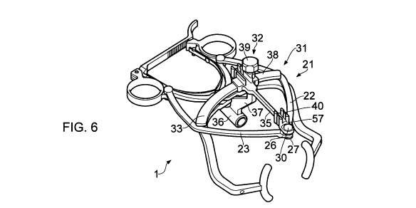
Figures 5 to 10 show an alignment means (31) according to some
embodiments of the present invention. The alignment means (31) comprises
a centring mechanism (21), an alignment guide (32) and a support arm (33).
The support arm (33) is pivotally connected to the clamp (2) by pivot (5). The
support arm (33) can be fixed in place using a fixing screw (34) at pivot (5).
In
the particular embodiment shown, the support arm (33) is crescent shaped.
The support arm may be any suitable shape.
The alignment guide (32) comprises an alignment arm (35) that is connected
to an alignment conduit (36) by body (37). Alignment arm (35) has a proximal
end (38) that is shaped so as to receive the support arm (33) such that the
alignment guide (32) is moveably connected to the support arm (33). The
alignment guide (32) can be reversibly locked in position on the support arm
(33) by means of locking screw (39) disposed at the proximal end (38) of
alignment arm (35). The alignment arm (35) has a distal end (57) that is
pivotally connected to the distal ends (26,27) of centring arms (22,23) by
pivot
(30). The alignment arm (35) has spring clips (40) disposed at the distal end
(57) for receiving a goneometer (41,42), as shown in figures 8 and 9.
A goneometer is a separate angle measuring device employed by the surgeon
to find the correct varus/ valgus angle. Those embodiments of the invention
that comprise a goneometer have the advantage that it facilitates hands free
use. ' Figures 8, 8a and 9 show two alternative designs of goneometer (41,42).
Goneometer (41) of-figures 8.and 8a comprises an angled, `V'-shaped rod that
defines the angle between the femoral implant component axis and the
femoral shaft. The rod may have any suitable angle. For example, the rod
may have an angle of 130 to 140 degrees. The rod may have an angle of
around 135 degrees. In use, a surgeon aligns the longer part of the shaft with
the femoral shaft by adjusting the alignment guide (32).
CA 02691184 2009-12-18
WO 2009/001109 PCT/GB2008/002244
Goneometer (42) of figure 9 comprises a distorted `T'-shaped rod that defines
the correct varus/ valgus angle from the leg alignment axis, which is a
straight
line between the centre of the hip and the centre of the knee. The rod may
have any suitable reference angle. In the example shown, the reference
angle is 128 degrees. In use, the surgeon aligns the intersection of the `T'
rod
over the centre of the femoral head and ensures that the distal end of the rod
points towards the centre of the knee. The difference between the femur shaft
angle and leg alignment axis is 7 degrees, therefore the difference between
the angles of the goneometers is also 7 degrees.
Alignment conduit (36) is shaped so as to receive a guide wire. As shown in
figure 8, in some embodiments of the present invention, the conduit (36)
comprises a fixation means (43). In the embodiment shown in figure 8, the
fixation means (43) is in the form of a cylindrical plunger having a plurality
of
spikes (44) arranged around a conduit (45) for receiving a guide wire (not
shown). In use, the surgeon can advance the plunger from a retracted,
disengaged position to an advanced, engaging position in which the spikes
(44) engage a bone. The fixation means has the advantage that it provides
additional stability to the device.
In an alternative embodiment of the invention (not shown), the alignment
means comprises an alignment guide (32) pivotally connected to the centring
mechanism (21) as in figure 6, but without a support arm (33).
Figure 8 shows an embodiment of the invention which comprises an additional
guide rod (46). In use, the guide rod (46) is received in a conduit (47) in
arm
(4). In use, the guide'rod (46) is aligned parallel to the guide wire,
providing
the surgeon with an additional point of reference when aligning the
device/guide wire.
In an alternative embodiment of the invention (not shown), alignment arm (35)
may receive a guide rod like guide rod (46) instead of a goneometer (41,42).
CA 02691184 2009-12-18
WO 2009/001109 PCT/GB2008/002244
In use, centring mechanism (21) works as follows. As shown in figures 9 and
10, the moveable arms (22,23) are positioned with respect to the jaws (12,13)
such that the pivot (30) is naturally aligned with the centre-point of the
jaws
(12,13) when the jaws of the device are attached to the neck of the femur
(48), for example. As a result,_the alignment guide is naturally aligned such
that the alignment arm (35) and conduit (36), and hence the goneometer
(41,42) and guide wire (not shown), are aligned with the centre-point of the
jaws (12,13) and hence the centre-point of the neck of the femur, for example.
Figures 11 and 13 show a centring mechanism (50) in which the arms (51,52)
are linear, instead of curved as in figures 5 to 10. The principle of
operation of
the centring mechanism (50) is the same as that described above in relation
to figures 9 and 10. Figure 12 shows a cross-section through the neck (54) of
a femur (53), with the jaws (12,13) of the device attached to the neck (54) in
an opposed position. From figures 11 and 12, it is clear how the centring
mechanism (50) locates the centre of the neck (54), in the same way as the
device shown in figures 9 and 10.
Figure 13 shows an assembled device having a linear arm (51,52) centring
mechanism (50), comprising an alignment guide (32) as in figure 6 (note that
the alignment conduit (36) is not shown, for reasons of clarity). The device
comprises a goneometer (41), which as shown is aligned with the central axis
(55) of the femur neck (54) and the femur axis (56).
Figures 14 and 15 show two alternative devices (60,61) to those shown in
figures 1 to 13. Alignment means (63) comprises an alignment guide (64) and
a support arm (65). The support arm (65) is fixedly attached to scissor clamp
(2) at fixation point (66). Support arm (65) may be fixed in place by a fixing
screw. The alignment guide (64) comprises a body (67) that is shaped so as
to receive support arm (65) such that the alignment guide (64) is moveably
connected to the support arm (65). Alignment guide (64) comprises an
alignment conduit (68) that is connected to body (67). Alignment conduit (68)
is shaped so as to receive a guide wire (not shown). The alignment guide
CA 02691184 2009-12-18
WO 2009/001109 PCT/GB2008/002244
(64) can be reversibly locked in position on the support arm (65) by means of
locking screw (69) disposed on body (67).
The devices (60,61) of figures 14 and 15 do not have a centring mechanism
like that shown in figures 5 to 13. Instead, as shown in figures 14 and 15,
the
arc of support arm (65) is defined by a radius R such that the main axis (70)
of
alignment conduit (68) intersects clamp axis (71) at the centre point of jaws
(12,13).
The devices (60,61) of figures 14 and 15 have alternative biasing~ means
(72,73) to the leaf springs (19,20) of the devices shown in figures 1 to 13.
Biasing means (72) of figure 14 comprises a body (74) moveably connected to
a rod (75) attached to scissor clamp (2). A first arm (76) is pivotally
connected
to body (74) at one end and pivotally connected to arm (4) of scissor clamp
(2)
at the other end near to the fixation point (66). A second arm (77) is
pivotally
connected to body (74) at one end and pivotally conriected to arm (3) of
scissor"clamp (2) at the other end near to the fixation point (66). A spring
(78)
is attached to the proximal end of rod (75), such that it biases scissor clamp
arms (3,4) apart via body (74) and arms (76,77).
Biasing means (73) of figure 15 comprises a body (79) moveably connected to
a rod (80) attached to scissor clamp '(2). A first arm (81) is pivotally
connected
to body (79) at ohe end and pivotally connected to arm (4) of scissor clamp
(2)
at the other end near to the grip (9). A second arm (82) is pivotally
connected
to body (79) at one end and pivotally connected to arm (3) of scissor clamp
(2)
at the other end near to the grip (8). A spring (83) is attached to the distal
end
of rod (80) near to fixation point (66), such that it biases scissor clamp
arms
(3,4) apart via body (79) and arms (81,82).
Figure 16 shows a section through a femur (84) having a resurfacing head
(85). Devices according to the present invention enable a surgeon to attach a
resurfacing head in the optimal position, as shown in figure 16.
CA 02691184 2009-12-18
WO 2009/001109 PCT/GB2008/002244
As shown in figure 17, with the device (1) in its open position, it is applied
to a
femur in the anterior-posterior (AP) direction and clamped securely about the
femoral neck (48) with the jaws (12,13) superior and inferior (figure 18). The
self-locking ratchet mechanism (14) maintains a secure grip. The device
shown has a centring mechanism (21) to locate the guide wire in the centre of
the femoral neck at all times irrespective of varus/vaigus adjustment. The
simple act of, clamping the device to the femoral neck establishes the neck
centre via the centring mechanism and makes the device stable.
As shown in figure 19, the removable goneometer (41) provides additional
alignment means and is incorporated within the device, thereby facilitating
hands free use and enabling the surgeon to determine the correct
varus/valgus angle.
As shown in figures 20 and 21, the varus/valgus angle alignment is adjusted
by loosening the locking screw (39) and swivelling the alignment guide with
goneometer attached until the main shaft of the goneometer is in line with the
femoral shaft. When content with the varus/vaigus angle, the surgeon
tightens the locking screw (39) leaving the goneometer attached. Note that a
useful angle reference may be made from the medial calcar:
As shown in figure 22, an additional guide rod (46) may be inserted into a
conduit (47) in the inferior arm. The surgeon can sight along the rod (46),
with
direct vision of the inferior neck. A guide wire is aligned with the inferior
neck
by toggling the entire device to select the appropriate anteversion angle.
With
the device aligned in both planes, the device may be fixed in position. As
shown in figure 23, spiked plunger (43) is engaged with the femoral head.
This may be assisted by the careful use of a hammer.
Figure 24 shows a guide wire (90) being drilled through the drill guide into
the
femoral head and neck. The device is then removed and the guide wire (90)
position may be verified using a conventional stylus (91), as shown in Figures
25 and 26. The resurfacing operation is then continued as per normal.
CA 02691184 2009-12-18
WO 2009/001109 PCT/GB2008/002244
Figure 27 shows a device (100) that is very similar to the device (1) shown in
Figures 1 to 10. Accordingly, corresponding parts are labelled the same as
those in Figures 1 to 10. In contrast to device (1), device (100) only has one
leaf spring (19), as opposed to two. As for device (1), the leaf spring (19)
of
device (100) biases the proximal ends (6,7) apart and hence biases distal
ends (10,11) apart. Furthermore, device (100) has a goneometer in the form
of a linear rod (101).
Figures 28 to 38 show various stages of operation of the device (100) shown
in Figure 27. As noted'above, device (100), is very similar to device (1) and
therefore the description supporting figures 17 to 26 applies to the operation
of the device (100) shown in figures 28 to 38.
The devices according to the present invention are of a size that is
compatible
with the dimensions of the bone that is operated on. For example, when used
in hip resurfacing, the device is sized so as to be complementary to the
dimensions of a femur. Devices according to the present invention may be
sized so that they are suitable for minimally invasive surgery.
Referring to figure 2, the length of the device measured from the ratchet arm
(15) to jaws (12,13) may be in the range 10 to 20 cm. The length may be in
the range 10 to 15 cm. Preferably, the length is in the range 11 to 14 cm.
Referring to figure 2, the width of the device measured between the outer
edges of grips (8,9) may be in the range 7 to 12 cm. The width may be in the
range 8 to 11 cm. Preferably, the width is in the range 8 to 10 cm.
Referring to figure 4, the depth of the device measured between the pivot (5)
and the point at which arm (4) attaches to jaw 13 may be in the range 3 to 6
cm. The depth may be in the range 3 to 5.5 cm. Preferably, the depth is in
the range 3 to 5 cm.
Devices according to the present invention may be made of metal. The metal
may be stainless steel. The metal may be titanium. The metal may be
CA 02691184 2009-12-18
WO 2009/001109 PCT/GB2008/002244
aluminium. The metal may be an alloy. Preferably, the metal is stainless
steel.
Devices according to the present invention may have dimensions and/or be
made of materials such that they have some flexibility, enabling enhanced
manipulation by the surgeon, particularly in minimally invasive surgery.